Embed Size (px)
Citation preview

THE USE OF INSECT CELLS TO IDENTIFY POTENT AND SELECTIVE INHIBITORS OF THE
REPLICATION OF THE DENGUE AND CHIKUNGUNYA VIRUSES AND UNRAVEL THEIR MOLECULAR
MECHANISM OF ACTION
NIDYA ALEXANDRA SEGURA GUERRERO
Doctoral Thesis in Biomedical Sciences
Universidad del Rosario, Bogotá, Colombia Katholieke Universiteit Leuven, Belgium
2016
2
THE USE OF INSECT CELLS TO IDENTIFY POTENT AND SELECTIVE INHIBITORS OF THE
REPLICATION OF THE DENGUE AND CHIKUNGUNYA VIRUSES AND UNRAVEL THEIR MOLECULAR
MECHANISM OF ACTION
NIDYA ALEXANDRA SEGURA GUERRERO
Doctoral thesis in Biomedical Sciences
Promoter Colombia: Felio Bello, MSc, PhD. Promoter Belgium: Prof. Dr. Johan Neyts, PhD
Jurymembers:
Professor: Kristel Van Laethem, Lab. of Clinical and Epidemiological Virology, KU Leuven Professor: Jose Manuel Lozano, Universidad Nacional de Colombia Professor: Jaime Eduardo Castellanos Parra, Universidad El Bosque
Universidad del Rosario, Bogotá, Colombia Katholieke Universiteit Leuven, Belgium
2016

3
To my son:
“The best and most beautiful things in the world cannot be seen or even touched
- they must be felt with the heart”
Hellen Adams Keller
American author, political activist, and lecturer.

4
Acknowledgments
First and foremost, I would like to thank Colciencias for giving me the opportunity to undertake my PhD studies and
researchthrough its scholarship program “Becas del Bicentenario Francisco José de Caldas”. I would also like to thank
Erasmus Mundus, who through the framework of the Eracol program gave me the opportunity to complete a great part
of my doctoral thesis in KU Leuven.I extend my thanks to Universidad del Rosario and KU Leuven as well.
I would like to express my most sincere gratitude to two researchers that have greatly influenced my life. Firstly, I would
like to thank Dr. Felio Bello from Universidad del Rosario, for encouraging me to begin my work as a researcher.Also,
thanks to him, I was able to begin working as a young researcher in Universidad del Rosario in 2003; without him, I
never would have been able to complete my PhD (I would not have been able to gotten to where I am today -
receiving my PhD in 2016). I would also like to extend my gratitude to professor Johan Neyts from KU Leuven because,
as I once told him, the opportunity to join his research group in Rega Institute for Medical Research allowed meevolve
in both my academic and personal capacities.
I would really like to thank greatly all the members of Johan Neyts group, especially Suzanne Kaptein for her constant
guidance andsupport during the DENV and USUV experiments; Kai Dallmeir for his guidance during all the molecular
work and seminars, to Pieter Leyssen for his excellent direction, patience and time devoted to CHIKV work. Likewise,
to Lotte Coelmont for her guidance during the final doctoral plan and the thesis manuscript writing, to Leen Delang and
Dirk Jochmans for their guidance during CHIKV work. I would like to give special thanks to my desk partner and laminar
flow chamber neighbor, Joanna Zmurko, for the discussions we had inside and outside of the lab. I would also like to
thank my colleagues Céline Lacroix, Kim Donckers, Yannick Debing, Joana Pereira and Mareike Grabner. And Stijn
Delmotte for resolving my doubts in P2 every time it was necessary, as well as Caroline Collar and Katelijne Haepers
and Dominique Brabants for their unbridled kindness to me.
My sincere thanks to Ali Tas, Martijn van Hemert and Eric Snijder from the Molecular Virology Laboratory in Leiden
University Medical Center; Gilles Quérat, Boris Pastorino and Xavier de Lamballerie from Aix-Marseille University, IRD
French Institute of Research for Development, and EHESP French School of Public Health; Byron Martina from the
Department of Viroscience, Erasmus Medical Center; and finally Mathy Froeyen and Piet Herdewijn from the Laboratory
of Medicinal Chemistry, Rega Institute for Medical Research, KU Leuven for their contribution during the chikungunya
work.
I would like to express my gratitude to the jury members, professorKristel Van Laethem, professor José Manuel
Lozanoand professor Jaime Eduardo Castellanos Parra, for their contributions in the completion of this project.

5
During the four years I was in Belgium, I met some very special people who I now consider my close friends. I was able
to count on these friendsduring the most challenging and difficult times, and theynever failed to make me smile and
leave me feeling motivated when I needed it most. I’m immensely grateful to Andrea Pineda, Jorge Ricardo Nova and
Adriana Moreno for sharing with me their advice and thoughts, and for making me part of their family. I want to thank
Annelies de Ceulaer for her invaluable friendship, our trips together and lovely chats in the Convent and via Skype;
JeroenSchouteeten for his friendship and for being the designated driver; Carme Nova for her advice, her delicious
food and wonderful conversations in KoffieOnan; Susan Obeid for her friendship and company, as well as for the
delicious dinners we had together in the Marokkaans restaurant Toeareg; Carolina Pinilla and Oscar for their
unconditional friendship and hospitality; and Juan Carlos Tinjaca for teaching me the value of kindness. To all of you:
thank you for making my experience in Belgium marvelous.
To doctor Luisa Mateus and Andrea Diaz, thanks for support me during my PhD.
Finally, To my parents and my sister Ángela Segura, who were largely my motivation during my PhD studies, my brother
Fabián Segura and my nephews, and lastly my husband Camilo Andrés Pico and my son Andrés Camilo, with whom I
have learned the value of love and who is the engine that propels me to be a better person every day, I’m forever
grateful for the love and support which you have shown me during this whole process; I could have never done it without
you.
Thank you.

6
Agradecimientos
Agradezco a Colciencias, que a través del programa Becas del Bicentenario “Francisco José de Caldas” me permitió
realizar mis estudios de doctorado; a Erasmus Mundus, que en el marco del programa Eracol, me dio la oportunidad
de realizar gran parte de mi tesis doctoral en KU Leuven. Así mismo a la Universidad del Rosario y a KU Leuven.
Durante el desarrollo de mi tesis doctoral tanto en Colombia como en Bélgica, muchas personas han influido
positivamente en el ámbito académico y personal.
Deseo expresar mis más sinceros agradecimientos a dos investigadores que han marcado grandemente mi vida.
Agradezco al Dr. Felio Bello de la Universidad del Rosario, por impulsar grandemente mi carrera como investigador,
pues fue gracias a él que me vinculé desde el año 2003 a la Universidad del Rosario como joven investigador, sin su
apoyo no habría sido capaz de completar mi PhD (No habría podido llegar hasta donde estoy hoy – recibiendo mi PhD
en 2016). Agradezco al profesor Johan Neyts de KU Leuven, pues como se lo dije alguna vez, gracias a la aceptación
en su grupo de investigación del Rega Institutefor Medical Research tuve la oportunidad de cambiar mi vida académica
y personal.
Agradezco inmensamente a todos los integrantes del grupo de Johan Neyts, especialmente a Suzanne Kaptein por su
guía y alegría constante durante los experimentos de DENV y USUV, a Kai Dallmeir por su guía durante todo el trabajo
molecular y durante los seminarios, a Pieter Leyssen por su excelente guía, su paciencia y el tiempo dedicado al trabajo
en CHIKV. Así mismo a Lotte Coelmont por su guía durante el final doctoral plan y el escrito de la tesis, a Leen Delang
y Dirk Jochmans por su guía durante el trabajo en CHIKV. Un agradecimiento especial a mi compañera de escritorio
y vecina de cabina de flujo laminar Joanna Zmurko por las jornadas de discusión dentro y fuera del laboratorio. A mis
colegas Céline Lacroix, Kim Donckers, Yannick Debing, Joana Pereira y Mareike Grabner. A Stijn Delmotte por resolver
mis dudas en P2 tantas veces como fuera necesario, así como a Caroline Collard y Katelijne Haepers, a Dominique
Brabants por su ilimitada amabilidad conmigo.
Mi agradecimiento sincero a Ali Tas, Martijn van Hemerty Eric Snijder del Laboratorio de Virología Molecular de Leiden
University Medical Center; a Gilles Quérat, Boris Pastorinoy Xavier de Lamballerie de Aix-Marseille University, IRD
French Institute of Research for Development, EHESP French School of Public Health; a Byron Martina del
Departmento de Virociencias, Erasmus Medical Center y finalmente a Mathy Froeyen y Piet Herdewijn del Laboratorio
de Medicina Química, Rega Institute for Medical Research, KU Leuven por su contribución durante el trabajo en
chikungunya.

7
Quisiera expresar mi gratitud a los miembros del jurado, Dra. Kristel Van Laethem, Dr. José Manuel Lozano y Dr. Jaime
Eduardo Castellanos Parra, por su contribución a la construcción del presente proyecto.
Durante los cuatro años que estuve en Bélgica personas muy importantes, que considero mis amigos hicieron mi vida
feliz y en otros momentos me hicieron recapacitar ante situaciones difíciles. Agradezco inmensamente a Andrea
Pineda, Jorge Ricardo Nova y Adriana Moreno por sus consejos, reflexiones y por hacerme parte de su familia. A
Annelies de Ceulaer por su invaluable amistad, nuestros viajes y amenas charlas en el Convento y vía Skype, a
JeroenSchouteeten por su amistad y por ser el conductor elegido, a Carme Nova por sus consejos, su deliciosa comida
y las amenas charlas en Koffie Onan, a Susan Obeid por su amistad y compañía, así como por nuestras deliciosas
cenas en el Marokkaans restaurant Toeareg, a Carolina Pinilla y Oscar por su amistad y hospitalidad de manera
incondicional, a Juan Carlos Tinjaca por enseñarme el valor de la bondad. A ustedes gracias por hacer que Bélgica
sea un país maravilloso.
A la doctora Luisa Mateus y a Andrea Diaz gracias por su apoyo durante mi doctorado.
A mis padres, a mi hermana Ángela Segura, quienes fueron en gran parte mi motivación durante el doctorado, a mi
hermano Fabián Segura y mis sobrinos, finalmente a mi esposo Camilo Andrés Pico a mi hijo Andrés Camilo, con
quienes entendí el valor del amor y quien es el motor que me impulsa a ser mejor cada día.
Gracias
8
Table of contents
THE USE OF INSECT CELLS TO IDENTIFY POTENT AND SELECTIVE INHIBITORS OF THE REPLICATION OF
THE DENGUE AND CHIKUNGUNYA VIRUSES AND UNRAVEL THEIR MOLECULAR MECHANISM OF ACTION .. 1
THE USE OF INSECT CELLS TO IDENTIFY POTENT AND SELECTIVE INHIBITORS OF THE REPLICATION OF
THE DENGUE AND CHIKUNGUNYA VIRUSES AND UNRAVEL THEIR MOLECULAR MECHANISM OF ACTION .. 2
Acknowledgments ............................................................................................................................................ 4
Agradecimientos .............................................................................................................................................. 6
List of abbreviations ........................................................................................................................................ 15
1. Summary ............................................................................................................................................... 19
2. General introduction .................................................................................................................................... 22
2.1. References .......................................................................................................................................... 23
3. State of knowledge ..................................................................................................................................... 24
3.1. Flaviviridae family ........................................................................................................................... 24
3.2. Dengue ............................................................................................................................................... 24
3.2.1. Dengue serotypes ......................................................................................................................... 24
3.2.2. Dengue transmission ..................................................................................................................... 24
3.2.3. Dengue classification ..................................................................................................................... 25
3.2.4. Dengue epidemiology .................................................................................................................... 26
3.2.5. Dengue in Colombia ...................................................................................................................... 28
3.2.6. Dengue genome ............................................................................................................................ 29
3.2.6.1. DENV Structural proteins ......................................................................................................... 30
3.2.6.2. DENV non-structural proteins ................................................................................................... 31
3.2.7. DENV life cycle ............................................................................................................................. 32
3.2.8. Antiviral therapy against DENV ....................................................................................................... 33
3.3. Togaviridae family ................................................................................................................................ 35
3.4. Chikungunya virus ................................................................................................................................ 36
3.4.1. Chikungunya epidemiology............................................................................................................. 37
3.4.2. Chikungunya in Colombia............................................................................................................... 40
3.4.3. Chikungunya disease..................................................................................................................... 41
3.4.4. CHIKV genome and life virus replication .......................................................................................... 42

9
3.4.5. Antiviral therapy against CHIKV ...................................................................................................... 45
3.5. Flavi and Alphavirus receptors for viral entry ....................................................................................... 46
3.6. Cell cultures ................................................................................................................................... 48
3.6.1. Types of cell cultures ..................................................................................................................... 49
3.6.2. Characterization of cell cultures ...................................................................................................... 50
3.6.3. Insect cell cultures ......................................................................................................................... 50
3.6.3.1. Insect cell cultures in virological studies .................................................................................... 51
3.7. References .......................................................................................................................................... 53
4. Establishment and Characterization of a New Cell Line Derived from Culex quinquefasciatus (Diptera: Culicidae) 67
4.1. Introduction ......................................................................................................................................... 67
4.2. Materials and methods ......................................................................................................................... 68
4.2.1. Sampling approach ........................................................................................................................ 68
4.2.2. Primary culture initiation ................................................................................................................. 68
4.2.3. Subcultures................................................................................................................................... 69
4.2.5. Cytogenetic characteristics ............................................................................................................. 69
4.2.6. Analyses of isozyme patterns ......................................................................................................... 69
4.2.7. Molecular characterization by Random Amplification of Polymorphic DNA (RAPD- PCR) ..................... 70
4.2.8. Cryopreservation ........................................................................................................................... 71
4.3. Results ................................................................................................................................................ 71
4.3.1. Primary culture initiation ................................................................................................................. 71
4.3.2. Morphological characteristics .......................................................................................................... 73
4.3.3. Cytogenetic characteristics ............................................................................................................. 73
4.3.4. Isoenzymatic profiles ..................................................................................................................... 74
4.3.5. RAPD-PCR analysis ...................................................................................................................... 74
4.4. Discussion ........................................................................................................................................... 77
4.5. References .............................................................................................................................................. 79
5. Differences in Replication efficiency of alpha-and flaviviruses in insect and mammalian cells ............................. 82
5.1. Introduction ......................................................................................................................................... 82
5.2. Materials and Methods ......................................................................................................................... 85
5.2.1. Cells and viruses ........................................................................................................................... 85
5.2.2. Viral infections in insects and mammalian cells ................................................................................ 85
5.2.3. DENV binding and entry assay ....................................................................................................... 86
10
5.2.4. Plaque assay ................................................................................................................................ 86
5.2.5. DENV2, YFV and CHIKV quantitative reverse transcription-PCR (qRT-PCR) ...................................... 87
5.2.6. Immunofluorescence Antibody assay (IFA) ...................................................................................... 87
5.3. Results ................................................................................................................................................ 87
5.3.1. Microscopical findings during viral infections .................................................................................... 87
5.3.2. Viral production of flaviviruses and alphaviruses in insect and mammalian cells .................................. 89
5.3.2.1. DENV2 production .................................................................................................................. 89
5.3.2.2. YFV production....................................................................................................................... 94
5.3.2.3. CHIKV production ................................................................................................................... 95
5.3.2.4. SINV production ..................................................................................................................... 98
5.4. Discussion ........................................................................................................................................... 98
5.5. References ........................................................................................................................................ 100
6. Differences in antiviral responses between Huh-7 and Vero cell lines treated with different dengue virus inhibitors
................................................................................................................................................................... 106
6.1. Introduction ....................................................................................................................................... 106
6.2. Materials and Methods ....................................................................................................................... 107
6.2.1. Virus and cells............................................................................................................................. 107
6.2.2. Compounds ................................................................................................................................ 108
6.2.3. CPE reduction assay ................................................................................................................... 109
6.2.4. Virus yield assay ......................................................................................................................... 110
6.2.5. DENV2 quantitative reverse transcription-PCR (qRT-PCR).............................................................. 110
6.3. Results .............................................................................................................................................. 110
6.4. Discussion ......................................................................................................................................... 114
6.5. References ........................................................................................................................................ 116
7. Mutations in the chikungunya virus non-structural proteins cause resistance to favipiravir (T-705), a broad-spectrum
antiviral ........................................................................................................................................................ 122
7.1. Introduction ....................................................................................................................................... 122
7.2. Materials and methods ....................................................................................................................... 124
7.2.1. Cells and virus strains .................................................................................................................. 124
7.2.2. Compounds ................................................................................................................................ 125
7.2.3. Cytopathic effect reduction assay .................................................................................................. 125

11
7.2.4. Virus yield assay ......................................................................................................................... 126
7.2.5. CHIKV quantitative reverse transcription-PCR ................................................................................ 127
7.2.6. Determination of 50% cell culture infective dose (CCID50) by titration .............................................. 128
7.2.7. Delay-of-treatment assay ............................................................................................................. 128
7.2.8. Reversal of anti-CHIKV activity ..................................................................................................... 128
7.2.9. Selection, purification and adaptation of T-705-resistant virus isolates .............................................. 129
7.2.10. Resistance and cross-resistance phenotyping .............................................................................. 130
7.2.11. Sequencing ............................................................................................................................... 130
7.2.12. Metabolic labeling with [3H]uridine, denaturing agarose electrophoresis and in-gel hybridization ....... 131
7.2.13. Reverse-engineering .................................................................................................................. 131
7.2.14. CHIKV mouse model ................................................................................................................. 132
7.2.15. 3D-model of the binding of T-705 to CHIKV nsP4 ......................................................................... 132
7.3. Results .............................................................................................................................................. 133
7.3.1. Favipiravir and T-1105 are selective inhibitors of CHIKV replication (and other alphaviruses) ............. 133
7.3.2. Favipiravir inhibits CHIKV infection at the replication stage .............................................................. 135
7.3.3. Favipiravir reduces CHIKV-induced disease in mice ....................................................................... 138
7.3.4. Favipiravir and T-1105 do not affect the specific infectivity of CHIKV ................................................ 139
7.3.5. Selection and characterization of favipiravir-resistant CHIKV isolates ............................................... 140
7.3.6. The K291R mutation in nsP4 confers resistance to favipiravir and T-1105 ........................................ 143
7.3.7. CHIKV nsP4 modeling ................................................................................................................. 145
7.4. Discussion ......................................................................................................................................... 147
7.5. References ........................................................................................................................................ 150
8. General Discussion ................................................................................................................................... 153
8.1. Dengue and Chikungunya as emerging viruses .................................................................................... 153
8.2. Cell cultures as systems for flavi- and alphavirus replication ................................................................... 154
8.3. Antiviral strategies against Dengue and Chikungunya ............................................................................ 155
8.4. Role of Favipiravir (T-705) against Chikungunya and others alphaviruses ............................................... 156
8.5. Final conclusion ................................................................................................................................. 157
8.6. References ........................................................................................................................................ 158

12
List of figures
Figure 1 - Classification and degrees of severity for suggested dengue cases ...................................... 26
Figure 2 - Average annual number of dengue infections ..................................................................... 27
Figure 3 - Global distribution of dengue ............................................................................................ 27
Figure 4 - Dengue infections in Colombia. Departments in red presented dengue infections between 2008-
2013.. ............................................................................................................................................ 29
Figure 5 - Representation of the DENV genome ................................................................................ 30
Figure 6 - Flavivirus life cycle ........................................................................................................... 33
Figure 7 - Phylogenetic tree of Alphaviruses species generated from partial E1 sequences ................... 36
Figure 8 - Geographical distribution of chikungunya cases in America as of June 2015 ......................... 38
Figure 9 - Contribution of each department to total cases of Chikungunya in Colombia as of April 2015. . 41
Figure 10 - Representation of CHIKV genome ................................................................................... 43
Figure 11 - CHIKV lifecycle .............................................................................................................. 44
Figure 12 -Types of cell cultures according to the number of passages ................................................ 49
Figure 13 - Vesicles in suspension during the initiation process of primary cell cultures from
Cx.quinquefasciatus embryonic tissues. ........................................................................................... 72
Figure 14 - Cx. quinquefasciatus confluent monolayer formed at 60 days after embryonic tissues were
explanted. ...................................................................................................................................... 72
Figure 15 -Cx. quinquefasciatus monolayer cells showing epithelioid cellular morphology ..................... 73
Figure 16 - Diploid chromosomes from Cx. quinquefasciatus cells cultures. ......................................... 73
Figure 17 -Random amplified polymorphic DNA (RAPD) profiles (Primer A2) ....................................... 75
Figure 18 -Random amplified polymorphic DNA (RAPD) (Primer A10). ................................................ 75
Figure 19 -Random amplified polymorphic DNA (RAPD) (Primer A20) ................................................. 76
Figure 20 -Random amplified polymorphic DNA (RAPD) profiles (Primer E07) .................................... 76
Figure 21 - CPE in insects and mammalian cells infected with CHIKV (MOI 0.001). .............................. 88
Figure 22 - In vitro DENV2 replication efficiency of insect and mammalian cells infected (MOI 0.001). .... 89
Figure 23 -C6/36 and Vero cells are able to produce infectious DENV2 ............................................... 90
Figure 24 - Viral infectious progeny from Lulo infected DENV2 cells developed by plaque assay in BHK
cells. ............................................................................................................................................. 90
Figure 25 - In vitro DENV2 replication efficiency in Lulo cells (MOI 0.05 and 0.01). ............................... 91
Figure 26 - Lulo cells are able to produce infectious DENV2 ............................................................... 92

13
Figure 27 - DENV2 binds and enters efficiently in insect cell cultures ................................................... 92
Figure 28 - Release of DENV2 RNA in infected insect and a mammalian cell cultures (MOI of 0.001). .... 93
Figure 29 - In vitro DENV2 replication efficiency in Lulo cell cultures ................................................... 94
Figure 30 -In vitro YFV replication efficiency in insects and a mammalian cell cultures .......................... 95
Figure 31 -In vitro CHIKV replication efficiency .................................................................................. 96
Figure 32 -In vitro CHIKV replication efficiency at high MOI. ............................................................... 97
Figure 33 -In vitro CHIKV replication efficiency at low MOI. ................................................................. 97
Figure 34 -In vitro SINV replication efficiency. ................................................................................... 98
Figure 35 - Structural formulae of the reference panel of compounds ................................................ 109
Figure 36 - Structural formulae of (A) T-705 and (B) T-1105. ............................................................ 125
Figure 37 - In vitro antiviral activity of T-705, T-1105, and chloroquine on CHIKV replication ................ 134
Figure 38 - A. Replication kinetics of CHIKV in Vero cells B. Comparison of the delay-of-treatment effect of
T-705 and chloroquine on intracellular viral RNA replication in CHIKV-infected Vero cells as quantified by
qRT-PCR. .................................................................................................................................... 136
Figure 39 A - CHIKV-infected cells treated or not treated with T-705 for 1 to 6 hours. B. Quantification of
the total amount of CHIKV RNA and CHIKV RNA synthesis activity in cells treated with T-705............. 137
Figure 40 - Reversal of anti-CHIKV activity of favipiravir by nucleosides............................................. 138
Figure 41 A - Survival curves of mice infected with CHIKV strain S27 and treated with 300 mg/kg.day of T-
705 or that received placebo treatment with PBS ............................................................................. 139
B - Average viral titers in the brain of treated or untreated mice ......................................................... 139
Figure 42 - CHIKV specific infectivity following treatment with different favipiravir, T-1105 and chloroquine
concentrations .............................................................................................................................. 140
Figure 43 - Sequence alignment of the RNA-dependent RNA polymerases of representative alphaviruses
(CHIKV, SFV, SINV), Flaviviridae (HCV, WNV), murine norovirus (MNV) and poliovirus (PV). ............. 144
Figure 44 A - Favipiravir and T-1105 resistance profiles of selected reverse-engineered CHIKV mutants
................................................................................................................................................... 145
B - Growth curves of the parent CHIKV LS3 and the reverse-engineered mutant viruses ..................... 145
Figure 45 - 3D-model of the binding of favipiravir to CHIKV nsP4. ..................................................... 147

14
List of tables
Table 1 - Cases of Chikungunya fever in America and the Caribbean from December 2013 to December
2014. ......................................................................................................................................... 39
Table 2 - Comparison of the chikungunya and dengue fever clinical features ................................... 42
Table 3 - Proposed Flavi and Alphavirus receptors in insects and mammalian cells .......................... 48
Table 4 - Diptera cell lines susceptible to Flavi and Alphavirus ........................................................ 52
Table 5 - Primer list used for RAPD-PCR characterization .............................................................. 70
Table 6 - Relative electrophoretic mobility for the four isoenzymes used in the study ........................ 74
Table 7 - Similarity coefficients for RAPD profiles using four different primers ................................... 77
Table 8 - Infections of flaviviruses and alphaviruses in insect and mammalian cells .......................... 86
Table 9 - Antiviral activity reference panel in Huh-7 and Vero cell lines .......................................... 114
Table 10 - Primer list used for sequencing wild-type CHIKV 899 lab strain, as well as T-705_res and T-
705_res_p7 CHIKV isolates. ...................................................................................................... 130
Table 11 - Effect of T-705, T-1105 and chloroquine on Chikungunya, Semliki forest and Sindbis virus-
induced cytopathic effect. .......................................................................................................... 134
Table 12 - Effect of T-705, T-1105 and chloroquine on the replication of an alphaviruses panel ....... 135
Table 13 - Antiviral phenotype of T-705-resistant CHIKV isolates .................................................. 141
Table 14 - Genotype of the T-705-resistant virus isolates. ............................................................ 142

15
List of abbreviations
aa aminoacid
ActD actinomycin D
ADE antibody-dependent enhancement
AP61 Aedes pseudoescutellaris cell line
ATC-15 Aedes albopictus cell line
ATP Adenosine triphosphate
ATP-lite Luminescene assay system
BALB/c albino, laboratory-bred strain of the House Mouse
BFV barmahforest virus
BHK baby hamster kidney cell line
BLAST Basic Local alignment Search Tool
BVDV bovine diarrhea virus
C capsid protein
C57BL/6 Black inbred mice
C6/36 Aedes albopictus cell line
CC50 50% cytotoxic concentration
CCID50 50% cell culture infective dose
CCL-125 Aedes aegypti cell line
CHIKV chikungunya virus
CHME-5 human embryonic fetal microglial cells
CPE cytopathic effect
CTP cytosine triphosphate
DAPI 4',6-diamino-2-fenilindol
DC-SIGN Dendritic Cell-Specific Intercellular adhesion molecule-3-Grabbing Non-integrin
DENV dengue virus
DF dengue fever
DHF dengue haemorrhagic fever
DMSO dimethyl sulfoxide
DNA Deoxyribonucleic acid
dsRNA double strand RNA
DSS dengue shock syndrome
E envelope protein
EC50 Compound concentration required to inhibit viral RNA replication by 50%
EDTA Ethylenediaminetetraacetic acid
EEE eastern equine encephalitis
EEEV eastern equine encephalitis virus

16
ER endoplasmic reticulum
favipiravir_res virus variants to further adapt to replicate in the presence of the compound
FBS Fetal bovine serum
FCS Fetal calf serum
G-6-PDH glucose-6-phosphate dehydrogenase
GRP78 78 kDa glucose-regulated protein
GTP Guanosine triphosphate
HB Hydrogen bound
HCV hepatitis C virus
HepG2 Human hepatic cell line
HIV human immunodeficiency virus
Hrz Hammerhead ribozyme
HS heparan sulfate
Huh-7 human hepatoma cell line
IFA immunofluorescence antibody assay
IFN Interferon
IgG Immunoglobulin G
ILHV ilheus virus
IMPDH inosine-5’-monophosphate dehydrogenase
JEV japanese encephalitis virus
JUNV junin virus
LACV la Crosse virus
LD50 50% lethal dose
L-SIGN Lymph-Specific Intercellular adhesion molecule-3-Grabbing Non-integrin
Lulo Lutzomyia longipalpis cell line
M membrane protein
Mab Monoclonal antibody
masl Metres above mean sea level
MDCK Madin-Darby canine kidney cell line
ME Malic dehydrogenase
MEB midgut escape barrier
MEF mice embryo fibroblast
MIB midgut infection barrier
MOI multipicity of infection
MTS/PMS 3-(4,5-dimethylthiazol-2-yl)-5-(3-carboxymethoxyphenyl)-2-(4-sulfophenyl)-2H-tetrazolium/phenazinemethosulfate
NGC New Guinea C
NS non-structural
nsP non-structural polyprotein
NWV norwalk virus

17
ONNV O’nyong-nyong virus
ORF open reading frame
p.i post infection
PFU plaque-forming units
PGI Phosphoglucose isomerase
PGM Phosphoglucose mutase
PICV pichinde virus
PrM pre-membrane protein
PTV Puntatoro
qRT-PCR Real-Time Quantitative Reverse Transcription
RAPD random amplified polymorphic DNA
RdRp RNA-dependent RNA polymerase
REM relative electrophoretic mobility
RML12 Aedes albopictus cell line
RMP ribofuranosyl monophosphate
RNA Ribonucleic acid
RNAi RNA interference
RRV ross river virus
RTP ribofuranosyl 5’-triphosphate
RVF rift Valley fever
SD Standard deviation
SEV st. Louis encephalitis virus
SFFV sandfly fever
SFV semlikiforest virus
SINV sindbis virus
siRNA small interfering RNA
+ssRNA Positive Single-stranded RNA
ST-148 Benzoxazole inhibitor
ST-610 Benzoxazole inhibitor
T-1105 3-hydroxy-2-pyrazinecarboxamide
T-705 6-fluoro-3-hydroxy-2-pyrazinecarboxamide
TBEV tick borne encephalitis virus
TCID50 50% infective tissue culture dose
TCRV tacaribe virus
TDV tetravalent dengue vaccine
UPR unfolding protein response
UTR untranslated terminal region
VEEV venezuelan equine encephalitis virus
VLP virus-like particle

18
VSV vesicular stomatitis virus
WEEV western equine encephalitis virus
WHO World Health Organization
WNV westnile virus
YFV yellow fever vaccine
YFV 17D yellow fever 17D vaccine
2’CMC 2’-C-methylcytidine
7D-2CMA 7-Deaza-2’-C-methyl-adenosine
19
1. Summary
The Dengue (DENV; flavivirus genus, Flaviviridae family) and chikungunya (CHIKV; alphavirus genus, Togaviridae
family) viruses cause the most important arthropod-borne viral infections for humans. These viruses comprise single
stranded (+) RNA and the same mosquito vectors (Aedes aegypti and Ae. albopictus) are able to transmit both viruses.
In addition, these viruses are predominant in tropical and subtropical regions, which are usually characterized byhigh
levels of poverty and lack of efficient health care systems. Dengue mortality rate is around 1.2 to 3.5% and deaths due
to chikungunya fever are around 1 in 1000; however, half of chikungunya-infected patients evolve into a chronic state
that can persist for months up to years. Although these viral diseases are highly prevalent in said regions, there are
neither vaccines nor specific antiviral drugs available for DENV and CHIKV treatment and prevention. Moreover, vector
control strategies have failed so far. Thus, the development of potent inhibitors for a broad spectrum of RNA viruses is
urgently needed.
In the fourth chapter of this study, we established and characterized a new embryonic insect cell line from Culex
quinquefasciatus mosquito. To this end, embryonated eggs were utilized as a source of tissue in order to make explants,
which were afterwards seeded in L-15, Grace, Grace/L-15, MM/VP12, Schneider and DMEM culture media and
incubated later at 28 °C. Morphological, cytogenetic, biochemical and molecular characteristics of cell cultures was
determined by observing cell shapes, obtaining the karyotypes and using both cellulose-acetate electrophoretic system
and random amplified polymorphic DNA analysis, respectively. The Grace/L-15 medium provided optimal nutritious
conditions for cell adhesion and proliferation. After 40 to 60 days of following explants, the confluent monolayer was
formed. Cell morphology in primary cultures and subcultures was heterogeneous, but in the monolayer formed,
epithelioid types predominated over other morphologies. The karyotype for these cells with a diploid number of six
chromosomes (2n=6) was determined. Isoenzymatic and molecular patterns of mosquito cell cultures matched those
obtained from immature and adult forms of the same species. Serial sub-cultures were obtained; however, after 37
serial passages, cells showed poor growth and attachment, entered in a period of cellular senescence and therefore,
the cell line died. Consequently, it was not possible to assay this cell line for arboviral replication studies (chapter 5).
In the fifth chapter of this study, we studied flaviviruses replication, such as in DENV and yellow fever virus (YFV), as
well as alphaviruses replication such as in CHIKV and sindbis virus (SINV), both in C6/36 and Lulo insect cell lines, as
well as in Vero mammalian cell line. We explored whether such cells are useful for antiviral studies. To this end, viral
infections were carried out in the three aforementioned cell lines at different multiplicities of infection (MOI); afterwards,
microscopic observations were conducted and supernatants were collected at different time post-infection times, aswell
as total and viral RNA were isolated. Viral production was assessed through qRT-PCR in order to establish viral RNA
production; in addition, the production of viral infectious progeny was established through plaque assay. As a result,
strong CPE was observed in Vero cells; meanwhile, CPE was moderate in C6/36 infected with alphaviruses and absent

20
when this cell line was infected with flaviviruses. Likewise, Lulo infected cells did not show any CPE signs. In general,
C6/36 presented the highest values of arboviral replication, especially during DENV, SINV and CHIKV infection;
however, it was demonstrated that Vero cell line constituted a very efficient system for arbovirus replication. On the
contrary, the Lulo cell line was barely susceptible to flavi- and alphavirus infections; its cell line needed a large MOI in
order to be able to produce infectious viral progeny. Surprisingly, virus- binding and virus-entry assays showed that
DENV can bind to and enter Lulo cells as efficiently as C6/36; therefore, the poor replication efficiency in Lulo cells
might be due to downstream events or the lack of proper host factors required for the efficient viral production.
Consequently, Lulo can constitute a helpful cell system in order to comprehend the mechanism(s) through which the
cell can evade viral replication.
Taking into account that Vero cells displayed CPE, which is a visible sign (microscopically) of viral infections and that
this cell line presented high values of flavi- and alphavirus replication, this cell culture was chosen for the succeeding
pair of virological studies (chapters 6 and 7).
In the sixth chapter of this study, we established a reference compound library and reference panel of assays and data
for DENV, which provides a benchmark for further studies. During this study, a panel of 9 antiviral molecules (ST-148,
celgosivir, ST-619, ivermectin, NITD-618, 2’CMC, 7-D-2’CMA, ribavirin and T-1105), with proven in vitro anti-dengue
virus activity and that act at different stages of the DENV life cycle, was selected. Antiviral activity for these molecules
was determined through viral CPE reduction, qRT-PCR and plaque assays. Likewise, the effect of these compounds
on cell viability was assessed by microscopic observations and ATP-lite assays, both in Vero (simian) and in Huh-7
(human) cell lines.
Both Huh-7 and Vero cell lines were sensitive to DENV2 infection, and all compounds were active against DENV in
these systems. However, EC50s and CC50s values obtained for each compound and each method showed differences
between these cell cultures. Usually, the highest EC50 values were obtained by CPE reduction assay, which was
assessed by microscopic observation and has the risk of observer bias due to CPE observation and quantification. In
contrast, when the antiviral activity was assessed by methods in which the observed variables had less intervention,
such as plaque assay and qRT-PCR, EC50 values were lower and, in addition, there was a higher similarity between
both methods for each compound. These methods should be assessed together in order to obtain more reliable results.
The reference panel indicates that both Vero and Huh-7 cell lines can be used to study the antiviral response of DENV
inhibitors that act at different points of the DENV life cycle in the host cell. In addition, different methods such as qRT-
PCR, plaque assay, microscopic observation and ATP-lite constitute valuable tools for characterizing in vitro the efficacy
of not yet discovered anti-DENV compounds.
In the seventh chapter of this study, Favipiravir or T-705, which was recently approved in Japan, and is currently in
phase III clinical trial in The USA for the treatment of influenza virus infections, was identified as an inhibitor of

21
alphaviruses and its mechanism of action in CHIKV was unraveled. Here, we demonstrate that T-705 inhibits the
replication of CHIKV laboratory strains and clinical isolates, as well as for O’Nyong Nyong virus (ONNV), Ross River
virus (RRV), Venezuelan equine encephalitis virus (VEEV), Western equine encephalitis virus (WEEV), Eastern equine
encephalitis virus (EEEV) and Barmah Forest virus (BFV). In addition, AG129 mice were infected with CHIKV and pre-
or post-treated orally with T-705, and showed a mortality reduction of 85% and 65%, respectively. Through a five-step
selection protocol, T-705 resistant CHIKV variants were selected independently, sequenced and compared with CHIKV
wild type; all resistant variants acquired the K291R mutation, located in nsP4, specifically in motif F1 of RNA- dependent
RNA polymerase (RdRp). Also, a BLAST analysis established that the arginine at this position in RdRp was not found
in any of the natural alphavirus isolates. Reverse-engineering of this mutation in an infectious clone of CHIKV
corroborated the link between the mutant genotype and the compound- resistant phenotype. Our results were confirmed
by a reversion of T-705 anti-CHIKV activity by nucleosides, showing that T-705 acts as a purine in the CHIKV infected
cells.
Interestingly, lysine in motif F1 is also highly conserved in positive-stranded RNA viruses in general and this might
explain the broad spectrum of T-705 antiviral activity. More importantly, deeper insights in the precise molecular
mechanism of action of favipiravir may be the key to designing novel molecules that can target the same position in the
viral polymerase. This may pave the way for the highly-needed development of potent inhibitors for a broad spectrum
of RNA viruses.
22
2. General introduction
The Dengue virus (DENV) causes the most important arthropod-borne viral infection for humans. According to Bhatt et
al., (2013) 390 million dengue infections occur annually. DENV infections may be asymptomatic, or they may lead to
undifferentiated fever, dengue fever or the most risky form known as dengue haemorrhagic fever, which may lead to
hypovolemic shock. The mortality rate varies from 1.2 – 3.5% (WHO, 2009). DENV (genus flavivirus, family Flaviviridae)
is a single stranded (+) RNA virus. Its genome is approximately 11kb in length, with a single open reading frame (ORF)
encoding three structural proteins (C, M and E) and seven non-structural proteins (NS1, NS2A, NS2B, NS3, NS4A,
NS4B and NS5).
Currently, the second most important arthropod-borne viral infection in humans is produced by the Chikungunya virus
(CHIKV). This virus causes chikungunya fever, which is an acute febrile illness associated with arthritis and arthralgia.
Since 1952, CHIKV outbreaks have occurred throughout Africa, Asia (Enserik 2006), Europe (Chen & Wilson 2010),
and recently in Central and South America, where the number of affected personshas been increasing dramatically
(Organización Panamericana de la Salud 2014). Although mortality rates due to chikungunya fever are around 1:1000
cases, around 50% of patients evolve in a chronic state, characterized by strong joint pains that can persist for months
(Manimunda et al. 2010). CHIKV (alphavirus genus; Togaviridae family) is a member of the Semliki Forest complex
(which include Semliki Forest virus, Sindbis virus, Ross River virus and O’Nyong Nyong virus, among others) (Powers
et al. 2001). CHIKV is a single stranded (+) RNA virus with a genome consisting of two sequential ORFs. The first ORF
encodes four non-structural proteins (nsP1, nsP2, nsP3 and nsP4) and the second ORF encodes five structural proteins
(C, E3, E2, 6K and E1) (Solignat, et al 2009). Both viruses, DENV and CHIKV, are transmitted by Ae. aegypti and Ae.
albopictus mosquito vectors (WHO, 2009).
Despite the high prevalence that dengue displays and the recent spread of the chikungunya fever, there are no specific
antiviral drugs available for the treatment of these viral diseases; yet, recently a new dengue vaccine was manufactured.
In addition, vector control strategies have not been successful to date. Therefore, it is of utmost urgency to develop
potent antivirals for prophylaxis and/or treatment of these infections and prevent their spread during an outbreak.
Possibly, one of the most economically feasible approaches towards developing antiviral treatments is to take
advantage of the antiviral activity of molecules that are currently on the market or in preclinical development for other
indications (the so called off-label use).
Following high arboviral replication efficiency in cell culture and the in vitro identification of potent inhibitors of flavi- or
alphaviruses, the selection of in vitro resistance towards these antiviral drug candidates is generally used as a tool in
revealing the molecular mechanism of action of such compounds. Once such virus variants are obtained, these viruses
23
are characterized both genotypically and phenotypically. However, the poor virus replication capacity of some
arboviruses in mammalian cells might complicate this selection process.
The objectives of this PhD thesis were: To (i) establish a new insect cell line from Culex quinquefasciatus that can
support arbovirus replication, (ii) study the replication of selected flavi- and alphaviruses in different insect cell lines and
explore whether such cells are useful for antiviral studies, (iii) establish a reference compound library and a reference
panel of assays and data for dengue that provides a benchmark for further studies, and (iv) identify novel inhibitors of
flavi- and alphaviruses and unravel their mechanism of action. Each objective is explained in detail in chapters 4 to 7,
respectively.
2.1. References
Chen, L.H. & Wilson, M.E., 2010. Dengue and chikungunya infections in travelers. Current opinion in infectious diseases, 23(5), pp.438–44. Available at: http://www.ncbi.nlm.nih.gov/pubmed/20581669 [Accessed July 10, 2013].
Enserik, M., 2006. Massive Outbreak Draws Fresh Attention to Little-Known Virus. Science, 311(February), p.2006.
Manimunda, S.P. et al., 2010. Clinical progression of chikungunya fever during acute and chronic arthritic stages and the changes in joint morphology as revealed by imaging. Transactions of the Royal Society of Tropical Medicine and Hygiene, 104(6), pp.392–9. Available at: http://www.ncbi.nlm.nih.gov/pubmed/20171708 [Accessed October 4, 2012].
Organización Panamericana de la Salud, 2014. Número de casos reportados de chikungunya en países o territorios de las Américas 2013-2014 (por semana),
Powers, A.M. et al., 2001. Evolutionary Relationships and Systematics of the Alphaviruses. Journal of virology, 75(21), pp.10118–10131.
Solignat, M. et al., 2009. Replication cycle of chikungunya: A re-emerging arbovirus. Virology, 393(2), pp.183–197.

24
3. State of knowledge
3.1. Flaviviridae family
The Flaviviridae family contains three genera: Flavivirus, Pestivirus and Hepacivirus, which are grouped together on
the basis of similar virion morphology and genome organization. The Flavivirus genus contains 67 known human and
animal viruses, such as dengue virus (DENV), West Nile virus (WNV), Yellow fever virus (YFV) and Japanese
encephalitis virus (JEV), among others. Most flaviviruses are arboviruses, which are transmitted by infected, blood-
sucking, arthropod vectors (arthropod borne: arbovirus). However, the vector for several other flaviviruses is currently
unknown.
3.2. Dengue
3.2.1. Dengue serotypes
There are four DENV serotypes (DENV1, DENV2, DENV3 and DENV4), each of which have different interactions with
the antibodies found in human blood serum. The fifth DENV serotype (DENV5) was discovered very recently (Normile
2013). This new DENV5 has only been implicated in one outbreak in humans in Malaysia and apparently; DENV5 does
not present a sustained transmission in humans. However, the virus might circulate among non-human primates on
Borneo(Normile 2013). The nomenclature is somewhat misleading because the five DENV serotypes are both
antigenically and genetically distinct. It is more accurate to consider DENV as five related viruses that cause very similar
diseases in humans. Each DENV shares around 65% of the genome, which is approximately the same degree of
genetic relatedness as WNV shares with JEV. Despite these differences, each serotype causes nearly identical
symptoms in humans (Beasley and Barret 2008 in Halstead 2008).
3.2.2. Dengue transmission
The DENV is transmitted to humans through the bites of infected mosquito vectors, principally Aedes aegypti (Diptera:
Culicidae). Additionally, transmission via Ae. albopictus, Ae. polynesiensis and several species of the Ae. scutellaris
complex has been reported. Each of these species has a particular ecology, behavior and geographical distribution.
Ae. Albopictus, for example, has spread from Asia all the way to Africa, the Americas and Europe. The mosquito
becomes infected when they feed on humans during the viraemia period (usually on the 5th day) (WHO 2009). There
are six main steps involving the infection process; the first two steps are associated with the virus crossing through the
midgut infection barrier (MIB). During these steps, the infection is established in the midgut epithelium and the virus
can replicate successfully in the midgut epithelium cells. The third and fourth steps are related to the midgut escape
barrier (MEB), in which the virus has to cross through the basal lamina, and then has to replicate in other organs and

25
tissues. Finally, the virus infects and escapes into the salivary glands lumen, crossing the transmission barriers. These
six steps can be completed in approximately 10 days (Black IV et al 2002).
3.2.3. Dengue classification
Accordingly to The World Health Organization (WHO), DENV infection can be classified into three categories:
undifferentiated fever, dengue fever (DF) and dengue haemorrhagic fever (DHF). Additionally, DHF was classified into
four degrees of severity, within which degrees III and IV are considered as dengue shock syndrome (DSS) (WHO,
1997). However, changes in the epidemiology of dengue lead to problems with the use of the previously mentioned
classification (WHO 2009). Although this classification is currently in use, several factors such as difficulties in applying
DHF criteria during the clinical situation and the increase of clinically severe dengue cases, which did not fulfill the strict
DHF criteria, have led to the request of reconsidering the classification previously established (WHO 2009).
Another classification, which takes into account different degrees of severity, has a high potential of being practical for
clinical use. This can facilitatedecision-making about how intensively the patient should be observed and treated. Under
this classification, patients are divided into three categories: patients with warning signs and those without them and
patients that already present severe dengue (Figure 1). However, it is imperative to keep in mind that even dengue
patients without warning signs may develop severe dengue (WHO 2009).

26
Figure 1 - Classification and degrees of severity for suggested dengue cases (WHO 2009)
3.2.4. Dengue epidemiology
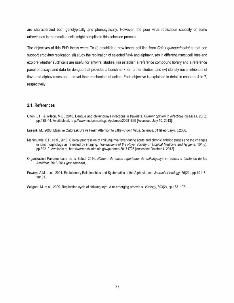
There is an alarming estimation of 390 million dengue infections per year, of which 96 million infections displayed typical
symptoms and were predicted by cartographic methods, considering local and spatial variations in risks that were
strongly influenced by rainfall, temperature, urbanization degree and socioeconomic variants(Bhatt et al. 2013). Taking
into account the number of cases manifesting the disease, the most affected regions are: Asia, whichaccounts for 70%
of total cases manifesting the disease; followed by India with 34%, Africa with 16%, the Americas with 14% and Oceania
with<0.2% (Bhatt et al. 2013)(Figure 2). In contrast, the WHO estimates that there are only between 50-100 million
cases per year (WHO 2009).
27
Figure 2 - Average annual number of dengue infections (Bhatt et al., 2013)
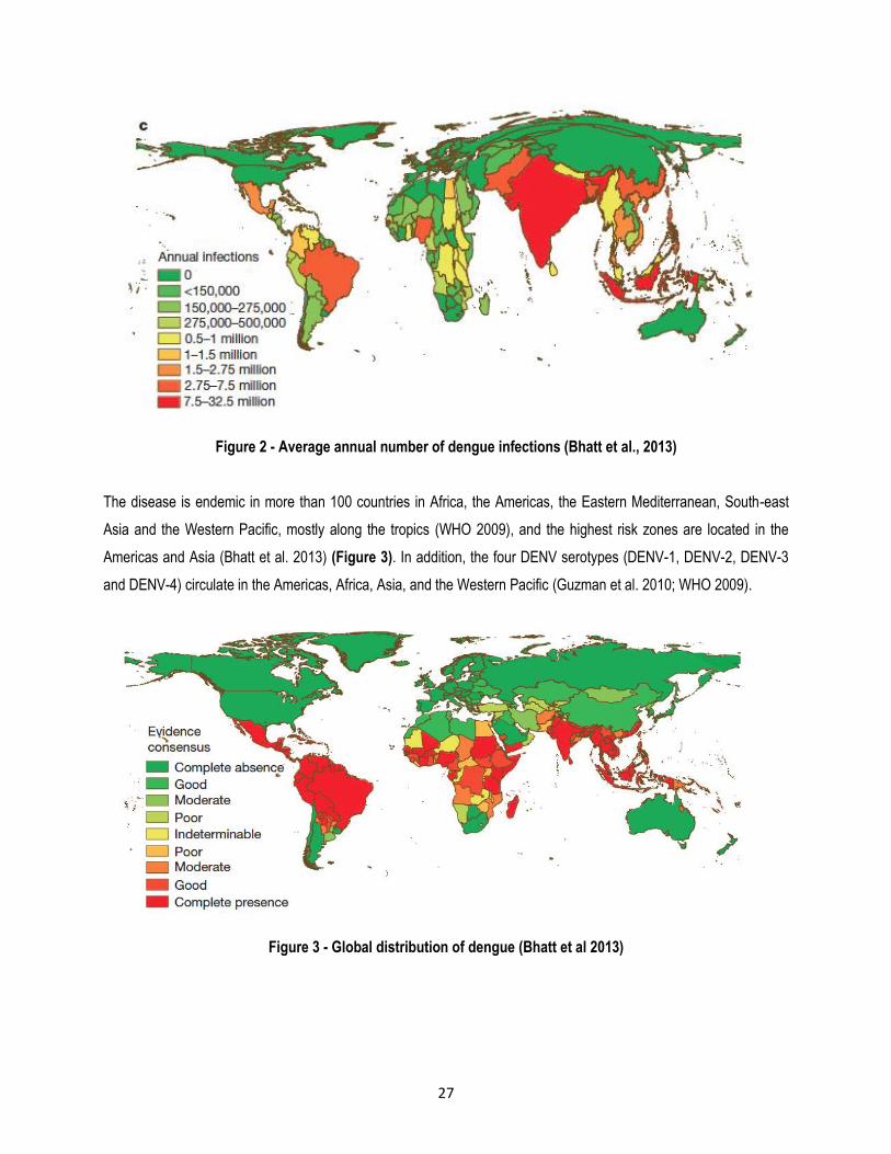
The disease is endemic in more than 100 countries in Africa, the Americas, the Eastern Mediterranean, South-east
Asia and the Western Pacific, mostly along the tropics (WHO 2009), and the highest risk zones are located in the
Americas and Asia (Bhatt et al. 2013) (Figure 3). In addition, the four DENV serotypes (DENV-1, DENV-2, DENV-3
and DENV-4) circulate in the Americas, Africa, Asia, and the Western Pacific (Guzman et al. 2010; WHO 2009).
Figure 3 - Global distribution of dengue (Bhatt et al 2013)
28
3.2.5. Dengue in Colombia
The increase of dengue in recent years has become a health problem. The National Health Institute (INS) in Colombia
reported 22.775 dengue cases in 2000, of which 1.093 corresponded to severe dengue; 14 people died due to the
disease. A few years later, in 2009, 55.592 cases were reported, of which 7.131 corresponded to severe dengue and
52 people from these cases died. Subsequently, more than 150.000 cases were reported in 2010, of which 6.209
corresponded to severe dengue;217 people from these cases died (Velandia & Castellanos 2011). In 2011, 29.389
cases were reported, of which 1.303 corresponded to severe dengue, and 53.258 cases were registered later in 2012,
of which 1.464 corresponded to severe dengue (MinSalud 2013). In 2013, 110.036 cases were reported, of which 3.000
corresponded to severe dengue and 129 people from these cases died. Dengue infections affect people of all ages,
but the mortality rate is higher in children under 14. In Colombia, the virus has reached a mortality rate of 4.7%
(Fernández & Linares 2013). Consequently, dengue constitutes a public health problem, since vector control strategies
have not yet been successful due to a variety of factors such as lack of quality education, displacement of communities,
conflict, and poverty, among others.
Between 2008 and 2013, 807 municipalities reported dengue infections. These municipalities were classified into
different transmissibility patterns, namely hypoendemic pattern - which means that there are no severe dengue cases
- mesoendemic pattern, indicating the presence of dengue and severe dengue cases and hyperendemic pattern, which
is characterized by an increase in individual susceptibility to severe dengue episodes due to the high prevalence of
severe dengue. 71.2% of municipalities were classified as mesoendemic, 21.3% as hypoendemic and 5.9% as
hyperendemic. The hypoendemic pattern is frequent in three departments: Amazonas, Guainía and San Andres,
whereas mesoendemic patterns is more frequent in 19 departments: Antioquia, Bolívar, Caquetá, Casanare, Cesar,
Chocó, Córdoba, Guajira, Huila, Magdalena, Meta, Norte De Santander, Putumayo, Risaralda, Santander, Sucre,
Tolima, Valle and Vichada (Figure 4).

29
Figure 4 - Dengue infections in Colombia. Departments in red presented dengue infections between 2008-
2013. Adapted from MinSalud 2013.
3.2.6. Dengue genome
The DENV genome is a positive single stranded RNA, which is approximately 11kb in length, its single open reading
frame (ORF) encodes three structural proteins (C, M and E) and seven non-structural proteins (NS1, NS2A, NS2B,
NS3, NS4A and NS5) (Figure 5).

30
Figure 5 - Representation of the DENV genome (Guzman et al. 2010)
3.2.6.1. DENV Structural proteins
Virions contain three structural proteins. The capsid protein (C) surrounds the genome of the virus, while the envelope
contains glycoprotein (E) and the membrane protein (M) (Hasteald 2008).
Membrane fusion is one of the most relevant events during the entry of enveloped viruses into cells (Modis et al. 2004).
However, the fusion is a complex process due to the fact that DENV has a great diversity in the cell tropism; in addition,
there are different receptors which are dependent on the type of the infected host cell (E. G. Acosta et al. 2008; Modis
et al. 2004). The envelope protein is responsible for the main steps in the entry process, which involves receptor
recognition and fusion between viral and cellular membranes (Rey 2003). In fact, E contains two putative N-linked
glycosylation sites: Asn-153, which is conserved among many flaviviruses and Asn-67, which is found only in DENV.
The presence of both N-linked carbohydrates is required for recognition by Dendritic Cell-Specific Intercellular adhesion
molecule-3-Grabbing Non-integrin (DC-SIGN) (Rey 2003).
In the host cells, it has been pointed out that the presence of mannose residues is important for viral entry (Hung et al.
1999). In addition, Heparan sulfate (HS), the most ubiquitous member of the glycosaminoglycan family, has been
identified both in Vero and human hepatoma cells (Huh-7) (Chen et al. 1997; Hilgard & Stockert 2000), and can act as
a receptor or concentrate the virus on the cell surface and facilitate the interaction with specific high-affinity receptors
(Germi et al. 2002). Moreover, different receptors of 74 and 44 kDa were described on Vero cells (Martínez-Barragán
& del Angel 2001).
The flavivirus C protein (12 kDa) forms homodimers in solution (Wang et al. 2004). This protein is essential in virus
assembly to ensure encapsidation of the viral genome. However, the mechanism by which encapsidation occurs has
not been well understood to date (Samsa et al. 2009). C also has been shown to interact with the heterogeneous
nuclear ribonucleoprotein K, which is a cellular regulatory protein, suggesting that C may also be involved in regulating
viral replication (Chang et al. 2001). Additionally, Samsa et al. (2012) demonstrated that basic residues within the
unstructured N-terminal region of C are required for DENV particle formation (Samsa et al. 2012).

31
Finally, maturation of flavivirus particles occurs during transport through the exocytic pathway. Prior to or during the
final release of virions, the cleavage of M protein precursor (prM) by furin transforms prM (18.44 kDa) into the M protein
(8.3 kDa) (van der Schaar et al. 2007) which allows the transformation from immature to mature viruses (Stadler et al.
1997); required for DENV infectivity (Zybert et al. 2008).
3.2.6.2. DENV non-structural proteins
NS1 is a 50-kDa glycoprotein that plays an essential role in viral replication (Mackenzie et al. 1996). NS1 is detectable
in plasma from patients; studies have shown that anti-NS1 antibody responses were found almost exclusively during
secondary infection, allowing the speculation that anti-NS1 antibody may play a role in DHF and DSS
immunopathogenesis (Avirutnan et al. 2006). However, Shu et al. (2000) showed that DF and DHF patients produced
significant NS1-specific antibody responses without having a direct correlation between this and DHF (Shu et al. 2000).
NS2A is a 22-kDa hydrophobic protein, it is implicated in the formation of virus-induced membranes (Leung et al. 2008)
that can be associated to virus assembly and RNA synthesis (Xie et al. 2013). In addition, this protein inhibits interferon
(IFN) and response (Muñoz-Jordan et al. 2003).
The viral protease activity lies within NS2B-NS3. NS2B is a 14-kDa hydrophobic protein; this protein is required for
NS3/NS4A cleavage and possibly also for the NS2A/NS2B, NS2B/NS3, and NS4B/NS5 cleavages, since these all
share the same amino acid sequence motif at the cleavage site. Meanwhile, NS3 is a 69.5-kDa protein and the 180
residues of this protein at the N-terminal contain a protease domain that is required for NS2A/NS2B and NS2B/NS3
cleavages. NS2B interacts with NS3 in order to promote the protease activity inherent in NS3. Both NS2B and NS3 are
required for protease activity that cleaves NS2A/NS2B, NS2B/NS3, NS4B/NS5 (Falgout et al. 1991), NS3/NS4A
(Cahour et al. 1992) and NS4B/NS5 (Yusof et al. 2000). The 440 amino acids at the C-terminal of NS3 protein constitute
a helicase region. NS3 is a multifunctional enzyme carrying out activities involved in viral RNA replication and capping:
helicase, nucleoside 5’-triphosphatase (NTPase), and RNA 5’-triphosphatase (RTPase) (Benarroch et al. 2004).
NS2A, NS4A and NS4B are IFN antagonists and might interact during DENV infection, resulting in a strong IFN inhibition
(Muñoz-Jordan et al. 2003). NS4A is a 16-kDa hydrophobic protein that is part of the viral replication complex, this
protein induces ER membrane rearrangements (Miller et al. 2007) and up-regulates autophagy, protecting the host cell
against death induced by the virus and providing a well-protected host cell for long-term virus replication (McLean et al.
2011).
NS4B is a 27-kDa transmembrane protein that participates in the viral replication complex formation (Miller et al. 2006).
This protein plays a role in viral RNA synthesis; NS4B enhance NS3 helicase activity, suggesting that this protein

32
modulates DENV replication via its interaction with NS3 (Umareddy et al. 2006). Moreover, NS4B is critical in DENV
virulence through the efficacy modulation of viral RNA synthesis in a mouse model (Grant et al. 2011).
NS5 is a 104-kDa protein from DENV. Residues 1 to 296 are associated with the S-adenosyl methionine transferase
(MTase) activity residing within its N-terminal domain. NS5 MTase activity is responsible for both guanine N-7 and
ribose 2’-O methylations; both methylations are required for 5’-cap formation (Ray et al. 2006). In addition, residues
270 to 900 contain the RNA-dependent RNA polymerase (RdRp) catalytic domain (Yap et al. 2007). NS5 also stimulates
NS3 nucleotide triphosphatase and RNA triphosphatase activities (Yon et al. 2005).
3.2.7. DENV life cycle
Flaviviruses enter host cells by receptor-mediated endocytosis. Following the attachment of virions to cell surface
receptors, the entry into the cell is achieved by endocytosis within clathrin-coated vesicles. These vesicles fuse with
endosomes, which subsequently undergo acidification triggering an irreversible E protein trimerization that allows the
fusion of viral and cell membranes (Allison et al. 1995). After the virus enters the cell and the nucleocapsid is uncovered,
the RNA molecule is translated as a single polyprotein. During this process, the polyprotein signal -and stop- transfer
sequences direct its back-and-forth translocation across the endoplasmic reticulum (ER) membrane. The polyprotein is
processed by cellular and virus-derived proteases into three structural proteins and seven non-structural proteins
(Rodenhuis-Zybert et al. 2010).
The coupling of protein synthesis, RNA synthesis, and the virion assembly on membranous structures assures that the
newly synthesized RNA genome can associate with C protein and initiate the assembly process. RNA encapsidation
initiates the budding of particles into the ER-derived membrane vesicles (Welsch et al. 2009). Particles that have
budded into the ER are then processed by carbohydrate addition and modification, as they proceed through the Golgi
membrane system. It is likely that transport into the trans-Golgi network requires the presence of the glycosylated prM
protein. Virions follow the exocytosis pathway and are released to the extracellular space by fusion of vesicles
containing virions with the plasma membrane (Figure 6). prM protein cleavage by host-encoded furin occurs just prior
to virion release and converts the particle to its mature form (Acheson 2007). Mature virus and subviral particles are
released from the host cell by exocytosis.

33
Figure 6 - Flavivirus life cycle (http://www.dsimb.inserm.fr/~debrevern/IDDT-
2009_in_silico_issue/iddt_2009_in_silico_issue.php#WATOWICH)
3.2.8. Antiviral therapy against DENV
There are no effective antiviral drugs for treating DENV infections, however very recently Sanofi licensed a tetravalent
dengue vaccine (TDV) against the viral disease. The vaccine comprises four recombinant, live-attenuated dengue
viruses (CYD-1-4), each of which have the DENV prM and E proteins of one of the four dengue serotypes, and in
addition, genes encoding NS and C proteins of the yellow fever 17D vaccine strain (YFV 17D) (Guy et al.
2011).Nevertheless, the TDV showed an efficacy of 30.2% (Sabchareon et al. 2012).
There are several compounds with anti-flavivirusesactivity, among them are:
Teicoplanin is a glycopeptide antibiotic that is used in the treatment of Gram-positive bacterial infections; it acts through
the biosynthesis inhibition of the bacterial cell wall. LCTA-949 is a teicoplanin-aglycone derivate that inhibits the
replication of human immunodeficiency virus (HIV) (Balzarini et al. 2003), hepatitis C virus (HCV) (Obeid et al. 2011),
DENV2, YFV, tick borne encephalitis virus (TBEV), WNV and the murine flavivirus, named Modoc virus (De
Burghgraeve et al. 2012). Obeid et al. (2011) reported that LCTA-949 inhibits the replication at a post-entry event in an

34
HCV replicon system. Meanwhile, De Burghraeve et al. (2012) demonstrated that this compound interferes with the
earliest stages of the DENV replication cycle, preventing virus-cell binding.
Castanospermine is a natural alkaloid derived from Castanospermumaustrale. This alkaloid is active against DENV,
but not against YFV and WNV (Whitby et al. 2005), and also acts as an inhibitor of ER -glucosidases. These molecules
block trims of N-linked carbohydrates, which directly affects DENV secretion and infectivity by preventing proper
processing of the envelope glycoproteins (Whitby et al. 2005; Courageot et al. 2000). Celgosivir is a pro-drug derivative
of castanospermine. This compound is an inhibitor of HIV (Taylor et al. 1994), bovine diarrhea virus (BVDV) and HCV
(Whitby et al. 2004). In addition, Celgosivir is around 100 times more effective against DENV2 than castanospermine
(Rathore et al. 2011). DENV treatment, testing castanospermine in a mouse model, resulted in a dose-dependent
response, where the lower dose (7.5 mg/kg) produced a reduction of 62% in the viraemia, and the higher dose (75
mg/kg) a reduction of 88%, respectively. In 2012, a clinical trial with castanospermine was started to treat DENV patients
in Singapore (Chang et al. 2013).
ST-610 is a benzoxazole inhibitor that is active against DENV and Venezuelan equine encephalitis virus (VEEV), but
does not inhibit YFV, HCV, WNV, BVDV, JEV and Modoc virus. ST-610 acts through the inhibition of ATP-dependent
helicase activity of DENV NS3 protein (Byrd et al. 2013). The compound can reduce virus replication and is well
tolerated in the mouse model (Byrd et al. 2013).
Ivermectin is an anthelmintic agent derived from Streptomyces avermitilis fermentation. Recently, it was discovered that
Ivermectin has antiviral activity against flaviviruses such as YFV, and also to a lesser degree against DENV, JEV and
TBEV (Mastrangelo et al. 2012). Two main mechanisms of action have been proposed for Ivermectin, (i) inhibition of
the viral helicase in flaviviruses (Mastrangelo et al. 2012), and (ii) disruption of the interaction between DENV NS5 and
importing /1, which is a nuclear import receptor (Wagstaff et al. 2012).
NITD-618 is an aminothiazole compound; this works as a selective inhibitor for DENV1 – DENV4, but not for WNV,
YFV, Chikungunya virus (CHIKV) and western equine encephalitis virus (WEEV). Sequencing of DENV2 resistant
replicons revealed mutations P104L and A119T in NS4B protein. The replicon analysis showed that together, these
mutations confer resistance to DENV2 inhibition by NITD-618. In addition, it was demonstrated that P104 mutation
abolished the interaction between NS3-NS4B, suggesting that this compound inhibits viral RNA synthesis; specifically,
the target is DENV NS4B protein (Xie et al. 2011).
2’-C-methylcytidine (2’CMC) is a nucleoside polymerase inhibitor that acts against RNA viruses such as HCV (Le
Pogam et al. 2006), YFV (Julander et al. 2010), Noroviruses (Rocha-Pererira et al. 2012) and foot and mouth disease
virus (Goris et al. 2007).

35
7-Deaza-2’-C-methyl-adenosine (7D-2CMA) is a nucleoside polymerase inhibitor that shares the same mechanism of
action than 2’CMC. 7D-2CMA has antiviral activity against BVDV, WNV, DENV, YFV, rhinovirus type 2, rhinovirus type
14, and poliovirus type 3, but none against WEEV and VEEV. Additionally, this compound did not have antiviral activity
against minus-stranded-RNA and double-stranded-DNA viruses. 7D-2CMA was tested in mice, rats, dogs and Rhesus
macaques. The 50% lethal dose (LD50) after 14 days of observation, when it was administrated as a single dose, was
2000 mg/kg in female mice. 7D-2CMC is less toxic than the related compound, which showed 30 fold lower than the
50% cytotoxic concentration (CC50) (i.e., the concentration that reduces the cells overall metabolic activity by 50%)
(Olsen et al. 2004).
The synthetic guanosine analogue, Ribavirin (1--D-ribofuranosyl-1,2,4-triazole-3-carboxamide) is a broad-spectrum
antiviral. Although ribavirin is active against a variety of RNA viruses as the pandemic H1N1 influenza (Rowe et al.
2010), Respiratory syncytial virus (Smith et al. 1991), Lassa fever virus (LFV) (Hadi et al. 2010) and Hanta virus
(Safronetz et al. 2011), the in vitro and in vivo activity of this compound against flaviviruses is very weak (Leyssen et
al. 2005). Leyssen et al. (2005) demonstrated that the predominant mechanism of action of ribavirin against flaviviruses
and paramyxoviruses is based on the inhibition of inosine-5’-monophosphate dehydrogenase (IMPDH).
Favipiravir (6-fluoro-3-hydroxy-2-pyrazinecarboxamide), also known as T-705, which is a broad-spectrum antiviral drug
that is in phase III clinical trials in The USA and was recently approved in Japan, has shown to be highly effective
against H1N1, H2N2, H3N2 (Furuta et al. 2005), H5N1 (Kiso et al. 2010) influenza A virus, influenza B and C viruses
(Furuta et al. 2002), as well as against Bunyaviruses [La Crosse (LACV), Punta Toro (PTV), Rift Valley fever (RFV) and
sandfly fever (SFFV)], Arenaviruses [Junin (JUNV), Pichinde (PICV), Tacaribe (TCRV)] (Gowen et al. 2007),
Flaviviruses (YFV and WNV) and Alphaviruses [CHIKV, Semliki Forest virus (SFV) and Sindbis virus (SINV)] (Delang
et al. 2014), but not against DNA viruses (Furuta et al. 2009). Although lethal mutagenesis was suggested as the
mechanism by which T-705 inhibits influenza virus replication (Baranovich et al. 2013), it was reported recently that two
consecutive substitutions of T705 ribofuranosyl monophosphate (RMP) allow complete inhibition of further nucleotide
incorporation in Influenza A virus (Jin et al. 2013). Delang et al. (2013) established through characterization of drug-
resistant variants that a mutation in CHIKV viral polymerase is the target of both T-705 and its defluorinated analog T-
1105.
3.3. Togaviridae family
The Togaviridae family comprises the Alphavirus and Rubivirus genera. The Rubivirus genus has a single species
member, Rubella virus, while the Alphavirus genus can be classified into at least 24 species and seven different
antigenic complexes (Barmah Forest, Ndumu, Middelburg, Semliki Forest, western equine encephalitis (WEE), eastern
equine encephalitis (EEE), and Venezuelan equine encephalitis (VEE) complex) (Figure 7) (Powers et al. 2001).

36
Additionally, alphaviruses have been classified as both Old World and New World viruses, depending on their
geographic distribution. Old World viruses can often cause fever, rash, and arthritic symptoms and diseases, while the
hosts infected with New World viruses may succumb to encephalitis. In humans and other mammals, alphavirus
infection is acute and in many cases characterized by high titer viraemia, rash, fever and encephalitis until the death of
the infected host or virus clearance by the immune system (Powers et al. 2001).
Figure 7 - Phylogenetic tree of Alphaviruses species generated from partial E1 sequences (Powers et al. 2001)
3.4. Chikungunya virus
CHIKV belongs, together with SFV, Ross River virus (RRV) and O’nyong-nyong virus (ONNV), to the Semliki Forest
complex of the Alphavirus genus (group A arboviruses), Togaviridae family. Other well-known viruses that belong to
this virus family are: the eastern equine encephalitis virus (representative of the EEEV complex Alphaviruses), VEEV
(VEEV complex), SINV and WEEV; the latter two belong to the WEE complex (Powers et al. 2001).
37
3.4.1. Chikungunya epidemiology
In 1953, CHIKV disease was first recorded in the Newala district of Tanzania bordering Mozambique (Ross 1956). The
name ‘chikungunya’ is derived from the root verb ‘kungunyala’ from the Ki Makonde language of the Makonde tribe and
means ‘that which bends up’, which refers to the stooped, bent posture CHIKV-infected patients present (Sudeep &
Parashar 2008). For a long time, very few cases or only small outbreaks of the disease had been reported. In the early
2000s, however, a sharp increase in CHIKV cases was observed in tropical areas surrounding the Indian Ocean. Large
outbreaks, affecting up to 70% of the local human population, started to become more and more frequent. In 2004 in
Lamu, Kenya, an estimated 13.500 people became infected. In 2005-2006, the number of diagnosed CHIKV infections
exceeded 266.000 in La Reunion island (Filleul et al. 2012). Since 2006, recurrent epidemics of this disease have
emerged in Africa, each time affecting approximately 15.000 people (Cavrini et al. 2009; WHO 2007) (Figure 8).
Between July and September 2007, the first outbreak of CHIKV disease occurred in the North-East of Italy, involving
over 205 cases (Rezza et al. 2007). The index case was a man that returned from Kerala, a region in India, which
presented an ongoing chikungunya epidemic. This man showed high titer of anti-CHIKV antibodies two days after his
arrival to Italy. The first autochthonous case was identified 13 days later and it appeared to be related to the index case.
Subsequently, the virus spread quickly in Castiglione, Cervia, Cesena, Ravenna, Rimini and Bologna. An 83-year old
man with severe underlying health conditions died as a result of complications induced by CHIKV infection (Liumbruno
et al. 2008). Ever since, multiple imported cases were documented in Asia, Australia, USA, Canada and Continental
Europe (Italy, Spain, Corsica, France, UK, Switzerland, Belgium, Czech Republic, Germany, and Norway) (ECDC
2012), and it is now well accepted that this virus has become endemic in several of these regions, as was evident from
two autochthonous cases presented in France in 2010 (Grandadam et al. 2011) (Figure 8).
Recently, CHIKV was reported for the first time in South America. Between December 2013 and December 12th, 2014,
the Chikungunya outbreak in America and the Caribbean produced 155 deaths, 20.209 confirmed cases and an
alarming number of 1.012.347 suspected cases (Organización Panamericana de la Salud 2014) (Figure 8 and Table
1).
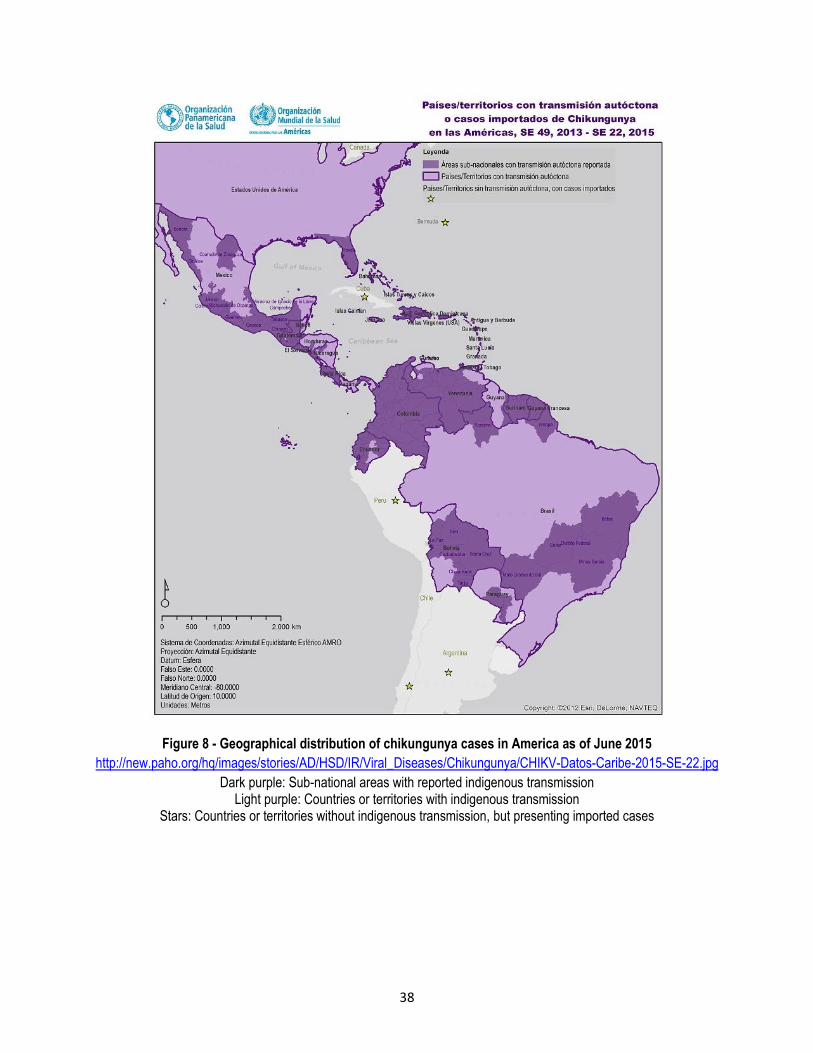
38
Figure 8 - Geographical distribution of chikungunya cases in America as of June 2015
http://new.paho.org/hq/images/stories/AD/HSD/IR/Viral_Diseases/Chikungunya/CHIKV-Datos-Caribe-2015-SE-22.jpg
Dark purple: Sub-national areas with reported indigenous transmission Light purple: Countries or territories with indigenous transmission
Stars: Countries or territories without indigenous transmission, but presenting imported cases

39
Table 1 - Cases of Chikungunya fever in America and the Caribbean from December 2013 to December 2014.
(Organización Panamericana de la Salud 2014)
Country Suspected cases Confirmed cases Deaths
No
rth
Am
eric
a
Bermuda 7 3
Canada
Mexico 74
USA 11
Cen
tral
Am
eric
a
Belize 3
Costa Rica 7
El Salvador 135226 157
Guatemala 579 49
Honduras 1381 9
Nicaragua 1598 542
Panama 32
Lat
in C
arib
bea
n
Cuba
Dominican Republic 524297 84 6
French Guayana 8172 5020
Guadalupe 80962 430 59
Haiti 64695 14
Martinique 78345 1515 76
Puerto Rico 22449 3732 5
San Bartolome 1014 142
San Martin 3771 793 3
An
dea
n a
rea
Bolivia
Colombia 45513 377 3
Ecuador
Peru
Venezuela 26451 1866
So
uth
ern
Co
ne
Argentina
Brazil 792 1303
Chile
Paraguay 1
no
n-L
atin
Car
ibb
ean
Anguila 31 49
Antigua and Barbuda 1399 18
Aruba 282 66
Bahamas 79
Barbados 1517 84
Cayman islands 148 37

40
Curazao 1838 835
Dominica 3588 173
Granada 3070 26
Guyana 76
Jamaica 1203 76
Montserrat 59 14
St. Kitts and Nevis 432 27
Santa Lucia 678 199
Saint Vincent and the Grenadines
1219 170
Saint Maarten 470
Suriname 1210 1
Trinidad and Tobago 177
Turks and Caigos 19
Virgin islands 1631 240 2
Total 1.012.347 20.209 155
3.4.2. Chikungunya in Colombia
In Colombia, CHIKV was recorded for the first time in September 2014, andto date 31 of 32 departments have registered
multiples case. However, the departments with the highest rates of infection are Valle, Tolima, Huila, Cundinamarca,
Córdoba, Antioquia, Sucre, Casanare, Norte de Santander and Atlántico; these departments together contain
approximately the 83% of CHIKV infections in Colombia. As of April 28 2015, 181.195 cases have been confirmed and
an additional 5.694 cases are suspected (INS 2015b) (Figure 9). Furthermore, 25 people have died due to CHIKV
infections and 17 additional deaths are under investigation(INS 2015a).

41
Figure 9 - Contribution of each department to total cases of Chikungunya in Colombia as of April
2015.Adapted from (Instituto Nacional de Salud 2015)
Ae. aegypti and Ae. albopictus mosquitoes primarily transmit CHIKV. However, other more geographically isolated
mosquito species can become infected as well (van den Hurk et al. 2010; Jupp et al. 1981). Taking into consideration
that mosquitoes have spread from tropical to more temperate regions (Charrel et al. 2007) and the importation of CHIKV
by infected travelers, an extensive surveillance network has been established for the virus (Chen & Wilson 2010; Tilston
et al. 2009; CDC & PAHO 2011).
3.4.3. Chikungunya disease
The onset of CHIKV disease is characterized by abrupt and sudden fever, chills, headache, nausea, photophobia,
vomiting, incapacitating joint pain and petechial or maculopapular rash. The acute phase may last for up to 10 days.
On occasion, neurological, haemorrhagic and ocular manifestations have also been described (Grandadam et al. 2011;
ECDC 2012). Although differential diagnosis between CHIKV and DENV infection appears to be quite difficult, the
clinical signs of arthralgia and myalgia, resulting from arthritis and tenosynovitis are typical of CHIKV disease, whereas
bleeding is very rare, so this symptom could be more indicative of a DENV infection (Table 2). With CHIKV, a rash is
usually observed between days 1 and 4 after disease onset, while for DENV, it is most pronounced between days 5
and 7. Finally, retro-orbital pain is a more common feature for DENV disease (Simon et al. 2011). After a week of
intense pain and incapacity, most of patients infected with chikungunya show a significant improvement of their
condition (Simon et al. 2011).

42
In about 49% of CHIKV-infected patients, the disease evolves into a chronic stage, which is characterized by persisting
polyarthralgia and stiffness (Manimunda et al. 2010), and which can severely incapacitate the patient for weeks to
several years after the initial infection (Simon et al. 2008). Although CHIKV infection is rarely fatal (Liumbruno et al.
2008), the viral disease might affect elderly people in a stronger way (Casolari et al. 2008). In addition, some atypical
cases of chikungunya fever have been reported very recently in Venezuela (Torres et al. 2015), these cases have been
characterized by hypotension, swelling, multi-organ failure and necrotic skin lesions followed by death (Torres et al.
2015). Laboratory diagnostic tools based on RT-PCR, haemaglutination or plaque neutralization significantly facilitate
the identification of the respective pathogens (Rezza et al. 2007).
Table 2 - Comparison of the chikungunya and dengue fever clinical features (Staples et al. 2009)
Clinical features Chikungunya
infection Dengue infection
Fever >39C +++ ++
Myalgias + ++
Arthralgias +++ +/-
Headache ++ ++*
Rash ++ +
Bleeding dyscrasias +/- ++
Shock - +/-
Leukopenia ++ +++
Neutropenia + +++
Lymphopenia +++ ++
Thrombocytopenia + +++
Symbols indicate the percentage of patients exhibiting each feature: +++, 70%-100% of patients; ++, 40-69%; +,
10%-39%; +/-, <10%; -, 0%. *Headache was often retro-orbital.
3.4.4. CHIKV genome and life virus replication
Alphaviruses are enveloped virions that contain one single-stranded, positive-sense RNA genome ranging from 11.44
kb (SFV and VEE) to 11.84 kb (ONN) in length. The CHIKV virion is about 60-70 nm in diameter (Powers et al. 2001),
and its genome is about 11.6 kb in length. It has two sequential ORFs of 7422 nt and 3744 nt - encoding the non-
structural polyproteins (nsP, 2474 aa) and the structural proteins (1248 aa) respectively -and a capped untranslated
terminal region (UTR) on its 5’ end and a poly-adenylated UTR on its 3’ end (Schuffenecker et al. 2006).
The first ORF encodes nsP1 (535 aa), which is associated with methyltransferase activity (Sreejith et al. 2012), nsP2
(798 aa) which has helicase and protease functions (Gomez de Cedrón et al., 1999; Takkinen & Kääriäinen, 1991),
nsP3 (530 aa) is a multifunctional protein which contains the ‘macro’ or ‘X’ domain (Malet et al. 2009) and the nsP4

43
(611 aa) has RNA-dependent RNA polymerases and poly-adenylated polymerase activities (Hahn et al., 1989; Tomar
et al., 2006).
The second ORF encodes the structural proteins (C, E3, E2, 6K and E1). C corresponds to the capsid protein (261 aa),
and there are three envelope proteins: E3 (64 aa), E2 (423 aa) and E1 (envelope protein 1, 439 aa). 6K corresponds
to an ion channel involved in the correct assembly of fully infectious virus particles, having 61 aa (Melton et al., 2002;
McInerney et al., 2004;Solignat, et al 2009) (Figure 10).
Figure 10 - Representation of CHIKV genome (Solignat et al. 2009)
CHIKV life cycle follows the typical Alphaviruses life cycle, starting with the attachment and fusion of E1 protein to
different combinations of cellular receptors (Sjöberg et al. 2011). The low pH in the endosome is essential for mediating
the fusion of the viral and host vesicle membranes in insects (Gay et al. 2012) as well as in mammalian cells (Sourisseau
et al. 2007), allowing the re-organization of the viral envelope complex made of E1 and E2 proteins, exposing the E1
fusion peptide (Schuffenecker et al. 2006). The E3 protein interacts with E2 protein, stabilizing a region called the “acid-
sensitive region”, facilitating indirectly E1 activation for membrane fusion (Sjöberg et al. 2011).
The C protein delivered into the cell cytoplasm binds to large ribosomal subunit and this might dissemble the
nucleocapsid and release genomic RNA (Singh & Helenius 1992) (Figure 11).
The composition of the replication complex varies throughout the infection. During the early stages of infection, minus
and plus-strand RNA are transcribed under nsP control. The nsP123 precursor is translated from the viral genome and

44
it binds to free nsP4 along with some host proteins to form the replication complex; this replication complex produces
the full length minus-strand (Barton et al. 1991), which is usually detected only during CHIKV replication early phases.
When nsP123 concentration is enough to support an efficient reaction, it is cleaved into mature non-structural proteins
nsP1, nsP2, nsP3 and nsP4 (Solignat et al. 2009). These proteins together with host cell proteins act as a plus-strand
RNA replicase, that produces the 26S sub-genomic plus-strand RNA using the negative-strand RNA as a template
(Shirako & Strauss 1994). nsP4 has the RdRP motif (Hahn et al., 1989; Tomar et al., 2006), and is expected, together
with nsP1, to catalyze the initiation or continuation of the negative-strand RNA synthesis (Sreejith et al. 2012). nsP3
participates in the transcription of negative strands during early events in replication (Wang et al. 1994). The 26S sub-
genomic RNA encodes the polyprotein precursor for structural proteins, which is cleaved to yield C, pE2, 6K and E1 by
an autoproteolytic serine protease (Solignat et al. 2009) (Figure 11).
Figure 11 - CHIKV lifecycle (Schwartz & Albert 2010)

45
3.4.5. Antiviral therapy against CHIKV
Currently, there is no licensed vaccine for the prevention of CHIKV disease. Significant strides have already been made
towards the development of a virus-like particle (VLP) system that has shown to be safe and effective in non-human
primates (Akahata et al. 2010), as well as an attenuated IRES-based vaccine (Plante et al. 2011). However, there is
still a large gap to be bridged before an effective vaccine is ready.
Similarly, there are no antiviral drugs available for the treatment or prevention of this viral disease. Chloroquine, a drug
that is commonly used for the treatment of malaria (Askling et al. 2012; Siswantoro et al. 2011), was demonstrated to
have a dose- and time-dependent antiviral effect on in vitro CHIKV replication (Khan et al. 2010). Chloroquine impairs
the replication of several enveloped viruses at an early stage by changing the intravesicular pH of endocytotic vesicles
and thus, preventing virus intrusion in the host cell (Ashfaq et al. 2011). It can also impair virus replication at late stages
by affecting the production of viral envelope glycoproteins at the cellular surface (Savarino et al. 2003; Savarino et al.
2001; Dille &Johnson 1982). In 1984, a patient reported an improvement of joint pain while taking chloroquine as a
prophylactic antimalarial drug. Brighton, subsequently, reported an improvement of arthritis symptoms in every 5 out of
10 patients with chronic CHIKV-induced joint symptoms that received a daily dose of 250 mg of chloroquine for 20
weeks (Brighton 1984). In an acute disease setting, however, De Lamballerie and colleagues did not observe any
statistical significant difference between placebo- and drug-treated patient groups (each of 27 individuals), of which the
latter received a short-term chloroquine treatment of 600 mg from day 1 to 3, and 300 mg on day 4 and 5 after fever
onset (De Lamballerie et al. 2008). At this time, it still remains a matter of discussion whether or not CHIKV-infected
patients may actually benefit from chloroquine treatment, either during the acute or the chronic stage of infection, or
both. Arbidol (Umifenovir®), a drug licensed for influenza A and B treatment, has been reported to have a selective
antiviral effect on CHIKV replication in Vero and MRC5 cells (human fetal lung fibroblasts). The compound interferes
with virus attachment and entry, which is corroborated by the observation of a single amino acid substitution (G407R) in
E2 protein, which renders the virus insensitive to the inhibitory effect of this compound (Delogu et al. 2011). No clinical
data is available of Arbidol against CHIKV infection. In addition to the compounds previously mentioned, only very few
other molecules have been reported to selectively inhibit in vitro CHIKV replication: 5,7-dihydroxyflavones (Pohjala et
al. 2011), ID 1452-2, a natural compound that partially blocks nsP2 (Lucas-Hourani et al. 2012) as well as an unusual
chlorinated daphnanediterpenoidorthoesther and some analogues isolated from the Trigonostemoncherrieri plant
(Allard et al. 2012). A moderate antiviral effect was observed in lupenone and -amyrone, two compounds that were
purified from the Anacolosapervilleanaplant (Bourjot et al. 2012). Even though the priority to develop a drug for the
treatment or prevention of CHIKV infection is currently not high from the business perspective, selective small-molecule
inhibitors are an important tool for studying the replication of this virus and will raise our preparedness level should the
need for an antiviral drug arise. As is evident from the studies with chloroquine and arbidol outlined above, the best
feasible approach towards an antiviral treatment for CHIKV today will be to take advantage of the anti-CHIKV activity

46
of compounds that are currently on the market or in preclinical development for another indication. The most promising
compound at present is Favipiravir, which will be the subject of the present study.
Favipiravir (6-fluoro-3-hydroxy-2-pyrazinecarboxamide), a mimetic nucleobase also known as T-705, was originally
discovered as a selective inhibitor of influenza A virus replication and is currently in phase III clinical trial in the USA
and recently approved in Japan, respectively. Furthermore, T-705 also inhibits the replication of various other RNA
viruses, including influenza A viruses such as H1N1, (Furuta et al. 2005)H2N2, H3N2, (Furuta et al. 2009) H5N1 (Kiso
et al. 2010), influenza B and C viruses (Furuta et al. 2002), as well as Bunyaviruses (LACV, PTV, RVF and SFFV),
Arenaviruses (JUNV, PICV, TCRV) (Gowen et al. 2007), Flaviviruses (YFV and WNV) (Julander et al. 2009) and
Noroviruses (Rocha-Pereira et al. 2012). Strong evidence, that was obtained from Madin-Darby canine kidney cells
(MDCK) (Furuta et al. 2005), as well as in human cells (Kiso et al. 2010), suggests that T-705 is metabolized to its
ribofuranosyl 5’-triphosphate form (T-705RTP). T-705RTP was shown to inhibit ATP and GTP incorporation in a
competitive manner, which suggests that T-705RTP is recognized as a purine nucleotide by the viral polymerase (Jin
et al. 2013; Sangawa et al. 2013).
However, the exact T-705 mechanism of action has not been elucidated yet. Two hypotheses are currently favored, i.e.
(i) the induction of lethal mutagenesis by ambiguous base pairing and/or (ii) chain termination by T-705RMP
incorporation into the nascent RNA strand. After serial passaging of influenza virus in the presence of T-705, the
infectious virus load was found to decrease disproportionally compared to the number of copies of RNA (Baranovich et
al. 2013) and sequence analysis also showed an increase in genotypes with a non-viable phenotype (Jin et al. 2013;
Baranovich et al. 2013). This data suggested that T-705 inhibits influenza virus (at least in part) through lethal
mutagenesis. However, it was also shown that the incorporation of a single T-705RTP molecule into a nascent RNA
strand caused inhibition of viral RNA extension, favoring the "chain terminator hypothesis". As the 3′-OH group in the
natural ribose is present in T-705RTP, the authors suggested that T-705 can be designated as a “non-obligate chain
terminator” (Sangawa et al. 2013). However, chain termination by T-705RMP could not be confirmed in another study,
in which at least two consecutive T-705RMP molecules were needed to be incorporated in order to arrest the extension
of the viral RNA (Jin et al. 2013). So far, however, no mutation(s) in the viral genome have been reported that could
prove the fact that the RNA-dependent polymerase or any other particular viral protein is involved in the mechanism of
action of this compound (Furuta et al. 2009).
3.5. Flavi and Alphavirus receptors for viral entry
During a natural in vivo infectionin mammals, cells like the mononuclear phagocyte lineage (macrophages, monocytes,
and dendritic cells) and Langerhans cells are the primary targets for DENV (Marovich et al. 2001; Noisakran et al. 2010)
and CHIKV infection (Rougeron et al. 2014). In contrast, DENV and CHIKV in insects usually infect vector’s midgut

47
epithelial cells. The infectious particles are spread and replicate in the body’s compartments and organs (Black et al.
2002; Coffey et al. 2014).
DENV and CHIKV infect target cells by attaching to various cell receptors, many of which are still unknown (Table 3).
In vitro DENV has shown to infect several cell lines from different origins (Barr & Anderson 2013). It could be possible
that the virus must bind to a ubiquitous cell-surface molecule, or exploit multiple receptors to mediate infection
(Rodenhuis-Zybert et al. 2010). Several candidate receptors have been identified, suggesting that flavi and alphaviruses
are capable of using different molecules to enter the cell (Table 3). For example, the Heat Shock Protein (HSP) HSP90
(84 kDa) and HSP70 (74 kDa) participate in DENV2 entry as a receptor complex in neuroblastoma and U937 cells
(Human leukemic monocyte lymphoma cell line), as well as in monocytes/macrophages. Both HSPs are associated
with membrane microdomains (lipid rafts) in response to DENV infection (Reyes-del Valle et al. 2005). Laminin was
also identified as a receptor for DENV1, DENV2 and DENV3 in porcine kidney cells (Tio et al. 2005). DC-SIGN is
expressed on dermal DCs and macrophages, which often encounter invading arboviruses in the skin. L-SIGN (Lymph-
Specific Intercellular adhesion molecule-3-Grabbing Non-integrin) is expressed on sinusoidal endothelial cells, which
would capture any virus inoculated in the small capillaries of the skin. Interaction with these receptors is presumably
mediated by the binding of DENV and CHIKV E protein N-linked carbohydrate modifications to carbohydrate recognition
domains of the lectin molecules (Klimstra et al. 2003; Alen et al. 2009; Voss et al. 2010). CHIKV has been less studied,
and in addition to DC-SIGN and L-SIGN, prohibitin, that is composed of two proteins of 30-kDa and 37-kDa, was
identified as a receptor in CHME-5 cells (human embryonic fetal microglial cells) (Wintachai et al. 2012). In addition,
HS could act as a receptor or help concentrate the virus on the cell surface in order to facilitate the interaction with
specific high-affinity receptors (Chen et al. 1997; Germi et al. 2002).
Several receptors have been described on insect cells, but in general these receptors differ from those found on
mammalian cells. The role of HS as a possible receptor on mosquito cells is controversial, as some groups could not
identify HS neither on Aedes albopictus C6/36 cells, nor on Aedes pseudoescutellaris (AP61) cell line (Thaisomboonsuk
et al. 2005). In contrast, other authors described this receptor in the Anopheles stephensi midgut and salivary glands
(a malaria mosquito vector) (Sinnis et al. 2007). In C6/36 cells and Ae.aegypti mosquito midgut, two proteins with
molecular masses of 80 and 67-kDa were suggested as receptors for DENV1 to DENV4 (Mercado-Curiel et al. 2006).
Additionally, DENV-4 bound to two glycoproteins of 40 and 45 kDa located on the cell surface in C6/36 cells. The 45-
kDa molecule was detected in total extracts from eggs, larvae and pupae as well as from midgut, ovary, and salivary
glands from Ae. aegypti, whereas it was absent in malpighian tubules. Therefore, the distribution of the 45-kDa protein
correlates with tissue tropism of DENV infection in mosquitoes. This protein was not detected in Anopheles albimanus
mosquito, which is not involved in DENV transmission (Mendoza et al. 2002). Prohibitin, that was described as CHIKV
receptor in mammalian cells has been characterized also in C6/36 and in Aedes aegypti cell line CCL-125, as a DENV-

48
2 receptor protein. It is possible that this highly conserved protein interacts with DENV infection in mammalian cells
(Kuadkitkan et al. 2010) (Table 3).
Table 3 - Proposed Flavi and Alphavirus receptors in insects and mammalian cells
Year Author Virus Cell line Receptor Type
1996 Ludwig et al VEEV C6/36 Laminin 32 kDa
Inse
cts
1997 Salas-Benito and Del Angel
DENV4 C6/36 40 and 45 kDa proteins
2001 Martinez-Barragan and del Angel
DENV4 C6/36 40 and 45 kDa proteins
2002 Mendoza et al DENV2 Aedes mosquitoes 45 kDa
2006 Mercado-Curiel et al DENV1 to DENV4 C6/36, MG A. aegypti R80, R67 proteins (80
and 67 kDa)
2007 Salas-Benito et al DENV2 C6/36 40 and 45 kDa proteins
2007 Sinnis et al ------- An. stephensi mosquitoes Heparan sulfate
2008 Mercado-Curiel et al DENV Aedes mosquitoes 67 kDa
2010 Kuadkitkan et al DENV2 C6/36, CCL-125 Prohibitin
1992 Wang et al SINV BHK, Vero, SW13, CEF,
CHO Laminin (67kDa protein)
Mam
mal
ian
1997 Chen et al DENV2 Vero Heparan sulfate
2000 Hilgard& Stocker DENV2 Huh-7 Heparan sulfate
2001 Bielefeldt-Ohmann et al DENV2, DENV3 Raji 34, 45 and 72 kDa
2001 Bielefeldt-Ohmann et al DENV2, DENV3 LK63 (lymphocyte B),
MOLT-4 (lymphocyte T) 45 and 72 kDa
2001 Martinez-Barragan and del Angel
DENV4 Vero 74 and 44 kDa proteins
2002 Germi et al DENV2, YFV Vero, CHO Heparan sulfate
2003 Klimstra et al SINV THP-1 transfected DC-SIGN, L-SIGN
2005 Tio et al DENV1 to DENV3 Porcine kidney cell line
clone D Laminin (37/67kDa
protein)
2005 Reyes-del Valle et al DENV2 Neuroblastoma, U937
human monocyte cell line HSP90 (84kDa), HSP70
(74k Da)
2009 Alen et al DENV2 Raji DC-SIGN+ DC-SIGN
2012 Wintachai et al CHIKV CHME-5 Prohibitin
3.6. Cell cultures
The possibility of culturing cells from diverse speciesoriginated over 100 years ago, when Harrison (1912) described
the first culture techniques in order to maintain neurons ex vivo alive and established the principal rules of maintenance.
Then, taking advantage of Harrison’s method, Lewis & Lewis (1911) made the first contribution to the knowledge of
artificial cell culture medium; they modified the amount of salts and animal extracts and fluids during growth and noticed
that the variability of physical conditions would improve tissues growing in culture. Subsequently, the first tumoral cell
culture was developed, improving the existing techniques and using cell culture flasks with a roller tube (Gey 1933).
Twenty years later, enzymatic tissue separation using enzymes as trypsin allowed the disintegration of the origin tissue.
Indeed, cellular isolates from chicken embryos were obtained and had the ability to grow in vitro on culture flasks
(Moscona 1951).

49
In recent years, different techniques and new applications of cell cultures have been developed. Nowadays, the use of
cell cultures is widespread and they are used in the development of vaccines, biological control, cellular signaling,
cellular differentiation and virology, among others (Gregersen et al 2011, Pedrini et al 2011, Acosta et al 2009, Tomokiyo
et al 2011, Patramool et al 2011).
3.6.1. Types of cell cultures
There are three principal types of cell cultures: primary cultures, secondary cultures and established
continuouslygrowingcell lines. Primary culture consists of a fragment of live tissue that is disaggregated using enzymes
or by mechanical action, which is cultured afterwards in an appropriated medium, and then it adheres to the growth
surface that usually will produce an outgrowth of cells. Cells in the outgrowth area are selected by their ability to migrate
from the explant and subsequently, once these cells proliferate they are subcultured, generating a cell line with cultures
of rapid growth. Most normal cell lines will undergo a limited number of subcultures, or passages, and are referred to
as finite cell lines. The number of doublings determines the limit that the cell population can overpass before it stops
growing because of senescence. Contrarily, an established cell line is characterized because it has been adapted by a
continued in vitro growth, and these cells maintain their characteristics in a constant and homogeneous way over time
(Freshney 2006) (Figure 12).
Figure 12 -Types of cell cultures according to the number of passages

50
3.6.2. Characterization of cell cultures
It is of great importance to establish the characteristics of each cell line used in research in order to ensure the identity
of each cell cultureand thereby, validate the research performed in vitro. Cell lines get mislabeled or contaminated with
fast-growing cells that can take over the original lines in no time (Chatterjee 2007). Misidentification or cross-
contamination of numerous cell lines have been reported; in addition, it is estimated that the incidence of research
papers flawed by the use of misidentified and cross-contaminated cell cultures is approximately 15-20% (Chatterjee
2007; Nardone 2007). The situations described above signify a great waste of time and resources. Additionally, the
results obtained usingmis-identified cell lines could invalidate research results.
When a cell line is established, it is crucial to determine the characterization of its cell culturein order to correlate it with
its tissue of origin, monitoring variation and excluding cross-contamination through unique features demonstration. In
order to establish the authenticity of cell lines and detect cross-contamination, there are several easily reproducible
techniques. These include: characterization using cell morphology (F Bello et al. 1997), isoenzymatic profiles (Losi et
al. 2008; F Bello et al. 1997), karyotype analysis (Bello et al. 2001) and molecular biological DNA analysis (Pan et al.
2007).
3.6.3. Insect cell cultures
The first arthropod cell line was established from tissues of Antherea eucalypti moth pupae in Australia, as described
by Grace (1962). Grace also obtained in 1966 the first mosquito cell line using Aedes aegypti larvae, other cell lines
were also obtained from this species (Singh 1967).
The modification of several techniques and the use of different types of tissues like insect embryos, ovaries, larvae,
hemocytes, fat bodies, imaginal discs, among others, allowed establishing insect cell lines as an in vitro system for
different studies. These include in vitro pathogen propagation (Lynn 2001), research on intracellular parasites
(Sakamoto & Azad 2007), viral infections (Sudeep et al. 2009), development of vaccines (Bonafé et al. 2009; Wang et
al. 2008), pest management, production of recombinant proteins (Cronin et al. 2007) and molecular studies (Acosta et
al. 2008). To date, more than 500 insect cell lines have been described, mainly from orders Diptera, Lepidoptera,
Hemiptera, Homoptera and Orthoptera (Lynn 2001).
Among the first relevant studies in regards to the establishment of insect vector cell cultures were (Singh 1967) for Ae.
aegypti and Ae. Albopictus,(Cahoon et al. 1978) for Aedes dorsalis, and (Tesh and Modi 1983) for Lutzomyia
longipalpis; a sand fly, which is a vector of Leishmania parasite.

51
In Colombia, the field of insect cell cultures has had great advances. Primary cell cultures from Anopheles albimanus
(Bello et al. 1995), Lu. Shannoni (Bello et al. 1997) Psorophora confinnis (Bello et al. 1999) and Lucilia sericata (Prieto
et al. 2009) were described. In addition, several continuous cell lines have been established and characterized
[morphologically, cytogenetically, biochemically and/or molecularly]. Among these cell lines, the most relevant
advances correspond to An. albimanus (Bello et al. 1997) and Psorophora confinnis (Bello et al. 2001), the sand flies
Lu. longipalpis (Rey et al. 2000), Lu. spinicrassa (Zapata et al. 2005), as well as to Sarconesiopsis magellanica blowfly
(Cruz & Bello 2013).
3.6.3.1. Insect cell cultures in virological studies
Many vertebrate and plant viruses replicate in insect cell lines. These generally involve cell lines developed from insect
vector species, which constitute a permissive host during part of the virus transmission cycle. Insect cell lines are
important tools in many aspects of virus-related research, including viral propagation and optimization in the
development of viral pesticides (Lynn 2001). Cell lines play key roles in the study of virus-cell interactions, viral entry
and replication processes (Acosta et al. 2008; Lannan et al. 2007; Chu et al. 2006).
The general characteristics of insect cell lines (to be) used for studying arboviruses could be summarized as follows:
The successful growth of arboviruses in dipteran cell lines rarely shows cytopathic effect (CPE), which can
vary from a limited structural change to cell lysis. Therefore mosquito cells are used for virus isolation.
The growth of arboviruses differs according to the species from which the cell line is derived.
The growth of arboviruses in insect cell lines is influenced principally by: pH, multiplicity of infection (MOI), viral
strain, cell type and composition of the cell culture medium (Yunker 1987).
Mosquito cell lines have been found to be effective in early detection and isolation due to their high sensitivity to
arboviruses (Lynn 2001), vertebrate viruses from a number of different families including Flaviviridae (Kuadkitkan et al.
2010; Rey et al. 2000), Reoviridae (Jia et al. 2012), Rhabdoviridae (Moraes 1990), Bunyaviridae (Schnettler et al. 2013)
and Togaviridae (Wintachai et al. 2012; Rey et al. 2000), have been propagated in dipteran cells. In addition, several
dipteran cell lines have been described to be susceptible to DENV (Table 4). Among these, C6/36 has been used
extensively to study DENV (Sakoonwatanyoo, 2006; Oliveira De Paula et al, 2003).
Singh & Paul (1969) found that ATC-15 cell line from Ae. albopictus was more susceptible to CHIKV, WNV and JEV
and also more susceptible to DENV2 regarding Vero cells. C6/36 cells, in addition to the ATC-15 cell line clone, are
susceptible to nineteen viruses (White 1987).

52
Table 4 - Diptera cell lines susceptible to Flavi and Alphavirus
Year Author Cell line Specie Origin
1969 Peleg Aag2 Aedes aegypti Embryos
1969 Paul and Sudeep ATC-15 Aedes albopictus Larva
1969 Paul and Sudeep ATC-10 Aedes aegypti Larva
1974 Varma AP-61 Aedes pseudoscutelaris
1978 Igarashi C6/36 Aedes albopictus Clon from CCL-125
1980 Tesh TA-9 Toxorhynchitesamboinensis First instar larvae
1980 Tesh TA-42 Toxorhynchitesamboinensis Embrionated eggs
1981 Kuno TRA-284 Toxorhynchitesamboinensis Larva
1981 Kuno et al TRA-171 Toxorhynchitesamboinensis Larva
1992 Morier et al CLA 1 Aedes pseudoscutelaris Clon from AP-61
1992 Pant et al NIVI-AK-455 Aedes krombeini Embryos
1992 Pant et al NIVI-AK-454 Aedes krombeini Embryos
1992 Pant et al NIVI-AK-453 Aedes krombeini Embryos
2009 Wikan CCL-125 Aedes aegypti Larva
2009 Sudeep et al Aedes aegypti Aedes aegypti Neonate larvae
Studies using insect cell lines have helped to elucidate antiviral strategies of host cells and the counterstrategies
evolved by their pathogens (Schütz & Sarnow 2006; Weaver 2006).
For example, it was established by the C6/36 cell line that during DENV2 infection, the viral RNA localized over the
rough ER and later formedvirus-induced smooth membrane structures. These formed within the ER and, when dense
areas were observed in close proximity to smooth membrane structures, encapsidation of the viral genome possibly
occurred (Grief et al. 1997).
On the other hand, C6/36 cells were transduced with vectors expressing hammerhead ribozymes (hRz), which are
small ribonucleic-bases enzymes that are capable of catalyzing target RNA cleavage in a sequence-specific manner.
These hRz act pairing of the 5’helix I and 3’helix III arms of the hRz to complementary 3’and 5’base pairs on the target
RNA. These ribozymes-transduced cells were challenged with DENV and through qRT-PCR, Northern analysis and
immunofluorescence, demonstrating that C6/36 cells expressing several hRz were able to suppress DENV replication
between 75% and 99%. It was shown that C6/36 provides alternative strategies targeting DENV in mosquito cells and
further on, transgenic mosquitoes tissues inhibiting the virus replication through ribozymes (Nawtaisong et al. 2009).

53
In another study, the infectivity entry pathways of DENV1-DENV4 into C6/36 cells was analyzed by biochemical and
molecular inhibitors, leading to the conclusion that pH dependent clathrin-mediated endocytosis is required for all DENV
serotypes in order to enter mosquito cells (Acosta et al. 2011; Acosta et al. 2008; Mosso et al. 2008).
In addition, using an Ae. aegypti cell line (Aag2) has revealed that persistent DENV2 infections generate small DENV2-
specific RNAs which are consistent in size and sequence with siRNAs (small interfering RNA) (Sánchez-Vargas et al.
2009). It is known that in Drosophila, RNA interference (RNAi) is a potent innate antiviral pathway that is triggered by
siRNA, forms in virus-infected cells and leads to viral RNA genome degradation (Steinert & Levashina 2011). On the
other hand, insect cell lines have been used in co-infection studies. For instance, a study showed how Ae. albopictus
RML12 and C6/36 cell lines were co-infected with Wolbachia and DENV-2, and results suggested that DENV-2
replication was reduced in both cell lines, especially in the most densely infected with Wolbachia. This favors the
hypothesis that viral inhibition may occur due to competition betweenWolbachia and DENV-2 for host cellular resources
(Frentiu et al. 2010).
3.7. References
Acosta, E., Castilla, V. & Damonte, E., 2008. Functional entry of dengue virus into Aedes albopictus mosquito cells is dependent on clathrin-mediated endocytosis. The Journal of general virology, 89(Pt 2), pp.474–84. Available at: http://www.ncbi.nlm.nih.gov/pubmed/18198378 [Accessed May 22, 2013].
Acosta, E.G., Castilla, V. & Damonte, E.B., 2011. Infectious dengue-1 virus entry into mosquito C6/36 cells. Virus research, 160(1-2), pp.173–9. Available at: http://www.ncbi.nlm.nih.gov/pubmed/21708195 [Accessed July 4, 2013].
Acosta, E.G., Talarico, L.B. & Damonte, E.B., 2008. Cell entry of dengue virus. Future Virology, 3(5), pp.471–479. Available at: http://www.futuremedicine.com/doi/abs/10.2217/17460794.3.5.471.
Acheson, N.H. Fundamentals of Molecular Virology. Ed. John Wiley and Sons. (United States of America, 2007). 181-190.
Akahata, W. et al., 2010. A virus-like particle vaccine for epidemic Chikungunya virus protects nonhuman primates against infection. Nature medicine, 16(3), pp.334–8. Available at: http://www.pubmedcentral.nih.gov/articlerender.fcgi?artid=2834826&tool=pmcentrez&rendertype=abstract [Accessed June 22, 2012].
Alen, M.M.F. et al., 2009. Antiviral activity of carbohydrate-binding agents and the role of DC-SIGN in dengue virus infection. Virology, 387(1), pp.67–75. Available at: http://www.ncbi.nlm.nih.gov/pubmed/19264337 [Accessed July 4, 2013].
Allard, P.-M., Leyssen, P., et al., 2012. Antiviral chlorinated daphnane diterpenoid orthoesters from the bark and wood of Trigonostemon cherrieri. Phytochemistry. Available at: http://www.ncbi.nlm.nih.gov/pubmed/22938995 [Accessed October 22, 2012].
Allard, P.-M., Martin, M.-T., et al., 2012. Trigocherrin A, the first natural chlorinated daphnane diterpene orthoester from Trigonostemon cherrieri. Organic letters, 14(1), pp.342–5. Available at: http://www.ncbi.nlm.nih.gov/pubmed/22182207.

54
Allison, S.L. et al., 1995. Oligomeric rearrangement of tick-borne encephalitis virus envelope proteins induced by an acidic pH . Oligomeric Rearrangement of Tick-Borne Encephalitis Virus Envelope Proteins Induced by an Acidic pH.
Ashfaq, U. a et al., 2011. Lysosomotropic agents as HCV entry inhibitors. Virology journal, 8(1), p.163. Available at: http://www.pubmedcentral.nih.gov/articlerender.fcgi?artid=3090357&tool=pmcentrez&rendertype=abstract [Accessed October 5, 2012].
Askling, H.H. et al., 2012. Management of imported malaria in Europe. Malaria journal, 11(1), p.328. Available at: http://www.ncbi.nlm.nih.gov/pubmed/22985344 [Accessed October 1, 2012].
Avirutnan, P. et al., 2006. Vascular leakage in severe dengue virus infections: a potential role for the nonstructural viral protein NS1 and complement. The Journal of infectious diseases, 193(8), pp.1078–88. Available at: http://www.ncbi.nlm.nih.gov/pubmed/16544248.
Balzarini, J. et al., 2003. Antiretroviral activity of semisynthetic derivatives of glycopeptide antibiotics. Journal of medicinal chemistry, 46(13), pp.2755–64. Available at: http://www.ncbi.nlm.nih.gov/pubmed/12801238.
Baranovich, T. et al., 2013. T-705 (favipiravir) induces lethal mutagenesis in influenza A H1N1 viruses in vitro. Journal of virology, 87(7), pp.3741–51. Available at: http://www.ncbi.nlm.nih.gov/pubmed/23325689 [Accessed August 12, 2013].
Barr, K. & Anderson, B., 2013. Dengue viruses exhibit strain-specific infectivity and entry requirements in vitro. Virus adaptation and treatment, 5, pp.1–9.
Barton, D.J., Sawicki, S.G. & Sawicki, D.L., 1991. Solubilization and Immunoprecipitation of Alphavirus Replication Complexes. Journal of virology, 65(3), pp.1496–1506.
Beasley, D.W. et al., 2008. The infecion agent. Dengue. Imperial College Press. London 485p.
Bello, F. et al., 1997. Establishment and characterization of a cell line from the mosquito Anopheles albimanus (Diptera: Culicidae). Memórias do Instituto Oswaldo Cruz, 92(1), pp.123–8. Available at: http://www.ncbi.nlm.nih.gov/pubmed/9302423.
Bello, F., Jiménez, M. & Ferro, C., 1997. Cultivos celulares primarios de Lutzomyia shannoni (Diptera: psychodidae) y estudio cariologico preliminar de la especie. Biomédica, 17, pp.49–55.
Bello, F.J. et al., 2001. A new continuous cell line from the mosquito Psorophora confinnis (Diptera: Culicidae) and its susceptibility to infections with some arboviruses. Memórias do Instituto Oswaldo Cruz, 96(6), pp.865–73. Available at: http://www.ncbi.nlm.nih.gov/pubmed/11562717.
Bello, F.J. et al., 1999. Estudio de cultivos celulares primarios de Psorophora confinnis (Diptera: Culicidae). Biomédica.
Bello, F.J. et al., 1995. Initiation of primary cell cultures from embryos Anopheles albimanus and Aedes taeniorhynchus (Diptera: Culicidae). Memórias do Instituto Oswaldo Cruz.
Benarroch, D. et al., 2004. The RNA helicase, nucleotide 5’-triphosphatase, and RNA 5'-triphosphatase activities of Dengue virus protein NS3 are Mg2+-dependent and require a functional Walker B motif in the helicase catalytic core. Virology, 328(2), pp.208–18. Available at: http://www.ncbi.nlm.nih.gov/pubmed/15464841 [Accessed July 29, 2013].
Bhatt, S. et al., 2013. The global distribution and burden of dengue. Nature, 496(7446), pp.504–7. Available at: http://www.ncbi.nlm.nih.gov/pubmed/23563266 [Accessed May 22, 2013].
Black, W.C. et al., 2002. Flavivirus susceptibility in Aedes aegypti. Archives of medical research, 33(4), pp.379–88. Available at: http://www.ncbi.nlm.nih.gov/pubmed/12234528.

55
Bonafé, N. et al., 2009. A recombinant West Nile virus envelope protein vaccine candidate produced in Spodoptera frugiperda expresSF+ cells. Vaccine, 27(2), pp.213–22.
Bourjot, M. et al., 2012. Chemical constituents of Anacolosa pervilleana and their antiviral activities. Fitoterapia, 83(6), pp.1076–80. Available at: http://www.ncbi.nlm.nih.gov/pubmed/22613073 [Accessed October 6, 2012].
Brighton, S.W., 1984. Chloroquine phosphate treatment of chronic Chikungunya arthritis. An open pilot study. South African medical journal, 66(6), pp.217–8. Available at: http://www.ncbi.nlm.nih.gov/pubmed/6087474.
De Burghgraeve, T. et al., 2012. An analogue of the antibiotic teicoplanin prevents flavivirus entry in vitro. PloS one, 7(5), p.e37244. Available at: http://www.pubmedcentral.nih.gov/articlerender.fcgi?artid=3356272&tool=pmcentrez&rendertype=abstract [Accessed July 2, 2013].
Byrd, C.M. et al., 2013. Novel benzoxazole inhibitor of dengue virus replication that targets the NS3 helicase. Antimicrobial agents and chemotherapy, 57(4), pp.1902–12. Available at: http://www.ncbi.nlm.nih.gov/pubmed/23403421 [Accessed July 4, 2013].
Cahoon, B., Hardy, J. & Reeves, W., 1978. Initiation and characterization of a diploid cell line from larval tissues of Aedes dorsalis (Meigen). In vitro, 14(3), pp.255–60.
Cahour, A., Falgout, B. & Lai, C.J., 1992. Cleavage of the dengue virus polyprotein at the NS3/NS4A and NS4B/NS5 junctions is mediated by viral protease NS2B-NS3, whereas NS4A/NS4B may be processed by a cellular protease. Journal of virology, 66(3), pp.1535–42. Available at: http://www.pubmedcentral.nih.gov/articlerender.fcgi?artid=240879&tool=pmcentrez&rendertype=abstract.
Casolari, S. et al., 2008. A fatal case of encephalitis associated with Chikungunya virus infection. Scandinavian journal of infectious diseases, 40(11-12), pp.995–6. Available at: http://www.ncbi.nlm.nih.gov/pubmed/18819040.
Cavrini, F. et al., 2009. Review Article Chikungunya : an emerging and spreading arthropod-borne viral disease. J Infect Dev Ctries, 3(February 2005), pp.744–752.
CDC & PAHO, 2011. Preparación y respuesta ante la eventual introducción del virus chikungunya en las Américas, Washington.
Chang, C.J. et al., 2001. The heterogeneous nuclear ribonucleoprotein K (hnRNP K) interacts with dengue virus core protein. DNA and cell biology, 20(9), pp.569–77. Available at: http://www.ncbi.nlm.nih.gov/pubmed/12466486.
Chang, J. et al., 2013. Small molecule inhibitors of ER α-glucosidases are active against multiple hemorrhagic fever viruses. Antiviral research, 98(3), pp.432–40. Available at: http://www.ncbi.nlm.nih.gov/pubmed/23578725 [Accessed August 17, 2013].
Charrel, R., De Lamballerie, X. & Raoult, D., 2007. Chikungunya Outbreaks — The Globalization of Vectorborne Diseases. N. Engl J Med, 356, pp.769–771.
Chatterjee, R., 2007. Cases of mistake identity. Science, 315(February), pp.928–31.
Chen, L.H. & Wilson, M.E., 2010. Dengue and chikungunya infections in travelers. Current opinion in infectious diseases, 23(5), pp.438–44. Available at: http://www.ncbi.nlm.nih.gov/pubmed/20581669 [Accessed July 10, 2013].
Chen, Y. et al., 1997. 1997 Chen et al DENV infectivity heparan sulfate.pdf. Nature medicine, 3(8), pp.866–871.
Chu, J.J.H., Leong, P.W.H. & Ng, M.L., 2006. Analysis of the endocytic pathway mediating the infectious entry of mosquito-borne flavivirus West Nile into Aedes albopictus mosquito (C6/36) cells. Virology, 349(2), pp.463–75. Available at: http://www.ncbi.nlm.nih.gov/pubmed/16490225 [Accessed July 4, 2013].

56
Coffey, L.L., Failloux, A. & Weaver, S.C., 2014. Chikungunya Virus–Vector Interactions. , pp.4628–4663.
Courageot, M.P. et al., 2000. Alpha-glucosidase inhibitors reduce dengue virus production by affecting the initial steps of virion morphogenesis in the endoplasmic reticulum. Journal of virology, 74(1), pp.564–72. Available at: http://www.pubmedcentral.nih.gov/articlerender.fcgi?artid=111573&tool=pmcentrez&rendertype=abstract.
Cronin, N.N. et al., 2007. Production of selenomethionyl-derivatized proteins in baculovirus-infected insect cells. Protein Science, 16, pp.2023–2029.
Cruz, M. & Bello, F., 2013. Establishment and characterization of an embryonic cell line from Sarconesiopsis magellanica. Journal of insect science, 13, p.130. Available at: http://www.pubmedcentral.nih.gov/articlerender.fcgi?artid=4014039&tool=pmcentrez&rendertype=abstract.
Delang, L. et al., 2014. Mutations in the chikungunya virus non-structural proteins cause resistance to favipiravir (T-705), a broad-spectrum antiviral. The Journal of antimicrobial chemotherapy, 69(10), pp.2770–84. Available at: http://www.ncbi.nlm.nih.gov/pubmed/24951535 [Accessed December 1, 2014].
Delogu, I. et al., 2011. In vitro antiviral activity of arbidol against Chikungunya virus and characteristics of a selected resistant mutant. Antiviral Research, 90(3), pp.99–107. Available at: http://www.ncbi.nlm.nih.gov/pubmed/21440006 [Accessed October 1, 2012].
Dille, B.J. & Johnson, T.C., 1982. Inhibition of vesicular stomatitis virus glycoprotein expression by chloroquine. The Journal of general virology, 62 (Pt 1), pp.91–103. Available at: http://www.ncbi.nlm.nih.gov/pubmed/6290597.
ECDC, 2012. Chikungunya fever. European Centre for Disease Prevention and Control. Chikungunya fever. Available at: http://www.ecdc.europa.eu/en/healthtopics/chikungunya_fever/basic_facts/Pages/factsheet_health_professionals.aspx.
Falgout, B. et al., 1991. Both nonstructural proteins NS2B and NS3 are required for the proteolytic processing of dengue virus nonstructural proteins. Journal of virology, 65(5), pp.2467–75. Available at: http://www.pubmedcentral.nih.gov/articlerender.fcgi?artid=240601&tool=pmcentrez&rendertype=abstract.
Fernández, C.F. & Linares, A., 2013. El dengue ya tiene nivel de epidemia en Colombia. El Tiempo. Available at: http://www.eltiempo.com/archivo/documento/CMS-13188599.
Filleul, L. et al., 2012. Chikungunya surveillance on Réunion Island between 2005 and 2011. 15th ICICD Abstracts / International Journal of Infectious Diseases, 16S, p.e256.
Frentiu, F.D. et al., 2010. Wolbachia-mediated resistance to dengue virus infection and death at the cellular level. PloS one, 5(10), p.e13398. Available at: http://www.pubmedcentral.nih.gov/articlerender.fcgi?artid=2955527&tool=pmcentrez&rendertype=abstract [Accessed May 30, 2013].
Freshney, I., 2006. Basic principles of cell culture. In G. Vunjak-Novakovic & I. Freshney, eds. Culture of Cells for Tissue Engineering. United States of America: Wiley-Liss, p. 512. Available at: http://www.ncbi.nlm.nih.gov/pubmed/14634214.
Furuta, Y. et al., 2002. In Vitro and In Vivo Activities of Anti-Influenza Virus Compound T-705. Antimicrobial agents and chemotherapy, 46(4), pp.977–981.
Furuta, Y. et al., 2005. Mechanism of Action of T-705 against Influenza Virus. Antimicrobial agents and chemotherapy, 49(3), pp.981–986.
Furuta, Y. et al., 2009. T-705 (favipiravir) and related compounds: Novel broad-spectrum inhibitors of RNA viral infections. Antiviral research, 82(3), pp.95–102. Available at: http://www.ncbi.nlm.nih.gov/pubmed/19428599 [Accessed July 15, 2012].

57
Gay, B. et al., 2012. pH-dependent entry of chikungunya virus into Aedes albopictus cells. Infection, genetics and evolution, 12(6), pp.1275–1281. Available at: http://www.ncbi.nlm.nih.gov/pubmed/22386853 [Accessed March 16, 2012].
Germi, R. et al., 2002. Heparan sulfate-mediated binding of infectious dengue virus type 2 and yellow fever virus. Virology, 292(1), pp.162–8. Available at: http://www.ncbi.nlm.nih.gov/pubmed/11878919 [Accessed June 7, 2013].
Gey, G.O., 1933. An Improved Technic for Massive Tissue Culture. Am J Cancer, 17, pp.752–56.
Gomez de Cedrón, M. et al., 1999. RNA helicase activity of Semliki Forest virus replicase protein NSP2. FEBS letters, 448(1), pp.19–22. Available at: http://www.ncbi.nlm.nih.gov/pubmed/10217401.
Goris, N. et al., 2007. 2 - C -Methylcytidine as a potent and selective inhibitor of the replication of foot-and-mouth disease virus. Antiviral research, 73, pp.161–168.
Gowen, B.B. et al., 2007. In vitro and in vivo activities of T-705 against arenavirus and bunyavirus infections. Antimicrobial agents and chemotherapy, 51(9), pp.3168–76. Available at: http://www.pubmedcentral.nih.gov/articlerender.fcgi?artid=2043187&tool=pmcentrez&rendertype=abstract [Accessed September 19, 2012].
Grandadam, M. et al., 2011. Chikungunya virus, southeastern France. Emerging infectious diseases, 17(5), pp.910–3. Available at: http://www.pubmedcentral.nih.gov/articlerender.fcgi?artid=3321794&tool=pmcentrez&rendertype=abstract [Accessed July 17, 2012].
Grant, D. et al., 2011. A single amino acid in nonstructural protein NS4B confers virulence to dengue virus in AG129 mice through enhancement of viral RNA synthesis. Journal of virology, 85(15), pp.7775–87. Available at: http://www.pubmedcentral.nih.gov/articlerender.fcgi?artid=3147917&tool=pmcentrez&rendertype=abstract [Accessed July 10, 2013].
Grief, C. et al., 1997. Intracellular localisation of dengue-2 RNA in mosquito cell culture using electron microscopic in situ hybridisation. Archives of virology, 142(12), pp.2347–57. Available at: http://www.ncbi.nlm.nih.gov/pubmed/9672599.
Guy, B. et al., 2011. From research to phase III: Preclinical, industrial and clinical development of the Sanofi Pasteur tetravalent dengue vaccine. Vaccine, 29(42), pp.7229–7241. Available at: http://dx.doi.org/10.1016/j.vaccine.2011.06.094.
Guzman, M.G. et al., 2010. Dengue: a continuing global threat. Nature reviews. Microbiology, 8(12 Suppl), pp.S7–16. Available at: http://www.ncbi.nlm.nih.gov/pubmed/21079655 [Accessed May 29, 2013].
Hadi, C.M. et al., 2010. Ribavirin for Lassa Fever Postexposure Prophylaxis. Letters, 16(12), pp.2009–2011.
Hahn, Y.S. et al., 1989. Mapping of RNA- temperature-sensitive mutants of Sindbis virus: complementation group F mutants have lesions in nsP4. Journal of virology, 63(3), pp.1194–202. Available at: http://www.pubmedcentral.nih.gov/articlerender.fcgi?artid=247815&tool=pmcentrez&rendertype=abstract.
Hilgard, P. & Stockert, R., 2000. Heparan sulfate proteoglycans initiate dengue virus infection of hepatocytes. Hepatology (Baltimore, Md.), 32(5), pp.1069–77. Available at: http://www.ncbi.nlm.nih.gov/pubmed/11050058 [Accessed June 10, 2013].
Hung, S. et al., 1999. Analysis of the Steps Involved in Dengue Virus Entry into Host Cells. Virology, 167, pp.156–167.
Van den Hurk, A.F. et al., 2010. Vector competence of Australian mosquitoes for chikungunya virus. Vector borne and zoonotic diseases (Larchmont, N.Y.), 10(5), pp.489–95. Available at: http://www.ncbi.nlm.nih.gov/pubmed/19877822.
INS, 2015a. Boletin Epidemiológico Evento prioritario, Bogotá.

58
INS, 2015b. Chikunguña a semana 15 de 2015, Bogotá.
Instituto Nacional de Salud, 2015. Chikunguña a semana 53, Bogotá.
Jia, D. et al., 2012. Development of an Insect Vector Cell Culture and RNA Interference System To Investigate the Functional Role of Fijivirus Replication. Journal of virology, 86(10), pp.5800–5807.
Jin, Z. et al., 2013. The Ambiguous Base-Pairing and High Substrate Efficiency of T-705 (Favipiravir) Ribofuranosyl 5’-Triphosphate towards Influenza A Virus Polymerase. PloS one, 8(7), p.e68347. Available at: http://www.pubmedcentral.nih.gov/articlerender.fcgi?artid=3707847&tool=pmcentrez&rendertype=abstract [Accessed August 12, 2013].
Julander, J.G. et al., 2009. Activity of T-705 in a hamster model of yellow fever virus infection in comparison with that of a chemically related compound, T-1106. Antimicrobial agents and chemotherapy, 53(1), pp.202–9. Available at: http://www.pubmedcentral.nih.gov/articlerender.fcgi?artid=2612161&tool=pmcentrez&rendertype=abstract [Accessed June 22, 2012].
Julander, J.G. et al., 2010. Efficacy of 2’-C-methylcytidine against yellow fever virus in cell culture and in a hamster model. Antiviral research, 86(3), pp.261–7. Available at: http://www.pubmedcentral.nih.gov/articlerender.fcgi?artid=2871769&tool=pmcentrez&rendertype=abstract [Accessed July 1, 2013].
Jupp, P.G. et al., 1981. Laboratory vector studies on six mosquito and one tick species with chikungunya virus. Transactions of the Royal Society of Tropical Medicine and Hygiene, 75(1), pp.15–9. Available at: http://www.ncbi.nlm.nih.gov/pubmed/6115488.
Khan, M. et al., 2010. Assessment of In Vitro Prophylactic and Therapeutic Efficacy of Chloroquine Against Chikungunya Virus in Vero Cells. Journal of Medical Virology, 824(August 2009), pp.817–824.
Kiso, M. et al., 2010. T-705 (favipiravir) activity against lethal H5N1 influenza A viruses. Proceedings of the National Academy of Sciences of the United States of America, 107(2), pp.882–7. Available at: http://www.pubmedcentral.nih.gov/articlerender.fcgi?artid=2818889&tool=pmcentrez&rendertype=abstract [Accessed September 16, 2012].
Klimstra, W. et al., 2003. DC-SIGN and L-SIGN Can Act as Attachment Receptors for Alphaviruses and Distinguish between Mosquito Cell- and Mammalian Cell-Derived Viruses DC-SIGN and L-SIGN Can Act as Attachment Receptors for Alphaviruses and Distinguish between Mosquito Cell- and Ma. Journal of virology, 77(22), pp.12022–12032.
Kuadkitkan, A. et al., 2010. Identification and characterization of prohibitin as a receptor protein mediating DENV-2 entry into insect cells. Virology, 406(1), pp.149–61. Available at: http://www.ncbi.nlm.nih.gov/pubmed/20674955 [Accessed May 21, 2013].
De Lamballerie, X. et al., 2008. On chikungunya acute infection and chloroquine treatment. Vector borne and zoonotic diseases, 8(6), pp.837–9. Available at: http://www.ncbi.nlm.nih.gov/pubmed/18620511 [Accessed March 20, 2012].
Lannan, E., Vandergaast, R. & Friesen, P.D., 2007. Baculovirus caspase inhibitors P49 and P35 block virus-induced apoptosis downstream of effector caspase DrICE activation in Drosophila melanogaster cells. Journal of virology, 81(17), pp.9319–30. Available at: http://www.pubmedcentral.nih.gov/articlerender.fcgi?artid=1951404&tool=pmcentrez&rendertype=abstract [Accessed July 24, 2014].
Leung, J.Y. et al., 2008. Role of nonstructural protein NS2A in flavivirus assembly. Journal of virology, 82(10), pp.4731–41. Available at: http://www.pubmedcentral.nih.gov/articlerender.fcgi?artid=2346727&tool=pmcentrez&rendertype=abstract [Accessed July 7, 2013].

59
Lewis, M.R. & Lewis, W.H., 1911. The cultivation of tissues from chick embryos in solutions of NaCl, CaCl2, KCl and NaHCO3. The Anatomical Record, 5(6), p.1911.
Leyssen, P. et al., 2005. The Predominant Mechanism by Which Ribavirin Exerts Its Antiviral Activity In Vitro against Flaviviruses and Paramyxoviruses Is Mediated by Inhibition of IMP Dehydrogenase The Predominant Mechanism by Which Ribavirin Exerts Its Antiviral Activity In Vitro. Journal of virology, 79(3), pp.1943–1947.
Liumbruno, G.M. et al., 2008. The Chikungunya epidemic in Italy and its repercussion on the blood system. Blood Transfus, 6, pp.199–210.
Losi, C.G. et al., 2008. An alternative method to isoenzyme profile for cell line identification and interspecies cross-contaminations: cytochrome b PCR-RLFP analysis. In vitro cellular & developmental biology. Animal, 44(8-9), pp.321–9. Available at: http://www.ncbi.nlm.nih.gov/pubmed/18594933 [Accessed July 4, 2013].
Lucas-Hourani, M. et al., 2012. A Phenotypic Assay to Identify Chikungunya Virus Inhibitors Targeting the Nonstructural Protein nsP2. Journal of biomolecular screening. Available at: http://www.ncbi.nlm.nih.gov/pubmed/22983165 [Accessed October 1, 2012].
Lynn, D., 2001. Novel techniques to establish new insect cell lines. In vitro cellular & developmental biology., 37(6), pp.319–21. Available at: http://www.ncbi.nlm.nih.gov/pubmed/11515961.
Mackenzie, J.M., Jones, M.K. & Young, P.R., 1996. SHORT COMMUNICATION Immunolocalization of the Dengue Virus Nonstructural Glycoprotein NS1 Suggests a Role in Viral RNA Replication. Virology, 240(220), pp.232–240.
Malet, H. et al., 2009. The crystal structures of Chikungunya and Venezuelan equine encephalitis virus nsP3 macro domains define a conserved adenosine binding pocket. Journal of virology, 83(13), pp.6534–45. Available at: http://www.pubmedcentral.nih.gov/articlerender.fcgi?artid=2698539&tool=pmcentrez&rendertype=abstract [Accessed September 27, 2012].
Manimunda, S.P. et al., 2010. Clinical progression of chikungunya fever during acute and chronic arthritic stages and the changes in joint morphology as revealed by imaging. Transactions of the Royal Society of Tropical Medicine and Hygiene, 104(6), pp.392–9. Available at: http://www.ncbi.nlm.nih.gov/pubmed/20171708 [Accessed October 4, 2012].
Marovich, M. et al., 2001. Human dendritic cells as targets of dengue virus infection. The journal of investigative dermatology. Symposium proceedings, 6(3), pp.219–224.
Martínez-Barragán, J. & del Angel, R., 2001. Identification of a Putative Coreceptor on Vero Cells That Participates in Dengue 4 Virus Infection. Journal of virology, 75(17), pp.7818–7827.
Mastrangelo, E. et al., 2012. Ivermectin is a potent inhibitor of flavivirus replication specifically targeting NS3 helicase activity: new prospects for an old drug. The Journal of antimicrobial chemotherapy, 67(8), pp.1884–94. Available at: http://www.ncbi.nlm.nih.gov/pubmed/22535622 [Accessed August 7, 2013].
McInerney, G.M. et al., 2004. Semliki Forest virus produced in the absence of the 6K protein has an altered spike structure as revealed by decreased membrane fusion capacity. Virology, 325(2), pp.200–6. Available at: http://www.ncbi.nlm.nih.gov/pubmed/15246260 [Accessed September 7, 2012].
McLean, J.E. et al., 2011. Flavivirus NS4A-induced autophagy protects cells against death and enhances virus replication. The Journal of biological chemistry, 286(25), pp.22147–59. Available at: http://www.pubmedcentral.nih.gov/articlerender.fcgi?artid=3121359&tool=pmcentrez&rendertype=abstract [Accessed July 29, 2013].

60
Melton, J. V et al., 2002. Alphavirus 6K proteins form ion channels. The Journal of biological chemistry, 277(49), pp.46923–31. Available at: http://www.ncbi.nlm.nih.gov/pubmed/12228229 [Accessed September 28, 2012].
Mendoza, M. et al., 2002. A putative receptor for dengue virus in mosquito tissues: localization of a 45-kDa glycoprotein. The American journal of tropical medicine and hygiene, 67(1), pp.76–84. Available at: http://www.ncbi.nlm.nih.gov/pubmed/12363068.
Mercado-Curiel, R.F. et al., 2006. The four serotypes of dengue recognize the same putative receptors in Aedes aegypti midgut and Ae. albopictus cells. BMC microbiology, 6, p.85. Available at: http://www.pubmedcentral.nih.gov/articlerender.fcgi?artid=1599738&tool=pmcentrez&rendertype=abstract [Accessed May 29, 2013].
Miller, S. et al., 2007. The non-structural protein 4A of dengue virus is an integral membrane protein inducing membrane alterations in a 2K-regulated manner. The Journal of biological chemistry, 282(12), pp.8873–82. Available at: http://www.ncbi.nlm.nih.gov/pubmed/17276984 [Accessed June 7, 2013].
Miller, S., Sandra, S. & Ralf, B., 2006. Subcellular Localization and Membrane Topology of the Dengue Virus Type 2 Non-structural Protein 4B. Journal of Biological Chemistry, 281(13), pp.8854–8863. Available at: http://www.jbc.org/cgi/doi/10.1074/jbc.M512697200 [Accessed June 19, 2013].
MinSalud, 2013. SITUACION ACTUAL DE DENGUE A SEMANA 12 DE 2013 PERIODO DE ANALISIS : 2008-2013, Bogotá.
Modis, Y. et al., 2004. Structure of the dengue virus envelope protein after membrane fusion. Nature, 427(6972), pp.313–9. Available at: http://www.ncbi.nlm.nih.gov/pubmed/14737159.
Moraes, L., 1990. Uso de células de Aedes albopictus C6/36 na propagação e classificação de arbovírus das famílias Togaviridae, Flaviviridae, Bunyaviridae e Rhabdoviridae. Revista da Sociedade Brasileira de Medicina Tropical, 23(1), pp.13–18.
Moscona, A., 1951. Cell suspensions from organ rudiments of chik embryos. Exp Cell Res, 3, pp.535–39.
Mosso, C. et al., 2008. Endocytic pathway followed by dengue virus to infect the mosquito cell line C6/36 HT. Virology, 378(1), pp.193–9. Available at: http://www.ncbi.nlm.nih.gov/pubmed/18571214 [Accessed July 4, 2013].
Muñoz-Jordan, J.L. et al., 2003. Inhibition of interferon signaling by dengue virus. Proceedings of the National Academy of Sciences of the United States of America, 100(24), pp.14333–8. Available at: http://www.ncbi.nlm.nih.gov/pubmed/23408610.
Nardone, R.M., 2007. Eradication of Cross-Contaminated Cell Lines: A Call for Action. , pp.1–9.
Nawtaisong, P. et al., 2009. Effective suppression of Dengue fever virus in mosquito cell cultures using retroviral transduction of hammerhead ribozymes targeting the viral genome. Virology journal, 6(73), pp.1–17. Available at: http://www.pubmedcentral.nih.gov/articlerender.fcgi?artid=2704196&tool=pmcentrez&rendertype=abstract [Accessed May 27, 2013].
Noisakran, S. et al., 2010. Cells in dengue virus infection in vivo. Advances in Virology, 2010.
Normile, D., 2013. Surprising New Dengue Virus Throws A Spanner in Disease Control Efforts. Science, 342, p.415.
Obeid, S. et al., 2011. Inhibition of hepatitis C virus replication by semi-synthetic derivatives of glycopeptide antibiotics. The Journal of antimicrobial chemotherapy, 66(6), pp.1287–94. Available at: http://www.ncbi.nlm.nih.gov/pubmed/21436155 [Accessed August 13, 2013].
Olsen, D.B. et al., 2004. A 7-Deaza-Adenosine Analog Is a Potent and Selective Inhibitor of Hepatitis C Virus Replication with Excellent Pharmacokinetic Properties. , 48(10), pp.3944–3953.

61
Organización Panamericana de la Salud, 2014. Número de casos reportados de chikungunya en países o territorios de las Américas 2013-2014 (por semana),
Pan, M.-H. et al., 2007. Establishment and characterization of two embryonic cell lines of Bombyx mori. In vitro cellular & developmental biology. Animal, 43(2), pp.101–4. Available at: http://www.ncbi.nlm.nih.gov/pubmed/17570024 [Accessed July 4, 2013].
Plante, K. et al., 2011. Novel chikungunya vaccine candidate with an IRES-based attenuation and host range alteration mechanism. PLoS pathogens, 7(7), p.e1002142. Available at: http://www.pubmedcentral.nih.gov/articlerender.fcgi?artid=3145802&tool=pmcentrez&rendertype=abstract [Accessed March 12, 2012].
Le Pogam, S. et al., 2006. In vitro selected Con1 subgenomic replicons resistant to 2’-C-methyl-cytidine or to R1479 show lack of cross resistance. Virology, 351(2), pp.349–59. Available at: http://www.ncbi.nlm.nih.gov/pubmed/16713611 [Accessed August 6, 2013].
Pohjala, L. et al., 2011. Inhibitors of alphavirus entry and replication identified with a stable Chikungunya replicon cell line and virus-based assays. PloS one, 6(12), p.e28923. Available at: http://www.pubmedcentral.nih.gov/articlerender.fcgi?artid=3242765&tool=pmcentrez&rendertype=abstract [Accessed November 10, 2012].
Powers, A.M. et al., 2001. Evolutionary Relationships and Systematics of the Alphaviruses. Journal of virology, 75(21), pp.10118–10131.
Prieto, L. et al., 2009. Estudio de cultivos celulares primarios derivados de Lucilia sericata (Diptera: Calliphoridae). Rev. Cienc. Salud, 7(3), pp.53–64.
Rathore, A.P.S. et al., 2011. Celgosivir treatment misfolds dengue virus NS1 protein , induces cellular pro-survival genes and protects against lethal challenge mouse model q. Antiviral Research, 92(3), pp.453–460. Available at: http://dx.doi.org/10.1016/j.antiviral.2011.10.002.
Ray, D. et al., 2006. West Nile virus 5’-cap structure is formed by sequential guanine N-7 and ribose 2'-O methylations by nonstructural protein 5. Journal of virology, 80(17), pp.8362–70. Available at: http://www.pubmedcentral.nih.gov/articlerender.fcgi?artid=1563844&tool=pmcentrez&rendertype=abstract [Accessed July 30, 2013].
Rey, A., 2003. Dengue virus envelope glycoprotein structure.100(12), pp.6899–6901.
Rey, G.J., Ferro, C. & Bello, F.J., 2000. Establishment and characterization of a new continuous cell line from Lutzomyia longipalpis (Diptera: psychodidae) and its susceptibility to infections with arboviruses and Leishmania chagasi. Memórias do Instituto Oswaldo Cruz, 95(1), pp.103–10. Available at: http://www.ncbi.nlm.nih.gov/pubmed/10656714.
Reyes-del Valle, J. et al., 2005. Heat Shock Protein 90 and Heat Shock Protein 70 Are Components of Dengue Virus Receptor Complex in Human Cells. , 79(8), pp.4557–4567.
Rezza, G. et al., 2007. Infection with chikungunya virus in Italy: an outbreak in a temperate region. Lancet, 370, pp.1840–46.
Rocha-Pereira, J. et al., 2012. Favipiravir (T-705) inhibits in vitro norovirus replication. Biochemical and biophysical research communications, 424(4), pp.777–80. Available at: http://www.ncbi.nlm.nih.gov/pubmed/22809499 [Accessed January 23, 2013].
Rocha-Pererira, J. et al., 2012. Inhibition of norovirus replication by the nucleoside analogue 2 0 - C -methylcytidine. Biochemical and biophysical research communications, (October), pp.1–5. Available at: http://dx.doi.org/10.1016/j.bbrc.2012.10.003.
62
Rodenhuis-Zybert, I. a, Wilschut, J. & Smit, J.M., 2010b. Dengue virus life cycle: viral and host factors modulating infectivity. Cellular and molecular life sciences : CMLS, 67(16), pp.2773–86. Available at: http://www.ncbi.nlm.nih.gov/pubmed/20372965 [Accessed May 25, 2014].
Ross, B.W., 1956. The Newala epidemic. III The virus: Isolation, pathogenic properties and relationship to the epidemic. J Hyg, 54(2), pp.177–191.
Rougeron, V. et al., 2014. Chikungunya, a paradigm of neglected tropical disease that emerged to be a new health global risk. Journal of Clinical Virology. Available at: http://dx.doi.org/10.1016/j.jcv.2014.08.032.
Rowe, T. et al., 2010. In vivo ribavirin activity against severe pandemic H1N1 Influenza A/Mexico/4108/2009. The Journal of general virology, 91(Pt 12), pp.2898–906. Available at: http://www.ncbi.nlm.nih.gov/pubmed/20797971 [Accessed August 6, 2013].
Sabchareon, A. et al., 2012. Dengue infection in children in Ratchaburi, Thailand: a cohort study. I. Epidemiology of symptomatic acute dengue infection in children, 2006-2009. PLoS neglected tropical diseases, 6(7), p.e1732. Available at: http://www.pubmedcentral.nih.gov/articlerender.fcgi?artid=3409110&tool=pmcentrez&rendertype=abstract [Accessed June 5, 2013].
Safronetz, D. et al., 2011. In vitro and in vivo activity of ribavirin against Andes virus infection. PloS one, 6(8), p.e23560. Available at: http://www.pubmedcentral.nih.gov/articlerender.fcgi?artid=3154477&tool=pmcentrez&rendertype=abstract [Accessed August 6, 2013].
Sakamoto, J.M. & Azad, A.F., 2007. Propagation of arthropod-borne Rickettsia spp. in two mosquito cell lines. Applied and environmental microbiology, 73(20), pp.6637–43. Available at: http://www.pubmedcentral.nih.gov/articlerender.fcgi?artid=2075076&tool=pmcentrez&rendertype=abstract [Accessed July 4, 2013].
Samsa, M.M. et al., 2009. Dengue virus capsid protein usurps lipid droplets for viral particle formation. PLoS pathogens, 5(10), p.e1000632. Available at: http://www.pubmedcentral.nih.gov/articlerender.fcgi?artid=2760139&tool=pmcentrez&rendertype=abstract [Accessed August 5, 2013].
Samsa, M.M. et al., 2012. Uncoupling cis-Acting RNA elements from coding sequences revealed a requirement of the N-terminal region of dengue virus capsid protein in virus particle formation. Journal of virology, 86(2), pp.1046–58. Available at: http://www.pubmedcentral.nih.gov/articlerender.fcgi?artid=3255831&tool=pmcentrez&rendertype=abstract [Accessed August 2, 2013].
Sánchez-Vargas, I. et al., 2009. Dengue virus type 2 infections of Aedes aegypti are modulated by the mosquito’s RNA interference pathway. PLoS pathogens, 5(2), p.e1000299. Available at: http://www.pubmedcentral.nih.gov/articlerender.fcgi?artid=2633610&tool=pmcentrez&rendertype=abstract [Accessed July 28, 2014].
Sangawa, H. et al., 2013. Mechanism of action of T-705 ribosyl triphosphate against influenza virus RNA polymerase. Antimicrobial agents and chemotherapy, 57(11), pp.5202–8. Available at: http://www.ncbi.nlm.nih.gov/pubmed/23917318 [Accessed February 24, 2014].
Savarino, a et al., 2001. Anti-HIV effects of chloroquine: mechanisms of inhibition and spectrum of activity. AIDS, 15(17), pp.2221–9. Available at: http://www.ncbi.nlm.nih.gov/pubmed/11698694.
Savarino, A. et al., 2003. Personal view Antiviral effects of chloroquine Effects of chloroquine on viral infections : an old drug against today ’ s diseases ? Personal view. The Lancet infectious diseases, 3(November), pp.722–727.

63
Van der Schaar, H.M. et al., 2007. Characterization of the early events in dengue virus cell entry by biochemical assays and single-virus tracking. Journal of virology, 81(21), pp.12019–28. Available at: http://www.pubmedcentral.nih.gov/articlerender.fcgi?artid=2168764&tool=pmcentrez&rendertype=abstract [Accessed August 5, 2013].
Schnettler, E. et al., 2013. RNA interference targets arbovirus replication in Culicoides cells. Journal of virology, 87(5), pp.2441–54. Available at: http://www.pubmedcentral.nih.gov/articlerender.fcgi?artid=3571378&tool=pmcentrez&rendertype=abstract [Accessed July 25, 2014].
Schuffenecker, I. et al., 2006. Genome microevolution of chikungunya viruses causing the Indian Ocean outbreak. PLoS medicine, 3(7), p.e263. Available at: http://www.pubmedcentral.nih.gov/articlerender.fcgi?artid=1463904&tool=pmcentrez&rendertype=abstract [Accessed June 5, 2014].
Schütz, S. & Sarnow, P., 2006. Interaction of viruses with the mammalian RNA interference pathway. Virology, 344(1), pp.151–7. Available at: http://www.ncbi.nlm.nih.gov/pubmed/16364746 [Accessed June 10, 2013].
Schwartz, O. & Albert, M.L., 2010. Biology and pathogenesis of chikungunya virus. Nature reviews. Microbiology, 8(7), pp.491–500. Available at: http://www.ncbi.nlm.nih.gov/pubmed/20551973 [Accessed May 22, 2013].
Shirako, Y. & Strauss, J.H., 1994. Regulation of Sindbis Virus RNA Replication : Uncleaved P123 and nsP4 Function in Minus-Strand RNA Synthesis , whereas Cleaved Products from P123 Are Required for Efficient Plus-Strand RNA Synthesis. Journal of virology, 68(3), pp.1874–85.
Shu, P.Y. et al., 2000. Dengue NS1-specific antibody responses: isotype distribution and serotyping in patients with Dengue fever and Dengue hemorrhagic fever. Journal of medical virology, 62(2), pp.224–32. Available at: http://www.ncbi.nlm.nih.gov/pubmed/11002252.
Simon, F. et al., 2011. Chikungunya virus infection. Current infectious disease reports, 13(3), pp.218–28. Available at: http://www.pubmedcentral.nih.gov/articlerender.fcgi?artid=3085104&tool=pmcentrez&rendertype=abstract [Accessed July 17, 2012].
Simon, F., Savini, H. & Parola, P., 2008. Chikungunya: a paradigm of emergence and globalization of vector-borne diseases. The Medical clinics of North America, 92(6), pp.1323–43. Available at: http://www.ncbi.nlm.nih.gov/pubmed/19061754 [Accessed July 23, 2012].
Singh, I. & Helenius, a, 1992. Role of ribosomes in Semliki Forest virus nucleocapsid uncoating. Journal of virology, 66(12), pp.7049–58. Available at: http://www.pubmedcentral.nih.gov/articlerender.fcgi?artid=240368&tool=pmcentrez&rendertype=abstract.
Singh, K., 1967. Cell cultures derived from larvae of Aedes albopictus (Skuse) and Aedes aegypti (L.). Curr Sci India, 36, pp.506–8.
Singh, K.R. & Paul, S.D., 1969. Isolation of Dengue viruses in Aedes albopictus cell cultures. Bulletin of the World Health Organization, 40(6), pp.982–3. Available at: http://www.pubmedcentral.nih.gov/articlerender.fcgi?artid=2554758&tool=pmcentrez&rendertype=abstract.
Sinnis, P. et al., 2007. NIH Public Access. J Biol Chem, 282(35), pp.25376–25384.
Siswantoro, H. et al., 2011. In vivo and in vitro efficacy of chloroquine against Plasmodium malariae and P. ovale in Papua, Indonesia. Antimicrobial agents and chemotherapy, 55(1), pp.197–202. Available at: http://www.pubmedcentral.nih.gov/articlerender.fcgi?artid=3019630&tool=pmcentrez&rendertype=abstract [Accessed October 1, 2012].

64
Sjöberg, M., Lindqvist, B. & Garoff, H., 2011. Activation of the alphavirus spike protein is suppressed by bound E3. Journal of virology, 85(11), pp.5644–5650.
Smee, D.F. et al., 2009. Intracellular metabolism of favipiravir (T-705) in uninfected and influenza A (H5N1) virus-infected cells. The Journal of antimicrobial chemotherapy, 64(4), pp.741–6. Available at: http://www.pubmedcentral.nih.gov/articlerender.fcgi?artid=2740635&tool=pmcentrez&rendertype=abstract [Accessed September 19, 2012].
Smith, D.W. et al., 1991. A controlled trial of aerosolized ribavirin in infants receiving mechanical ventilation for severe respiratory syncytial virus infection. The New England journal of medicine, 325(1), pp.24–29.
Solignat, M. et al., 2009. Replication cycle of chikungunya: A re-emerging arbovirus. Virology, 393(2), pp.183–197.
Sourisseau, M. et al., 2007. Characterization of Reemerging Chikungunya Virus. PLoS pathogens, 3(6), p.e89.
Sreejith, R. et al., 2012. Mapping interactions of Chikungunya virus nonstructural proteins. Virus research, pp.6–11. Available at: http://www.ncbi.nlm.nih.gov/pubmed/22951312 [Accessed September 21, 2012].
Stadler, K. et al., 1997. Proteolytic activation of tick-borne encephalitis virus by furin. Journal of virology, 71(11), pp.8475–81. Available at: http://www.pubmedcentral.nih.gov/articlerender.fcgi?artid=192310&tool=pmcentrez&rendertype=abstract.
Staples, J.E., Breiman, R.F. & Powers, A.M., 2009. Chikungunya fever: an epidemiological review of a re-emerging infectious disease. Clinical infectious diseases, 49(6), pp.942–8. Available at: http://www.ncbi.nlm.nih.gov/pubmed/19663604 [Accessed March 10, 2012].
Steinert, S. & Levashina, E. a, 2011. Intracellular immune responses of dipteran insects. Immunological reviews, 240(1), pp.129–40. Available at: http://www.ncbi.nlm.nih.gov/pubmed/21349091.
Sudeep, a B. et al., 2009. Establishment and characterization of a new Aedes aegypti (L.) (Diptera: Culicidae) cell line with special emphasis on virus susceptibility. In vitro cellular & developmental biology. Animal, 45(9), pp.491–5. Available at: http://www.ncbi.nlm.nih.gov/pubmed/19533252 [Accessed July 4, 2013].
Sudeep, a B. & Parashar, D., 2008. Chikungunya: an overview. Journal of biosciences, 33(4), pp.443–9. Available at: http://www.ncbi.nlm.nih.gov/pubmed/19208970.
Takkinen, K., Peränen, J. & Kääriäinen, L., 1991. Proteolytic processing of Semliki Forest virus-specific non-structural polyprotein. The Journal of general virology, 72 ( Pt 7), pp.1627–33. Available at: http://www.ncbi.nlm.nih.gov/pubmed/1856693.
Taylor, D.L. et al., 1994. Inhibition of alpha-glucosidase I of the glycoprotein-processing enzymes by 6-O-butanoyl castanospermine (MDL 28,574) and its consequences in human immunodeficiency virus-infected T cells. Antimicrobial agents and chemotherapy, 38(8), pp.1780–7. Available at: http://www.pubmedcentral.nih.gov/articlerender.fcgi?artid=284636&tool=pmcentrez&rendertype=abstract.
Thaisomboonsuk, B.K. et al., 2005. Characterization of dengue-2 virus binding to surfaces of mammalian and insect cells. The American journal of tropical medicine and hygiene, 72(4), pp.375–83. Available at: http://www.ncbi.nlm.nih.gov/pubmed/15827273.
Tilston, N., Skelly, C. & Weinstein, P., 2009. Pan-European Chikungunya surveillance: designing risk stratified surveillance zones. International journal of health geographics, 8, p.61. Available at: http://www.pubmedcentral.nih.gov/articlerender.fcgi?artid=2776014&tool=pmcentrez&rendertype=abstract [Accessed September 14, 2012].

65
Tio, P.H., Jong, W.W. & Cardosa, M.J., 2005. Two dimensional VOPBA reveals laminin receptor (LAMR1) interaction with dengue virus serotypes 1, 2 and 3. Virology journal, 2, p.25. Available at: http://www.pubmedcentral.nih.gov/articlerender.fcgi?artid=1079963&tool=pmcentrez&rendertype=abstract [Accessed June 14, 2013].
Tomar, S. et al., 2006. Catalytic core of alphavirus nonstructural protein nsP4 possesses terminal adenylyltransferase activity. Journal of virology, 80(20), pp.9962–9. Available at: http://www.pubmedcentral.nih.gov/articlerender.fcgi?artid=1617302&tool=pmcentrez&rendertype=abstract [Accessed September 21, 2012].
Torres, J. et al., 2015. IDCases Chikungunya fever : Atypical and lethal cases in the Western hemisphere A Venezuelan experience. IDCases, 2, pp.6–10.
Umareddy, I. et al., 2006. Dengue virus NS4B interacts with NS3 and dissociates it from single-stranded RNA. Journal of general virology, 87(Pt 9), pp.2605–14. Available at: http://www.ncbi.nlm.nih.gov/pubmed/16894199 [Accessed July 10, 2013].
Velandia, M.L. & Castellanos, J.E., 2011. Virus del dengue : estructura y ciclo viral Dengue virus : structure and viral cycle. Revista Infectio, (571), pp.1–11.
Voss, J.E. et al., 2010. Glycoprotein organization of Chikungunya virus particles revealed by X-ray crystallography. Nature, 468(7324), pp.709–12. Available at: http://www.ncbi.nlm.nih.gov/pubmed/21124458 [Accessed June 28, 2013].
Wagstaff, K.M. et al., 2012. Ivermectin is a specific inhibitor of importin α/β-mediated nuclear import able to inhibit replication of HIV-1 and dengue virus. The Biochemical journal, 443(3), pp.851–6. Available at: http://www.pubmedcentral.nih.gov/articlerender.fcgi?artid=3327999&tool=pmcentrez&rendertype=abstract [Accessed August 8, 2013].
Wang, E. et al., 2008. Chimeric alphavirus vaccone candidates for chikungunya. Vaccine, 26(39), pp.747–758.
Wang, S.-H., Syu, W.-J. & Hu, S.-T., 2004. Identification of the homotypic interaction domain of the core protein of dengue virus type 2. The Journal of general virology, 85(Pt 8), pp.2307–14. Available at: http://www.ncbi.nlm.nih.gov/pubmed/15269372 [Accessed August 2, 2013].
Wang, Y., Sawicki, S.G. & Sawicki, D.L., 1994. Alphavirus nsP3 Functions To Form Replication Complexes Transcribing Negative-Strand RNA. Journal of virology, 68(10), pp.6466–6475.
Weaver, S.C., 2006. Evolutionary influences in arboviral disease. In Current topics in microbiology and immunology. Berlin: Springer-Verlag Berlin Heidelberg, pp. 285–314. Available at: http://www.ncbi.nlm.nih.gov/pubmed/16568903.
Welsch, S. et al., 2009. Composition and three-dimensional architecture of the dengue virus replication and assembly sites. Cell host & microbe, 5(4), pp.365–75. Available at: http://www.ncbi.nlm.nih.gov/pubmed/19380115 [Accessed June 8, 2013].
Whitby, K. et al., 2004. Action of celgosivir (6 O-butanoyl castanospermine) against the pestivirus BVDV: implications for the treatment of hepatitis C. Antiviral chemistry & chemotherapy, 15(3), pp.141–51. Available at: http://www.ncbi.nlm.nih.gov/pubmed/15266896.
Whitby, K. et al., 2005. Castanospermine, a Potent Inhibitor of Dengue Virus Infection In Vitro and In Vivo. Journal of virology, 79(14), pp.8698–8706.
White, L., 1987. Susceptibility of Aedes albopictus C6/36 cells to viral infection. Journal of clinical microbiology, 25(7), pp.1221–4. Available at: http://www.pubmedcentral.nih.gov/articlerender.fcgi?artid=269180&tool=pmcentrez&rendertype=abstract.
WHO, 2009. Dengue: Guidelines for diagnosis, treatment, prevention and control, France.

66
WHO, 2007. Weekly epidemiological record Relevé épidémiologique hebdomadaire. Wkly Epidemiol Rec, (82), pp.409–416.
Wintachai, P. et al., 2012. Identification of Prohibitin as a Chikungunya Virus Receptor Protein. Journal of Medical Virology, 84, pp.1757–1770.
Xie, X. et al., 2011. Inhibition of dengue virus by targeting viral NS4B protein. Journal of virology, 85(21), pp.11183–95. Available at: http://www.pubmedcentral.nih.gov/articlerender.fcgi?artid=3194949&tool=pmcentrez&rendertype=abstract [Accessed August 8, 2013].
Xie, X. et al., 2013. Membrane topology and function of dengue virus NS2A protein. Journal of virology, 87(8), pp.4609–22. Available at: http://www.ncbi.nlm.nih.gov/pubmed/23408612 [Accessed May 28, 2013].
Yap, T.L. et al., 2007. Crystal structure of the dengue virus RNA-dependent RNA polymerase catalytic domain at 1.85-angstrom resolution. Journal of virology, 81(9), pp.4753–65. Available at: http://www.pubmedcentral.nih.gov/articlerender.fcgi?artid=1900186&tool=pmcentrez&rendertype=abstract [Accessed November 19, 2012].
Yon, C. et al., 2005. Modulation of the nucleoside triphosphatase/RNA helicase and 5’-RNA triphosphatase activities of Dengue virus type 2 nonstructural protein 3 (NS3) by interaction with NS5, the RNA-dependent RNA polymerase. The Journal of biological chemistry, 280(29), pp.27412–9. Available at: http://www.ncbi.nlm.nih.gov/pubmed/15917225 [Accessed July 30, 2013].
Yunker C.E. 1987. Arboviruses in Arthropod Cells in Vitro CRC Press, Boca Ratón, Florida. 304 p.
Yusof, R., Clum, S. & Wetzel, M., 2000. Purified NS2B/NS3 Serine Protease of Dengue Virus Type 2 Exhibits Cofactor NS2B Dependence for Cleavage of Substrates with Dibasic Amino Acids in Vitro. Journal of Biological Chemistry, 275(14), pp.9963–9969. Available at: http://www.jbc.org/cgi/doi/10.1074/jbc.275.14.9963 [Accessed July 14, 2013].
Zapata, A.C., Cárdenas, E. & Bello, F., 2005. Characterization of cell cultures derived from Lutzomyia spinicrassa (Diptera: Psychodidae) and their susceptibility to infection with Leishmania (Viannia) braziliensis. Medical science monitor, 11(12), pp.BR457–64. Available at: http://www.ncbi.nlm.nih.gov/pubmed/16319783.
Zybert, I. a et al., 2008. Functional importance of dengue virus maturation: infectious properties of immature virions. The Journal of general virology, 89(Pt 12), pp.3047–51. Available at: http://www.ncbi.nlm.nih.gov/pubmed/19008392 [Accessed August 5, 2013].

67
4. Establishment and Characterization of a New Cell Line Derived from Culex quinquefasciatus
(Diptera: Culicidae)
4.1. Introduction
Culex quinquefasciatus, Say 1823 (Diptera: Culicidae), is an anthropophilic mosquito (Forattini et al. 2000) with
cosmopolitan distribution which inhabits tropical and subtropical regions and is abundant in the southern region of The
United States, Central and South America, Tropical Africa, Middle and Far East, South Asia, New Guinea and Australia.
The wide distribution of this mosquito both in the northern and southern hemispheres exposes it to a variety of climates
and conditions that challenge its survival. Worldwide, this species has been related to the transmissionof different
philaria such as Wuchereria bancrofti and Dirofilaria immitis, as well as flaviviruses transmission such as WNV and St.
Louis encephalitis virus (SEV), as well as VEEV Alphavirus (Rivas et al. 1997, Goddard et al. 2002).
The first arthropod cell line was established from Antheraea eucalypti pupae moth tissues (Lepidoptera: Saturniidae) in
Australia, as described by Grace (1962). The study of insect cell cultures progressed in the following years and more
than 500 cell lines of different insect species have been described to date, mainly those corresponding to the Diptera,
Lepidoptera, Hemiptera, Homoptera and Orthoptera orders (Lynn 2001). These cell lines have become an important
tool in different studies such as in vitro pathogen propagation, research on intracellular parasites, viral infections,
development of vaccines, pest management, production of recombining proteins and molecular studies (Hoshino et al.
2009; Sudeep et al. 2009; Acosta et al. 2011; Jia et al. 2012; Arif & Pavlik 2013). In Colombia, different cell lines mainly
from the Diptera order have been obtained and morphologically, cytogenetically, biochemically and/or molecularly
characterized (Cruz & Bello 2013; Bello 2009; Prieto et al. 2009; Bello et al. 2001; Zapata et al. 2005; Rey et al. 2000)
Likewise, some of these works assessed the susceptibility that insect cell lines have regarding infections from
arboviruses and parasites.
Cell lines derived from mosquitoes which have public health importance are considered important tools in basic and
applied biomedical studies (Arif & Pavlik 2013). Despite the fact that currently there is a significant amount of cell lines
established from different mosquito species, theydo not cover all the requirements for solving research problems or
utilization in biotechnological processes, where these cells are indeed required. Besides, mosquito cell cultures are not
always useful as substrate for these purposes, even when they are derived from the same species but different tissues.
Hsu et al. (1970) established a cell line derived from Cx. quinquefasciatus ovarian tissues, which was morphologically
and cytogenetically characterized, showing notable particularities in its growth pattern and cell shapes. Also, Hsu (1971)
studied the cell line susceptibility to infection with 9 arboviruses and showed that all the viruses tested, except EEEV
68
and SINV, replicated in C. quinquefasciatus cell cultures in different degrees. The present chapter describes, for the
first time, the establishment and morphological, cytogenetic, biochemical and molecular characteristics of a new cell
line derived from C. quinquefasciatus embryonic tissues.
4.2. Materials and methods
4.2.1. Sampling approach
Cx. quinquefasciatus embryonated eggs were taken from a colony in the Entomology Group insectary, at the National
Health Institute in March 2009, from adults collected in an urban area of Bogotá. This area is located at 4º36’43”N and
74º04’07”W at 2600 masl.
4.2.2. Primary culture initiation
Eight to ten rafts were used for each explant of embryonated tissues which werecomposed ofapproximately 600 eggs
in total. After a 12 to 20-hour incubation, eggs were refrigerated for 24 h and then incubated at 28 ºC for 6 h.
Subsequently; embryonated eggs were manually separated using a soft bristle paintbrush inside a laminar flow
chamber. Then, the eggs were placed into a 50 mL centrifuge tube, which previously containeda 95% ethanol solution,
which were rinsed twice for one min with the same solution.During this period, the tube was stirred repeatedly.
Afterwards, the ethanol solution was removed and 0.5% sodium hypochlorite was addedwhich was stirred continuously
for five min. Later, the eggs were washed three times with sterile distilled water. After sterilization, embryonated eggs
were rinsed with the growth medium which was to be used. Finally, one mL of medium was placed into a 2 mL ”Ten
Brock homogenizer” and the eggs were disrupted mechanically (Oelofsen et al. 1990). The resultant solution was placed
in a 25 cm2 plastic tissue culture flask (Corning) containing 10 ml of the growth medium.
In order to assess the best culture medium, the seeding of embryonic tissues was carried out separately in the following
culture media: L-15 (Gibco), Grace (Gibco), Grace/L-15, MM/VP12 (Varina & Pudney 1969), Schneider (Sigma) and
DMEM (Gibco), supplemented with 20% fetal bovine serum (FBS, Gibco), and a mixture of penicillin (100 units/mL)
(Sigma-Aldrich), streptomycin (100 units/mL) (Sigma-Aldrich), and antimycotics (2.5 μg/mL amphotericin B) (Sigma-
Aldrich). The pH of the medium was adjusted in the range of 6.7 and 6.9. The culture flasks, each containing both
selected growth medium and cells, were incubated at 28 ºC without CO2 atmosphere.

69
4.2.3. Subcultures
After the first subculture was obtained, several serial passages were obtained. The seperation of confluent monolayers
in the cultures was carried out mechanically using a scrapper. After the cells were re-suspended, they were spread
through vigorous pipetting and then, the cell solution was transferred to a new flask that previously had contained 5 mL
of fresh medium. The first five subcultures were developed at a 1:1 ratio; each subculture lasting an average of 30 days.
From the sixth to the twelfth passages, subcultures were carried out in a 1:1 passage split ratio at 15-day intervals.
Afterwards, the split ratio was increased gradually and carried out ata ratio of 1:3 every 8 days. However, after 37
passages the cell line started to show poor attachment and growth and consequently, the cells died.
4.2.4. Morphological characteristics
Cell shapes were determined through daily observation, using an inverted microscope with a microphotographic system
(Leica DMLI) set to100 to 400x zoom.
4.2.5. Cytogenetic characteristics
Samples of subcultures (passage 15) were used to obtain metaphasic chromosomes. 0.6 µg/ml colchicine was added
to the cultures for three hours. Then, cells were removed and the resulting solution was centrifuged to 800xg for 10 min.
The supernatant was discarded and 0.56% KCl was added to the precipitate. The mixture was stirred by flushing with
a Pasteur pipette and left for 30 min. Afterwards, the mixture was centrifuged again and Carnoy fixative (methanol and
acetic acid, 3:1) was added for 15 min. Three successive washings with Carnoy were carried out. One ml of cell
suspension was dropped onto clean and degreased slides. The dried preparation was stained with 2% Giemsa.
4.2.6. Analyses of isozyme patterns
The isoenzymatic phenotypes of four enzyme systems were examined: malic dehydrogenase (ME-1.1.1.37), glucose
6 phosphate dehydrogenase (G-6PDH-1.1.1.49), phosphoglucose isomerase (PGI-5.3.1.9) and phosphoglucose
mutase (PGM-2.7.5.1). Isoenzymes were separated using electrophoresis on cellulose acetate (Brown & Knudson
1980). For this, cell samples were run simultaneously with Cx. quinquefasciatus larvae and pupae extracts from the
same colony. The four-isozymatic patterns from cell cultures were compared with Lutzomyia longipalpis cell line
(Diptera: Psychodidae), named Lulo cell line (Rey et al. 2000) and two isozymatic patterns were compared with
Lutzomyia spinicrassa cell line (Diptera: Psychodidae) (Zapata et al. 2005). These two cell lines were maintained at 28
C without CO2 atmosphere in L-15 (Gibco) and a mix of Grace/L-15 (Gibco) medium supplemented with 10% FBS,
respectively.

70
Relative electrophoretic mobility (REM) was calculated using the following formula:
𝑅𝐸𝑀 =𝑒
𝑎. 100
Where e corresponds to the distance run in mm by each enzyme for the Cx. quinquefasciatus sample regarding Lulo
sample standard, and a corresponds to the distance run in mm by each enzyme in the Lulo sample. Migration was
measured from the edge of the well, where the sample had been applied to the corresponding band midpoint (Zapata
et al. 2005).
4.2.7. Molecular characterization by Random Amplification of Polymorphic DNA (RAPD- PCR)
DNA extraction from Cx. quinquefasciatus confluent monolayers, as well as from Lulo and Lutzomyia spinicrassa cell
lines was carried out (Landry et al. 1993). In addition, DNA extraction from Cx. quinquefasciatus adults was carried out
following another procedure (Coen et al. 1982).
The PCR technique was standardized considering the substances present in the reaction mixture that could affect the
amplification process more directly. The reaction mixture was prepared as follows: 2.5 µl Buffer A (10X), 1 µl dNTP (10
mM), 1.25 µl MgCl2 (50 mM), 1 µl primer (10 µM), 0.2 µl Taq DNA polymerase (1 U/µl), 5 µl DNA (10 ng/µl) and 14.05
µl nuclease-free water for a final volume of 25 µl per sample. Four primers, synthesized by InvitrogenTM, were selected
(Table 5). The reaction mixture was set in a thermal cycler (MJ Research). The PCR was run first at 94°C for 4 min
(denaturation step), followed by 45 cycles, each consisting of 94°C for 1 min, 36 °C for 1 min (annealing step), 72 °C
for 2 min (extension step), and 5 min at 72 °C for the last cycle (Williams et al. 1990; Kawai & Mitsuhashi 1997).
Table 5 - Primer list used for RAPD-PCR characterization
Primer Sequence
A2 5’-TGCCGAGCTG-3’
A10 5’-ACGGCGTATG-3’
A20 5’- GTTGCGATCC- 3’
E07 5’- AGATGCAGCC- 3’
The reaction products were electrophoresed on 1.4% agarose gel at 35 mA for 120 min. Then, the agarose plate was
stained using 0.5 µg/mL ethidium bromide in TAE buffer (40 mMTris, 20 mM acetic acid, 1 mM Na2EDTA). Individual

71
bands were scored as present or absent in the amplification profile of each sample (Williams et al. 1990; Stevens &
Wall 1997).
Band patterns were compared by using the similarity coefficient ofNei & Li (1979), which is represented by the following
formula:
𝑆𝐴𝐵 = 2𝑁𝐴𝐵/(𝑁𝐴 + 𝑁𝐵)
Where, NA and NB correspond to the total number of bands shown by individuals A and B, respectively, while
NABestablishes the number of shared bands.
4.2.8. Cryopreservation
Culture semiconfluent monolayers were separated and then, detached cells were adjusted to 5×10⁶/ml in freezing
medium, that contains 50% Grace/L15, 40% FBS and 10% Dimethyl sulfoxide (DMSO). Cell suspension was dispensed
into sterile cryotubes and refrigerated at 5ºC for 20 min, then frozen at –70ºC overnight and, finally, placed in liquid
nitrogen for permanent storage.
4.3. Results
4.3.1. Primary culture initiation
Primary cultures from embryonic tissues were successfully obtained using the Grace/L15 medium. There was no cell
growth in Grace, L-15, Schneider, MM, VP12, MM/VP12 or DMEM media. At the initiation of cell cultures the best results
were achieved using eggs with an incubation period between 16 and 20 hours, followed by a refrigeration time of up to
24 hours at 4ºC, and then an additional incubation of 4 to 6 hours at 28ºC. Cell growth during the development of
primary cultures was slow and during the first weeks of adhesion and cell growth, some tissue fragments with pulsating
movement were observed, which continued sometimes for more than three weeks. The presence of vesicles was also
observed (Figure 13); while many of them were in suspension, others adhered to the cells present in the monolayer.
An inverted microscope revealed that vesicles were characteristically formed by a monolayer of epithelioid cells
surrounding an empty space. As time passed in cultures, the vesicles greatly increased in size and number.
Subsequently, these vesicles ruptured and constituted an important source of cell release which contributed to cell
proliferation in the primary cell culture, and hence the formation of the confluent monolayer (Figure 14).
The confluent monolayer was observed after 40 – 60 incubation days (Figure 14) and its cells grew and adhered firmly
to the flask surface. Serial sub-cultures were obtained, which at the beginning displayed very slow growth; these

72
subcultures showed similar characteristics to the primary cultures that showed low cell proliferation. However, after the
sixth passage, the cultures increased cell division and were subcultured at a ratio of 1:3 once per week. However, after
37 serial passages, the cells showed poor growth and attachment to the flask, entered a period of cellular senescence
and the cell line died. The viability of frozen cells was shown five months after freezing.
Figure 13 - Vesicles in suspension during the initiation process of primary cell cultures from Cx.quinquefasciatus embryonic tissues. Bar = 200 μm
Figure 14 - Cx. quinquefasciatus confluent monolayer formed at 60 days after embryonic tissues were explanted. Bar = 200 μm

73
4.3.2. Morphological characteristics
Cx. quinquefasciatus cell cultures showed heterogeneous cell morphology, consisting in spherical, elongated, irregular
and, occasionally, giant shapes at the initial growth stages. However, the predominant type at the confluent monolayer
was the epithelioid type (Figure 15). Epithelioid shapes also predominated in subcultures.
Figure 15 -Cx. quinquefasciatus monolayer cells showing epithelioid cellular morphology (subculture 5). Bar = 200 μm
4.3.3. Cytogenetic characteristics
The metaphases obtained from Cx. quinquefasciatus primary cultures and sub-cultures exhibited a diploid
chromosomal number 2n=6 (Figure 16). The classification of chromosomes was carried out in ascending order in terms
of size and the position of the centromeres. Thus, pairs 1 and 2 were metacentric, whereas pair 3 was sub-metacentric.
Figure 16 - Diploid chromosomes from Cx. quinquefasciatus cells cultures. Bar = 25 m

74
4.3.4. Isoenzymatic profiles
The four isoenzymatic systems used to characterize Cx. quinquefasciatus cell cultures allowed establishing the identity
of the new cell line by comparing their bands with those from mosquito larvae and pupae, whereas band patterns
exhibited by Lulo and L. spinicrassa cell lines differed from each other as well as the new cell line. The four systems
analyzed (ME, G-6-PDH, PGI and PGM) showed 1 band both in the cell line and in the C. quinquefasciatus larvae and
pupae. On the other hand, the Lulo cell line also showed a zymogram with one band in each of the isoenzymatic
systems assessed, but with different relative mobility values, whereas L. spinicrassa cell line was heterozygote for the
PGM system (Table 6).
Table 6 - Relative electrophoretic mobility for the four isoenzymes used in the study
Lutzomyia longipalpis
Lutzomyia spinicrassa
Cx. quinquefasciatus
Systems Cells Cells Larvae Pupa Cells
Malic dehydrogenase 100 -- 111 111 111 Glucose-6-phosphate dehydrogenase 100 -- 108 108 108 Phosphoglucose isomerase 100 133 166 166 166
Phosphoglucose mutase 100 102.5 112.5 112.5 112.5
4.3.5. RAPD-PCR analysis
Using the primer A2, seven DNA fragments were amplified in Lulo and Cx. quinquefasciatus adult cell cultures and
there were 13 DNA fragments obtained (Figure 17). Likewise, the primer A10 allowed us to obtain 17 DNA fragments
from cell cultures and adults, while 13 fragments were obtained from the Lulo cell line (Figure 18). In contrast, by using
primer A20, eleven DNA fragments were obtained from Lulo and six from adult mosquito samples and cell cultures
(Figure 19). Finally, primer EO7 allowed obtaining eight DNA fragments for the species (cell cultures and adults), nine
for L. spinicrassa cell line and six for the Lulo cell line (Figure 20). Generally, DNA fragments obtained covered a range
from 300 to 1100 bp.
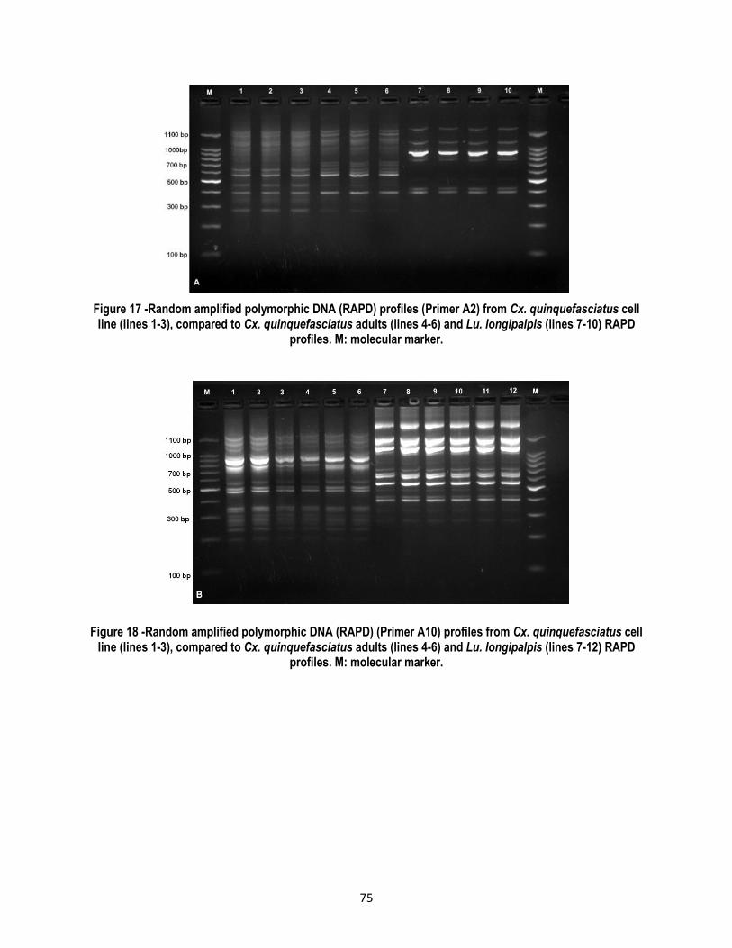
75
Figure 17 -Random amplified polymorphic DNA (RAPD) profiles (Primer A2) from Cx. quinquefasciatus cell line (lines 1-3), compared to Cx. quinquefasciatus adults (lines 4-6) and Lu. longipalpis (lines 7-10) RAPD
profiles. M: molecular marker.
Figure 18 -Random amplified polymorphic DNA (RAPD) (Primer A10) profiles from Cx. quinquefasciatus cell line (lines 1-3), compared to Cx. quinquefasciatus adults (lines 4-6) and Lu. longipalpis (lines 7-12) RAPD
profiles. M: molecular marker.

76
Figure 19 -Random amplified polymorphic DNA (RAPD) (Primer A20) profiles from Cx. quinquefasciatus cell line (lines 1-3), compared to Cx. quinquefasciatus adults (lines 4-6) and Lu. longipalpis (lines 7-12) RAPD
profiles. M: molecular marker.
Figure 20 -Random amplified polymorphic DNA (RAPD) profiles (Primer E07) from Cx. quinquefasciatus cell line (lines 1-3), compared to Cx. quinquefasciatus adults (lines 4-6), Lu spinicrassa (lines 7-9) and Lu.
longipalpis (lines 10-12) RAPD profiles. M: molecular marker.
The similarity coefficient values between DNA bands from cell cultures and Cx. quinquefasciatus adults were identical.
In contrast, values obtained by comparing patterns from the Cx. quinquefasciatus and Lulo cell lines, and for one case
in L. spinicrassa cell line (primer E07), showed different trends (Table 7).

77
Table 7 - Similarity coefficients for RAPD profiles using four different primers
Primer Cx. quinquefasciatus cell
line compared to Cx. quinquefasciatus adults
Cx. quinquefasciatus cell line compared to
Lutzomyia longipalpis cell line
Cx. quinquefasciatus cell line compared to
Lutzomyia spinicrassa cell line
A2 1 0.3 --
A10 1 0.2 --
A20 1 0.35 --
E07 1 0.28 0.35
4.4. Discussion
One of the most important variables to be taken into account in order to achieve the initiation of cell cultures is the
embryogenesis time of the eggs used in tissue explants. In the present study, an optimal incubation time of 16 to 20 h
was determined for the eggs in order to obtain cell cultures, which corresponded approximately to 2/3 parts of the total
time used for embryo formation before egg hatching (Meillon et al. 1967). A similar situation has been reported in
previous works on the establishment of cell lines derived from mosquitoes (Bello et al. 1997). However, the
embryonated eggs that produced the best results at the initiation of the cell cultures were those that received, besides
the first incubation period, an additional incubation period set at 28°C for 4 to 6 h. It was also evidenced that eggs
retained their viability for up to 24 h when they were refrigerated at 4 °C, and also, that after leaving them at room
temperature for 30 min and incubating them under the additional conditions previously indicated, they produced better
results at obtaining primary cell cultures. Similar results using this same technique were reported before, suggesting
that it is possible that such temperature changes stimulate cell division (Oelofsen et al. 1990).
Cell growth evolution in primary cell cultures before the formation of the confluent monolayer was relatively slow, as
shown in other works on Diptera and Hymenoptera orders (Goblirsch et al. 2013; Cruz & Bello 2013; Sudeep et al.
2009). Also, the cell division and growth patterns were characteristic of insect cell cultures (Goblirsch et al. 2013),which
depended initially on the embryonic tissue fragments that adhered firmly to the flask surface after 12 hours of being
explanted.Subsequently, cells began migrating and proliferating from these tissues.
Additionally, during the initial stages, the pulsating movement of cell cultures allowed us to infer that the activity of
muscle tissue was dependent on contractile proteins. Also, there were fiber connections observed from several
fragments. This is one reason which explainswhy the movement in covered areas appeared more extensively; this
matches previous observations (Hsu et al. 1970) in ovaric cell cultures of Cx. quinquefasciatus, as well as in others
species form the same order (Cruz & Bello 2013). Nevertheless, in the present study, cells during the active growth
state detached from fragments in order to constitute cell focalized colonies, facilitating the formation of the the confluent
monolayer.

78
Another important characteristic ofthe cell growth pattern was the occurrence of vesicles surrounded by cells, which
facilitate the confluent monolayer formation. The occurrence of vesicles in insect cell cultures, as well as in other
arthropods, has been a very common characteristic found in different studies (Charpentier et al. 1995; Rey et al. 2000;
Silva et al. 2008).
The culture medium, where cells were able to adapt, grow and proliferate, corresponded to the mixture of equal parts
of Grace and L-15 media, which is an indication that this medium provided the substances, both in quantity and quality
that were needed for the initiation of Cx. quinquefasciatus primary cell cultures. However, cell growth and division of
the first five subcultures were slow, entailing duration of 30 days between one sub-culture and the other, and displayed
similar characteristics as the primary cultures. Furthermore, some particles were observed in the cells from the cultures
and medium, which possibly correspond to metabolites produced in response to excess or deficiency of some
substances; nevertheless, after the sixth sub-culture these metabolites were not observed.
Cell morphology at the beginning of the primary cell cultures was heterogeneous, but later in the confluent monolayer
and subcultures, the predominant cell shape was epithelioid. This is explained by different tissues that proliferated and
initiated cell growth in accordance with the nature of the mixed culture. The predominant cell shapewas the sameas in
many other studies since the establishment of mosquito cell cultures (Bello et al. 2001; Zapata et al. 2005; Sudeep et
al. 2009).
The number of chromosomes in insect cell lines canrange from a few pairs in dipterous (flies) (Cruz & Bello 2013) to
hundreds in lepidopterous (butterflies) (Zhang et al. 2012). In relation to the karyological analysis of Cx.
quinquefasciatus cell culture, the number of chromosomes matches the previous reports on other cell lines for the
Culicidae family, including species from the Culex genus (Sudeep et al. 2009; Athawale et al. 2002; Hsu et al. 1970).
In addition, the number and morphology of the Culicidae chromosomes is highly retained in the whole family (2n=6)
(Kitzmiller 1976). Additionally, the absence of polyploidy demonstrates the integrity of this cell line, showing that no
transformations occurred, such as was reported in a cell line from Aedes aegypti neonate larvae, which showed more
than 75% of cells containing a diploid number of chromosomes (Sudeep et al. 2009). Taking into account that
subcultures from passages 15 were evaluated for karyological analysis and not at passage 37, which corresponded to
the last passage that was carried out, it is unlikely that this cell line had reached spontaneous transformation, as has
been describe before in other cell lines (Rey et al. 2000; Hoshino et al. 2009).
Isoenzymatic profiles for Cx. quinquefasciatus cell line totally match samples from the immature forms of the same
species, showing in all cases the same mobility which serves as indication of a common origin. However, when
comparing isoenzymatic profiles for the four systems assessed with patterns obtained from Lulo and L. spinicrassa-
derived cell lines, differences in the donor isoenzymatic profile were observed indicating diverse origins, and rejecting
cross contamination among cell lines used in the Medical and Forensic Entomology Laboratory from Universidad del

79
Rosario. This methodology is a common practice in characterizing and authenticating different cell lines from insects
(Rey et al. 2000; Zapata et al. 2005; Cruz & Bello 2013); in addition, this methodology could be used in order to
periodically verify that each cell line continues as a pure cell culture.
The identity of the new cell line was also determined using RAPD profiles. Differences were not observed between Cx.
quinquefasciatus cell line and its adult samples, showing a similarity of 1 in the four primers used, confirming the cell
line identity. This result suggests that cell cultures did not lose genetic material and that the molecular composition,
according to these markers, reflected the low allelic diversity of the colonized Cx. quinquefasciatus strain (Léry et al.
2003). In contrast, comparing RAPD profiles of the new cell line with Lulo and L. spinicrassa-derived cell lines, a
similarity coefficient lower than 0.3 was found, corresponding to the homology of some bands (2 in most cases). This
indicated that cell lines come from different insect species. The four primers used were able to differentiate and confirm
the identity of the original sources of Cx. quinquefasciatus, L. longipalpis and L. spinicrassa cell lines and determine
that there was not cross contamination. The efficiency of this technique has been previously reported, and it has been
established that it can differentiate related species but not clones of cell lines (Ahmen et al. 2014). In L. longipalpis and
L. spinicrassacases, it was observed that primer E07 was able to differentiate both species from the same genus;
therefore, it became an effective tool to authenticate cell lines of taxonomical proximity (McIntosh et al. 1996; Kawai &
Mitsuhashi 1997).
4.5. References
Acosta, E.G., Castilla, V. & Damonte, E.B., 2011. Infectious dengue-1 virus entry into mosquito C6/36 cells. Virus research, 160(1-2), pp.173–9. Available at: http://www.ncbi.nlm.nih.gov/pubmed/21708195 [Accessed July 4, 2013].
Ahmen, I., Huebner, H. & Buchholz, R., 2014. Establishment and Characterization of three New Embryonic Spodoptera littoralis Cell Lines and Testing their Susceptiblity to SpliMNPV. JSM Biotechnologu & Biomedical Engineering, 2(1), pp.1–9.
Arif, B. & Pavlik, L., 2013. Insect cell culture: virus replication and applications in biotechnology. Journal of invertebrate pathology, 112 Suppl, pp.S138–41. Available at: http://www.ncbi.nlm.nih.gov/pubmed/22841637 [Accessed August 9, 2014].
Athawale, S.S. et al., 2002. A new cell line from the embryonic tissues of Culex tritaeniorhynchus and its susceptibility to certain flaviviruses. Acta virologica, 46(4), pp.237–40. Available at: http://www.ncbi.nlm.nih.gov/pubmed/12693860.
Bello, F. et al., 1997. Establishment and characterization of a cell line from the mosquito Anopheles albimanus (Diptera: Culicidae). Memórias do Instituto Oswaldo Cruz, 92(1), pp.123–8. Available at: http://www.ncbi.nlm.nih.gov/pubmed/9302423.
Bello, F., 2009. Estudio de cultivos celulares primarios derivados de Lucilia sericata ( Diptera : Calliphoridae )*.
Bello, F.J. et al., 2001. A new continuous cell line from the mosquito Psorophora confinnis (Diptera: Culicidae) and its susceptibility to infections with some arboviruses. Memórias do Instituto Oswaldo Cruz, 96(6), pp.865–73. Available at: http://www.ncbi.nlm.nih.gov/pubmed/11562717.
Brown, S.E. & Knudson, D.L., 1980. Characterization of invertebrate cell lines. In vitro, 16(10), pp.829–832.

80
Charpentier, G. et al., 1995. Establishment and characterization of three cell lines from Aedes triseriatus (Diptera: Culicidae). Journal of medical entomology, 32(6), pp.793–800. Available at: http://www.ncbi.nlm.nih.gov/pubmed/8551501.
Coen, E., Strachan, T. & Dover, G., 1982. Dynamics of concerted evolution of ribosomal DNA and histone gene families in the melanogaster species subgroup of Drosophila. Journal of molecular biology, 158(1), pp.17–35. Available at: http://www.ncbi.nlm.nih.gov/pubmed/6811755.
Cruz, M. & Bello, F., 2013. Establishment and characterization of an embryonic cell line from Sarconesiopsis magellanica. Journal of insect science, 13, p.130. Available at: http://www.pubmedcentral.nih.gov/articlerender.fcgi?artid=4014039&tool=pmcentrez&rendertype=abstract.
Forattini, O., Kakitani, I., La Corte Dos Santos, R., Kobayashi, K., Ueno, H. et al., 2000. Potencial sinantrópico de mosquitos Kertesia e Culex (Diptera: Culicidae) no Sudeste do Brasil. Rev Saúde Pública 34: 565-569.
Goblirsch, M.J., Spivak, M.S. & Kurtti, T.J., 2013. A cell line resource derived from honey bee (Apis mellifera) embryonic tissues. PloS one, 8(7), p.e69831. Available at: http://www.pubmedcentral.nih.gov/articlerender.fcgi?artid=3720946&tool=pmcentrez&rendertype=abstract [Accessed August 22, 2014].
Goddard, L.B., Roth, A.R., Reisen, W.K., Scott, T.W., 2002. Vector competence of California mosquitoes for West Nile Virus. Em Infect Dis 8: 1385-1391.
Grace, T.D.C., 1962. Establishment of four strains of cells from insect tissues grown in vitro. Nature 195: 788-789.
Hoshino, K., Hirose, M. & Iwabuchi, K., 2009. A new insect cell line from the longicorn beetle Plagionotus christophi (Coleoptera: Cerambycidae). In vitro cellular & developmental biology. Animal, 45(1-2), pp.19–22. Available at: http://www.ncbi.nlm.nih.gov/pubmed/18946707 [Accessed July 4, 2013].
Hsu, S., Mao, W., & Cross, J., 1970. Establishment of a line of cells derived from ovarian tissue of. Jornal of medical entomology, 7(December 1969), pp.703–7.
Jia, D. et al., 2012. Development of an Insect Vector Cell Culture and RNA Interference System To Investigate the Functional Role of Fijivirus Replication. Journal of virology, 86(10), pp.5800–5807.
Kawai, Y. & Mitsuhashi, J., 1997. An insect cell line discrimination method by RAPD-PCR. In vitro cellular & developmental biology. Animal, 33(7), pp.512–5. Available at: http://www.ncbi.nlm.nih.gov/pubmed/9282311.
Kitzmiller, J.B., 1976. Genetics, cytogenetics, and evolution of mosquitoes. Advances in genetics, 18, pp.315–433. Available at: http://www.ncbi.nlm.nih.gov/pubmed/7931.
Landry, B., Dextraze, L. & Boivin, G., 1993. Random amplified polymorphic DNA markers for DNA fingerprinting and genetic variability assessment of minute parasitic wasp species (Hymenoptera: Mymaridae and Trichogrammatidae) used in biological control programs of phytophagous insects. Genome, 36(3), pp.580–7. Available at: http://www.ncbi.nlm.nih.gov/pubmed/8349128.
Léry, X. et al., 2003. Characterization and authentication of insect cell lines using RAPD markers. Insect Biochemistry and Molecular Biology, 33(10), pp.1035–1041. Available at: http://linkinghub.elsevier.com/retrieve/pii/S0965174803001188 [Accessed September 4, 2014].
Lynn, D., 2001. Novel techniques to establish new insect cell lines. In vitro cellular & developmental biology., 37(6), pp.319–21. Available at: http://www.ncbi.nlm.nih.gov/pubmed/11515961.

81
McIntosh, a H., Grasela, J.J. & Matteri, R.L., 1996. Identification of insect cell lines by DNA amplification fingerprinting (DAF). Insect molecular biology, 5(3), pp.187–95. Available at: http://www.ncbi.nlm.nih.gov/pubmed/8799737.
Meillon, B., Sebastian, A. & Khan, Z.H., 1967. The duration of egg larval and pupal stages Culex pipiens fatigans in Rangoon, Burma. Bull. Org. mond. Santé, 36, pp.7–14.
Nei, M. & Li, W.H., 1979. Mathematical model for studying genetic variation in terms of restriction endonucleases. Proceedings of the National Academy of Sciences of the United States of America, 76(10), pp.5269–73. Available at: http://www.pubmedcentral.nih.gov/articlerender.fcgi?artid=413122&tool=pmcentrez&rendertype=abstract.
Oelofsen, M.J. et al., 1990. Establishment and characterization of a cell line from the mosquito Culex (Culex) theileri (Diptera: Culicidae) and its susceptibility to infection with arboviruses. Journal of medical entomology, 27(6), pp.939–44. Available at: http://www.ncbi.nlm.nih.gov/pubmed/2280394.
Prieto, L. et al., 2009. Estudio de cultivos celulares primarios derivados de Lucilia sericata (Diptera: Calliphoridae). Rev. Cienc. Salud, 7(3), pp.53–64.
Rey, G.J., Ferro, C. & Bello, F.J., 2000. Establishment and characterization of a new continuous cell line from Lutzomyia longipalpis (Diptera: psychodidae) and its susceptibility to infections with arboviruses and Leishmania chagasi. Memórias do Instituto Oswaldo Cruz, 95(1), pp.103–10. Available at: http://www.ncbi.nlm.nih.gov/pubmed/10656714.
Rivas, F., Diaz, L., Cárdenas, V., Daza, E., Bruzón, et al., 1997. Epidemic Venezuelan Equine Encephalitis in La Guajira, Colombia 1995. J Infect Dis 175: 828-832.
Silva, L.M. et al., 2008. Primary culture of venom glands from the Brazilian armed spider, Phoneutria nigriventer (Araneae, Ctenidae). Toxicon, 51(3), pp.428–34. Available at: http://www.ncbi.nlm.nih.gov/pubmed/18068746 [Accessed September 1, 2014].
Stevens, J. & Wall, R., 1997. the Blowflies. Biochemical Systematics and Ecology, 25(2), pp.81–97.
Sudeep, a B. et al., 2009. Establishment and characterization of a new Aedes aegypti (L.) (Diptera: Culicidae) cell line with special emphasis on virus susceptibility. In vitro cellular & developmental biology. Animal, 45(9), pp.491–5. Available at: http://www.ncbi.nlm.nih.gov/pubmed/19533252 [Accessed July 4, 2013].
Varina, M. & Pudney, M., 1969. The growth and seriala passages of cell lines from Aedes aegypti (L.). Journal of medical entomology, 6(4), pp.432–39.
Williams, J. et al., 1990. DNA polymorphisms amplified by arbitrary primers are useful as genetic markers. Nucleic acids research, 18(22), pp.6531–5. Available at: http://www.pubmedcentral.nih.gov/articlerender.fcgi?artid=332606&tool=pmcentrez&rendertype=abstract.
Zapata, A.C., Cárdenas, E. & Bello, F., 2005. Characterization of cell cultures derived from Lutzomyia spinicrassa (Diptera: Psychodidae) and their susceptibility to infection with Leishmania (Viannia) braziliensis. Medical science monitor, 11(12), pp.BR457–64. Available at: http://www.ncbi.nlm.nih.gov/pubmed/16319783.
Zhang, X. et al., 2012. Characterization of a new insect cell line that is derived from the neonate larvae of Papilio xuthus (Lepidoptera: Papilionidae) and its susceptibility to AcNPV. Tissue & cell, 44(3), pp.137–42. Available at: http://www.ncbi.nlm.nih.gov/pubmed/22386749 [Accessed September 4, 2014].

82
5. Differences in Replication efficiency of alpha-and flaviviruses in insect and mammalian cells
5.1. Introduction
DENV and CHIKV are the most common vector-born viral diseases in humans. Both viruses are distributed in tropical
and subtropical regions, and both are transmitted by Aedes aegypti and Aedes albopictus (Chen & Wilson 2010).
According to The WHO, between 50-100 million infections of dengue occur annually (WHO 2009). However, Bhatt et
al. (2013) recently estimated, based on cartographic methods that 390 million dengue infections occur each year. The
dengue mortality rate varies from 1.2 – 3.5% (WHO 2009). In contrast, fatality rates for CHIKV infections are around 1
in 1000; however, about half of patients evolve into a chronic state. During this state, patients may present signs of
persistent polyarthralgia and stiffness that can persist for months to years (Brighton et al. 1983; Simon et al. 2008;
Manimunda et al. 2010).
There are five different DENV serotypes; however, only serotypes 1-4 have been found in humans (Normile 2013). A
primary infection by DENV induces protective immunity only against the infecting serotype; whereas a secondary
infection with another serotype might produce DHF/DSS (WHO 2009) due to an immunopathological process of
antibody-dependent enhancement (ADE) (Rothman 2004). In contrast, protective antibodies for CHIKV infection, once
developed, produce life-long immunity (Nitatpattana et al. 2014). Despite the fact that control of main mosquito vectors
has failed and the alarming increasing number of dengue and chikungunya fever cases, there are no antiviral therapy
against these viruses and the only option for dengue is vaccination.
The viral genome of flaviviruses such as DENV and YFV constitutes a positive single stranded RNA, which is
approximately 10-11kb in length. The viral genome has a 5' m7GpppN cap like, but lack a 3' poly (A) tail (Chiu et al.
2005). Its single ORF encodes structural proteins such as C, M and E, as well as seven non-structural proteins (NS1,
NS2A, NS2B, NS3, NS4A, NS4B and NS5). Structural proteins, especially E protein,are associated with virion binding
and attachment to the host cell (Rey 2003); meanwhile, non-structural proteins are responsible for the replication
complex (Guzman et al. 2010). NS1 is associated with the activation of human complement, probably at the sites of
vascular leakage (Avirutnan et al. 2006); NS2A is an IFN antagonist and possesses helicase activity (Lindenbach &
Rice 2007; Rodenhuis-Zybert et al. 2010); NS3 also has helicase activity, but together with NS2B it has protease action
(Acheson 2007); NS4A and NS4B are IFN antagonists (Muñoz-Jordan et al. 2003) and finally, NS5 exerts RNA
polymerase activity (Acheson 2007).
Alphaviruses, such as CHIKV, SINV, SFV, and ONNV are enveloped virions that contain one single-stranded, positive-
sense RNA genome ranging from 11.44 Kb (SFV and VEE) to 11.84Kb (ONN) bases in length. The viral genome has
two sequential ORFs; it has a capped UTR on its 5’ end, and a poly-adenylated UTR on its 3’ end (Strauss & Strauss
1994). The first ORF, which covers around 2/3 of the genome, encodes non-structural proteins nsP1, nsP2, nsP3 and

83
nsP4 and the second ORF encodes structural proteins C, E3, E2, 6K and E1. nsP1 carry out methyltransferase activity
(Sreejith et al. 2012), nsP2 has helicase and protease tasks (Takkinen & Kääriäinen, 1991; Gomez de Cedrón et al.,
1999;), nsP3 is a multifunctional protein containing the ‘macro’ or ‘X’ domain, (Malet et al. 2009) and nsP4 which acts
as RdRp and polyadenylate polymerase (Hahn et al., 1989; Tomar et al., 2006). The recognition and attachment of the
viral particle to the host cell is mainly a function of E1 and E2 proteins, meanwhile E3 stabilizes E1 domains respect to
E2 (Voss et al. 2010), finally the ion channel 6K is involved in the correct assembly of fully-infectious virus particles
(Melton et al., 2002; McInerney et al., 2004).
The events involved in virus entry comprise virion binding to the cell surface, followed by endocytosis and uncoating; in
addition, there are some key factors for viral pathogenesis, such as host range and tissue tropism, which include the
great diversity of in vivo and in vitro cellular tropism, as well a different receptor(s) dependent on the type of the host
cell infected (Modis et al. 2004, Acosta et al. 2008); the aforementioned factores increase the complexity of this process.
DENV and CHIKV infect target cells by attaching to different cell receptors, many of which are still unknown. In vitro
DENV has shown to infect lots of cell lines from different origins (Barr & Anderson 2013). It could be that the virus must
bind to a ubiquitous cell-surface molecule, or exploit multiple receptors to mediate infection (Rodenhuis-Zybert et al.
2010). Several candidate receptors have been identified, suggesting that flavi and alphaviruses are capable of using
different molecules in order to enter the cell.
Several DENV receptors have been described on insect cells, but in general these receptors differ from those on
mammalian cells. On C6/36 cells, two proteins with molecular masses of 80 and 67-kDa were suggested as receptors
for DENV1 to DENV4 (Mercado-Curiel et al. 2006). Additionally, for this cell line, DENV2 and DENV4 bound to two
glycoproteins of 40 and 45 kDa located on the cell surface (Martínez-Barragán & del Angel 2001; Salas-Benito et al.
2007). Also, Prohibitin has been characterized as a DENV2 receptor protein on C6/36 and CCL-125 (Kuadkitkan et al.
2010). Regarding to DENV receptors on Vero cells, HS has been identified. It can act as a receptor or concentrate
DENV2 on the cell surface and facilitate the interaction with specific high-affinity receptors (Chen et al. 1997; Germi et
al. 2002). Moreover, two proteins of 74 and 44 kDa were described as DENV4 receptors on this cell line (Martínez-
Barragán & del Angel 2001). There is no information about CHIKV receptors on Vero cells; however, prohibitin, that
was also characterized on C6/36 insect cells and CCL-125 cells as DENV-2 receptor protein, was also identified as a
receptor for CHIKV on CHME-5 cells (Kuadkitkan et al. 2010).
In nature, mosquitoes and mammals play an essential role in propagating arboviruses such as DENV, YFV, CHIKV and
SINV among many others. Regarding the successful viral propagation of arboviruses in insect and mammalian cells,
both types of cells can share or present unique host factors (Sessions et al. 2009) and, at least in part, these host
factors might explain similarities as well as differences in the replication efficiency of these viruses.

84
The Vero cell line was derived from the kidney of a Cercopithecus aethiops adult (Afrikan green monkey) in 1962 by
Yasamura at the Chiba University in Japan (Sheets 2000). However, this species was re-classified into four different
species and using mitochondrial DNA analysis, it was established that Vero cells come from C. sabaeus (Osada et al.
2014). The characteristics that make Vero cells one of the most frequently used cell lines in virology are: the highly
susceptibility to arboviruses (Acosta et al. 2009; Nougairede et al. 2013), adenoviruses (Damen et al. 2008),
enteroviruses (Liu et al. 2011) and influenza viruses (Zhai et al. 2012). This cell line has shown different karyotypes
with chromosomes ranging from 52 to 62 (Osada et al. 2014). In addition, Vero cell line has beenbroadly used for the
development of vaccines (Murakami et al. 2007; Tseng et al. 2011).
C6/36 is a genetical homogeneus clonal cell line (Igarashi 1978) generated from a cell line derived of Aedes albopictus
larval tissues (Singh 1967). C6/36 clonal cell line is higly susceptible to different arboviruses, such as flaviviruses
(DENV, Rocio virus (ROC), SLEV); alphaviruses (CHIKV, SINV, EEE) and bunyaviruses (LACV (Igarashi 1978; White
1987). This clonal cell line showed reduced susceptibility to Adenoviruses, Coxsakieviruses, Enteroviruses and Herpes
simplex virus and no viral infection was detected with Influenza, Parainfluenza and Measles viruses (White 1987). The
predominant number of cromosomes in these cells was 2n=6, with some tetraploid types (Igarashi 1978). This cell line
is easy to handle in a laboratory and can be stored at room temperature for up to two weeks without changing the
medium cell culture; also, it has a high split ratio of 1:10 and grows into a confluent monolayer in 4 days (White 1996).
Lulo is a cell line derived from Lutzomyia spinicrasa neonate larvae (a sand fly). Although Lulo has been used mainly
for studying parasites from Leishmania genera (Zapata et al. 2005), it has been established that this cell line is
susceptible to the infection of different arboviruses, such as the alphavirus Mayaro virus (MV), the flavivirus Ilheus virus
(ILHV), the orvivirus Changuinola virus, the phlebovirus PTV and the vesiculovirus Vesicular stomatitis virus (VSV) (Rey
et al. 2000). The karyologycal analysis showed the presence of haploid, aneuploid and tetraploid cells; however, around
85% of the cells presented 2n=8 (Rey et al. 2000). Similarly to C6/36, this cell line is easy to handle in a laboratory and
can be maintained at room temperature.
In this study, the relative replication fitness of flaviviruses DENV and YFV and alphaviruses CHIKV and SINV was
assessed in Vero and C6/36 cells, which are commonly used for viral propagation, as well as in the Lulo cell line, which
has hardly been studied at the virological level.

85
5.2. Materials and Methods
5.2.1. Cells and viruses
Vero-A (ATCC CCL-81) cells were maintained in a minimal essential medium (MEM Rega-3, Gibco, Belgium),
supplemented with 10% FBS (Integro, The Netherlands), 1% L-glutamine (Gibco) and 1% sodium bicarbonate (Gibco).
Baby hamster kidney cells (BHK-21; ATCC CCL10) were cultured in Dulbecco’s modified Eagle’s medium (DMEM;
Invitrogen), supplemented with 10% FBS and 1mM sodium pyruvate (Invitrogen). C6/36 (ATCC CRL-1660) cells were
maintained in Grace's Insect Medium (Grace Life Technologies, Belgium), supplemented with 10% FBS and 1% L-
glutamine; Lulo cells were maintained in a mix 1:1 of Grace and Leibovitz media (L-15 medium, Gibco, Belgium) and
supplemented with 10% FBS. Insect cell cultures were maintained at 28°C without CO2 atmosphere, the mammalian
cell culture was maintained at 37°C in an atmosphere of 5% CO2 and 95%–99% humidity.
V. Deubel (formerly at Institut Pasteur, Lyon, France) kindly provided DENV serotype 2 New Guinea C (NGC) strain
which was cultured in C6/36 cells. YFV-17D vaccine strain (Stamaril) (Aventis Pasteur MSD, Brussels, Belgium) was
passaged into Vero cells in order to prepare a working virus stock. Professor S. Gunther (Bernhard Nocht Institute for
Tropical Medicine, Hamburg, Germany) generously provided CHIKV Indian Ocean strain 899 (GenBank FJ959103.1)
which was cultured in Vero cells. SFV (SFV; Vietnam strain, GenBank EU350586.134) belongs to the collection of the
Rega Institute of Medical Research, Belgium.
5.2.2. Viral infections in insects and mammalian cells
Vero, C6/36 and Lulo cells were seeded in 12.5 mL flasks (Falcon®) using growth media. The number of cells seeded
for each cell line was different in order to reach 70 – 80% confluence during the following 24h. The number of cells
seeded per line was: Vero cells, 600.000 cells/flask; Lulo cells, 900.000 cells/flask and C6/36 cells, 1 × 10⁶ cells/flask.
Following 24h of incubation, the growth media was replaced with 2 mL of assay medium, which is the respective cell
growth media, supplemented with 2% instead of 10% FBS and in addition, containing the appropriated virus dilution
(Table 8). After 2h of incubation at room temperature with continuous shaking, cell monolayers were washed 3 times
with PBS in order to remove non-adsorbed viruses and cultures were further incubated (Table 8). Flasks were monitored
daily for CPE development and cells and supernatants were harvested. Total and viral RNA were isolated using the
NucleoSpin ® RNA (Macherey Nagel Düren, Germany) and the NucleoSpin 96 virus kit (Macherey Nagel Düren,
Germany), respectively.
Each assay was carried out by triplicate in three independent assays and mock-infected cell cultures were used as
control.

86
Table 8 - Infections of flaviviruses and alphaviruses in insect and mammalian cells
Virus Cell line MOI Supernatant collected
DENV2 NGC Vero, C6/36, Lulo 0.001 Each 48h
DENV2 NGC Lulo 0.1 and 0.5 Each 48h
YFV 17D Vero, C6/36, Lulo 0.01 Each 48h
CHIKV 899 Vero, C6/36, Lulo 0.001 Each 24h
CHIKV 899 Vero, C6/36, Lulo 0.00001 Each 6, 16, 28, 40, 52, 64 and 76h
SINV Vero, C6/36, Lulo 0.00001 Each 6, 16, 28, 40, 52, 64 and 76h
5.2.3. DENV binding and entry assay
A binding assay was carried out in order to determine whether the viral infection detected by the plaque assay and by
the immunofluorescence antibody assay (IFA) in the supernatant from Lulo infected at high MOI was due to virus
replication and not carry-over. For this, 1 x 10⁶ C6/36 cells and 900.000 Lulo cells were seeded in 12.5 mL flasks
(Falcon®) using the growth medium. Following 24h of incubation, the growth medium was replaced with 2 mL of assay
medium at 4C. DENV2 NGC infections were carried out at 4C using a MOI of 0.05, and then cells were incubated at
the same temperature for 20 minutes. Later, monolayers were extensively washed with PBS, cells were immediately
harvested and total RNA was isolated using the NucleoSpin® RNA (Macherey Nagel Düren, Germany). In order to
establish the successful entry of DENV in these cell lines the same protocol was followed but this time, the assay was
carried out at room temperature and the time of incubation was extended to 45 minutes.
5.2.4. Plaque assay
BHK cells were seeded in 12-well plates (IWAKI) at a density of 5 × 10⁵ cells/well in 10% FBS medium. Following 24h
of incubation, monolayers were washed 3 times with PBS and cells were infected with 500 µl of serial 1:10 supernatant
dilutions, previously prepared in the assay medium. After an hour of infection with continuous shaking, monolayers
were washed 3 times with PBS in order to remove non-adsorbed viruses. Each monolayer was coated with 1.5 mL of
a sterile solution containing 2% Avicel PH-101 (Sigma-Aldrich) and 2% FBS medium. For DENV, monolayers were
carefully washed 3 times with PBS after 6 days of incubation and for CHIKV, monolayers were washed after 5 days of
incubation. Finally, monolayers were fixed with 70% ethanol and stained with blue methylene in order to visualize and
count the plaque-forming units (PFU). Each assay was carried out by triplicate.

87
5.2.5. DENV2, YFV and CHIKV quantitative reverse transcription-PCR (qRT-PCR)
For DENV primers and probe sequences were as described earlier: DENV-For 5’TCGGAGCCGGAGTTTACAAA3’,
DENV-Rev 5’TCTTAACGTCCGCCCATGAT3’, DENV-Probe FAM-5’ATTCCACACAATGTGGCAT-MGB3’ (Kaptein et
al. 2010); CHIKV primers and probe sequences selected were as published before: ChikSII 5’-
CCGACTCAACCATCCTGGAT-3’, ChikAsII 5’-GGCAGACGCAGTGGTACTTCCT-3’ and ChikProbe 5’-FAM-
TCCGACATCATCCTCCTTGCTGGC-TAMRA (Panning et al. 2008) and YFV primers and probe sequences were: YFV-
For 5’TGGCATATTCCAGTCAACCTTCT3’, YFV-Rev 5’GAAGCCCAAGATGGAATCAACT3’ and YFV-Probe FAM-
TTCCACACAATGTGGCATG-MGB (Kaptein et al. 2010). One-step, quantitative RT-PCR was carried out in a total
volume of 25 µl, containing 13.94 µl H2O, 6.25 µl master mix (Eurogentec, Seraing, Belgium), 0.375 µl of 60 µM
forward primer, 0.375 µl of 60 µM reverse primer, 1 µl of 10 µM probe, 0.0625 µl reverse transcriptase (Eurogentec)
and 3 µl RNA sample. qRT-PCR was carried out using the ABI 7500 Fast Real-Time PCR System (Applied Biosystems,
Branchburg, NJ) under the following conditions: 30 min at 48C and 10 min at 95C, followed by 40 cycles of 15 s at
95C and 1 min at 60C. Data were analyzed using the ABI PRISM 7500 SDS software (version 1.3.1; Applied
Biosystems). For absolute quantification, standard curves were generated using 10-fold dilutions of template
preparation of known concentrations.
5.2.6. Immunofluorescence Antibody assay (IFA)
BHK cells were seeded in a 8-well chamber slide (Lab-teck, II, Nune, Germany) at a density of 8000 cells/well; 24h
later, cells were infected using 1:10 serial dilutions of DENV2 NGC supernatants obtained from infection assays in the
three cell lines at a MOI of 0.001; furthermore, supernatants obtained from Lulo infection assays at a MOI of 0.01 and
0.05 were used. The virus inoculum was removed after 1h, cells were washed and then incubated for 72h. Cells were
stained with the anti-dengue E protein antibody (Ab) clone 3H5 (Millipore, Billerica, MA) and the secondary antibody
AbAlexa Fluor 488 (Millipore). Following DAPI (4',6-diamino-2-fenilindol) staining, the cultures were visualized using a
confocal laser-scanning microscope (LCSM, Leica Microsystems, Germany).
5.3. Results
5.3.1. Microscopical findings during viral infections
When C6/36 and Lulo cells were infected with DENV2 (MOI 0.001, 0.01 and 0.05), they did not show CPE signs;
however, C6/36 infected cells started to die sooner than the mock-infected C6/36 cells. During DENV2 infection, a state
of “crisis” was observed in the C6/36 culture on day 6 p.i., during which the cells showed poor growth. However, the

88
cell line overcame this state on day 8 p.i. In contrast, due to over confluence, Lulo-infected cells and mock-infected
cells started to form cell clumps by day 10 p.i., Contrarily, Vero infected cell behavior (MOI 0.001) was completely
different and a strong CPE was observed after day 4 p.i., and this cell culture started to die on day 6 p.i. C6/36, all Vero
cells had died by day 10 p.i. and Lulo cell cultures were not in good conditions. Whereas, the CPE observed during
YFV infection was very similar to the CPE observed when cells were infected with DENV2.
In contrast, when the three cell cultures were infected with CHIKV, CPE was extremely strong in Vero cells and was
evident in C6/36 cells, but not in Lulo cells. When cells were infected at a MOI of 0.00001, Vero cells started to die as
soon as day 2 p.i., and by day 4 p.i., around 70% of the cell culture had died; in contrast, C6/36 cells showed slight
signs of CPE as soon as day 3 p.i. CPE was much more observable when Vero cells were infected at a MOI of 0.001
and on day 2 p.i., around 50% of the cells of this cell culture died due to the strong viral infection. C6/36 cells showed
perceptible CPE signs that became more evident over time. Contrarily, Lulo cells never showed CPE signs (Figure 21).
In contrast, when the cells were infected at a low MOI, CPE appearance was observable in Vero cells after 40 hours
p.i., and in C6/36 cells it was almost observable by 76 hours p.i., but Lulo cells did not show any CPE signs. During
SINV infection, three cell cultures presented a very similar CPE response to the viral infection, which was observable
during CHIKV infection.
Figure 21 - CPE in insects and mammalian cells infected with CHIKV (MOI 0.001).

89
5.3.2. Viral production of flaviviruses and alphaviruses in insect and mammalian cells
5.3.2.1. DENV2 production
The viral progeny able to infect new cells was quantified trough plaque assays using 1:10 dilutions of supernatants
previously obtained (Table 8). In general, during DENV2 infection (MOI 0.001), C6/36 culture was able to produce a
higher number of infectious viral particles when compared with the supernatant obtained from Vero cells, and the higher
DENV2 titer was obtained on day 4 p.i. in C6/36 and on day 6 in Vero cells. Also, a state of “crisis” was observed in the
C6/36 culture on day 6 p.i., during which cells showed poor growth. However, the cell line overcame this state by day
8 p.i., and the cells were able to produce DENV2 in far higher titers than Vero. The Lulo insect cell line was almost non-
infectable under these conditions (Figure 22).
Figure 22 - In vitro DENV2 replication efficiency of insect and mammalian cells infected (MOI 0.001).
These findings were confirmed by IFA, which showed that the higher DENV2 titers were produced by C6/36, followed
by Vero and finally, that Lulo cells were not infected. Therefore, infectious DENV2 were not produced in the latter cells
(Figure 23).
0
1
2
3
4
5
2 4 6 8 10
Log 1
0P
FU/m
l DEN
V2
days p.i.
C6/36 Vero M LULO

90
Figure 23 -C6/36 and Vero cells are able to produce infectious DENV2 Vero cells infected with DENV2 1:10 dilutions from C6/36, Vero and Lulo cell cultures infected at MOI of 0.001, mock-infected cells were used as uninfected cell controls. DENV2 E protein expression was visualized on day 3 p.i. (anti-
dengue E protein antibody clone 3H5, antibody AbAlexa Fluor 488 (green) and DAPI staining (blue)).
When Lulo cells were infected at high MOIs (0.01 and 0.05) with DENV2, infectious DENV2 was produced as soon as
day 2 p.i., and these viral productions showed a slow increase every day up to day 10 p.i. (Figure 24 and 25). In
addition, viral production was a bit higher when cells where infected at a MOI of 0.05 than a MOI of 0.01. When
comparing DENV2 production in both Lulo and C6/36 insect cells, remarkable viral production was observed in C6/36
cells (MOI 0.001) and it was 3 to 4 log10 higher than in Lulo (MOIs 0.01 and 0.05) (Figure 22 and 25).
Figure 24 - Viral infectious progeny from Lulo infected DENV2 cells developed by plaque assay in BHK cells.

91
Figure 25 - In vitro DENV2 replication efficiency in Lulo cells (MOI 0.05 and 0.01). DENV2 production was calculated as PFU/ml by plaque assays. Data comprise mean values of three independent
experiments.
Altogether, the infectious virus titer in the supernatant from Lulo infected cell cultures was determined by IFA, which
further confirmed the production of infectious virus particles (Figure 26). In addition, a binding assay was carried out in
order to determine whether the viral infection, detected by plaque assay and by IFA in the supernatant from Lulo cells
infected at high MOI, was due to virus replication and not carry-over. Surprisingly, DENV2 was found to attach more
efficiently to Lulo cells than to C6/36 cells (Figure 27). In general, virion binding to the cell surface (attachment) and
subsequent uptake into the cytoplasm are key factors for the virus to enter a specific host cell type. In Lulo cells, the
low efficiency of viral progeny production is not due to poor attachment; in fact, DENV2 binds more efficiently to Lulo
cells than to C6/36 cells, although DENV2 entry was very similar between both insect cell cultures, being a little bit
higher in C6/36 cells. Therefore, the inefficient viral production in Lulo cells could be explained by other downstream
events.
0
1
2
3
4
5
2 4 6 8 10
Log 1
0P
FU/m
l DEN
V2
days p.i.
0.05 0.01

92
Figure 26 - Lulo cells are able to produce infectious DENV2 BHK cells infected with DENV2 1:10 serial dilutions from Lulo cell cultures infected at MOIs of 0.01 (panels B, C and
D) and 0.05 (F, G and H), mock-infected cells were used as uninfected cell controls (panels A and E). DENV2 E protein expression was visualized on day 3 p.i., (anti-dengue E protein antibody clone 3H5, antibody AbAlexa Fluor
488 (green) and DAPI staining (blue)).
Figure 27 - DENV2 binds and enters efficiently in insect cell cultures Lulo cells were infected with DENV2 at a MOI of 0.05. Following infection, monolayers were extensively washed with PBS. Immediately thereafter, RNA was isolated from each cell culture and analysed by qRT-PCR as a control, C6/36
cells were included in the assay. Data comprise mean values of three independent experiments.
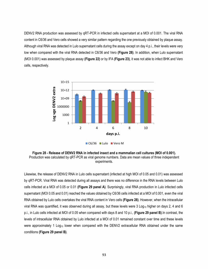
93
DENV2 RNA production was assessed by qRT-PCR in infected cells supernatant at a MOI of 0.001. The viral RNA
content in C6/36 and Vero cells showed a very similar pattern regarding the one previously obtained by plaque assay.
Although viral RNA was detected in Lulo supernatant cells during the assay except on day 4 p.i., their levels were very
low when compared with the viral RNA detected in C6/36 and Vero (Figure 28). In addition, when Lulo supernatant
(MOI 0.001) was assessed by plaque assay (Figure 22) or by IFA (Figure 23), it was not able to infect BHK and Vero
cells, respectively.
Figure 28 - Release of DENV2 RNA in infected insect and a mammalian cell cultures (MOI of 0.001). Production was calculated by qRT-PCR as viral genome numbers. Data are mean values of three independent
experiments.
Likewise, the release of DENV2 RNA in Lulo cells supernatant (infected at high MOI of 0.05 and 0.01) was assessed
by qRT-PCR. Viral RNA was detected during all assays and there was no difference in the RNA levels between Lulo
cells infected at a MOI of 0.05 or 0.01 (Figure 29 panel A). Surprisingly, viral RNA production in Lulo infected cells
supernatant (MOI 0.05 and 0.01) reached the values obtained by C6/36 cells infected at a MOI of 0.001, even the viral
RNA obtained by Lulo cells overtakes the viral RNA content in Vero cells (Figure 28). However, when the intracellular
viral RNA was quantified, it was observed during all assay, but these levels were 3 Log10 higher on days 2, 4 and 6
p.i., in Lulo cells infected at MOI of 0.05 when compared with days 8 and 10 p.i., (Figure 29 panel B).In contrast, the
levels of intracellular RNA obtained by Lulo infected at a MOI of 0.01 remained constant over time and these levels
were approximately 1 Log10 lower when compared with the DENV2 extracellular RNA obtained under the same
conditions (Figure 29 panel B).
1
1000
1000000
1E+09
1E+12
1E+15
2 4 6 8 10
Log
vge
DEN
V2
ext
ra
days p.i.
C6/36 Lulo Vero M

94
Figure 29 - In vitro DENV2 replication efficiency in Lulo cell cultures Panel A: DENV2 released during the infection at a MOI of 0.05 and 0.01 in Lulo cells; Panel B: Intracellular DENV2
RNA produced during the infection at a MOI of 0.05 and 0.01 in Lulo cells. Production was calculated by qRT-PCR as viral genome numbers. Data comprise mean values of three independent experiments.
5.3.2.2. YFV production
Supernatants from Vero, Lulo and C6/36 infected at a MOI of 0.01 were assessed by plaque assay; however, after
several attempts it was not possible to obtain plaques. Nevertheless, intracellular and extracellular RNA from these
infected cell cultures were quantified by qRT-PCR.
During the first 4 days of infection the amount of viral RNA released was undetectable in the three cell cultures and it
remained undetectable during all the assays in Lulo cells; however, after day 6 p.i., the number of YFV genomes
increased in C6/36 and Vero cultures until reaching reach high levels (Figure 30 panel A). In line with results obtained
for DENV2, Lulo cell cultures showed the lowest number of viral genomes when these were detected by qRT-PCR. In
contrast, when intracellular YFV RNA was assessed, it was detected in a relatively low amount in the three cell cultures
as soon as day 2 p.i., but after day 4 p.i., there was a high increase in the number of viral genomes produced by Vero
and C6/36, but not by Lulo cells (Figure 30 panel B). There was no difference between the amount of viral genomes
(intra and extracellular) that were detected in C6/36 and Vero cells. However, it is worth noting that Lulo cells were not
able to produce extracellular viral RNA, despite that viral RNA was detected intracellularly.
1
1000
1000000
1E+09
1E+12
1E+15
2 4 6 8 10
vge
DEN
V2
ext
ra
days p.i
0.05 0.01 A
1
1000
1000000
1E+09
1E+12
1E+15
2 4 6 8 10
vge
DEN
V2
intr
a
days p.i.
0.05 0.01 B
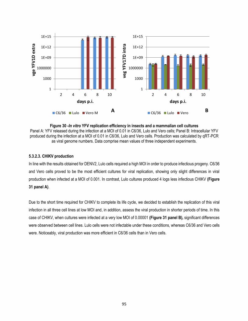
95
Figure 30 -In vitro YFV replication efficiency in insects and a mammalian cell cultures Panel A: YFV released during the infection at a MOI of 0.01 in C6/36, Lulo and Vero cells; Panel B: Intracellular YFV produced during the infection at a MOI of 0.01 in C6/36, Lulo and Vero cells. Production was calculated by qRT-PCR
as viral genome numbers. Data comprise mean values of three independent experiments.
5.3.2.3. CHIKV production
In line with the results obtained for DENV2, Lulo cells required a high MOI in order to produce infectious progeny. C6/36
and Vero cells proved to be the most efficient cultures for viral replication, showing only slight differences in viral
production when infected at a MOI of 0.001. In contrast, Lulo cultures produced 4 logs less infectious CHIKV (Figure
31 panel A).
Due to the short time required for CHIKV to complete its life cycle, we decided to establish the replication of this viral
infection in all three cell lines at low MOI and, in addition, assess the viral production in shorter periods of time. In this
case of CHIKV, when cultures were infected at a very low MOI of 0.00001 (Figure 31 panel B), significant differences
were observed between cell lines. Lulo cells were not infectable under these conditions, whereas C6/36 and Vero cells
were. Noticeably, viral production was more efficient in C6/36 cells than in Vero cells.
1
1000
1000000
1E+09
1E+12
1E+15
2 4 6 8 10
vge
YFV
1D
ext
ra
days p.i.
C6/36 Lulo Vero M A
1
1000
1000000
1E+09
1E+12
1E+15
2 4 6 8 10
veg
YFV
17
D in
tra
days p.i.
C6/36 Lulo Vero B

96
Figure 31 -In vitro CHIKV replication efficiency Panel A: CHIKV released during the infection at a MOI of 0.001 evaluated every two days; Panel B: CHIKV released during the infection at a MOI of 0.00001 evaluated per hours. CHIKV production was calculated as PFU/ml by plaque
assays. Data comprise mean values of three independent experiments.
In addition, CHIKV RNA production was assessed by qRT-PCR when cells were infected at high or low MOI.
Extracellular viral RNA content in C6/36 and Vero cells was very similar from day 2 – 8; however, on day 10 p.i., viral
RNA was only detectable at low levels in C6/36 cells. Viral RNA in Lulo was detectable at low levels only during the first
days of infection. In addition, in line with findings for DENV2, Lulo needs to be infected at high MOIs in order to be
detected by qRT-PCR (Figure 32 panel A). The highest levels of intracellular CHIKV RNA corresponded to Vero cells
on days 6 – 10 p.i.; nevertheless, there was no difference between C6/36 and Vero cells on days 2 – 4. In contrast, as
it was shown before, Lulo cell culture was almost non-infectable under these conditions (Figure 32 panel B). Contrary
to previous findings, when cell cultures were infected at a low MOI and assessed by qRT-PCR, C6/36 showed the
highest intra and extracellular values of viral genomes, followed by Vero cells and, as it has been observed before in
DENV2 and YFV, Lulo cells showed a very low number of CHIKV genomes (Figure 33). Moreover, over time increasing
viral RNA production (intra and extracellular) was observed in C6/36 and Vero cultures, and as opposed to the previous
trend, Lulo cells showed only slight differences in CHIKV RNA production.
0
1
2
3
4
5
6
7
2 4 6 8 10
Log 1
0P
FU/m
l CH
IKV
days p.i.
C6/36 Vero M LULO A
0
1
2
3
4
5
6
7
6 16 28 40 52 64 76
Log 1
0P
FU/m
l CH
IKV
hours p.i.
C6/36 Vero M LULO B

97
Figure 32 -In vitro CHIKV replication efficiency at high MOI. Panel A: CHIKV RNA released during the infection at a high MOI of 0.001 in C6/36, Lulo and Vero cells; Panel B:
Intracellular CHIKV RNA produced during the infection at a MOI of 0.001 in C6/36, Lulo and Vero cells. Production was calculated by qRT-PCR as viral genome numbers. Data comprise mean values of three independent
experiments.
Figure 33 -In vitro CHIKV replication efficiency at low MOI. Panel A: CHIKV RNA released during the infection at a low MOI of 0.00001 in C6/36, Lulo and Vero cells; Panel B:
Intracellular CHIKV RNA produced during the infection at a MOI of 0.00001 in C6/36, Lulo and Vero cells. Production was calculated by qRT-PCR as viral genome numbers. Data comprise mean values of three independent
experiments.
1
1000
1000000
1E+09
1E+12
1E+15
2 4 6 8 10
vge
CH
IKV
ext
ra
days p.i
C6/36 Lulo Vero A
1
1000
1000000
1E+09
1E+12
1E+15
2 4 6 8 10
vge
CH
IKV
intr
a
days p.i.
C6/36 Lulo Vero B
1
1000
1000000
1E+09
1E+12
6 16 28 40 52 64 76
vge
CH
IKV
ext
ra
hours p.i.
C6/36 Lulo Vero A
1
1000
1000000
1E+09
1E+12
6 16 28 40 52 64 76
vge
CH
IKV
intr
a
hours p.i.
C6/36 Lulo Vero B

98
5.3.2.4. SINV production
SINV production was assessed by plaque assay and in general, results showed the same pattern that was observed
when cells were infected at low MOI with CHIKV. C6/36 was 3 Logs more efficient in the production of SINV infectious
progeny when compared to Vero. Although Vero cells produced SINV less efficiently than C6/36, when the time of
appearance of plaques between Vero infected with CHIKV or SINV was contrasted, a delay in the appearance of CHIKV
plaques was observed. The SINV production in both C6/36 and Vero cell cultures increased up to 64 h p.i., and later
the viral production decreased. This result is in accordance with CPE appearance, due to the fact that it appeared just
before 40 hours p.i., in Vero and by 76 hour in C6/36 cells. Finally, as was found previously for Lulo cells infected at
low MOIs with DENV2 and CHIKV, this cell culture was not able to produce SINV capable of infecting new cells (Figure
34).
Figure 34 -In vitro SINV replication efficiency. Panel A: SINV released during the infection at a MOI of 0.00001, assessed per hours. Viral production was calculated
as PFU/ml by plaque assays. Data comprise mean values of three independent experiments.
5.4. Discussion
Of the various studies that are conducted in a laboratory, insect and mammalian cell lines are used for different
purposes such as viral propagation, the study of antiviral strategies (Scott et al. 2010; Delang et al. 2014), the
characterization of proteins (Samsa et al. 2009), vaccines (Murakami et al. 2007; Tseng et al. 2011), the study of host
factors (Sessions et al. 2009; Lee & Chu 2015) among others. However, there are several essential factors that may
influence the replication efficiency of arboviruses in cell cultures. Among these are: Type (serotype, strain) of virus
(Bielefeldt-Ohmann et al. 2001; Barr & Anderson 2013), virion binding, cell receptors (Thaisomboonsuk et al. 2005;
0
1
2
3
4
5
6
7
6 16 28 40 52 64 76
log 1
0P
FU/m
l SIN
V
hours p.i.
C6/36 Vero M LULO

99
Tio et al. 2005), endocytosis of the viral particles (Acosta et al. 2012) and host factors that might enable successful
viral replication (Sessions et al. 2009), viral production and release of new infectious viral progeny.
Although both insect and mammalian cell lines presented different characteristics when infected with flavi or
alphaviruses, one of the most notable characteristics was the appearance of CPE in Vero cells when they were infected
with the arboviruses assessed. In contrast, CPE was moderate in C6/36 cells when they were infected with CHIKV or
SINV and imperceptible when the cell culture was infected with DENV or YFV. In the case of Lulo cell culture, CPE was
not present when cells were infected with flavi or alphaviruses. These findings are in agreement with previous reports
that indicate that CPE is almost absent in insect cells (Chen et al. 2011; Li et al. 2012). In both insect and mammalian
cells, this can be explained due to the fact that DENV infection activates the unfolding protein response (UPR) in order
to overcome with ER stress. This is a protective mechanism that might protect cells from apoptosis, allowing viral
replication (Umareddy et al. 2007; Courageot et al. 2003). When UPR is activated in DENV infected cells, they ultimately
face apoptosis through effect of ER stress or a mitochondrion-mediated caspases pathway. This might result in changes
in the mitochondrial membrane potential and the appearance of reactive oxygen species (ROS), given that mosquito
but not mammalian cells increase the activities of enzymes, such as glutathione S-transferase, that play a role in cellular
detoxification (Chen et al. 2011). During CHIKV infection it is suggested that C6/36, but not Vero cells may carry some
host factors that are able to protect this cell line against apoptosis (Li et al. 2012).
In the present study, C6/36 showed the highest values of viral RNA production as well as infectious progeny production
which could be associated, at least in part, to the fact that the origin of this cell line constitutes the natural vector of
DENV and CHIKV, which is Ae. Albopictus. In addition, C6/36 is a clonal cell line, which can afford to have auniform
host cell system instead of a mixed population (Igarashi 1978). Another explanation for the dissimilar response in insect
and mammalian cells during arboviral infections correspond to the exogenous interference RNA (RNAi) pathway, which
is an important antiviral defense against arboviruses in insects of the Diptera order (Steinert & Levashina 2011). When
C6/36 cells are infected with DENV2, they exhibit ineficient Dicer-2 cleavage of long double strand RNA (dsRNA), that
recognize and cleaves dsRNA into siRNA in order to initiate the RNAi pathway. The ability of C6/36 to support robust
arbovirus replication may be due to lack of a complete, functional RNAi pathway (Scott et al. 2010). This disfunctional
patter has been also found when C6/36 cells are infected with bunyaviruses (Léger et al. 2013), other flaviviruses, and
alphaviruses (Brackney et al. 2010).
Despite the strong CPE observed during the arboviral infection in Vero cells, the cell culture showed efficient replication
of flavi and alphaviruses; this finding had been previously reported and it might have been strongly influenced by the
inability of this cell line to produce IFN type 1 and antiviral factors knows as IFN-stimuated genes (Desmyter et al.
1968; Chew et al. 2009), that allows extreme permissive of Vero to viral infections.

100
In the case of the viral production by Lulo cells, there were surprising findings: it was observed that when Lulo cell
cultures were infected at high MOI, the cell culture was able to produce infectious viral particles; along the same lines,
when DENV binding and entry was compared with Lulo and C6/36 cells, only slight differences were found, suggesting
that Lulo has an appropriate cell receptor and that the inefficient arboviral replication can be associated with
downstream events or the lack of proper host factors required for efficient viral production. In addition, the quantification
of the viral RNA in Lulo showed high levels of viral genomes which had no correlation to the infectious viral particles
(plaque assay), opening the possibility of the production of defective viral particles. In constrast, although Lu.
longipalpis is not a natural vector of flavi- and alphaviruses, the cell line derived from this species has been reported
to be susceptible to the Mayaro alphavirus (Rey et al. 2000), showing that related viruses cannot necessarily replicate
efficiently in the same cell culture (Bielefeldt-Ohmann et al. 2001; Barr & Anderson 2013).
In conclusion, C6/36 was the most efficient cell line in the flavi and alphavirus production, followed by Vero cells; these
cell cultures can be used efficiently in DENV, YFV, CHIKV and SINV productions. However, it is important to consider
their characteristics for virological studies as both cell lines do not represent an accurate model for molecular
interactions. In the case of C6/36 due to lack of a functional RNAi pathway (Scott et al. 2010) and in the case of Vero
due to the inability to produce IFN type 1 (Chew et al. 2009). Although the viral particles can bind to and enter Lulo
cells, this cell line was almost unable to produce high levels of infectious viral progeny, showing that it is not suitable
for viral production. Despite this, Lulo can be considered a tool for understanding the mechanism(s) through which the
cell can evade viral replication.
5.5. References
Acosta, E., Castilla, V. & Damonte, E., 2008. Functional entry of dengue virus into Aedes albopictus mosquito cells is dependent on clathrin-mediated endocytosis. The Journal of general virology, 89(Pt 2), pp.474–84. Available at: http://www.ncbi.nlm.nih.gov/pubmed/18198378 [Accessed May 22, 2013].
Acosta, E.G., Castilla, V. & Damonte, E.B., 2009. Alternative infectious entry pathways for dengue virus serotypes into mammalian cells. Cellular microbiology, 11(10), pp.1533–49. Available at: http://www.ncbi.nlm.nih.gov/pubmed/19523154 [Accessed July 2, 2013].
Acosta, E.G., Castilla, V. & Damonte, E.B., 2012. Differential requirements in endocytic trafficking for penetration of dengue virus. PloS one, 7(9), p.e44835. Available at: http://www.pubmedcentral.nih.gov/articlerender.fcgi?artid=3436767&tool=pmcentrez&rendertype=abstract [Accessed May 29, 2013].
Avirutnan, P. et al., 2006. Vascular leakage in severe dengue virus infections: a potential role for the nonstructural viral protein NS1 and complement. The Journal of infectious diseases, 193(8), pp.1078–88. Available at: http://www.ncbi.nlm.nih.gov/pubmed/16544248.

101
Barr, K. & Anderson, B., 2013. Dengue viruses exhibit strain-specific infectivity and entry requirements in vitro. Virus adaptation and treatment, 5, pp.1–9.
Bielefeldt-Ohmann, H. et al., 2001. Dengue virus binding to human leukocyte cell lines: receptor usage differs between cell types and virus strains. Virus research, 73(1), pp.81–9. Available at: http://www.ncbi.nlm.nih.gov/pubmed/11163646.
Brackney, D.E. et al., 2010. C6/36 Aedes albopictus cells have a dysfunctional antiviral RNA interference response. PLoS Neglected Tropical Diseases, 4(10), pp.24–27.
Brighton, S.W., Prozesky, O.W. & De La Harpe, A.L., 1983. Chikungunya virus infection. A retrospective study of 107 cases. South African medical journalMedical Journal, 63(February), pp.313–315.
Chen, L.H. & Wilson, M.E., 2010. Dengue and chikungunya infections in travelers. Current opinion in infectious diseases, 23(5), pp.438–44. Available at: http://www.ncbi.nlm.nih.gov/pubmed/20581669 [Accessed August 3, 2012].
Chen, T.H. et al., 2011. Antioxidant defense is one of the mechanisms by which mosquito cells survive dengue 2 viral infection. Virology, 410(2), pp.410–417. Available at: http://dx.doi.org/10.1016/j.virol.2010.12.013.
Chen, Y. et al., 1997. Chen et al DENV infectivity heparan sulfate.pdf. Nature medicine, 3(8), pp.866–871.
Chew, T. et al., 2009. Characterization of the interferon regulatory factor 3-mediated antiviral response in a cell line deficient for IFN production. Molecular Immunology, 46(3), pp.393–399.
Chiu, W., Kinney, R.M. & Dreher, T.W., 2005. Control of Translation by the 5 ′ - and 3 ′ -Terminal Regions of the Dengue Virus Genome Control of Translation by the 5’- and 3-Terminal Regions of the Dengue Virus Genome. Journal of Virology, 79(13), pp.8303–8315.
Courageot, M.P., Catteau, A. & Desprès, P., 2003. Mechanisms of Dengue virus-induced cell death. Advances in Virus Research, 60, pp.157–186.
Damen, M. et al., 2008. Real-time PCR with an internal control for detection of all known human adenovirus serotypes. Journal of Clinical Microbiology, 46(12), pp.3997–4003.
Delang, L. et al., 2014. Mutations in the chikungunya virus non-structural proteins cause resistance to favipiravir (T-705), a broad-spectrum antiviral. The Journal of antimicrobial chemotherapy, 69(June), pp.1–15. Available at: http://www.ncbi.nlm.nih.gov/pubmed/24951535.
Desmyter, J., Melnick, J.L. & Rawls, W.E., 1968. Defectiveness of interferon production and of rubella virus interference in a line of African green monkey kidney cells (Vero). Journal of virology, 2(10), pp.955–61. Available at: http://www.pubmedcentral.nih.gov/articlerender.fcgi?artid=375423&tool=pmcentrez&rendertype=abstract.
Germi, R. et al., 2002. Heparan sulfate-mediated binding of infectious dengue virus type 2 and yellow fever virus. Virology, 292(1), pp.162–8. Available at: http://www.ncbi.nlm.nih.gov/pubmed/11878919 [Accessed June 7, 2013].
Gomez de Cedrón, M. et al., 1999. RNA helicase activity of Semliki Forest virus replicase protein NSP2. FEBS letters, 448(1), pp.19–22. Available at: http://www.ncbi.nlm.nih.gov/pubmed/10217401.
Guzman, M.G. et al., 2010. Dengue: a continuing global threat. Nature reviews. Microbiology, 8(12 Suppl), pp.S7–16. Available at: http://www.ncbi.nlm.nih.gov/pubmed/21079655 [Accessed May 29, 2013].
Hahn, Y.S. et al., 1989. Mapping of RNA- temperature-sensitive mutants of Sindbis virus: complementation group F mutants have lesions in nsP4. Journal of virology, 63(3), pp.1194–202. Available at: http://www.pubmedcentral.nih.gov/articlerender.fcgi?artid=247815&tool=pmcentrez&rendertype=abstract.

102
Igarashi, a., 1978. Isolation of a Singh’s Aedes albopictus Cell Clone Sensitive to Dengue and Chikungunya Viruses. Journal of General Virology, 40(3), pp.531–544. Available at: http://vir.sgmjournals.org/cgi/doi/10.1099/0022-1317-40-3-531.
Kaptein, S.J.F. et al., 2010. A derivate of the antibiotic doxorubicin is a selective inhibitor of dengue and yellow fever virus replication in vitro. Antimicrobial agents and chemotherapy, 54(12), pp.5269–80. Available at: http://www.pubmedcentral.nih.gov/articlerender.fcgi?artid=2981273&tool=pmcentrez&rendertype=abstract [Accessed December 20, 2013].
Kuadkitkan, A. et al., 2010. Identification and characterization of prohibitin as a receptor protein mediating DENV-2 entry into insect cells. Virology, 406(1), pp.149–61. Available at: http://www.ncbi.nlm.nih.gov/pubmed/20674955 [Accessed May 21, 2013].
Lee, R.C.H. & Chu, J.J.H., 2015. Proteomics Profiling of Chikungunya-Infected Aedes albopictus C6/36 Cells Reveal Important Mosquito Cell Factors in Virus Replication. PLOS Neglected Tropical Diseases, 9(3), p.e0003544. Available at: http://dx.plos.org/10.1371/journal.pntd.0003544.
Léger, P. et al., 2013. Dicer-2- and Piwi-mediated RNA interference in Rift Valley fever virus-infected mosquito cells. Journal of virology, 87(3), pp.1631–48. Available at: http://www.ncbi.nlm.nih.gov/pubmed/23175368.
Li, Y.G. et al., 2012. Chikungunya virus induces a more moderate cytopathic effect in mosquito cells than in mammalian cells. Intervirology, 56(1), pp.6–12.
Lindenbach, B.D. & Rice, C.M., 2007. Flaviviridae: The Viruses and Their Replication. In Fields Virology. Philadelphia: Lippincott-Raven Publishers, pp. 1101–52.
Liu, C.C. et al., 2011. Purification and characterization of enterovirus 71 viral particles produced from vero cells grown in a serum-free microcarrier bioreactor system. PLoS ONE, 6(5), pp.1–9.
Malet, H. et al., 2009. The crystal structures of Chikungunya and Venezuelan equine encephalitis virus nsP3 macro domains define a conserved adenosine binding pocket. Journal of virology, 83(13), pp.6534–45. Available at: http://www.pubmedcentral.nih.gov/articlerender.fcgi?artid=2698539&tool=pmcentrez&rendertype=abstract [Accessed September 27, 2012].
Manimunda, S.P. et al., 2010. Clinical progression of chikungunya fever during acute and chronic arthritic stages and the changes in joint morphology as revealed by imaging. Transactions of the Royal Society of Tropical Medicine and Hygiene, 104(6), pp.392–9. Available at: http://www.ncbi.nlm.nih.gov/pubmed/20171708 [Accessed October 4, 2012].
Martínez-Barragán, J. & del Angel, R., 2001. Identification of a Putative Coreceptor on Vero Cells That Participates in Dengue 4 Virus Infection. Journal of virology, 75(17), pp.7818–7827.
McInerney, G.M. et al., 2004. Semliki Forest virus produced in the absence of the 6K protein has an altered spike structure as revealed by decreased membrane fusion capacity. Virology, 325(2), pp.200–6. Available at: http://www.ncbi.nlm.nih.gov/pubmed/15246260 [Accessed September 7, 2012].
Melton, J. V et al., 2002. Alphavirus 6K proteins form ion channels. The Journal of biological chemistry, 277(49), pp.46923–31. Available at: http://www.ncbi.nlm.nih.gov/pubmed/12228229 [Accessed September 28, 2012].
Mercado-Curiel, R.F. et al., 2006. The four serotypes of dengue recognize the same putative receptors in Aedes aegypti midgut and Ae. albopictus cells. BMC microbiology, 6, p.85. Available at: http://www.pubmedcentral.nih.gov/articlerender.fcgi?artid=1599738&tool=pmcentrez&rendertype=abstract [Accessed May 29, 2013].
Muñoz-Jordan, J.L. et al., 2003. Inhibition of interferon signaling by dengue virus. Proceedings of the National Academy of Sciences of the United States of America, 100(24), pp.14333–8. Available at: http://www.ncbi.nlm.nih.gov/pubmed/23408610.

103
Murakami, S. et al., 2007. Neuraminidase inhibitors and their role in avian and pandemic influenza. Antiviral Therapy, 12(4 B), pp.593–602.
Nitatpattana, N. et al., 2014. Long-term persistence of Chikungunya virus neutralizing antibodies in human populations of North Eastern Thailand. Virology Journal, 11(1), p.183. Available at: http://www.virologyj.com/content/11/1/183.
Normile, D., 2013. Surprising New Dengue Virus Throws A Spanner in Disease Control Efforts. Science, 342, p.415.
Nougairede, A. et al., 2013. Random Codon Re-encoding Induces Stable Reduction of Replicative Fitness of Chikungunya Virus in Primate and Mosquito Cells. PLoS Pathogens, 9(2), pp.1–18.
Osada, N.A. et al., 2014. The Genome Landscape of the African Green Monkey Kidney-Derived Vero Cell Line. DNA Research, pp.1–11.
Panning, M. et al., 2008. Chikungunya Fever in Travelers Returning to Europe from the Indian Ocean Region, 2006. Emerging infectious diseases, 14(3), pp.416–22.
Rey, A., 2003. Dengue virus envelope glycoprotein structure: New insight into its interactions during viral entry. Nature, 100(12), pp.6899–6901.
Rey, G.J., Ferro, C. & Bello, F.J., 2000. Establishment and characterization of a new continuous cell line from Lutzomyia longipalpis (Diptera: psychodidae) and its susceptibility to infections with arboviruses and Leishmania chagasi. Memórias do Instituto Oswaldo Cruz, 95(1), pp.103–10. Available at: http://www.ncbi.nlm.nih.gov/pubmed/10656714.
Rodenhuis-Zybert, I. a, Wilschut, J. & Smit, J.M., 2010. Dengue virus life cycle: viral and host factors modulating infectivity. Cellular and molecular life sciences : CMLS, 67(16), pp.2773–86. Available at: http://www.ncbi.nlm.nih.gov/pubmed/20372965 [Accessed May 25, 2014].
Rothman, A.L., 2004. Dengue: Defining protective versus pathologic immunity. Journal of Clinical Investigation, 113(7), pp.946–951.
Salas-Benito, J. et al., 2007. Evidence that the 45-kD glycoprotein, part of a putative dengue virus receptor complex in the mosquito cell line C6/36, is a heat-shock related protein. The American journal of tropical medicine and hygiene, 77(2), pp.283–90. Available at: http://www.ncbi.nlm.nih.gov/pubmed/17690400.
Samsa, M.M. et al., 2009. Dengue virus capsid protein usurps lipid droplets for viral particle formation. PLoS pathogens, 5(10), p.e1000632. Available at: http://www.pubmedcentral.nih.gov/articlerender.fcgi?artid=2760139&tool=pmcentrez&rendertype=abstract [Accessed August 5, 2013].
Scott, J.C. et al., 2010. Comparison of dengue virus type 2-specific small RNAs from RNA interference-competent and - incompetent mosquito cells. PLoS Neglected Tropical Diseases, 4(10).
Sessions, O.M. et al., 2009. Discovery of insect and human dengue virus host factors. Nature, 458(7241), pp.1047–1050. Available at: http://dx.doi.org/10.1038/nature07967.
Sheets, R., 2000. History and Characterization of the Vero Cell Line. Open session, pp.1–12.
Simon, F., Savini, H. & Parola, P., 2008. Chikungunya: a paradigm of emergence and globalization of vector-borne diseases. The Medical clinics of North America, 92(6), pp.1323–43. Available at: http://www.ncbi.nlm.nih.gov/pubmed/19061754 [Accessed July 23, 2012].

104
Singh, K., 1967. Cell cultures derived from larvae of Aedes albopictus (Skuse) and Aedes aegypti (L.). Curr Sci India, 36, pp.506–8.
Sreejith, R. et al., 2012. Mapping interactions of Chikungunya virus nonstructural proteins. Virus research, pp.6–11. Available at: http://www.ncbi.nlm.nih.gov/pubmed/22951312 [Accessed September 21, 2012].
Steinert, S. & Levashina, E. a, 2011. Intracellular immune responses of dipteran insects. Immunological reviews, 240(1), pp.129–40. Available at: http://www.ncbi.nlm.nih.gov/pubmed/21349091.
Strauss, J.H. & Strauss, E.G., 1994. The alphaviruses: gene expression, replication, and evolution. Microbiological reviews, 58(3), pp.491–562. Available at: http://www.pubmedcentral.nih.gov/articlerender.fcgi?artid=372977&tool=pmcentrez&rendertype=abstract.
Takkinen, K., Peränen, J. & Kääriäinen, L., 1991. Proteolytic processing of Semliki Forest virus-specific non-structural polyprotein. The Journal of general virology, 72 ( Pt 7), pp.1627–33. Available at: http://www.ncbi.nlm.nih.gov/pubmed/1856693.
Thaisomboonsuk, B.K. et al., 2005. Characterization of dengue-2 virus binding to surfaces of mammalian and insect cells. The American journal of tropical medicine and hygiene, 72(4), pp.375–83. Available at: http://www.ncbi.nlm.nih.gov/pubmed/15827273.
Tio, P.H., Jong, W.W. & Cardosa, M.J., 2005. Two dimensional VOPBA reveals laminin receptor (LAMR1) interaction with dengue virus serotypes 1, 2 and 3. Virology journal, 2, p.25. Available at: http://www.pubmedcentral.nih.gov/articlerender.fcgi?artid=1079963&tool=pmcentrez&rendertype=abstract [Accessed June 14, 2013].
Tomar, S. et al., 2006. Catalytic core of alphavirus nonstructural protein nsP4 possesses terminal adenylyltransferase activity. Journal of virology, 80(20), pp.9962–9. Available at: http://www.pubmedcentral.nih.gov/articlerender.fcgi?artid=1617302&tool=pmcentrez&rendertype=abstract [Accessed September 21, 2012].
Tseng, Y.F. et al., 2011. Adaptation of high-growth influenza H5N1 vaccine virus in Vero cells: Implications for pandemic preparedness. PLoS ONE, 6(10), pp.3–7.
Umareddy, I. et al., 2007. Dengue virus serotype infection specifies the activation of the unfolded protein response. Virology journal, 4, p.91.
Voss, J.E. et al., 2010. Glycoprotein organization of Chikungunya virus particles revealed by X-ray crystallography. Nature, 468(7324), pp.709–12. Available at: http://www.ncbi.nlm.nih.gov/pubmed/21124458 [Accessed June 28, 2013].
White, J.N., 1996. The treatment of malaria. N Engl J Med, 335, pp.800–6.
White, L., 1987. Susceptibility of Aedes albopictus C6/36 cells to viral infection. Journal of clinical microbiology, 25(7), pp.1221–4. Available at: http://www.pubmedcentral.nih.gov/articlerender.fcgi?artid=269180&tool=pmcentrez&rendertype=abstract.
WHO, 1997. Dengue haemorrhagic fever: Diagnosis, treatment, prevention and control P. Jevon, ed. , pp.12–23. Available at: http://doi.wiley.com/10.1002/9781444340051.
WHO, 2009. Dengue: Guidelines for diagnosis, treatment, prevention and control, France.
Zapata, A.C., Cárdenas, E. & Bello, F., 2005. Characterization of cell cultures derived from Lutzomyia spinicrassa (Diptera: Psychodidae) and their susceptibility to infection with Leishmania (Viannia) braziliensis. Medical science monitor, 11(12), pp.BR457–64. Available at: http://www.ncbi.nlm.nih.gov/pubmed/16319783.

105
Zhai, W. et al., 2012. Comparison of Different Cell Substrates on the Measurement of Human Influenza Virus Neutralizing Antibodies. PLoS ONE, 7(12), pp.1–13.

106
6. Differences in antiviral responses between Huh-7 and Vero cell lines treated with different dengue
virus inhibitors
6.1. Introduction
Dengue is the most important arthropod-borne viral infection of humans. This disease is caused by a single-stranded
positive RNA virus belonging to the Flavivirus genus (Flaviviridae family), which includes five distinct serotypes.
According to the WHO, between 50 - 100 million infections occur annually (WHO 2009). However, Bhatt et al. (2013)
recentlyestimated 390 million dengue infections per year based on cartographic approaches. Additionally, according to
the geographical region, mortality rate might vary from 1.2 – 3.5% (WHO 2009).
DENV infections may be asymptomatic or may lead to undifferentiated fever, dengue fever (DF) or dengue
haemorrhagic fever (DHF) with plasma leakage that may evolve to hypovolemic shock (DSS). Although most patients
recover following a self-limiting non-severe clinical course, a small proportion progress to severe state characterized by
plasma leakage with or without haemorrhage. Clinical manifestations include severe headache, retro-orbital pain,
muscle and bone or joint pains, nausea, vomiting and rashes; additionally, skin haemorrhages are common (WHO
1997).
Aedes aegypti and Aedes albopictus are the main vectors for DENV (Chen & Wilson 2010). Ae. aegypti is distributed
in tropical regions, meanwhile Ae. albopictus is distributed in regions that are tropical to temperate. Once these
mosquitoes get the infection, these are infected for life (Higa 2011). The broad distribution of these species and the
failure in attempts to control these vectors has allowed the expansion of this viral disease.
The DENV genome is a single-positive strand RNA, which is approximately 11kb in length, it single ORF encodes three
structural proteins: Capsid (C), membrane (M) and envelope (E) and seven non-structural proteins (NS1, NS2A, NS2B,
NS3, NS4A, NS4B and NS5).
E protein (53-kDa) is responsible for the main steps involved in the entry process, which involves receptor recognition
and fusion between viral and cellular membranes (Rey 2003). The C protein (12-kDa) is essential in virus assembly to
ensure encapsidation of the viral genome. However, the mechanism by which encapsidation occurs is not well
understood (Samsa et al. 2009).Maturation of flavivirus particles occurs during transport through the exocytic pathway,
through the cleavage of the M protein precursor (prM) by furin, that transforms the prM (18.44 kDa) into M protein (8.3
kDa) (van der Schaar et al. 2007) which is required for DENV infectivity (Zybert et al. 2008).NS1 glycoprotein (50-kDa)
plays an essential role in viral replication (Mackenzie et al. 1996) and might play a function in DHF and DSS
immunopathogenesis (Avirutnan et al. 2006). NS2A hydrophobic protein (22-kDa) is implicated in the formation of virus-

107
induced membranes (Leung et al. 2008) that can be associated in virus assembly, RNA synthesis (Xie et al. 2013), and
also inhibits IFN and response (Muñoz-Jordan et al. 2003). Both NS2B (14-kDa) and NS3 (69.5-kDa) proteins are
required for protease activity (Falgout et al. 1991; Cahour et al. 1992; Yusof et al. 2000). Furthermore, the NS3 carrying
activities involved in helicase and capping (Benarroch et al. 2004). NS4A hydrophobic protein (16-Kda) and NS4B
transmembrane protein (27 kDa) are IFN antagonist (Muñoz-Jordan et al. 2003) and also are part of the replication
complex (Miller et al. 2006; Miller et al. 2007). In addition, NS4A induces ER membrane rearrangements (Miller et al.
2007) and NS4B enhances the helicase activity of NS3 (Umareddy et al. 2006). Finally, NS5 protein (104-Kda) is
involved in 5’-cap formation (Ray et al. 2006), and contains RdRp catalytic domain (Yap et al. 2007). Both structural
and non-structural proteins can be potential targets for antiviral intervention.
We conducted a comparative study of the antiviral activity of a panel of 9 antiviral molecules, with has been proven in
vitro for anti-dengue virus activity and that act at different stages of the dengue viral life cycle. Their antiviral activity
was determined through viral CPE reduction (evaluated microscopically by CPE scoring), qRT-PCR and plaque assays.
Likewise, the effect of these compounds on cell viability was evaluated by microscopic observation and ATP-lite assays.
Both, the human hepatoma cell line Huh-7 and the cell line Vero from African green monkey were used to set up these
antiviral assays. This dataset may represent a reference panel and serve to compare the activity of molecules not yet
discovered.
6.2. Materials and Methods
6.2.1. Virus and cells
V. Deubel (Institur Pasteur, Lyon, France) provided DENV serotype 2 (DENV2) New Guinea C strain. DENV2 was
propagated in the Ae. albopictus cell line named C6/36 [C6/36 (ATCC CRL-1660)] in Leibovitz (L-15, Gibco, Belgium)
cell growth medium supplemented with 2% FBS and 2% non-essential amino acids. The virus was allowed to grow in
accordance with the requirement of the cell culture at 28 C without CO2 atmosphere.
Vero-F cells were maintained in cell growth medium composed of minimum essential medium (MEM Rega-3, Gibco,
Belgium) supplemented with 10% FBS, 1% L-glutamine (Gibco), and 1% sodium bicarbonate (Gibco). Huh-7 cells were
maintained in Dulbecco's Modified Eagle's Medium (DMEM, Gibco, Belgium) supplemented with 10% FBS, 1% HEPES
(Gibco), and was adapted to slow growth. BHK cells were maintained in DMEM supplemented with 10% FBS, 1%
Sodium bicarbonate and 1% L-glutamine. Antiviral assays were carried out in virus growth medium, which is the
respective cell growth medium supplemented with 2% (instead of 10%) FBS. All cell cultures were maintained at 37°C
in an atmosphere of 5% CO2 and 95-99% humidity.

108
6.2.2. Compounds
ST-148 is a selective DENV inhibitor (Byrd et al 2013). The compound was purchased from Sigma-Aldrich (St. Lous,
Mo.). Celgosivir is a pro-drug derivative of castanospermine, which was purchased from Dalton Pharma Services
(Canada) ST-610 is a benzoxazole inhibitor obtained from ChemBridge (San Diego, CA. Ivermectin is an anthelmintic
agent that was purchased from ICN Pharmaceuticals (Costa Mesa, CA, USA). NITD-618 inhibits viral RNA synthesis;
and specifically targets DENV NS4B protein (Xie et al 2011). The nucleoside polymerase inhibitors 2’CMC was
synthetized as described before (Pierra et al. 2006). 7D-2CMA was purchased from Sigma-Aldrich (St. Lous, Mo.),
respectively. Ribavirin (1--D-ribofuranosyl-1, 2, 4-triazole-3-carboxamide) was purchased from ICN Pharmaceuticals
(Costa Mesa, CA). T-1105 is a defluorinated analogue of T-705; it was obtained as a custom synthesis product from
ABCR (Karlsrushe, Germany).
ST-148
Celgosivir
ST-610
Ivermectin
NITD-618
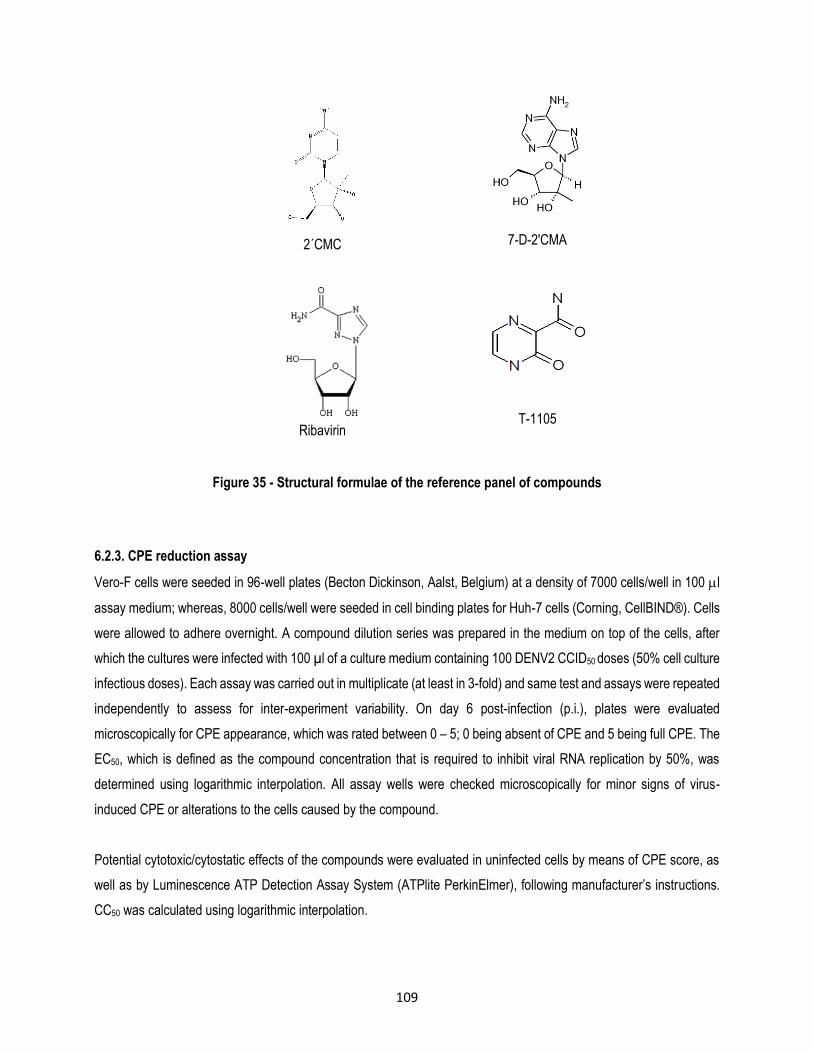
109
2´CMC
7-D-2'CMA
Ribavirin
T-1105
Figure 35 - Structural formulae of the reference panel of compounds
6.2.3. CPE reduction assay
Vero-F cells were seeded in 96-well plates (Becton Dickinson, Aalst, Belgium) at a density of 7000 cells/well in 100 l
assay medium; whereas, 8000 cells/well were seeded in cell binding plates for Huh-7 cells (Corning, CellBIND®). Cells
were allowed to adhere overnight. A compound dilution series was prepared in the medium on top of the cells, after
which the cultures were infected with 100 µl of a culture medium containing 100 DENV2 CCID50 doses (50% cell culture
infectious doses). Each assay was carried out in multiplicate (at least in 3-fold) and same test and assays were repeated
independently to assess for inter-experiment variability. On day 6 post-infection (p.i.), plates were evaluated
microscopically for CPE appearance, which was rated between 0 – 5; 0 being absent of CPE and 5 being full CPE. The
EC50, which is defined as the compound concentration that is required to inhibit viral RNA replication by 50%, was
determined using logarithmic interpolation. All assay wells were checked microscopically for minor signs of virus-
induced CPE or alterations to the cells caused by the compound.
Potential cytotoxic/cytostatic effects of the compounds were evaluated in uninfected cells by means of CPE score, as
well as by Luminescence ATP Detection Assay System (ATPlite PerkinElmer), following manufacturer’s instructions.
CC50 was calculated using logarithmic interpolation.
110
6.2.4. Virus yield assay
Vero-F cells were seeded in 96-well plates (Becton Dickinson, Aalst, Belgium) at a density of 4 × 10⁴ cells/well.
Meanwhile, 1 × 10⁴ cells/well were seeded for Huh-7 cells in binding plates (Corning, CellBIND®). Following 24h of
incubation, culture medium was replaced with 100 µl assay medium containing a serial dilution of the compound and
100 µl of culture medium containing 100 DENV2 CCID50 for each well. After 2h of incubation, the cell monolayer was
washed 3 times with assay medium to remove non-adsorbed viruses and cultures were incubated for a further 2 days
in the presence of the compound. Supernatant was harvested and viral RNA was isolated using Macherey Nagel
“NucleoSpin 96 virus kit”. The Viral load was determined by real-time quantitative RT-PCR. Additionally, the amount of
infectious progeny from virus -infected compound - treated cells was assessed by plaque assay. Briefly, BHK cells were
seeded in 12-well plates (IWAKI) at a density of 5 × 10⁵ cells/well in 10% FBS medium. Following 24h of incubation,
the monolayers were washed 3 times with PBS and cells were infected with 500 µl of serial 1:10 supernatant dilutions,
which have been previously prepared in assay medium. After an hour of infection with continuous shaking, the
monolayers were washed 3 times with PBS in order to remove non-adsorbed viruses. Each monolayer was coated with
3 mL of a sterile solution of 2% Avicel PH-101 (Sigma-Aldrich) and 2% FBS medium. After 6 days of incubation the
monolayers were carefully washed 3 times with PBS, fixed with 70% ethanol and stained with blue methylene in order
to visualize and count the plaque-forming units (PFU).
6.2.5. DENV2 quantitative reverse transcription-PCR (qRT-PCR)
Viral RNA was isolated from 100 µl supernatant using the NucleoSpin® 96 RNA / Core Kit (Macherey-Nagel, Düren,
Germany). Primers and probe sequences were used as described earlier: DENV-For
5’TCGGAGCCGGAGTTTACAAA3’, DENV-Rev 5’TCTTAACGTCCGCCCATGAT3’ and DENV-Probe FAM-
5’ATTCCACACAATGTGGCAT-MGB3’ (Kaptein et al. 2010). One-step, quantitative RT-PCR was carried out in a total
volume of 25 µl, containing 13.94 µl H2O, 6.25 µl master mix (Eurogentec, Seraing, Belgium), 0.375 µl of 60 µM
forward primer, 0.375 µl of 60 µM reverse primer, 1 µl of 10 µM probe, 0.0625 µl reverse transcriptase (Eurogentec)
and 3 µl RNA sample. qRT-PCR was carried out using the ABI 7500 Fast Real-Time PCR System (Applied Biosystems,
Branchburg, NJ) using the following conditions: 30 min at 48C and 10 min at 95C, followed by 40 cycles of 15 s at
95C and 1 min at 60C. Data were analyzed using the ABI PRISM 7500 SDS software (version 1.3.1; Applied
Biosystems). For absolute quantification, standard curves were generated using 10-fold dilutions of template
preparation of known concentrations.
6.3. Results
ST-148 is a novel small-molecule compound that is a potent inhibitor of the four in vitro DENV serotypes; this compound
has shown substantial activity against Modoc virus and weak activity against YFV and HCV. Although the mechanism
of action of this SIGA compound is not very clear, evidence suggests that ST-148 could alter the interaction between

111
lipid droplets and DENV C protein, inhibiting the viral replication (Byrd et al. 2013). In our study, the EC50s obtained
through plaque assay in BHK cells for ST-148 in Vero cells (1.85 ± 0.07 M), as well as in Huh-7 cells (2.13 ± 0.02
M) (Table 9), were higher compared to the results obtained by Byrd et al. (2013) in these cell lines (0.016 0.01 M
in Vero and 0.012 0.01 M in Huh-7 cells) using DENV2 NGC. However, according to Byrd et al., (2013) ST-148 can
show different activity against other DENV serotypes, such as the case of DENV1 (2.83 ± 1.13 M), whose value is
more similar to with the value obtained in our study for DENV2.
Celgosivir is a pro-drug of the natural alkaloid castanospermine derived from the tree Castanospermunaustrale. This
compound is an inhibitor of HIV (Taylor et al. 1994), BVDV, HCV (Whitby et al. 2004). In DENV, Celgosivir acts as an
inhibitor of ER α-glucosidases, preventing proper processing of NS1 (Rathore et al. 2011). The EC50 obtained in BHK-
21 cell based on flavivirus immunodetection assays (CFI) in a DENV2 clinical isolate was 0.22 ± 0.01 M, and it was
three times higher in DENV3 (Rathore et al. 2011). However, Celgosivir was tested in the same cell line, but in lab
strains the EC50s were slightly different and the compound was more sensitive to DENV4 and DENV3 (Rathore et al.
2011). In our study, Vero cells were more sensitive to the antiviral activity of Celgosivir than to Huh-7 cells (Table 9).
Nevertheless, EC50 values obtained by three different methods were higher than the EC50 obtained by Rathore et al.
(2011) using CFI. On the other hand, CC50 values obtained in both Vero and Huh-7 cells were above 200 M, confirming
that this compound is safe.
ST-610 is a benzoxazole inhibitor that is active against DENV and VEEV, but does not inhibit YFV, HCV, WNV, BVDV,
JEV and Modoc virus. ST-610 acts through the inhibition of the ATP-dependent helicase activity of DENV NS3 protein
(Byrd et al. 2013). In our study, ST-610 inhibits DENV2 in Vero as well as in Huh-7 cells; the EC50s obtained by three
different methods were similar between both cell lines; however, the EC50s obtained by CPE reduction scoring was
three times higher than by plaque assay or by qRT-PCR (Table 9). Our results are partially in agreement with Byrd et
al., (2013), who obtained through a viral titer reduction assay an EC50s of 0.27 0.05 M and 3.59 M for Vero and
Huh-7 cells and an EC90 of 3.59 M in Vero cells, respectively. These differences can be explained, at least in part,
due to the different conditions of the assay, because the viral titers were established by plaque assay in Vero cells
instead of BHK cells. This compound was safe in both Huh-7 and Vero cell lines; however, Huh-7 was 2.5 times more
cytotoxic in Vero cells.
Ivermectin is an anthelmintic agent. Recently, it was discovered that ivermectin has antiviral activity against YFV and
DENV, JEV and tick borne encephalitis virus (Mastrangelo et al. 2012). Two main mechanisms of action have been
proposed for Ivermectin: the inhibition of viral helicase in flaviviruses (Mastrangelo et al. 2012) and the disruption of the
interaction between DENV NS5 and importin /1, which is a nuclear import receptor (Wagstaff et al. 2012). Although
we found that ivermectin is a DENV2 inhibitor in Vero and Huh-7 cells, we obtained a low SI of 2 for both Vero and Huh-

112
7 cell lines, showing that ivermectin is highly cytotoxic in these cell cultures, especially in Huh-7 cells. On the other
hand, Mastrangelo et al. (2012) established an EC50 of 0.7 M in Vero cell cultures infected with DENV2 NGC. This
result is comparable to our results due to the similarity in the virus yield assay, as assessed by qRT-PCR. In addition,
the results obtained by qRT-PCR were very similar between both cell lines; on the other hand, the EC50 obtained by
plaque assay from Huh-7 cells supernatants was the lowest in the study (Table 9). Moreover, the CC50 obtained by
Mastrangelo in Vero cells was 2 times higher than the CC50 obtained in our study for the same cell line.
NITD-618 is a selective inhibitor of the four DENV serotypes, but not against related and non-related flaviviruses (Xie
et al. 2011). This compound inhibits viral RNA synthesis, specifically the target is DENV NS4B protein (Xie et al. 2011).
In our study, NITD-618 inhibits DENV2 in Huh-7 as well as in Vero cells, being more active against DENV in Huh-7
cells (Table 9). However, although the compound is less cytotoxic in Vero cells, a higher compound concentration is
required in order to inhibit DENV2. The EC50values obtained by the three methods, as well as the CC50 values obtained
by two methods in Huh-7 cells were comparable with the data obtained by Xie et al., (2011), who reported an EC50 of
1.6 M and a CC50 of 40 M in BHK-21 cells.
2’CMC is a nucleoside polymerase inhibitor that acts as a non-obligate chain terminator (Carroll et al. 2003). This
compound is active against RNA viruses such as hepatitis C virus (Le Pogam et al. 2006), DENV2 (Pierra et al. 2006),
YFV (Julander et al. 2010), Noroviruses (Rocha-Pererira et al. 2012) and foot and mouth disease virus (Goris et al.
2007). The prodrug, Valopicitabine (NM283), from 2’CMC is in phase IIb clinical trial. In our study, data from visual
inspection in Vero cells are comparable with the data obtained by Julander et al. (2010) in YFV-17D CPE reduction (9.7
2.7 M), which are similar to the CC50 (85.3 13.5 M). Although in our study 2’CMC was active against DENV2 in
both cell cultures, it was usually more active in Vero cells than in Huh-7 cells, except corresponding to the EC50 obtained
by plaque assay (Table 9).
7D-2CMA is a nucleoside polymerase inhibitor that shares the same mechanism of action than 2’CMC. 7D-2’CMA has
antiviral activity against BVDV, WNV, DENV, YFV, rhinovirus type 2, rhinovirus type 14, and poliovirus type 3. This
compound did not have antiviral activity against minus-stranded-RNA and double-stranded-DNA viruses (Olsen et al.
2004). In the present study, 7D-2’CMA was highly active in Vero and in Huh-7 cell cultures against DENV2, protecting
them from the virus at low concentrations in comparison with CC50 (Table 9). However, 7D-2’CMA was more active
against DENV2 in Huh-7; and in addition, the compound was less toxic in Huh-7 than in Vero cells. Our results regarding
CPE reduction and cytotoxicity in Vero cells are comparable to the results obtained by Olsen et al. (2004) by neutral
red (EC50 15 M, CC50>320 M). However, we obtained a standard deviation of 7.4 M in the CPE reduction assay.
In contrast, the results corresponding to qRT-PCR and plaque assay showed higher values, which are similar to each
other.

113
Ribavirin (1--D-ribofuranosyl-1, 2, 4-triazole-3-carboxamide) is a broad-spectrum synthetic guanosine analogue,
which is active against influenza H1N1 (Rowe et al. 2010), Respiratory syncytial virus (Smith et al. 1991), Lassa fever
virus (Hadi et al. 2010) and Hanta virus (Safronetz et al. 2011). Leyssen et al (2005) demonstrated that the predominant
mechanism of action of ribavirin against flaviviruses is based on inhibition of IMPDH (Leyssen et al. 2005). In our study,
Ribavirin was more active against DENV in Huh-7 cells than in Vero cells (Table 9), and we obtained lower EC50
compared to Leyssen et al., (2001) which showed a weak activity of Ribavirin against flaviviruses and an EC50 of 155.6
45 M (Leyssen et al. 2001) through a CPE reduction test in Vero cells. In contrast, Takhampunya et al. (2006) found
an IC50 of 50.9 18 M in LLC-MK cells. However, although our results showed lower EC50s, the CC50 values through
ATPlite assay in Vero cells showed values closer to the EC50 obtained by Leyssen et al., (2001). In addition, although
Ribavirin was active against DENV in Huh-7 cells, the compound was highly cytotoxic in this cell line (Table 9).
T-1105 is a defluorinated analogue that lacks the 6-fluoro atom of T-705 or Favipiravir, which is a pyrazinecarboxamide
compound. This compound is a selective inhibitor of influenza A virus replication and is currently in Phase III clinical
trial in the USA and approved in Japan. This compound inhibits influenza B and C (Furuta et al. 2005; Furuta et al.
2002), Bunyaviruses (Gowen et al. 2007), Arenaviruses (Gowen et al. 2007), Flaviviruses (YFV and WNV) (Furuta et
al. 2009; Julander et al. 2010), Alphaviruses (Julander et al. 2010) and Norovirus (Rocha-Pereira et al. 2012). There
are no reports on the assessment of these compounds against DENV. We found that T-1105 was moderately active
against DENV; in addition, T-1105 was much more effective in Vero cells than in Huh-7 cells, but it was slightly more
cytotoxic in Vero cells (Table 9). A recent paper demonstrated through the selection of CHIKV resistant variants, that
T-705 acts in the RdRP; likewise, Delang et al. (2014) suggested that T-1105 might have a similar mechanism of action
as T-705. Contrarily, lethal mutagenesis was proposed as another mechanism of action for T-705 (Baranovich et al.
2013).

114
Table 9 - Antiviral activity reference panel in Huh-7 and Vero cell lines
Target Cell type Compound
CPE reduction Microscopic observation*
Microscopic observation*
RT-qPCR+ Plaque assay+
ATP-lite
EC50 (µM) CC50 (µM) EC50 (µM) EC50 (µM) CC50 (µM)
C Huh-7
ST-148 5.2 ± 1.4 128.1 ± 1.1 1.42 ± 0.07 1.85 ± 0.07 103.05
Vero 4.7 ± 0.2 89.3 ± 1.4 2.04 ± 0.11 2.13 ± 0.02 91.4
NS1 Huh-7
Celgosivir 6.98 ± 0.03 >387.7 3.56 ± 1 3.25 ± 0.23 248.2
Vero 6.98 ± 0.7 >387.7 3.02 ± 1.1 2.67 ± 1.7 251.2
NS3
Huh-7 ST-610
12.1 ± 0.9 202.5 ± 0.7 2.86 ± 0.86 3.38 ± 1.3 344.4
Vero 12.1 ± 5.2 >496.8 2.06 ± 0.09 2.83 ± 0.12 134.1
Huh-7 Ivermectin
1.02 ± 0.007 2.6 ± 0.4 0.94 ± 0.27 0.33 ± 0.05 1.7
Vero 0.68 ± 0.2 2.9 ± 0.3 1.3 ± 0.07 1.02 ± 0.09 2.5
NS4B Huh-7
NITD-618 1.89 ± 0.02 41.1 ± 1.1 1.41 ± 0.07 2.15 ± 0.23 35.6
Vero 4.49 ± 0.7 86.8 ± 0.9 3.38 ± 1.1 2.67 ± 0.52 152.3
NS5
Huh-7 2'CMC
25.6 ± 1.9 172.9 ± 0.4 28.9 ± 2.3 13.5 ± 2.6 176.8
Vero 10.4 ± 2.3 75.4 ± 8.9 20.1 ± 2.3 26.3 ± 3.2 79.5
Huh-7 7D-2’CMA
4.99 ± 1.4 >356.7 9.8 ± 0.5 6.4 ± 1.0 630.8
Vero 17.4 ± 7.4 >713.5 30.6 ± 2.0 26.3 ± 8.2 325.4
Huh-7 Ribavirin
12.6 ± 7.7 >409 16.3 ± 0.7 6.9 ± 2.5 42.5
Vero 49.2 ± 12.6 >409 18.2 ± 4.1 11.7 ± 3.7 170.4
Huh-7 T-1105
104.9 ± 21.5 >1437 76.4 ± 22.1 184.7 ± 46.8 >1437
Vero 62.5 ± 7.9 >1437 39.6 ± 9.6 19.1 ± 4.45 904.7
*Data are for the mean ± SD (3 independent experiments performed in triplicate)
+Data are for the mean ± SD (2 independent experiments performed in triplicate) EC50: Compound concentration required to inhibit viral RNA synthesis by 50% in Vero cells or Huh-7 cells infected with the DENV serotype 2 New Guinea C.
CC50: Compound concentration required to reduce the viability of Vero and Huh-7 cells by 50%.
6.4. Discussion
Vero cell line has been used as a vaccine cell substrate for virus replication studies and plaque assays. This cell line is
interferon-deficient; unlike normal mammalian cells, they do not secrete IFN or when infected by viruses (Desmyter
et al. 1968). This characteristic makes Vero cells a good substrate for antiviral studies, which is highly sensitive to
infection with Polyomaviridae, arboviruses, Reoviridae, Togaviridae, Adenoviridae, Picornaviridae, Orthomyxoviridae,
Paramyxoviridae, Poxviridae and others viruses (Sheets 2000). Although this cell line has huge advantages for antiviral
studies, the tissue of origin is non-human, and therefore, the results may not always accurately reflect the human
115
response. In contrast, Huh-7 is a well-differentiated hepatocyte-derived carcinoma cell line from a human liver tumor
(Nakabayashi et al. 1982). This cell line produces plasma proteins and two liver-specific enzymes (Nakabayashi et al.
1982). In addition, the Huh-7 cell line is highly permissive to Flaviviridae (Sainz et al. 2012), Herpesviridae,
Polyomaviridae, Parvoviridae, Hepadnaviridae, Retroviridae and Papillomaviridae [Cell bank JCRB0403] (Nakabayashi
et al. 1982). Consequently, Huh-7 cells constitute a valuable tool for virological studies that resemble the original
hepatoma tissue.
In the present study, both Huh-7 and Vero cell lines were sensitive to DENV2 infection, and all compounds were active
against the virus. However, EC50 and CC50 values obtained for each compound and each method showed differences
between these cell cultures. In addition, DENV2 response in these cell lines to all compounds did not show a pattern,
that is to say none of the cell lines showed to be more sensitive to the action of all compounds against the virus. These
variants might be associated to intrinsic heterogeneity in the drug sensitivity of the two cell lines that can be related, for
example, to differences in receptors in each one. Binding of viruses to the cell surface occurs as a result of an adhesion
receptor-like interaction between a viral ectodomain molecule and a co-receptor which appears on the surface of the
target cells (Chen et al. 1997). In the case of DENV, a variety of receptors have been described in mammalian cells,
these receptors vary according to the cell line and DENV serotype (Jindadamrongwech & Smith 2004). In Vero cells,
the putative primary receptor HS has been associated to the virus concentration on the cell surface (Chen et al. 1997;
Hilgard & Stockert 2000). In addition, 74 and 44 kDa proteins have been describe as receptors for DENV4 (Martínez-
Barragán & del Angel 2001). In contrast, only HS was identified as a receptor or primary receptor in Huh-7 cells for
DENV (Hilgard & Stockert 2000). In addition, GRP78 was described as a minor receptor in DENV internalization for the
human hepatic cell line HepG2 (Cabrera-Hernandez et al. 2007).
In contrast, differences might exist in genetically determined host factors that may affect downstream events and
therefore, the specific manner in which DENV2 proteins are processed by Vero or Huh-7 cell lines, as well as the
antiviral activity of the compounds that were evaluated. This fact was described during the susceptibility of Herpes
simplex virus (HSV), in which, vidarabine was 1.6 and 2.7 times more active in Vero and BALB/c mice embryo fibroblast
(MEF), respectively, regarding C57BL/6 MEF. Contrarily, acyclovir was tree times more effective in C57BL/6 MEF than
in BALB/c MEF and five times more effective than in Vero cells. Abghari et al., (1994) suggested that these differences
may be explained, at least in part, by the ability of structural cells to support virus replication (Abghari et al. 1994).
In relation to the previous paragraph, Byrd et al., (2013) obtained differences up to 6 times in ST-148 EC50 values for
Vero, Huh-7, C6/36, BHK and L929 cell lines infected with DENV2. However, in agreement with our results, ST-148
was slightly more active in DENV2 infected Huh-7 cells than in infected Vero cells (Byrd et al. 2013). In addition, in our
study, DENV2 was less sensitive to Celgosivir antiviral activity in Huh-7 cells than in Vero cells. This finding was

116
described previously by Whitby et al., (2005), which suggested that a high concentration of castanospermine is required
to inhibit the production of infectious DENV2 in Huh-7, with an IC50 of 85.7 M, compared to the IC50 of 1 M in infected
BHK21 cells (Whitby et al. 2005).
In addition, differences in the EC50 and CC50 values that were observed between the present study and the values
reported by other authors might be associated with differences in the batches of compounds used, as well as to
methodological aspects. This could be due to a variety of assays that were used by other authors in order to assess
the antiviral activity or the cytotoxicity of this compounds (qRT-PCR, CFI, immunofluorescence, plaque assay using
several cells, luminescence cell viability, and microscopic observation). In addition, the varying time of incubation for
the virus yield assay could increase or decrease the DENV production.
In the present study, varying results were obtained by three methods of measuring the antiviral activity of different
compounds in both Vero and Huh-7 cells. Usually, the higher EC50 values were obtained by CPE reduction assay that
was assessed by microscopic observation and increases the risk of observer bias due to CPE observation and
quantification. In contrast, when the antiviral activity was assessed by methods in which the observed variables had
less intervention, such as plaque assay and qRT-PCR, the EC50 values were lower and in addition, they were more
similar between both methods for each compound. The last two methods measure different parameters, such as
infectious viral progeny production (plaque assay) and viral RNA content (qRT-PCR), consequently; these methods
should be assessed together in order to obtain more reliable results.
In conclusion, we have described the antiviral activity of a panel of DENV inhibitors with different mechanisms of action
in two different cell lines - a human cell line and a simian cell line. The data indicate that both, Vero and Huh-7 cell lines
can be used to study the antiviral response of DENV inhibitors that act at different points of the DENV life cycle in the
host cell. In addition, different methods such as qRT-PCR, plaque assay, microscopical observation and ATP-lite
constitute valuable tools for characterizing in vitro the efficacy of not yet discovered anti DENV compounds.
6.5. References
Abghari, S.Z. et al., 1994. Effect of genetically determined host factors on the efficacy of vidarabine, acyclovir and 5-trifluorothymidine in herpes simplex virus type 1 infection. Ophtalmic Res, 26, pp.95–104.
Avirutnan, P. et al., 2006. Vascular leakage in severe dengue virus infections: a potential role for the nonstructural viral protein NS1 and complement. The Journal of infectious diseases, 193(8), pp.1078–88. Available at: http://www.ncbi.nlm.nih.gov/pubmed/16544248.
Baranovich, T. et al., 2013. T-705 (favipiravir) induces lethal mutagenesis in influenza A H1N1 viruses in vitro. Journal of virology, 87(7), pp.3741–51. Available at: http://www.ncbi.nlm.nih.gov/pubmed/23325689 [Accessed August 12, 2013].

117
Benarroch, D. et al., 2004. The RNA helicase, nucleotide 5’-triphosphatase, and RNA 5'-triphosphatase activities of Dengue virus protein NS3 are Mg2+-dependent and require a functional Walker B motif in the helicase catalytic core. Virology, 328(2), pp.208–18. Available at: http://www.ncbi.nlm.nih.gov/pubmed/15464841 [Accessed July 29, 2013].
Byrd, C.M. et al., 2013. Novel benzoxazole inhibitor of dengue virus replication that targets the NS3 helicase. Antimicrobial agents and chemotherapy, 57(4), pp.1902–12. Available at: http://www.ncbi.nlm.nih.gov/pubmed/23403421 [Accessed July 4, 2013].
Cabrera-Hernandez, A. et al., 2007. Dengue Virus Entry Into Liver ( HepG2 ) Cells Is Independent of hsp90 and hsp70. Journal of Medical Virology, 392(November), pp.386–392.
Cahour, A., Falgout, B. & Lai, C.J., 1992. Cleavage of the dengue virus polyprotein at the NS3/NS4A and NS4B/NS5 junctions is mediated by viral protease NS2B-NS3, whereas NS4A/NS4B may be processed by a cellular protease. Journal of virology, 66(3), pp.1535–42. Available at: http://www.pubmedcentral.nih.gov/articlerender.fcgi?artid=240879&tool=pmcentrez&rendertype=abstract.
Carroll, S.S. et al., 2003. Inhibition of hepatitis C virus RNA replication by 2’-modified nucleoside analogs. The Journal of biological chemistry, 278(14), pp.11979–84. Available at: http://www.ncbi.nlm.nih.gov/pubmed/12554735 [Accessed January 30, 2014].
Chen, L.H. & Wilson, M.E., 2010. Dengue and chikungunya infections in travelers. Current opinion in infectious diseases, 23(5), pp.438–44. Available at: http://www.ncbi.nlm.nih.gov/pubmed/20581669 [Accessed August 3, 2012].
Chen, Y. et al., 1997. 1997 Chen et al DENV infectivity heparan sulfate.pdf. Nature medicine, 3(8), pp.866–871.
Delang, L. et al., 2014. Mutations in the chikungunya virus non-structural proteins cause resistance to favipiravir (T-705), a broad-spectrum antiviral. The Journal of antimicrobial chemotherapy, 69(10), pp.2770–84. Available at: http://www.ncbi.nlm.nih.gov/pubmed/24951535 [Accessed December 1, 2014].
Desmyter, J., Melnick, J.L. & Rawls, W.E., 1968. Defectiveness of interferon production and of rubella virus interference in a line of African green monkey kidney cells (Vero). Journal of virology, 2(10), pp.955–61. Available at: http://www.pubmedcentral.nih.gov/articlerender.fcgi?artid=375423&tool=pmcentrez&rendertype=abstract.
Falgout, B. et al., 1991. Both nonstructural proteins NS2B and NS3 are required for the proteolytic processing of dengue virus nonstructural proteins. Journal of virology, 65(5), pp.2467–75. Available at: http://www.pubmedcentral.nih.gov/articlerender.fcgi?artid=240601&tool=pmcentrez&rendertype=abstract.
Furuta, Y. et al., 2002. In Vitro and In Vivo Activities of Anti-Influenza Virus Compound T-705. Antimicrobial agents and chemotherapy, 46(4), pp.977–981.
Furuta, Y. et al., 2005. Mechanism of Action of T-705 against Influenza Virus. Antimicrobial agents and chemotherapy, 49(3), pp.981–986.
Furuta, Y. et al., 2009. T-705 (favipiravir) and related compounds: Novel broad-spectrum inhibitors of RNA viral infections. Antiviral research, 82(3), pp.95–102. Available at: http://www.ncbi.nlm.nih.gov/pubmed/19428599 [Accessed July 15, 2012].
Goris, N. et al., 2007. 2 - C -Methylcytidine as a potent and selective inhibitor of the replication of foot-and-mouth disease virus. Antiviral research, 73, pp.161–168.
Gowen, B.B. et al., 2007. In vitro and in vivo activities of T-705 against arenavirus and bunyavirus infections. Antimicrobial agents and chemotherapy, 51(9), pp.3168–76. Available at: http://www.pubmedcentral.nih.gov/articlerender.fcgi?artid=2043187&tool=pmcentrez&rendertype=abstract [Accessed September 19, 2012].

118
Hadi, C.M. et al., 2010. Ribavirin for Lassa Fever Postexposure Prophylaxis. Letters, 16(12), pp.2009–2011.
Higa, Y., 2011. Dengue Vectors and their Spatial Distribution. Tropical medicine and health, 39(4 Suppl), pp.17–27. Available at: http://www.pubmedcentral.nih.gov/articlerender.fcgi?artid=3317606&tool=pmcentrez&rendertype=abstract [Accessed May 30, 2013].
Hilgard, P. & Stockert, R., 2000. Heparan sulfate proteoglycans initiate dengue virus infection of hepatocytes. Hepatology (Baltimore, Md.), 32(5), pp.1069–77. Available at: http://www.ncbi.nlm.nih.gov/pubmed/11050058 [Accessed June 10, 2013].
Jindadamrongwech, S. & Smith, D.R., 2004. Virus Overlay Protein Binding Assay (VOPBA) reveals serotype specific heterogeneity of dengue virus binding proteins on HepG2 human liver cells. Intervirology, 47(6), pp.370–3. Available at: http://www.ncbi.nlm.nih.gov/pubmed/15564750 [Accessed July 4, 2013].
Julander, J. et al., 2010. Effect of T-705 treatment on western equine encephalitis in a mouse model. Antiviral research, 82(3), pp.169–171.
Julander, J.G. et al., 2010. Efficacy of 2’-C-methylcytidine against yellow fever virus in cell culture and in a hamster model. Antiviral research, 86(3), pp.261–7. Available at: http://www.pubmedcentral.nih.gov/articlerender.fcgi?artid=2871769&tool=pmcentrez&rendertype=abstract [Accessed July 1, 2013].
Kaptein, S.J.F. et al., 2010. A derivate of the antibiotic doxorubicin is a selective inhibitor of dengue and yellow fever virus replication in vitro. Antimicrobial agents and chemotherapy, 54(12), pp.5269–80. Available at: http://www.pubmedcentral.nih.gov/articlerender.fcgi?artid=2981273&tool=pmcentrez&rendertype=abstract [Accessed December 20, 2013].
Leung, J.Y. et al., 2008. Role of nonstructural protein NS2A in flavivirus assembly. Journal of virology, 82(10), pp.4731–41. Available at: http://www.pubmedcentral.nih.gov/articlerender.fcgi?artid=2346727&tool=pmcentrez&rendertype=abstract [Accessed July 7, 2013].
Leyssen, P. et al., 2001. A novel model for the study of the therapy of flavivirus infections using the Modoc virus. Virology, 279(1), pp.27–37. Available at: http://www.ncbi.nlm.nih.gov/pubmed/11145886 [Accessed January 22, 2014].
Leyssen, P. et al., 2005. The Predominant Mechanism by Which Ribavirin Exerts Its Antiviral Activity In Vitro against Flaviviruses and Paramyxoviruses Is Mediated by Inhibition of IMP Dehydrogenase The Predominant Mechanism by Which Ribavirin Exerts Its Antiviral Activity In Vitro. Journal of virology, 79(3), pp.1943–1947.
Mackenzie, J.M., Jones, M.K. & Young, P.R., 1996. SHORT COMMUNICATION Immunolocalization of the Dengue Virus Nonstructural Glycoprotein NS1 Suggests a Role in Viral RNA Replication. Virology, 240(220), pp.232–240.
Martínez-Barragán, J. & del Angel, R., 2001. Identification of a Putative Coreceptor on Vero Cells That Participates in Dengue 4 Virus Infection. Journal of virology, 75(17), pp.7818–7827.
Mastrangelo, E. et al., 2012. Ivermectin is a potent inhibitor of flavivirus replication specifically targeting NS3 helicase activity: new prospects for an old drug. The Journal of antimicrobial chemotherapy, 67(8), pp.1884–94. Available at: http://www.ncbi.nlm.nih.gov/pubmed/22535622 [Accessed August 7, 2013].
Miller, S. et al., 2007. The non-structural protein 4A of dengue virus is an integral membrane protein inducing membrane alterations in a 2K-regulated manner. The Journal of biological chemistry, 282(12), pp.8873–82. Available at: http://www.ncbi.nlm.nih.gov/pubmed/17276984 [Accessed June 7, 2013].

119
Miller, S., Sandra, S. & Ralf, B., 2006. Subcellular Localization and Membrane Topology of the Dengue Virus Type 2 Non-structural Protein 4B. Journal of Biological Chemistry, 281(13), pp.8854–8863. Available at: http://www.jbc.org/cgi/doi/10.1074/jbc.M512697200 [Accessed June 19, 2013].
Muñoz-Jordan, J.L. et al., 2003. Inhibition of interferon signaling by dengue virus. Proceedings of the National Academy of Sciences of the United States of America, 100(24), pp.14333–8. Available at: http://www.ncbi.nlm.nih.gov/pubmed/23408610.
Nakabayashi, H. et al., 1982. Growth of Human Hepatoma Cell Lines with Differentiated Functions in Chemically Defined Medium Growth of Human Hepatoma Cell Lines with Differentiated Chemically Defined Medium. Cancer Research, 42, pp.3858–3863.
Olsen, D.B. et al., 2004. A 7-Deaza-Adenosine Analog Is a Potent and Selective Inhibitor of Hepatitis C Virus Replication with Excellent Pharmacokinetic Properties. , 48(10), pp.3944–3953.
Pierra, C. et al., 2006. Synthesis and pharmacokinetics of valopicitabine (NM283), an efficient prodrug of the potent anti-HCV agent 2’-C-methylcytidine. Journal of medicinal chemistry, 49(22), pp.6614–20. Available at: http://www.ncbi.nlm.nih.gov/pubmed/17064080.
Le Pogam, S. et al., 2006. In vitro selected Con1 subgenomic replicons resistant to 2’-C-methyl-cytidine or to R1479 show lack of cross resistance. Virology, 351(2), pp.349–59. Available at: http://www.ncbi.nlm.nih.gov/pubmed/16713611 [Accessed August 6, 2013].
Rathore, A.P.S. et al., 2011. Celgosivir treatment misfolds dengue virus NS1 protein , induces cellular pro-survival genes and protects against lethal challenge mouse model. Antiviral Research, 92(3), pp.453–460. Available at: http://dx.doi.org/10.1016/j.antiviral.2011.10.002.
Ray, D. et al., 2006. West Nile virus 5’-cap structure is formed by sequential guanine N-7 and ribose 2'-O methylations by nonstructural protein 5. Journal of virology, 80(17), pp.8362–70. Available at: http://www.pubmedcentral.nih.gov/articlerender.fcgi?artid=1563844&tool=pmcentrez&rendertype=abstract [Accessed July 30, 2013].
Rey, A., 2003. Dengue virus envelope glycoprotein structure: New insight into its interactions during viral entry. Nature, 100(12), pp.6899–6901.
Rocha-Pereira, J. et al., 2012. Favipiravir (T-705) inhibits in vitro norovirus replication. Biochemical and biophysical research communications, 424(4), pp.777–80. Available at: http://www.ncbi.nlm.nih.gov/pubmed/22809499 [Accessed January 23, 2013].
Rocha-Pererira, J. et al., 2012. Inhibition of norovirus replication by the nucleoside analogue 2 0 - C -methylcytidine. Biochemical and biophysical research communications, (October), pp.1–5. Available at: http://dx.doi.org/10.1016/j.bbrc.2012.10.003.
Rowe, T. et al., 2010. In vivo ribavirin activity against severe pandemic H1N1 Influenza A/Mexico/4108/2009. The Journal of general virology, 91(Pt 12), pp.2898–906. Available at: http://www.ncbi.nlm.nih.gov/pubmed/20797971 [Accessed August 6, 2013].
Safronetz, D. et al., 2011. In vitro and in vivo activity of ribavirin against Andes virus infection. PloS one, 6(8), p.e23560. Available at: http://www.pubmedcentral.nih.gov/articlerender.fcgi?artid=3154477&tool=pmcentrez&rendertype=abstract [Accessed August 6, 2013].
Sainz, B. et al., 2012. Permissiveness of human hepatoma cell lines for HCV infection. Virology journal, 9(1), p.30. Available at: http://www.pubmedcentral.nih.gov/articlerender.fcgi?artid=3317838&tool=pmcentrez&rendertype=abstract [Accessed March 17, 2014].

120
Samsa, M.M. et al., 2009. Dengue virus capsid protein usurps lipid droplets for viral particle formation. PLoS pathogens, 5(10), p.e1000632. Available at: http://www.pubmedcentral.nih.gov/articlerender.fcgi?artid=2760139&tool=pmcentrez&rendertype=abstract [Accessed August 5, 2013].
Van der Schaar, H.M. et al., 2007. Characterization of the early events in dengue virus cell entry by biochemical assays and single-virus tracking. Journal of virology, 81(21), pp.12019–28. Available at: http://www.pubmedcentral.nih.gov/articlerender.fcgi?artid=2168764&tool=pmcentrez&rendertype=abstract [Accessed August 5, 2013].
Sheets, R., 2000. History and Characterization of the Vero Cell Line. Open session, pp.1–12.
Smith, D.W. et al., 1991. A controlled trial of aerosolized ribavirin in infants receiving mechanical ventilation for severe respiratory syncytial virus infection. The New England journal of medicine, 325(1), pp.24–29.
Taylor, D.L. et al., 1994. Inhibition of alpha-glucosidase I of the glycoprotein-processing enzymes by 6-O-butanoyl castanospermine (MDL 28,574) and its consequences in human immunodeficiency virus-infected T cells. Antimicrobial agents and chemotherapy, 38(8), pp.1780–7. Available at: http://www.pubmedcentral.nih.gov/articlerender.fcgi?artid=284636&tool=pmcentrez&rendertype=abstract.
Umareddy, I. et al., 2006. Dengue virus NS4B interacts with NS3 and dissociates it from single-stranded RNA. Journal of general virology, 87(Pt 9), pp.2605–14. Available at: http://www.ncbi.nlm.nih.gov/pubmed/16894199 [Accessed July 10, 2013].
Wagstaff, K.M. et al., 2012. Ivermectin is a specific inhibitor of importin α/β-mediated nuclear import able to inhibit replication of HIV-1 and dengue virus. The Biochemical journal, 443(3), pp.851–6. Available at: http://www.pubmedcentral.nih.gov/articlerender.fcgi?artid=3327999&tool=pmcentrez&rendertype=abstract [Accessed August 8, 2013].
Whitby, K. et al., 2004. Action of celgosivir (6 O-butanoyl castanospermine) against the pestivirus BVDV: implications for the treatment of hepatitis C. Antiviral chemistry & chemotherapy, 15(3), pp.141–51. Available at: http://www.ncbi.nlm.nih.gov/pubmed/15266896.
Whitby, K. et al., 2005. Castanospermine, a Potent Inhibitor of Dengue Virus Infection In Vitro and In Vivo. Journal of virology, 79(14), pp.8698–8706.
WHO, 1997. Dengue haemorrhagic fever: Diagnosis, treatment, prevention and control P. Jevon, ed. , pp.12–23. Available at: http://doi.wiley.com/10.1002/9781444340051.
WHO, 2009. Dengue: Guidelines for diagnosis, treatment, prevention and control, France.
Xie, X. et al., 2011. Inhibition of dengue virus by targeting viral NS4B protein. Journal of virology, 85(21), pp.11183–95. Available at: http://www.pubmedcentral.nih.gov/articlerender.fcgi?artid=3194949&tool=pmcentrez&rendertype=abstract [Accessed August 8, 2013].
Xie, X. et al., 2013. Membrane topology and function of dengue virus NS2A protein. Journal of virology, 87(8), pp.4609–22. Available at: http://www.ncbi.nlm.nih.gov/pubmed/23408612 [Accessed May 28, 2013].
Yap, T.L. et al., 2007. Crystal structure of the dengue virus RNA-dependent RNA polymerase catalytic domain at 1.85-angstrom resolution. Journal of virology, 81(9), pp.4753–65. Available at: http://www.pubmedcentral.nih.gov/articlerender.fcgi?artid=1900186&tool=pmcentrez&rendertype=abstract [Accessed November 19, 2012].

121
Yusof, R., Clum, S. & Wetzel, M., 2000. Purified NS2B/NS3 Serine Protease of Dengue Virus Type 2 Exhibits Cofactor NS2B Dependence for Cleavage of Substrates with Dibasic Amino Acids in Vitro. Journal of Biological Chemistry, 275(14), pp.9963–9969. Available at: http://www.jbc.org/cgi/doi/10.1074/jbc.275.14.9963 [Accessed July 14, 2013].
Zybert, I. a et al., 2008. Functional importance of dengue virus maturation: infectious properties of immature virions. The Journal of general virology, 89(Pt 12), pp.3047–51. Available at: http://www.ncbi.nlm.nih.gov/pubmed/19008392 [Accessed August 5, 2013].

122
7. Mutations in the chikungunya virus non-structural proteins cause resistance to favipiravir (T-705),
a broad-spectrum antiviral
7.1. Introduction
Over the last decade, CHIKV has developed from being a rather unknown pathogen to a virus having an
impact worldwide. CHIKV belongs to the Semliki Forest complex of the Alphavirus genus (arboviruses group
A) in the Togaviridae family (Powers et al. 2001). It is primarily transmitted by Aedes aegypti and Aedes
albopictus mosquitoes. The onset of CHIKV disease is characterized by abrupt fever, chills, headache,
nausea, photophobia, vomiting, incapacitating joint pain and a petechial or maculopapular rash. The acute
phase may last for up to 10 days (Grandadam et al. 2011). Although CHIKV infections are rarely fatal (a
fatality rate of 1 in 1000), in 50% of infected patients they evolve into a chronic disease, which is characterized
by persisting polyarthralgia and stiffness (Manimunda et al. 2010) that can severely incapacitate the patient
for weeks up to several years after the initial infection (Brighton et al. 1983; Simon et al. 2008).
CHIKV was recorded for the first time in Tanzania in 1953, and, in the following 50 years, relatively small-
scale and mainly isolated outbreaks were reported at intervals of many years. In the early 2000s, however, a
very steep increase in the number of CHIKV cases was observed in both Africa and Asia, specifically in the
tropical areas surrounding the Indian Ocean (Burt et al. 2012). Currently, CHIKV is endemic in many tropical
regions in Africa and Asia. The recent spread of in particular A. albopictus mosquitoes to more temperate
regions such as Southern Europe, Northern Asia and the Northern Americas, and the high viraemia in infected
travellers returning from endemic areas, increase the risk that CHIKV might become endemic in new regions
(Tilston et al. 2009; Chen & Wilson 2010). From July to September 2007, the first outbreak of CHIKV disease
in Europe occurred at the North East of Italy, involving at least 205 cases (Rezza et al. 2007). Since then,
multiple imported cases have been documented in Asia, Australia, the USA, Canada and continental Europe
(Rezza et al. 2007; Odolini et al. 2012). Obviously, CHIKV has become a substantial new public health
problem and is expected to continue expanding to all regions that sustain mosquito vectors, which are able
of transmitting CHIKV to humans. This is highlighted by locally transmitted infections in the Americas that
have been reported since December 2013 in the Caribbean and Andean regions (Organización
Panamericana de la Salud 2014).

123
There is no licensed vaccine for the prevention of CHIKV infections nor are there any antiviral drugs available
for the treatment or prevention of this viral disease. The current therapy aims to alleviate the symptoms of the
disease and consists of analgesics, antipyretics and anti-inflammatory agents. Chloroquine, a drug commonly
used for the treatment of malaria (White 1996) was demonstrated to have a dose- and time-dependent
inhibitory effect on in vitro CHIKV replication, (Khan et al. 2010) but clinical studies have reported contradictory
results (Brighton 1984; De Lamballerie et al. 2008). Other molecules have been shown to have anti- in vitro
CHIKV activity, but no (pre) clinical data are available (Delogu & De Lamballerie 2011; Bourjot et al. 2012;
Kaur & Chu 2013). The fastest and perhapsmost economically viable approach for developing an antiviral
treatment for CHIKV might be to take advantage of the (potential) anti-CHIKV activity of molecules that are
currently on the market or in development for other indications.
Favipiravir (6-fluoro-3-hydroxy-2-pyrazinecarboxamide), a mimetic nucleobase also known as T-705, was
originally discovered as a selective inhibitor of influenza A virus replication, is currently in Phase III clinical
trials in the USA, and was recently approved in Japan. Favipiravir also inhibits the replication of different RNA
viruses, including influenza virus B and C (Furuta et al. 2002; Furuta et al. 2005; Furuta et al. 2009; Kiso et
al. 2010), the more distantly related Bunyaviruses (Gowen et al. 2007), arenaviruses (Gowen et al. 2007),
flavi- viruses (YFV and WNV) (Furuta et al. 2009; Julander et al. 2009), alphaviruses (WEEV) (Julander et
al. 2010) and Noroviruses (Rocha-Pereira et al. 2012). In the cell, favipiravir is metabolized to its ribofuranosyl
5′ -triphosphate form (favipiravir-RTP). Favipiravir-RTP has been shown to inhibit the incorporation of ATP
and GTP in a competitive manner, which suggests that favipiravir-RTP is recognized as a purine nucleotide
by the viral polymerase (Furuta et al. 2005; Jin et al. 2013; Sangawa et al. 2013). However, the exact
mechanism of action of favipiravir has not yet been elucidated. Two hypotheses are currently favored: (i) the
induction of lethal mutagenesis by ambiguous base pairing; and/or (ii) chain termination by the incorporation
of favipiravir-RMP into the nascent RNA strand. After the serial passaging of influenza virus in the presence
of favipiravir, the infectious virus load was found to decrease disproportionately compared with the RNA copy
number (Baranovich et al. 2013) and additionally, sequence analysis showed an increase in genotypes with
a non-viable phenotype (Jin et al. 2013; Baranovich et al. 2013). These data suggested that favipiravir inhibits
influenza virus (at least in part) through lethal mutagenesis. However, it was also shown that the incorporation
of a single molecule of favipiravir-RMP into a nascent RNA strand causes an inhibition of viral RNA extension,
favoring the ‘chain terminator hypothesis’. As the 3′ OH group in the natural ribose is present in favipiravir-
RTP, the authors suggested that favipiravir could be designated as a ‘non-obligate chain terminator’
(Sangawa et al. 2013). However, chain termination by favipiravir-RMP could not be confirmed in another

124
study, in which at least two consecutive molecules of favipiravir-RMP needed to be incorporated to arrest the
extension of the viral RNA (Jin et al. 2013). A further study of the mechanism of action of favipiravir is thus
required, which, among other things, demands the isolation and study of compound-resistant virus variants,
which, has not been accomplished (Furuta et al. 2009).
In this study, we describe in general terms the selective antiviral activity of favipiravir on the replication of
CHIKV and alphaviruses and demonstrate that this molecule protects against lethal CHIKV infection in mice.
Furthermore, we have succeeded for the first time in isolating favipiravir-resistant CHIKV variants. The
characterization of these virus variants in cell culture suggests that a highly conserved part of the viral
polymerase in positive-strand RNA viruses is the favipiravir target and that the mechanism of action might be
different to lethal mutagenesis.
7.2. Materials and methods
7.2.1. Cells and virus strains
Professor S. Gunther (Bernhard Nocht Institute for Tropical Medicine, Hamburg, Germany) generously
provided CHIKV Indian Ocean strain 899 (GenBank FJ959103.1). CHIKV strain LR2006_OPY1 (GenBank
DQ443544.2) and the clinical isolates Venturini, Bianchi (Italy 2008) and Congo 95 (2011) belong to the
collection of viruses at the UMR 190, Marseille, France, as do ONN strain IPD A234, Mayaro strain TC625,
Barmah Forest strain BH2193, Venezuelan equine encephalitis virus vaccine strain TC83, WEEV strain 47a
and Eastern equine encephalitis virus strain H178/99. RRV 5281v was received from the National Collection
of Pathogenic Viruses (UK). CHIKV LS3 (GenBank KC149888) was used for reverse genetics studies and is
derived from an infectious clone belonging to the collection of the Leiden University Medical Center, The
Netherlands. The CHIKV S27-strain (GenBank AF36902433) belongs to the collection of viruses at Erasmus
Medical Center, department of Viroscience, The Netherlands. SINV (SINV; strain HRsp, GenBank J02363.1)
and the SFV (SFV; Vietnam strain, GenBank EU350586.134) belong to the collection of the Rega Institute of
Medical Research, Belgium. All viruses were propagated in African green monkey kidney cells [Vero cells
(ATCC CCL-81)].
Vero-A cells were maintained in minimal essential medium (MEM Rega-3, Gibco, Belgium) supplemented
with 10% FBS, 1% L-glutamine (Gibco) and 1% sodium bicarbonate (Gibco). Vero E6 cells were maintained
in Eagle’s MEM (Gibco) supplemented with non-essential amino acids (Gibco) and 7.5% FBS. Human fetal
lung fibroblast cells (MRC-5, ATCC CCL-171) were kept in MEM (Gibco) supplemented with 10% FBS. The
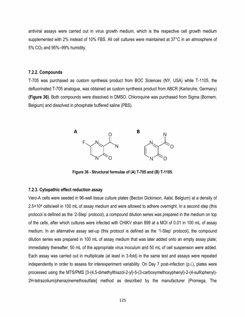
125
antiviral assays were carried out in virus growth medium, which is the respective cell growth medium
supplemented with 2% instead of 10% FBS. All cell cultures were maintained at 37°C in an atmosphere of
5% CO2 and 95%–99% humidity.
7.2.2. Compounds
T-705 was purchased as custom synthesis product from BOC Sciences (NY, USA) while T-1105, the
defluorinated T-705 analogue, was obtained as custom synthesis product from ABCR (Karlsruhe, Germany)
(Figure 36). Both compounds were dissolved in DMSO. Chloroquine was purchased from Sigma (Bornem,
Belgium) and dissolved in phosphate buffered saline (PBS).
Figure 36 - Structural formulae of (A) T-705 and (B) T-1105.
7.2.3. Cytopathic effect reduction assay
Vero-A cells were seeded in 96-well tissue culture plates (Becton Dickinson, Aalst, Belgium) at a density of
2.5×10⁴ cells/well in 100 mL of assay medium and were allowed to adhere overnight. In a second step (this
protocol is defined as the ‘2-Step’ protocol), a compound dilution series was prepared in the medium on top
of the cells, after which cultures were infected with CHIKV strain 899 at a MOI of 0.01 in 100 mL of assay
medium. In an alternative assay set-up (this protocol is defined as the ‘1-Step’ protocol), the compound
dilution series was prepared in 100 mL of assay medium that was later added onto an empty assay plate;
immediately thereafter, 50 mL of the appropriate virus inoculum and 50 mL of cell suspension were added.
Each assay was carried out in multiplicate (at least in 3-fold) in the same test and assays were repeated
independently in order to assess for interexperiment variability. On Day 7 post-infection (p.i.), plates were
processed using the MTS/PMS [3-(4,5-dimethylthiazol-2-yl)-5-(3-carboxymethoxyphenyl)-2-(4-sulfophenyl)-
2H-tetrazolium/phenazinemethosulfate] method as described by the manufacturer (Promega, The

126
Netherlands). The 50% effective concentration (EC50), which is defined as the concentration of compound
that is required to inhibit virus-induced cell death by 50%, was determined using logarithmic interpolation.
Potential cytotoxic/cytostatic effects of the compound were evaluated in uninfected cells using the MTS/PMS
method. The 50% cytotoxic concentration (CC50; i.e. the concentration that reduces the overall metabolic
activity of the cells by 50%) was calculated using logarithmic interpolation. All the assay wells were checked
microscopically for minor signs of a virus-induced CPE or alterations to the cells caused by the compound.
A variant of this protocol was used for the reverse-engineered viruses. In summary, 1×10⁴ Vero E6 cells were
seeded per well in a 96-well tissue culture plate and infected the next day at a MOI of 0.001. On Day 4 p.i.,
plates were processed using a viability assay as has been described (Scholte et al. 2013). EC50 and CC50
values were calculated using non-linear regression with GraphPad Prism.
7.2.4. Virus yield assay
Vero-A cells were seeded in 96-well plates at a density of 5×10⁴ cells/well in 2% FBS medium. Following 24
h of incubation, the culture medium was replaced with 100 mL of assay medium containing a serial dilution of
the compound and 100 mL of CHIKV 899 inoculum (MOI = 0.01). After 2 h of incubation, cell monolayer was
washed three times with assay medium to remove non-adsorbed virus and the cultures were further incubated
for 2 days in the presence of the compound. Chloroquine was included as a reference compound. Supernatant
was harvested and viral RNA was isolated using Macherey Nagel NucleoSpin 96 virus kit. Viral load was
determined by qRT–PCR. Additionally, the amount of infectious progeny virus from virus-infected, compound
treated cells was assessed by plaque assay. Briefly, BHK cells were seeded in 12-well plates (IWAKI) at a
density of 5×10⁵ cells/well in 10% FBS medium. Following 24 h of incubation, monolayers were washed three
times with PBS and cells were infected with 500 mL of 10-fold serial dilutions of the harvested supernatants.
After 1 h of incubation with continuous shaking, monolayers were washed three times with PBS and overlaid
with 3 mL of 2% Avicel PH-101 (Sigma-Aldrich) in medium with 2% FBS. After 4 days at 37°C, monolayers
were carefully washed three times with PBS, fixed with 70% ethanol and stained with methylene blue to
visualize and count PFU.
A variant of this protocol was used for the other CHIKV strains, as well as for the other alphaviruses. For
these, cells were seeded in 2.5% supplemented fetal calf serum medium (FCS). The next day, 2-fold serial
dilutions of the compounds were added to cells (25 mL/well). Fifteen minutes later, 25 mL of a virus mix

127
containing the appropriate amount of viral stock dilution in medium was added to the 96-well plates. Cells
were cultivated for 2 days and viral RNA was isolated as was previously described.
7.2.5. CHIKV quantitative reverse transcription-PCR
Viral RNA was isolated from 150 µl supernatant using the NucleoSpin RNA virus kit (Macherey-Nagel, Düren,
Germany). Primers and probe sequences were as published before: ChikSII 5’-
CCGACTCAACCATCCTGGAT-3’, ChikAsII 5’-GGCAGACGCAGTGGTACTTCCT-3’, ChikProbe 5’-FAM-
TCCGACATCATCCTCCTTGCTGGC-TAMRA (Panning et al. 2008). One-step, quantitative RT-PCR was
carried out in a total volume of 25 µl, containing 13.94 µl H2O, 6.25 µl master mix (Eurogentec, Seraing,
Belgium), 0.375 µl of 60 µM forward primer, 0.375 µl of 60 µM reverse primer, 1 µl of 10 µM probe, 0.0625
µl reverse transcriptase (Eurogentec) and 3 µl RNA sample. qRT-PCR was carried out using the ABI 7500
Fast Real-Time PCR System (Applied Biosystems, Branchburg, NJ) under the following conditions: 30 min at
48C and 10 min at 95C, followed by 40 cycles of 15 s at 95C and 1 min at 60C. The data were analyzed
using the ABI PRISM 7500 SDS software (version 1.3.1; Applied Biosystems). For absolute quantification,
standard curves were generated using CHIKV cDNA 10-fold dilutions. Alternatively, CHIKV LR2006_OPY1
and clinical isolates from Italy and Congo were quantified by real time RT-PCR to determine viral RNA yield
(SuperScript III Platinium one-step RT-PCR with Rox from Invitrogen), using Chik-F2
TGGAATGGCTGGTTAACAAGATAA, CHIK-R2 CTCCGCGGACACCTAACG (except Congo strain: CHIK-
R3 CTCCGCGGACACCTAWSG) and probe FAM-CTACTAAGAGAGTCACTTGGGTAG-MGB.
Other Alphaviruses were amplified using: O’Nyong Nyong; ONN-F2: GGAGCGGGCATAGTCGAA; ONNR2:
CGCGTGAATCAGACTGAGTTTT, ONN-P2 FAM-TCAGACCTTGTTGTCGGAG-MGB, Mayaro: Maya-F2:
CGCCCGCCTACAATTCAA; Maya-R2: GTACTGACCGCAGCAATCAACT; Maya-P2: FAM-
CAGATCAACAGGCCCG-MGB, Barmah Forest: Barm-F: CCGATCCAAAGCTGCTATGC; Barm-R:
TTGCCAATAAACCTGGGCTTA; Barm-P: FAM-ACACCATTCTCCC-MGB, Ross River Virus: RRV-F2:
CCGTGGCGGGTATYATCAAT; RRV-R2: AACACYCCCGTCGACAACAGA, RRV-P FAM-
ATTAAGAGTGTAGCCATCC-MGB, Venezuelian Equine Encephalitis virus; VEEV-F:
AACTGGGCCGACGAAACC; VEEV-R: CAGAGAATAGAACATTGTTGGATGGT; VEEV-P: FAM-
ACGGCTCGTAACATAGG-MGB, Western Equine Encephalitis virus; WEEV-F:
AGGGATMCCCCCGAAGGTT; WEEV-R: GTGAATAGCACACGGGTGGTT; WEEV-P: FAM-
CTTTCGAATGTCACGTTCCCATGCG-TAMRA and Eastern Equine Encephalitis virus; EEV-F:

128
TGTGCGTACCTCCTCATCGTT; EEEV-R: GACTGGCGTGAATCTCWGCTT; EEEV-P: -FAM-
AGCAGCCTACCTTTCCGACAATGGTTGTC-TAMRA. For absolute quantification, standard curves were
generated using T7 polymerase-generated RNA 100-fold dilutions of known quantities for each virus. qRT-
PCR was carried out on a ABI 7900 HT Fast Real-Time PCR System, using 20 min at 50°C and 3 min at
95°C, followed by 40 cycles of 15 s at 95C and 1 min at 60C.
7.2.6. Determination of 50% cell culture infective dose (CCID50) by titration
To titrate the virus stocks resulting from the virus yield assay, 96-well microtiter plates were filled with 100 µL
of assay medium. Next, 25 µL of the viral supernatant was added to six wells of the microtiter plate. Fivefold
serial dilutions were prepared and 100 µL of Vero-A cell suspension (2.5 × 10⁴ cells/well) was added to the
wells. Following 5 days of incubation at 37°C, cells were examined microscopically for CHIKV-induced CPE.
A well was scored positive if any trace of CPE was observed, as compared to uninfected controls. The CCID50
was calculated with the Reed and Muench method (Reed & Muench 1938).
7.2.7. Delay-of-treatment assay
Vero-A cells were seeded in 96-well plates at a density of 5 × 10⁴ cells/well in assay medium and incubated
overnight. Two hours prior to CHIKV infection 50 µM of T-705 or chloroquine was added in 200 µL of assay
medium at the condition -2 hours. Subsequently, at time point 0, the medium of all wells was removed and
cells were infected with CHIKV 899 at 0.1 MOI during 1 hour at 37°C. Next, 50 µM of T-705 or chloroquine
was added at 2, 4, 8 and 12 hours after infection. Following 48 hours of incubation, supernatants were
collected and RNA was isolated from 150 µl supernatant using the NucleoSpin RNA virus kit. Viral RNA was
quantified by qRT-PCR as described above.
7.2.8. Reversal of anti-CHIKV activity
Vero-A cells were seeded in 96-well tissue culture plates at a density of 2.5×10⁴ cells/well in 100 mL of assay
medium and were allowed to adhere overnight. The following day, 2-fold dilution series of the
nucleobases/nucleosides adenosine, guanosine, cytosine, thymine and uracil were added to the cells, starting
at 764M. Immediately afterwards, 50 L of favipiravir solution was added to each well, resulting in a final
concentration of 127 M, a concentration sufficient enough to completely inhibit CHIKV replication in the
absence of the added nucleobases/nucleosides. Subsequently, cells were infected with CHIKV strain 899 at
a MOI of 0.05. In the same test, multiplicates were included and at least two independent assays were carried

129
out. The effect of the nucleobases/nucleosides was assessed during the MTS/PMS method as described
above. In addition, all assays were checked microscopically for minor signs of CPE and possible adverse
effects that might have been induced by the nucleic acids/nucleosides.
7.2.9. Selection, purification and adaptation of T-705-resistant virus isolates
To select for T-705 resistant virus isolates a 5-step protocol was designed. In a first step, Vero-A cells, seeded
in 100µl of assay medium in 96-well microtiter plates at a density of 2.5 × 10⁴ cells/well, were allowed to
adhere overnight. Subsequently and in duplicate, 6 antiviral assays with T-705 were set up using dilutions
with different CCID50 of CHIKV 899 (ranging from 10 to 1000 CCID50). After 7 days of incubation, all assay
wells were checked microscopically and quantitative data on cell survival were collected using the MTS/PMS
method. Based on these data, the lowest concentration of compound and the highest virus input at which
complete and reproducible inhibition of virus-induced CPE was observed were selected. In a second step,
three 96-well plates (a total of 144 assay wells) containing adherent Vero cells were infected with the most
optimal virus dilution (1000 CCID50) and compound concentration (127 M). As expected, after seven days
of incubation, most assay wells did not show any signs of virus-induced cell death. However, in some of the
assay wells, virus break-through could be observed and the supernatant of the 3 wells with the most
pronounced signs of virus-induced CPE was collected. These samples, which are supposed to contain virus
variants that are capable of replicating in the presence of T-705, were purified in 6-fold by titration (1:5 dilution
series) in presence of 127 µM T-705. Again, after 7 days of incubation and based on microscopic observations
as well as MTS/PMS data, three virus isolates (one from each original sample) that produced the most
pronounced signs of CPE in the presence of T-705 at the lowest virus input possible were selected (in order
to obtain a virus population as pure as possible). Step four encompassed growing these selected virus
variants in 25 cm2 flasks in presence of T-705 to produce reference virus stocks for further experiments. After
day 5 post-infection, the cell culture medium was collected, cell debris removed by centrifugation, the
supernatant aliquoted, and stored at -80°C. Subsequently, the resistant phenotype of the selected virus
isolates was determined in comparison with the wild-type virus essentially by repeating step 1 as described
above (in triplicate). In parallel, the genotype was determined by full genome sequencing. The virus isolates
obtained at this stage will be referred to as T-705_res isolates. In order to allow the virus to have the
opportunity to acquire additional mutations required for a more efficient replication in presence of T-705, the
three T-705_res virus isolates were further cultured in presence of T-705 (127 M for passage 1-5 and 159

130
M for passages 6 and 7). These virus isolates will be referred to as passage 7 (T-705_res_p7) isolates.
Again, the resistant phenotype was determined, as well as the sequence of the full genome.
7.2.10. Resistance and cross-resistance phenotyping
Essentially, the protocol used to determine the resistant phenotype was identical to the ‘2-Step’ protocol
described above. The resistance and cross-resistance phenotyping assays were standardized by using 100
CCID50 of virus inoculum of CHIKV 899 wild-type and both passage 0 and passage 7 virus isolates.
Resistance was evaluated against T-705; T-1105 was included to assess cross-resistance.
7.2.11. Sequencing
Eight overlapping PCR amplicons were generated from viral RNAs previously extracted from the wild-type
CHIKV 899 strain and from the p0 and p7 virus isolates. Amplicons were generated by the OneStep RT-PCR
kit (Qiagen) and were gel purified and sequenced (BigDye® Terminator v3.1 Cycle Sequencing Kit ABI) using
primers listed in table 10. The complete nucleotide sequences were assembled in ContigExpress (VNTI,
Invitrogen) and the genomes of the resistant isolates were compared to the wild-type genome. Finally, BLAST
analyses were carried out for the sequences of interest (Blast 2.2.26+, (Altschul et al. 1997)).
Table 10 - Primer list used for sequencing wild-type CHIKV 899 lab strain, as well as T-705_res and T-
705_res_p7 CHIKV isolates.
Protein Position Sequence
nsP1 CHIKV(+)1 ATGGCTGCGTGTGACACAC
CHIKV(+)600 GGGTTGGGTTCGACACAAC
CHIKV(+)1450 CGGGTTGTCAATCCCTTTG
CHIKV(-)1800 GACTGAGCTTCTGGCTACG
CHIKV(+)1450 CGGGTTGTCAATCCCTTTG
nsP2 CHIKV(+)2200 TGCCTGCCCATACAAAATTG
CHIKV(-)2407 GCATCCATTCAAGAGCAGC
CHIKV(-)3000 TCGGTGGGTTCTGCAGCGTCT
CHIKV(+)2800 ATACGAGGTCATGACAGCA
CHIKV(+)3200 GCATACTCACCTGAAGTAGCC
CHIKV(-)4200 GCCATTTTTTGTATACTGCCT
CHIKV(+)4000 AAGGAATTTCACAACTCATGTC
nsP3 CHIKV(+)4700 ACGGCTGTGGATATGGC
CHIKV(-)5000 CTTGGGTCCGCATCTGTATGG
nsP4 CHIKV(-)5800 TCCTTTGCTTCATCCAGCT
CHIKV(+)5600 ACAGACAGCGACTGGTCCA

131
CHIKV(+)6100 TGATGCATATCTAGACATGGT
CHIKV(-)7100 CTTCCATGTTCATCCAAGTGGC
CHIKV(-)7600 ATTGTAAAAAGTTTGGGTTGGGA
CHIKV(+)7400 ATATCAGTTGTGGTAATGTC
Core CHIKV(+)7750 CGCAGGAATCGGAAGAATAAG
CHIKV(+)8100 CACCCATGAGAAACTGGAG
E2 CHIKV(-)9000 CAAGGTAGCTCTTTACCGTG
CHIKV(+)8800 ACATCAGCACCGTGTACGA
CHIKV(+)9500 CCGTGCCGACTGAAGGGCT
E1 CHIKV(-)10000 TTCGTACGCGCTCACAGTGTG
CHIKV(+)10200 GGACAAAAACCTACCTGACTACAG
CHIKV(+)11000 CCGTCACTATTCGGGAAG
CHIKV(-)11300 ACGACACGCATAGCACCAC
CHIKV(-)11832 TTTTTTTTTTTTTTTTTTTTGAAATATTAAAAACAAAATAACATCTCC
7.2.12. Metabolic labeling with [3H]uridine, denaturing agarose electrophoresis and in-gel hybridization
Actinomycin D (ActD; Sigma-Aldrich) was added to a final concentration of 5mg/mL to 2.8×10⁵CHIKV infected or
mock-infected Vero E6 cells in12-well plates at 5.5 hp.i. At 6h p.i., the viral RNA that had been synthesized was
labeled by adding 40μCiof [3H]uridine to the medium. At 7h p.i.,the tota lRNA was isolated and separated in
denaturing agarose gels as described (Scholte et al. 2013).3H-labelled RNA was visualized by fluorography. To
correct for variations in loading, the gel was hybridized with a 32
P-labelled oligonucleotide probe recognizing 18S
ribosomal RNA. The detection of positive-sense strand CHIKV RNA by in gel hybridization with a 32
P-labelled probe
complementary to the 3´end of the genome was carried out as previously described (Scholte et al. 2013).3H-labelled
RNA was quantified by direct scintillation counting of RNA samples and by densitometry of scanned films after
fluorography of agarose gels. Hybridized gels were analysed using PhosphorImagerscreensanda Typhoon 9410
imager (GEHealthcare), followed by quantification with Quantity One (Biorad).
7.2.13. Reverse-engineering
Mutations were introduced in the infectious CHIKV LS3 cDNA clones using the QuikChange mutagenesis kit (Agilent),
according to the manufacturer’s instructions. The constructs were verified by sequencing using the BigDye Terminator
Cycle Sequencing Kit v1.1 (Applied Biosystems) and a 3130 Genetic Analyzer automatic sequencer (Applied
Biosystems). CHIKV was produced from these plasmids as described by (Scholte et al. 2013). In summary, RNA was
transcribed using the AmpliScribe T7 high yield transcription kit (Epicenter), the m7GpppA RNA cap structure analogue
(NEB) and 0.7 mg of linearized template DNA. After digestion of the template DNA with DnaseI and precipitation with
7.5 M LiCl (Ambion), the concentration of the in vitro transcribed RNA was determined with a NanoDropspectro-

132
photometer (Thermo Scientific) and its integrity was checked by agarose gel electrophoresis. BHK-21 cells were
electrophorated with in vitro transcribed RNA using the AmaxaNucleofector, according to the manufacturers’
instructions. Infectious CHIKV from the supernatant of transfected cells (p0) was used to grow working stocks (p1) of
mutant viruses, which were used for further experiments. To confirm the presence of the introduced mutations (and
the absence of other mutations), viral RNA was extracted from virus stocks using the QIAamp Viral RNA Mini Kit
(Qiagen). This RNA was used to generate four overlapping amplicons by RT–PCR amplification that were used for
sequencing. One-step growth curves were generated by infecting Vero E6 cells at a MOI of 3, after which the CHIKV
titre in the medium was determined at various time-points after infection by plaque assay as previously described
(Scholte et al. 2013).
7.2.14. CHIKV mouse model
Three groups of 6-week-old AG129 mice (B&K Universal Limited; n= 6 per group) were used in this experiment
(Couderc et al. 2008). The animals in Group 1 were treated with favipiravir 24 h prior to infection. The animals in Group
2 received treatment 4 h after challenge and the animals in Group 3 received mock treatment. All of the animals were
treated twice a day with 300 mg/kg/day via oral gavage. Favipiravir was suspended in a sterile 0.4% sodium
carboxymethyl cellulose solution as previously described (Julander et al. 2009). At timepoint 0, the mice were
challenged intraperitoneally with 100 CHIKV strain S27 median infective tissue culture dose (TCID50). Mice were
euthanized by cervical dislocation under isofluraneanaesthesia when they reached humane endpoints (immobility and
paralysis), after which the brains were immediately collected for further processing. To quantify the viral loads in the
brain, half of the brain was weighed and homogenized using a metal bead in 1 mL of DMEM containing antibiotics
(100 U of penicillin, 100 mg/mL of streptomycin) using a tissue homogenizer. The CHIKV S27 RNA load was quantified
using a one-step RT–PCR TaqMan protocol (EZ-kit, Applied Biosystems) and an ABI PRISM 7500 detection
instrument. The primers and probe used for CHIKV S27 RNA quantification were: forward primer
AAGCTCCGCGTCCTTTACCAAG; reverse primer CCAAATTGTCCTGGTCTTCCT; probe FAM-CCAATGTCTTCAG
CCTGGACACCTTT-TAMRA. The results were expressed as TCID50 equivalents per gram of brain tissue. For this
purpose, a log10 dilution of the virus stock was prepared, which was 109 TCID50/mL. Next, a reference line containing
virus titre on the x-axis and the Ct value on the y-axis was generated. The Ct value of the sample was then interpolated
to estimate the TCID50 equivalents. All the animal experiments were conducted in accordance with the Dutch guidelines
for animal experimentation and were approved by the Animal Welfare Committee of the Erasmus Medical Center,
Rotterdam, The Netherlands (protocol number DEC122-12-20).
7.2.15. 3D-model of the binding of T-705 to CHIKV nsP4
The CHIKV nsP4 homology model created by Kumar and colleagues (Kumar et al. 2012) was superimposed on protein
databank structure 3BSO of the Norwalk virus (NWV) polymerase containing an RNA template/primer with entering

133
cytosine triphosphate (CTP) using the Dali server (Holm & Sander 1996; Castrignanò et al. 2006; Zamyatkin et al. 2008;
Kumar et al. 2012). To obtain further insight into the mechanism of action of favipiravir on CHIKV nsP4, the structure of
favipiravir was superimposed onto the cytosine base of CTP bound in the active site of the NWV polymerase structure
using Quatfit (D.J. Heisterberg, 1990, unpublished results). The favipiravir base was entered in the anti-conformation.
However, this way, it clashed with the pairing template G nucleotide; only C and U nucleotides in the template strand
produced matching hydrogen bonds with favipiravir (Jin et al. 2013). Therefore G in the template was replaced by C.
As a result, favipiravir-RTP acts as a purine in the model, which is also suggested by the nucleoside competition
experiments that were carried out (Figure 40). Replacement of amino acids in the homology model was done using
Edpdb (Zhang Xue-Jun & Matthews 1995). The model was created using Chimera (Pettersen et al. 2004).
7.3. Results
7.3.1. Favipiravir and T-1105 are selective inhibitors of CHIKV replication (and other alphaviruses)
Favipiravir and its defluorinated analogue T-1105 (Figure 36) were evaluated in parallel with the reference compound
chloroquine for their ability to inhibit the CPE induced by CHIKV laboratory- adapted strains and clinical isolates, as
well as the closely related viruses SINV and SFV. The antiviral (i.e. cell-protective) effect was quantified by means of
a colorimetric method [using different assay set-ups (‘1-Step’ versus ‘2-Step’)] (Table 11) and was confirmed by
microscopic inspection. At appropriate levels of concentration, favipiravir, T-1105 and chloroquine fully protected the
cells from CHIKV, SINV or SFV induced CPE. T-1105 proved to be 2 to 5 fold more potent than favipiravir. At
concentrations lower than 500 mM, favipiravir and T-1105 did not cause notable changes to the cell, monolayer
morphology (microscopic inspection) or cell viability (as measured with an MTS/PMS assay).
The antiviral effect of favipiravir and T-1105 was next validated by quantifying (i) the release of viral RNA by qRT–PCR
and (ii) the production of infectious progeny virus by plaque assay (Figure 37, Table 12). The EC50 values calculated
from these dose–response curves were comparable to those from the CPE reduction assays (Tables 11 and 12). Both
compounds also inhibited other laboratory adapted strains and clinical isolates of CHIKV (with geographically different
origins), as well as seven other alphaviruses including both New and Old World pathogens (Table 12).

134
Table 11 - Effect of T-705, T-1105 and chloroquine on Chikungunya, Semliki forest and Sindbis virus-induced
cytopathic effect.
EC50 (µM)
Virus species Strain T-705 T-1105 Chloroquine
CHIKV
Indian Ocean 899 (lab) 25 ± 3a
60 ± 10b
7.0 ± 1a
47 ± 12b
11 ± 7a, c
28 ± 1b, c
LR2006-OPY1 (lab) 25 ± 1 (MOI 0.1)b
48 ± 2 (MOI 1)b ND ND
Italy 2008 (clinical) 16 ± 6 (MOI 0.1)b
48 ± 1 (MOI 1)b ND ND
SFV Vietnam (lab) 29 ± 14a
48 ± 10b
6.2 ± 0.4a
32 ± 4b
14 ± 2a
69 ± 1b
SINV HRsp (lab) 37 ± 6a
28 ± 6b
17 ± 2a
23 ± 7b
11 ± 2a
30 ± 5b
a ‘1-Step’ protocol, b ‘2-Step’ protocol (see Materials and Methods), c previously published (Bourjot et al. 2012). ND = not determined. CC50 values are >636, > 571 and 89 ± 28 µM for T-705, T-1105 and chloroquine, respectively.
Figure 37 - In vitro antiviral activity of T-705, T-1105, and chloroquine on CHIKV replication Dose-response effects of T-705 (blue), T-1105 (orange) and chloroquine (black) on CHIKV replication quantified by
qRT-PCR (panel A) and by plaque assay (panel B). Data comprise mean values of at least three independent experiments.
0
20
40
60
80
100
0,1 1 10 100 1000
% C
HIK
V R
NA
of
un
trea
ted
co
ntr
ols
Log10 Concentration (µM)
Chloroquine T-1105 T-705 A
0
20
40
60
80
100
0,1 10 1000% P
FU o
f u
ntr
eate
d c
on
tro
ls
Log10 Concentration µM
Chloroquine T-1105 T-705

135
Table 12 - Effect of T-705, T-1105 and chloroquine on the replication of an alphaviruses panel
Virus species
Strain
EC50 (µM) T-705 T-1105
CHIKV
Indian Ocean 899 (lab) 5.9 ± 3.3a
4.7 ± 1.5b 2.8 ± 0.3a
0.7 ± 0.07b
LR2006-OPY1 (lab) 9.8 ± 0.1b 7.3 ± 0.8b
Venturini (Italy 2008) 12 ± 0.3b 13 ± 2.9b
Bianchi (Italy 2008) 6.1 ± 0.1b 11 ± 4.5b
Congo 95 (2011) 1.9 ± 1.1b 1.6 ± 0.3b
ONNV IPD A234 9.7 ± 3.8b 4.9 ± 1.4b
Mayaro NCPV TC652 16 ± 1.2b 11 ± 1.4b
RRV NCPV 5281v 3.5 ± 0.5b 3.0 ± 1.0b
VEEV TC83 11 ± 4.3b 13 ± 2.9b
WEEV 47a 7.5 ± 1.8b 12 ± 4.0b
EEEV H178/99 18 ± 1.7b 25 ± 1.9b
BFV BH2193 18 ± 5.5b 5.4 ± 4.4b
a determined by plaque assay, b determined by RT-qPCR. ND = not determined, ONNV= O’Nyong Nyong virus, RRV= Ross River virus; VEEV= Venezuelan equine encephalitis virus; WEEV= Western equine encephalitis virus; EEEV= Eastern equine encephalitis virus; BFV= Barmah Forest virus.
7.3.2. Favipiravir inhibits CHIKV infection at the replication stage
A delay-of-treatment experiment was carried out with favipiravir and chloroquine to obtain an initial idea of the stage
in the viral replication cycle at which favipiravir acts. The addition of favipiravir to the infected cultures resulted in an
almost complete inhibition of viral replication when added at 22, 0 or 2 h p.i. (Figure 38B). A further delay of treatment
resulted in a gradual loss of antiviral activity, and no antiviral effect was observed when the compound was added at
12 h p.i. These results, interpreted in the context of the production and release of progeny virus (Figure 38A, blue
bars measured as CHIKV RNA levels in the supernatant) and intracellular RNA replication (Figure 38A, red bars),
suggest that favipiravir acts during the stage of viral RNA synthesis, which is consistent with the hypothesis that
favipiravir targets the viral RdRp. In cells treated with chloroquine, viral RNA levels were 87% lower when the

136
compound was added at 22 h, and 32% lower when added at time-point 0, compared with untreated controls (Figure
38B). These results confirm that chloroquine primarily exerts its antiviral effect at an early stage of the CHIKV
replication cycle (Khan et al. 2010).
Figure 38 - A. Replication kinetics of CHIKV in Vero cells B. Comparison of the delay-of-treatment effect of T-705 and chloroquine on intracellular viral RNA replication in CHIKV-infected Vero cells as quantified by qRT-
PCR. Data shown comprise mean values ± SD of at least three independent experiments.
To corroborate these observations, 3H-uridine labelling experiments were conducted to assess the effect of favipiravir
on CHIKV RNA synthesis. First, the virus was allowed to adsorb the cells for 1 h, after which the inoculum (MOI 5)
was removed and favipiravir was added to the medium at different time-points p.i., ultimately resulting in 1 – 6 h
treatments. At 5.5 h p.i. ActD was added to arrest cellular transcription, and at 6 h p.i., 3H-uridine was added to the
medium to selectively label the newly synthesized CHIKV RNA. Uninfected cultures were included to demonstrate the
specificity of the metabolic labeling for viral RNA synthesis (Figure 39A). At 7 h p.i., cells were lysed and total RNA
was isolated. Short favipiravir treatments of up to 4 h hardly affected the accumulation of CHIKV RNA as detected by
hybridization, while longer treatments of 5-6 h resulted in a 20%–25% reduction of intracellular CHIKV RNA levels
compared with untreated infected control cells (Figure 39B). However, CHIKV RNA synthesis, measured by the
quantification of 3H-uridine incorporation, was strongly reduced with increasing treatment times (which is in line with
the observation that favipiravir must be converted to its triphosphorylated ribonucleoside) (Figure 39B). The treatment
of CHIKV-infected cells with favipiravir for 6 h resulted in an 80% reduction in the rate of viral RNA synthesis. A
reduction in RNA synthesis of ≥50% required a favipiravir treatment of at least 4 h. This also explains why only
treatments longer than 4 h resulted in having an antiviral effect (reduction of CHIKV RNA) (Figure 39B).
-6
-4
-2
0
2
4
2 4 8 12 24 48
CH
IKV
RN
A q
uan
tity
(n
g, lo
g10
)
Time post infection (h)
extracellular intracellular A
0
20
40
60
80
100
-2 0 2 4 8 12
% o
f vi
ral R
NA
of
un
trtr
eate
d
con
tro
ls
Time of drug addition
Chloroquine T-705B

137
Figure 39 A - CHIKV-infected cells treated or not treated with T-705 for 1 to 6 hours, as schematically
depicted. B. Quantification of the total amount of CHIKV RNA and CHIKV RNA synthesis activity in cells treated with T-705.
To assess whether the inhibitory effect of favipiravir could be reversed by nucleosides, CHIKV infected cells were
incubated in a medium with 127 mM favipiravir and different concentrations of nucleosides, after which favipiravir
antiviral effect was determined by CPE reduction assays. The purine nucleosides adenosine and guanosine were able
to revert the antiviral activity of 127 mM favipiravir at concentrations between 24 – 764 mM, and 96 – 764 mM,
respectively (Figure 40), with adenosine being more potent than guanosine. In contrast, the addition of pyrimidines
did not affect the antiviral activity of favipiravir on the replication of CHIKV. None of the nucleosides themselves
inhibited CHIKV replication at any of the concentrations tested (data not shown). This competition experiment
demonstrates that favipiravir acts as a purine nucleoside in CHIKV RNA replication. Similar data were obtained for
influenza virus (Furuta et al. 2005).
A B

138
Figure 40 - Reversal of anti-CHIKV activity of favipiravir by nucleosides.
Data shown comprise mean values +SD of multiplicates from at least two independent experiments.
7.3.3. Favipiravir reduces CHIKV-induced disease in mice
AG129 mice were infected with 100 CHIKV strain S27 TCID50 (37). Mice that received placebo treatment (PBS)
developed severe signs of neurological infection on Day 3 and Day 4 p.i., and were euthanized at that time on humane
grounds (Figure 41A, black circles). Only one of the six infected animals that were treated orally twice daily with 300
mg/kg/day of favipiravir (starting either 24 h before infection or 4 h after infection) for 7 consecutive days had to be
euthanized due to severe clinical signs before day 7 (Figure 41A, grey and white circles). One other mouse (from the
group whose treatment started at 4 h p.i.) had to be euthanized on day 10. All the other mice remained healthy until
day 14 (the end of the experiment). After euthanasia, the brain tissue from the euthanized mice was collected to
determine viral loads by qRT–PCR (Figure 41B). The viral loads of CHIKV were 3.2+0.1 and 2.3+0.3 log10 TCID50
equivalents/g brain lower for the mice that received treatment 24 h prior to infection and 4 h after infection, respectively,
compared with the placebo controls.
0
20
40
60
80
100
120
1 10 100 1000
% o
f T
-70
5 a
nti
CH
IKV
act
ivit
y
Log10 concentration (µM)
Adenosine Guanosine Cytosine Thymine Uracil

139
Figure 41 A - Survival curves of mice infected with CHIKV strain S27 and treated with 300 mg/kg.day of T-705 or that received placebo treatment with PBS
() 6 mice infected with 100 TCID50 of CHIKV strain S27 and that received placebo treatment with PBS (BID for 7 days), () 6 mice that were infected with 100 TCID50 of CHIKV strain S27 and that received the first oral dosing of a 7-day BID 300 mg/kg.day treatment schedule with T-705 starting 4h after infection, () 6 mice that received the first
oral dosing of a 7-day BID 300 mg/kg.day treatment schedule with T-705 starting 24h prior to infection with 100 TCID50 of CHIKV strain S27. The animals were treated for 7 days and were kept for another 7 days for additional
observation.
B - Average viral titers in the brain of treated or untreated mice Viral titers are expressed as log10TCID50 equivalents per gram of brain tissue (bars). Viral loads in the brain were
determined by qRT-PCR. 7.3.4. Favipiravir and T-1105 do not affect the specific infectivity of CHIKV
An induction of error-prone replication has been suggested as the mechanism by which favipiravir elicits its antiviral
effect against influenza virus (Baranovich et al. 2013). To study whether favipiravir and its analogue T-1105 inhibit
CHIKV infection by inducing error-prone replication, the specific infectivity of CHIKV (strain 899) was determined,
calculated as the ratio of infectious virus yield×1023 to the genome copy number, in the presence of favipiravir, T-1105
or chloroquine (Figure 42). The calculated specific infectivity values were between 0.5 and 1.1 for all concentrations
of the studied compounds. The decrease in infectivity upon treatment was proportional to the decrease in viral RNA
content. In contrast, the specific infectivity of influenza virus was found to be more than 25-fold lower upon treatment
with 10 mM favipiravir compared with that of the untreated controls, while the RNA copy numbers remained
comparable, which suggests an inhibition of virus replication through error catastrophe (Baranovich et al. 2013). Under
the same conditions, the specific infectivity ratio for CHIKV remained at 1.0+0.06, suggesting that error catastrophe is
not the mechanism underlying the inhibitory effect of favipiravir for this virus.

140
Figure 42 - CHIKV specific infectivity following treatment with different favipiravir, T-1105 and chloroquine
concentrations Data were normalized to that of untreated control samples (1.0) as described by Baranovich et al., 2013.
7.3.5. Selection and characterization of favipiravir-resistant CHIKV isolates
By using the resistance selection protocol described before, three putative favipiravir-resistant virus isolates were
independently obtained from a heterogeneous wild-type (quasi species) CHIKV population (strain 899). These virus
variants were designated favipiravir_res. Subsequently, each of these isolates was further cultured for seven additional
passages in the presence of 127–159 mM favipiravir to allow the respective virus variants to further adapt upon
replication in the presence of the compound (designated favipiravir_res_p7). Overall, the favipiravir_res virus variants
(Clones 1–3) were slightly less susceptible to the antiviral effect of favipiravir, with a modest but significant shift in EC50
values (Table 13).

141
Table 13 - Antiviral phenotype of T-705-resistant CHIKV isolates
Averages and standard deviations were calculated from data obtained from at least 6 independent antiviral experiments. FR= fold resistance; NA= not applicable; c1, c2 and c3 are the enriched/purified virus isolates that were obtained independently.
Up to six mutations were observed in ORF 1, i.e. in nsP2, nsP3 and nsP4 (Table 14). In contrast, in ORF 2 that
encodes the structural proteins, only silent mutations were detected (data not shown). Mutation K291R in nsP4
(encoding the RdRp) was the only mutation that was detected in all three of the favipiravir_res isolates. Respectively,
none of the 149 or 382 sequences in the CHIKV or alphavirus BLAST analysis carried an arginine at position 291 in
nsP4 (Table 14). Four of the other ORF 1 mutations were also shown to be unique for the favipiravir-resistant CHIKV
virus variants. Furthermore, T-1105 proved cross-resistance with favipiravir, since T-1105 also elicited reduced
antiviral activity with the favipiravir-resistant isolates (2.5-fold).
The EC50 values for the favipiravir_res_p7 isolates ranged from 4.0- up to 9.6-fold higher than for the wild-type (WT)
(240–577 mM) (Table 13). In addition to the K291R mutation in nsP4, four additional mutations were now detected in
the sequence of all three of the p7 virus isolates (Y543C in nsP2, and D31G, F345S and S471P in nsP3). Two of these
mutations are unique to favipiravir-resistant CHIKV variants, as shown by the CHIKV BLAST analysis (Table 14). A
number of other mutations were identified in one or two isolates, as well as reversions to the wild-type (K150R and
W524Opal). The fitness of the favipiravir_res_p7 virus variants was similar or slightly (5-to 10-fold) lower than that of
wild-type virus (calculated as TCID50/mL values; data not shown). Based on information of the resistant phenotype and
genotype of the favipiravir_res and the favipiravir_res_p7 virus variants, strong evidence was obtained that shows that
the nsP4 K291R mutation in particular is the key candidate to be linked to the observed phenotypic resistance against
favipiravir, as it is the only mutation that is present in the genome of all favipiravir-resistant CHIKV isolates.
T-705 T-1105
EC50 (µM) FR EC50 (µM) FR
Wild-type 60 ± 10 NA 47 ± 12 NA
T-705_res c1 116 ± 10 1.9 109 ± 11 2.3
c2 82 ± 15 1.4 75 ± 4 1.6
c3 175 ± 10 2.9 170 ± 12 3.6
T-705_res_p7 c1 577 ± 52 9.6 306 ± 3 6.5
c2 240 ± 1 4.0 318 ± 4 6.8
c3 295 ± 31 4.9 306 ± 15 6.5
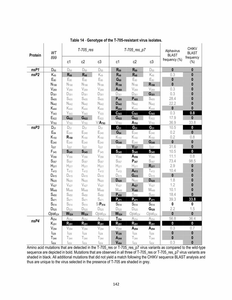
142
Table 14 - Genotype of the T-705-resistant virus isolates.
Protein
WT 899
T-705_res T-705_res_p7
Alphavirus BLAST
frequency (%)
CHIKV BLAST
frequency (%) c1 c2 c3 c1 c2 c3
nsP1 D89 D89 D89 D89 R89 R89 D89 0 0
nsP2 K49 R49 R49 K49 R49 R49 K49 0.3 0 E80 E80 E80 E80 G80 E80 E80 0 0 N198 N198 N198 N198 R198 N198 R198 0 0 V269 V269 V269 V269 A269 V269 V269 0.3 0 D351 D351 D351 D351 D351 D351 G351 0.3 0 S405 S405 S405 S405 P405 P405 S405 28.4 0 N442 N442 N442 N442 D442 N442 N442 22.2 0 K444 K444 K444 K444 R444 K444 K444 0 0 Y543 Y543 Y543 Y533 C543 C543 C543 0.3 0.8 E622 G622 G622 E622 G622 G622 E622 17.9 0 V793 V793 V793 V/A793 V793 A793 V793 36.9 33.8
nsP3 D31 D31 D31 D31 G31 G31 G31 10.5 0 E84 E240 E240 E240 G84 E240 E240 0.2 0 K150 R150 K150 K150 K150 K150 K150 0.2 0.8 E240 E240 E240 E240 G240 E240 G240 0 0 I337 I337 I337 I337 I337 V337 I337 31.6 0 F345 S345 S345 F345 S345 S345 S345 10.5 0 V356 V356 V356 V356 V356 A356 V356 11.1 0.8 S357 S357 S357 S357 S357 P357 S357 73.4 98.5 H377 H377 H377 H377 H377 H377 R377 2.9 0 T413 T413 T413 T413 T413 A413 T413 10.4 0 D415 D415 D415 D415 D415 G415 D415 0 0 N420 N420 N420 N420 D420 N420 D420 1.8 0 V427 V427 V427 V427 V427 A427 V427 1.2 0 M449 M449 M449 M449 M449 V449 M449 10.1 0 S450 S450 S450 S450 P450 S450 S450 18.4 0 S471 S471 S471 S471 P471 P471 P471 39.3 33.8 S514 S514 S514 S/P514 S514 S514 S514 0 0 D520 D520 D520 D520 D520 D520 G520 2.2 1.5 Opal524 W524 W524 Opal524 W524 Opal524 Opal524 0 0
nsP4 A254 A254 A254 A254 T254 A254 A254 56.8 36.4 K291 R291 R291 R291 R291 R291 R291 0 0
V294 V294 V294 V294 V294 A294 A294 0.3 0.7 I326 I326 I326 I326 V326 I326 I326 0 0 T344 T344 T344 T344 A344 T344 T344 0 0 I404 I404 I404 I404 V404 I404 I404 0.3 0
Amino acid mutations that are detected in the T-705_res or T-705_res_p7 virus variants as compared to the wild-type sequence are depicted in bold. Mutations that are observed in all three of T-705_res or T-705_res_p7 virus variants are shaded in black. All additional mutations that did not yield a match following the CHIKV sequence BLAST analysis and thus are unique to the virus selected in the presence of T-705 are shaded in grey.

143
7.3.6. The K291R mutation in nsP4 confers resistance to favipiravir and T-1105
Because of the large number of mutations that were identified in the compound-resistant virus variants, only the
mutations that were shared by multiple variants and that are not observed at a high frequency in natural CHIKV strains
were selected for further analysis. These mutations were reverse-engineered into an infectious CHIKV clone and the
susceptibility to favipiravir and T-1105 of the resulting recombinant viruses was determined. Since an infectious clone
of CHIKV strain 899 is not available, the reverse-genetics system of CHIKV strain LS3 was used, (Scholte et al. 2013)
which also allowed an analysis of the mutations in an independent genetic background.
An alignment of the polymerase sequences for different viruses showed that Lys-291 is located in motif F1 of nsP4
protein from the alphavirus RNA-dependent RNA polymerase, but, interestingly, also in that of other +ssRNA viruses
[i.e. alphaviruses (CHIKV, SFV, SINV), Flaviviridae (HCV, West Nile virus), noroviruses (murine norovirus) and
picornaviruses (poliovirus)] (Figure 43). This motif is believed to be involved in the binding and positioning of the
incoming nucleotide substrate (Lesburg et al. 1999). Therefore, the nsP4 K291R mutation was considered to be the
prime candidate for investigating the link between genotype and phenotypic resistance. In addition, several other
mutants were engineered (Figure 44). The reverse-engineered nsP4_K291R virus mutant was 1.6-fold less susceptible
to the antiviral effect of favipiravir and 1.8-fold less to that of T-1105 (Figure 44A), a statistically significant shift in EC50
that is comparable to what was observed for the favipiravir_res virus isolates of CHIKV strain 899 (Table 13). In contrast,
the nsP3_Opal524W and nsP2_K49R_E622G mutations did not increase the resistance to the antiviral effect of
favipiravir and T-1105. Virus derived from a reverse-engineered clone that carried all four of the identified mutations
(LS3_res_nsP2_K49R_E622G_nsP3_Opal524W_nsP4_K291R) was 3.8-fold more resistant to favipiravir than wild-
type CHIKV LS3 (Figure 44A). Interestingly, when the nsP4_K291R mutant was passaged in absence of antiviral
pressure, the mutant virus acquired the nsP2_Y543C mutation, a mutation that is also present in the viral genome of
all three clones of favipiravir_res_p7 (Table 14).
A comparison of the growth curves of the wild-type CHIKV and the reverse-engineered mutants revealed that the K291R
mutation in nsP4 (in the absence of favipiravir) negatively affected the replication kinetics (Figure 44B, black squares
versus black circles). The triple mutant virus (with the nsP2 and nsP4 mutations) also exhibited a reduced growth rate
compared with wild-type virus (Figure 44B, black squares versus open circles). The nsP3_Opal524W mutant as well
as the mutants in which the nsP4 mutation was combined with either the nsP3_Opal mutation (double mutant), the
nsP2 mutations (triple mutant) or a combination of all of these mutations (quadruple mutant) had replication rates that
were only slightly lower than that of wild-type virus. These results, together with the evaluation of the resistance
phenotype, suggest that the mutations in nsP2 and nsP3 have been acquired to compensate (at least in the absence
of the antiviral compound) for a negative effect of the nsP4 mutation on viral replication fitness.

144
Figure 43 - Sequence alignment of the RNA-dependent RNA polymerases of representative alphaviruses (CHIKV, SFV, SINV), Flaviviridae (HCV, WNV), murine norovirus (MNV) and poliovirus (PV).
Multiple alignment of RdRp sequences of CHIKV strain 899 (ACV88658.1), SFV (NP_740668.1), SINV (NP_740669.1), HCV (CAB46913.1), WNV (2HCN_A), MNV (3NAH_C) and poliovirus (AAV87643.1) using the PROMALS server (http://prodata.swmed.edu/promals) (Pei & Grishin 2007). The first line in each block shows
conservation indices for positions with a conservation index above 5. Strictly conserved residues are highlighted in yellow. The residues corresponding to K291 in CHIKV are highlighted in blue. Conserved polymerase motifs are
indicated by black boxes. Each sequence is colored according to PSIPRED secondary structure predictions (red: alpha-helix, blue: beta-strand) (Jones 1999). The consensus predicted secondary structure is shown in the last line in
each block. If the fraction of helix or strand predictions among representative sequences in a position is larger than 0.5, the consensus letter is "h" or "e", respectively.

145
Figure 44 A - Favipiravir and T-1105 resistance profiles of selected reverse-engineered CHIKV mutants Average EC50 values+SD of favipiravir (white bars) and T-1105 (grey bars) on a panel of reverse-engineered CHIKV mutants were calculated from data obtained from at least two independent antiviral experiments. FR fold resistance. p values reaching statistical significance (calculated with the unpaired Student’s test) are marked only on the favipiravir
graph; similar results were obtained with T-1105. *p,0.05, **p,0.01, ***p,0.005, ****p,0.001. Double mutant=nsP3_Opal524W_nsP4_K291R; triple mutant=nsP2_K49R_E622G_ nsP4_K291R; quadruple
mutant=nsP2_K49R_E622G_ nsP3_Opal524W_nsP4_K291R.
B - Growth curves of the parent CHIKV LS3 and the reverse-engineered mutant viruses Vero E6 cells were infected at an MOI of 3 and viral progeny titres in the supernatant were determined at various
timepoints p.i. Data points represent the mean+SD of two independent experiments.
7.3.7. CHIKV nsP4 modeling
To obtain further insight into the mechanism of action of favipiravir on CHIKV nsP4, the structure of favipiravir onto the
cytosine base of CTP bound was superimposed in the active site of the NWV polymerase. The favipiravir base was
entered in the anti-conformation. However, this way, it clashes with the pairing template G nucleotide; only C and U
nucleotides in the template strand produce matching hydrogen bonds with favipiravir (Jin et al. 2013). Therefore, G in
the template was replaced by C. As a result, favipiravir-RTP acts as a purine in the model, which is also suggested by
nucleoside competition experiments that were carried out (Figure 40).
CHIKV residue Asp-226 (corresponding to Asp-247 of NWV) clashes with the sugar O2’ of favipiravir-RTP, the active
triphosphate form of favipiravir. Choosing the same rotamer as residue Asp-247 in NMV remediates this problem. The
NWV residue Ser-300 makes a hydrogen bond (HB) to O2’ of CTP. In the CHIKV homology model, the Ser-430 CA-CB
bond needed to be rotated from -54 to 168 degrees to keep this HB interaction. The side chain of residue Asn-439 had
to be changed to conserve the HB from Asn-309 to O2’ in the NWV structure. The side chains of residues Asp-371,
A B

146
Asp-466, and Asp-467 in the CHIKV homology model had to be adjusted to avoid clashes with the introduced
triphosphate. The ionic bonds of the two Mn2+ with residues Asp-242, Asp-343 and Asp-344 in the NWV structure are
replaced by interactions with CHIKV homology model residues Asp-371, Asp-466, and Asp-467. Arg-182 making a salt
bridge/HB-bond to the first phosphate group is replaced by a Gln-160 that has no interaction with favipiravir-RTP. The
main chain nitrogen atoms of Arg-245, Trp-246 and Asp-247 make hydrogen bonds to different oxygens of the
phosphate groups. Those interactions are conserved in the homology model by residues Ser-374, Phe-375 and Asp-
376 respectively. The Asp-376side chain acid group forms an additional HB with O3’.
Because the relative position of the triphosphate and Mn2+ ions did not change, the ionic interactions of Mn-511 and
Mn-512 with the GTP triphosphate group are conserved in favipiravir-RTP. The ionic bonds of the two Mn2+ with Asp-
242, Asp-343 and Asp-344 in the NWV structure are replaced by interactions with CHIKV homology model residues
Asp-371, Asp-466, and Asp-467. Arg-182 making a salt bridge/HB-bond to the first phosphate group is replaced by a
Gln-160 that has no interaction with favipiravir-RTP. The main chain of nitrogen for Arg-245, Trp-246 and Asp-247 make
HB to different oxygen’s of the phosphate groups. Those interactions are conserved in the homology model with
residues Ser-374, Phe-375 and Asp-376, respectively. The Asp-376 side chain acid group forms an additional HB with
O3’.
In the NWV polymerase structure, the binding of the CTP is stabilized by stacking with the first base in the primer strand,
many ionic interactions, Watson Crick HB from a template guanine base to the cytosine base of CTP and HB to 2’OH
and the 3’OH of the sugar moiety. The favipiravir-RTP triphosphate makes 2 HB interactions with the complementary
cytosine in the template strand. Lys-291 (corresponding with Lys-166 in NWV) shows no specific interactions with
favipiravir-RTP. At position Lys-291 (residue number 141 in the homology structure), an arginine was introduced to
investigate the effect of this mutation (Zhang Xue-Jun & Matthews 1995). The charged arginine side chain occupies a
space closer to the favipiravir-RTP inhibitor then the original Lys-291 (Figure 45). However, no specific interaction or
steric repulsion is observed between K291R and the favipiravir-RTP inhibitor, making it still difficult to offer a satisfactory
explanation for the K291R resistance mutation.

147
Figure 45 - 3D-model of the binding of favipiravir to CHIKV nsP4. Homology model of the CHIKV nsP4 structure (fingers = blue, palm = green, thumb = red ribbons) superimposed onto
the NWV polymerase structure (grey ribbons). The primer RNA strand has yellow carbons and ribbon, the template has brown carbons and ribbon. The favipiravir triphosphate (favipiravir-RTP) has purple carbons. The mutation
K291R is highlighted by a color change from green to cyan carbons. CHIKV conserved residues Asp-371, Asp-466, Asp-467 (binding the 2 Mn++ ions that also stabilize the triphosphate group) and residues Asn-439, Ser-430 and Asp-376 important for the catalytic reaction have green carbons. Hydrogen bonds are shown as black dashed lines. The
figure was created using Chimera (Pettersen et al. 2004).
7.4. Discussion
Highly efficient antivirals and combinations thereof are available today for the treatment of infections with herpesviruses,
HIV and hepatitis B virus. For hepatitis C virus and influenza virus, novel antivirals have recently been approved or are
currently in clinical development. Besides a number of neuraminidase inhibitors, the most advanced anti-influenza drug
(currently in Phase III clinical studies in the USA and approved in Japan) is favipiravir (T-705). However, antiviral drugs
are not available for the treatment of infections with many other, often emerging and/or neglected, RNA viruses that
pose a serious threat to human health. It is also expected that new, potentially highly pathogenic viruses will continue
to emerge in the future. As it may not be economically feasible to develop specific antivirals for each individual pathogen,
the development of broad-spectrum, anti-RNA virus drugs and strategies is urgently needed. As well as being an active
inhibitor of influenza, favipiravir has also been shown to inhibit the replication of various other RNA virus families
[including Bunyaviruses, (Gowen et al. 2007) arenaviruses, (Gowen et al. 2007) flaviviruses (YFV and WNV), (Furuta
et al. 2009; Julander et al. 2009) alphaviruses (WEEV) (Julander et al. 2010) and norovirus (Rocha-Pereira et al. 2012)].
Here, we demonstrated that favipiravir and its defluorinated analogue T-1105 inhibit the replication of (i) different
laboratory strains and clinical isolates of CHIKV and (ii) all other alphaviruses tested (SINV, SFV, ONNV, Mayaro virus,

148
RRV, VEEV, WEEV, Eastern equine encephalitis virus, BFV). The treatment of CHIKV-infected AG129 mice with
favipiravir decreased mortality by more than 50% and protected the animals from severe neurological disease. The
broad-spectrum anti-alphavirus activity of a molecule such as favipiravir, which is in advanced clinical development for
another indication (flu), and whose activity in a lethal mouse model of CHIKV infection, suggests that it may potentially
be utilized for the treatment of CHIKV infections, as well as infections with other alphaviruses.
Little is known about the precise molecular mechanism by which favipiravir inhibits the replication of RNA viruses. In
particular, it is unknown how it interacts with the viral target protein(s). Delayed-time-of-treatment experiments
suggested that favipiravir inhibits CHIKV infection at a stage that coincides with the onset of viral RNA synthesis, and
3H-uridine labelling experiments confirmed that favipiravir inhibits CHIKV RNA synthesis. Favipiravir needs to be
present in the infected cultures for at least 4 h to have a pronounced antiviral effect (Figure 39B), which falls in line
with the observation that favipiravir needs to be metabolized into its active metabolite favipiravir-RTP for several hours
to reach an effective concentration (Furuta et al. 2005; Smee et al. 2009). Furthermore, the anti-CHIKV activity of
favipiravir could be reversed by adenosine and guanosine, but not by pyrimidines (Figure 40), thereby extending the
results obtained with influenza virus (Furuta et al. 2005).
Recently, a favipiravir-induced increase in the error-rate of replication was put forward as the mechanism by which the
compound exerts its anti-influenza virus activity (Baranovich et al. 2013). An inhibition of virus replication by inducing
lethal mutagenesis has also been suggested for ribavirin and 5-fluorouracil (Coffey et al. 2011). The incorporation of
such compounds into the growing viral RNA chain by the RdRp was suggested to result in hypermutation by ambiguous
base pairing which in the end results in a ‘collapse’ of the viability of the virus population (Perales et al. 2011). For
influenza, it was shown that, upon treatment with favipiravir, the reduction of viral infectivity in vitro was
disproportionately larger than the decrease in viral RNA titres (Baranovich et al. 2013). Here, we show that the decrease
in CHIKV RNA titres by favipiravir correlates with a decrease in infectivity, which suggests that an increase in the error
rate of replication is probably not a key mechanism of the anti-CHIKV activity of the molecule.
To gain further insight into its mechanism of action, obtaining favipiravir-resistant virus variants is of utmost importance.
To the best of our knowledge, favipiravir-resistant variants have not been obtained so far for any of the viruses that are
susceptible to favipiravir, despite several efforts to do so (Furuta et al. 2009; Baranovich et al. 2013). Here we report
that favipiravir-resistant CHIKV variants were obtained, which all carry a K291R mutation in the RdRp nsP4. The
introduction of this mutation into an infectious CHIKV clone corroborated the link between the nsP4_K291R mutation
and the favipiravir/T-1105-resistant phenotype and proved that this mutation iskey to the low-level resistance to
favipiravir. Furthermore, reverse genetics showed that the nsP2 (K49R and E622G) and nsP3 (Opal524W) mutations,
which were also found in favipiravir resistant CHIKV variants, did not result in phenotypic resistance to favipiravir. The
growth curves of these mutant viruses suggest that these mutations probably compensate for detrimental effects of the
nsP4_K291R mutation in the absence of the antiviral compound (Figure 44B).

149
The lysine at position 291 is located in a region of nsP4 that has a very high degree of conservation among alphaviruses
(Table 14). By the alignment of polymerase sequences of different +ssRNA viruses, all motifs (A–F) were also
delineated in the RdRp of CHIKV (Figure 43). In motif F, ‘sub’ motif F1 could also be clearly delineated (Bruenn 2003).
Lys-291 is located in this ‘sub’ motif F1, which is believed to be involved in the binding and positioning of the incoming
nucleotide substrate (Lesburg et al. 1999). Furthermore, Lys-291 proved to be strictly conserved in the polymerases of
all +ssRNA viruses (of different virus families) that were included in our analysis, which may provide an explanation for
the broad-spectrum antiviral activity of favipiravir (at least for +ssRNA viruses).
The structural impact of an arginine residue at position 291 on the potential interaction with favipiravir-RTP was explored
by modeling, since a crystal structure for the CHIKV RdRp is unavailable (Figure 45). To this end, a CHIKV RdRp
homology model was superimposed on the structure of the NWV polymerase containing an RNA template/primer with
entering CTP (Kumar et al. 2012; Castrignanò et al. 2006; Zamyatkin et al. 2008). The structure of favipiravir was
superimposed onto the cytosine base of CTP bound in the active site of the NWV polymerase structure using Quatfit.
The favipiravir base was entered in the anti-conformation. However, this way, it clashed with the pairing template G
nucleotide; only the C and U nucleotides in the template strand produced matching hydrogen bonds with favipiravir (Jin
et al. 2013). Therefore, G in the template was replaced by C, allowing favipiravir-RTP to incorporate opposite a cytidine
in the growing RNA chain, as is also suggested by the nucleoside competition experiments that were conducted (Figure
40). Lys-291 (corresponding with Lys-166 in NWV) showed no specific interactions with favipiravir-RTP. To investigate
the effect of the K291R mutation, an arginine was introduced at this position in the model (residue number 141 in the
homology structure) (Zhang Xue-Jun & Matthews 1995). The charged arginine side chain occupies a space closer to
the favipiravir-RTP inhibitor than the original Lys-291 (Figure 45). However, no specific interaction or steric repulsion
is observed between K291R and favipiravir-RTP, making it difficult to propose a satisfactory explanation for the effect
of the K291R mutation on resistance to favipiravir based on this model.
In conclusion, we here demonstrate that favipiravir exerts broad-spectrum anti-alphavirus activity and provides
protection of lethal CHIKV infectionin a mouse model. Moreover, we have described for the first time the isolation of
favipiravir-resistant viruses and confirm the link between a mutant genotype and phenotypic resistance to favipiravir by
reverse-engineering. The K291R mutation in CHIKV nsP4 was demonstrated as the key mutation that is responsible
for the low-level resistance of the virus to favipiravir and its analogue T-1105. Interestingly, the lysine at position 291 of
the CHIKV nsP4 is highly conserved in the polymerases of other +ssRNA viruses, which may provide an explanation
for the broad-spectrum antiviral activity of favipiravir and the high barrier to resistance. Once approved for the treatment
of influenza virus infections, favipiravir may possibly be used off-label for the treatment of alpha- and other RNA virus
infections. Importantly, deeper insights in the precise molecular mechanism of action of favipiravir may be key to
designing new molecules that target the same position in the viral polymerase. This may pave the way for the much-
needed development of potent inhibitors of a broad spectrum of RNA viruses.

150
7.5. References
Altschul, S. et al., 1997. Gapped BLAST and PSI- BLAST: a new generation of protein database search programs. Nucleic acids Res, 25(17), pp.3389–3402. Available at: http://nar.oxfordjournals.org/content/25/17/3389.short.
Baranovich, T. et al., 2013. T-705 (favipiravir) induces lethal mutagenesis in influenza A H1N1 viruses in vitro. Journal of virology, 87(7), pp.3741–51. Available at: http://www.ncbi.nlm.nih.gov/pubmed/23325689 [Accessed August 12, 2013].
Bourjot, M. et al., 2012. Prostratin and 12-O-tetradecanoylphorbol 13-acetate are potent and selective inhibitors of chikungunya virus replication. Journal of Natural Products, 75, pp.2183–2187.
Brighton, S.W., 1984. Chloroquine phosphate treatment of chronic Chikungunya arthritis. An open pilot study. South African medical journal, 66(6), pp.217–8. Available at: http://www.ncbi.nlm.nih.gov/pubmed/6087474.
Brighton, S.W., Prozesky, O.W. & De La Harpe, A.L., 1983. Chikungunya virus infection. A retrospective study of 107 cases. South African medical journalMedical Journal, 63(February), pp.313–315.
Bruenn, J. a., 2003. A structural and primary sequence comparison of the viral RNA-dependent RNA polymerases. Nucleic Acids Research, 31(7), pp.1821–1829.
Burt, F.J. et al., 2012. Chikungunya: A re-emerging virus. The Lancet, 379(9816), pp.662–671. Available at: http://dx.doi.org/10.1016/S0140-6736(11)60281-X.
Castrignanò, T. et al., 2006. The PMDB Protein Model Database. Nucleic acids research, 34, pp.D306–D309.
Chen, L.H. & Wilson, M.E., 2010. Dengue and chikungunya infections in travelers. Current opinion in infectious diseases, 23(5), pp.438–44. Available at: http://www.ncbi.nlm.nih.gov/pubmed/20581669 [Accessed August 3, 2012].
Coffey, L.L. et al., 2011. Arbovirus high fidelity variant loses fitness in mosquitoes and mice. Proceedings of the National Academy of Sciences, 108(38), pp.16038–16043.
Couderc, T. et al., 2008. A mouse model for Chikungunya: Young age and inefficient type-I interferon signaling are risk factors for severe disease. PLoS Pathogens, 4(2).
Delogu, I. & De Lamballerie, X., 2011. Chikungunya Disease and Chloroquine Treatment. Journal of Medical Virology, 1059(February), pp.1058–1059.
Furuta, Y. et al., 2002. In Vitro and In Vivo Activities of Anti-Influenza Virus Compound T-705. Antimicrobial agents and chemotherapy, 46(4), pp.977–981.
Furuta, Y. et al., 2005. Mechanism of Action of T-705 against Influenza Virus. Antimicrobial agents and chemotherapy, 49(3), pp.981–986.
Furuta, Y. et al., 2009. T-705 (favipiravir) and related compounds: Novel broad-spectrum inhibitors of RNA viral infections. Antiviral research, 82(3), pp.95–102. Available at: http://www.ncbi.nlm.nih.gov/pubmed/19428599 [Accessed July 15, 2012].
Gowen, B.B. et al., 2007. In vitro and in vivo activities of T-705 against arenavirus and bunyavirus infections. Antimicrobial agents and chemotherapy, 51(9), pp.3168–76. Available at: http://www.pubmedcentral.nih.gov/articlerender.fcgi?artid=2043187&tool=pmcentrez&rendertype=abstract [Accessed September 19, 2012].

151
Grandadam, M. et al., 2011. Chikungunya virus, southeastern France. Emerging infectious diseases, 17(5), pp.910–3. Available at: http://www.pubmedcentral.nih.gov/articlerender.fcgi?artid=3321794&tool=pmcentrez&rendertype=abstract [Accessed July 17, 2012].
Holm, L. & Sander, C., 1996. Alignment of three-dimensional protein structures: network server for database searching. Methods in enzymology, 266, pp.653–662.
Jin, Z. et al., 2013. The Ambiguous Base-Pairing and High Substrate Efficiency of T-705 (Favipiravir) Ribofuranosyl 5’-Triphosphate towards Influenza A Virus Polymerase. PloS one, 8(7), p.e68347. Available at: http://www.pubmedcentral.nih.gov/articlerender.fcgi?artid=3707847&tool=pmcentrez&rendertype=abstract [Accessed August 12, 2013].
Jones, D., 1999. Protein secondary structure prediction based on position-specific scoring matrices. J. Mol. Biol., 292, pp.195–202.
Julander, J. et al., 2010. Effect of T-705 treatment on western equine encephalitis in a mouse model. Antiviral research, 82(3), pp.169–171.
Julander, J.G. et al., 2009. Activity of T-705 in a hamster model of yellow fever virus infection in comparison with that of a chemically related compound, T-1106. Antimicrobial agents and chemotherapy, 53(1), pp.202–9. Available at: http://www.pubmedcentral.nih.gov/articlerender.fcgi?artid=2612161&tool=pmcentrez&rendertype=abstract [Accessed June 22, 2012].
Kaur, P. & Chu, J.J.H., 2013. Chikungunya virus: An update on antiviral development and challenges. Drug Discovery Today, 18(19-20), pp.969–983. Available at: http://dx.doi.org/10.1016/j.drudis.2013.05.002.
Khan, M. et al., 2010. Assessment of In Vitro Prophylactic and Therapeutic Efficacy of Chloroquine Against Chikungunya Virus in Vero Cells. Journal of Medical Virology, 824(August 2009), pp.817–824.
Kiso, M. et al., 2010. T-705 (favipiravir) activity against lethal H5N1 influenza A viruses. Proceedings of the National Academy of Sciences of the United States of America, 107(2), pp.882–7. Available at: http://www.pubmedcentral.nih.gov/articlerender.fcgi?artid=2818889&tool=pmcentrez&rendertype=abstract [Accessed September 16, 2012].
Kumar, S.P. et al., 2012. Exploring the polymerase activity of chikungunya viral non structural protein 4 (nsP4) using molecular modeling, e-pharmacophore and docking studies. International Journal of Pharmacy & Life Sciences, 3(6), pp.1752–1765.
De Lamballerie, X. et al., 2008. On chikungunya acute infection and chloroquine treatment. Vector borne and zoonotic diseases, 8(6), pp.837–9. Available at: http://www.ncbi.nlm.nih.gov/pubmed/18620511 [Accessed March 20, 2012].
Lesburg, C. a et al., 1999. Crystal structure of the RNA-dependent RNA polymerase from hepatitis C virus reveals a fully encircled active site. Nature structural biology, 6(10), pp.937–943.
Manimunda, S.P. et al., 2010. Clinical progression of chikungunya fever during acute and chronic arthritic stages and the changes in joint morphology as revealed by imaging. Transactions of the Royal Society of Tropical Medicine and Hygiene, 104(6), pp.392–9. Available at: http://www.ncbi.nlm.nih.gov/pubmed/20171708 [Accessed October 4, 2012].
Odolini, S. et al., 2012. Travel-related imported infections in Europe, EuroTravNet 2009. Clinical Microbiology and Infection, 18, pp.468–474.
Organización Panamericana de la Salud, 2014. Número de casos reportados de chikungunya en países o territorios de las Américas 2013-2014 (por semana),

152
Panning, M. et al., 2008. Chikungunya Fever in Travelers Returning to Europe from the Indian Ocean Region, 2006. Emerging infectious diseases, 14(3), pp.416–22.
Pei, J. & Grishin, N., 2007. PROMALS: Towards accurate multiple sequence alignments of distantly related proteins. Bioinformatics, 23(7), pp.802–808.
Perales, C., Martín, V. & Domingo, E., 2011. Lethal mutagenesis of viruses. Current Opinion in Virology, 1(5), pp.419–422.
Pettersen, E.F. et al., 2004. UCSF Chimera - A visualization system for exploratory research and analysis. Journal of Computational Chemistry, 25(13), pp.1605–1612.
Powers, A.M. et al., 2001. Evolutionary Relationships and Systematics of the Alphaviruses. Journal of virology, 75(21), pp.10118–10131.
Reed, L.. & Muench, H., 1938. A simple method of estimating fifty per cent end points. American journal of epidemiology, 3, pp.493–7.
Rezza, G. et al., 2007. Infection with chikungunya virus in Italy: an outbreak in a temperate region. Lancet, 370, pp.1840–46.
Rocha-Pereira, J. et al., 2012. Favipiravir (T-705) inhibits in vitro norovirus replication. Biochemical and biophysical research communications, 424(4), pp.777–80. Available at: http://www.ncbi.nlm.nih.gov/pubmed/22809499 [Accessed January 23, 2013].
Sangawa, H. et al., 2013. Mechanism of action of T-705 ribosyl triphosphate against influenza virus RNA polymerase. Antimicrobial agents and chemotherapy, 57(11), pp.5202–8. Available at: http://www.ncbi.nlm.nih.gov/pubmed/23917318 [Accessed February 24, 2014].
Scholte, F.E.M. et al., 2013. Characterization of Synthetic Chikungunya Viruses Based on the Consensus Sequence of Recent E1-226V Isolates. PloS one, 8(8), p.e71047. Available at: http://www.pubmedcentral.nih.gov/articlerender.fcgi?artid=3731263&tool=pmcentrez&rendertype=abstract [Accessed September 25, 2013].
Simon, F., Savini, H. & Parola, P., 2008. Chikungunya: a paradigm of emergence and globalization of vector-borne diseases. The Medical clinics of North America, 92(6), pp.1323–43. Available at: http://www.ncbi.nlm.nih.gov/pubmed/19061754 [Accessed July 23, 2012].
Smee, D.F. et al., 2009. Intracellular metabolism of favipiravir (T-705) in uninfected and influenza A (H5N1) virus-infected cells. The Journal of antimicrobial chemotherapy, 64(4), pp.741–6. Available at: http://www.pubmedcentral.nih.gov/articlerender.fcgi?artid=2740635&tool=pmcentrez&rendertype=abstract [Accessed September 19, 2012].
Tilston, N., Skelly, C. & Weinstein, P., 2009. Pan-European Chikungunya surveillance: designing risk stratified surveillance zones. International journal of health geographics, 8, p.61. Available at: http://www.pubmedcentral.nih.gov/articlerender.fcgi?artid=2776014&tool=pmcentrez&rendertype=abstract [Accessed September 14, 2012].
White, J.N., 1996. The treatment of malaria. N Engl J Med, 335, pp.800–6.
Zamyatkin, D.F. et al., 2008. Structural insights into mechanisms of catalysis and inhibition in Norwalk virus polymerase. The Journal of biological chemistry, 283(12), pp.7705–7712.
Zhang Xue-Jun & Matthews, B.W., 1995. EDPDB: A multifunctional tool for protein structure analysis. Journal of Applied Crystallography, 28(5), pp.624–630.

153
8. General Discussion
8.1. Dengue and Chikungunya as emerging viruses
DENV and CHIKV are the most common vector-born viral diseases in humans. Both viruses are distributed in tropical
and subtropical regions, and both are transmitted by Ae. aegypti and Ae. albopictus (Chen & Wilson, 2010). The DENV
(genus flavivirus) has a single ORF encoding three structural proteins (C, M and E) and seven non-structural proteins
(NS1, NS2A, NS2B, NS3, NS4A, NS4B and NS5). Approximately 390 million dengue infections occur annually (Bhatt
et al. 2013). Dengue infections may be asymptomatic, but may also lead to undifferentiated fever, dengue fever or
dengue haemorrhagic fever, which is often associated with plasma leakage and may lead to hypovolemic shock. The
mortality rate varies from 1.2 – 3.5% (WHO 2009).The CHIKV (genus alphavirus), in contrast, consists of two sequential
ORFs. The first ORF encodes the non-structural proteins (nsP1, nsP2, nsP3 and nsP4) and the second ORF encodes
the structural proteins (C, E3, E2, 6K and E1) (Solignat, et al 2009). This virus causes chikungunya fever, which is an
acute febrile illness, associated with arthritis and arthralgia. Large outbreaks of chikungunya fever have occurred
throughout Africa, Asia, (Enserik 2006), Europe (Cavrini et al. 2009) and very recently in Central and South America
(Organización Panamericana de la Salud, 2014), where poverty and the lack of effective health-care systems worsen
the problem. The fatality rate of CHIKV infections is around of 1 in 1000; however, about half of patients evolve into a
chronic state, during which patients might present signs of persistent polyarthralgia that can persist for months to years
(Manimunda et al. 2010). Currently, there are no vaccines or antiviral drugs availablefor the prevention or treatment
these viral diseases, and the only strategy available is vector control through different methods; however, it is a fact
that this strategy has failed due to insufficient education in the most affected populations, as well as the resistance to
insecticides that are used (WHO 1992; Marcombe et al. 2012). Besides, the lack of good public services such as
functioning sewer systems and water treatment plants forces populations to store water and thus increasing the number
of vector breeding sites.
This PhD thesis was focused on the exploration of insect cell cultures for antiviral studies (chapters IV and V); although
C6/36 showed the highest replication capacity of flavi- and alphaviruses, Vero cells showed an efficient viral replication
and in addition, the CPE resulting from Vero infected cells was useful in order to establish a reference compound library
and a reference panel of assays and data for DENV that provides a benchmark for further studies (chapter VI), as well
as for identifying the molecular mechanism of action of T-705 through the in vitro selection of CHIKV variants resistant
to the drug (chapter VII).

154
8.2. Cell cultures as systems for flavi- and alphavirus replication
A limited number of cell lines derived from insect vectors (AP-61, TRA-284 and C6/36) that support flavi- and alphavirus
replication have been reported; therefore, it is important to increase the number of available insect cell lines for different
virological studies. First we established and characterized a new mosquito cell line from embryonic tissue of Culex
quinquefasciatus.
The key factors for initiating and obtaining cell cultures are: (i) the embryogenesis time of eggs used in the tissue
explants, in our case it was optimal between 16 - 20 h and this time corresponds to 2/3 of the time used in the formation
of the embryo before the egg hatching (Meillon et al. 1967). Similar results were obtained in primary insect cell cultures
of Culex theileri (Oelofsen et al. 1990) and Anopheles albimanus (Bello et al. 1997); and (ii) the choice of the appropriate
culture medium, in the present case, the cells were able to adapt, growth and proliferate in a mixture (1:1) of Grace and
L-15 media, which is an indication that this medium provided different substances in proper quantities needed to begin
Cx. quinquefasciatus primary cell cultures. During the cell line characterization, it was established that the predominant
cell shape was epithelioid, which is the predominant form in insect cell cultures (Igarashi 1978; Bello et al. 2001; Rey
et al. 2000; Sudeep et al. 2009). Regarding the karyological analysis of Cx. quinquefasciatus cell culture, the number
of chromosomes (2n=6) matches the previous reports on other cell lines of Culicidae (Sudeep et al. 2009; Athawale et
al. 2002). Cx. quinquefasciatus cell line isoenzymatic profiles perfectly matched samples of the immature forms of the
same specie, showing in all cases the same mobility as an indication of a common origin and eliminating the risk of
cross contamination among the cell lines used in the laboratory. In addition, when RAPD profiles where assessed, no
difference was observed between the Cx. quinquefasciatus cell line and its adult samples, indicating that the cell
cultures did not lose genetic material and that the molecular composition reflected the low allelic diversity of the
colonized Cx. quinquefasciatus strain (Léry et al. 2003).
The evolution of the Cx. quinquefasciatus cell line presented the typical curve in the establishment of cell cultures
(Freshney 2006); during this process, the embryonic tissue had to go through explantation, afterwards to primary
culture, and starting from the previous phase some cells migrated in order to produce a confluent monolayer that
subsequently gave way to the first subculture and subcultures. In addition, in order to become an established cell line,
the subcultures with increasing frequency took the way of senescence and death, instead of transformation. Therefore
it was not possible to assay this cell line for arboviral replication.
The replication efficiency of flavi- and alphavirus in insects and mammalian cell lines is under control of factors such as
cell type, virus strain (Bielefeldt-Ohmann et al. 2001; Barr & Anderson 2013), virion binding, cell receptors
(Thaisomboonsuk et al. 2005; Tio et al. 2005), endocytosis of viral particles (Acosta et al. 2012) and host factors
(Sessions et al. 2009). Although both insect and mammalian cell lines presented different characteristics when they
155
were infected with flavi- or alphaviruses, one of the most remarkable was the presence of strong CPE in Vero cells;
although, it was moderate in C6/36 infected with flaviviruses and absent when this cell line was infected with flaviviruses.
Likewise, Lulo infected cells did not show any signs of CPE signs. Our findings are in line with previous reports that
indicate CPE is almost absent in insect cells (Chen et al. 2011; Li et al. 2012). The high replication efficiency in C6/36
followed by Vero cells can be explained by UPR response in order to overcome with ER stress, protecting the cells
from apoptosis and in turn allowing viral replication (Umareddy et al. 2007; Courageot et al. 2003). In addition only
mosquito cells increase the activities of glutathione S-transferase that play a role in cellular detoxification (Chen et al.
2011). Other reasons that makes C6/36 and Vero good cell systems for viral replication are, respectively, the lack of a
functional RNAi pathway (Scott et al. 2010) and the inhability to produce IFN type 1 (Desmyter et al. 1968; Chew et al.
2009). Therefore, it is not surprising that C6/36 is the most efficient cell culture in the replication of flavi- and
alphaviruses. In contrast, even though Lulo cell culture has some exceptions (DENV high MOI) which make them unable
to produce infectious viral particles, DENV was able to bind and enter this cell line as efficiently as it was able to enter
C6/36 cells. Taking into account this information, Lulo can constitute a helpful cell system in order to understand the
mechanism(s) through which the cell can evade viral replication.
8.3. Antiviral strategies against Dengue and Chikungunya
A panel of 9 antiviral molecules, with proven in vitro anti-dengue virus activity and that act at different stages of the
dengue viral life cycle, was selected. Their antiviral activity was determined through viral CPE reduction, qRT-PCR and
plaque assays. Also the effect of these compounds on cell viability was evaluated by microscopical observation and
ATP-lite assays. Both Huh-7 and Vero cell lines were sensitive to DENV2 infection, and all compounds were active
against the DENV. EC50s and CC50s values obtained for each compound and each method showed differences between
these cell cultures. They did not display a general pattern in the response of Vero or Huh-7 due to the fact that none of
the cell lines was more sensitive to the action of the panel of compounds against DENV. These variances might be
associated to intrinsic heterogeneity in the drug sensitivity of the two cell lines that can be related, for example, to the
different mechanisms of action of the inhibitors assessed (Carroll et al. 2003; Leyssen et al. 2005; Rathore et al. 2011;
Xie et al. 2011; Byrd et al. 2013; Delang et al. 2014). In addition, differences in cellular receptors might affect the amount
of DENV particles that could be processed by each cell culture and therefore, it might affect the response of the antiviral
compounds. HS was identified as primary receptor for DENV on Vero as on Huh-7 cells (Chen et al. 1997; Hilgard &
Stockert 2000). But only the 74 and 44 kDa proteins have been describe as DENV receptors on Vero cells (Martínez-
Barragán & del Angel 2001). Likewise, differences in host factors might affect the specific manner at which DENV
proteins are processed by the cellular machinery of Vero or Huh-7 cells, which could therefore affect the antiviral activity
of the assessed compounds (Abghari et al. 1994).

156
In addition, differences in the EC50 and CC50 values that were observed between the present study and the values
reported by other authors might be associated at least in part to differences in the batches of compounds used, but
mainly to differences in the assays used to assess new compounds. In the literature, diverse assays (immunodetection
assays, luminescence cell viability, immunofluorescence, plaque assay using several cell cultures, qRT-PCR and
microscopic observation) were observed. Here, the antiviral activity of the different compounds was assessed by three
different methods (qRT-PCR, microscopic observation and plaque assay), obtaining dissimilar results. The highest
EC50s values were obtained by CPE reduction assay (microscopic observation). Nevertheless, when the antiviral activity
was evaluated by qRT-PCR and plaque assay, EC50s values were lower and they were similar in value. Consequently,
qRT-PCR, plaque assay, microscopic observation and ATP-lite methods constitute valuable tools for characterizing the
efficacy in vitro of not yet discovered anti DENV compounds. The dataset presented in this PhD thesis may represent
a reference panel and serve to compare the activity of molecules not yet discovered.
8.4. Role of Favipiravir (T-705) against Chikungunya and others alphaviruses
Favipiravir (T-705) is a mimetic nucleobase which was originally discovered as a selective inhibitor of influenza A virus
replication and is currently in Phase III clinical trials in the USA and also was recently approved in Japan. T-705 also
inhibits the replication of others RNA viruses (Furuta et al. 2005; Gowen et al. 2007; Furuta et al. 2009; Kiso et al. 2010;
Julander et al. 2010; Rocha-Pereira et al. 2012). Emerging viruses such as CHIKV are causing large outbreaks in
tropical and subtropical regions (Organización Panamericana de la Salud 2014). However, antiviral drugs are not
available for its treatment or that of many other infections. Taking advantage of the broad antiviral spectrum of T-705,
we demonstrated that favipiravir and its defluorinated analogue T-1105 inhibit the replication of different laboratory
strains and clinical isolates of CHIKV and all other alphaviruses (SINV, SFV, ONNV, Mayaro virus, RRV, VEEV, WEEV,
EEEV, and BFV). The treatment of CHIKV-infected AG129 mice with favipiravir decreased mortality by more than 50%.
Regarding the mechanism of action of T-705, we describe for the first time the isolation of favipiravir-resistant viruses;
despite several efforts to obtain resistant viruses to T-705, other research groups have not yet obtained them for any
of the viruses that are susceptible to favipiravir. (Furuta et al. 2009; Baranovich et al. 2013). We confirmed the link
between a mutant genotype and phenotypic resistance to favipiravir by reverse-engineering. The K291R mutation in
CHIKV nsP4 was demonstrated to be the key mutation that is responsible for the low-level resistance of the virus to
favipiravir and its analogue T-1105. Also, the lysine at position 291 of CHIKV nsP4 is highly conserved in the
polymerases of other +ssRNA viruses. Lys-291 is located specifically in motif F1, which is involved in the binding and
positioning of the incoming nucleotide substrate (Lesburg et al. 1999), which may provide an explanation for the
favipiravir broad-spectrum antiviral activity for +ssRNA viruses. Our findings are in line with previous reports that
suggest that the polymerase is the target of T-705 in influenza virus (Furuta et al. 2005; Smee et al. 2009). In contrast,
an increase in the error-rate of replication was proposed as the molecular mechanism of action of T-705 in influenza

157
virus (Baranovich et al. 2013). Nevertheless, we demonstrated that the decrease in CHIKV RNA titres by T-705 is
correlated with a decrease in infectivity, showing that an increase in the error-rate of replication is not the key
mechanism of the anti-CHIKV activity of the molecule.
Once approved for the treatment of influenza virus infections, favipiravir may possibly be used off-label for the treatment
of alpha- and other RNA virus infections.
8.5. Final conclusion
We established, characterized and authenticated through cellular morphology, cytogenetic analysis, isozymatic patterns
and RAPD-PCR a new cell line from Cx. quinquefasciatus. The evolution of the cell line followed the typical curve of
the establishment of cell cultures (Freshney 2006) which go through the following process: explantation, primary culture,
first subculture, subcultures, senescence and death.
The manuscript “Establishment and characterisation of a new cell line derived from Culex quinquefasciatus (Diptera:
Culicidae)”. Mem Inst Oswaldo Cruz, Rio de Janeiro, 107(1): 89. http://www.ncbi.nlm.nih.gov/pubmed/22310541 was
published.
The susceptibility and replication efficiency of flavi- and alphaviruses were explored in the insect cells C6/36 and Lulo,
and compared to Vero cell cultures. The Lulo cell line proved to be poorly susceptible to both arboviruses. Surprisingly,
DENV bound more efficiently to the Lulo cells than to the C6/36 cells. Thus, the poor permissiveness of Lulo cells (that
translates into low efficiency in viral progeny production) is not due to poor attachment or entry, but rather likely to other
downstream events.
We performed a comparative study of the antiviral activity of a panel of in vitro inhibitors of DENV replication. This
dataset may represent a reference panel and serve to compare the activity of molcules not yet discovered.
We described the selective antiviral activity of T-705 on CHIKV replication and demonstrated that this molecule partially
protects against a lethal CHIKV infection in mice. Furthermore, we demonstrated for the first time, by characterization
of drug-resistant variants, that the viral polymerase is, in the context of the infected cell, the target of T-705 and we
provided rationale for the broad-spectrum anti-RNA virus activity.
The manuscript entitled “Mutations in the chikungunya virus non-structural proteins cause resistance to favipiravir (T-
705), a broad-spectrum antiviral” J. Antimicrob. Chemother. Oct;69(10):2770-84. doi: 10.1093/jac/dku209.
http://www.ncbi.nlm.nih.gov/pubmed/24951535 was published.

158
8.6. References
Abghari, S.Z. et al., 1994. Effect of genetically determined host factors on the efficacy of vidarabine, acyclovir and 5-trifluorothymidine in herpes simplex virus type 1 infection. Ophtalmic Res, 26, pp.95–104.
Acosta, E.G., Castilla, V. & Damonte, E.B., 2012. Differential requirements in endocytic trafficking for penetration of dengue virus. PloS one, 7(9), p.e44835. Available at: http://www.pubmedcentral.nih.gov/articlerender.fcgi?artid=3436767&tool=pmcentrez&rendertype=abstract [Accessed May 29, 2013].
Athawale, S.S. et al., 2002. A new cell line from the embryonic tissues of Culex tritaeniorhynchus and its susceptibility to certain flaviviruses. Acta virologica, 46(4), pp.237–40. Available at: http://www.ncbi.nlm.nih.gov/pubmed/12693860.
Baranovich, T. et al., 2013. T-705 (favipiravir) induces lethal mutagenesis in influenza A H1N1 viruses in vitro. Journal of virology, 87(7), pp.3741–51. Available at: http://www.ncbi.nlm.nih.gov/pubmed/23325689 [Accessed August 12, 2013].
Barr, K. & Anderson, B., 2013. Dengue viruses exhibit strain-specific infectivity and entry requirements in vitro. Virus adaptation and treatment, 5, pp.1–9.
Bello, F. et al., 1997. Establishment and characterization of a cell line from the mosquito Anopheles albimanus (Diptera: Culicidae). Memórias do Instituto Oswaldo Cruz, 92(1), pp.123–8. Available at: http://www.ncbi.nlm.nih.gov/pubmed/9302423.
Bello, F.J. et al., 2001. A new continuous cell line from the mosquito Psorophora confinnis (Diptera: Culicidae) and its susceptibility to infections with some arboviruses. Memórias do Instituto Oswaldo Cruz, 96(6), pp.865–73. Available at: http://www.ncbi.nlm.nih.gov/pubmed/11562717.
Bhatt, S. et al., 2013. The global distribution and burden of dengue. Nature, 496(7446), pp.504–7. Available at: http://www.ncbi.nlm.nih.gov/pubmed/23563266 [Accessed May 22, 2013].
Bielefeldt-Ohmann, H. et al., 2001. Dengue virus binding to human leukocyte cell lines: receptor usage differs between cell types and virus strains. Virus research, 73(1), pp.81–9. Available at: http://www.ncbi.nlm.nih.gov/pubmed/11163646.
Brighton, S.W., Prozesky, O.W. & De La Harpe, A.L., 1983. Chikungunya virus infection. A retrospective study of 107 cases. South African medical journalMedical Journal, 63(February), pp.313–315.
Byrd, C.M. et al., 2013. Novel benzoxazole inhibitor of dengue virus replication that targets the NS3 helicase. Antimicrobial agents and chemotherapy, 57(4), pp.1902–12. Available at: http://www.ncbi.nlm.nih.gov/pubmed/23403421 [Accessed July 4, 2013].
Carroll, S.S. et al., 2003. Inhibition of hepatitis C virus RNA replication by 2’-modified nucleoside analogs. The Journal of biological chemistry, 278(14), pp.11979–84. Available at: http://www.ncbi.nlm.nih.gov/pubmed/12554735 [Accessed January 30, 2014].
Cavrini, F. et al., 2009. Review Article Chikungunya : an emerging and spreading arthropod-borne viral disease. J Infect Dev Ctries, 3(February 2005), pp.744–752.
Chen, L.H. & Wilson, M.E., 2010. Dengue and chikungunya infections in travelers. Current opinion in infectious diseases, 23(5), pp.438–44. Available at: http://www.ncbi.nlm.nih.gov/pubmed/20581669 [Accessed August 3, 2012].
Chen, T.H. et al., 2011. Antioxidant defense is one of the mechanisms by which mosquito cells survive dengue 2 viral infection. Virology, 410(2), pp.410–417. Available at: http://dx.doi.org/10.1016/j.virol.2010.12.013.

159
Chen, Y. et al., 1997. 1997 Chen et al DENV infectivity heparan sulfate.pdf. Nature medicine, 3(8), pp.866–871.
Chew, T. et al., 2009. Characterization of the interferon regulatory factor 3-mediated antiviral response in a cell line deficient for IFN production. Molecular Immunology, 46(3), pp.393–399.
Courageot, M.P., Catteau, A. & Desprès, P., 2003. Mechanisms of Dengue virus-induced cell death. Advances in Virus Research, 60, pp.157–186.
Delang, L. et al., 2014. Mutations in the chikungunya virus non-structural proteins cause resistance to favipiravir (T-705), a broad-spectrum antiviral. The Journal of antimicrobial chemotherapy, 69(June), pp.1–15. Available at: http://www.ncbi.nlm.nih.gov/pubmed/24951535.
Desmyter, J., Melnick, J.L. & Rawls, W.E., 1968. Defectiveness of interferon production and of rubella virus interference in a line of African green monkey kidney cells (Vero). Journal of virology, 2(10), pp.955–61. Available at: http://www.pubmedcentral.nih.gov/articlerender.fcgi?artid=375423&tool=pmcentrez&rendertype=abstract.
Enserik, M., 2006. Massive Outbreak Draws Fresh Attention to Little-Known Virus. Science, 311(February), p.2006.
Freshney, I., 2006. Basic principles of cell culture. In G. Vunjak-Novakovic & I. Freshney, eds. Culture of Cells for Tissue Engineering. United States of America: Wiley-Liss, p. 512. Available at: http://www.ncbi.nlm.nih.gov/pubmed/14634214.
Furuta, Y. et al., 2005. Mechanism of Action of T-705 against Influenza Virus. Antimicrobial agents and chemotherapy, 49(3), pp.981–986.
Furuta, Y. et al., 2009. T-705 (favipiravir) and related compounds: Novel broad-spectrum inhibitors of RNA viral infections. Antiviral research, 82(3), pp.95–102. Available at: http://www.ncbi.nlm.nih.gov/pubmed/19428599 [Accessed July 15, 2012].
Hilgard, P. & Stockert, R., 2000. Heparan sulfate proteoglycans initiate dengue virus infection of hepatocytes. Hepatology (Baltimore, Md.), 32(5), pp.1069–77. Available at: http://www.ncbi.nlm.nih.gov/pubmed/11050058 [Accessed June 10, 2013].
Hsu, S.., Mao, W.. & Cross, J.., 1970. Establishment of a line of cells derived from ovarian tissue of. Jornal of medical entomology, 7(December 1969), pp.703–7.
Igarashi, A., 1978. Isolation of a Singh’s Aedes albopictus cell clone sensitive to dengue and Chikungunya viruses. Journal of General Virology, 40(3), pp.531–544.
INS, 2015. Chikunguña a semana 15 de 2015, Bogotá.
Léry, X. et al., 2003. Characterization and authentication of insect cell lines using RAPD markers. Insect Biochemistry and Molecular Biology, 33(10), pp.1035–1041. Available at: http://linkinghub.elsevier.com/retrieve/pii/S0965174803001188 [Accessed September 4, 2014].
Lesburg, C. a et al., 1999. Crystal structure of the RNA-dependent RNA polymerase from hepatitis C virus reveals a fully encircled active site. Nature structural biology, 6(10), pp.937–943.
Leyssen, P. et al., 2005. The Predominant Mechanism by Which Ribavirin Exerts Its Antiviral Activity In Vitro against Flaviviruses and Paramyxoviruses Is Mediated by Inhibition of IMP Dehydrogenase The Predominant Mechanism by Which Ribavirin Exerts Its Antiviral Activity In Vitro. Journal of virology, 79(3), pp.1943–1947.
Li, Y.G. et al., 2012. Chikungunya virus induces a more moderate cytopathic effect in mosquito cells than in mammalian cells. Intervirology, 56(1), pp.6–12.

160
Manimunda, S.P. et al., 2010. Clinical progression of chikungunya fever during acute and chronic arthritic stages and the changes in joint morphology as revealed by imaging. Transactions of the Royal Society of Tropical Medicine and Hygiene, 104(6), pp.392–9. Available at: http://www.ncbi.nlm.nih.gov/pubmed/20171708 [Accessed October 4, 2012].
Marcombe, S. et al., 2012. Insecticide resistance in the dengue vector aedes aegypti from martinique: Distribution, mechanisms and relations with environmental factors. PLoS ONE, 7(2), pp.1–11.
Martínez-Barragán, J. & del Angel, R., 2001. Identification of a Putative Coreceptor on Vero Cells That Participates in Dengue 4 Virus Infection. Journal of virology, 75(17), pp.7818–7827.
Meillon, B., Sebastian, A. & Khan, Z.H., 1967. The duration of egg larval and pupal stages Culex pipiens fatigans in Rangoon, Burma. Bull. Org. mond. Santé, 36, pp.7–14.
Oelofsen, M. et al., 1990. Establishment and characterization of a cell line from the mosquito Culex (Culex) theileri (Diptera: Culicidae) and its susceptibility to infection with arboviruses. Journal of medical entomology, 27(6), pp.939–44. Available at: http://www.ncbi.nlm.nih.gov/pubmed/2280394.
Organización Panamericana de la Salud, 2014. Número de casos reportados de chikungunya en países o territorios de las Américas 2013-2014 (por semana),
Rathore, A.P.S. et al., 2011. Celgosivir treatment misfolds dengue virus NS1 protein , induces cellular pro-survival genes and protects against lethal challenge mouse model. Antiviral Research, 92(3), pp.453–460. Available at: http://dx.doi.org/10.1016/j.antiviral.2011.10.002.
Rey, G.J., Ferro, C. & Bello, F.J., 2000. Establishment and characterization of a new continuous cell line from Lutzomyia longipalpis (Diptera: psychodidae) and its susceptibility to infections with arboviruses and Leishmania chagasi. Memórias do Instituto Oswaldo Cruz, 95(1), pp.103–10. Available at: http://www.ncbi.nlm.nih.gov/pubmed/10656714.
Scott, J.C. et al., 2010. Comparison of dengue virus type 2-specific small RNAs from RNA interference-competent and - incompetent mosquito cells. PLoS Neglected Tropical Diseases, 4(10).
Sessions, O.M. et al., 2009. Discovery of insect and human dengue virus host factors. Nature, 458(7241), pp.1047–1050. Available at: http://dx.doi.org/10.1038/nature07967.
Simon, F., Savini, H. & Parola, P., 2008. Chikungunya: a paradigm of emergence and globalization of vector-borne diseases. The Medical clinics of North America, 92(6), pp.1323–43. Available at: http://www.ncbi.nlm.nih.gov/pubmed/19061754 [Accessed July 23, 2012].
Smee, D.F. et al., 2009. Intracellular metabolism of favipiravir (T-705) in uninfected and influenza A (H5N1) virus-infected cells. The Journal of antimicrobial chemotherapy, 64(4), pp.741–6. Available at: http://www.pubmedcentral.nih.gov/articlerender.fcgi?artid=2740635&tool=pmcentrez&rendertype=abstract [Accessed September 19, 2012].
Solignat, M. et al., 2009. Replication cycle of chikungunya: A re-emerging arbovirus. Virology, 393(2), pp.183–197.
Sudeep, a B. et al., 2009. Establishment and characterization of a new Aedes aegypti (L.) (Diptera: Culicidae) cell line with special emphasis on virus susceptibility. In vitro cellular & developmental biology. Animal, 45(9), pp.491–5. Available at: http://www.ncbi.nlm.nih.gov/pubmed/19533252 [Accessed July 4, 2013].
Thaisomboonsuk, B.K. et al., 2005. Characterization of dengue-2 virus binding to surfaces of mammalian and insect cells. The American journal of tropical medicine and hygiene, 72(4), pp.375–83. Available at: http://www.ncbi.nlm.nih.gov/pubmed/15827273.

161
Tio, P.H., Jong, W.W. & Cardosa, M.J., 2005. Two dimensional VOPBA reveals laminin receptor (LAMR1) interaction with dengue virus serotypes 1, 2 and 3. Virology journal, 2, p.25. Available at: http://www.pubmedcentral.nih.gov/articlerender.fcgi?artid=1079963&tool=pmcentrez&rendertype=abstract [Accessed June 14, 2013].
Umareddy, I. et al., 2007. Dengue virus serotype infection specifies the activation of the unfolded protein response. Virology journal, 4, p.91.
WHO, 2009. Dengue: Guidelines for diagnosis, treatment, prevention and control, France.
WHO, 1992. Vector Resistance to Insecticides, Geneva. Available at: http://whqlibdoc.who.int/trs/WHO_TRS_818.pdf.
Xie, X. et al., 2011. Inhibition of dengue virus by targeting viral NS4B protein. Journal of virology, 85(21), pp.11183–95. Available at: http://www.pubmedcentral.nih.gov/articlerender.fcgi?artid=3194949&tool=pmcentrez&rendertype=abstract [Accessed August 8, 2013].