Embed Size (px)
Citation preview
Clin Sports Med 24 (2005) 133–152
The Use of Arthroscopy in the Athlete with
Knee Osteoarthritis
George T. Calvert, MD, Rick W. Wright, MD*
Department of Orthopaedic Surgery, Washington University School of Medicine, Campus Box 8233,
St. Louis, MO 63110, USA
The use of arthroscopy in the classification, diagnosis, and treatment of knee
osteoarthritis (OA) is currently a source of considerable debate among sports
medicine practitioners as well as the general medical community [1–10]. No
consensus guidelines for when or how to use arthroscopy in the treatment of
OA have been developed. This article summarizes the current evidence regarding
the use of arthroscopy in knee OA. Data from the most recent articles, European
publications, and the rheumatology literature has been included. The author’s
current treatment algorithm for the athlete with OA is presented.
Arthroscopic classification
Multiple classification systems for assessing articular cartilage lesions have
been developed [11–16] (Table 1). Of particular interest to the arthroscopist,
many of the commonly used systems were developed at open arthrotomy [11–13]
or cadaver dissection [14], and several systems were designed for evaluation of
the patella alone [11–13]. The commonly used system of Outerbridge [11] was
developed based upon incidental observation of patellar articular cartilage defects
in 196 cases of open medial meniscectomy. The more recently developed clas-
sifications of Noyes [15] and the French Society of Arthroscopists (SFA) [16]
were created specifically for arthroscopic assessment of the entire knee.
Little has been published on the validity, accuracy, and reliability of the ar-
throscopic classifications [16–19]. Validity describes how well a system actu-
0278-5919/05/$ – see front matter D 2004 Elsevier Inc. All rights reserved.
doi:10.1016/j.csm.2004.08.004 sportsmed.theclinics.com
* Corresponding author.
E-mail address: [email protected] (R.W. Wright).
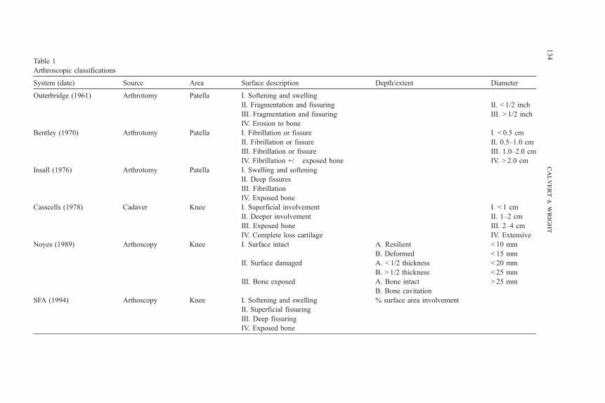
Table 1
Arthroscopic classifications
System (date) Source Area Surface description Depth/extent Diameter
Outerbridge (1961) Arthrotomy Patella I. Softening and swelling
II. Fragmentation and fissuring II. b 1/2 inch
III. Fragmentation and fissuring III. N 1/2 inch
IV. Erosion to bone
Bentley (1970) Arthrotomy Patella I. Fibrillation or fissure I. b 0.5 cm
II. Fibrillation or fissure II. 0.5–1.0 cm
III. Fibrillation or fissure III. 1.0–2.0 cm
IV. Fibrillation m/� exposed bone IV. N 2.0 cm
Insall (1976) Arthrotomy Patella I. Swelling and softening
II. Deep fissures
III. Fibrillation
IV. Exposed bone
Casscells (1978) Cadaver Knee I. Superficial involvement I. b 1 cm
II. Deeper involvement II. 1–2 cm
III. Exposed bone III. 2–4 cm
IV. Complete loss cartilage IV. Extensive
Noyes (1989) Arthoscopy Knee I. Surface intact A. Resilient b 10 mm
B. Deformed b 15 mm
II. Surface damaged A. b 1/2 thickness b 20 mm
B. N 1/2 thickness b 25 mm
III. Bone exposed A. Bone intact N 25 mm
B. Bone cavitation
SFA (1994) Arthoscopy Knee I. Softening and swelling % surface area involvement
II. Superficial fissuring
III. Deep fissuring
IV. Exposed bone
calvert
&wright
134
arthroscopy in athlete with knee oa 135
ally measures what it purports to measure. The SFA group provided internal and
external validation data for their system [16]. They first showed correlation
between visual analog scale scoring of the arthroscopist’s overall assessment
of cartilage damage with the surface characteristics and depth of the lesions
(intrinsic validity). Patient age and extent of radiographic changes were also
correlated (extrinsic validity). The SFA grading system (Table 1) was then vali-
dated using the visual analog scale in a multicenter study of 755 knee arthroscopy
cases. Accuracy is the degree to which a measurement represents the true value
of the attribute being measured. This can be determined only if a ‘‘gold standard’’
reference value is accepted. Cameron et al [19] used postarthroscopy dissection
of cadaver knees in their determination of the accuracy of the Outerbridge
classification. They found an accuracy of 68% among nine orthopedic surgeons
with decreasing accuracy for low-grade lesions.
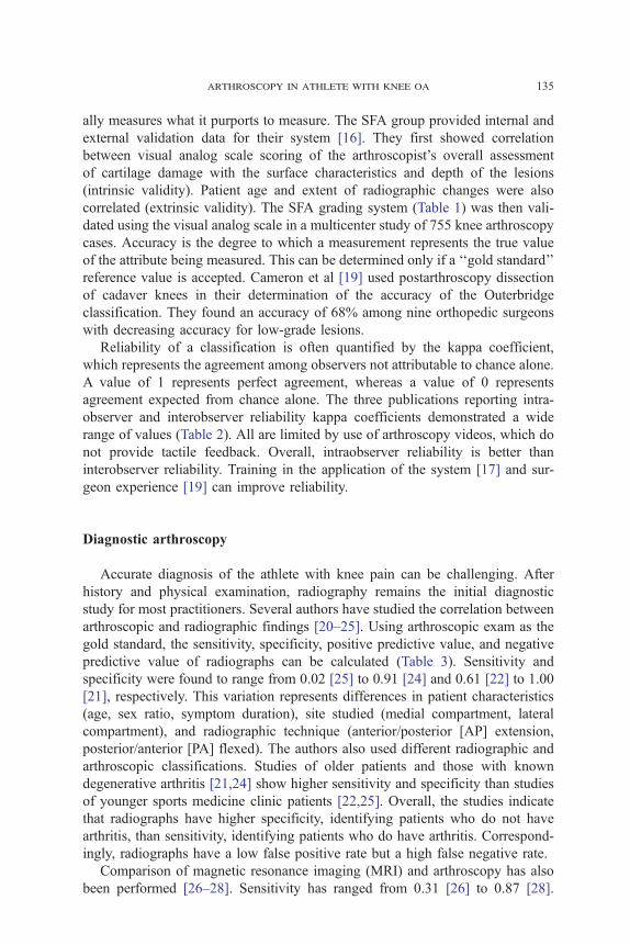
Reliability of a classification is often quantified by the kappa coefficient,
which represents the agreement among observers not attributable to chance alone.
A value of 1 represents perfect agreement, whereas a value of 0 represents
agreement expected from chance alone. The three publications reporting intra-
observer and interobserver reliability kappa coefficients demonstrated a wide
range of values (Table 2). All are limited by use of arthroscopy videos, which do
not provide tactile feedback. Overall, intraobserver reliability is better than
interobserver reliability. Training in the application of the system [17] and sur-
geon experience [19] can improve reliability.
Diagnostic arthroscopy
Accurate diagnosis of the athlete with knee pain can be challenging. After
history and physical examination, radiography remains the initial diagnostic
study for most practitioners. Several authors have studied the correlation between
arthroscopic and radiographic findings [20–25]. Using arthroscopic exam as the
gold standard, the sensitivity, specificity, positive predictive value, and negative
predictive value of radiographs can be calculated (Table 3). Sensitivity and
specificity were found to range from 0.02 [25] to 0.91 [24] and 0.61 [22] to 1.00
[21], respectively. This variation represents differences in patient characteristics
(age, sex ratio, symptom duration), site studied (medial compartment, lateral
compartment), and radiographic technique (anterior/posterior [AP] extension,
posterior/anterior [PA] flexed). The authors also used different radiographic and
arthroscopic classifications. Studies of older patients and those with known
degenerative arthritis [21,24] show higher sensitivity and specificity than studies
of younger sports medicine clinic patients [22,25]. Overall, the studies indicate
that radiographs have higher specificity, identifying patients who do not have
arthritis, than sensitivity, identifying patients who do have arthritis. Correspond-
ingly, radiographs have a low false positive rate but a high false negative rate.
Comparison of magnetic resonance imaging (MRI) and arthroscopy has also
been performed [26–28]. Sensitivity has ranged from 0.31 [26] to 0.87 [28].
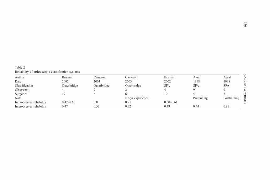
Table 2
Reliability of arthroscopic classification systems
Author Brismar Cameron Cameron Brismar Ayral Ayral
Date 2002 2003 2003 2002 1998 1998
Classification Outerbridge Outerbridge Outerbridge SFA SFA SFA
Observers 4 9 2 4 9 9
Surgeries 19 6 6 19 5 5
Note N 5-yr experience Pretraining Posttraining
Intraobserver reliability 0.42–0.66 0.8 0.91 0.50–0.61
Interobserver reliability 0.47 0.52 0.72 0.49 0.44 0.87
calvert
&wright
136
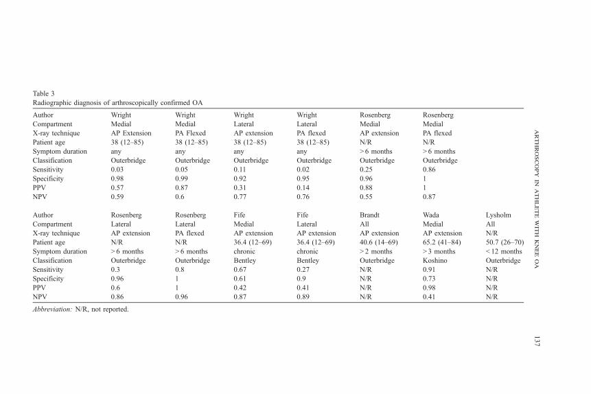
Table 3
Radiographic diagnosis of arthroscopically confirmed OA
Author Wright Wright Wright Wright Rosenberg Rosenberg
Compartment Medial Medial Lateral Lateral Medial Medial
X-ray technique AP Extension PA Flexed AP extension PA flexed AP extension PA flexed
Patient age 38 (12–85) 38 (12–85) 38 (12–85) 38 (12–85) N/R N/R
Symptom duration any any any any N 6 months N 6 months
Classification Outerbridge Outerbridge Outerbridge Outerbridge Outerbridge Outerbridge
Sensitivity 0.03 0.05 0.11 0.02 0.25 0.86
Specificity 0.98 0.99 0.92 0.95 0.96 1
PPV 0.57 0.87 0.31 0.14 0.88 1
NPV 0.59 0.6 0.77 0.76 0.55 0.87
Author Rosenberg Rosenberg Fife Fife Brandt Wada Lysholm
Compartment Lateral Lateral Medial Lateral All Medial All
X-ray technique AP extension PA flexed AP extension AP extension AP extension AP extension N/R
Patient age N/R N/R 36.4 (12–69) 36.4 (12–69) 40.6 (14–69) 65.2 (41–84) 50.7 (26–70)
Symptom duration N 6 months N 6 months chronic chronic N 2 months N 3 months b 12 months
Classification Outerbridge Outerbridge Bentley Bentley Outerbridge Koshino Outerbridge
Sensitivity 0.3 0.8 0.67 0.27 N/R 0.91 N/R
Specificity 0.96 1 0.61 0.9 N/R 0.73 N/R
PPV 0.6 1 0.42 0.41 N/R 0.98 N/R
NPV 0.86 0.96 0.87 0.89 N/R 0.41 N/R
Abbreviation: N/R, not reported.
arthroscopyinathletewithkneeoa
137
calvert & wright138
Again, differences in patient characteristics, imaging techniques, and classifi-
cation schemes likely explain much of the difference. MRI interpretation is
also more difficult and thus susceptible to interobserver variation based upon
experience and training. Similar to radiographs, MRI generally has higher
specificity than sensitivity.
As indicated by the above discussion, arthroscopy remains the gold standard
for diagnosis of articular cartilage lesions. Like all diagnostic tools, its use de-
pends on the individual clinical situation and the index of suspicion of the
clinician. Advantages in comparison to noninvasive imaging include direct
visualization, tactile feedback, and ability to simultaneously provide treatment.
Disadvantages include the risk associated with invasive procedure and cost.
Arthroscopic lavage
The oldest and simplest arthroscopic treatment of OA is lavage. Burman
reported symptomatic relief in a series of 30 patients in 1934 [29]. Subsequently,
numerous studies have evaluated lavage alone [30,31], lavage in combination
with other treatments [32,33], and lavage in comparison with other treatments
[34–41]. Differences in surgical techniques, patient populations, outcome mea-
sures, and statistical analyses have resulted in variable findings. Consequently,
different recommendations have been made. Despite this, some general princi-
ples are evident. All studies show improvement after lavage in comparison to
preoperative pain and function. The reason for improvement is unknown. The
amount of improvement, percentage of patients improved, and duration of
improvement varies between studies.
Surgical technique can vary by the amount of irrigation fluid and the mode of
delivery. Dawes et al performed an observer-blinded, randomized controlled trial
comparing needle lavage of 2 L saline with injection of 10 mL saline in
20 patients [35]. Although both groups showed improvement in pain and func-
tion, the only significant difference between groups was decreased thigh circum-
ference in the lavage group at 12 weeks. The power of this study to prove
equivalence was obviously limited by sample size. Kalunian et al performed a
double-blind randomized controlled trial comparing visually guided needle
irrigation with 3 L and 250 mL. Neither intervention generated a statistically
significant improvement in overall outcome as judged by Western Ontario and
McMaster University Osteoarthritis Index (WOMAC) at 12 months. However,
the 3-L lavage group did have statistically significant improvement in the
WOMAC pain subscale and visual analog pain scale (VAS) at 12 months. Chang
et al performed an observer blind randomized controlled trial comparing
arthroscopic debridement with office-based needle lavage with 1-L saline in 32
patients. Differences in technique included lavage versus debridement and needle
irrigation versus formal arthroscopy. No differences between groups were found
at 3 months or 1 year. Again, sample size limits the ability to assess equivalence
between groups.
arthroscopy in athlete with knee oa 139
Arthroscopic lavage and conservative medical management have been com-
pared. Livesley et al performed a nonrandomized, nonblinded prospective study
comparing 37 patients receiving lavage and physical therapy with 24 patients
receiving physical therapy alone [36]. Both groups displayed improvement in
pain and physical signs of knee irritation. The lavage group showed greater and
longer lasting improvement. A double-blind randomized placebo-controlled trial
of arthroscopy plus corticosteroid injection versus arthroscopy plus placebo
injection in 71 patients was recently published [33]. The corticosteroid group
showed statistically significant greater response to therapy on the Osteoarthritis
Research Society International criteria than placebo at 4 weeks. No differences
were noted at greater than 4 weeks. Ravaud et al performed a multicenter
randomized controlled trial comparing needle arthroscopic lavage plus cortico-
steroid injection, needle arthroscopic lavage alone, corticosteroid alone, and
placebo injection in 98 patients [32]. The trial was double blind for injection
but open for arthroscopy. Lavage improved VAS pain scale up to the trial
endpoint of 24 weeks. Corticosteroid injection improved VAS pain scale only at
weeks 1 and 4. The combination lavage plus injection group showed an additive
but not synergistic response of the two treatments.
Arthroscopic lavage and arthroscopic debridement have been compared in
multiple studies [34,37–41]. Study design, patient characteristics, and study
results are summarized in Table 4. Overall, two studies demonstrated increased
benefit with debridement [34,39] and three studies failed to show major
differences between interventions [37,38,41]. The two studies favoring debride-
ment have design characteristics worthy of specific mention. The study of
Jackson et al was retrospective, and therefore susceptible to selection bias. The
Table 4
Comparison studies of lavage and debridement
Author Jackson Gibson Chang Hubbard Moseley
Date 1986 1992 1993 1996 2002
Study design RCT PRCT PRCT PRCT PRCT
Total patients 207 20 32 76 180
Follow-up (months) 39.6 12 12 54 24
Percent withdrawal 20% 0 0 24% 8%
Lavage group 53 10 14 36 61
debridement group 113 10 18 40 59
Age lavage N/R 53 +/� 10 (38–68) 65 +/� 13 N/R 51.2 +/� 10.5
Age debridement N/A 57 +/� 7(45–69) 61 +/� 11 N/R 53.6 +/� 12.2
Sex lavage M/F N/R 6/4 4/10 N/R 54/7
Sex debridement M/F N/A 8/2 5/13 N/R 57/2
Symptom lavage (month) N/R 120+/�108 51 N 12 N 6
Symptom control (month) N/A 96+/�84 53 N 12 N 6
Blind assessor N/A N/R Yes No Yes
% Good/excellent lavage 45% N/R 58% 12% N/R
% Good/excellent debridement 68% N/R 44% 59% N/R
Abbreviations: N/A, not applicable; N/R, not reported; PRCT, prospective randomized controlled trial;
RCT, retrospective control trial.
Table 5
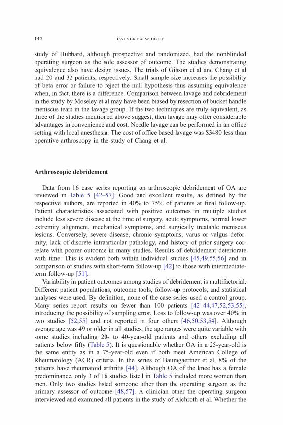
Arthroscopic debridement case series
Author Sprague Salisbury Baumgaertner Timoney McLaren Gross Aichroth Olgilvie-Harris
Date 1981 1985 1990 1990 1991 1991 1991 1991
Study design RCS RCS RCS RCS RCS RCS PCS RCS
Total patients 72 41 44 108 170 40 280 441
Total knees 78 48 49 111 170 43 280 441
Follow-up (months) 13.6 27.5 33 50.6 25 24 44 48
Withdrawal 12% 21% 7% 8% N/R 20% 9% 20%
Age 56 (24–78) N/R 63 (51–76) 58.1 (40–81) 54 (23–82) 54 (40–71) 49 (28–82) 58 (28–92)
Sex M/F 45/27 N/R N/R 75/33 119/51 28/22 184/70 N/R
Symptoms (month) N/R N/R all N 6 avg 48.9 N/R all N 6 N/R N/R
Outcome device Author’s Author’s Author’s HSS Author’s HSS Author’s Author’s
Assessor Surgeon Surgeon Surgeon Surgeon Surgeon Surgeon Physician Surgeon
Follow-up method phone clinic clinic phone mail clinic clinic clinic
Xrays N 50% JSN N/R N/R N 90% N/R N/R 4.70% 39% N/R
% Outerbridge 4 61% N/R N 80% N 50% N/R N/R 26% 37%
% Good/excellent 74% N/R 40% 45% 65% 72.10% 75% 53%
% Meniscus tear 81% N/R 84% N 75% N/R 70% 81% 35% unstable
% Further surgery N/R N/R 29% N/R 12.40% N/R 14% N/R
calvert
&wright
140
Author Yang Harwin McGinley Shannon Bohnsack Fond Jackson Dervin
Date 1995 1999 1999 2001 2002 2002 2003 2003
Study design RCS RCS RCS RCS RCS RCS RCS PCS
Total patients 103 190 77 54 104 36 121 126
Total knees 105 204 91 55 104 36 121 126
Follow-up (months) 11.7 88.8 158.4 29.6 64.8 60 48–72 24
Withdrawal N/R 14% 52% N/R N/R 44% 2.50% 19.20%
Age 64.2 (60–81) 62.1 (32–88) 62.6 (55–82) 60.9 (48–83) 60 (50–83) 64.8 (50–82) 56.4 (22–85) 61.7 (43–75)
Sex M/F 83/20 81/109 N/R 24/30 50/54 N/R N/R 59/67
Symptoms (month) 82% N 1mo N/R N/R 37% N 1 yr N/R 60 N/R N/R
Outcome device Author’s Author’s Tegner Duke Lysholm HSS Author’s SF-36, WOMAC
Assessor Surgeon Surgeon Surgeon Surgeon Surgeon Surgeon Surgeon Patient
Follow-up method N/R N/R Phone Phone N/R Clinic Clinic Mail
X-rays N 50% JSN N/R N/R N/R N/R 100% N/R N/R N/R
% Outerbridge 4 N/R N/R 100% N/R N/R N/R N/R N 30%
% Good/excellent 64.80% 63.20% N/R 48% 65% 69.40% 50.40% 44%
% Meniscus tear 96% N/R N/R 22% treated N 82% 94.40% 52.10% 63% unstable
% Further surgery 4.80% 26.50% 37% 25.90% 20% 22% 28.90% N/R
Abbreviations: avg, average; N/R, Not reported; PCS, Prospective case series; RCS, Retrospective case series; yr, year.
arthroscopyinathletewithkneeoa
141
calvert & wright142
study of Hubbard, although prospective and randomized, had the nonblinded
operating surgeon as the sole assessor of outcome. The studies demonstrating
equivalence also have design issues. The trials of Gibson et al and Chang et al
had 20 and 32 patients, respectively. Small sample size increases the possibility
of beta error or failure to reject the null hypothesis thus assuming equivalence
when, in fact, there is a difference. Comparison between lavage and debridement
in the study by Moseley et al may have been biased by resection of bucket handle
meniscus tears in the lavage group. If the two techniques are truly equivalent, as
three of the studies mentioned above suggest, then lavage may offer considerable
advantages in convenience and cost. Needle lavage can be performed in an office
setting with local anesthesia. The cost of office based lavage was $3480 less than
operative arthroscopy in the study of Chang et al.
Arthroscopic debridement
Data from 16 case series reporting on arthroscopic debridement of OA are
reviewed in Table 5 [42–57]. Good and excellent results, as defined by the
respective authors, are reported in 40% to 75% of patients at final follow-up.
Patient characteristics associated with positive outcomes in multiple studies
include less severe disease at the time of surgery, acute symptoms, normal lower
extremity alignment, mechanical symptoms, and surgically treatable meniscus
lesions. Conversely, severe disease, chronic symptoms, varus or valgus defor-
mity, lack of discrete intraarticular pathology, and history of prior surgery cor-
relate with poorer outcome in many studies. Results of debridement deteriorate
with time. This is evident both within individual studies [45,49,55,56] and in
comparison of studies with short-term follow-up [42] to those with intermediate-
term follow-up [51].
Variability in patient outcomes among studies of debridement is multifactorial.
Different patient populations, outcome tools, follow-up protocols, and statistical
analyses were used. By definition, none of the case series used a control group.
Many series report results on fewer than 100 patients [42–44,47,52,53,55],
introducing the possibility of sampling error. Loss to follow-up was over 40% in
two studies [52,55] and not reported in four others [46,50,53,54]. Although
average age was 49 or older in all studies, the age ranges were quite variable with
some studies including 20- to 40-year-old patients and others excluding all
patients below fifty (Table 5). It is questionable whether OA in a 25-year-old is
the same entity as in a 75-year-old even if both meet American College of
Rheumatology (ACR) criteria. In the series of Baumgaertner et al, 8% of the
patients have rheumatoid arthritis [44]. Although OA of the knee has a female
predominance, only 3 of 16 studies listed in Table 5 included more women than
men. Only two studies listed someone other than the operating surgeon as the
primary assessor of outcome [48,57]. A clinician other the operating surgeon
interviewed and examined all patients in the study of Aichroth et al. Whether the
arthroscopy in athlete with knee oa 143
evaluating clinician was blinded to the findings of surgery was not reported. The
study of Devin et al used validated patient-reported quality-of-life indices.
Follow-up data collection also varied between case series. Four studies used
phone interviews [42,45,52,53], and two series used mailed surveys [46,57].
Arthroscopic debridement has been compared with medical management in at
least one prospective trial [58]. Eighty consecutive patients over age 50 were
randomized to surgery or medical management. Age, sex ratio, and disease
severity were similar between groups. Medical management consisted of
nonsteroidal antiinflammatory drugs (NSAIDs) and activity modification. All
patients received comparable physical therapy. The Hospital for Special Surgery
Knee Rating Score was used to assess outcome. Operated patients had sta-
tistically significant greater improvement at final follow-up. At 3-year follow-up
67% of operated patients were improved and 45% of medically treated patients
were improved. Use of NSAIDs in the operative group and use of corticosteroid
injections in either group was not reported.
Simultaneous comparison of lavage, debridement, and sham surgery was
reported in the previously mentioned trial of Moseley et al (Table 4). This and
the pilot study that preceded it are the only reports in the literature including an
operative placebo control group [40,41]. The trial showed significant improve-
ment in pain and function in the three groups, and failed to show any difference
in outcome among the three groups. Strengths of study include prospective
analysis, randomization, single surgeon, double-blind analysis, low withdrawal,
and use of validated patient-reported outcome measures. The treatment groups
had similar age, sex ratio, symptoms, and radiographic disease severity. Extra-
polation from this study of a predominantly male Veteran’s Administration
patient population to the general population has been questioned. Also, 44% of
eligible patients refused to participate introducing the possibility of selection
bias. Although validated outcome instruments including the Short Form 36 and
Arthritis Impact Measurement Scales were used, the primary outcome scale was
nonvalidated and created for the study. Even if the criticisms of equivalence
between treatment groups are accepted, the dramatic response of patients to
placebo surgery remains. When patients were questioned as to which procedure
had been performed, they answered no more frequently than they would by
chance alone.
All of the preceding studies report data from selected surgeons publishing in
academic journals. Use of arthroscopic debridement in the province of Ontario
between 1992 and 1996 has been reported [59]. This is the only population
research available. On average, 1.4 arthroscopic debridements were performed
per 1000 residents. The complication rate was 1.9%. At 3-year follow-up, 2.9%
underwent high tibial osteotomy, 7.7% underwent repeat arthroscopy, and
18.4% underwent total knee arthroplasty (TKA). Thirty-three percent over
age 70 underwent TKA. Geographic regions within the province in which the
rate of debridement was higher than average, and also had higher rates of sub-
sequent TKA among patients over 70. This suggests overuse of the procedure in
elderly patients in some areas.
calvert & wright144
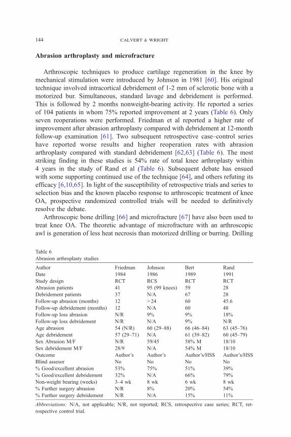
Abrasion arthroplasty and microfracture
Arthroscopic techniques to produce cartilage regeneration in the knee by
mechanical stimulation were introduced by Johnson in 1981 [60]. His original
technique involved intracortical debridement of 1-2 mm of sclerotic bone with a
motorized bur. Simultaneous, standard lavage and debridement is performed.
This is followed by 2 months nonweight-bearing activity. He reported a series
of 104 patients in whom 75% reported improvement at 2 years (Table 6). Only
seven reoperations were performed. Friedman et al reported a higher rate of
improvement after abrasion arthroplasty compared with debridement at 12-month
follow-up examination [61]. Two subsequent retrospective case–control series
have reported worse results and higher reoperation rates with abrasion
arthroplasty compared with standard debridement [62,63] (Table 6). The most
striking finding in these studies is 54% rate of total knee arthroplasty within
4 years in the study of Rand et al (Table 6). Subsequent debate has ensued
with some supporting continued use of the technique [64], and others refuting its
efficacy [6,10,65]. In light of the susceptibility of retrospective trials and series to
selection bias and the known placebo response to arthroscopic treatment of knee
OA, prospective randomized controlled trials will be needed to definitively
resolve the debate.
Arthroscopic bone drilling [66] and microfracture [67] have also been used to
treat knee OA. The theoretic advantage of microfracture with an arthroscopic
awl is generation of less heat necrosis than motorized drilling or burring. Drilling
Table 6
Abrasion arthroplasty studies
Author Friedman Johnson Bert Rand
Date 1984 1986 1989 1991
Study design RCT RCS RCT RCT
Abrasion patients 41 95 (99 knees) 59 28
Debridement patients 37 N/A 67 28
Follow-up abrasion (months) 12 N 24 60 45.6
Follow-up debridement (months) 12 N/A 60 48
Follow-up loss abrasion N/R 9% 9% 18%
Follow-up loss debridement N/R N/A 9% N/R
Age abrasion 54 (N/R) 60 (29–88) 66 (46–84) 63 (45–76)
Age debridement 57 (29–71) N/A 61 (39–82) 60 (45–79)
Sex Abrasion M/F N/R 59/45 58% M 18/10
Sex debridement M/F 28/9 N/A 54% M 18/10
Outcome Author’s Author’s Author’s/HSS Author’s/HSS
Blind assesor No No No No
% Good/excellent abrasion 53% 75% 51% 39%
% Good/excellent debridement 32% N/A 66% 79%
Non-weight bearing (weeks) 3–4 wk 8 wk 6 wk 8 wk
% Further surgery abrasion N/R 8% 20% 54%
% Further surgery debridement N/R N/A 15% 11%
Abbreviations: N/A, not applicable; N/R, not reported; RCS, retrospective case series; RCT, ret-
rospective control trial.
arthroscopy in athlete with knee oa 145
in 77 knees of 73 patient has been compared with an age-matched control group
of 16 arthroscopic lavage patients [66]. Relief of pain was reported in 69% of
drilling patients and 19% of lavage patients. Steadman et al report 75% of
patients in their series had improved pain at 3- to 5-year follow-up. Again,
prospective randomized controlled trials will be needed to assess benefit of these
procedures in comparison to lavage, debridement, and placebo.
Partial meniscectomy in patients with osteoarthritis
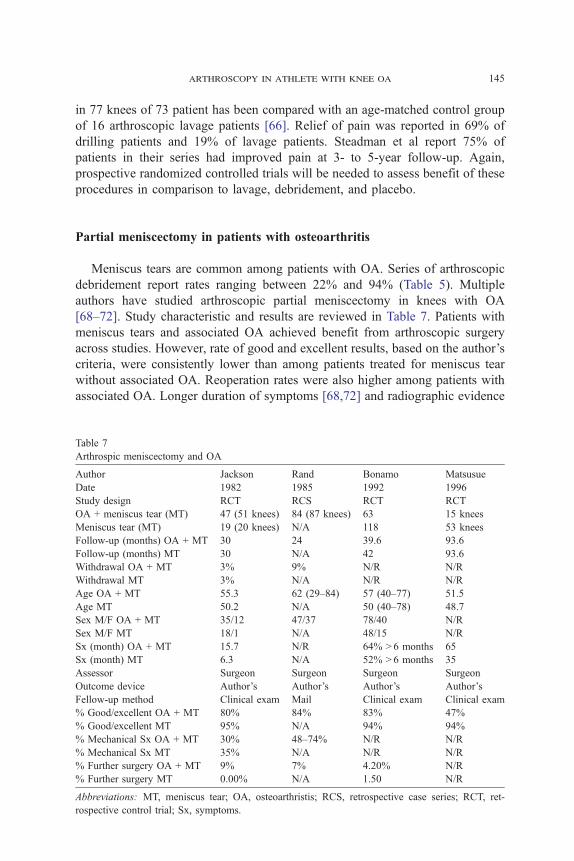
Meniscus tears are common among patients with OA. Series of arthroscopic
debridement report rates ranging between 22% and 94% (Table 5). Multiple
authors have studied arthroscopic partial meniscectomy in knees with OA
[68–72]. Study characteristic and results are reviewed in Table 7. Patients with
meniscus tears and associated OA achieved benefit from arthroscopic surgery
across studies. However, rate of good and excellent results, based on the author’s
criteria, were consistently lower than among patients treated for meniscus tear
without associated OA. Reoperation rates were also higher among patients with
associated OA. Longer duration of symptoms [68,72] and radiographic evidence
Table 7
Arthrospic meniscectomy and OA
Author Jackson Rand Bonamo Matsusue
Date 1982 1985 1992 1996
Study design RCT RCS RCT RCT
OA + meniscus tear (MT) 47 (51 knees) 84 (87 knees) 63 15 knees
Meniscus tear (MT) 19 (20 knees) N/A 118 53 knees
Follow-up (months) OA + MT 30 24 39.6 93.6
Follow-up (months) MT 30 N/A 42 93.6
Withdrawal OA + MT 3% 9% N/R N/R
Withdrawal MT 3% N/A N/R N/R
Age OA + MT 55.3 62 (29–84) 57 (40–77) 51.5
Age MT 50.2 N/A 50 (40–78) 48.7
Sex M/F OA + MT 35/12 47/37 78/40 N/R
Sex M/F MT 18/1 N/A 48/15 N/R
Sx (month) OA + MT 15.7 N/R 64% N 6 months 65
Sx (month) MT 6.3 N/A 52% N 6 months 35
Assessor Surgeon Surgeon Surgeon Surgeon
Outcome device Author’s Author’s Author’s Author’s
Fellow-up method Clinical exam Mail Clinical exam Clinical exam
% Good/excellent OA + MT 80% 84% 83% 47%
% Good/excellent MT 95% N/A 94% 94%
% Mechanical Sx OA + MT 30% 48–74% N/R N/R
% Mechanical Sx MT 35% N/A N/R N/R
% Further surgery OA + MT 9% 7% 4.20% N/R
% Further surgery MT 0.00% N/A 1.50 N/R
Abbreviations: MT, meniscus tear; OA, osteoarthristis; RCS, retrospective case series; RCT, ret-
rospective control trial; Sx, symptoms.
calvert & wright146
of degenerative disease [69] adversely affected outcome. Sex, age, and a history
of precipitating trauma did not have significant prognostic value.
Recently, MRI evaluation of meniscus tears among patients older than
45 years with and without OA, as defined by ACR criteria, was studied [73].
Meniscus tear was defined as signal change extending to the articular sur-
face. Comparison of 154 OA patients with 49 asymptomatic controls was made.
Validated outcome measures were used (WOMAC and pain visual analog scale).
Meniscus tears were common among patients with symptomatic OA (86%) and
asymptomatic volunteers (67%). Prevalence of meniscus tears increased with
increasing radiographic severity of OA. Individuals with severe radiographic
change as graded by Kellgren-Lawrence criteria had a 100% rate of meniscus
tear. Within the symptomatic OA group, there was no difference in pain or
WOMAC score between those with and without meniscus tears. Limitations of
the study include lack of arthroscopic confirmation of the MRI findings, lack of
clinical symptomatology data (locking, catching), and difference in weight be-
tween the study groups. Despite these limitations, one can conclude that menis-
cus tears identifiable by MRI are extremely common among individuals over
age 45. The role of meniscus tears in the causation of symptoms among patients
with OA is called into question.
Anterior cruciate ligament reconstruction in the arthritic patient
Symptomatic knee instability combined with arthrosis in the active patient is a
challenging combination. Arthroscopy can be used for diagnosis, debridement,
and assistance with reconstruction. Short and intermediate term follow-up of
arthritic patients treated with arthroscopically assisted or endoscopic anterior
cruciate ligament (ACL) reconstruction reveals significant improvements in pain,
stability, and function [74,75]. The authors of both studies counseled patients to
avoid high-impact activities such as distance running and cutting sports. Shel-
bourne and Gray have reported retrospective objective follow-up of 482 patients
at 7.6 years post-op and retrospective subjective follow-up of 928 patients at
8.6 years [76]. All patients underwent ACL reconstruction; status of the menisci
and articular cartilage was noted at surgery. Patients with articular cartilage
damage had lower subjective scores, lower modified International Knee Docu-
mentation Committee scores, and a higher rate of radiographic abnormalities.
The authors concluded that in order of importance, articular cartilage damage,
partial or total medial meniscectomy, and partial or total lateral meniscectomy
adversely affect the outcome of ACL reconstruction.
Arthroscopy and high tibial osteotomy
Arthroscopy has been combined with high tibial osteotomy (HTO) for both
diagnostic and therapeutic goals. Keene et al have evaluated short- [77] and
arthroscopy in athlete with knee oa 147
intermediate-term [78] results of diagnostic arthroscopy combined with HTO.
Arthroscopy failed to provide prognostic information in either study. Presence
of bicompartmental and tricompartmental OA at arthroscopy did not predict worse
outcome. Adequate valgus correction was the primary determinant of outcome in
both studies. Other authors recommend routine arthroscopy before HTO [79,80].
They stress the low morbidity of the procedure and the ability to treat con-
comitant intraarticular pathology. Finally, operative indications and contraindi-
cations for HTO are not yet strictly defined. The use of diagnostic arthroscopy to
exclude candidates remains undefined.
Arthroscopic debridement, drilling, and abrasion arthroplasty have been com-
bined with HTO [79,81]. One prospective, nonrandomized, comparison trial has
been performed [81]. Patients were evaluated by repeat arthroscopy at 1 year and
Japanese Orthopaedic Association knee score at 4.8 years in the abrasion group
and 3.5 years in the HTO only group. Repeat arthroscopy revealed statistically
significant more fibrocartilage filling of articular surface defects in the abrasion
plus HTO group. However, there were no differences in clinical outcome between
groups. Schultz et al reported similar finding in a retrospective comparison of
diagnostic arthroscopy, drilling, and abrasion arthroplasty in conjunction with
HTO [79]. No difference in clinical outcome was found at 1-year follow-up.
Author’s preferred method
A few data points are critical during the workup and evaluation of the patient
considering arthroscopic treatment of their knee pain. During the physical ex-
amination and history we want to elicit any symptoms of mechanical locking
or catching. In addition, joint line tenderness and a palpable clunk during a
McMurray exam are valuable information. All patients evaluated in our clinics
for knee pain either bring or undergo bilateral weight-bearing X-rays. We use the
Rosenberg view, but either view is acceptable in a sports medicine clinic.
If the patient has a history and physical examination consistent with a me-
niscal tear and no joint space narrowing is noted on standing X-rays then we will
discuss obtaining an MRI to verify the diagnosis. If the patient has classic locking
symptoms and does not desire to undergo an MRI, then we will discuss pro-
ceeding with arthroscopic evaluation. If the patient has joint space narrowing on
weight-bearing X-rays then we typically begin treatment by maximizing con-
servative management of their arthritis. This includes recommending NSAIDs,
ice, and physical therapy especially for anterior knee pain symptoms. If this fails
to control their symptoms, then we will discuss corticosteroid or hyaluronic acid
injections. Alignment X-rays to include the hip, knee, and ankle for consideration
of an osteotomy will be obtained at this time.
Patients with normal alignment and greater than 50% joint space remaining
who undergo corticosteroid or hyaluronic acid injections with quick return of
their pain in less than a month following injection are counseled regarding
arthroscopic debridement as a treatment option. This is especially true if they

calvert & wright148
have continuing mechanical symptoms and joint line tenderness. If they obtain
several months relief following injection then we believe that the majority of their
symptoms can be controlled with conservative measures. Patients considering
arthroscopic debridement are counseled regarding potential benefit and the fact
that the procedure will not be curative.
At the time of arthroscopy all meniscal tears are debrided. Chondral flaps and
loose articular cartilage are debrided with mechanical shavers and biters. If there
are focal areas of complete articular cartilage loss surrounded by relatively
normal articular cartilage the patient may be a candidate for microfracture. If the
articular cartilage wear is diffuse throughout the compartment or entire knee, then
simple debridement is performed. We do not inject the knee with corticosteroids
at the time of arthroscopy, to avoid any increased chance of infection. Following
arthroscopy, the patients undergo routine postoperative management with
physical therapy for range of motion and strengthening and ice therapy to
minimize swelling. Patients that undergo microfracture are nonweight bearing
for the initial 6 weeks.
Summary
Arthroscopy is an important technique in the diagnosis, classification, and
treatment of the athlete with OA. Reliability of the current classification systems
improves with training and experience. Arthroscopy remains superior to imaging
in the diagnosis of OA. Arthroscopic lavage and debridement provide benefit in a
significant percentage of patients. The reasons for improvement are not fully
defined. Arthroscopic treatment of OA is not curative, and results deteriorate with
time. Variability in the use of medical management, arthroscopy, osteotomy, and
arthroplasty remains among different practitioners. Indications for arthroscopy
require further clarification based upon empiric evidence.
References
[1] Jackson RW. The role of arthroscopy in the management of the arthritic knee. Clin Orthop 1974;
101(1):28–35.
[2] Schonholtz GJ. Arthroscopic debridement of the knee joint. Orthop Clin North Am 1989;20(2):
257–63.
[3] Burks RT. Arthroscopy and degenerative arthritis of the knee: a review of the literature.
Arthroscopy 1990;6(1):43–7.
[4] Ike RW. The role of arthroscopy in the differential diagnosis of osteoarthritis of the knee.
Rheum Dis Clin North Am 1993;19(3):673–96.
[5] Novak PJ, Bach Jr BR. Selection criteria for knee arthroscopy in the osteoarthritic patient.
Orthop Rev 1993;22(7):798–804.
[6] Jackson RW, Gilbert JE, Sharkey PF. Arthroscopic debridement versus arthroplasty in the
osteoarthritic knee. J Arthroplasty 1997;12(4):465–9 [discussion 469–70].
[7] Goldman RT, Scuderi GR, Kelly MA. Arthroscopic treatment of the degenerative knee in older
athletes. Clin Sports Med 1997;16(1):51–68.
arthroscopy in athlete with knee oa 149
[8] Buckwalter JA, Lane NE. Athletics and osteoarthritis. Am J Sports Med 1997;25(6):873–81.
[9] Hanssen AD, Stuart MJ, Scott RD, Scuderi GR. Surgical options for the middle-aged patient
with osteoarthritis of the knee joint. Instr Course Lect 2001;50:499–511.
[10] Hunt SA, Jazrawi LM, Sherman OH. Arthroscopic management of osteoarthritis of the knee.
J Am Acad Orthop Surg 2002;10(5):356–63.
[11] Outerbridge RE. The etiology of chondromalacia patellae. J Bone Joint Surg Br 1961;43-B:
752–7.
[12] Bentley G, Dowd G. Current concepts of etiology and treatment of chondromalacia patellae.
Clin Orthop 1984;189:209–28.
[13] Insall J, Falvo KA, Wise DW. Chondromalacia Patellae. A prospective study. J Bone Joint Surg
Am 1976;58(1):1–8.
[14] Casscells SW. Gross pathological changes in the knee joint of the aged individual: a study of
300 cases. Clin Orthop 1978;132:225–32.
[15] Noyes FR, Stabler CL. A system for grading articular cartilage lesions at arthroscopy. Am J
Sports Med 1989;17(4):505–13.
[16] Dougados M, Ayral X, Listrat V, Gueguen A, Bahuaud J, Beaufils P, et al. The SFA system for
assessing articular cartilage lesions at arthroscopy of the knee. Arthroscopy 1994;10(1):69–77.
[17] Ayral X, Gueguen A, Ike RW, Bonvarlet JP, Frizziero L, Kalunian K, et al. Inter-observer re-
liability of the arthroscopic quantification of chondropathy of the knee. Osteoarthritis Cartilage
1998;6(3):160–6.
[18] Brismar BH, Wredmark T, Movin T, Leandersson J, Svensson O. Observer reliability in the
arthroscopic classification of osteoarthritis of the knee. J Bone Joint Surg Br 2002;84(1):42–7.
[19] Cameron ML, Briggs KK, Steadman JR. Reproducibility and reliability of the outerbridge
classification for grading chondral lesions of the knee arthroscopically. Am J Sports Med 2003;
31(1):83–6.
[20] Lysholm J, Hamberg P, Gillquist J. The correlation between osteoarthrosis as seen on radio-
graphs and on arthroscopy. Arthroscopy 1987;3(3):161–5.
[21] Rosenberg TD, Paulos LE, Parker RD, Coward DB, Scott SM. The forty-five-degree pos-
teroanterior flexion weight-bearing radiograph of the knee. J Bone Joint Surg Am 1988;70(10):
1479–83.
[22] Fife RS, Brandt KD, Braunstein EM, et al. Relationship between arthroscopic evidence
of cartilage damage and radiographic evidence of joint space narrowing in early osteoarthritis
of the knee. Arthritis Rheum 1991;34(4):377–82.
[23] Brandt KD, Fife RS, Braunstein EM, Katz B, Shelbourne KD, Kalasinski LA, et al. Ra-
diographic grading of the severity of knee osteoarthritis: relation of the Kellgren and Lawrence
grade to a grade based on joint space narrowing, and correlation with arthroscopic evidence of
articular cartilage degeneration. Arthritis Rheum 1991;34(11):1381–6.
[24] Wada M, Baba H, Imura S, Morita A, Kusaka Y. Relationship between radiographic
classification and arthroscopic findings of articular cartilage lesions in osteoarthritis of the
knee. Clin Exp Rheumatol 1998;16(1):15–20.
[25] Wright RW, Spindler KP, Boyce RH, Michener T, Shyr Y, McCarty EC. The sensitivity
and specificity of standing knee radiographs (AP vs. PA flexion) in arthroscopically confirmed
early arthritis. Clin Orthop, in press.
[26] Speer KP, Spritzer CE, Goldner JL, Garrett Jr WE. Magnetic resonance imaging of traumatic
knee articular cartilage injuries. Am J Sports Med 1991;19(4):396–402.
[27] Broderick LS, Turner DA, Renfrew DL, Schnitzer TJ, Huff JP, Harris C. Severity of articular
cartilage abnormality in patients with osteoarthritis: evaluation with fast spin-echo MR vs
arthroscopy. AJR Am J Roentgenol 1994;162(1):99–103.
[28] Potter HG, Linklater JM, Allen AA, Hannafin JA, Haas SB. Magnetic resonance imaging
of articular cartilage in the knee. An evaluation with use of fast-spin-echo imaging. J Bone
Joint Surg Am 1998;80(9):1276–84.
[29] Burman MS. Arthroscopy or the direct visualization of joints: an experimental cadaver study.
1931. Clin Orthop 2001;390:5–9.
calvert & wright150
[30] Edelson R, Burks RT, Bloebaum RD. Short-term effects of knee washout for osteoarthritis.
Am J Sports Med 1995;23(3):345–9.
[31] Kalunian KC, Moreland LW, Klashman DJ, Brion PH, Concoff AL, Myers S, et al. Visually-
guided irrigation in patients with early knee osteoarthritis: a multicenter randomized, controlled
trial. Osteoarthritis Cartilage 2000;8(6):412–8.
[32] Ravaud P, Moulinier L, Giraudeau B, Ayral X, Guerin C, Noel E, et al. Effects of joint lavage
and steroid injection in patients with osteoarthritis of the knee: results of a multicenter, ran-
domized, controlled trial. Arthritis Rheum 1999;42(3):475–82.
[33] Smith MD, Wetherall M, Darby T, Esterman A, Slavotinek J, Roberts-Thomson P, et al. A
randomized placebo-controlled trial of arthroscopic lavage versus lavage plus intra-articular
corticosteroids in the management of symptomatic osteoarthritis of the knee. Rheumatology
(Oxford) 2003;42(12):1477–85.
[34] Jackson RW, Silver R, Marans H. Arthroscopic treatment of degenerative joint disease.
Arthroscopy 1986;2(2):114.
[35] Dawes PT, Kirlew C, Haslock I. Saline washout for knee osteoarthritis: results of a controlled
study. Clin Rheumatol 1987;6(1):61–3.
[36] Livesley PJ, Doherty M, Needoff M, Moulton A. Arthroscopic lavage of osteoarthritic knees.
J Bone Joint Surg Br 1991;73(6):922–6.
[37] Gibson JN, White MD, Chapman VM, Strachan RK. Arthroscopic lavage and debridement for
osteoarthritis of the knee. J Bone Joint Surg Br 1992;74(4):534–7.
[38] Chang RW, Falconer J, Stulberg SD, Arnold WJ, Manheim LM, Dyer AR. A randomized,
controlled trial of arthroscopic surgery versus closed-needle joint lavage for patients with
osteoarthritis of the knee. Arthritis Rheum 1993;36(3):289–96.
[39] Hubbard MJ. Articular debridement versus washout for degeneration of the medial femoral
condyle. A five-year study. J Bone Joint Surg Br 1996;78(2):217–9.
[40] Moseley Jr JB, Wray NP, Kuykendall D, Willis K, Landon G. Arthroscopic treatment of
osteoarthritis of the knee: a prospective, randomized, placebo-controlled trial. Results of a
pilot study. Am J Sports Med 1996;24(1):28–34.
[41] Moseley JB, O’Malley K, Petersen NJ, Menke TJ, Brody BA, Kuykendall DH, et al. A con-
trolled trial of arthroscopic surgery for osteoarthritis of the knee. N Engl J Med 2002;347(2):81–8.
[42] Sprague III NF. Arthroscopic debridement for degenerative knee joint disease. Clin Orthop 1981;
160:118–23.
[43] Salisbury RB, Nottage WM, Gardner V. The effect of alignment on results in arthroscopic
debridement of the degenerative knee. Clin Orthop 1985;198:268–72.
[44] Baumgaertner MR, Cannon Jr WD, Vittori JM, Schmidt ES, Maurer RC. Arthroscopic de-
bridement of the arthritic knee. Clin Orthop 1990;253:197–202.
[45] Timoney JM, Kneisl JS, Barrack RL, Alexander AH. Arthroscopy update #6. Arthroscopy in
the osteoarthritic knee. Long-term follow-up. Orthop Rev 1990;19(4):371–3 [376–9].
[46] McLaren AC, Blokker CP, Fowler PJ, Roth JN, Rock MG. Arthroscopic debridement of the
knee for osteoarthrosis. Can J Surg 1991;34(6):595–8.
[47] Gross DE, Brenner SL, Esformes I, Gross ML. Arthroscopic treatment of degenerative joint
disease of the knee. Orthopedics 1991;14(12):1317–21.
[48] Aichroth PM, Patel DV, Moyes ST. A prospective review of arthroscopic debridement for
degenerative joint disease of the knee. Int Orthop 1991;15(4):351–5.
[49] Ogilvie-Harris DJ, Fitsialos DP. Arthroscopic management of the degenerative knee. Arthros-
copy 1991;7(2):151–7.
[50] Yang SS, Nisonson B. Arthroscopic surgery of the knee in the geriatric patient. Clin Orthop
1995;316:50–8.
[51] Harwin SF. Arthroscopic debridement for osteoarthritis of the knee: predictors of patient
satisfaction. Arthroscopy 1999;15(2):142–6.
[52] McGinley BJ, Cushner FD, Scott WN. Debridement arthroscopy. 10-year followup. Clin Orthop
1999;367:190–4.
[53] Shannon FJ, Devitt AT, Poynton AR, Fitzpatrick P, Walsh MG. Short-term benefit of
arthroscopic washout in degenerative arthritis of the knee. Int Orthop 2001;25(4):242–5.
arthroscopy in athlete with knee oa 151
[54] Bohnsack M, Lipka W, Ruhmann O, Peters G, Schmolke S, Wirth CJ. The value of knee
arthroscopy in patients with severe radiological osteoarthritis. Arch Orthop Trauma Surg 2002;
122(8):451–3.
[55] Fond J, Rodin D, Ahmad S, Nirschl RP. Arthroscopic debridement for the treatment of
osteoarthritis of the knee: 2- and 5-year results. Arthroscopy 2002;18(8):829–34.
[56] Jackson RW, Dieterichs C. The results of arthroscopic lavage and debridement of osteoar-
thritic knees based on the severity of degeneration: a 4- to 6-year symptomatic follow-up.
Arthroscopy 2003;19(1):13–20.
[57] Dervin GF, Stiell IG, Rody K, Grabowski J. Effect of arthroscopic debridement for osteoar-
thritis of the knee on health-related quality of life. J Bone Joint Surg Am 2003;85-A(1):10–9.
[58] Merchan EC, Galindo E. Arthroscope-guided surgery versus nonoperative treatment for limited
degenerative osteoarthritis of the femorotibial joint in patients over 50 years of age: a prospective
comparative study. Arthroscopy 1993;9(6):663–7.
[59] Wai EK, Kreder HJ, Williams JI. Arthroscopic debridement of the knee for osteoarthritis
in patients fifty years of age or older: utilization and outcomes in the Province of Ontario.
J Bone Joint Surg Am 2002;84-A(1):17–22.
[60] Johnson LL. Arthroscopic abrasion arthroplasty historical and pathologic perspective: present
status. Arthroscopy 1986;2(1):54–69.
[61] Friedman MJ, Berasi CC, Fox JM, Del Pizzo W, Snyder SJ, Ferkel RD. Preliminary results
with abrasion arthroplasty in the osteoarthritic knee. Clin Orthop 1984;182:200–5.
[62] Bert JM, Maschka K. The arthroscopic treatment of unicompartmental gonarthrosis: a five-year
follow-up study of abrasion arthroplasty plus arthroscopic debridement and arthroscopic
debridement alone. Arthroscopy 1989;5(1):25–32.
[63] Rand JA. Role of arthroscopy in osteoarthritis of the knee. Arthroscopy 1991;7(4):358–63.
[64] Johnson LL. Arthroscopic abrasion arthroplasty: a review. Clin Orthop 2001;391(Suppl):
S306–17.
[65] Bert JM. Role of abrasion arthroplasty and debridement in the management of osteoarthritis of
the knee. Rheum Dis Clin North Am 1993;19(3):725–39.
[66] Pedersen MS, Moghaddam AZ, Bak K, Koch JS. The effect of bone drilling on pain in
gonarthrosis. Int Orthop 1995;19(1):12–5.
[67] Steadman JR, Rodkey WG, Briggs KK. Microfracture to treat full-thickness chondral defects:
surgical technique, rehabilitation, and outcomes. J Knee Surg 2002;15(3):170–6.
[68] Jackson RW, Rouse DW. The results of partial arthroscopic meniscectomy in patients over
40 years of age. J Bone Joint Surg Br 1982;64(4):481–5.
[69] Rand JA. Arthroscopic management of degenerative meniscus tears in patients with degenerative
arthritis. Arthroscopy 1985;1(4):253–8.
[70] Bonamo JJ, Kessler KJ, Noah J. Arthroscopic meniscectomy in patients over the age of 40.
Am J Sports Med 1992;20(4):422–8 [discussion 428–9].
[71] Rangger C, Klestil T, Gloetzer W, Kemmler G, Benedetto KP. Osteoarthritis after arthroscopic
partial meniscectomy. Am J Sports Med 1995;23(2):240–4.
[72] Matsusue Y, Thomson NL. Arthroscopic partial medial meniscectomy in patients over 40 years
old: a 5- to 11-year follow-up study. Arthroscopy 1996;12(1):39–44.
[73] Bhattacharyya T, Gale D, Dewire P, Totterman S, Gale ME, McLaughlin S, et al. The clinical
importance of meniscal tears demonstrated by magnetic resonance imaging in osteoarthritis of
the knee. J Bone Joint Surg Am 2003;85-A(1):4–9.
[74] Shelbourne KD, Wilckens JH. Intraarticular anterior cruciate ligament reconstruction in the
symptomatic arthritic knee. Am J Sports Med 1993;21(5):685–8 [discussion 688–9].
[75] Noyes FR, Barber-Westin SD. Arthroscopic-assisted allograft anterior cruciate ligament
reconstruction in patients with symptomatic arthrosis. Arthroscopy 1997;13(1):24–32.
[76] Shelbourne KD, Gray T. Results of anterior cruciate ligament reconstruction based on meniscus
and articular cartilage status at the time of surgery. Five- to fifteen-year evaluations. Am J Sports
Med 2000;28(4):446–52.
[77] Keene JS, Dyreby Jr JR. High tibial osteotomy in the treatment of osteoarthritis of the knee.
The role of preoperative arthroscopy. J Bone Joint Surg Am 1983;65(1):36–42.
calvert & wright152
[78] Keene JS, Monson DK, Roberts JM, Dyreby Jr JR. Evaluation of patients for high tibial
osteotomy. Clin Orthop 1989;243:157–65.
[79] Schultz W, Gobel D. Articular cartilage regeneration of the knee joint after proximal tibial valgus
osteotomy: a prospective study of different intra- and extra-articular operative techniques. Knee
Surg Sports Traumatol Arthrosc 1999;7(1):29–36.
[80] Williams III RJ, Wickiewicz TL, Warren RF. Management of unicompartmental arthritis in
the anterior cruciate ligament-deficient knee. Am J Sports Med 2000;28(5):749–60.
[81] Akizuki S, Yasukawa Y, Takizawa T. Does arthroscopic abrasion arthroplasty promote carti-
lage regeneration in osteoarthritic knees with eburnation? A prospective study of high tibial
osteotomy with abrasion arthroplasty versus high tibial osteotomy alone. Arthroscopy 1997;
13(1):9–17.