Embed Size (px)
Citation preview
Thorax (1967), 22, 497.
The use of a cusp-bearing homograft patch to theoutflow tract and pulmonary artery in Fallot's
tetralogy and pulmonary valvular stenosisPAUL MARCHAND
From the Cardiovascular Research Unit, University of the Witwatersrand, and Department of ThoracicSurgery, Johannesburg General Hospital
A technique is described for maintaining pulmonary valve competence whilst widening thehypoplastic pulmonary artery ring found invariably in Fallot's tetralogy and pulmonary valvestenosis. A homograft composed of the aortic cusps, a portion of ascending aorta, and theanterior mitral leaflet is used. One, two, or three cusps may be replaced or added by variationsof technique. The right ventricular outflow tract and the pulmonary trunk are also widened.The operation is applicable to children, and adult homografts can be used. The ring shouldbe widened to a circumference of from 6 to 8 cm. so as to prevent residual pressure gradients.One or two cusp replacements are favoured in the hope that growth will proceed because acontinuous portion of natural pulmonary artery and ring remains untouched. An advantage ofthe technique is the small ventriculotomy required to excise the infundibular obstruction andclose the ventricular septal defect. Four case histories are presented. Three patients have survivedoperation and are doing well. Up to six months post-operatively no serious pulmonary incom-petence has developed.
Experience with total surgical correction forFallot's tetralogy soon showed that the commonestcause of failure was incomplete relief of thepulmonary obstruction (Lillehei, Cohen, Warden,and Varco, 1956; Kirklin, Donald, Harshbarger,Hetzel, Patrick, Swan, and Wood, 1956). In anattempt to improve the results of operation,Lillehei and his co-workers (1956) introduced out-flow tract patching in addition to wide resectionof the infundibular muscle. This group has alsoemphasized that, in some cases, it is necessary todivide the ring and occasionally to extend thepatch up to the bifurcation of the pulmonaryartery (Lillehei et al., 1956; and Lillehei, Morris,Adams, and Anderson, 1964). Barnard and Schrire(1961) found it necessary to cross the valve ringin about half of their cases, but most surgeons aremore conservative because they fear that pul-monary incompetence may be as great a burdenfor the right ventricle as residual stenosis. Lilleheiet al. (1964) have suggested that a prostheticpulmonary valve would be desirable in some casesand they mention experimental and limited clinicalexperience with this technique. Our group hasdescribed the use of a homograft pulmonary valvein a patient with massive pulmonary incompetence
following pulmonary valvotomy (Fuller, Mar-chand, Zion, and Zwi, 1966). At catheterization ayear later the valve was found to function satis-factorily. We were thereby encouraged to usevalve homografts in four further cases, three ofFallot's tetralogy and one of so-called trilogy ofFallot. Unlike the published case, these all hadsmall pulmonary rings and it was not possible tointroduce a homograft valve without at the sametime enlarging both the ventricular outflow andthe pulmonary artery. It was proposed to use anaortic valve homograft with a short attached seg-ment of ascending aorta to enlarge the pulmonarytrunk and to widen the outflow tract with aTeflon patch. Shortly before surgery my colleague,Mr. L. du Plessis, suggested that the anteriorleaflet of the mitral valve be left on the homo-graft for use as an outflow patch. The first case,though finally fatal, encouraged us to persevere,and the next three patients have done so well thatit is felt that the technique should be reported.
PREPARATION OF THE HOMOGRAFT
The base of the heart is removed in the post-mortem room and placed in a dilute solution of
497
on 31 May 2018 by guest. P
rotected by copyright.http://thorax.bm
j.com/
Thorax: first published as 10.1136/thx.22.6.497 on 1 N
ovember 1967. D
ownloaded from
Paul Marchand
aqueous chlorhexidine (Hibitane). It is imme-diately dissected under clean but not sterile con-ditions. If early dissection is not possible thespecimen is rapidly frozen to - 720 C. and storeduntil dissection is convenient.The ascending aorta is freed and both coronary
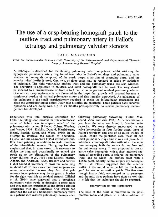
arteries are dissected and ligated flush with theaortic wall. The left atrium is opened and theanterior and posterior mitral leaflets are separatedby dividing the junctional tissue. The annulus iscrossed and the incisions are carried through theleft and right trigones aiming to include both non-coronary and left coronary cusps (Fig. 1). Theright coronary cusp is freed from the inter-ventricular septum by cutting the muscle beneaththe base of that cusp along a line level with themitral annulus (E-D in Fig. 1). This linecorresponds with the basal ring described byBarratt-Boyes in his preparation of aortic valvegrafts (1964). The mitral annulus lies at the baseof the left coronary cusp and half the non-coronary cusp (du Plessis and Marchand, 1964),but, by retaining some of the firm fibrous trigone,it is possible to conserve continuity between themitral leaflet and two complete cusps. After the
chordae tendineae have been cut from the anteriormitral leaflet the aortic root, ascending aorta, andthe anterior leaflet of the mitral valve can beremoved en bloc.A Tubb's dilator is introduced into the aortic
ring and the blades are expanded until resistanceis encountered. This measures half the circum-ference of the ring and is recorded as an indexof the size of the graft. The circumference of theadult aortic ring has been found to vary between6 5 and 9 cm. (Table 1). This agrees with Barratt-Boyes' (1965) finding of an internal diameter rangefrom 20 to 30 mm. (circumference 6 3 to 9 5 cm.).Loose tissue is removed from the adventitia, andthe septal muscle beneath the right coronary cuspis trimmed to the minimum consistent with main-tenance of a straight subvalvular border. Thehomograft is sterilized with ethylene oxide, freeze-dried, and stored (Marchand, 1958).When, at operation, it is decided to widen the
pulmonary ring the homograft is re-hydrated in an
antibiotic solution (Marchand, 1958). If threecusps are to be retained the aortic wall is excisedfrom the right coronary sinus leaving a 1-mm.fringe (Figs 1 and 2). At the apices of the com-
FIG. 1. 'Opened' specimen of mitral and aortic valves. The interrupted linemarks the incisions described in the text for a three-cusp homograft. Non-coronary cusp (N); right coronary cusp (R); left cusp (L). In actual preparation ofthe homograft the left and right cusps are not separated. M, anterior mitralleaflet; P, posterior mitral leaflet; T, trigones; A, B, C, mitral annulus; A, B,D, base of homograft; E, D, base ofmuscular interventricular septum.
498
on 31 May 2018 by guest. P
rotected by copyright.http://thorax.bm
j.com/
Thorax: first published as 10.1136/thx.22.6.497 on 1 N
ovember 1967. D
ownloaded from
Use of a cusp-bearing homograft patch in Fallot's tetralogy
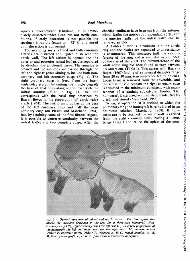
TABLE IMEASUREMENTS OF INTERNAL CIRCUMFERENCES OFTHE AORTIC AND PULMONARY ARTERY RINGS OFSEVEN FRESH HEARTS USED FOR VALVE HOMOGRAFTS
Age of Subject Aorta Pulmonary Ratio Aorta %(years) (cm.) Artery (cm.) P.A.
7 5.5 7.5 738 55 7-1 77
15 7-2 93 I 77
Average (child) 76
17 6-9 8-5 8124 7 1 8-5 8328 6-9 8-1 8535 8-1 9-5 85
Average (adult) 82
The aortic ring circumference of 45 other adult grafts collectedfor aortic valve replacement has varied from 6-5 to 9 cm.~~~~~~~~~~~~~C
B D
FIG. 2. Diagrams of the homograft. The stippled areas are
the parts of the aorta and mitral leaflet used to enlarge thepulmonary artery and right ventricular outflow respectively.The cross-hatched line (B-D) represents the muscularremnant cf the ventricular septum. (A) Viewedfrom behindwith aortic wall excisedfrom non-coronary cusp. (B) Viewedfrom the side: a, tongue of aorta; b, mitral leaflet; c, leftcoronary artery.
missural pillars of that cusp, two incisions aremade to veer away from each other and obliquelytransect the aorta. They meet about 1-5 cm. abovethe commissural pillars in line with the commis-sure between the left and non-coronary cusps(Fig. 2). The specimen now consists of tworoughly triangular tongues, the anterior leaflet ofthe mitral valve, and a portion of the aortic walltogether with the intact aortic ring and the valvecusps (Fig. 2). Should two cusps be required fortransplantation the right coronary is excised (Fig.3). For a single-cusp patch the non-coronary cusp
is retained and parts of the aorta and anteriormitral leaflet have to be cut away (Fig. 4).
OPERATIVE TECHNIQUE
The heart is exposed through a vertical sterno-tomy. It is prepared for cardiopulmonary bypasswith reduction of body temperature to 30° C.No attempt is made to separate the pulmonaryartery from the aorta. On bypass, the pulmonaryartery is incised longitudinally from its bifurcationto the valve ring. The cusps are inspected and anycommissural adhesions are divided. The circum-ference of the ring is measured by expanding theblades of a Tubb's dilator until resistance is en-countered. If the valve is bicuspid the ring isdivided through the anterior commissure so as topreserve both cusps. If tricuspid, the incision iscarried through the anterior cusp, which is therebydestroyed. The incision is continued through theright ventricular outflow far enough to permitexcision of the obstructing muscle bands andexposure of the ventricular septal defect. Theinfundibular obstruction and septal defect can bedealt with through a limited ventriculotomybecause of the additional access provided by thearteriotomy and ring division. Once the septaldefect is closed, attention is directed towards thevalve.
SINGLE-CUSP TRANSPLANT If the valve is bicuspidand the leaflets well formed they are bothpreserved and a single cusp is introduced. The firststep is to fix accurately the lateral angles of thepatch, which correspond with the base of the cusp(A-B in Fig. 4), to the divided host's arterial ring.This will line up the base of the new cusp withthat of the natural ones. The homograft cusp willprobably always be deeper and larger than thoseof the host, so that the height of the commissuralpillars will not match. Once the bases of the cuspsare correctly aligned the homograft is treatedsimply as an anterior patch. A continuous 4/0 silksuture joins the aortic portion to the edge of theincised pulmonary artery, and this suture lineapproximates the commissures of host and donoras closely as possible. The apex of the anastomosisextends as far as necessary to widen the pul-monary artery, but I have always taken it up tothe bifurcation. The anterior leaflet of the mitralvalve is the proximal patch. This is tough andfibrous and is readily sewn with 3/0 silk to theedges of the myocardium.
THREE-CUSP TRANSPLANT If all three cusps are tobe replaced the natural cusps are excised. The
499
on 31 May 2018 by guest. P
rotected by copyright.http://thorax.bm
j.com/
Thorax: first published as 10.1136/thx.22.6.497 on 1 N
ovember 1967. D
ownloaded from
Paul Marchand
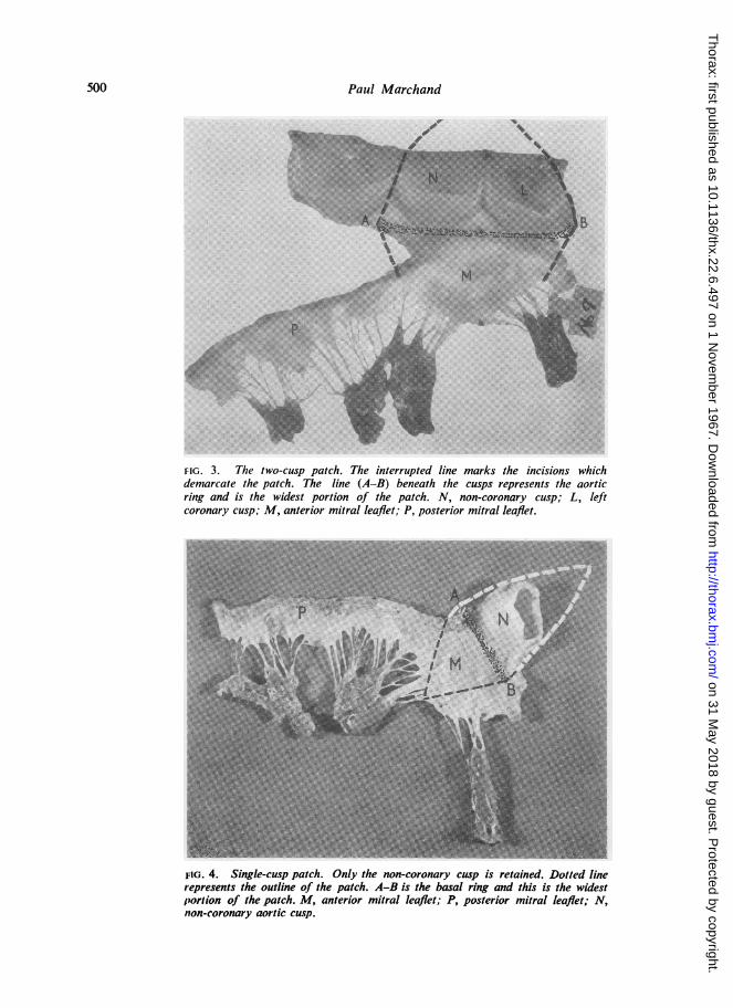
FIG. 3. The two-cusp patch. The interrupted line marks the incisions whichdemarcate the patch. The line (A-B) beneath the cusps represents the aorticring and is the widest portion of the patch. N, non-coronary cusp; L, leftcoronary cusp; M, anterior mitral leaflet; P, posterior mitral leaflet.
FIG. 4. Single-cusp patch. Only the non-coronary cusp is retained. Dotted linerepresents the outline of the patch. A-B is the basal ring and this is the widestporlion of the patch. M, anterior mitral leaflet; P, posterior mitral leaflet; N,non-coronary aortic cusp.
500
on 31 May 2018 by guest. P
rotected by copyright.http://thorax.bm
j.com/
Thorax: first published as 10.1136/thx.22.6.497 on 1 N
ovember 1967. D
ownloaded from

Use of a cusp-bearing homograft patch in Fallot's tetralogy
intact ring of the host graft is placed within thegutter formed by the opened-out pulmonarytrunk and, posteriorly, the aortic fringe of theright coronary cusp is sewn to the endotheliumof the host artery. It must lie in an undistortedmanner with its base accurately aligned with thehost ring. Stitches then join the edges of theobliquely cut aortic margins to the endotheliumof the pulmonary artery until the cut edges of thelatter are reached. Thereafter suturing edge toedge between aortic graft and pulmonary artery iscarried out distally. The basal ring of the homo-graft is sewn to the endothelium of the pulmonaryartery ring until the cut edge of the pulmonaryartery is reached, and thereafter the anterior mitralleaflet is sewn proximally to the myocardium towiden the outflow tract.
TWO-CUSP TRANSPLANT If one host cusp is to beretained and two homograft cusps transplantedthe suturing is similar to that for total replacement.After two cusps have been excised the remainingone occupies only a third of the circumference ofthe pulmonary artery and the homograft has to befolded into the guttered pulmonary artery to fillthe gaps created. The commissures will extend toa more distal level than those of the natural cusp,but the sutures pick up the apices of the naturalcommissures in an attempt to ensure cusp apposi-tion during closure. The external surface of thehomograft ring is sewn to the endothelium of thehost ring up to its cut edges. Portions of theaortic and leaflet triangles will remain foldedwithin the guttered pulmonary artery and ventri-cular cavity and these are sewn to the endotheliumof the lattzr structures until their cut edges arereached. Thereafter edge to edge suturing com-pletes the widening of the pulmonary artery andoutflow tract of the right ventricle.
In all cases the anastomosis has been simple andthe homograft has joined well to the thin pul-monary artery. Haemostasis has been no problem,but, as an insurance, the mitral patch has beenreinforced with a covering of Teflon felt.The aim is to enlarge the ring circumference
without causing pulmonary incompetence. De-pending upon the size of the patient, a ringcircumference between 6 and 8 cm. should beobtained. If the stretched width of the host ringis 1l5 cm., then the host will contribute 3 cm.towards the final circumference. One adult aorticcusp will provide about 2-5 cm., so that with abicuspid host valve where both cusps are retained,one additional adult cusp should provide acircumference of 5-5 cm. With a tricuspid valve
the host's contribution to the circumference of thering will be reduced by the size of the cuspsremoved and more will be gained if two, ratherthan one, are replaced by the larger adult leafle-ts.
CASE REPORTS
CASE 1 S. S., a girl aged 5 with Fallot's tetralogy,weighed 30 lb. (13 kg.).
This little girl had been blue since birth. Fromthe time she began to walk at 2-1 years she had beenbreathless and had squatted. Cyanotic attacks becamefrequent after the age of 4, and when admitted tohospital she was severely incapacitated and wasunable to walk more than a few steps.She was a thin, emaciated, intensely cyanosed
child. She was investigated and catheterized by Dr.C. Rainier-Pope, who confirmed the diagnosis ofFallot's tetralogy and commented on the shortness ofthe ejection systolic murmur.On 6 August 1966 operation was performed. A
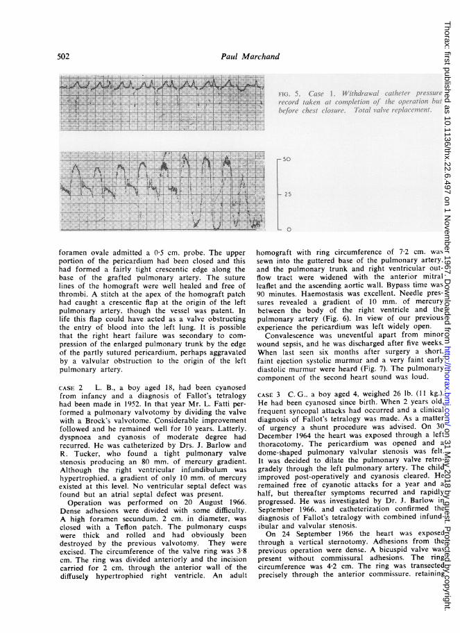
patent foramen ovale was confirmed but not closed.The pulmonary trunk was of reasonable size and theaorta was large. Three pulmonary cusps were presentand these were formed but minute in size. The cir-cumference of the valve ring was 2-2 cm. The incisionwas carried across the ring and for 1 cm. throughthe anterior wall of the right ventricle. The infun-dibular obstruction was excised and the ventricularseptal defect was closed with a Teflon patch. Thevalve cusps were excised and replaced with a homo-graft obtained from an 8-year-old child. The diameterof the new pulmonary ring was 5-6 cm. The homo-graft anterior mitral leaflet was used to enlarge theoutflow tract, and the triangular tongue of aortawidened the pulmonary trunk up to its bifurcation.Total by-pass time was 72 minutes. The heart main-tained the burden of the circulation well and haemo-stasis was no problem. Needle pressures wererecorded (Fig. 5). A gradient of 5 mm. of mercury waspresent between the body of the right ventricle andthe main pulmonary artery. The upper half of thepericardium was closed with catgut.The child remained pink and well for the first 10
days, though a long, loud ejection systolic murmurwas present. Radiographically, the lungs appeared tobe well vascularized. From the tenth day onwardsthe jugular venous pressure began to rise and theliver to enlarge and eventually pulsate. Cyanosis re-appeared on the fourteenth day. By the sixteenth daythe child was severely dyspnoeic and 300 ml. of trans-udate was aspirated from the left pleura. Despiteintensive therapy the congestive failure persisted anddaily paracenteses of the left pleura were required.She died on the twenty-third day.At necropsy no myocardial or pulmonary infarcts
were present and the pulmonary arteries and veinswere free of clot. The homograft cusps were healthyand mobile and histologically virtually normal. Theventricular septal defect was securely closed. The
501
on 31 May 2018 by guest. P
rotected by copyright.http://thorax.bm
j.com/
Thorax: first published as 10.1136/thx.22.6.497 on 1 N
ovember 1967. D
ownloaded from
Paul Marchand
*~~ ~ ~ ~ ~ ~ b/i''.t..t.iduu.a/h.. ..
t..i.......s ....ee .. .. .... ...... X. +. -.-------------------.t. 1F .^..
I)~~~~~~~~~~~~~~~~~~~~9
______ X ---- r - - ::;t7,t-., _ s-1,--.. .....r.;~~~~~~~~~~~~~~~~~~~~~~~~~~~~~ *-.t I....
t~~~~~~~~~~~~~~~~~~~~~4 .-
,*..,... .. .. .1 ......... 1. -t ~~~~~.
2 5___ L.. .* ... .. S . ...* .- ..*-
------̂. . .. .. ,t_... 0F 4^. 4 . ,.__ __ 4 ^ +A
*8s' ''. - T-. -' b-t .-- __ s_
foramen ovale admitted a 0-5 cm. probe. The upper
portion of the pericardium had been closed and thishad formed a fairly tight crescentic edge along thebase of the grafted pulmonary artery. The suture
lines of the homograft were well healed and free ofthronmbi. A stitch at the apex of the homograft patchhad caught a crescentic flap at the origin of the leftpulmonary artery, though the vessel was patent. Inlife this flap could have acted as a valve obstructingthe entry of blood into the left lung. It is possiblethat the right heart failure was secondary to com-
pression of the enlarged pulmonary trunk by the edgeof the partly sutured pericardium, perhaps aggravatedby a valvular obstruction to the origin of the leftpulmonary artery.
CASE 2 L. B., a boy aged 18, had been cyanosedfrom infancy and a diagnosis of Fallot's tetralogyhad been made in 1952. In that year Mr. L. Fatti per-
formed a pulmonary valvotomy by dividing the valvewith a Brock's valvotome. Considerable improvementfollowed and he remained well for 10 years. Latterly.dyspnoea and cyanosis of moderate degree hadrecurred. He was catheterized by Drs. J. Barlow andR. Tucker, who found a tight pulmonary valvestenosis producing an 80 mm. of mercury gradient.Although the right ventricular infundibulum was
hypertrophied. a gradient of only 10 mm. of mercury
existed at this level. No ventricular septal defect was
found but an atrial septal defect was present.Operation was performed on 20 August 1966.
Dense adhesions were divided with some difficulty.A high foramen secundum. 2 cm. in diameter, was
closed with a Teflon patch. The pulmonary cusps
were thick and rolled and had obviously beendestroyed by the previous valvotomy. They were
excised. The circumference of the valve ring was 3-8cm. The ring was divided anteriorly and the incisioncarried for 2 cm. through the anterior wall of thediffusely hypertrophied right ventricle. An adult
homograft with ring circumference of 72 cm. wassewn into the guttered base of the pulmonary artery.and the pulmonary trunk and right ventricular out-flow tract were widened with the anterior mitralleaflet and the ascending aortic wall. Bypass time was90 minutes. Haemostasis was excellent. Needle pres-sures revealed a gradient of 10 mm. of mercurybetween the body of the right ventricle and thepulmonary artery (Fig. 6). In view of our previousexperience the pericardium was left widely open.
Convalescence was uneventful apart from minorwound sepsis, and he was discharged after five weeks.When last seen six months after surgery a short.faint ejection systolic murmur and a very faint earlydiastolic murmur were heard (Fig. 7). The pulmonarycomponent of the second heart sound was loud.
CASE 3 C. G.. a boy aged 4, weighed 26 lb. (11 kg.).He had been cyanosed since birth. When 2 years old.frequent syncopal attacks had occurred and a clinicaldiagnosis of Fallot's tetralogy was made. As a matterof urgency a shunt procedure was advised. On 30December 1964 the heart was exposed through a leftthoracotomy. The pericardium was opened and adome-shaped pulmonary valvular stenosis was felt.It was decided to dilate the pulmonary valve retro-gradely through the left pulmonary artery. The childimproved post-operatively and cyanosis cleared. Heremained free of cyanotic attacks for a year and ahalf, but thereafter symptoms recurred and rapidlyprogressed. He was investigated by Dr. J. Barlow inSeptember 1966, and catheterization confirmed thediagnosis of Fallot's tetralogy with combined infund-ibular and valvular stenosis.On 24 September 1966 the heart was exposed
through a vertical sternotomy. Adhesions from theprevious operation were dense. A bicuspid valve waspresent without commissural adhesions. The ringcircumference was 4-2 cm. The ring was transectedprecisely through the anterior commissure. retaining
502
on 31 May 2018 by guest. P
rotected by copyright.http://thorax.bm
j.com/
Thorax: first published as 10.1136/thx.22.6.497 on 1 N
ovember 1967. D
ownloaded from
Use of a cusp-bearing homograft patch in Fallot's tetralogy
I
\4.\..u ! ~~~~~t \~
FIG. 6. Case 2. Needle pressures recorded between thebody of the right ventricle and the pulmonary trunk atcompletion of surgery for total valve replacement.
two effective cusps. The right ventricle was cut for15 cm. to just short of a large coronary arterycrossing the outflow tract. The fibrous stricture andconsiderable infundibular muscle were excised. Theventricular septal defect was closed with a patch.The right and left coronary cusps were then removedfrom an adult homograft with a ring circumferenceof 7.5 cm. and the non-coronary cusp was interposedbetween the two natural cusps. The outflow tract wasenlarged with the anterior mitral leaflet, and thepulmonary artery, as far as its bifurcation, was
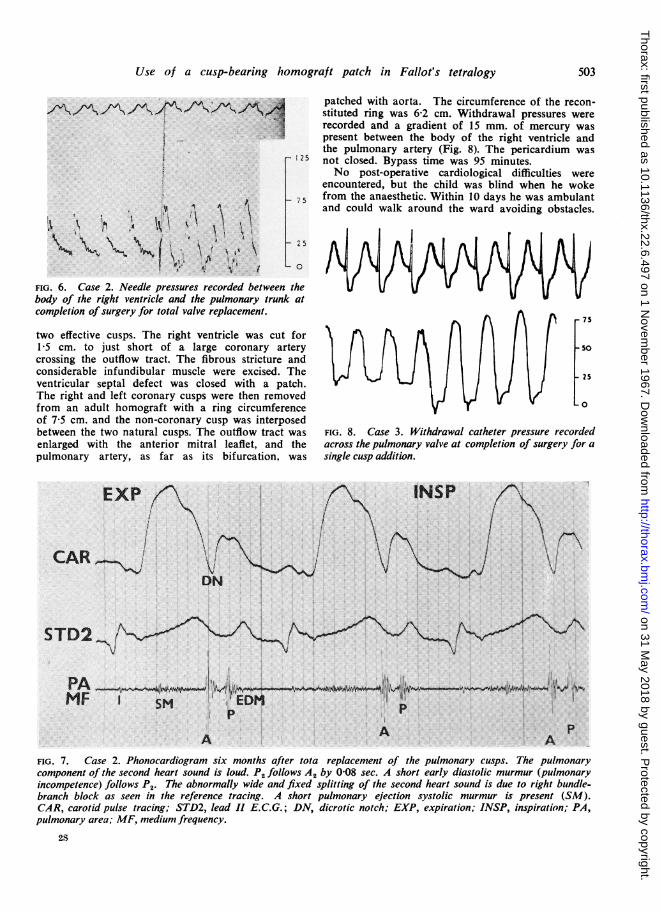
patched with aorta. The circumference of the recon-stituted ring was 6-2 cm. Withdrawal pressures wererecorded and a gradient of 15 mm. of mercury waspresent between the body of the right ventricle andthe pulmonary artery (Fig. 8). The pericardium was
125 not closed. Bypass time was 95 minutes.No post-operative cardiological difficulties were
encountered, but the child was blind when he woke7 5 from the anaesthetic. Within 10 days he was ambulant
and could walk around the ward avoiding obstacles.
0=
- 75
-so
25
L 0
FIG. 8. Case 3. Withdrawal catheter pressure recordedacross the pulmonary valve at completion of surgery for asingle cusp addition.
EXP IS
CAR_ 'DN
STD2\
PA
MEF EDMP
A A Ap
FIG. 7. Case 2. Phonocardiogram six months after tota replacement of the pulmonary cusps. The pulmonarycomponent of the second heart sound is loud. P2 follows A2 by 0-08 sec. A short early diastolic murmur (pulmonaryincompetence) follows P2. The abnormally wide and fixed splitting of the second heart sound is due to right bundle-branch block as seen in the reference tracing. A short pulmonary ejection systolic murmur is present (SM).CAR, carotid pulse tracing; STD2, lead II E.C.G.; DN, dicrotic notch; EXP, expiration; INSP, inspiration; PA,pulmonary area; MF, medium frequency.
28
503
it4
i
II
/%A-t/V. '111. I/V./V\ /-IN-:/. :.,I
on 31 May 2018 by guest. P
rotected by copyright.http://thorax.bm
j.com/
Thorax: first published as 10.1136/thx.22.6.497 on 1 N
ovember 1967. D
ownloaded from
Paul Marchand
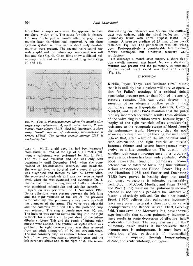
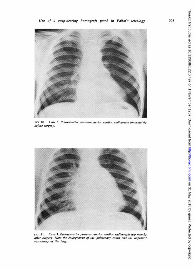
No retinal changes were seen. He appeared to haveperipheral vision only. The cause for this is obscure.He was discharged a month after surgery. Fourmonths later his vision had improved. A soft shortejection systolic murmur and a short early diastolicmurmur were present. The second heart sound waswidely split and the pulmonary component was softbut audible (Fig. 9). Chest films show a dilated pul-monary trunk and well vascularized lung fields (Figs10 and I1).
A
CAR1 i\
STD2
3 LI S A
A A
FIG. 9. Case 3. Phonocardiogram taken five months afiersingle cusp replacement. A, aortic valve closutre; P, pul(-*nonary valve closure; 3LIS, third left interspace. A sholrtearly diastolic murmutr of puilmonary incompetence ispresent (EDM). The ejection systolic muirmutr (SM) ismoderately loud.
CASE 4 M. E., a girl aged 18, had been cyanosedfrom birth. In 1954, at the age of 6, a Brock's pul-monary valvotomy was performed by Mr. L. Fatti.The result was excellent and she was only seenoccasionally until December 1962, when she com-plained of breathlessness, dizziness, and headache.She was admitted to hospital and a cerebral abscesswas diagnosed and treated by Mr. K. Lewer-Allen.She recovered completely and was next seen in April1966. when she was cyanosed and dyspnoeic. Dr. J.Barlow confirmed the diagnosis of Fallot's tetralogywith combined infundibular and valvular stenosis.
Operation was performed on 1 November 1966.Dense adhesions were present between the left lungand the right ventricle at the site of the previousventriculotomy. The pulmonary artery trunk was halfthe diameter of tho aorta. The valve was tricuspidand the commissures fused. Only the posterior cuspwas retained. The ring circumference was 4-2 cm.The incision was carried across the ring into the rightventricle for about 2 cm. to just short of the infun-dibular stricture. This and the bands of infundibularmuscle were excised. The ventricular septal defect waspatched. The right coronary cusp was then removedfrom an adult homograft of 72 cm. circumference.The non-coronary cusp was sutured below and to theleft of the remaining natural posterior cusp and theleft coronary above and to the right of it. The recon-
structed ring circumference was 6 5 cm. The outflowtract was widened with the mitral leaflet and thepulmonary trunk with aorta. Bypass lasted 100minutes. A pressure gradient of 25 mm. of mercuryremained (Fig. 12). The pericardium was left wideopen. Post-operatively a considerable left haemo-thorax developed, but otherwise recovery wassatisfactory.On discharge a month after surgery a short ejec-
tion systolic murmur was heard. No early diastolicmurmur was present and the pulmonary componentof the second heart sound was loud and clear(Fig. 13).
DISCUSSION
Kirklin, Payne. Theye, and DuShane (1960) statethat it is unlikely that a patient will survive opera-tion for Fallot's tetralogy if a residual rightventricular pressure greater than 50"!, of the aorticpressure remains. This can occur despite theinsertion of an adequate outflow patch if thepulmonary ring is hypoplastic. Edwards, Carey,Neufeld, and Lester (1965) maintain that the pul-monary incompetence which results from divisionof the valve ring is seldom severe, because hyper-trophy of the right ventricle offers considerableresistance to the retrograde escape of blood fromthe pulmonary trunk. However, they do notadvocate routine division of the ring, because theybelieve that, after relief of the infundibular andcusp obstruction, the right ventricular musclebecomes thinner and severe incompetence mayevolve as a late complication. The question ofwhether pulmonary incompetence is a progres-sively serious lesion has been widely debated. Withgood myocardial function, pulmonary incom-petence can be tolerated for a long time withoutserious consequence, and Ellison, Brown, Hague,and Hamilton (1955) and Fowler and Duchesne(1958) have proved in healthy dogs that totalpulmonary valvectomy is tolerated remarkablywell. Blount, McCord, Mueller, and Swan (1954)and Price (1961) maintain that pulmonary incom-petence is not serious provided the obstructivefactor is effectively relieved. On the other hand,Brock (1959) believes that pulmonary incompe-tence may present as great a threat as other valveincompetences, and Bender, Austen, Ebert, Green-field, Tsunekawa, and Morrow (1963) have shownexperimentally that sudden pulmonary incomp-tence results in acute depression of effective rightventricular function. The very existence of thepulmonary valve makes it inconceivable thatincompetence is unimportant. It must have adeleterious effect, particularly if myocardialfunction is impaired through long-standingdisease, the ventriculotomy, or bypass.
504
on 31 May 2018 by guest. P
rotected by copyright.http://thorax.bm
j.com/
Thorax: first published as 10.1136/thx.22.6.497 on 1 N
ovember 1967. D
ownloaded from
Use of a cusp-bearing homograft patch in Fallot's tetralogy
ly.t.......... . ---; :?< ... <.-5>:X8.a: ':. ; . '. ' ei'&9:;a. ' f 8f 9 . . ;.D F, g.eYae .! B X: x .O :_ 80Et
*.: ': 'Bi'L::
:6|Zzw l==_g.
..... :o i1 l _XZ ......... ............ :.-_ i. i_SS, B .X:
5 B.MNX;Z --.;6.F t_ ! 5 ............................... .._ ' z.;. .Bt/ - _ .: . .y ........................................ .-0 w_ _ s. _ F_.,'' __*6 _ > . - * N N-#*.N s. E .' ..... r ... ,, ..... -
:;= ...
.x:
I 1|. ................................b :;o_- l | .s., :_r s - .............. E ! .. ... ! _. ...... J_L ..................... fi.:4&e; WA =: ' ?_. ... >.E_ =1 F.or* _E j. i _ Sx, ,s.___W : I*|_ _.'_ I
ra. I
w s
FIG. 1O. Case 3. Pre-operative postero-anterior cardiac radiograpAl immediatelybefore surgery.................. ...... . a_ .... _ - _ - s.U* :.:.. ::.o.o'.E_ ., ':i* . ::sc... ::: .
e _
.r' '..:.. 'q'
i'Es; 'ffi nrb Xt .. _R;:;q .................................................':"gy 'o S. , W¢ i
#,o. _fi_i: :. .. : co#!.:. .......................................... . <s } g _- a f.. _ ......... _ fi_ :. ']I
£ sR. sd. ^
FIG. 11. Case-3. Post-operative postero-anterior cardiac radiograph two monthsafter surgery. Note the enlargement of the pulmonary conus and the improvedvascularity of the lungs.
505
on 31 May 2018 by guest. P
rotected by copyright.http://thorax.bm
j.com/
Thorax: first published as 10.1136/thx.22.6.497 on 1 N
ovember 1967. D
ownloaded from
Paiul Marchand
-I00
1 4~~~~~~-75
~~,kII~~~~~~~-4 ~~~~~~~~~~~~~~-25
-4eoo
INSP
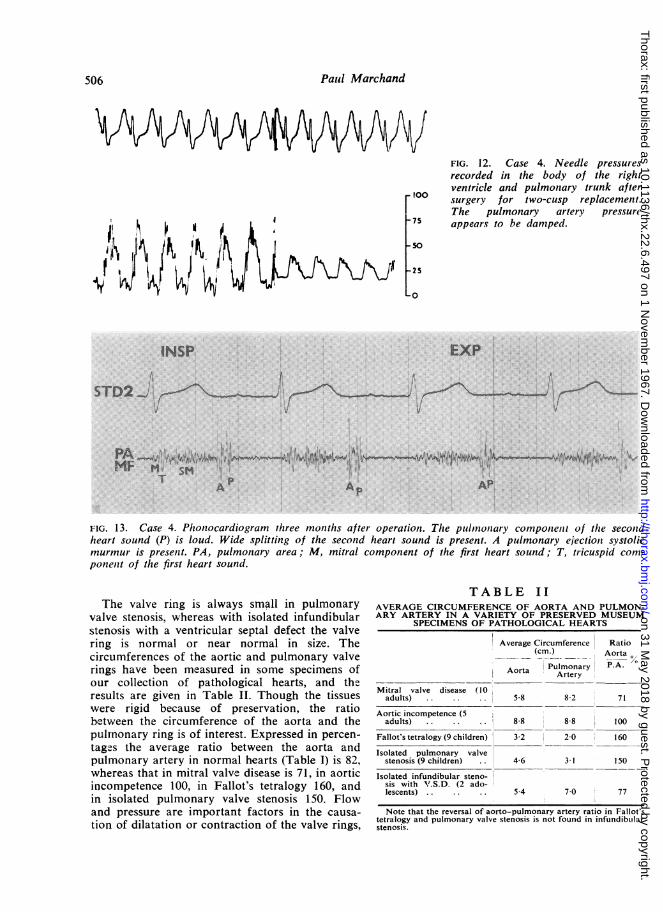
FIG. 12. Case 4. Needle pressures
recorded in the body of the rightventricle and pulmonary trunk aftersurgery for two-cusp replacement.The pulmonary artery pressure
appears to be damped.
EXP
STD21; 'v ' y~~~~~~~~~~~~~~~~~
mA~ ~ ~~~AMF M M' 1,' 9 r { *, ^ /j, ,'T
AA AP.
FiG. 13. Case 4. Phonocardiogram three mouzths after operation. The pulunouiary componienit of the secotidheart sound (P) is loud. Wide splitting of the second heart sound is present. A pulmonary ejectioni systolicmurmur is preselzt. PA, pulmonary area; M, mitral component of the first heart sound; T, tricuspid com-
ponenit of the first heart sound.
TABLE II
The valve ring is always simall in pulmonaryvalve stenosis, whereas with isolated infundibularstenosis with a ventricular septal defect the valvering is normal or near normal in size. Thecircumferences of the aortic and pulmonary valverings have been measured in some specimens ofour collection of pathological hearts, and theresults are given in Table II. Though the tissueswere rigid because of preservation, the ratiobetween the circumference of the aorta and thepulmnonary ring is of interest. Expressed in percen-
tages the average ratio between the aorta andpulmonary artery in normal hearts (Table I) is 82,whereas that in mitral valve disease is 71, in aorticincompetence 100, in Fallot's tetralogy 160, andin isolated pulmonary valve stenosis 150. Flowand pressure are important factors in the causa-
tion of dilatation or contraction of the valve rings,
AVERAGE CIRCUMFERENCE OF AORTA AND PULMON-ARY ARTERY IN A VARIETY OF PRESERVED MUSEUM
SPECIMENS OF PATHOLOGICAL HEARTS
Average Circumference Ratio(cm.) Aorta
Aorta Pulmonary P.A.Artery
Mitral valve disease (10adults). 58 8-2 71
Aortic incompetence (5adults) .. .. .. 8-8 8-8 100
Fallot's tetralogy (9 children) 3-2 2-0 160
Isolated pulmonary valvestenosis (9 children) .. 4-6 3-1 150
Isolated infundibular steno-sis with ".S.D. (2 ado-lescents). 54 7 0 77
Note that the reversal of aorto-pulmonary artery ratio in Fallot'stetralogy and pulmonary valve stenosis is not found in infundibtilarstenosis.
506
on 31 May 2018 by guest. P
rotected by copyright.http://thorax.bm
j.com/
Thorax: first published as 10.1136/thx.22.6.497 on 1 N
ovember 1967. D
ownloaded from
Use of a cusp-bearing homograft patch in Fallot's tetralogy
but, whatever the cause, serious hypoplasia of thevalve ring exists in all cases with pulmonary valvestenosis. Not infrequently, a nairrowing in theimmediate subvalvular position is present.Edwards et al. (1965) have demonstrated that,even after complete resection of the infundibulum,residual obstruction may remain immediatelybelow the cusps. Furthermore, the pulmonaryartery trunk is seldom of normal calibre in Fallot'stetralogy. Sabiston, Cornell, Criley, Neill, Ross,and Bahnson (1964) examined the pulmonaryarteries of 95 specimens of Fallot's tetralogy andfound pulmonary atresia in 22, severe hypoplasiain 19, and absence in one. The pulmonaryarteries were of normal calibre in only two cases.It therefore appears desirable in many cases notonly to widen the outflow tract but also to dividethe ring and often to enlarge the main pulmonarytrunk. Similarly, in pulmonary stenosis with intactseptum the valve ring is invariably hypoplastic,though, as Brock (1957) points out, the lumen ofthe pulmonary artery above the valve is alwaysdilated.
Ideally, every case of pulmonary valvularstenosis should have the ring enlarged to acircumference of from 7 to 9 cm., provided pul-monary incompetence is not induced. It is notalways possible to accomplish this even with acusp-bearing homograft patch. In our first patientthe hypoplastic cusps were excised and replacedwith a homograft of an 8-year-old child. The finalring circumference was 5 2 cm., and, had this childsurvived, relative stenosis might have recurredlater in life through failure of growth of thehomograft. Adult homografts were used for theother cases and the cusp commissures reachedalmost to the pulmonary artery bifurcation.Because it seemed possible that a circumferentialgraft, though confined to the valve-bearing area,might impede growth of the natural pulmonaryartery and also because of difficulty in obtainingtissue from children, it was decided in the last twopatients to preserve posterior continuity of thepulmonary trunk and to use anterior patchescarrying either one or two cusps. As it happened,the third case had a bicuspid valve, and a singleadult cusp was easily inserted. The final circum-ference of the orifice was 6-2 cm., which willprobably be adequate for this patient throughoutlife even if the natural pulmonary artery fails togrow. In Fallot's tetralogy it may often be possibleto introduce a patch carrying only one cusp, for,as Edwards et al. (1965) point out, the valve inthis condition is frequently bicuspid. It remainsto be seen whether the discrepancy between the
small host cusps and the large adult homograftcusps will result in increasing pulmonary incom-petence. At present the three survivors have littleor no incompetence, and the important fact is thatno serious incompetence was present during thecritical early post-operative phase.When planning the first of these operations
consideration was given to excising the pulmonaryvalves and artery and performing an end-to-endanastomosis between a cylindrical homograft andthe transected pulmonary trunk. The homograftring would have been fixed with posterior stitchesand the outflow tract widened with the mitralleaflet. The plan was rejected because it wasthought that the risks of haemorrhage would beserious and control of a possible posterior leakwould be difficult. Furthermore, the procedurewould depend on accurate matching of the hostand homograft, and a considerable number ofgrafts from which to select would be required.In most cases this would exclude the use of anadult vessel. These serious limiting factors werefelt to outweigh the advantage of certain com-petency. As it proved, competence of high degreehas been attained by all three methods used.A further distinct advantage of the technique is
that only a small ventriculotomy is requiredbecause the ring division enhances the exposureof the infundibular stricture and septal defect.Gerbode and Kerth (1963) have cautioned againstlarge ventriculotomies in Fallot's tetralogy andhave advocated the use of a transverse incisionso as to limit the number of muscle bundlesdivided. The short ventriculotomy also lessens therisk of cutting anomalous coronary arteries whichso frequently cross the outflow tract towards theanterior interventricular sulcus.One of the present imponderables is the long-
term fate of homografts in this situation. The casewe have described (Fuller et al., 1966) has a well-functioning valve more than a year after surgery.Already considerable information is available onthe fate of homografts in the aortic position upto five years after surgery (Barratt-Boyes, 1964,1965; Barratt-Boyes, Lowe, Cole, and Kelly,1965; Ross, 1966) and the results are begin-ning to vindicate the enterprise of Murray (1956).Conditions are not necessarily the same in thepulmonary position, but the possible adverseeffect of a venous environment may be balancedby the low diastolic pressures which the homografthas to withstand.
I have to thank Mr. D. Fuller, of our department,for stimulating my interest in the use of homograftsin the pulmonary position, and Mr. L. du Plessis for
507
on 31 May 2018 by guest. P
rotected by copyright.http://thorax.bm
j.com/
Thorax: first published as 10.1136/thx.22.6.497 on 1 N
ovember 1967. D
ownloaded from
Paul Marchand
his assistance. Drs. J. Barlow and I. Obel haveshouldered the burden of pre-operative assessment andpost-operative management, and I am deeply indebtedto them for their skill and co-operation. The work ofthe Cardiovascular Research Unit is subsidized by theC.S.I.R. (S.A.), the Johannesburg City Council, andthe Wellcome Foundation.
REFERENCES
Barnard, C. N., and Schrire, V. (1961). The surgical treatment of thetetralogy of Fallot. Thorax, 16. 346.
Barratt-Boyes, B. G. (1964). Homograft aortic valve replacement inaortic incompetence and stenosis. Ibid., 19, 131.
- (1965). A method for preparing and inserting a homograftaortic valve. Brit. J. Surg., 52, 847.Lowe, J. B., Cole, D. S., and Kelly, D. T. (1965). Homograftvalve replacement for aortic valve. Thorax, 20, 495.
Bender, H. W., Austen, W. G., Ebert, P. A., Greenfield, L. J.,Tsunekawa, T., and Morrow, A. G. (1963). Experimentalpulmonic regurgitation. J. thorac. cardiovasc. Surg., 45, 451.
Blount, S. G., McCord, M. C., Mueller, H., and Swan, H. (1954).Isolated valvular pulmonic stenosis-clinical and physiologicresponses to open valvuloplasty. Circulation, 10, 161.
Brock, R. (1957). The Anatomy oj Congenital Pulnionary Stenosis.Cassell, London.(1959). The surgical treatment of Fallot's tetralogy. Guy's Hosp.Rep., 108, 314.
du Plessis, L. A., and Marchand. P. (1964). The anatomy of the mitralvalve and its associated structures. Thorax, 19, 221.
Edwards, J. E., Carey, L. S., Neufeld, H. N., and Lester, R. G. (1965).Congenital Heart Disease, Vol. 2. W. B. Saunders, Philadelphiaand London.
Ellison, R. G., Brown, W. J., Hague, E. E., and Hamilton, W. F.(1955). Physiologic observations in experimental pulmonaryinsufficiency. J. thorac. Surg., 30, 633.
Fowler, N. O., and Duchesne, E. R. (1958). Effect of experimentalpulmonary valvular insufficiency on the circulation. Ibid., 35, 643.
Fuller, D. N., Marchand, P., Zion, M. M., and Zwi, S. (1966).Homograft replacement of the pulmonary valve. Thlorax, 21, 337.
Gerbode, F., and Kerth, W. J. (1963). Technical considerations inthe treatment of tetralogy of Fallot-the transverse ventriculo-tomy. Ann. Surg., 158, 975.
Kirklin, J. W., Payne, W. S., Theye, R. A., and DuShane. J. W.(1960). Factors affecting survival after open operation fortetialogy of Fallot. Ibid., 152, 485.
--Donald, D. E., Harshbarger, H. G., Hetzel, P. S., Patrick, R. T.,Swan, H. J. C., and Wood, E. H. (1956). Studies in extra-corporeal circulation. Applicability of Gibbon-type pumrp-oxygenator to human intracardiac surgery: 40 cases. Ibid.,144, 2.
Lillehei, C. W., Cohen, M., Warden, H. E., and Varco, R. L. (1956).Complete anatomical correction of the tetralogy of Fallotdefects. Report of successful case. Arch. Surg., 73, 526.
--Morris, M. J., Adams, P., and Anderson, R. C. (1964). Cor-rective surgery for tetralogy of Fallot. J. thorac. cardiovase.Surg., 48, 556.
Marchand, P. (1958). The management of a blood vessel bank. S. Afr.mned. J., 32, 423.
Murray, G. (1956). Homologous aortic-valve-segment transplants assurgical treatment for aortic and mitral insufficiency. Angiology,7, 466.
Price, B. 0. (1961). Isolated incompetence of the pulmonic valve.Circulation. 23, 596.
Ross, D. N. (1966). Aor-tic valve surgery. Ann. roy. Coll. Siurg. Engi.,39, 192.
Sabiston, D. C., Cornell, W. P., Criley, J. M., Neill, C. A., Ross,R. S., and Bahnson, H. T. (1964). The diagnosis and surgicalcorrection of total obstruction of the right ventricle. J. thorac.cardiovase. Surg.. 48, 577.
ADDENDUM
The three surviving patients were seen by Drs. J.Barlow and I. Obel at the Cardiac Clinic of the
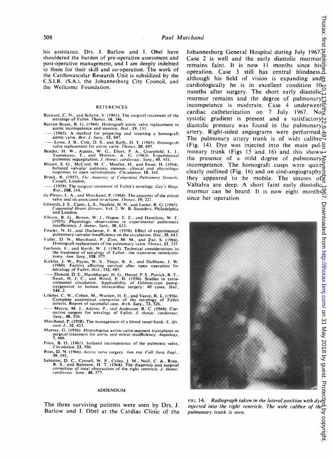
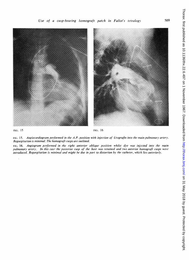
Johannesburg General Hospital during July 1967.Case 2 is well and the early diastolic murmurremains faint. It is now 11 months since hisoperation. Case 3 still has central blindness.although his field of vision is expanding andcardiologically he is in excellent condition 10months after surgery. The short early diastolicmurmur remains and the degree of pulmonaryincompetence is moderate. Case 4 underwentcardiac catheterization on 7 July 1967. Nosystolic gradient is present and a satisfactorydiastolic pressure was found in the pulmonaryartery. Right-sided angiograms were performed.The pulmonary artery trunk is of wide calibre(Fig. 14). Dye was injected into the main pul-monary trunk (Figs 15 and 16) and this showsthe presence of a mild degree of pulmonaryincompetence. The homograft cusps were quiteclearly outlined (Fig. 16) and on cine-angiographythey appeared to be mobile. The sinuses ofValsalva are deep. A short faint early diastolicmurmur can be heard. It is now eight monthssince her operation.
FIG. 14. Radiograph taken in the lateral position with dyeinjected into the right ventricle. The wide calibre of thepulmonary trunk is seen.
508
on 31 May 2018 by guest. P
rotected by copyright.http://thorax.bm
j.com/
Thorax: first published as 10.1136/thx.22.6.497 on 1 N
ovember 1967. D
ownloaded from
Use of a cusp-bear-ing homograft patch in Fallot's tetralog0
FIG. 15 FIG. 16
FIG. 15. Angiocardiogram performed in the A.P. position with injection of Urografin into the main pulmonary arterY.Regurgitation is minimal. The homograft cusps are outlined.FIG. 16. Angiogram performed in the right anterior oblique position whilst dye was injected into the mainpulmonary artery. In this case the posterior cusp of the host was retained and two anterior homograft cusps wereintroduced. Regurgitation is minimal and might be due in part to distortion by the catheter, which lies anteriorly.
509
on 31 May 2018 by guest. P
rotected by copyright.http://thorax.bm
j.com/
Thorax: first published as 10.1136/thx.22.6.497 on 1 N
ovember 1967. D
ownloaded from