Embed Size (px)
Citation preview
Washington University in St. LouisWashington University Open Scholarship
Arts & Sciences Electronic Theses and Dissertations Arts & Sciences
Summer 8-15-2013
The Uropathogenic Escherichia coli Effector YbcLModulates the Innate Immune Response in theUrinary TractMegan Elizabeth LauWashington University in St. Louis
Follow this and additional works at: https://openscholarship.wustl.edu/art_sci_etds
Part of the Biology Commons
This Dissertation is brought to you for free and open access by the Arts & Sciences at Washington University Open Scholarship. It has been acceptedfor inclusion in Arts & Sciences Electronic Theses and Dissertations by an authorized administrator of Washington University Open Scholarship. Formore information, please contact [email protected].
Recommended CitationLau, Megan Elizabeth, "The Uropathogenic Escherichia coli Effector YbcL Modulates the Innate Immune Response in the UrinaryTract" (2013). Arts & Sciences Electronic Theses and Dissertations. 1042.https://openscholarship.wustl.edu/art_sci_etds/1042
WASHINGTON UNIVERSITY IN ST. LOUIS
Division of Biology and Biomedical Sciences
Molecular Microbiology and Microbial Pathogenesis
Dissertation Examination Committee: David A. Hunstad, Chair
Douglas E. Berg Michael G. Caparon Daniel E. Goldberg David B. Haslam
Jeffrey P. Henderson Scott J. Hultgren
The Uropathogenic Escherichia coli Effector YbcL Modulates the Innate Immune Response in the Urinary Tract
by
Megan Elizabeth Lau
A dissertation presented to the Graduate School of Arts and Sciences
of Washington University in partial fulfillment of the
requirements for the degree of Doctor of Philosophy
August 2013
St. Louis, Missouri

copyright by
Megan Elizabeth Lau
2013
ii
TABLE OF CONTENTS List of Tables and Figures............................................................................................................ iv Acknowledgements ........................................................................................................................ v Abstract ......................................................................................................................................... vi Chapter 1 Introduction The Urinary Tract .......................................................................................................................... 1 Urinary Tract Infections ................................................................................................................. 2
Classifications .................................................................................................................... 2 Symptomology and Diagnosis ........................................................................................... 3 Treatment ........................................................................................................................... 4 Recurrence ......................................................................................................................... 4 Predisposing Factors .......................................................................................................... 5 Etiology .............................................................................................................................. 6
Uropathogenic Escherichia coli Pathogenesis ............................................................................... 7 Binding and Invasion ......................................................................................................... 7 Intracellular Replication..................................................................................................... 8 Fluxing and Filamentation ................................................................................................. 9 Quiescent Reservoir Formation ....................................................................................... 10 Chronic and Recurrent Cystitis ........................................................................................ 11
Innate Immune Response to UPEC .............................................................................................. 11 Immune Evasion by UPEC .......................................................................................................... 13 Summary ...................................................................................................................................... 15 References .................................................................................................................................... 19 Chapter 2 YbcL of Uropathogenic Escherichia coli Suppresses Transepithelial Neutrophil Migration Abstract ........................................................................................................................................ 31 Introduction .................................................................................................................................. 32 Materials and Methods ................................................................................................................. 35 Results .......................................................................................................................................... 41 Discussion .................................................................................................................................... 48 Acknowledgements ...................................................................................................................... 53 References .................................................................................................................................... 63 Chapter 3 Liberation of YbcL from the Bacterial Periplasm Abstract ........................................................................................................................................ 69 Introduction .................................................................................................................................. 69 Materials and Methods ................................................................................................................. 72
iii
Results .......................................................................................................................................... 76 Discussion .................................................................................................................................... 80 Acknowledgements ...................................................................................................................... 84 References .................................................................................................................................... 91 Chapter 4 Effects of YbcL on Bladder Epithelial Cells and Neutrophils Abstract ........................................................................................................................................ 94 Introduction .................................................................................................................................. 94 Materials and Methods ................................................................................................................. 98 Results ........................................................................................................................................ 101 Discussion .................................................................................................................................. 106 Acknowledgements .................................................................................................................... 110 References .................................................................................................................................. 115 Chapter 5 Concluding Remarks and Future Directions Allelic Variation in ybcL Loci ................................................................................................... 121 Release of YbcL from the Periplasm ......................................................................................... 123 Suppression of Transuroepithelial PMN Migration by YbcL.................................................... 129 Concluding Remarks .................................................................................................................. 133 References .................................................................................................................................. 135 Appendix 1 Quantitative Assessment of Human Neutrophil Migration across a Cultured Bladder Epithelium Short Abstract ............................................................................................................................ 139 Long Abstract............................................................................................................................. 139 Introduction ................................................................................................................................ 140 Protocol ...................................................................................................................................... 142 Representative Results ............................................................................................................... 148 Discussion .................................................................................................................................. 150 Acknowledgements .................................................................................................................... 154 References .................................................................................................................................. 157
iv
LIST OF TABLES AND FIGURES Chapter 1 Introduction Figure 1 - Schematic of the Urinary Tract System ...................................................................... 17 Figure 2 - UPEC Infectious Cycle in the Bladder........................................................................ 18 Chapter 2 YbcL of Uropathogenic Escherichia coli Suppresses Transepithelial Neutrophil Migration Table 1 - Primers used in this study ............................................................................................. 54 Figure 1 - UPEC YbcL suppresses transepithelial PMN migration in vitro ................................ 55 Figure 2 - Threonine 78 is required for suppression of PMN migration by YbcLUTI ......................... 56 Figure 3 - YbcLUTI confers suppressive activity on nonpathogenic E. coli ................................ 58 Figure 4 - YbcLUTI is secreted ..................................................................................................... 60 Figure 5 - YbcLUTI suppresses acute PMN migration in vivo ..................................................... 62 Chapter 3 Liberation of YbcL from the Bacterial Periplasm Table 1 - Primers used in this study ............................................................................................. 85 Figure 1 - YbcLUTI is not packaged into OMVs .......................................................................... 86 Figure 2 - UPEC release intracellular contents during infection of bladder epithelial cells........ 87 Figure 3 - Liberation of YbcL is dose-, time- and bladder epithelial cell-dependent .................. 88 Figure 4 - Selective release of bacterial intracellular proteins ..................................................... 90 Chapter 4 Effects of YbcL on Bladder Epithelial Cells and Neutrophils Figure 1 - UTI89 inhibits NF-κB signaling independent of YbcL ............................................ 111 Figure 2 - Epithelial integrity is maintained during PMN migration......................................... 112 Figure 3 - PMN movement is unaffected by YbcL.................................................................... 113 Figure 4 - YbcL does not affect the production of chemotactic molecules by bladder epithelial cells or E. coli ............................................................................................................................ 114 Appendix 1 Quantitative Assessment of Human Neutrophil Migration across a Cultured Bladder Epithelium Figure 1 - Schematic of experimental design ............................................................................ 155 Figure 2 - PMN migrate across bladder epithelia in response to various stimuli ...................... 156
v
ACKNOWLEDGEMENTS
This work was supported by NIH grants DK076556, DK080752, and DK064540.
To the many people who supported and encouraged me along the way…
- David and My Colleagues -
- My Friends, Family and Ryan -
Thank You.
vi
ABSTRACT OF THE DISSERTATION
The Uropathogenic Escherichia coli Effector YbcL Modulates the Innate Immune Response in
the Urinary Tract
By
Megan Elizabeth Lau
Doctor of Philosophy in Molecular Microbiology and Microbial Pathogenesis
Washington University in St. Louis, 2013
Assistant Professor David A. Hunstad, Chair
Uropathogenic Escherichia coli (UPEC) are the primary etiology of urinary tract
infections (UTIs), one of the most common bacterial infections afflicting the human population.
While UPEC cause disease throughout the urinary tract, bladder infection, or cystitis, is most
prevalent. A key aspect of UPEC pathogenesis in the bladder is the modulation of the host
inflammatory response. At acute time points, UPEC delay the arrival of immune cells, such as
neutrophils, to the bladder. The lack of neutrophils in the bladder lumen enables UPEC to
replicate freely in the urine and invade the bladder epithelium, a requirement for bacterial
persistence, in the absence of immune pressure. The UPEC products responsible for delaying the
arrival of immune cells to the bladder had not been identified.
This thesis work identified a bacterial protein, YbcL, that was modestly up-regulated
upon UPEC exposure to either cultured bladder epithelial cells or human neutrophils. We
demonstrated that YbcL suppressed the migration of neutrophils across bladder epithelia in an in
vitro model of transuroepithelial neutrophil migration and an in vivo murine model of cystitis.
Suppression of PMN migration by YbcL was dependent upon the presence of threonine at
position 78 (T78). In fact, T78 in YbcL is highly conserved in clinical UPEC isolates,
vii
suggesting that inhibition of neutrophil migration across epithelial barriers by YbcL is a
conserved mechanism of immune modulation among UPEC.
Using a number of complementary approaches, we demonstrated that liberation of YbcL
from the bacterial periplasm was required for suppression of neutrophil migration across a
bladder epithelium. YbcL was detected in the supernatant and in association with bladder
epithelial cells and neutrophils. Release of YbcL from the periplasm occurred in a manner that
was dependent upon the concentration of YbcL in the periplasm, the duration of the infection and
the presence of bladder epithelial cells. Although YbcL was soluble in the supernatant, we
demonstrated that YbcL was not secreted from the periplasm by a canonical secretion system.
Despite the apparent absence of a dedicated secretion system, these findings demonstrate that
YbcL functions as an exoprotein.
Investigations into the mechanism underlying suppression of neutrophil migration by
YbcL revealed that YbcL did not influence the production of chemoattractant molecules by
bladder epithelial cells or bacteria or the ability of neutrophils to chemotax in response to stimuli,
requirements for neutrophils to traverse epithelial barriers. This work identified and began the
characterization of a bacterial protein, YbcL, that contributes to modulation of the innate
immune response by UPEC. Additional experimentation is required to elucidate the importance
of T78, the mode of delivery of YbcL from the periplasm, and the mechanism of action of YbcL.
By delaying the arrival of immune cells, the activity of YbcL likely facilitates formation of the
acute intracellular niche occupied by UPEC and required for persistence in the urinary tract.
1
CHAPTER 1
INTRODUCTION TO THE DISSERTATION
The Urinary Tract
The organs and tissues that comprise the urinary tract (i.e., the kidneys, ureters, bladder,
sphincter muscles and urethra) are responsible for the production, storage and removal of waste
in the form of urine from the body (1) (Figure 1). Urea, a component of urine, is produced when
foods containing protein are broken down in the gastrointestinal tract and is carried in the
bloodstream to the kidneys. The kidneys filter urea and other waste products from the blood,
regulate electrolyte balance, control blood volume and maintain blood pressure through the
removal of excess fluid from the bloodstream. The urine formed in the kidneys travels through
two thin tubes, termed ureters, and into the bladder where it is stored. The bladder is a hollow
muscular organ that expands and contracts to hold changing volumes of urine, while the
sphincter muscles at the base of the bladder prevent urine from leaking out. During urination, or
micturition, urine is expelled from the bladder through the urethra to the outside of the body by
the coordinated relaxation of the sphincter muscles and contraction of smooth muscle in the
bladder.
The primary function of the bladder is to store urine, often for long periods of time. To
prevent waste products in the urine from damaging tissue or reentering the bloodstream, the
bladder must be impermeable to urine contents. To that end, the epithelium that lines the bladder
lumen functions as a barrier to ions, solutes and water, which can vary greatly in concentration
and volume (2). This epithelium, or mucosal layer, is composed of an umbrella cell layer, an
intermediate cell layer and a basal cell layer. The umbrella cells, also referred to as facet cells or
2
superficial cells, are highly differentiated and polarized with distinct apical and basolateral
membranes separated by tight junctions (3). The luminal, or apical, plasma membranes of these
cells bear hexagonal arrays of uroplakin complexes that confer membrane impermeability to
urine and membrane integrity during mechanical stress (4, 5). To change the surface area of the
bladder during filling and voiding, fusiform vesicles containing uroplakins are recycled to and
from the plasma membrane through endocytosis and exocytosis (6-8). The urinary tract
represents a highly evolved organ system that efficiently filters waste products from the blood
and permits waste elimination from the body at convenient intervals.
Urinary Tract Infections
As the urinary tract is exposed to the environment, these organs are susceptible to foreign
threats such as pathogenic bacteria. In fact, urinary tract infections (UTIs) are among the most
common bacterial infections afflicting the human population. In the United States alone, there
are more than 14 million medical visits prompted by UTI, and medical expenditures reach almost
$4 billion each year (9). In addition to community-acquired infections, UTIs account for 40% of
all nosocomial, or hospital-acquired, infections (10). There are 1 million cases of nosocomial
UTIs in the US per year, and 80% of these can be attributed to catheterization (10, 11). UTIs
have a low mortality rate. Given a high incidence in addition to a high rate of recurrence, UTIs
represent a significant health burden and result in staggering health care costs.
Classifications
Bacterial pathogens access the urinary tract through the urethra which is exposed to the
external environment. These organisms can replicate in the urine and colonize the bladder.
3
They can also ascend the ureters and colonize the kidneys. From the kidneys, bacteria can enter
the bloodstream and cause sepsis. One classification scheme for UTIs is based on the infected
organs. For example, infection of the bladder, or cystitis, is considered a lower UTI, while
infection of the kidneys, or pyelonephritis, is termed an upper UTI. Additionally, UTIs can be
described as uncomplicated or complicated. Uncomplicated UTIs typically occur in otherwise
healthy individuals with no structural or functional abnormalities of the urinary tract. All other
UTIs, including patients that are pregnant or have been catheterized, are considered complicated
(12). These classifications can influence the choice and duration of antimicrobial therapy
prescribed.
Symptomology and Diagnosis
UTIs are frequently diagnosed based on symptomology. Symptoms of cystitis include
increased frequency and urgency of urination, painful urination, cloudy urine and pelvic pain.
Symptoms of pyelonephritis include fever, chills, flank pain and nausea or vomiting. UTIs can
also be diagnosed by culture of a clean catch urine specimen. To support a diagnosis of UTI, the
urine culture must yield a known uropathogen above a certain threshold (e.g., 103 colony
forming units (CFU) per ml of urine, although this threshold varies widely) (10, 13, 14). In
addition to confirming bacteriuria, or the presence of bacteria in the urine, urine cultures also
help to determine the antimicrobial susceptibility of the organism. The dipstick urinalysis, a
commercially available test that detects the presence of leukocyte esterase, an enzyme released
by leukocytes, and nitrites, generated by the reduction of nitrates by bacteria, represents an
additional diagnostic tool (12). However, this test provides little additional information when
symptoms and patient history suggest UTI. Because symptoms and bacteriuria can occur
4
independently and urine culture can delay diagnosis, lower UTIs are often treated based on
symptomology alone.
Treatment
Lower UTIs are generally self-limiting and very rarely progress to pyelonephritis, but
because of debilitating symptoms, antibiotics are usually prescribed. Antibiotic treatment of
uncomplicated cystitis can range between a single dose and a ten-day course (12). Broader
spectrum antimicrobial agents and longer regimens are prescribed for patients with complicated
UTIs. Although antibiotics lead to faster resolution of urinary symptoms, they also have
profound adverse effects on the microbiota of the gastrointestinal tract and vagina (15, 16).
Antibiotics also select for resistant pathogens and commensal organisms. Despite some variation
in resistance patterns, overall antibiotic resistance among pathogenic bacteria is becoming a
major problem worldwide. These alarming observations are prompting physicians and
researchers to reconsider standard UTI therapies. Alternative treatment and prevention options,
such as the use of probiotics, adhesion inhibitors, and vaccines, are also being explored (17-19).
Recurrence
Despite effective antibiotic therapies that speed resolution of acute infection, the rate of
recurrent infection is high. For example, approximately 50% of women will experience a UTI at
some point in their lifetime. Of these women, 25% will experience a second UTI and 3% will
experience a third in the six months after treatment of the initial UTI (20). For women who
experience multiple recurrences, treatment options aside from continuous antibiotic prophylaxis
are practically nonexistent. Traditionally, UTIs have been thought to be initiated by
5
contamination of the urinary tract with fecal flora, as the gastrointestinal tract serves as a
reservoir for uropathogens and the urethral opening and rectum are in close proximity in women
(20, 21). However, recent experimental evidence also suggests the existence of an intracellular
reservoir formed by some uropathogens in the bladder epithelium that is resistant to antibiotic
and immune clearance and is capable of initiating recurrent infection (22, 23). It is likely that
both reservoirs, the gastrointestinal tract and the bladder epithelium, contribute to the high rate of
recurrence. In support of these observations, about two thirds of recurrent infections in healthy
women are caused by the same bacterial strain that caused the initial infection (24). Given the
gastrointestinal reservoir, it is not surprising that uropathogens are thought to be transmitted
directly from person to person and indirectly through contaminated food or water (10). A more
thorough understanding of these bacterial reservoirs may facilitate intervention in the cycle of
recurrence.
Predisposing Factors
A number of factors influence susceptibility to UTI. The female and male urinary
systems are very similar except for the length of the urethra. In women, the shorter distance
between the urethral opening and the bladder and the proximity of the urethral opening to the
vagina and rectum, which both harbor large microbial communities, facilitates colonization of
the urinary tract by pathogenic microbes. In fact, women between the ages of 15 and 30 have the
highest frequency of symptomatic infection (25). Structural and functional abnormalities of the
urinary tract, particularly those that affect urine flow and bladder emptying, also increase
susceptibility to UTI (20, 26). In addition to anatomy, certain behaviors, such as frequent sexual
intercourse and the use of spermicides, increase the likelihood of infection (27, 28). Women
6
with diabetes, who are pregnant, or have undergone bladder catheterization are also more prone
to UTI (29-31). Previous UTIs significantly increase the likelihood of subsequent UTIs. Lastly,
genetic variation in genes involved in the immune response has been correlated to increased
susceptibility (32). In agreement with those findings, a history of UTIs in a first-degree female
relative increases the likelihood of UTI (33). In spite of these predisposing factors, the bladder is
well-equipped to prevent and clear bacterial infection.
Etiology
In addition to host factors, bacterial factors also influence whether colonization of the
urinary tract results in disease or clearance. A number of bacterial species can cause UTIs,
including Klebsiella, Proteus, Staphylococcus, and Enterococcus species, to name a few (10).
However, the vast majority of community-acquired UTIs, greater than 80%, are caused by the
gram-negative organism uropathogenic Escherichia coli (UPEC) (20). UPEC are a
heterogeneous group of E. coli that are highly adapted to colonizing the urinary tract and evading
clearance by immune mechanisms. These organisms encode a number of fitness and virulence
factors including adhesins, capsule, fimbriae, flagella, siderophores, and toxins (34). Classified
as extraintestinal pathogenic Escherichia coli (ExPEC), UPEC are distinct from commensal E.
coli and from E. coli that cause disease in the gastrointestinal tract (35). Just as multiple E. coli
pathotypes cause distinct gastrointestinal diseases, UPEC isolates are associated with different
clinical manifestations including acute cystitis, recurrent cystitis, pyelonephritis and
asymptomatic bacteriuria, or the presence of bacteria in the urine in the absence of symptoms
(36). Research has focused on understanding how UPEC strains cause disease throughout the
urinary tract.
7
Uropathogenic Escherichia coli Pathogenesis
As UPEC are responsible for the majority of community-acquired UTIs, researchers have
dedicated significant resources to investigating the mechanisms by which UPEC survive and
persist in the urinary tract. Our current understanding of UPEC pathogenesis is the result of in
vitro experimentation, a well-characterized murine model of cystitis (37), and clinical studies
analyzing samples from human patients.
Binding and Invasion
Micturition is a powerful host defense that eliminates the majority of bacteria that gain
access to the lumen of the bladder. To persist in the bladder in spite of urine flow, UPEC adhere
to the bladder epithelium using proteinaceous surface organelles, termed pili (Figure 2A) (38).
Pili are composed of multiple protein subunits and are assembled by the chaperone/usher
secretion pathway at the bacterial outer membrane. These highly stable fibers contain an
adhesion at their tip that mediates binding to biotic and abiotic surfaces. While UPEC encode a
number of different pili systems, two have been shown to be essential for colonization of the
urinary tract. Type 1 pili interact with mannosylated uroplakins on the luminal surface of the
bladder epithelium via the tip adhesin FimH (39). Pap, or P, pili use PapG to interact with
globoseries glycolipids on the surface of kidney epithelial cells (40). These virulence organelles
mediate the first, and arguably the most important, step in colonization of the urinary tract,
namely adherence. UPEC strains that do not produce type 1 pili are unable to adhere to the
epithelium and are eliminated from the bladder during micturition (41). It follows that small
molecule inhibitors of the type 1 pilus-uroplakin interaction have been shown to treat and
prevent infection in a murine model of cystitis (42, 43).
8
After adherence to the bladder epithelium, UPEC invade the umbrella cells and gain
access to the cytoplasm (Figure 2A) (44). Host proteins (e.g., clathrin and dynamin) have been
implicated in the invasion process (45-47). In contrast to other bacterial pathogens, UPEC
invasion appears to be a passive process, as no bacterial effectors required for invasion have been
identified, aside from pili. Furthermore, UPEC have been detected in fusiform vesicles, which
are transported to and from the apical plasma membrane allowing changes in the surface area of
the umbrella cells to accommodate changing urine volumes (48). UPEC may co-opt this cellular
process to mediate their internalization, though internalized bacteria may also be expelled back
into the lumen through exocytosis (49). While bacterial binding to the epithelium does not
guarantee internalization, expelled bacteria may also contribute to the observation that two
orders of magnitude fewer bacteria are detected intracellularly than extracellularly (50, 51). The
mechanisms underlying the presumed escape of UPEC from vesicles remain unclear.
Intracellular Replication
After UPEC gain access to the cytoplasm, a phase of exponential growth begins that
results in the formation of intracellular bacterial communities, or IBCs (Figure 2B). IBCs are
large, globular masses of bacteria that have biofilm-like properties and are thought to contain
between 105 and 106 organisms (52-54). IBC formation by UPEC was initially observed in a
murine model of cystitis (54). In fact, multiple clinical UPEC isolates have been shown to form
IBCs in multiple murine backgrounds, suggesting that IBC formation is a conserved mechanism
of pathogenesis (55). Confirming the relevance of this intracellular reservoir, IBCs have been
observed in urine samples from women with cystitis (56). The number of bacteria required to
initiate a bladder infection in humans is unclear, though it may be relatively low. Formation of
9
the IBC serves to amplify the initial bacterial inoculum, and bacteria that comprise the IBC will
perpetuate the infection.
The importance of IBC formation for the propagation of infection is demonstrated by the
observation that UPEC strains unable to form IBCs do not persist in the murine model of cystitis.
Bacterial factors involved in IBC formation have been identified, although more certainly exist.
In addition to mediating binding to the bladder epithelium, type 1 pili also aid in IBC formation
(51). This finding is not surprising, as surface organelles have been implicated in biofilm
formation and IBCs resemble biofilms (57). In addition to type 1 pili, a UPEC strain lacking
surA expression was also defective in IBC formation in a murine model of cystitis (50). As SurA
encodes a periplasmic chaperone that aids outer membrane protein (OMP) biogenesis, it is likely
that the defect in intracellular growth was due to the absence of one or more OMPs from the
outer membrane. In agreement with that hypothesis, a UPEC strain lacking ompA, a SurA-
dependent OMP, was also unable to form IBCs (58). It is unclear how the presence of OmpA
promotes IBC formation. The identification of additional bacterial products that facilitate IBC
formation would illuminate the cellular processes required for the development of these complex
communities.
Fluxing and Filamentation
Upon maturation of the IBC, bacteria detach from the community and flux from the
infected umbrella cell via cell lysis (Figure 2C) (53). Though some of the extracellular bacteria
will be removed by micturition, other bacteria will initiate binding and invasion of neighboring
naïve umbrella cells, prompting additional rounds of IBC formation and egress. A fraction of the
bacteria flux from the ruptured umbrella cell as filamentous organisms. Filamentous bacteria are
10
more resistant to phagocytosis by immune cells than bacillary-shaped bacteria, although these
organisms appear to have unique anti-phagocytosis qualities in addition to cell shape (59). The
ability to form filaments is an important aspect of pathogenesis, as a UPEC mutant that was
unable to filament did not form second-round IBCs (59).
Quiescent Reservoir Formation
A host defense against bacterial invasion and intracellular replication is the exfoliation of
infected umbrella cells as a result of apoptosis (60-62). A toxin encoded by UPEC, α-hemolysin
(HlyA), may promote this process by forming pores in bladder epithelial cell membranes (63).
Umbrella cells containing IBCs have been detected in the urine of both mice and humans with
cystitis (54, 56). However, the exfoliation of umbrella cells exposes the underlying intermediate
cells. Bacteria present in the bladder may bind and invade intermediate cells, forming quiescent
intracellular reservoirs, or QIRs (Figure 2D). In a murine model, UPEC in QIRs were detected
in membrane-bound compartments and did not appear to exhibit growth, in stark contrast to the
IBC (22). This quiescent reservoir is refractory to antibiotic and immune clearance (23).
Bacteria remain viable for months in the bladder epithelium despite the absence of bacteriuria
and are thought to emerge from the QIR in response to specific, though not entirely clear, signals
(e.g., epithelial turnover) and initiate recurrent infection. Despite data supporting this
hypothesis, direct evidence for QIRs in women is lacking. Identification of the QIR has
revolutionized thinking about sources of recurrent infection. Characterization of the QIR with
regard to bacterial factors required for formation and maintenance could lead to the identification
of new drug targets, and disruption of QIR formation may prevent recurrent infection in some
women.
11
Chronic and Recurrent Cystitis
Using the murine model of cystitis, researchers have demonstrated that some mouse
strains (e.g., C57BL/6J) spontaneously resolve acute bladder infection, with or without the
formation of QIRs, in the absence of intervention. In comparison, other murine backgrounds
(e.g., C3H/HeN) are susceptible to the development of chronic cystitis, the severity of which is
dependent upon the host strain and the infectious dose (64). Chronic cystitis is defined by high
bacterial titers (> 104 CFU/ ml) in the urine and bladder at 4 weeks post infection (p.i.) and
results in chronic inflammation, though sterilization of the bladder is not achieved. Biomarkers
of chronic cystitis in mice at the local and systemic level were identified at 24 hours p.i. and
included elevated levels of specific cytokines and chemokines, weight lost and severe pyuria
(i.e., PMN in the urine) (64). After antibiotic therapy to resolve infection, mice that had
experienced chronic cystitis were more susceptible to recurrent chronic cystitis upon inoculation
with a different UPEC strain (65). These data suggest that the acute inflammatory response may
predispose to chronic bacterial infection and may dictate susceptibility to recurrent cystitis.
These in vivo findings mirror clinical observations in women who experience persistent recurrent
infections despite appropriate antibiotic therapy. Additionally, these observations suggest that
the magnitude of the inflammatory response must be properly regulated to achieve bacterial
clearance without predisposing to chronic cystitis.
Innate Immune Response to UPEC
Mucosal tissues such as the gastrointestinal tract and the vagina harbor robust microbial
communities that contribute to the overall health of the organism. In contrast, the urinary tract,
aside from the urethra, has traditionally been considered a sterile mucosa. Consequently,
12
colonization by bacteria elicits a robust inflammatory response that is thought to cause the
symptoms associated with disease. An innate immune response is initiated when pathogen-
associated molecular patterns (PAMPs) are recognized by pattern recognition receptors (PRRs)
on epithelial cells and resident immune cells. Recognition of gram-negative lipopolysaccharide
(LPS) by Toll-like receptor 4 (TLR4) is an essential step in the pro-inflammatory response to
UPEC in the bladder (66). In addition to LPS, TLR4 also appears to recognize type 1 and P pili
(67-70). Stimulation of TLR2 and TLR5 by bacterial lipoproteins and flagellin, respectively,
also contributes to the pro-inflammatory response (71, 72). Upon ligand binding, Toll-like
receptors activate signaling cascades, including the NF-κB pathway, that result in changes in
gene expression. Among the genes up-regulated, cytokines and chemokines such as IL-6 and IL-
8 are produced and secreted (73-75). In response to the chemoattractant gradient, leukocytes,
primarily polymorphonuclear leukocytes (PMN; neutrophils), migrate from the bloodstream
across the epithelium and into the bladder lumen (75). Antimicrobial activities of the epithelium
and innate immune cells coordinate to clear the bladder of bacteria in many cases of cystitis.
Aspects of the innate immune response in the bladder were elucidated using a murine
model of cystitis and have been confirmed by human studies. Compared to the C3H/HeN
background, C3H/HeJ mice had higher bacterial titers in the bladder and kidneys upon UPEC
infection (76). Increased susceptibility in these mice was shown to be primarily the result of a
mutation in the Tlr4 gene (77, 78). In the absence of a TLR4 response, C3H/HeJ mice failed to
recruit PMN to the bladder and, consequently, failed to clear infection (79). Analogous to the
phenotype observed in C3H/HeJ mice, humans suffering from asymptomatic bacteriuria had
lower TLR4 expression on their neutrophils compared to healthy controls (80). Genetic variation
in Cxcr1, an IL-8 receptor, is correlated with increased incidence of pyelonephritis, and mice
13
deficient in CXCR1 are more susceptible to UTI than wild-type controls (81, 82). Lastly, IL-6,
IL-8 and PMN can be detected in the urine of both mice and humans with cystitis (75, 83, 84).
Murine models and human studies highlight the importance of a tightly regulated innate immune
response in clearing the urinary tract of bacterial pathogens.
Immune Evasion by UPEC
UPEC are highly adapted to colonize and persist in the urinary tract, evidenced by the
complex pathogenic cascade driven by UPEC in the bladder. In contrast to laboratory or
commensal strains of E. coli, UPEC have evolved multiple, often overlapping, strategies to
attenuate and subvert the innate immune response. At early time points, UPEC manipulate
eukaryotic signaling cascades to delay the initiation of a pro-inflammatory response. In contrast
to nonpathogenic E. coli, UPEC elicit significantly lower cytokine levels (e.g., IL-6 and IL-8) in
culture supernatants during in vitro infection of cultured bladder epithelial cells (85-87). Low
cytokine levels were observed even upon addition of known TLR agonists (e.g., LPS) during
UPEC infection, suggesting active suppression of signaling cascades upstream of cytokine
production. Genes involved in LPS biosynthesis have been implicated in this phenotype, though
alterations to LPS structure appear to be only one aspect of a complex phenotype (85, 88).
Klumpp and colleagues demonstrated that the clinical UPEC isolate NU14 stabilized an inhibitor
of NF-κB, preventing NF-κB activation even in the presence of activating stimuli, although the
mechanism underlying this phenotype is unclear (89). Cirl et al. identified a Toll/IL-1 receptor
(TIR) domain-containing protein TcpC encoded by clinical isolate CFT073 that interacts with
MyD88, inhibiting signaling through any pathway that requires this adaptor (90). However,
TcpC homologs in clinical UPEC isolates are rare. Recently, Dhakal and Mulvey implicated α-
14
hemolysin (HlyA) in suppression of eukaryotic signaling during UPEC infection. HlyA
indirectly mediated the degradation of many cellular proteins, including components of the NF-
κB pathway (91). Corroborating these in vitro findings, a human study demonstrated an absence
of IL-6 and IL-8 in the urine immediately before the onset of symptomatic recurrent UTI (92).
These observations may be partly due to the heterogeneous nature of UPEC, but may also
represent multiple complementary strategies that can be employed by a single UPEC strain to
achieve the same result, namely inhibition of eukaryotic signaling pathways that initiate a pro-
inflammatory response.
UPEC-mediated suppression of signaling in bladder epithelial cells delays the production
of cytokines and chemokines. Compared to nonpathogenic E. coli, UPEC also delay the
recruitment of PMN to the bladder lumen during infection (87, 93). It is unclear if this delay in
PMN arrival is a consequence of inhibited chemokine production or if UPEC employ additional
factors to prevent PMN from transiting the bladder epithelium. Eventually, PMN infiltrate the
bladder in great numbers. In fact, PMN are readily detectable in the urine of patients with
cystitis. Nevertheless, UPEC have evolved to persist in spite of a robust inflammatory response.
Replication within IBCs protects UPEC from infiltrating immune cells, and filamentation allows
extracellular UPEC to resist PMN engulfment. Analogous to suppression of signaling in bladder
epithelial cells, UPEC also manipulate the activities of PMN. Cytotoxic necrotizing factor 1
(CNF1), a toxin secreted by UPEC, was shown to inhibit the chemotactic and phagocytic
capabilities of PMN (94, 95). In addition, UPEC were shown to be less susceptible to PMN
killing and elicited a less robust ROS response compared to nonpathogenic E. coli, though the
mechanisms underlying these phenotypes are unclear (93).
15
In total, the activities of UPEC in the bladder promote colonization and propagation of
infection. Our understanding of these processes is incomplete. It is unclear how UPEC products
(e.g., LPS, TcpC, HlyA) coordinate to inhibit eukaryotic signaling, whether cascades in addition
to the NF-κB pathway are blocked, and if there are additional bacterial products involved in
these phenotypes. The mechanisms underlying the delayed migration of PMN to the bladder are
also unclear. These initial events likely influence the outcome of infection and warrant further
investigation. Suppression of the inflammatory response by UPEC is an important strategy for
colonization of the bladder during acute and recurrent cystitis, but it may also facilitate
asymptomatic bacteriuria and pyelonephritis.
Summary
UTIs are among the most common bacterial infections in humans. Due to the high
incidence and high rate of recurrence, UTIs impose significant financial and health burdens.
Because of the paucity of treatment options aside from antibiotics, treatment of UTIs is
contributing to the growing global problem of antibiotic resistant organisms, which, in turn,
threatens the effective treatment of UTIs. The majority of UTIs are caused by UPEC.
Investigations into UPEC pathogenesis in the urinary tract may enable the identification of novel
bacterial targets and the development of new anti-infective therapeutics.
During cystitis, UPEC direct a complex cascade that involves the formation of
intracellular reservoirs and the manipulation of many eukaryotic processes. Intracellular
replication, or IBC formation, not only allows UPEC to replicate in the presence of an abundant
nutrient supply, but also serves to protect the community from infiltrating PMN and other
16
immune cells. Formation of IBCs is essential for the propagation of infection. Therefore, the
events that precede and facilitate IBC formation are important steps in the pathogenic cascade.
At early time points, UPEC delay the arrival of PMN to the bladder, allowing invasion of
the bladder epithelium and establishment of the intracellular niche in the absence of immune
pressure. In other words, the delayed arrival of PMN promotes IBC formation. It would stand to
reason that accelerated PMN arrival to the bladder would limit bacterial invasion into the
epithelium through bacterial killing and, consequently, disrupt formation of the intracellular
reservoir. Interference in UPEC-mediated suppression of PMN migration may aid in bacterial
clearance.
UPEC inhibit the production of cytokines and chemokines by the bladder epithelium at
early time points, and some of the bacterial products involved in this phenotype have been
identified. It is unclear if delayed PMN recruitment is due to the absence of epithelial-derived
chemokines. Given the overlapping functions of previously identified bacterial products, it is
likely that UPEC encode additional factors that specifically mediate inhibition of PMN
migration. Such proteins have not been identified thus far. Suppression of the acute
inflammatory response by UPEC represents an important step in the pathogenic cascade that
could be manipulated via therapeutic intervention, and consequently, deserves investigation.
17
Tables and Figures
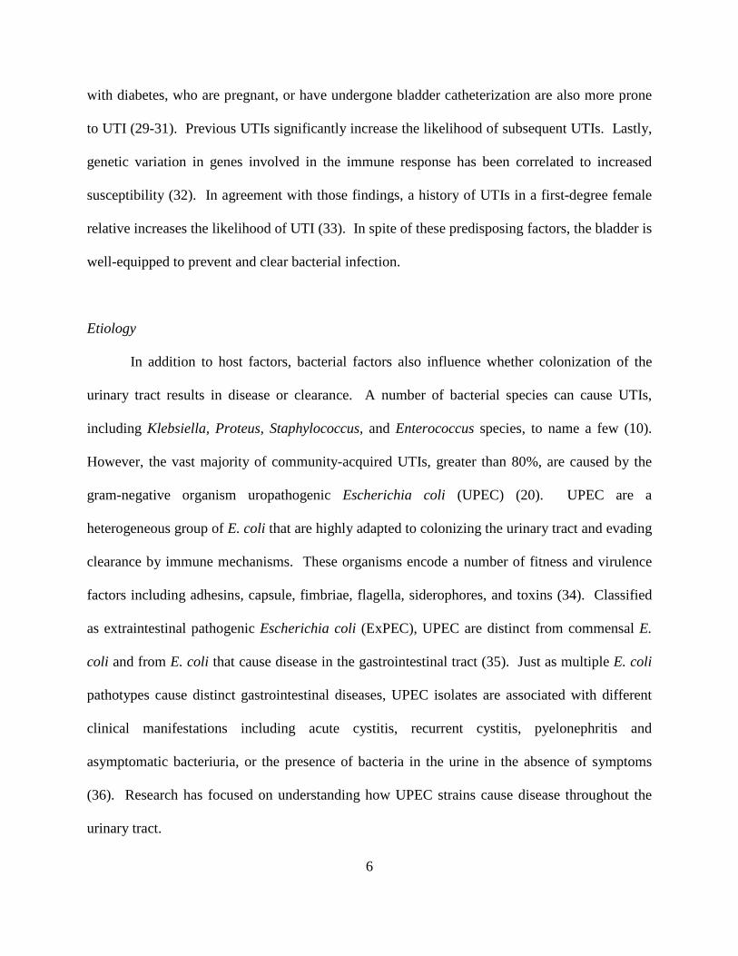
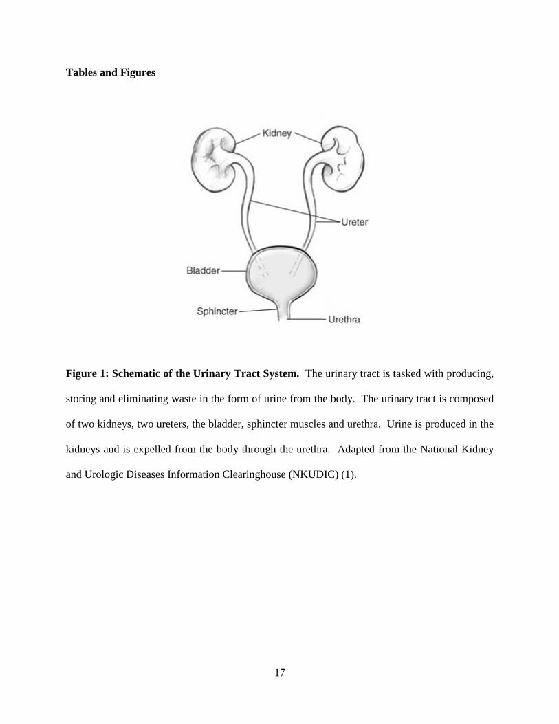
Figure 1: Schematic of the Urinary Tract System. The urinary tract is tasked with producing,
storing and eliminating waste in the form of urine from the body. The urinary tract is composed
of two kidneys, two ureters, the bladder, sphincter muscles and urethra. Urine is produced in the
kidneys and is expelled from the body through the urethra. Adapted from the National Kidney
and Urologic Diseases Information Clearinghouse (NKUDIC) (1).
18
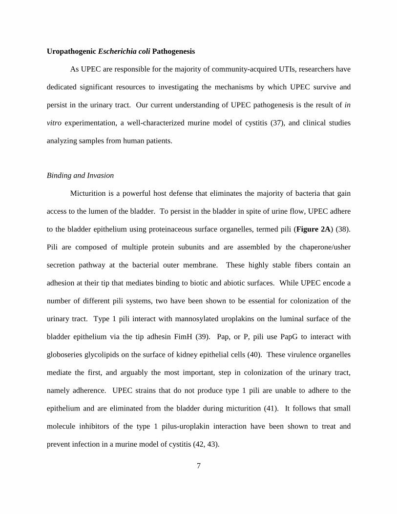
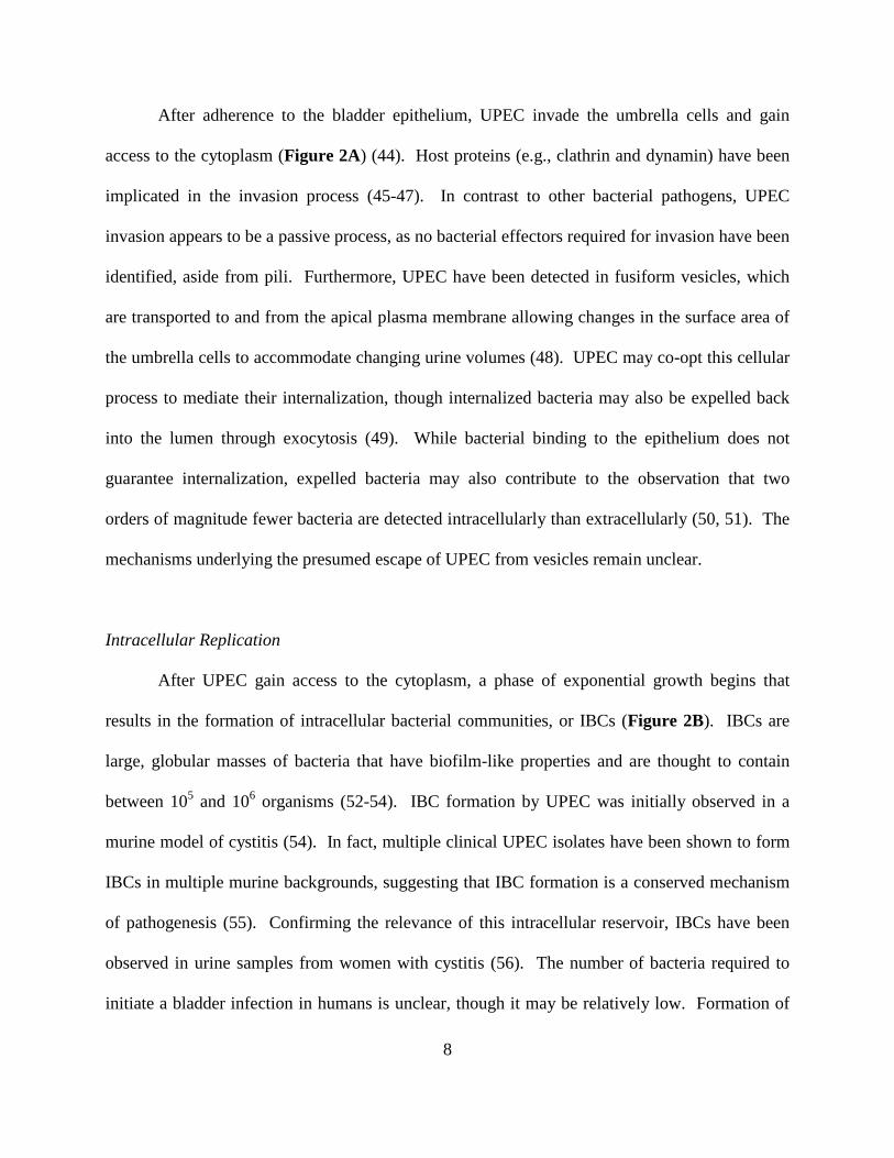
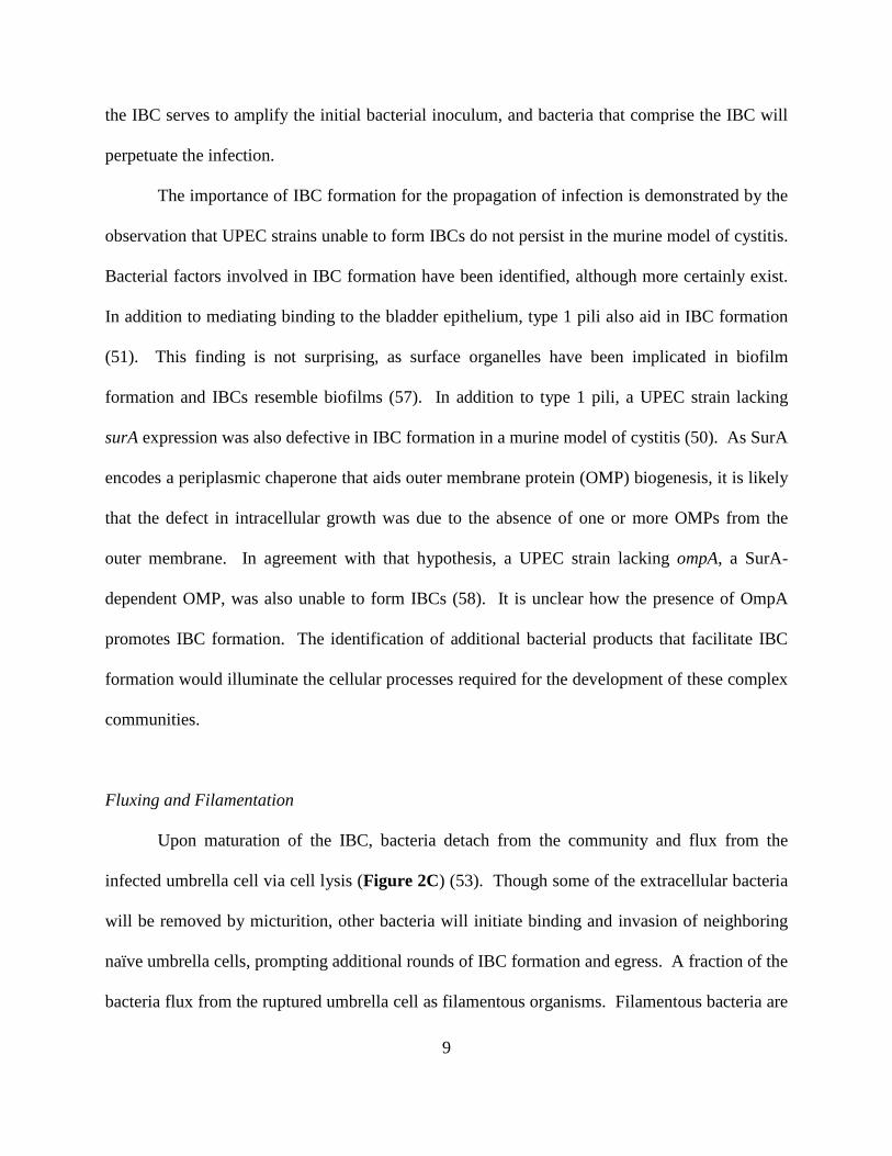
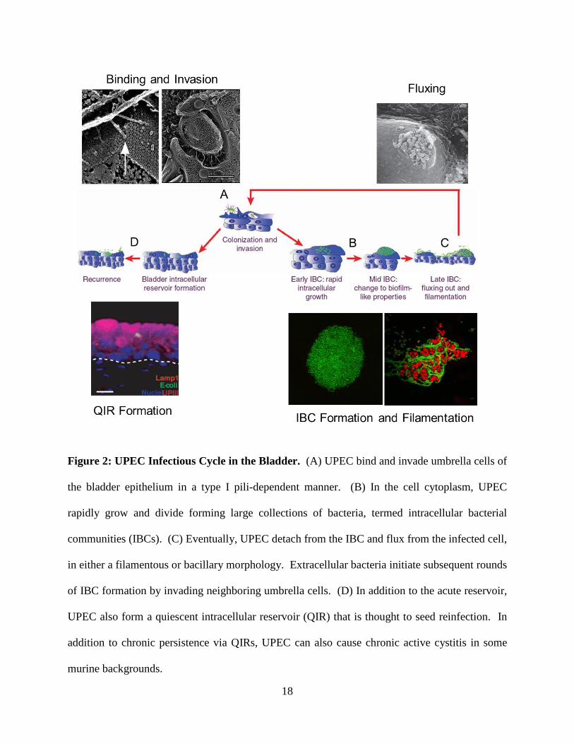
Figure 2: UPEC Infectious Cycle in the Bladder. (A) UPEC bind and invade umbrella cells of
the bladder epithelium in a type I pili-dependent manner. (B) In the cell cytoplasm, UPEC
rapidly grow and divide forming large collections of bacteria, termed intracellular bacterial
communities (IBCs). (C) Eventually, UPEC detach from the IBC and flux from the infected cell,
in either a filamentous or bacillary morphology. Extracellular bacteria initiate subsequent rounds
of IBC formation by invading neighboring umbrella cells. (D) In addition to the acute reservoir,
UPEC also form a quiescent intracellular reservoir (QIR) that is thought to seed reinfection. In
addition to chronic persistence via QIRs, UPEC can also cause chronic active cystitis in some
murine backgrounds.
19
References
1. (NKUDIC) NKUDIC June 29, 2012, posting date. Your Urinary System and How It
Works. National Institute of Diabetes and Digestive and Kidney Diseases, National
Institutes of Health. [Online.]
2. Khandelwal P, Abraham SN, Apodaca G. 2009. Cell biology and physiology of the
uroepithelium. Am J Physiol Renal Physiol 297:F1477-1501.
3. Acharya P, Beckel J, Ruiz WG, Wang E, Rojas R, Birder L, Apodaca G. 2004.
Distribution of the tight junction proteins ZO-1, occludin, and claudin-4, -8, and -12 in
bladder epithelium. Am J Physiol Renal Physiol 287:F305-318.
4. Hu P, Meyers S, Liang FX, Deng FM, Kachar B, Zeidel ML, Sun TT. 2002. Role of
membrane proteins in permeability barrier function: uroplakin ablation elevates urothelial
permeability. Am J Physiol Renal Physiol 283:F1200-1207.
5. Negrete HO, Lavelle JP, Berg J, Lewis SA, Zeidel ML. 1996. Permeability properties of
the intact mammalian bladder epithelium. Am J Physiol 271:F886-894.
6. Apodaca G. 2004. The uroepithelium: not just a passive barrier. Traffic 5:117-128.
7. Kreft ME, Jezernik K, Kreft M, Romih R. 2009. Apical plasma membrane traffic in
superficial cells of bladder urothelium. Ann N Y Acad Sci 1152:18-29.
8. Truschel ST, Wang E, Ruiz WG, Leung SM, Rojas R, Lavelle J, Zeidel M, Stoffer D,
Apodaca G. 2002. Stretch-regulated exocytosis/endocytosis in bladder umbrella cells.
Mol Biol Cell 13:830-846.
9. Litwin MS, Saigal CS, Yano EM, Avila C, Geschwind SA, Hanley JM, Joyce GF,
Madison R, Pace J, Polich SM, Wang M, Urologic Diseases in America P. 2005.
Urologic diseases in America Project: analytical methods and principal findings. J Urol
173:933-937.
20
10. Foxman B. 2010. The epidemiology of urinary tract infection. Nat Rev Urol 7:653-660.
11. Tambyah PA, Maki DG. 2000. Catheter-associated urinary tract infection is rarely
symptomatic: a prospective study of 1,497 catheterized patients. Arch Intern Med
160:678-682.
12. Hooton TM. 2012. Clinical practice. Uncomplicated urinary tract infection. N Engl J Med
366:1028-1037.
13. Rubin RH, Shapiro ED, Andriole VT, Davis RJ, Stamm WE. 1992. Evaluation of new
anti-infective drugs for the treatment of urinary tract infection. Infectious Diseases
Society of America and the Food and Drug Administration. Clin Infect Dis 15 Suppl
1:S216-227.
14. Warren JW, Abrutyn E, Hebel JR, Johnson JR, Schaeffer AJ, Stamm WE. 1999.
Guidelines for antimicrobial treatment of uncomplicated acute bacterial cystitis and acute
pyelonephritis in women. Infectious Diseases Society of America (IDSA). Clin Infect Dis
29:745-758.
15. Dethlefsen L, Huse S, Sogin ML, Relman DA. 2008. The pervasive effects of an
antibiotic on the human gut microbiota, as revealed by deep 16S rRNA sequencing. PLoS
Biol 6:e280.
16. Tempera G, Furneri PM, Cianci A, Incognito T, Marano MR, Drago F. 2009. The impact
of prulifloxacin on vaginal lactobacillus microflora: an in vivo study. J Chemother
21:646-650.
17. Barrons R, Tassone D. 2008. Use of Lactobacillus probiotics for bacterial genitourinary
infections in women: a review. Clin Ther 30:453-468.
21
18. Cusumano CK, Hultgren SJ. 2009. Bacterial adhesion--a source of alternate antibiotic
targets. IDrugs 12:699-705.
19. Sivick KE, Mobley HL. 2010. Waging war against uropathogenic Escherichia coli:
winning back the urinary tract. Infect Immun 78:568-585.
20. Foxman B. 2003. Epidemiology of urinary tract infections: incidence, morbidity, and
economic costs. Dis Mon 49:53-70.
21. Brauner A, Jacobson SH, Kuhn I. 1992. Urinary Escherichia coli causing recurrent
infections--a prospective follow-up of biochemical phenotypes. Clin Nephrol 38:318-323.
22. Mysorekar IU, Hultgren SJ. 2006. Mechanisms of uropathogenic Escherichia coli
persistence and eradication from the urinary tract. Proc Natl Acad Sci U S A 103:14170-
14175.
23. Schilling JD, Lorenz RG, Hultgren SJ. 2002. Effect of trimethoprim-sulfamethoxazole on
recurrent bacteriuria and bacterial persistence in mice infected with uropathogenic
Escherichia coli. Infect Immun 70:7042-7049.
24. Russo TA, Stapleton A, Wenderoth S, Hooton TM, Stamm WE. 1995. Chromosomal
restriction fragment length polymorphism analysis of Escherichia coli strains causing
recurrent urinary tract infections in young women. J Infect Dis 172:440-445.
25. Foxman B, Brown P. 2003. Epidemiology of urinary tract infections: transmission and
risk factors, incidence, and costs. Infect Dis Clin North Am 17:227-241.
26. Raz R, Gennesin Y, Wasser J, Stoler Z, Rosenfeld S, Rottensterich E, Stamm WE. 2000.
Recurrent urinary tract infections in postmenopausal women. Clin Infect Dis 30:152-156.
22
27. Foxman B, Gillespie B, Koopman J, Zhang L, Palin K, Tallman P, Marsh JV, Spear S,
Sobel JD, Marty MJ, Marrs CF. 2000. Risk factors for second urinary tract infection
among college women. Am J Epidemiol 151:1194-1205.
28. Scholes D, Hooton TM, Roberts PL, Stapleton AE, Gupta K, Stamm WE. 2000. Risk
factors for recurrent urinary tract infection in young women. J Infect Dis 182:1177-1182.
29. Andriole VT, Patterson TF. 1991. Epidemiology, natural history, and management of
infections in pregnancy. Med Clin North Am 75: 359-373.
30. Nicolle LE. 2005. Catheter-related urinary tract infection. Drugs Aging 22:627-639.
31. Ronald A, Ludwig E. 2001. Urinary tract infections in adults with diabetes. Int J
Antimicrob Agents 17:287-292.
32. Ragnarsdottir B, Lutay N, Gronberg-Hernandez J, Koves B, Svanborg C. 2011. Genetics
of innate immunity and UTI susceptibility. Nat Rev Urol 8:449-468.
33. Scholes D, Hawn TR, Roberts PL, Li SS, Stapleton AE, Zhao LP, Stamm WE, Hooton
TM. 2010. Family history and risk of recurrent cystitis and pyelonephritis in women. J
Urol 184:564-569.
34. Wiles TJ, Kulesus RR, Mulvey MA. 2008. Origins and virulence mechanisms of
uropathogenic Escherichia coli. Exp Mol Pathol 85:11-19.
35. Kohler CD, Dobrindt U. 2011. What defines extraintestinal pathogenic Escherichia coli?
Int J Med Microbiol 301:642-647.
36. Croxen MA, Finlay BB. 2010. Molecular mechanisms of Escherichia coli pathogenicity.
Nat Rev Microbiol 8:26-38.
37. Hung CS, Dodson KW, Hultgren SJ. 2009. A murine model of urinary tract infection.
Nat Protoc 4:1230-1243.
23
38. Wright KJ, Hultgren SJ. 2006. Sticky fibers and uropathogenesis: bacterial adhesins in
the urinary tract. Future Microbiol 1:75-87.
39. Zhou G, Mo WJ, Sebbel P, Min G, Neubert TA, Glockshuber R, Wu XR, Sun TT, Kong
XP. 2001. Uroplakin Ia is the urothelial receptor for uropathogenic Escherichia coli:
evidence from in vitro FimH binding. J Cell Sci 114:4095-4103.
40. Dodson KW, Jacob-Dubuisson F, Striker RT, Hultgren SJ. 1993. Outer-membrane PapC
molecular usher discriminately recognizes periplasmic chaperone-pilus subunit
complexes. Proc Natl Acad Sci U S A 90:3670-3674.
41. Hultgren SJ, Porter TN, Schaeffer AJ, Duncan JL. 1985. Role of type 1 pili and effects of
phase variation on lower urinary tract infections produced by Escherichia coli. Infect
Immun 50:370-377.
42. Cusumano CK, Pinkner JS, Han Z, Greene SE, Ford BA, Crowley JR, Henderson JP,
Janetka JW, Hultgren SJ. 2011. Treatment and prevention of urinary tract infection with
orally active FimH inhibitors. Sci Transl Med 3:109ra115.
43. Han Z, Pinkner JS, Ford B, Chorell E, Crowley JM, Cusumano CK, Campbell S,
Henderson JP, Hultgren SJ, Janetka JW. 2012. Lead optimization studies on FimH
antagonists: discovery of potent and orally bioavailable ortho-substituted biphenyl
mannosides. J Med Chem 55:3945-3959.
44. Martinez JJ, Mulvey MA, Schilling JD, Pinkner JS, Hultgren SJ. 2000. Type 1 pilus-
mediated bacterial invasion of bladder epithelial cells. Embo J 19:2803-2812.
45. Eto DS, Gordon HB, Dhakal BK, Jones TA, Mulvey MA. 2008. Clathrin, AP-2, and the
NPXY-binding subset of alternate endocytic adaptors facilitate FimH-mediated bacterial
invasion of host cells. Cell Microbiol 10:2553-2567.
24
46. Eto DS, Jones TA, Sundsbak JL, Mulvey MA. 2007. Integrin-mediated host cell invasion
by type 1-piliated uropathogenic Escherichia coli. PLoS Pathog 3:e100.
47. Martinez JJ, Hultgren SJ. 2002. Requirement of Rho-family GTPases in the invasion of
Type 1-piliated uropathogenic Escherichia coli. Cell Microbiol 4:19-28.
48. Bishop BL, Duncan MJ, Song J, Li G, Zaas D, Abraham SN. 2007. Cyclic AMP-
regulated exocytosis of Escherichia coli from infected bladder epithelial cells. Nat Med
13:625-630.
49. Song J, Bishop BL, Li G, Grady R, Stapleton A, Abraham SN. 2009. TLR4-mediated
expulsion of bacteria from infected bladder epithelial cells. Proc Natl Acad Sci U S A
106:14966-14971.
50. Justice SS, Lauer SR, Hultgren SJ, Hunstad DA. 2006. Maturation of intracellular
Escherichia coli communities requires SurA. Infect Immun 74:4793-4800.
51. Wright KJ, Seed PC, Hultgren SJ. 2007. Development of intracellular bacterial
communities of uropathogenic Escherichia coli depends on type 1 pili. Cell Microbiol
9:2230-2241.
52. Anderson GG, Palermo JJ, Schilling JD, Roth R, Heuser J, Hultgren SJ. 2003.
Intracellular bacterial biofilm-like pods in urinary tract infections. Science 301:105-107.
53. Justice SS, Hung C, Theriot JA, Fletcher DA, Anderson GG, Footer MJ, Hultgren SJ.
2004. Differentiation and developmental pathways of uropathogenic Escherichia coli in
urinary tract pathogenesis. Proc Natl Acad Sci U S A 101:1333-1338.
54. Mulvey MA, Schilling JD, Hultgren SJ. 2001. Establishment of a persistent Escherichia
coli reservoir during the acute phase of a bladder infection. Infect Immun 69:4572-4579.
25
55. Garofalo CK, Hooton TM, Martin SM, Stamm WE, Palermo JJ, Gordon JI, Hultgren SJ.
2007. Escherichia coli from urine of female patients with urinary tract infections is
competent for intracellular bacterial community formation. Infect Immun 75:52-60.
56. Rosen DA, Hooton TM, Stamm WE, Humphrey PA, Hultgren SJ. 2007. Detection of
intracellular bacterial communities in human urinary tract infection. PLoS Med 4:e329.
57. Pratt LA, Kolter R. 1998. Genetic analysis of Escherichia coli biofilm formation: roles of
flagella, motility, chemotaxis and type I pili. Mol Microbiol 30:285-293.
58. Nicholson TF, Watts KM, Hunstad DA. 2009. OmpA of uropathogenic Escherichia coli
promotes postinvasion pathogenesis of cystitis. Infect Immun 77:5245-5251.
59. Justice SS, Hunstad DA, Seed PC, Hultgren SJ. 2006. Filamentation by Escherichia coli
subverts innate defenses during urinary tract infection. Proc Natl Acad Sci U S A
103:19884-19889.
60. Klumpp DJ, Rycyk MT, Chen MC, Thumbikat P, Sengupta S, Schaeffer AJ. 2006.
Uropathogenic Escherichia coli induces extrinsic and intrinsic cascades to initiate
urothelial apoptosis. Infect Immun 74:5106-5113.
61. Mulvey MA, Lopez-Boado YS, Wilson CL, Roth R, Parks WC, Heuser J, Hultgren SJ.
1998. Induction and evasion of host defenses by type 1-piliated uropathogenic
Escherichia coli. Science 282:1494-1497.
62. Thumbikat P, Berry RE, Schaeffer AJ, Klumpp DJ. 2009. Differentiation-induced
uroplakin III expression promotes urothelial cell death in response to uropathogenic E.
coli. Microbes Infect 11:57-65.
63. Smith YC, Rasmussen SB, Grande KK, Conran RM, O'Brien AD. 2008. Hemolysin of
uropathogenic Escherichia coli evokes extensive shedding of the uroepithelium and
26
hemorrhage in bladder tissue within the first 24 hours after intraurethral inoculation of
mice. Infect Immun 76:2978-2990.
64. Hannan TJ, Mysorekar IU, Hung CS, Isaacson-Schmid ML, Hultgren SJ. 2010. Early
severe inflammatory responses to uropathogenic E. coli predispose to chronic and
recurrent urinary tract infection. PLoS Pathog 6.
65. Hannan TJ, Totsika M, Mansfield KJ, Moore KH, Schembri MA, Hultgren SJ. 2012.
Host-pathogen checkpoints and population bottlenecks in persistent and intracellular
uropathogenic Escherichia coli bladder infection. FEMS Microbiol Rev 36:616-648.
66. Schilling JD, Martin SM, Hunstad DA, Patel KP, Mulvey MA, Justice SS, Lorenz RG,
Hultgren SJ. 2003. CD14- and Toll-like receptor-dependent activation of bladder
epithelial cells by lipopolysaccharide and type 1 piliated Escherichia coli. Infect Immun
71:1470-1480.
67. Ashkar AA, Mossman KL, Coombes BK, Gyles CL, Mackenzie R. 2008. FimH adhesin
of type 1 fimbriae is a potent inducer of innate antimicrobial responses which requires
TLR4 and type 1 interferon signalling. PLoS Pathog 4:e1000233.
68. Bergsten G, Wullt B, Schembri MA, Leijonhufvud I, Svanborg C. 2007. Do type 1
fimbriae promote inflammation in the human urinary tract? Cell Microbiol 9:1766-1781.
69. Hedlund M, Frendeus B, Wachtler C, Hang L, Fischer H, Svanborg C. 2001. Type 1
fimbriae deliver an LPS- and TLR4-dependent activation signal to CD14-negative cells.
Mol Microbiol 39:542-552.
70. Fischer H, Ellstrom P, Ekstrom K, Gustafsson L, Gustafsson M, Svanborg C. 2007.
Ceramide as a TLR4 agonist; a putative signalling intermediate between sphingolipid
receptors for microbial ligands and TLR4. Cell Microbiol 9:1239-1251.

27
71. Andersen-Nissen E, Hawn TR, Smith KD, Nachman A, Lampano AE, Uematsu S, Akira
S, Aderem A. 2007. Cutting edge: Tlr5-/- mice are more susceptible to Escherichia coli
urinary tract infection. J Immunol 178:4717-4720.
72. Jeannin P, Magistrelli G, Goetsch L, Haeuw JF, Thieblemont N, Bonnefoy JY, Delneste
Y. 2002. Outer membrane protein A (OmpA): a new pathogen-associated molecular
pattern that interacts with antigen presenting cells-impact on vaccine strategies. Vaccine
20 Suppl 4:A23-27.
73. Hedges S, Svensson M, Svanborg C. 1992. Interleukin-6 response of epithelial cell lines
to bacterial stimulation in vitro. Infect Immun 60:1295-1301.
74. de Man P, van Kooten C, Aarden L, Engberg I, Linder H, Svanborg Eden C. 1989.
Interleukin-6 induced at mucosal surfaces by gram-negative bacterial infection. Infect
Immun 57:3383-3388.
75. Agace WW, Hedges SR, Ceska M, Svanborg C. 1993. Interleukin-8 and the neutrophil
response to mucosal gram-negative infection. J Clin Invest 92:780-785.
76. Hagberg L, Hull R, Hull S, McGhee JR, Michalek SM, Svanborg Eden C. 1984.
Difference in susceptibility to gram-negative urinary tract infection between C3H/HeJ
and C3H/HeN mice. Infect Immun 46:839-844.
77. Poltorak A, He X, Smirnova I, Liu MY, Huffel CV, Du X, Birdwell D, Alejos E, Silva
M, Galanos C, Freudenberg M, Ricciardi-Castagnoli P, Layton B, Beutler B. 1998.
Defective LPS signaling in C3H/HeJ and C57BL/10ScCr mice: mutations in Tlr4 gene.
Science 282:2085-2088.
78. Vogel SN, Johnson D, Perera PY, Medvedev A, Lariviere L, Qureshi ST, Malo D. 1999.
Cutting edge: functional characterization of the effect of the C3H/HeJ defect in mice that
28
lack an Lpsn gene: in vivo evidence for a dominant negative mutation. J Immunol
162:5666-5670.
79. Shahin RD, Engberg I, Hagberg L, Svanborg Eden C. 1987. Neutrophil recruitment and
bacterial clearance correlated with LPS responsiveness in local gram-negative infection. J
Immunol 138:3475-3480.
80. Ragnarsdottir B, Samuelsson M, Gustafsson MC, Leijonhufvud I, Karpman D, Svanborg
C. 2007. Reduced toll-like receptor 4 expression in children with asymptomatic
bacteriuria. J Infect Dis 196:475-484.
81. Lundstedt AC, Leijonhufvud I, Ragnarsdottir B, Karpman D, Andersson B, Svanborg C.
2007. Inherited susceptibility to acute pyelonephritis: a family study of urinary tract
infection. J Infect Dis 195:1227-1234.
82. Svensson M, Irjala H, Alm P, Holmqvist B, Lundstedt AC, Svanborg C. 2005. Natural
history of renal scarring in susceptible mIL-8Rh-/- mice. Kidney Int 67:103-110.
83. Hedges S, Anderson P, Lidin-Janson G, de Man P, Svanborg C. 1991. Interleukin-6
response to deliberate colonization of the human urinary tract with gram-negative
bacteria. Infect Immun 59:421-427.
84. Samuelsson P, Hang L, Wullt B, Irjala H, Svanborg C. 2004. Toll-like receptor 4
expression and cytokine responses in the human urinary tract mucosa. Infect Immun
72:3179-3186.
85. Hunstad DA, Justice SS, Hung CS, Lauer SR, Hultgren SJ. 2005. Suppression of bladder
epithelial cytokine responses by uropathogenic Escherichia coli. Infect Immun 73:3999-
4006.
29
86. Hilbert DW, Pascal KE, Libby EK, Mordechai E, Adelson ME, Trama JP. 2008.
Uropathogenic Escherichia coli dominantly suppress the innate immune response of
bladder epithelial cells by a lipopolysaccharide- and Toll-like receptor 4-independent
pathway. Microbes Infect 10:114-121.
87. Billips BK, Forrestal SG, Rycyk MT, Johnson JR, Klumpp DJ, Schaeffer AJ. 2007.
Modulation of host innate immune response in the bladder by uropathogenic Escherichia
coli. Infect Immun 75:5353-5360.
88. Billips BK, Schaeffer AJ, Klumpp DJ. 2008. Molecular basis of uropathogenic
Escherichia coli evasion of the innate immune response in the bladder. Infect Immun
76:3891-3900.
89. Klumpp DJ, Weiser AC, Sengupta S, Forrestal SG, Batler RA, Schaeffer AJ. 2001.
Uropathogenic Escherichia coli Potentiates Type 1 Pilus-Induced Apoptosis by
Suppressing NF-kappa B. Infect. Immun. 69:6689-6695.
90. Cirl C, Wieser A, Yadav M, Duerr S, Schubert S, Fischer H, Stappert D, Wantia N,
Rodriguez N, Wagner H, Svanborg C, Miethke T. 2008. Subversion of Toll-like receptor
signaling by a unique family of bacterial Toll/interleukin-1 receptor domain-containing
proteins. Nat Med 14:399-406.
91. Dhakal BK, Mulvey MA. 2012. The UPEC pore-forming toxin alpha-hemolysin triggers
proteolysis of host proteins to disrupt cell adhesion, inflammatory, and survival
pathways. Cell Host Microbe 11:58-69.
92. Czaja CA, Stamm WE, Stapleton AE, Roberts PL, Hawn TR, Scholes D, Samadpour M,
Hultgren SJ, Hooton TM. 2009. Prospective cohort study of microbial and inflammatory
30
events immediately preceding Escherichia coli recurrent urinary tract infection in
women. J Infect Dis 200:528-536.
93. Loughman JA, Hunstad DA. 2011. Attenuation of human neutrophil migration and
function by uropathogenic bacteria. Microbes Infect 13:555-565.
94. Davis JM, Rasmussen SB, O'Brien AD. 2005. Cytotoxic necrotizing factor type 1
production by uropathogenic Escherichia coli modulates polymorphonuclear leukocyte
function. Infect Immun 73:5301-5310.
95. Davis JM, Carvalho HM, Rasmussen SB, O'Brien AD. 2006. Cytotoxic necrotizing factor
type 1 delivered by outer membrane vesicles of uropathogenic Escherichia coli attenuates
polymorphonuclear leukocyte antimicrobial activity and chemotaxis. Infect Immun
74:4401-4408.
31
CHAPTER TWO
YBCL OF UROPATHOGENIC ESCHERICHIA COLI SUPPRESSES TRANSEPITHELIAL
NEUTROPHIL MIGRATION
Megan E. Lau,1 Jennifer A. Loughman,1 and David A. Hunstad1,2*
Departments of Pediatrics1 and Molecular Microbiology2,
Washington University School of Medicine, St. Louis, Missouri, 63100
* For correspondence. US Mail: Department of Pediatrics, Washington University School of
Medicine, 660 S. Euclid Ave., Campus Box 8208, Saint Louis, MO 63110-1093.
Phone: (314) 286-2710. Fax: (314) 286-2895. E-mail: [email protected].
Modified from Lau et al. (2012) Infect Immun.
Abstract
Uropathogenic E. coli (UPEC) suppress the acute inflammatory response in the urinary
tract to ensure access to the intracellular uroepithelial niche that supports the propagation of
infection. Our understanding of this initial cross-talk between host and pathogen is incomplete.
Here we report the identification of a previously uncharacterized periplasmic protein, YbcL,
encoded by UPEC that contributes to immune modulation in the urinary tract by suppressing
acute neutrophil migration. In contrast to wild-type UPEC, an isogenic strain lacking ybcL
expression (UTI89 ΔybcL) failed to suppress transepithelial PMN migration in vitro, a defect
32
complemented by expressing ybcL episomally. YbcL homologs are present in many E. coli
genomes; expression of the YbcL variant encoded by nonpathogenic E. coli K-12 strain MG1655
(YbcLMG) failed to complement the UTI89 ΔybcL defect, whereas expression of the UPEC YbcL
variant (YbcLUTI) in MG1655 conferred the capacity for suppressing PMN migration. This
phenotypic difference was due to a single amino acid difference (V78T) between the two YbcL
homologs, and a majority of clinical UPEC strains examined were found to encode the
suppressive YbcL variant. Purified YbcLUTI protein suppressed PMN migration in response to
live or killed MG1655, and YbcLUTI was detected in the supernatant during UPEC infection of
bladder epithelial cells or PMN. Lastly, early PMN influx to murine bladder tissue was
augmented upon in vivo infection with UTI89 ΔybcL compared with wild-type UPEC. Our
findings demonstrate a role for UPEC YbcL in suppression of the innate immune response
during urinary tract infection.
Introduction
Urinary tract infections (UTI) are among the most common bacterial infections in the
United States, resulting in over $2 billion in direct and indirect costs (11). Uncomplicated UTI
primarily afflict otherwise healthy women, though anatomical and urodynamic abnormalities,
genetic variation and behavior can predispose individuals to infection. Despite appropriate
antibiotic therapy, resolution is often short-lived, and recurrent UTI are a major problem (25% of
women experience recurrent infection within six months of initial infection) (11). As the
gastrointestinal (GI) tract serves as a reservoir for uropathogenic bacteria, recurrent infections
are typically thought to arise through reinoculation of the urinary tract with fecal flora.
However, recent investigations have identified a bacterial reservoir within the bladder epithelium
33
that is refractory to antibiotic and immune clearance and may also contribute to recurrence (28,
31). The recent emergence of antibiotic-resistant isolates further complicates the effective
treatment of UTI (37).
The majority of community-onset UTI are caused by a heterogeneous group of
uropathogenic Escherichia coli (UPEC) that employ a variety of strategies to effectively colonize
and persist within the urinary tract. This is evidenced by an array of disease manifestations that
include asymptomatic bacteriuria, acute and recurrent cystitis, and pyelonephritis. Investigations
using a murine model of cystitis and UPEC isolate UTI89 have revealed a complex pathogenic
cascade that begins with bacterial binding and invasion of the superficial umbrella cells of the
bladder epithelium through type 1 pili – uroplakin interactions (24, 25, 38). Internalized bacteria
rapidly multiply within the epithelial cell cytoplasm to form intracellular bacterial communities
(IBCs) that are protected from the mounting immune response (2, 26). Expansion of the IBC and
associated epithelial cell rupture release UPEC to initiate binding and invasion events with
neighboring cells, leading to additional rounds of IBC formation and propagating the infection
(19). The importance of bacterial amplification within the intracellular niche for UPEC
pathogenesis is demonstrated by the attenuation of UPEC mutants unable to form mature IBCs
(1, 29), the conservation of IBC formation among clinical UPEC isolates in multiple murine
backgrounds (12), and the presence of IBCs in samples from human patients (30). Given the
significance of the IBC, the events that precede bacterial invasion facilitating intracellular
replication likely dictate disease outcome.
As the urinary tract is typically a sterile environment, the proliferation of UPEC within
the bladder elicits a robust inflammatory response characterized by the production of cytokines
and chemokines and the recruitment of leukocytes, primarily polymorphonuclear leukocytes
34
(PMN) or neutrophils, which are essential for clearance of bacteria from the urinary tract (13).
UPEC have acquired mechanisms to modulate the innate immune response during acute
infection to access the intracellular niche (reviewed in (17)). Recent studies have demonstrated
inhibition of pro-inflammatory signaling pathways and attenuated cytokine production by
cultured bladder epithelial cells during infection with UPEC relative to nonpathogenic E. coli (3,
15, 18, 20). Similarly, UPEC inhibit PMN functions such as production of reactive oxygen
species, phagocytosis and chemotaxis (9, 10, 23). Though bacterial effectors responsible for
some of these phenotypes have been identified in some UPEC strains, the conservation of innate
immune modulation (3, 15) and the considerable genome plasticity among UPEC (5, 6, 33)
suggest that additional mechanisms of immune modulation exist.
In this study, we identified a previously uncharacterized bacterial protein, YbcL, that
contributes to modulation of the host immune response by UPEC during acute UTI. While both
nonpathogenic and uropathogenic E. coli encode YbcL homologs, only the uropathogenic
variant, YbcLUTI, suppressed PMN migration in an in vitro model of acute inflammation,
dependent upon a threonine at amino acid 78 (where the nonpathogenic allele encodes a valine).
The suppressive phenotype was conferred upon the nonpathogenic strain of E. coli K-12
MG1655 by episomal expression of the YbcLUTI variant or by addition of purified YbcLUTI
protein to the bacterial inoculum. Furthermore, YbcLUTI was detected in the supernatant during
UPEC infection of bladder epithelial cells and PMN in vitro, and YbcLUTI suppressed PMN
migration to the bladder at early time points in a murine cystitis model. Taken together, these
results describe a novel bacterial product that contributes to UPEC pathogenesis by influencing
the innate immune response in the urinary tract.
35
Materials and Methods
Bacterial strains and culture.
E. coli strains were grown statically in Luria-Bertani (LB) broth at 37°C for 18 h. Where
indicated, chloramphenicol, ampicillin, or isopropyl β-D-1-thiogalactopyranoside (IPTG) was
added at 20 µg/ml, 100 µg/ml, or 100 µM, respectively. UPEC strain UTI89 was isolated from a
patient with cystitis (6), and MG1655 is a well-characterized K-12 laboratory strain that is type 1
piliated (4). Heat-killed bacterial suspensions were generated by 30-min incubation at 55°C, and
an aliquot of the suspension was plated to confirm bacterial death. UTI89 ybcL::cat (also
denoted UTI89 ΔybcL) was created by linear transformation of UTI89/pKM208 (27) with a
fragment amplified from template plasmid pKD3 (8) using the primers JLP266 and JLP267
(primer sequences are given in Table 1); the deletion was verified by direct sequencing. For
complementation experiments, a plasmid encoding YbcL with a C-terminal FLAG tag under the
control of an IPTG-inducible promoter was constructed. The ybcL open reading frame (ORF)
encoded by UTI89 was amplified from genomic DNA using primers MEL23 and MEL24, with
the reverse primer containing the FLAG epitope sequence. The fragment was digested with
BamHI and XbaI and then ligated into pTRC99A (Ampr) which had been similarly digested.
Transformed clones of E. coli Top10 (Invitrogen) were selected on ampicillin plates and tested
by colony PCR. Accuracy of the resulting pYbcLUTI construct was confirmed by direct
sequencing. Using a similar strategy, ybcL encoded by MG1655 was amplified using primers
MEL62 and MEL24 and ligated into pTRC99A to generate pYbcLMG. The QuikChange Site-
Directed Mutagenesis kit (Stratagene) was used to generate point mutations at the ybcL codon for
residue 78. Primers MEL69 and MEL70 and template plasmid pYbcLUTI were used to generate
pYbcLUTI(T78V). Primers MEL237 and MEL238 and template plasmid pYbcLUTI were used to
36
generate pYbcLUTI(T78A). Primers MEL67 and MEL68 and template plasmid pYbcLMG were
used to generate pYbcLMG(V78T). The expected mutations were verified by direct sequencing.
Membrane-tethered YbcL variants were designed according to the findings of Yamaguchi
and colleagues (34). To tether YbcL to the bacterial inner membrane (YbcLIM), we generated a
fusion protein between NlpA (an inner membrane lipoprotein) and the mature form of YbcL.
Using UTI89 genomic DNA as template, code for the signal sequence and the first 12 amino
acids of NlpA as well as a region homologous to the N-terminus of YbcL was amplified by
primers MEL286 and MEL287. Sequence encoding the mature form of YbcL including a region
homologous to NlpA was amplified by primers MEL288 and MEL24. These PCR products were
annealed and extended by PCR, digested with BamHI and XbaI and ligated into pTRC99A.
Using a similar approach, we generated an NlpA-YbcL fusion protein that localized to the outer
membrane (YbcLOM) by mutating the second amino acid in NlpA from an aspartic acid to a
serine (34). Primers MEL286 and MEL289 were used to amplify the NlpA product containing
the amino acid mutation, and primers MEL290 and MEL24 were used to amplify the YbcL
product. The PCR products were cloned into pTRC99A as described above. The two constructs
were verified by direct sequencing. Equivalent expression of all YbcL variants after IPTG
induction was confirmed by sodium dodecyl sulfate-polyacrylamide gel electrophoresis (SDS-
PAGE) and Western blot analysis of bacterial fractions.
Tissue culture.
The 5637 bladder epithelial cell line (derived from bladder carcinoma; ATCC HTB-9) was
obtained from the American Type Culture Collection. Cells were cultured and experiments were
conducted in RPMI 1640 medium (Gibco) supplemented with 10% fetal bovine serum (Sigma)
37
at 37°C in a humidified atmosphere with 5% CO2 unless otherwise noted. Preparation of
inverted epithelial monolayers has been previously described (23). Briefly, ~ 105 5637 cells
were seeded on an inverted Transwell insert (0.33cm2 polycarbonate membranes with 3-µm
diameter pores, Corning #3472) and allowed to adhere to the membrane for 16 h. Transwells
were then moved to a 24-well plate containing tissue culture medium, and additional medium
was added to the upper reservoir. Fresh medium was applied every 2 days until the epithelial
monolayers reached confluence, assessed by impermeability to liquid (21).
Human PMN isolation.
In accordance with a protocol approved by the Washington University Human Research
Protection Office, PMN were isolated from venous blood of healthy adult volunteers after verbal
consent was obtained. The isolation of human PMN from blood was adapted from a previously
published protocol (14). In short, erythrocyte numbers were reduced by dextran sedimentation,
contaminating immune cells (other than PMN) were removed using a Ficoll density gradient
(Ficoll-Paque Plus, GE Healthcare), and the remaining erythrocytes were lysed hypotonically.
PMN viability was >99% as assessed by trypan blue exclusion, and purity was >99% as
determined by visualization of nuclear morphology after staining (Hema3, Fisher Scientific).
Purified PMN were resuspended in RPMI 1640 medium (Gibco) to a concentration of 107
PMN/ml and used immediately.
Transepithelial PMN migration assay.
Transepithelial PMN migration assays were conducted in accordance with previously published
protocols (23). Briefly, Transwells with confluent 5637 monolayers were washed three times in
38
RPMI. Bacterial cells were washed in PBS and diluted in RPMI. A bacterial inoculum of 6 ×
106 CFU/ml (a multiplicity of infection (MOI) of 40 CFU/cell) or equivalent volume of RPMI
was applied to the apical side of inverted Transwells and incubated for 1 h at 37°C. The
Transwells were then righted into 24-well plates (Ultra Low Attachment plates, Corning #3473)
containing 0.6 ml RPMI and 106 PMN were added to the upper reservoir. After 1 h at 37°C,
PMN in the lower reservoir were collected and enumerated using a hemacytometer, and the
number of PMN recruited into the lower reservoir was normalized to input PMN. Data represent
the mean and standard deviation of at least three independent experiments. Statistically
significant differences were determined using an unpaired Student’s t test.
To generate conditioned media, 5637 cells grown to confluence in 15-cm dishes were
infected with the indicated strains of E. coli at a MOI 40. After 1 h incubation at 37°C, the
supernatant was collected and filter sterilized using syringe-driven filter units (0.22μm pore size,
Millipore). The filter-sterilized supernatant (conditioned media) was used as the inoculum and
replaced 0.6 ml RPMI in the lower reservoir in the transepithelial PMN migration assay. PMN
migration in response to the conditioned media was assessed as described above.
YbcL localization by Western blot.
To mimic the transepithelial PMN migration assay, 5637 cells or freshly isolated PMN were
infected with the indicated strains of E. coli at a MOI of 40 or 10, respectively. After 1 h
incubation at 37°C, the supernatant was collected and the eukaryotic cells were washed with PBS
and lysed using 0.1% Triton X-100 containing protease inhibitors (Roche). The supernatant and
cell lysate samples were filter sterilized using syringe-driven filter units (0.22µm pore size,
Millipore), and protein was precipitated using 15% trichloroacetic acid (TCA) (Sigma). Samples
39
were separated by SDS-PAGE using 4-20% precast gels (Bio-Rad) and transferred to
polyvinylidene difluoride (PVDF) membrane (Millipore). After blocking with 2% nonfat milk +
2% bovine serum albumin (BSA) (Sigma), blots were probed with mouse anti-FLAG antibody
(1:1000, Sigma) followed by goat anti-mouse IgG antibody (1:2000, Sigma) and were developed
using Tropix CDP-Star (Applied Biosystems).
Sequencing ybcL alleles in clinical isolates.
A collection of 74 UPEC isolates, including strains from women with acute cystitis, recurrent
cystitis, asymptomatic bacteriuria, or pyelonephritis, was obtained from Dr. Scott Hultgren (6,
12, 30). Chromosomal DNA was isolated from each UPEC strain using the Wizard Genomic
DNA Purification kit (Promega) according to the manufacturer’s instructions. Primers MEL231
and MEL232 were designed to bind conserved regions within the ybcL ORF identified through
nucleotide alignment of ybcL alleles present in sequenced E. coli genomes. PCR was conducted
using Pfu DNA polymerase (Stratagene), and product formation was assessed by agarose gel
electrophoresis. Amplicons of the predicted size were purified with the QIAquick PCR
Purification kit (Qiagen) and submitted for sequencing (SeqWright). Nucleotide alignments
were performed using Vector NTI software (Invitrogen). The prevalence of specific amino acids
at position 78 in YbcL from various E. coli groups was compared using Fisher’s exact test.
Purification of YbcL variants.
ybcL alleles were amplified from the constructs described above using the following primer sets:
pYbcLUTI and pYbcLUTI(T78V), MEL23 and MEL30; and pYbcLMG and pYbcLMG(V78T), MEL62
and MEL30, where the reverse primer contains a sequence encoding a 6-histidine tag (6xHis) in
40
place of the FLAG epitope. The amplicons were cloned into pTRC99A as described above. The
constructs were confirmed by direct sequencing, and expression of the YbcL variants was
confirmed by SDS-PAGE. Periplasms were prepared from E. coli Top10 carrying these
plasmids and dialyzed overnight in PBS before being applied to a Ni-NTA column (Qiagen).
Protein purification was conducted according to the instructions of the manufacturer, using an
elution buffer containing 200 mM imidazole. Protein concentrations were determined using a
bicinchoninic acid protein assay (Thermo Scientific).
Murine cystitis and tissue myeloperoxidase activity assay.
All animal procedures were approved in advance by the Animal Studies Committee at
Washington University. In accordance with a well-described model of murine cystitis (16), 8-
week-old female C3H/HeN mice (Harlan) were transurethrally inoculated with 50 µl of bacterial
suspension (2.5 × 10⁷ CFU) or sterile PBS. At 1 h post infection (p.i.), animals were sacrificed,
bladders were harvested and homogenized in 1 ml PBS, and an aliquot of each bladder
homogenate was plated on LB agar to determine tissue bacterial burden. The myeloperoxidase
(MPO) content of bladder tissue was measured as described previously (23). Aliquots of
undiluted bladder homogenates were transferred to a 96-well plate, and a standard curve was
generated using purified MPO. Samples were incubated with the reaction buffer for 1 h (Fluoro
MPO, Cell Technology) according to the manufacturer’s instructions. Enzyme activity was
measured by fluorescent detection of an MPO product using a microplate reader (Synergy 2;
BioTek). MPO activity in the bladder samples is reported in units/ml, and data points represent
the mean of triplicate measurements from individual bladders. At least 12 mice were infected for
41
each bacterial strain tested. Differences in MPO levels were examined for significance using an
unpaired Student’s t test, and bacterial loads were compared using the Mann-Whitney U test.
Results
YbcL encoded by UTI89 suppresses transepithelial PMN migration.
Given the ability of UPEC strain UTI89 to suppress innate immune responses by undefined
mechanisms (17, 18, 23), we sought to further characterize the early host-pathogen interaction.
Guided by preliminary transcriptional profiling data ((23) and J. Loughman, unpublished data),
we identified a periplasmic protein YbcL with structural homology to mammalian Raf-1 kinase
inhibitory protein (RKIP) (32), a modulator of eukaryotic signal transduction pathways (22, 35,
36). To investigate a role for YbcL in suppression of innate responses by UTI89, we utilized an
in vitro model of acute inflammation that quantifies PMN migration across a bladder epithelial
monolayer. Transwells bearing confluent 5637 uroepithelial monolayers were infected with E.
coli strains or mock infected for 1 h before freshly isolated human PMN were applied to the
upper reservoir, and PMN migration into the lower reservoir was enumerated using a
hemacytometer. Consistent with our prior results (23), the nonpathogenic E. coli strain MG1655
stimulated robust PMN migration, while infection with the UPEC strain UTI89 resulted in
significantly fewer PMN in the lower reservoir (Figure 1; p < 0.0001). The low level of PMN
migration upon UPEC infection reflects active suppression of the inflammatory response by
UPEC rather than failure to induce inflammatory signaling, as co-infection with MG1655 plus
UTI89 yields the uropathogenic phenotype (23). In contrast to wild-type UTI89, UTI89 ΔybcL
elicited significantly more PMN (p < 0.0001), and episomal expression of YbcL in the ybcL
mutant restored wild-type levels of PMN migration (Figure 1). The differential PMN migration
42
observed was not the result of differences in either 5637 or PMN viability; both cell types
survived equally well in the presence of the E. coli strains used at early time points, as assessed
by lactate dehydrogenase (LDH) release (data not shown). These data suggest that YbcL
encoded by UTI89 contributes to UPEC-mediated suppression of PMN migration.
Suppression of PMN migration by YbcLUTI relies on threonine 78.
To investigate the properties of YbcL responsible for UPEC-specific suppression of the
innate immune response, we first explored sequence conservation among YbcL homologs
encoded by E. coli. The nonpathogenic strain MG1655 (4) contains a ybcL allele that is 95%
identical at the nucleotide level to the UTI89 allele (6), resulting in six predicted amino acid
differences. Four are contained within the mature protein, and three of these amino acid
differences represent conservative or semi-conservative changes (Figure 2A and B, blue). In
the single non-conservative difference, the UTI89 variant (denoted YbcLUTI) contains a
threonine at position 78, while the MG1655 variant (YbcLMG) contains a valine (Figure 2A and
B, green). The crystal structure of YbcL encoded by K-12 strain W3110 (100% identical at the
amino acid level to YbcLMG) has been solved (32). However, any effect these amino acid
differences may have on the tertiary structure of YbcLUTI is unclear.
Because MG1655 was unable to suppress in vitro PMN migration and YbcL contributed
to this phenotype during infection with UTI89, we hypothesized that the YbcL variants encoded
by these E. coli strains were functionally divergent. In accordance with this hypothesis,
episomal expression of the YbcLMG variant failed to complement UTI89 ΔybcL in the
transepithelial PMN migration model (Figure 2C; p < 0.0001 compared to UTI89). To assess
the importance of the non-conservative amino acid substitution in suppression of PMN migration
43
by YbcLUTI, we generated additional YbcL variants. Expression of YbcLUTI(T78V) (containing a
threonine-to-valine mutation at position 78) in UTI89 ΔybcL did not suppress PMN migration
(Figure 2C; p < 0.005), demonstrating that this mutation resulted in a loss of function for the
uropathogenic variant. Conversely, expression of YbcLMG(V78T) (containing a valine-to-
threonine mutation at position 78) in UTI89 ΔybcL reduced PMN levels in the lower reservoir
(Figure 2C), demonstrating a gain of function for the nonpathogenic variant. These data
demonstrate the functional divergence of the YbcL variants encoded by nonpathogenic and
uropathogenic E. coli and highlight the importance of threonine 78 in YbcL for UPEC-mediated
suppression of PMN migration.
Given the functional consequence of the non-conservative amino acid difference between
MG1655 and UTI89 YbcL variants, we hypothesized that threonine 78 would be conserved
among UPEC. We therefore assessed the distribution of YbcL homologs among sequenced E.
coli strains, focusing on the amino acid at position 78. A BLAST search using the full UTI89
YbcL amino acid sequence revealed YbcL homologs in many but not all sequenced E. coli
genomes including laboratory strains, uncharacterized fecal isolates, and human pathogens
classified as either gastrointestinal E. coli ((GIPEC); including adherent-invasive E. coli (AIEC),
enteroaggregative E. coli (EAEC) and enterohemorrhagic E. coli (EHEC)) or extraintestinal E.
coli ((ExPEC); including neonatal meningitis E. coli (NMEC), avian pathogenic E. coli (APEC)
and UPEC). Among the sequenced strains encoding YbcL homologs, position 78 contained a
threonine in 100% of ExPEC compared to 39% of uncharacterized fecal isolates, 14% of GIPEC
(all that contained T78 were AIEC), and 0% of laboratory strains (Figure 2D; p < 0.05 for
ExPEC versus each group). To further examine the correlation between threonine 78 in YbcL
and ExPEC, we amplified and sequenced ybcL alleles from clinical UPEC isolates associated
44
with a range of disease manifestations (6, 12, 30). We were unable to generate an amplicon from
26 of the 74 isolates despite using multiple primer sets, suggesting that like sequenced E. coli
strains, clinical isolates also vary in their genomic content. Among 48 clinical isolates from
which a ybcL homolog could be amplified, 39 (81%) contained a threonine at position 78
(Figure 2E). In total, 83% of UPEC (including both sequenced and clinical strains) compared to
25% of other E. coli encoded a threonine at position 78 (p < 0.0001).
In addition to threonine and valine, alanine was also found at position 78 in some YbcL
homologs encoded by these various E. coli strains. As with valine, episomal expression of the
alanine-containing variant YbcLUTI(T78A) failed to complement UTI89 ΔybcL in the
transepithelial PMN migration model (Figure 2C; p < 0.0001). Taken together, these data
demonstrate the prevalence of threonine 78 in YbcL among UPEC and illustrate its importance
in suppression of the innate immune response by these diverse uropathogens.
YbcLUTI confers suppressive activity on nonpathogenic E. coli MG1655.
We next aimed to investigate whether other UPEC-encoded factors were required for
YbcLUTI-mediated suppression of PMN migration. To define the bacterial context required for
this phenotype, we assessed PMN migration in response to MG1655 episomally expressing a
panel of YbcL variants. Expression of YbcLUTI or YbcLMG(V78T) in MG1655 yielded PMN
migration levels similar to wild-type UTI89 (Figure 3A), demonstrating conferral of the
uropathogenic phenotype upon the nonpathogenic strain. In contrast, episomal expression of
YbcLMG or YbcLUTI(T78V) in MG1655 allowed significantly more PMN migration than UTI89
(Figure 3A; p < 0.05), consistent with the nonpathogenic phenotype.
45
To demonstrate that suppression of PMN migration was mediated directly by YbcL, we
added purified YbcL protein to MG1655 immediately before infection of the epithelial layer. An
initial concentration of 225 ng/ml was chosen to approximate the amount of YbcL present in
bacterial inocula used above that contained pYbcLUTI, as determined by Western blot (data not
shown). The addition of purified YbcLUTI or YbcLMG(V78T) upon infection with MG1655
resulted in PMN levels similar to infection with UTI89 (Figure 3B). Conversely, MG1655 plus
purified YbcLMG or YbcLUTI(T78V) stimulated significantly more PMN migration than the
uropathogen UTI89 (Figure 3B; p < 0.01). Analogous experiments conducted using these
purified YbcL variants and UTI89 ΔybcL as the bacterial stimulus resulted in the same trends in
PMN migration (data not shown). Furthermore, YbcLUTI maintained migration-suppressing
potency at concentrations as low as 150 pg/ml or 8 pM, and a decrement in effect was observed
with further dilution (Figure 3C).
To explore whether YbcL activity required live bacteria (i.e., an intact periplasm), we
next used heat-killed MG1655 (HKMG) as the bacterial stimulus, which elicited robust PMN
migration in contrast to UTI89 in the transepithelial PMN migration model (Figure 3D; p <
0.05). Infection with HKMG plus purified YbcLUTI or YbcLMG(V78T) yielded the uropathogenic
phenotype, eliciting low levels of PMN migration similar to UTI89 (Figure 3D), while the
addition of YbcLMG or YbcLUTI(T78V) to the same bacterial stimulus had no effect on PMN
migration (Figure 3D; p < 0.005), in agreement with data generated using live MG1655. Taken
together, these data demonstrate that YbcLUTI confers the capacity to suppress PMN migration
upon nonpathogenic E. coli, and that this activity is independent of other pathogen-specific
attributes or active bacterial processes.
46
YbcL is secreted from the bacterial periplasm.
As purified YbcLUTI suppressed PMN migration elicited by both live and heat-killed
bacteria in the transepithelial PMN migration model, we hypothesized that YbcLUTI was secreted
from the bacterial periplasm during UPEC infection. To demonstrate a requirement for YbcLUTI
secretion for suppression of PMN migration by UTI89, we engineered two fusion proteins
composed of the lipoprotein NlpA and YbcL (36; see Materials and Methods) to tether YbcL to
the inner or outer bacterial membrane (YbcLIM and YbcLOM, respectively), and assessed the
ability of these variants to complement UTI89 ΔybcL in the transepithelial PMN migration
model. UTI89 ΔybcL episomally expressing either YbcLIM or YbcLOM stimulated significantly
more PMN migration than wild-type UTI89 (Figure 4A; p ≤ 0.005). The membrane-tethered
YbcL variants failed to complement the ybcL mutation, suggesting that YbcLUTI does not act to
suppress PMN migration from within the bacterial periplasm.
To support these data, we sought to demonstrate secretion of YbcLUTI by wild-type
UTI89 during infection of bladder epithelial cells using a biochemical approach. 5637 cells in
10-cm dishes were infected with the indicated strains of E. coli at a MOI 40 for 1 h at 37°C. The
supernatant (conditioned media) was filter sterilized and used in place of the bacterial inoculum
in the transepithelial PMN migration model. Conditioned media from infection of 5637 cells
with UTI89 stimulated a low level of PMN migration (Figure 4B). In contrast, conditioned
media generated during UTI89 ΔybcL infection stimulated significantly more PMN migration
(Figure 4B; p < 0.01). This phenotype could be reversed by expression of YbcLUTI in UTI89
ΔybcL, but not expression of either of the membrane-tethered YbcL variants, YbcLIM or YbcLOM
(p < 0.01 compared to UTI89). These data suggest that YbcLUTI is secreted from the bacterial
periplasm and mediates suppression of PMN migration from the exterior of the bacterial cell.
47
To corroborate evidence from the transepithelial PMN migration model suggesting that
YbcLUTI is secreted, we assessed localization of the YbcL variants during UPEC infection of
bladder epithelial cells or neutrophils. 5637 cells or PMN were infected with the indicated
strains of E. coli at a MOI of 40 or 10, respectively, for 1 h at 37°C. The supernatant and
eukaryotic cell lysate fractions were filter sterilized, concentrated by TCA precipitation and
resolved using SDS-PAGE. During UPEC infection of 5637 cells or PMN, YbcLUTI and
YbcLOM were clearly detected in the supernatant, in contrast to YbcLIM which was minimally
detected in that fraction (Figure 4C). All three YbcL variants were detected in the PMN lysate.
However, only YbcLUTI was detected in the 5637 cell lysate (Figure 4C). When these cell types
were infected with either MG1655 or UTI89 ΔybcL episomally expressing the MG1655 YbcL
variant, YbcLMG exhibited the same localization pattern as YbcLUTI (data not shown),
confirming that the differential PMN migration observed in the transepithelial PMN migration
model was not the result of differences in secretion of the YbcL variants. These data
demonstrate that YbcLUTI is secreted from the bacterial periplasm during infection of bladder
epithelial cells and PMN. Although it was detected in the supernatant, YbcLOM did not
complement the ybcL mutant in the transepithelial PMN migration model, suggesting that
localization to the supernatant is not sufficient for suppression of PMN migration by YbcLUTI.
YbcLUTI suppresses acute PMN migration in vivo.
We used a murine model of cystitis to assess a potential contribution by YbcLUTI to
UPEC-mediated suppression of the innate response in vivo (16, 23). Female C3H/HeN mice
were transurethrally inoculated with the indicated strains of E. coli or PBS, and myeloperoxidase
(MPO) activity in bladder homogenates was determined at 1 h p.i. as a surrogate for PMN influx
48
into bladder tissue. In accordance with our in vitro observations, MG1655 and UTI89 ΔybcL
elicited significantly more PMN than wild-type UTI89 (Figure 5A; p < 0.0001). Suppression of
PMN migration was nearly completely restored to the ybcL mutant upon complementation with
pYbcLUTI (Figure 5A). Modestly lower bacterial titers were recovered after infection with the
ybcL mutant or the complemented strain compared to wild-type UTI89 (Figure 5B; p < 0.05). It
is unlikely that these two strains exhibited a defect in colonization at this early time point, as
both assembled levels of type 1 pili similar to wild-type UTI89 as assessed by microscopy,
hemagglutination titers, and in vitro binding and invasion assays using 5637 cells (data not
shown). In addition, UTI89 ΔybcL formed IBCs that were indistinguishable from wild-type
UTI89 as assessed by confocal fluorescent microscopy (data not shown), and bacterial titers
recovered from wild-type or ybcL mutant-infected mice were similar at 6, 16, 24, 48, hours p.i., 1
and 2 weeks p.i. (data not shown). In agreement with results obtained using the in vitro model of
inflammation, these in vivo data argue that UPEC-encoded YbcL suppresses early PMN
migration in a murine model of cystitis.
Discussion
The present study identifies a novel bacterial protein encoded by UPEC that contributes
to modulation of the innate immune response during UTI. UPEC-encoded YbcL suppressed
early PMN migration in an in vitro model of acute inflammation and an in vivo model of murine
cystitis. Examination of the YbcL homolog encoded by the nonpathogenic E. coli K-12 strain
MG1655 revealed three conservative or semi-conservative and one non-conservative amino acid
difference compared to the UTI89 homolog. We demonstrated that threonine at the non-
conservative position 78 is required for suppression of PMN migration by the uropathogenic
49
variant YbcLUTI. In contrast, the nonpathogenic variant YbcLMG contains a valine at this
position and has no effect on PMN migration. We hypothesize that threonine 78 is required
directly or indirectly for protein-protein interactions. Future work will investigate how the
identity of a single amino acid dictates YbcL functionality in this model of transepithelial PMN
migration.
The presence of YbcL homologs in many but not all E. coli strains exemplifies both the
genomic heterogeneity within the species and the variation in mechanisms of immune
modulation among pathogenic strains. We were not surprised to find YbcL homologs containing
threonine 78 in some uncharacterized fecal isolates and GIPEC strains, as the GI tract serves as a
reservoir for UPEC in addition to the resident (commensal) microflora and supports a
considerable amount of horizontal gene transfer. Like UPEC in the urinary tract, GIPEC
influence local immune responses within the GI tract (7), although it is unclear if YbcL
homologs contribute to this phenotype in the gut. Given that the majority of GIPEC encode
valine- or alanine-containing YbcL homologs, immune modulation in the GI tract by these
strains more likely occurs independently of YbcL. In contrast, the prevalence of threonine 78
among UPEC-encoded YbcL homologs suggests that suppression of PMN migration by YbcL is
a conserved mechanism of innate immune modulation within the urinary tract. As threonine-
containing YbcL homologs are present in UPEC strains that cause asymptomatic bacteriuria and
pyelonephritis in addition to acute and recurrent cystitis, it is likely that YbcL contributes to
pathogenesis throughout the urinary tract.
Using a murine cystitis model, we demonstrated that YbcL encoded by UTI89 suppresses
acute PMN migration to the bladder. Compared to wild-type UTI89, both the ybcL mutant and
the complemented strain yielded modestly lower bacterial titers at 1 h p.i. We hypothesize that
50
the lower ybcL mutant titers may be the result of increased PMN recruitment to those bladders,
as evidenced by elevated MPO levels. In agreement with that hypothesis, MG1655 titers also
trended lower than UTI89 at 1 h p.i. The slightly lower titers in the complement-infected
bladders might relate to decreased bacterial fitness caused by maintenance of the plasmid or
overexpression of YbcLUTI, as PMN levels were similar to those measured in wild-type
infection. Examination of IBC formation and bacterial titers at subsequent time points revealed
no significant differences between wild-type and ΔybcL-infected mice, suggesting that YbcL
facilitates the establishment of UTI rather than persistence. Considering the large bacterial
inoculum (~107 CFU) and the capacity of IBCs to amplify and propagate infection, it is not
surprising that increased PMN recruitment in the UTI89 ΔybcL-infected bladders early did not
adversely affect bacterial titers at later time points. In the human urinary tract, where the
inoculum is likely to be significantly lower and varying host genetics influence susceptibility to
UTI, the activity of YbcLUTI may significantly favor bacterial survival prior to epithelial
invasion, tipping the balance toward infection rather than clearance.
Suppression of PMN migration by YbcLUTI was conferred by episomal expression or the
addition of purified protein to either live or nonviable MG1655, demonstrating that YbcLUTI
functions independently of bacterial context. Using multiple approaches, we demonstrated that
YbcL was secreted by UTI89 during infection of bladder epithelial cells or PMN. We were
unable to detect YbcLUTI by Western blot in filter-sterilized, TCA-precipitated conditioned
media from UTI89/pYbcLUTI ΔybcL grown in LB (M. Lau and D. Hunstad, unpublished data),
suggesting that secretion of YbcLUTI is regulated. Given that the localization pattern of YbcLMG
mimicked the pattern of YbcLUTI during infection of eukaryotic cells, it is unlikely that the
amino acid at position 78 regulates secretion. While YbcL was detected in the supernatant, the
51
mode of delivery from the bacterial cell remains unclear, although it is unlikely pathogen-
specific as the localization pattern of the YbcL variants (YbcLUTI and YbcLMG) was independent
of the bacterial strain, MG1655 or UTI89 ΔybcL. In light of these observations, we hypothesize
that secretion of YbcLUTI, a periplasmic protein, occurs through outer membrane proteins (such
as secretins) or via outer membrane vesicles (OMVs). Given its presence in the bacterial outer
membrane, we hypothesize that YbcLOM in the supernatant fraction during UPEC infection is
associated with OMVs and the membrane tether prevents that YbcL variant from suppressing
PMN migration. As periplasmic proteins as well as outer membrane proteins are packaged in
OMVs and precedent exists for the delivery of UPEC effectors via OMVs (e.g., cytotoxic
necrotizing factor 1) (9), it is possible that these vesicles mediate YbcLUTI secretion. Future
work will address these hypotheses.
In addition to localization to the supernatant during UPEC infection, similar levels of the
three YbcL variants were also detected in the filtered PMN lysate. It is unlikely that the PMN-
associated YbcL signal originated from internalization of supernatant YbcL, as YbcLIM was not
present in the supernatant but was detected in the PMN lysate. Rather, as PMN are professional
phagocytes, we hypothesize that the PMN-associated YbcL signal was generated via bacterial
lysis within the phagolysosome. In addition to the supernatant and PMN lysate, YbcLUTI also
was detected in the 5637 cell lysate. As the membrane-tethered YbcL variants were not 5637
cell-associated and were unable to complement the ybcL mutant in the transepithelial PMN
migration model, it is possible that association with epithelial cells is required for suppression of
PMN migration by YbcLUTI. Future experimentation will focus on specifying the relative
contribution of YbcL activity on these cell types to the suppression of PMN migration in our
models.
52
Elucidation of the YbcL crystal structure by Serre and colleagues revealed structural
homology to the mammalian protein RKIP (32), which modulates signal transduction pathways
including the MAP kinase and NF-κB pathways (35, 36). Klumpp and colleagues demonstrated
that UPEC strain NU14 inhibits signaling through the MAP kinase and NF-κB pathways during
in vitro infection of cultured bladder epithelial cells (20), although the mechanism underlying
this inhibition remains unclear. Like NU14, UTI89 also inhibits signaling through these
pathways, though this occurs independent of YbcLUTI (M. Lau and D. Hunstad, unpublished
data), demonstrating that YbcLUTI and RKIP have distinct functions despite their structural
homology. Furthermore, UTI89 ΔybcL, like wild-type UTI89, elicits minimal IL-6 and IL-8
from cultured bladder epithelial cells or human PMN relative to MG1655 (M. Lau and D.
Hunstad, unpublished data), suggesting that differences in the induction of these cytokines are
not responsible for the increased PMN migration observed with ybcL deletion. Given the
structural homology between YbcL and RKIP, the low concentration of YbcLUTI required to
suppress PMN migration, and the presence of YbcLUTI in eukaryotic cell lysates, we hypothesize
that YbcLUTI inhibits a eukaryotic signaling cascade that promotes transepithelial PMN
migration. Ongoing work aims to elucidate the mechanism underlying the differential PMN
migration and the role that YbcLUTI plays in mediating this phenotype, with specific attention to
the importance of threonine 78.
The success of many mucosal pathogens relies on strategies to modulate host immune
processes at the epithelial interface. By suppressing acute PMN recruitment, YbcL may extend
the window in which UPEC can accomplish epithelial invasion and establish the protected
intracellular niche required for propagating infection. YbcL represents a novel example of a
bacterial exoprotein that influences early host-pathogen interactions within the urinary tract.
53
Acknowledgements
This work was supported by National Institutes of Health (NIH) grants R01-DK080752
and R03-DK076556 (to D.A.H.). Clinical isolates were collected under NIH grant P50-
DK064540. We thank K. Tiemann for technical assistance, and D. Haslam and D. Berg for
critical review of the manuscript.
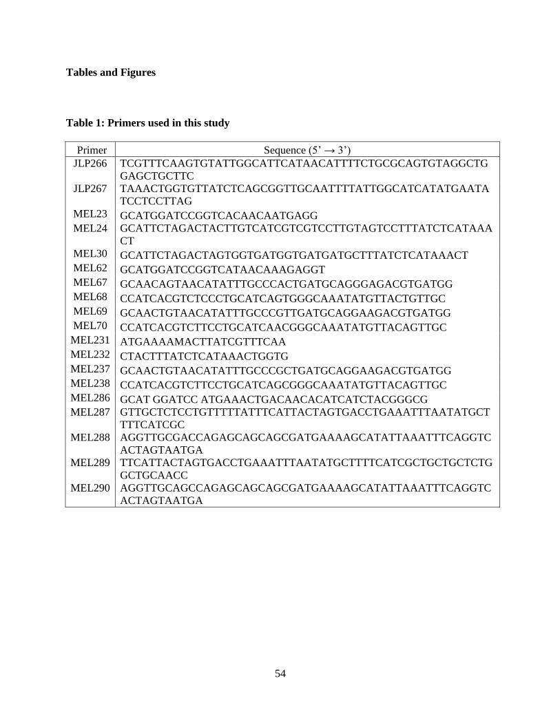
54
Tables and Figures
Table 1: Primers used in this study
Primer Sequence (5’ → 3’) JLP266 TCGTTTCAAGTGTATTGGCATTCATAACATTTTCTGCGCAGTGTAGGCTG
GAGCTGCTTC JLP267 TAAACTGGTGTTATCTCAGCGGTTGCAATTTTATTGGCATCATATGAATA
TCCTCCTTAG MEL23 GCATGGATCCGGTCACAACAATGAGG MEL24 GCATTCTAGACTACTTGTCATCGTCGTCCTTGTAGTCCTTTATCTCATAAA
CT MEL30 GCATTCTAGACTAGTGGTGATGGTGATGATGCTTTATCTCATAAACT MEL62 GCATGGATCCGGTCATAACAAAGAGGT MEL67 GCAACAGTAACATATTTGCCCACTGATGCAGGGAGACGTGATGG MEL68 CCATCACGTCTCCCTGCATCAGTGGGCAAATATGTTACTGTTGC MEL69 GCAACTGTAACATATTTGCCCGTTGATGCAGGAAGACGTGATGG MEL70 CCATCACGTCTTCCTGCATCAACGGGCAAATATGTTACAGTTGC MEL231 ATGAAAAMACTTATCGTTTCAA MEL232 CTACTTTATCTCATAAACTGGTG MEL237 GCAACTGTAACATATTTGCCCGCTGATGCAGGAAGACGTGATGG MEL238 CCATCACGTCTTCCTGCATCAGCGGGCAAATATGTTACAGTTGC MEL286 GCAT GGATCC ATGAAACTGACAACACATCATCTACGGGCG MEL287 GTTGCTCTCCTGTTTTTATTTCATTACTAGTGACCTGAAATTTAATATGCT
TTTCATCGC MEL288 AGGTTGCGACCAGAGCAGCAGCGATGAAAAGCATATTAAATTTCAGGTC
ACTAGTAATGA MEL289 TTCATTACTAGTGACCTGAAATTTAATATGCTTTTCATCGCTGCTGCTCTG
GCTGCAACC MEL290 AGGTTGCAGCCAGAGCAGCAGCGATGAAAAGCATATTAAATTTCAGGTC
ACTAGTAATGA
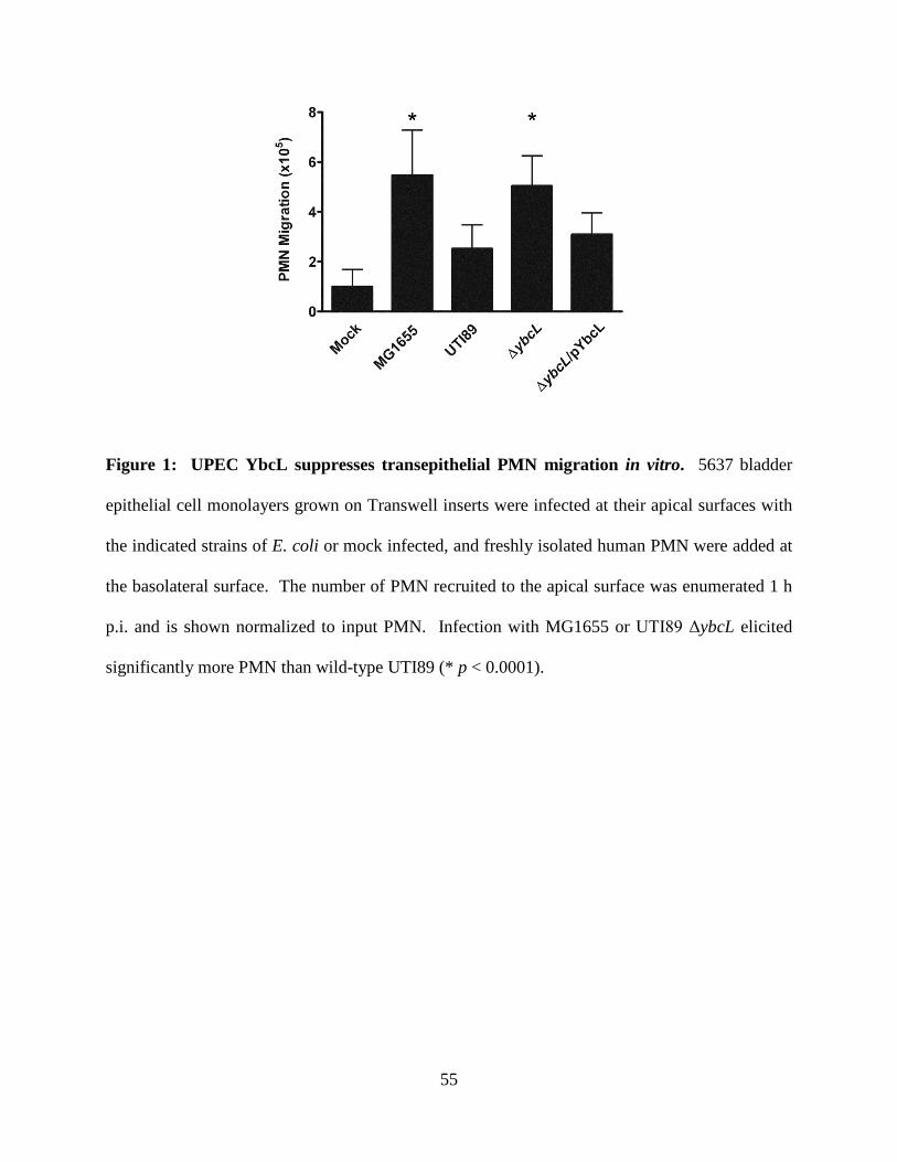
55
Figure 1: UPEC YbcL suppresses transepithelial PMN migration in vitro. 5637 bladder
epithelial cell monolayers grown on Transwell inserts were infected at their apical surfaces with
the indicated strains of E. coli or mock infected, and freshly isolated human PMN were added at
the basolateral surface. The number of PMN recruited to the apical surface was enumerated 1 h
p.i. and is shown normalized to input PMN. Infection with MG1655 or UTI89 ΔybcL elicited
significantly more PMN than wild-type UTI89 (* p < 0.0001).
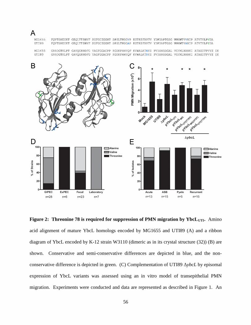
56
Figure 2: Threonine 78 is required for suppression of PMN migration by YbcLUTI. Amino
acid alignment of mature YbcL homologs encoded by MG1655 and UTI89 (A) and a ribbon
diagram of YbcL encoded by K-12 strain W3110 (dimeric as in its crystal structure (32)) (B) are
shown. Conservative and semi-conservative differences are depicted in blue, and the non-
conservative difference is depicted in green. (C) Complementation of UTI89 ΔybcL by episomal
expression of YbcL variants was assessed using an in vitro model of transepithelial PMN
migration. Experiments were conducted and data are represented as described in Figure 1. An
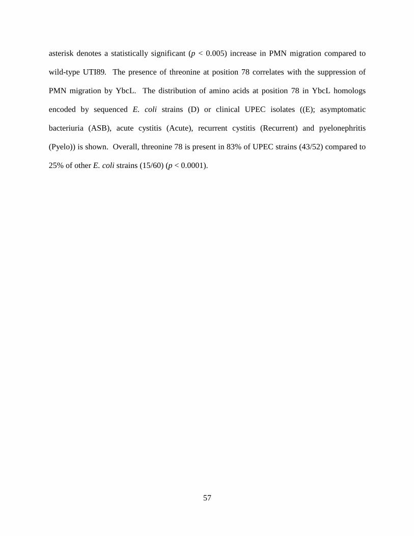
57
asterisk denotes a statistically significant (p < 0.005) increase in PMN migration compared to
wild-type UTI89. The presence of threonine at position 78 correlates with the suppression of
PMN migration by YbcL. The distribution of amino acids at position 78 in YbcL homologs
encoded by sequenced E. coli strains (D) or clinical UPEC isolates ((E); asymptomatic
bacteriuria (ASB), acute cystitis (Acute), recurrent cystitis (Recurrent) and pyelonephritis
(Pyelo)) is shown. Overall, threonine 78 is present in 83% of UPEC strains (43/52) compared to
25% of other E. coli strains (15/60) (p < 0.0001).
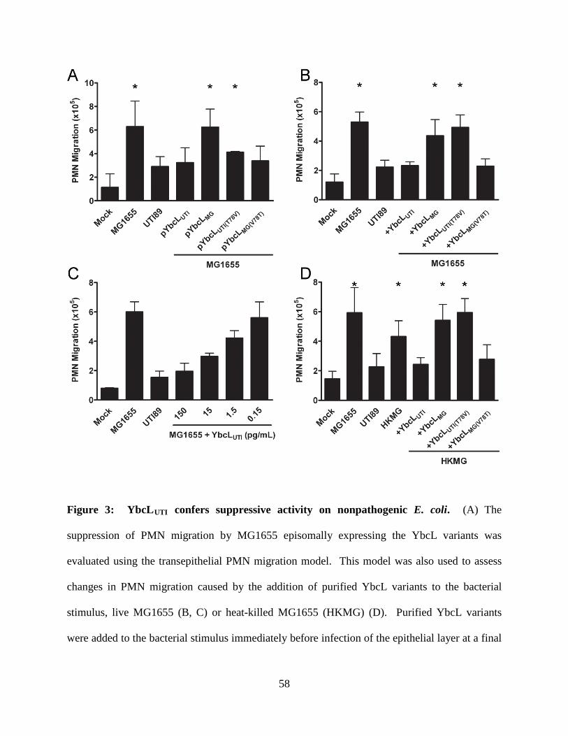
58
Figure 3: YbcLUTI confers suppressive activity on nonpathogenic E. coli. (A) The
suppression of PMN migration by MG1655 episomally expressing the YbcL variants was
evaluated using the transepithelial PMN migration model. This model was also used to assess
changes in PMN migration caused by the addition of purified YbcL variants to the bacterial
stimulus, live MG1655 (B, C) or heat-killed MG1655 (HKMG) (D). Purified YbcL variants
were added to the bacterial stimulus immediately before infection of the epithelial layer at a final
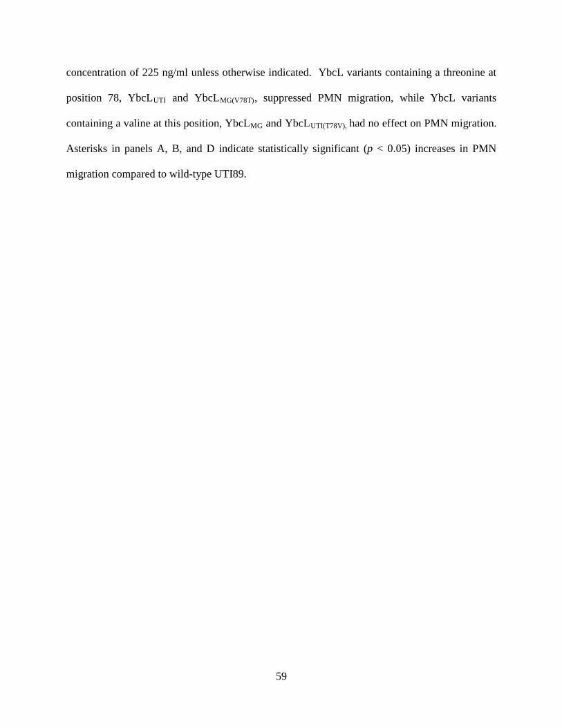
59
concentration of 225 ng/ml unless otherwise indicated. YbcL variants containing a threonine at
position 78, YbcLUTI and YbcLMG(V78T), suppressed PMN migration, while YbcL variants
containing a valine at this position, YbcLMG and YbcLUTI(T78V), had no effect on PMN migration.
Asterisks in panels A, B, and D indicate statistically significant (p < 0.05) increases in PMN
migration compared to wild-type UTI89.
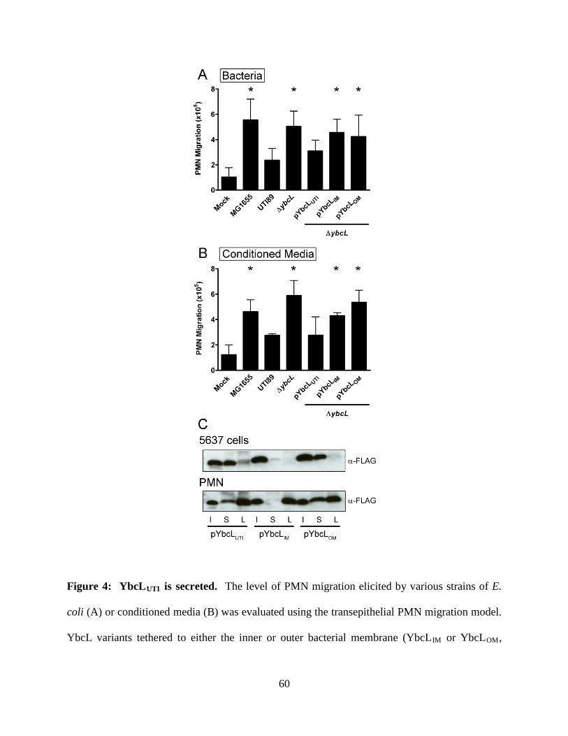
60
Figure 4: YbcLUTI is secreted. The level of PMN migration elicited by various strains of E.
coli (A) or conditioned media (B) was evaluated using the transepithelial PMN migration model.
YbcL variants tethered to either the inner or outer bacterial membrane (YbcLIM or YbcLOM,
61
respectively) were unable to suppress PMN migration when expressed episomally in UTI89
ΔybcL. The trends in PMN migration observed with conditioned media mimicked those
observed when the inoculum included live bacteria. Asterisks in panels A and B indicate
statistically significant (p < 0.05) increases in PMN migration compared to wild-type UTI89.
(C) Localization of YbcL variants was assessed by Western blot. After 1 h infection of 5637
cells or PMN with the indicated strains of E. coli, the supernatant (S) and eukaryotic cell lysate
(L) fractions were filter sterilized, TCA precipitated and resolved by SDS-PAGE. During
infection of either cell type, YbcLUTI and YbcLOM were clearly detected in the supernatant
fractions, while YbcLIM was minimally detected in those fractions. All three variants were
detected in the PMN lysate; however, only YbcLUTI was detected in the 5637 cell lysate. An
equivalent volume of each bacterial inoculum (I) is shown for comparison across strains.
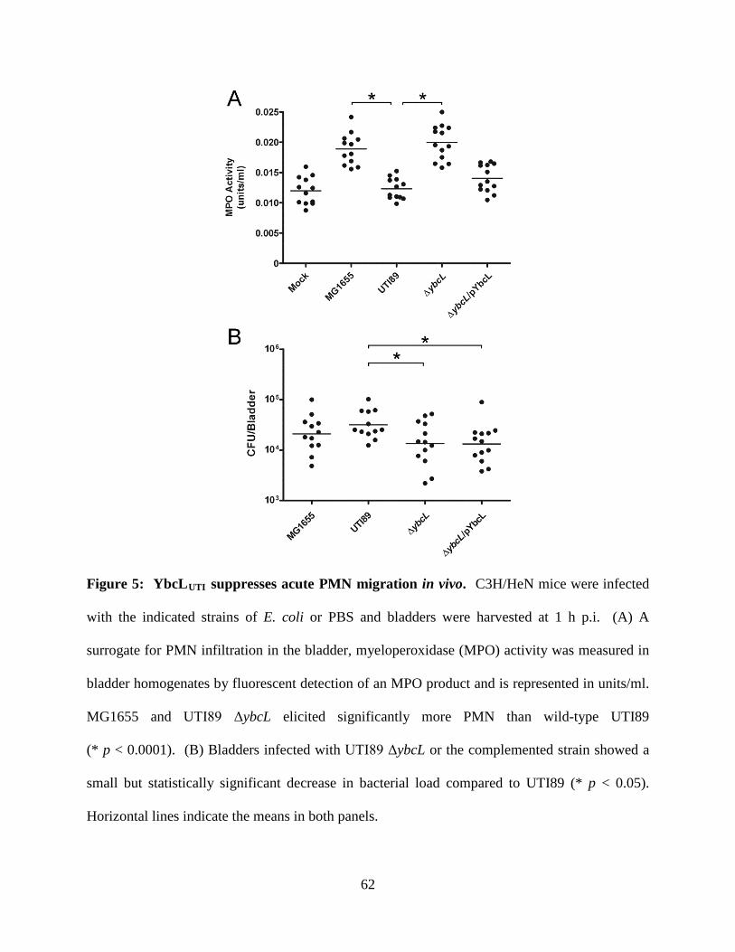
62
Figure 5: YbcLUTI suppresses acute PMN migration in vivo. C3H/HeN mice were infected
with the indicated strains of E. coli or PBS and bladders were harvested at 1 h p.i. (A) A
surrogate for PMN infiltration in the bladder, myeloperoxidase (MPO) activity was measured in
bladder homogenates by fluorescent detection of an MPO product and is represented in units/ml.
MG1655 and UTI89 ΔybcL elicited significantly more PMN than wild-type UTI89
(* p < 0.0001). (B) Bladders infected with UTI89 ΔybcL or the complemented strain showed a
small but statistically significant decrease in bacterial load compared to UTI89 (* p < 0.05).
Horizontal lines indicate the means in both panels.
63
References
1. Anderson, G. G., C. C. Goller, S. Justice, S. J. Hultgren, and P. C. Seed. 2010.
Polysaccharide capsule and sialic acid-mediated regulation promote biofilm-like
intracellular bacterial communities during cystitis. Infect Immun 78:963-975.
2. Anderson, G. G., J. J. Palermo, J. D. Schilling, R. Roth, J. Heuser, and S. J. Hultgren.
2003. Intracellular bacterial biofilm-like pods in urinary tract infections. Science
301:105-107.
3. Billips, B. K., S. G. Forrestal, M. T. Rycyk, J. R. Johnson, D. J. Klumpp, and A. J.
Schaeffer. 2007. Modulation of host innate immune response in the bladder by
uropathogenic Escherichia coli. Infect Immun 75:5353-5360.
4. Blattner, F. R., G. Plunkett, 3rd, C. A. Bloch, N. T. Perna, V. Burland, M. Riley, J.
Collado-Vides, J. D. Glasner, C. K. Rode, G. F. Mayhew, J. Gregor, N. W. Davis, H. A.
Kirkpatrick, M. A. Goeden, D. J. Rose, B. Mau, and Y. Shao. 1997. The complete
genome sequence of Escherichia coli K-12. Science 277:1453-1474.
5. Brzuszkiewicz, E., H. Bruggemann, H. Liesegang, M. Emmerth, T. Olschlager, G. Nagy,
K. Albermann, C. Wagner, C. Buchrieser, L. Emody, G. Gottschalk, J. Hacker, and U.
Dobrindt. 2006. How to become a uropathogen: comparative genomic analysis of
extraintestinal pathogenic Escherichia coli strains. Proc Natl Acad Sci U S A 103:12879-
12884.
6. Chen, S. L., C. S. Hung, J. Xu, C. S. Reigstad, V. Magrini, A. Sabo, D. Blasiar, T. Bieri,
R. R. Meyer, P. Ozersky, J. R. Armstrong, R. S. Fulton, J. P. Latreille, J. Spieth, T. M.
Hooton, E. R. Mardis, S. J. Hultgren, and J. I. Gordon. 2006. Identification of genes
subject to positive selection in uropathogenic strains of Escherichia coli: A comparative
genomics approach. Proc Natl Acad Sci U S A 103:5977-5982.
64
7. Croxen, M. A., and B. B. Finlay. 2010. Molecular mechanisms of Escherichia coli
pathogenicity. Nat Rev Microbiol 8:26-38.
8. Datsenko, K. A., and B. L. Wanner. 2000. One-step inactivation of chromosomal genes
in Escherichia coli K-12 using PCR products. Proc Natl Acad Sci U S A 97:6640-6645.
9. Davis, J. M., H. M. Carvalho, S. B. Rasmussen, and A. D. O'Brien. 2006. Cytotoxic
necrotizing factor type 1 delivered by outer membrane vesicles of uropathogenic
Escherichia coli attenuates polymorphonuclear leukocyte antimicrobial activity and
chemotaxis. Infect Immun 74:4401-4408.
10. Davis, J. M., S. B. Rasmussen, and A. D. O'Brien. 2005. Cytotoxic necrotizing factor
type 1 production by uropathogenic Escherichia coli modulates polymorphonuclear
leukocyte function. Infect Immun 73:5301-5310.
11. Foxman, B. 2010. The epidemiology of urinary tract infection. Nat Rev Urol 7:653-660.
12. Garofalo, C. K., T. M. Hooton, S. M. Martin, W. E. Stamm, J. J. Palermo, J. I. Gordon,
and S. J. Hultgren. 2007. Escherichia coli from urine of female patients with urinary tract
infections is competent for intracellular bacterial community formation. Infect Immun
75:52-60.
13. Haraoka, M., L. Hang, B. Frendus, G. Godaly, M. Burdick, R. Strieter, and C. Svanborg.
1999. Neutrophil recruitment and resistance to urinary tract infection. J Infect Dis
180:1220-1229.
14. Henson, P. M., and Z. G. Oades. 1975. Stimulation of human neutrophils by soluble and
insoluble immunoglobulin aggregates. Secretion of granule constituents and increased
oxidation of glucose. J Clin Invest 56:1053-1061.
65
15. Hilbert, D. W., K. E. Pascal, E. K. Libby, E. Mordechai, M. E. Adelson, and J. P. Trama.
2008. Uropathogenic Escherichia coli dominantly suppress the innate immune response
of bladder epithelial cells by a lipopolysaccharide- and Toll-like receptor 4-independent
pathway. Microbes Infect 10:114-121.
16. Hung, C. S., K. W. Dodson, and S. J. Hultgren. 2009. A murine model of urinary tract
infection. Nat Protoc 4:1230-1243.
17. Hunstad, D. A., and S. S. Justice. 2010. Intracellular lifestyles and immune evasion
strategies of uropathogenic Escherichia coli. Annu Rev Microbiol 64:203-221.
18. Hunstad, D. A., S. S. Justice, C. S. Hung, S. R. Lauer, and S. J. Hultgren. 2005.
Suppression of bladder epithelial cytokine responses by uropathogenic Escherichia coli.
Infect Immun 73:3999-4006.
19. Justice, S. S., C. Hung, J. A. Theriot, D. A. Fletcher, G. G. Anderson, M. J. Footer, and S.
J. Hultgren. 2004. Differentiation and developmental pathways of uropathogenic
Escherichia coli in urinary tract pathogenesis. Proc Natl Acad Sci U S A 101:1333-1338.
20. Klumpp, D. J., A. C. Weiser, S. Sengupta, S. G. Forrestal, R. A. Batler, and A. J.
Schaeffer. 2001. Uropathogenic Escherichia coli potentiates type 1 pilus-induced
apoptosis by suppressing NF-κB. Infect Immun 69:6689-6695.
21. Lipschutz, J. H., L. E. O'Brien, Y. Altschuler, D. Avrahami, Y. Nguyen, K. Tang, and K.
E. Mostov. 2001. Analysis of membrane traffic in polarized epithelial cells. Curr Protoc
Cell Biol Chapter 15:Unit 15.5.
22. Lorenz, K., M. J. Lohse, and U. Quitterer. 2003. Protein kinase C switches the Raf kinase
inhibitor from Raf-1 to GRK-2. Nature 426:574-579.
66
23. Loughman, J. A., and D. A. Hunstad. 2011. Attenuation of human neutrophil migration
and function by uropathogenic bacteria. Microbes Infect 13:555-565.
24. Martinez, J. J., M. A. Mulvey, J. D. Schilling, J. S. Pinkner, and S. J. Hultgren. 2000.
Type 1 pilus-mediated bacterial invasion of bladder epithelial cells. EMBO J 19:2803-
2812.
25. Mulvey, M. A., Y. S. Lopez-Boado, C. L. Wilson, R. Roth, W. C. Parks, J. Heuser, and
S. J. Hultgren. 1998. Induction and evasion of host defenses by type 1-piliated
uropathogenic Escherichia coli. Science 282:1494-1497.
26. Mulvey, M. A., J. D. Schilling, and S. J. Hultgren. 2001. Establishment of a persistent
Escherichia coli reservoir during the acute phase of a bladder infection. Infect Immun
69:4572-4579.
27. Murphy, K. C., and K. G. Campellone. 2003. Lambda Red-mediated recombinogenic
engineering of enterohemorrhagic and enteropathogenic E. coli. BMC Mol Biol 4:11.
28. Mysorekar, I. U., and S. J. Hultgren. 2006. Mechanisms of uropathogenic Escherichia
coli persistence and eradication from the urinary tract. Proc Natl Acad Sci U S A
103:14170-14175.
29. Nicholson, T. F., K. M. Watts, and D. A. Hunstad. 2009. OmpA of uropathogenic
Escherichia coli promotes postinvasion pathogenesis of cystitis. Infect Immun 77:5245-
5251.
30. Rosen, D. A., T. M. Hooton, W. E. Stamm, P. A. Humphrey, and S. J. Hultgren. 2007.
Detection of intracellular bacterial communities in human urinary tract infection. PLoS
Med 4:e329.
67
31. Schilling, J. D., R. G. Lorenz, and S. J. Hultgren. 2002. Effect of trimethoprim-
sulfamethoxazole on recurrent bacteriuria and bacterial persistence in mice infected with
uropathogenic Escherichia coli. Infect Immun 70:7042-7049.
32. Serre, L., K. Pereira de Jesus, C. Zelwer, N. Bureaud, F. Schoentgen, and H. Benedetti.
2001. Crystal structures of YbhB and YbcL from Escherichia coli, two bacterial
homologues to a Raf kinase inhibitor protein. J Mol Biol 310:617-634.
33. Welch, R. A., V. Burland, G. Plunkett, 3rd, P. Redford, P. Roesch, D. Rasko, E. L.
Buckles, S. R. Liou, A. Boutin, J. Hackett, D. Stroud, G. F. Mayhew, D. J. Rose, S. Zhou,
D. C. Schwartz, N. T. Perna, H. L. Mobley, M. S. Donnenberg, and F. R. Blattner. 2002.
Extensive mosaic structure revealed by the complete genome sequence of uropathogenic
Escherichia coli. Proc Natl Acad Sci U S A 99:17020-17024.
34. Yamaguchi, K., F. Yu, and M. Inouye. 1988. A single amino acid determinant of the
membrane localization of lipoproteins in E. coli. Cell 53:423-432.
35. Yeung, K., T. Seitz, S. Li, P. Janosch, B. McFerran, C. Kaiser, F. Fee, K. D. Katsanakis,
D. W. Rose, H. Mischak, J. M. Sedivy, and W. Kolch. 1999. Suppression of Raf-1 kinase
activity and MAP kinase signalling by RKIP. Nature 401:173-177.
36. Yeung, K. C., D. W. Rose, A. S. Dhillon, D. Yaros, M. Gustafsson, D. Chatterjee, B.
McFerran, J. Wyche, W. Kolch, and J. M. Sedivy. 2001. Raf kinase inhibitor protein
interacts with NF-κB-inducing kinase and TAK1 and inhibits NF-κB activation. Mol Cell
Biol 21:7207-7217.
37. Zhanel, G. G., T. L. Hisanaga, N. M. Laing, M. R. DeCorby, K. A. Nichol, L. P. Palatnik,
J. Johnson, A. Noreddin, G. K. Harding, L. E. Nicolle, and D. J. Hoban. 2005. Antibiotic
resistance in outpatient urinary isolates: final results from the North American Urinary
68
Tract Infection Collaborative Alliance (NAUTICA). Int J Antimicrob Agents 26:380-
388.
38. Zhou, G., W. J. Mo, P. Sebbel, G. Min, T. A. Neubert, R. Glockshuber, X. R. Wu, T. T.
Sun, and X. P. Kong. 2001. Uroplakin Ia is the urothelial receptor for uropathogenic
Escherichia coli: evidence from in vitro FimH binding. J Cell Sci 114:4095-4103.
69
CHAPTER 3
LIBERATION OF YBCL FROM THE BACTERIAL PERIPLASM
Abstract
Uropathogenic Escherichia coli (UPEC) suppress neutrophil migration across bladder epithelia
in vitro and in vivo through the action of a conserved protein, YbcL. Using varied approaches,
we demonstrated that YbcL was released from UPEC during infection of bladder epithelial cells
and neutrophils and that liberation of YbcL from the periplasm was required for suppression of
neutrophil migration. Here we report our initial findings on the mode of YbcL release. During
in vitro infection of bladder epithelial cells, YbcL was released into the supernatant in soluble
form. Bacterial cytoplasmic proteins, GroEL and RNA Polymerase α subunit, were also detected
in the supernatant, suggesting partial lysis of the bacterial inoculum during infection. The
involvement of a type 2 secretion system (T2SS) or a type 4 pilus system (T4PS) in YbcL release
was excluded, as the level of YbcL in the supernatant was unaffected by mutation of the outer
membrane components of these systems. Development of a β–lactamase (TEM-1) reporter assay
facilitated the characterization of YbcL release. YbcL::TEM-1, a translational fusion, was
released by UPEC into the supernatant in a dose-, time-, and bladder epithelial cell-dependent
fashion. Another periplasmic fusion, Skp::TEM-1, was detected in the supernatant; however, a
cytoplasmic fusion, Gst::TEM-1, was noticeably absent. Taken together, these findings begin to
illuminate the manner by which YbcL is released from the periplasm during UPEC infection of
eukaryotic cells.
Introduction
70
Uropathogenic Escherichia coli (UPEC) manipulate the acute inflammatory response in
the bladder, favoring the formation of the intracellular niche that propagates the infection over
bacterial clearance by immune cells. An essential aspect of the innate immune response,
migration of polymorphonuclear leukocytes (PMN; neutrophils) across the bladder epithelium
into the lumen, is suppressed by a conserved UPEC protein YbcL (1). YbcL contains a
canonical signal sequence and is targeted initially to the periplasm by the Sec system located in
the inner membrane. Using a number of different approaches, we previously demonstrated that
YbcL was released from the periplasm during UPEC infection (1). In contrast to the wild-type
YbcL variant, YbcL variants that were unable to leave the periplasm did not complement the
ybcL mutation in the transuroepithelial PMN migration assay. Additionally, purified YbcL
protein suppressed PMN migration elicited by live and heat-killed bacterial stimuli as well as a
peptide chemoattractant. YbcL was not detected in the supernatant during logarithmic phase
growth of UPEC in nutrient rich media. However, YbcL was detected in the supernatant during
UPEC infection of cultured bladder epithelial cells and PMN. Taken together, these data
demonstrate that YbcL functions to suppress PMN migration from outside the bacterial
periplasm. However, the mode of delivery of YbcL remains unclear.
Secretion, the transport of molecules (e.g., DNA and proteins) from the interior of the
bacterial cell to the external environment, is a fundamental process of bacterial survival as well
as pathogenesis. Gram-negative bacteria employ a variety of complex secretion systems. For
example, type three secretion systems (T3SS), sometimes referred to as injectisomes or
molecular syringes, are multiprotein complexes that span the inner and outer bacterial
membranes delivering proteins directly from the bacterial cytoplasm into eukaryotic cells (2).
Type four secretion systems (T4SS) are homologous to conjugation machinery and can transport
71
both DNA and protein (3). In contrast to T3SS, T4SS transport proteins from either the
cytoplasm or the periplasm across the outer membrane (4). Unlike other bacterial pathogens,
UPEC do not encode T3SS or T4SS. Like T3SS, type one secretion systems (T1SS) are also
composed of a contiguous channel that translocates proteins from the cytoplasm to the
extracellular space in a single step (5). Discovered recently, type six secretion systems (T6SS)
deliver proteins to either prokaryotic or eukaryotic cells in a contact-dependent manner (6).
Preliminary work suggests that T6SS substrates transit the apparatus in a one-step mechanism
that avoids periplasmic intermediates. For this reason, neither T1SS nor T6SS mediate release of
YbcL. Autotransporters, or type five secretion systems (T5SS), are typically multi-domain
proteins that rely on the Sec machinery to reach the periplasm. The C-terminus of the
autotransporter, after insertion into the bacterial outer membrane, transports the passenger
domain across the membrane where it is cleaved and released (7, 8). The crystal structure of
YbcL demonstrates that it is a globular protein and is unlikely to be a member of the T5SS
family (9).
Type two secretion systems (T2SS) transport periplasmic proteins across the outer
membrane through the secretin, a complex composed of a multimeric protein (10, 11). Type four
pilus systems (T4PS) are structurally and functionally related to T2SS and have been shown, in
some cases, to function as secretion systems (12, 13). Both T2SS and T4PS have been
implicated in UPEC pathogenesis (14). Lastly, outer membrane vesicles (OMVs) are liberated
by all Gram-negative bacteria and have been shown to deliver effector proteins of several
pathogens, including UPEC, to eukaryotic cells (15-17). OMVs form when portions of the outer
membrane pinch off and are composed of periplasmic contents surrounded by a lipid outer
membrane bilayer (15). These secretion systems, T2SS/T4PS and OMVs, can be distinguished
72
by a characteristic of the secreted proteins, exposed versus protected from external elements,
which may have implications for protein stability and targeting, for example. T2SS, T4PS, and
OMVs contribute to colonization of the urinary tract by UPEC (14, 17).
In this study, we report our initial findings on the mode of delivery of YbcL from the
bacterial periplasm during UPEC infection. The wild-type YbcL variant was released into the
supernatant in a soluble form but was not dependent upon the T2SS or T4PS known to be
encoded by the UPEC strain UTI89. By Western blot, bacterial cytoplasmic proteins were also
detected in the supernatant during UPEC infection of bladder epithelial cells. Using a β-
lactamase reporter assay, we demonstrated that release of YbcL::TEM-1, a translational fusion,
into the supernatant was dose-, time-, and bladder epithelial cell-dependent. Finally, Skp::TEM-
1, another periplasmic fusion, was present in the supernatant, but Gst::TEM-1, a cytoplasmic
fusion, was not. These results suggest that release of YbcL from the periplasm is not mediated
by a canonical secretion system, but rather through the release of intracellular contents into the
supernatant, possibly in a selective fashion.
Materials and Methods
Bacterial strains and culture.
E. coli strains were grown statically in Luria-Bertani (LB) broth for 18 h at 37°C.
Chloramphenicol and isopropyl β-D-1-thiogalactopyranoside (IPTG) were added at 20 µg/ml
and 100 µM, respectively, where indicated. UTI89 is a UPEC isolate from a patient with
cystitis (18). UTI89 ybcL::cat, UTI89/pYbcLUTI ybcL::cat, and UTI89/pYbcLOM ybcL::cat
were created as previously described (1). UTI89 yheF::cat was created by linear transformation
of UTI89/pKM208 (19) with a fragment amplified from template plasmid pKD3 (20) using the
73
primers MEL296 and MEL297 (primer sequences are given in Table 1). UTI89 hofQ::cat was
created similarly using primers MEL300 and MEL301. The deletions were verified by direct
sequencing.
Plasmid pTRC99A-Chl was generated from plasmid pTRC99A by replacing the
ampicillin resistance cassette with a chloramphenicol resistance cassette. Briefly, pTRC99A was
amplified using primers MEL245 and MEL246, and the chloramphenicol resistance cassette was
amplified from template plasmid pKD3 (20) using primers MEL247 and MEL248. The PCR
products were digested with PacI and SpeI and then ligated. Transformed clones of E. coli
Top10 (Invitrogen) were selected on chloramphenicol plates and tested by colony PCR and
restriction enzyme digestion. To assess release of YbcL from the bacterial periplasm using a β–
lactamase substrate, we generated a fusion protein between YbcL and TEM-1, a β–lactamase
variant, including a FLAG tag at the C-terminus. The ybcL gene was amplified from UTI89
genomic DNA using primers MEL253 and MEL275 and digested with SacI and NotI; TEM-1
sequence was amplified from plasmid pBR322 using primers MEL278 and MEL284 and
digested with NotI and BamHI. The digested PCR products were ligated into pTRC99A-Chl
(Chlr) that had been digested with SacI and BamHI. We also generated control fusion proteins,
Skp::TEM-1 and Gst::TEM-1, that localize to the periplasm and cytoplasm, respectively. Using
a similar strategy, skp and gst were amplified from UTI89 genomic DNA using primer sets
MEL285, 273 and MEL261, 277, respectively. The PCR products were cloned into pTRC99A-
Chl as described above. Accuracy of the resulting constructs, pYbcL::TEM-1, pSkp::TEM-1 and
pGst::TEM-1, was confirmed by direct sequencing. Expression of the fusion proteins upon
IPTG induction was confirmed by sodium dodecyl sulfate-polyacrylamide gel electrophoresis
(SDS-PAGE) and Western blot analysis of bacterial lysates.
74
Tissue culture.
The 5637 bladder epithelial cell line (derived from bladder carcinoma; ATCC HTB-9) was
obtained from the American Type Culture Collection. Cells were cultured in RPMI 1640 media
(Gibco) supplemented with 10% fetal bovine serum (Sigma) at 37°C in a humidified atmosphere
with 5% CO2 unless otherwise noted.
Protease protection and ultracentrifugation assays.
The protease protection assay was developed based on previously published protocols (17, 21).
To generate sterile conditioned media containing YbcL variants, 5637 cells in 15-cm dishes were
infected with UTI89/pYbcLUTI ybcL::cat or UTI89/pYbcLOM ybcL::cat at a MOI of 40 at 37°C.
After 1 h, the supernatant, or conditioned media, was sterilized using syringe-driven filter units
(0.22µm pore size, Millipore), and separated into four aliquots. Proteinase K (Sigma), Triton X-
100, and phenylmethylsulfonyl fluoride (PMSF) (Sigma), a protease inhibitor, were added to the
sterile conditioned media at 200 µg/ml, 0.1%, and 5 mM, respectively, where indicated. After
incubation at 37°C for 45 min, protein was precipitated from the reactions using 15%
trichloroacetic acid (TCA) (Sigma).
In parallel experiments, sterile conditioned media, generated as described above, was
ultracentrifuged at 245,000 × g for 1 h at 4°C. Protein was precipitated from control conditioned
media and the ultracentrifuged supernatant using 15% TCA (Sigma), while the ultracentrifuged
pellet was resuspended using Laemmli sample buffer.
Western blotting.
75
Proteins were separated by SDS-PAGE using 9-12% polyacrylamide gels and transferred to
polyvinylidene difluoride (PVDF) membrane (Millipore). After blocking with 2% nonfat milk +
2% bovine serum albumin (BSA) (Sigma), blots were probed with mouse anti-FLAG (1:1000,
Sigma), rabbit anti-GroEL (1:400000, Sigma) or mouse anti-RNA Polymerase α subunit (1:4000,
Neoclone) antibodies followed by goat anti-mouse or goat anti-rabbit IgG antibodies (1:2000,
Sigma) and were developed using Tropix CDP-Star (Applied Biosystems).
β-lactamase reporter assays.
5637 cells in 6-well plates were infected with the indicated strains of E. coli at a MOI of 40 at
37°C. After 1 h, the supernatants were cleared by centrifugation at 16,000 x g for 5 min, and
then moved to clean microfuge tubes. The cleared supernatant was aliquotted into a 96-well
plate, 200 µl per well in triplicate, and CCF2-FA (Invitrogen), a β–lactamase substrate, was
added at a final concentration of 500 nM. The reactions were incubated in the dark at 37°C with
shaking for 1 h, before fluorescence (excitation at 409 nm and emission at 447 nm and 520 nm)
was measured using a microtiter plate reader (Synergy 2; BioTek). Fluorescence is represented
as a ratio of 447 nm to 520 nm, and the mean and standard deviation of replicates is shown.
Statistically significant differences were evaluated using an unpaired Student’s t test.
In analogous experiments, 5637 cells in 15-cm dishes were infected with the indicated
strains of E. coli for 1 h at 37°C. The supernatant was sterilized using syringe-driven filter units
(0.22µm pore size, Millipore), and concentrated 10-fold, from 9 ml to 0.9 ml, using centrifugal
filter units according to the manufacturer’s instructions (10 kDa, Millipore). CCF2 was added to
200 µl concentrated supernatant and fluorescence was measured after 1 h at 37°C as described
above. The mean and standard deviation of replicates is shown.
76
Results
YbcL is released into the supernatant in soluble form.
To explore the mode of delivery of YbcL from the bacterial periplasm, we examined the
characteristics of the extracellular fraction of YbcL. To clarify the relationship between YbcL
localization and suppression of PMN migration, we included a bacterial outer membrane-
tethered YbcL variant in these analyses (1). 5637 cells were infected with the ybcL mutant
expressing either the wild-type UTI89 YbcL variant, YbcLUTI, or the outer membrane-tethered
YbcL variant, YbcLOM, at a MOI of 40 for 1 h. The supernatant, or conditioned media, was
filter sterilized and subjected to protease (Proteinase K) treatment in the presence and absence of
detergent (Triton X-100) and protease inhibitor (PMSF). Protein was precipitated from the
reactions using 15% TCA and resolved using SDS-PAGE. The wild-type YbcL variant was
susceptible to digestion by protease in the absence of detergent (Figure 1A). In contrast, the
outer membrane-tethered YbcL variant was present in 2 distinct fractions: one fraction was
susceptible to degradation by protease in the absence of detergent, while the second fraction was
only susceptible in the presence of detergent (Figure 1A). Levels of both YbcL variants were
restored upon addition of protease inhibitor. The concentration of Proteinase K used in these
reactions did not compromise OMV integrity in the absence of detergent, assessed using purified
OMVs (data not shown).
To confirm these observations using an alternative approach, we ultracentrifuged sterile
conditioned media, at a speed previously determined to pellet OMVs, to separate soluble and
OMV fractions. The pellet, or insoluble fraction, was resuspended in Laemmli buffer, while
protein remaining in the supernatant after ultracentrifugation, the soluble fraction, was
precipitated using 15% TCA. Samples were resolved as described above. YbcLUTI was found
77
exclusively in the ultracentrifuged supernatant, while YbcLOM was found in both the
ultracentrifuged supernatant and pellet (Figure 1B). The fraction of YbcLOM that is resistant to
protease digestion in the absence of detergent and is present in the ultracentrifuged pellet is
consistent with OMV cargo. In contrast, these data indicate that the wild-type YbcL variant is
released into the supernatant in free, soluble form and is not packaged within OMVs.
Bacterial intracellular contents are released during infection of bladder epithelial cells.
The origin of YbcLOM in the conditioned media that was susceptible to protease in the
absence of detergent and present in the ultracentrifuged supernatant was unclear, given the
covalent bond tethering YbcLOM to the inner face of the bacterial outer membrane. We
hypothesized that a low level of bacterial lysis occurred during infection of the bladder epithelial
cells and that bacterial lysis supplied this fraction of YbcLOM. To address this hypothesis, we
infected bladder epithelial cells with UTI89/pYbcLUTI ybcL::cat as described above and resolved
the filter sterilized, TCA precipitated supernatant by SDS-PAGE. In addition to YbcLUTI,
bacterial cytoplasmic proteins GroEL and the α subunit of RNA Polymerase were detected in the
supernatant (Figure 2). To ensure that the cytoplasmic proteins present in the supernatant were
the result of bacterial lysis and not carryover from the bacterial overnight culture, the bacterial
inoculum was washed repeatedly in PBS before infection of the bladder epithelial cells. The
presence of bacterial cytoplasmic proteins in the supernatant may suggest that UPEC lyse and
release internal contents, including YbcLUTI, during infection of bladder epithelial cells.
To support this hypothesis and eliminate a role for the T2SS or T4PS in release of YbcL
from the periplasm, we generated insertional mutations in the outer membrane secretin
components of these systems in UTI89, yheF and hofQ, respectively. We found that the level of
78
YbcL in the supernatant, assessed by Western blot of filter sterilized, TCA precipitated
supernatant, was not decreased in the absence of either secretin (data not shown). These results
were confirmed using the β-lactamase reporter assay described below (data not shown). These
data suggest that neither the T2SS nor the T4PS encoded by UTI89 mediates delivery of YbcL
into the supernatant and support the hypothesis that YbcL may be released via bacterial lysis.
YbcL is released from the periplasm in a dose-, time- and bladder epithelial cell-dependent
manner.
To further examine release of YbcL into the supernatant by UPEC, we developed an
alternative, quantitative assay with increased accuracy and reproducibility that relies on the
enzyme activity of β–lactamase (TEM-1). By generating a translational fusion between full-
length YbcL and the mature form of TEM-1 (denoted YbcL::TEM-1), the level of β–lactamase
activity in the supernatant can be used as a surrogate for the amount of YbcL::TEM-1 present.
To detect β–lactamase activity, we chose a fluorescent substrate, CCF2, which consists of a
cephalosporin core linking a 7-hydroxycoumarin to a fluorescein. In the absence of β-lactamase
activity, excitation of the coumarin at 409 nm results in FRET to the fluorescein which emits a
green signal (520 nm). In the presence of β-lactamase activity, CCF2 is cleaved, spatially
separating the two fluorophores such that excitation of the coumarin results in emission of a blue
fluorescent signal (447 nm). In accordance with previous experiments, 5637 cells in 6-well
plates were infected with UTI89 pYbcL::TEM-1 at a MOI 40 for 1 h, unless otherwise indicated,
before the supernatant was cleared by centrifugation and aliquotted into a 96-well plate. CCF2
was added to each well at a final concentration of 500 nM. Fluorescence was measured after 1 h
at 37°C and is represented as a ratio of blue to green fluorescence (447 nm/520 nm).
79
5637 cells were infected with UTI89 pYbcL::TEM-1 induced overnight with varying
concentrations of IPTG. Measurement of β-lactamase activity in the cleared supernatant
revealed that the level of fluorescence positively correlated with the level of IPTG induction
(Figure 3A). Therefore, the extent to which YbcL::TEM-1 is released into the supernatant is
dependent upon the amount present in the bacterial periplasm. When 5637 cells were infected
with UTI89 pYbcL::TEM-1 for varying times before the supernatant was cleared, the level of
fluorescence in the supernatant increased with increasing duration of infection (Figure 3B).
These data suggest that liberation of YbcL::TEM-1 from the bacterial periplasm occurs over
time. Finally, UTI89 pYbcL::TEM-1 was incubated in the presence and absence of 5637 cells
for 1 h before the level of YbcL::TEM-1 in the supernatant was evaluated using the β-lactamase
substrate. Significantly higher levels of fluorescence were detected in supernatant generated in
the presence of bladder epithelial cells compared to supernatant generated in the absence of
epithelial cells (Figure 3C; p < 0.0001). These data suggest that release of YbcL::TEM-1 occurs
either as a bacterial response to or as a consequence of exposure to bladder epithelial cells.
Taken together, these data demonstrate that YbcL::TEM-1 is released from the periplasm in a
dose-, time- and bladder epithelial cell-dependent manner.
Release of bacterial intracellular contents may be selective.
The presence of bacterial cytoplasmic proteins in the supernatant suggested that
intracellular contents were released, possibly via lysis, during UPEC infection of 5637 cells. To
evaluate the ability of the β-lactamase reporter assay to detect the release of intracellular
contents, we generated 2 additional fusion proteins, Skp::TEM-1 and Gst::TEM-1, that localize
to the bacterial periplasm and the cytoplasm, respectively, and expressed these fusions in UTI89.
80
Initial attempts to detect Skp::TEM-1 and Gst::TEM-1 in cleared supernatant after 1 h infection
of 5637 cells in 6-well plates using CCF2 were unsuccessful (data not shown). The absence of a
fluorescent signal could indicate that these fusion proteins were not released into the supernatant
or that their levels in the supernatant were too low to detect using the initial experimental
parameters. To distinguish between these possibilities, we modified the protocol.
5637 cells in 15-cm dishes were infected with the indicated strains of E. coli at a MOI of
40 for 1 h. Then, the supernatant was filter sterilized and concentrated 10-fold using centrifugal
filter units with a 10 kDa cutoff. Control supernatant (input) and concentrated supernatant
(retentate) were assayed for β-lactamase activity using CCF2 as described above. Upon
concentration, Skp::TEM-1 was detected in the supernatant, although the level of fluorescence
was lower than in retentate containing YbcL::TEM-1 (Figure 4). This disparity is likely due to
differences in expression of the 2 fusion proteins (data not shown). In contrast, Gst::TEM-1 was
not detected in the supernatant even after this 10-fold concentration (Figure 4). The absence of
Gst::TEM-1 from the supernatant cannot be explained by low expression levels as Gst::TEM-1
expressed to levels similar to YbcL::TEM-1. In agreement with the β-lactamase reporter assay,
Skp::TEM-1 and YbcL::TEM-1, but not Gst::TEM-1, were detected in filter sterilized, TCA
precipitated supernatant by Western blot (data not shown). The presence of periplasmic proteins,
Skp and YbcL, and the absence of cytoplasmic protein, Gst, from the supernatant suggests that
the release of intracellular contents from bacteria may be selective. Future work will attempt to
clarify these observations and elucidate the mechanism underlying release of YbcL from the
bacterial periplasm.
Discussion
81
In this study, we investigated the mode of release of YbcL from the bacterial periplasm
during UPEC infection of bladder epithelial cells. We demonstrated that YbcL was released into
the supernatant in soluble form. Given this finding, we developed a β-lactamase reporter assay
to further probe the dynamics of YbcL release. We demonstrated that the level of YbcL in the
supernatant was influenced by the amount of YbcL in the periplasm, the duration of the
infection, and the presence or absence of bladder epithelial cells. Additionally, we were able to
detect a second, unrelated periplasmic protein in the supernatant. However, detection of
cytoplasmic proteins in the supernatant was variable. In total, these findings inform our
understanding of how bacterial exoproteins, specifically YbcL, can be released into the
extracellular milieu. Additional work is required to clarify these observations.
O’Brien and colleagues demonstrated that cytotoxic necrotizing factor 1 (Cnf1) was
initially targeted to the periplasm, subsequently packaged into OMVs, and then delivered to
PMN (17). In contrast to purified Cnf1, Cnf1 in complex with OMVs attenuated PMN function,
suggesting that delivery via OMVs was required for Cnf1 to reach the eukaryotic cytoplasm
where it exerts its effect (17). In contrast, wild-type YbcL did not appear to be packaged into
OMVs, as it was susceptible to protease in the absence of detergent and remained in the soluble
fraction after ultracentrifugation. The outer membrane-tethered YbcL variant, on the other hand,
appeared to be at least partially packaged within OMVs, as a fraction of YbcLOM was resistant to
protease in the absence of detergent and was detected in the pellet after ultracentrifugation. As
YbcLOM was unable to complement the ybcL mutation in the transuroepithelial PMN migration
assay (1), association with OMVs, in this case, may preclude YbcL activity. As OMV cargo,
YbcLOM may be inaccessible to binding partners or unable to reach its final destination possibly
due to the lipid tether. YbcLUTI was detected in the supernatant and in bladder epithelial cell
82
lysate during UPEC infection, while YbcLOM was present only in the supernatant (1). The mode
of delivery of YbcLUTI did not aid in the determination of site of action. Additional experiments
will attempt to clarify whether YbcL localization to the supernatant or association with
eukaryotic cells is required for suppression of PMN migration.
While one fraction of YbcLOM in the supernatant was consistent with OMV cargo, the
origin of the second fraction was unclear. The presence of bacterial cytoplasmic proteins, GroEL
and RNA Polymerase α subunit, in the supernatant suggested that intracellular contents were
released during infection of bladder epithelial cells. We hypothesize that the release of
intracellular contents, possibly through bacterial lysis, is responsible for the presence of YbcLUTI
and YbcLOM in the supernatant. To support the hypothesis that bacterial lysis was solely
responsible for YbcLUTI in the supernatant by excluding other secretion systems, we performed
densitometry on the Western blots, comparing the level of GroEL in the supernatant to the level
present in the input. Using these calculations and the number of CFU in the input, we estimate
that less than 0.1% of the bacterial inoculum lysed during the 1 h infection. The ratio of YbcL in
the supernatant to YbcL in the input was less than the same ratio calculated for GroEL,
suggesting that YbcL was released into the supernatant solely via bacterial lysis and not through
an alternative or additional mechanism. Given the low level of YbcL required to suppress PMN
migration in the in vitro transuroepithelial PMN migration assay (<10 pM) (1), lysis of a
minority of the bacterial inoculum may be sufficient to release an effective amount of YbcL.
To support the hypothesis that YbcL release is mediated by bacterial lysis, we examined
the potential involvement of other secretion systems. We demonstrated previously that the level
of the wild-type YbcL variant in the supernatant was similar independent of the bacterial strain
used in the infection, MG1655 pYbcLUTI or UTI89/pYbcLUTI ybcL::cat (1), suggesting that the
83
mode of delivery of YbcLUTI is conserved between nonpathogenic and uropathogenic E. coli.
By generating mutations in the secretins of the T2SS and T4PS, loci that are highly conserved
between MG1655 and UTI89 (14), we demonstrated that neither secretion system mediated
YbcL release. These data demonstrate that YbcL does not access the extracellular space via any
of the canonical secretion systems.
To further our understanding of the parameters of YbcL release, we developed a reporter
assay that relies on the detection of β-lactamase activity in the cleared supernatant using a
fluorescent substrate. Compared to examining YbcL levels in filter sterilized, TCA precipitated
supernatant by Western blot, the reporter assay is quantitative and highly reproducible. Using a
translational fusion composed of YbcL and TEM-1, a β-lactamase variant, we demonstrated that
release of YbcL::TEM-1 into the supernatant was dependent upon the concentration of the fusion
present in the periplasm and increased over time. Furthermore, we demonstrated that release of
YbcL::TEM-1 was significantly higher in the presence of bladder epithelial cells than in their
absence. Using the β-lactamase reporter assay to examine the hypothesis that bacterial lysis
mediates YbcL release, we found that Skp::TEM-1 was present in the supernatant, but
Gst::TEM-1 was not. The release of intracellular proteins did not correlate with overall
expression, nor with molecular weight as the translational fusions are equivalent in size (50
kDa). These results may suggest that release of intracellular contents is selective, although it is
unclear how selectivity would be achieved during bacterial lysis. Additional experimentation is
required to understand these seemingly incongruent pieces of data. The β-lactamase reporter
assay will be invaluable in further exploring release of YbcL from the bacterial periplasm during
infection.
84
The results presented herein suggest that YbcL is not released from the bacterial
periplasm through a canonical secretion system or via OMVs. YbcL appears to be released
along with other intracellular proteins in a dose-, time-, and bladder epithelial cell-dependent
process. Although we cannot exclude the possibility that these observations are the result of
multiple processes acting simultaneously, we hypothesize that the release of YbcL is mediated
by bacterial lysis. Future work will attempt to determine the aspects of bladder epithelial cell
exposure that stimulate or mediate YbcL release from the periplasm.
Acknowledgements
This work was supported by National Institutes of Health (NIH) grants R01-DK080752,
R03-DK076556, and P50-DK064540. We thank K. Tiemann for technical assistance.
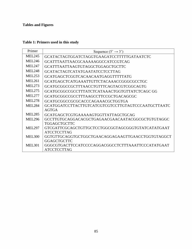
85
Tables and Figures
Table 1: Primers used in this study
Primer Sequence (5’ → 3’) MEL245 GCATACTAGTGGATCTAGGTGAAGATCCTTTTTGATAATCTC MEL246 GCATTTAATTAACGCAAAAAGGCCATCCGTCAG MEL247 GCATTTAATTAAGTGTAGGCTGGAGCTGCTTC MEL248 GCATACTAGTCATATGAATATCCTCCTTAG MEL253 GCATGAGCTCGGTCACAACAATGAGGTTTTTATG MEL261 GCATGAGCTCATGAAATTGTTCTACAAACCGGGCGCCTGC MEL273 GCATGCGGCCGCTTTAACCTGTTTCAGTACGTCGGCAGTG MEL275 GCATGCGGCCGCCTTTATCTCATAAACTGGTGTTATCTCAGC GG MEL277 GCATGCGGCCGCCTTTAAGCCTTCCGCTGACAGCGC MEL278 GCATGCGGCCGCGCACCCAGAAACGCTGGTGA MEL284 GCATGGATCCTTACTTGTCATCGTCGTCCTTGTAGTCCCAATGCTTAATC
AGTGA MEL285 GCATGAGCTCGTGAAAAAGTGGTTATTAGCTGCAG MEL296 GCCTTGTGCAGGACACGCTGAGAACGAACAATACGGCGCTGTGTAGGC
TGGAGCTGCTTC MEL297 GTCGATTCGCAGCTGTTGCTCCTGGCGGTAGCGGGTGTATCATATGAAT
ATCCTCCTTAG MEL300 GGTGTTGCAGGTGCTGGCTGAACAGGAGAAGTTGAACCTGGTGTAGGCT
GGAGCTGCTTC MEL301 GGGCGTGACTTCCATCCCCAGGACGGCCTCTTTAAATTCCCATATGAAT
ATCCTCCTTAG
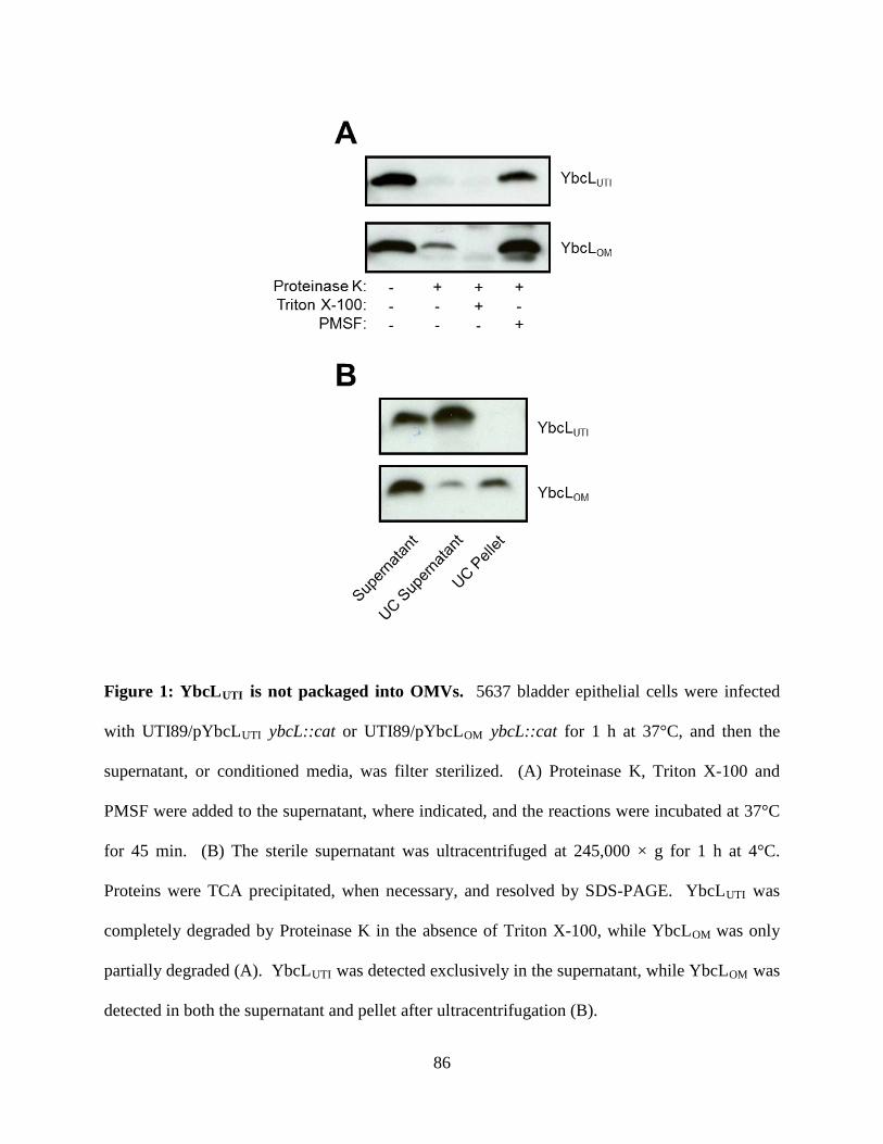
86
Figure 1: YbcLUTI is not packaged into OMVs. 5637 bladder epithelial cells were infected
with UTI89/pYbcLUTI ybcL::cat or UTI89/pYbcLOM ybcL::cat for 1 h at 37°C, and then the
supernatant, or conditioned media, was filter sterilized. (A) Proteinase K, Triton X-100 and
PMSF were added to the supernatant, where indicated, and the reactions were incubated at 37°C
for 45 min. (B) The sterile supernatant was ultracentrifuged at 245,000 × g for 1 h at 4°C.
Proteins were TCA precipitated, when necessary, and resolved by SDS-PAGE. YbcLUTI was
completely degraded by Proteinase K in the absence of Triton X-100, while YbcLOM was only
partially degraded (A). YbcLUTI was detected exclusively in the supernatant, while YbcLOM was
detected in both the supernatant and pellet after ultracentrifugation (B).
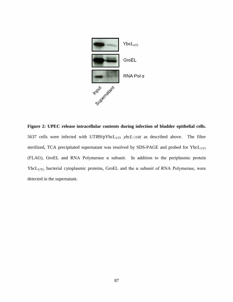
87
Figure 2: UPEC release intracellular contents during infection of bladder epithelial cells.
5637 cells were infected with UTI89/pYbcLUTI ybcL::cat as described above. The filter
sterilized, TCA precipitated supernatant was resolved by SDS-PAGE and probed for YbcLUTI
(FLAG), GroEL and RNA Polymerase α subunit. In addition to the periplasmic protein
YbcLUTI, bacterial cytoplasmic proteins, GroEL and the α subunit of RNA Polymerase, were
detected in the supernatant.
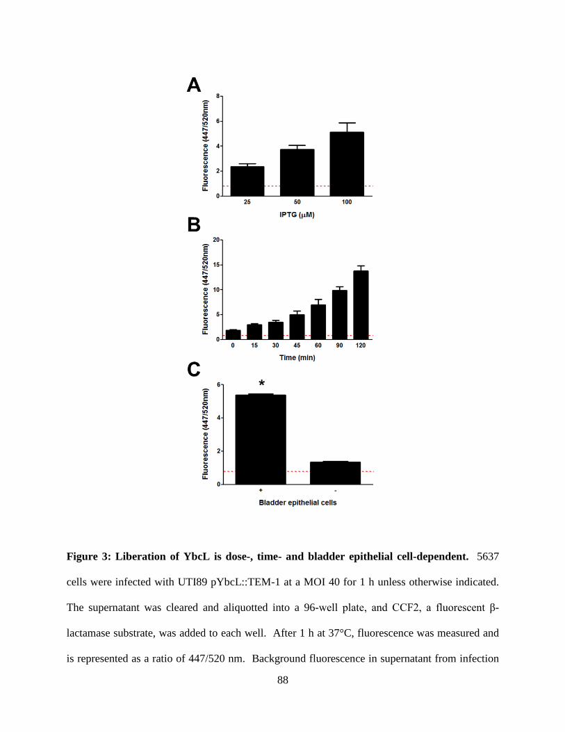
88
Figure 3: Liberation of YbcL is dose-, time- and bladder epithelial cell-dependent. 5637
cells were infected with UTI89 pYbcL::TEM-1 at a MOI 40 for 1 h unless otherwise indicated.
The supernatant was cleared and aliquotted into a 96-well plate, and CCF2, a fluorescent β-
lactamase substrate, was added to each well. After 1 h at 37°C, fluorescence was measured and
is represented as a ratio of 447/520 nm. Background fluorescence in supernatant from infection
89
of 5637 cells with wild-type UTI89 is indicated by the red dashed line. The level of β–lactamase
activity in the supernatant is dependent upon the concentration of YbcL::TEM-1 in the periplasm
(A), the duration of the infection (B), and the presence of 5637 cells (* p < 0.0001) (C).
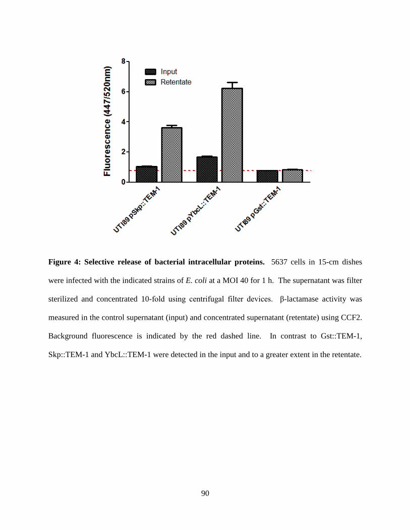
90
Figure 4: Selective release of bacterial intracellular proteins. 5637 cells in 15-cm dishes
were infected with the indicated strains of E. coli at a MOI 40 for 1 h. The supernatant was filter
sterilized and concentrated 10-fold using centrifugal filter devices. β-lactamase activity was
measured in the control supernatant (input) and concentrated supernatant (retentate) using CCF2.
Background fluorescence is indicated by the red dashed line. In contrast to Gst::TEM-1,
Skp::TEM-1 and YbcL::TEM-1 were detected in the input and to a greater extent in the retentate.
91
References
1. Lau ME, Loughman JA, Hunstad DA. 2012. YbcL of uropathogenic Escherichia coli
suppresses transepithelial neutrophil migration. Infect Immun 80:4123-4132.
2. Buttner D. 2012. Protein export according to schedule: architecture, assembly, and
regulation of type III secretion systems from plant- and animal-pathogenic bacteria.
Microbiol Mol Biol Rev 76:262-310.
3. Voth DE, Broederdorf LJ, Graham JG. 2012. Bacterial Type IV secretion systems:
versatile virulence machines. Future Microbiol 7:241-257.
4. Thanassi DG, Bliska JB, Christie PJ. 2012. Surface organelles assembled by secretion
systems of Gram-negative bacteria: diversity in structure and function. FEMS Microbiol
Rev 36:1046-1082.
5. Gentschev I, Dietrich G, Goebel W. 2002. The E. coli alpha-hemolysin secretion system
and its use in vaccine development. Trends Microbiol 10:39-45.
6. Silverman JM, Brunet YR, Cascales E, Mougous JD. 2012. Structure and regulation of
the type VI secretion system. Annu Rev Microbiol 66:453-472.
7. Henderson IR, Navarro-Garcia F, Desvaux M, Fernandez RC, Ala'Aldeen D. 2004. Type
V protein secretion pathway: the autotransporter story. Microbiol Mol Biol Rev 68:692-
744.
8. Benz I, Schmidt MA. 2011. Structures and functions of autotransporter proteins in
microbial pathogens. Int J M Microbiol 301:461-468.
9. Serre L, Pereira de Jesus K, Zelwer C, Bureaud N, Schoentgen F, Benedetti H. 2001.
Crystal structures of YBHB and YBCL from Escherichia coli, two bacterial homologues
to a Raf kinase inhibitor protein. J Mol Biol 310:617-634.
92
10. Korotkov KV, Sandkvist M, Hol WG. 2012. The type II secretion system: biogenesis,
molecular architecture and mechanism. Nat Rev Microbiol 10:336-351.
11. McLaughlin LS, Haft RJ, Forest KT. 2012. Structural insights into the Type II secretion
nanomachine. Curr Opin Struct Biol 22:208-216.
12. Mattick JS. 2002. Type IV pili and twitching motility. Annu Rev Microbiol 56:289-314.
13. Hager AJ, Bolton DL, Pelletier MR, Brittnacher MJ, Gallagher LA, Kaul R, Skerrett SJ,
Miller SI, Guina T. 2006. Type IV pili-mediated secretion modulates Francisella
virulence. Mol Microbiol 62:227-237.
14. Kulkarni R, Dhakal BK, Slechta ES, Kurtz Z, Mulvey MA, Thanassi DG. 2009. Roles of
putative type II secretion and type IV pilus systems in the virulence of uropathogenic
Escherichia coli. PLoS One 4:e4752.
15. Kulp A, Kuehn MJ. 2010. Biological functions and biogenesis of secreted bacterial outer
membrane vesicles. Annu Rev Microbiol 64:163-184.
16. Ellis TN, Kuehn MJ. 2010. Virulence and immunomodulatory roles of bacterial outer
membrane vesicles. Microbiol Mol Biol Rev 74:81-94.
17. Davis JM, Carvalho HM, Rasmussen SB, O'Brien AD. 2006. Cytotoxic necrotizing factor
type 1 delivered by outer membrane vesicles of uropathogenic Escherichia coli attenuates
polymorphonuclear leukocyte antimicrobial activity and chemotaxis. Infect Immun
74:4401-4408.
18. Chen SL, Hung CS, Xu J, Reigstad CS, Magrini V, Sabo A, Blasiar D, Bieri T, Meyer
RR, Ozersky P, Armstrong JR, Fulton RS, Latreille JP, Spieth J, Hooton TM, Mardis ER,
Hultgren SJ, Gordon JI. 2006. Identification of genes subject to positive selection in
93
uropathogenic strains of Escherichia coli: A comparative genomics approach. Proc Natl
Acad Sci U S A 103:5977-5982.
19. Murphy KC, Campellone KG. 2003. Lambda Red-mediated recombinogenic engineering
of enterohemorrhagic and enteropathogenic E. coli. BMC Mol Biol 4:11.
20. Datsenko KA, Wanner BL. 2000. One-step inactivation of chromosomal genes in
Escherichia coli K-12 using PCR products. Proc Natl Acad Sci U S A 97:6640-6645.
21. Cheng LW, Schneewind O. 2000. Yersinia enterocolitica TyeA, an intracellular regulator
of the type III machinery, is required for specific targeting of YopE, YopH, YopM, and
YopN into the cytosol of eukaryotic cells. J Bacteriol 182:3183-3190.
94
CHAPTER FOUR
EFFECTS OF YBCL ON BLADDER EPITHELIAL CELLS AND NEUTROPHILS
Abstract
Uropathogenic Escherichia coli (UPEC) manipulate the acute inflammatory response in the
bladder, delaying the arrival of immune cells and facilitating invasion into the bladder
epithelium, a requirement for the propagation of infection. Previously, we demonstrated that a
protein encoded by UPEC, YbcL, suppressed the migration of polymorphonuclear leukocytes
(PMN) or neutrophils across bladder epithelia in vitro and in vivo. To understand how UPEC
employ YbcL to influence PMN migration, we examined a number of processes required for
PMN to transit an epithelial barrier. We demonstrated that UPEC-mediated inhibition of the NF-
κB pathway, which plays a pivotal role in initiating the pro-inflammatory response during
infection, did not require YbcL. Production of PMN chemoattractants by bacteria and bladder
epithelial cells, likely in an NF-κB-independent manner, was not disrupted in the presence of
YbcL. Suppression of PMN migration by YbcL required a bladder epithelium on the Transwell
inserts; YbcL did not inhibit PMN movement elicited by chemotactic molecules. Finally,
integrity of the bladder epithelium was maintained even after high levels of PMN traversal,
confirming that PMN migration in the in vitro model of transuroepithelial PMN migration is an
active process. These findings contribute to our understanding of suppression of PMN migration
by UPEC and further validate the in vitro model as a means to investigate events that occur in
vivo.
Introduction
95
Mucosal surfaces of the human body are constantly exposed to external insults. The
epithelial and resident immune cells that comprise the mucosa constitute the first line of defense
again foreign threats such as bacterial pathogens. These cells are also responsible for initiating a
pro-inflammatory response that elicits immune effector cells to aid in bacterial clearance.
Infection of the bladder (i.e., cystitis) or kidneys (i.e., pyelonephritis), mucosal surfaces of the
urinary tract, results in a characteristic inflammatory response. Aspects of the innate immune
response to uropathogenic Escherichia coli (UPEC), the primary etiology of urinary tract
infections (UTIs), have been elucidated using a variety of techniques and validated using
samples from human patients. For example, interleukin (IL)-6, IL-8 and polymorphonuclear
leukocytes (PMN; neutrophils) can be detected in the urine of patients with cystitis (1-3).
Additionally, genetic polymorphisms in Toll-like receptors (TLRs) and IL-8 receptors within the
human population influence disease outcome (4-6), further implicating these pathways in the
innate immune response to UTI. Preliminary work has begun to characterize the acute
inflammatory response in the bladder and to define how UPEC manipulate this response to cause
disease. Given the complexity of these processes, further investigation is required.
In contrast to other mucosal surfaces like the gastrointestinal tract, the urinary tract, aside
from the urethra, has been considered devoid of resident microbes and intolerant of bacterial
colonization. Consequently, the presence of UPEC in the bladder lumen initiates a pro-
inflammatory response. Specifically, bacterial products such as lipopolysaccharide (LPS), type I
and P pili, and flagellin are recognized by pathogen recognition receptors (PRRs) including
TLR4 and TLR5 expressed by bladder epithelial cells and resident immune cells (7-11). The
engagement of PRRs results in the activation of pro-inflammatory signaling pathways such as
the NF-κB and MAPK pathways. Sequential phosphorylation events result in the activation of
96
transcription factors that mediate the pro-inflammatory response through changes in gene
expression. Among other things, cytokines and chemokines such as IL-6 and IL-8, respectively,
are produced and secreted to form a chemoattractant gradient across the bladder epithelium (12,
13). In addition to chemotactic cytokines (e.g., IL-8), complement components (e.g., C5a),
bioactive lipids (e.g., arachidonic acid metabolites) and bacterial products (e.g., N-formylated
peptides) also participate in the formation of chemoattractant gradients (14). These chemically
diverse molecules orchestrate the recruitment of immune cells, primarily neutrophils but also
macrophages and monocytes, to the bladder.
The movement of PMN along a chemical gradient requires the temporal and spatial
regulation of multiple intracellular signaling cascades that allow the cell to detect a gradient,
polarize, and then migrate rapidly toward the highest concentration of the chemoattractant (15).
The perception of chemoattractant substances by PMN is mediated primarily by G-protein
coupled receptors (GPCRs) present in the cellular membrane. Upon ligand binding, GPCRs
activate signaling pathways responsible for the reorganization of the cytoskeleton and membrane
lipids. PMN exit the circulation via transendothelial migration and cross the extracellular matrix
to reach the epithelium. To traverse the epithelial barrier, adhesins expressed by PMN (e.g.,
CD11b/CD18 and SIRPα) interact with adhesins expressed by epithelial cells (e.g., CD47,
ICAM-1) in a step-wise process that allows PMN to first interact with the basolateral surface of
the epithelium, navigate through cell-cell junctions, and then remain attached to the apical face
of the epithelium or detach into the lumen (14, 16, 17). The serial engagement of adhesins is
thought to initiate signaling cascades that facilitate this process. Once in the lumen, bacterial
products stimulate the antimicrobial activities of PMN including phagocytosis, degranulation and
ROS production (18). Much of the work defining PMN transit of epithelial barriers has been
97
conducted in the gastrointestinal tract and lung. Many of the mechanisms underlying these
complex processes, especially in the urinary tract, are incompletely defined.
In vitro and in vivo models of cystitis have demonstrated that infection with
nonpathogenic E. coli, laboratory or commensal strains, results in a robust inflammatory
response at early time points (19). In contrast, UPEC elicit a muted inflammatory response as
these strains have evolved mechanisms to manipulate processes within bladder epithelial and
immune cells (19-22). Compared to nonpathogenic E. coli, UPEC delay the arrival of PMN to
the bladder (19, 20), which affords a period without immune pressure during which UPEC can
accomplish invasion into bladder epithelial cells. Suppression of PMN migration by UPEC has
been demonstrated using an in vitro model of transuroepithelial PMN migration and an in vivo
murine model of cystitis (19, 23). Additionally, we have demonstrated that a UPEC protein,
YbcL, is involved in this phenotype, as a strain lacking ybcL expression is unable to suppress
PMN migration (23). In spite of these findings, the mechanism underlying the low level of PMN
migration elicited by UPEC and the role that YbcL plays in this phenotype remains unclear.
In this study, we examined multiple cellular processes involved in the recruitment of
PMN across a bladder epithelium during UPEC infection. We demonstrated that UPEC inhibit
NF-κB signaling independent of YbcL. Despite inhibition of the NF-κB pathway,
chemoattractant molecules were produced during UPEC infection of bladder epithelial cells; the
levels of chemoattractants were not influenced by YbcL. Importantly, YbcL was unable to
inhibit PMN movement in the absence of a bladder epithelium. Finally, the integrity of the
bladder epithelium was maintained even after high levels of PMN migration, demonstrating that
traversal of the bladder epithelium in this model is not a passive process. Taken together, these
98
findings contribute to our understanding of PMN migration across a bladder epithelial barrier
and the strategies employed by UPEC to influence this response.
Material and Methods
Bacterial strains and culture.
MG1655 is a K-12 laboratory strain of E. coli (24), and UTI89 is a UPEC isolate from a patient
with cystitis (25). UTI89 ybcL::cat was created as previously described (23) by linear
transformation of UTI89/pKM208 (26) with a product amplified from plasmid pKD3 (27). E.
coli strains were grown statically in Luria-Bertani (LB) broth for 18 h at 37°C, and
chloramphenicol was added at 20 µg/ml where indicated.
Tissue culture.
The 5637 bladder epithelial cell line (derived from bladder carcinoma; ATCC HTB-9) was
obtained from the American Type Culture Collection. Cells were cultured in RPMI 1640 media
(Gibco) supplemented with 10% fetal bovine serum (Sigma) at 37°C in a humidified atmosphere
with 5% CO2 unless otherwise noted.
Western blotting.
5637 cells in serum-free RPMI media were infected with E. coli strains at a MOI 40 or mock
infected for 2 h at 37°C. The cells were washed with PBS, and cell lysate was prepared by the
addition of Laemmli sample buffer. Cell lysates were separated by SDS-PAGE using 9%
polyacrylamide gels and transferred to polyvinylidene difluoride (PVDF) membrane (Millipore).
After blocking with 2% nonfat milk + 2% bovine serum albumin (BSA) (Sigma), blots were
99
probed with mouse anti-β-actin, rabbit anti-NF-κB p65, or rabbit anti-phospho-NF-κB p65
(Ser536) (1:1000, Cell Signaling Technology), followed by goat anti-mouse or anti-rabbit IgG
antibody (1:2000, Sigma), and were developed using Tropix CDP-Star (Applied Biosystems).
ELISA.
ELISAs were conducted as previously described (22). In short, 5637 cells were infected with E.
coli at a MOI 40 or mock infected for 2 h at 37°C. Culture supernatants were removed, cleared
by centrifugation at 16000 × g for 5 min, and stored at -80°C until IL-6 determination. ELISAs
were conducted using Immulon 4 HBX microtiter plates (Thermo Scientific), anti-human IL-6
capture and detection antibodies and recombinant human IL-6 (R&D Systems), streptavidin-
horseradish peroxidase (Zymed), 3, 3’, 5, 5’-tetramethylbenzidine (TMB) (Sigma), and a
microtiter plate reader (Synergy 2; BioTek). The mean and standard deviation from 3
independent experiments is shown.
Human PMN isolation.
In accordance with a protocol approved by the Washington University Human Research
Protection Office, PMN were isolated from venous blood of healthy adult volunteers according
to a previously established protocol (19). Briefly, dextran sedimentation was used to reduce
erythrocytes, leukocytes were separated using a Ficoll density gradient (Ficoll-Paque Plus, GE
Healthcare), and remaining erythrocytes were lysed hypotonically. Purified PMN were
resuspended in serum-free RPMI media to a concentration of 107 PMN/ml and used
immediately. PMN viability was >99% as assessed by trypan blue exclusion, and purity was
100
>99% as determined by visualization of nuclear morphology after staining (Hema3, Fisher
Scientific).
Transuroepithelial PMN migration.
The transuroepithelial PMN migration assay has been previously described (19, 23). Briefly,
~105 5637 cells were seeded and grown on Transwell inserts (0.33cm2 polycarbonate membranes
with 3-µm diameter pores, Corning #3472) until the cells reached confluence, assessed by
impermeability to liquid. Transwell inserts bearing confluent 5637 cells were inverted, and the
epithelium was infected with E. coli at a MOI 40 or mock infected for 1 h. Transwell inserts
were then righted into a 24-well plate (Ultra Low Attachment plates, Corning # 3473) and 106
freshly isolated human PMN, prepared as described above, were applied to the upper reservoir.
After 1 h, the number of PMN in the lower reservoir was enumerated using a hemacytometer and
is represented normalized to 106 input PMN. The mean and standard deviation from 3
independent experiments is shown.
Macromolecular permeability of the bladder epithelium.
To assess permeability of the bladder epithelium during the transuroepithelial PMN migration
assay, 1 mg/ml fluorescein isothiocyanate-conjugated dextran (FITC-dextran) (10kDa,
Invitrogen) was added to the PMN suspension before application to the upper reservoirs of the
Transwell inserts. After 1 h, the level of FITC-dextran in the lower reservoir was quantified in
triplicate using a microtiter plate reader (Synergy 2; BioTek) and is shown as a percentage of
input FITC-dextran. The mean and standard deviation from 3 independent experiments is
shown.
101
PMN migration.
To evaluate PMN movement in response to chemoattractant substances, 100 nM N-formyl-Met-
Leu-Phe (fMLF; Sigma) was added to 0.6 ml serum-free RPMI in a 24-well low attachment
plate. YbcL variants, YbcLUTI and YbcLMG, encoded by UTI89 and MG1655, respectively,
were purified as previously described (23) and were added to the 24-well plate at a concentration
of 225 ng/ml. Transwell inserts containing confluent 5637 epithelial layers or empty, uncoated
Transwell inserts were added to each well and 106 PMN were applied to the upper reservoir.
PMN migration across the Transwell insert into the lower reservoir was enumerated using a
hemacytometer and is shown normalized to 106 input PMN. The mean and standard deviation
from at least 3 independent experiments is shown.
Generation of conditioned media.
5637 cells grown to confluence in 10-cm dishes were infected with E. coli strains at a MOI 40 or
mock infected for 1 h at 37°C in 5 ml serum-free RPMI. The supernatant was removed,
sterilized using syringe-driven filter units (0.22μm pore size, Millipore) and 0.6 ml filter
sterilized supernatant (conditioned media) was applied to the lower reservoir in the PMN
migration assays described above. Conditioned media was also generated by incubating E. coli
in serum-free RPMI in 10-cm dishes in the absence of 5637 cells.
Statistical analysis.
Statistically significant differences were evaluated using an unpaired Student’s t test.
Results
102
UPEC inhibit NF-κB signaling independent of YbcL.
Despite the heterogeneous nature of UPEC, many UPEC strains suppress NF-κB
signaling during in vitro infection of cultured bladder epithelial cells, evidenced by lower
cytokine and chemokine levels (e.g., IL-6 and IL-8) in culture supernatants compared to cells
infected with nonpathogenic E. coli (20-22). Given the prevalence of YbcL among UPEC
isolates and the structural homology between YbcL and Raf kinase inhibitory protein (RKIP)
(23, 28), a mammalian protein that inhibits numerous signaling cascades (29, 30), we
hypothesized that YbcL functioned analogous to RKIP by suppressing NF-κB signaling during
UPEC infection. To address this hypothesis, we infected 5637 bladder epithelial cells with
various strains of E. coli for 2 h, and then probed the bladder cell lysates by Western blot for a
subunit of NF-κB, p65, phosphorylated at serine 536, a post-translational modification that
increases NF-κB activity (31). Infection with nonpathogenic E. coli MG1655 resulted in a
modest increase in phospho-NF-κB p65, suggesting activation of the NF-κB pathway (Figure
1A). In contrast, infection with wild-type UTI89, a cystitis isolate, or UTI89 ybcL::cat resulted
in lower levels of both the phosphorylated and unphosphorylated forms of NF-κB p65 (Figure
1A), suggesting abrogation of NF-κB signaling. To confirm these observations, we quantified
IL-6 levels in culture supernatants, an indirect assessment of NF-κB activation. Infection with
MG1655 resulted in significantly more IL-6 in the supernatant than infection with either wild-
type UTI89 or the ybcL mutant (Figure 1B; p < 0.05). Furthermore, episomal expression of the
UTI89 YbcL variant in MG1655 had no effect on the levels of phospho-NF-κB p65 or IL-6 (data
not shown). Taken together, these data demonstrate that UTI89 inhibits NF-κB signaling at early
time points independent of YbcL.
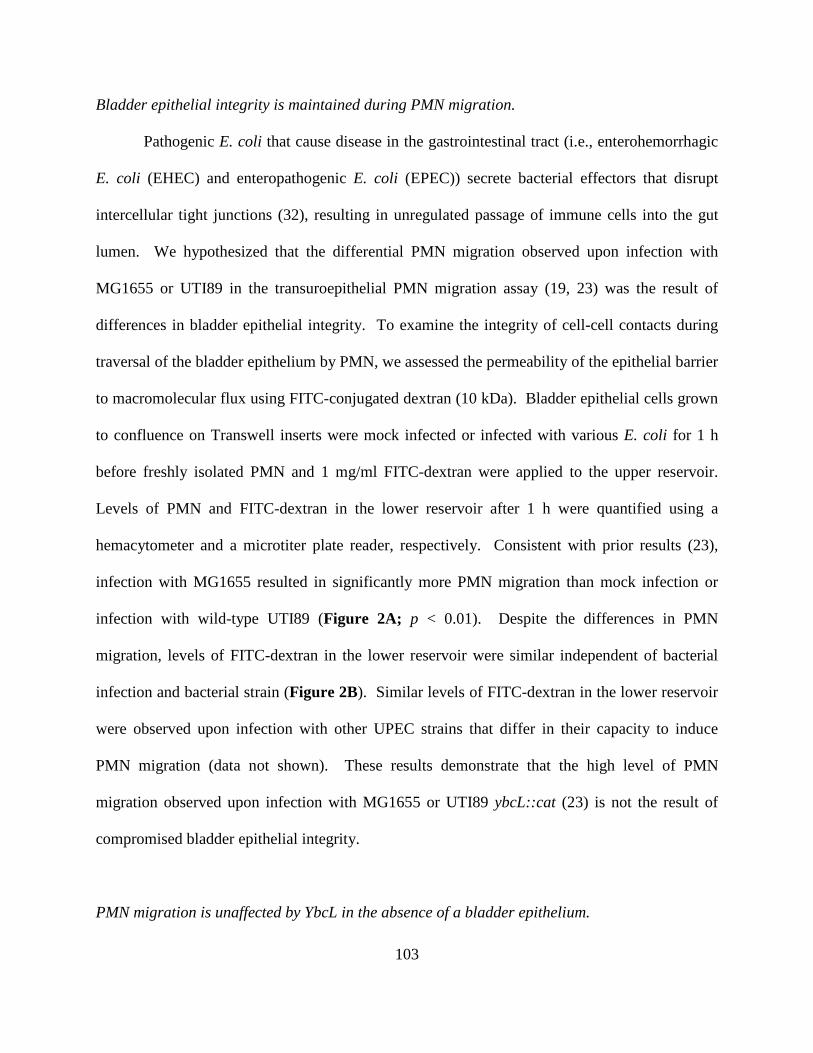
103
Bladder epithelial integrity is maintained during PMN migration.
Pathogenic E. coli that cause disease in the gastrointestinal tract (i.e., enterohemorrhagic
E. coli (EHEC) and enteropathogenic E. coli (EPEC)) secrete bacterial effectors that disrupt
intercellular tight junctions (32), resulting in unregulated passage of immune cells into the gut
lumen. We hypothesized that the differential PMN migration observed upon infection with
MG1655 or UTI89 in the transuroepithelial PMN migration assay (19, 23) was the result of
differences in bladder epithelial integrity. To examine the integrity of cell-cell contacts during
traversal of the bladder epithelium by PMN, we assessed the permeability of the epithelial barrier
to macromolecular flux using FITC-conjugated dextran (10 kDa). Bladder epithelial cells grown
to confluence on Transwell inserts were mock infected or infected with various E. coli for 1 h
before freshly isolated PMN and 1 mg/ml FITC-dextran were applied to the upper reservoir.
Levels of PMN and FITC-dextran in the lower reservoir after 1 h were quantified using a
hemacytometer and a microtiter plate reader, respectively. Consistent with prior results (23),
infection with MG1655 resulted in significantly more PMN migration than mock infection or
infection with wild-type UTI89 (Figure 2A; p < 0.01). Despite the differences in PMN
migration, levels of FITC-dextran in the lower reservoir were similar independent of bacterial
infection and bacterial strain (Figure 2B). Similar levels of FITC-dextran in the lower reservoir
were observed upon infection with other UPEC strains that differ in their capacity to induce
PMN migration (data not shown). These results demonstrate that the high level of PMN
migration observed upon infection with MG1655 or UTI89 ybcL::cat (23) is not the result of
compromised bladder epithelial integrity.
PMN migration is unaffected by YbcL in the absence of a bladder epithelium.
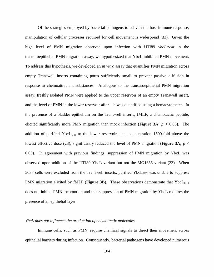
104
Of the strategies employed by bacterial pathogens to subvert the host immune response,
manipulation of cellular processes required for cell movement is widespread (33). Given the
high level of PMN migration observed upon infection with UTI89 ybcL::cat in the
transuroepithelial PMN migration assay, we hypothesized that YbcL inhibited PMN movement.
To address this hypothesis, we developed an in vitro assay that quantifies PMN migration across
empty Transwell inserts containing pores sufficiently small to prevent passive diffusion in
response to chemoattractant substances. Analogous to the transuroepithelial PMN migration
assay, freshly isolated PMN were applied to the upper reservoir of an empty Transwell insert,
and the level of PMN in the lower reservoir after 1 h was quantified using a hemacytometer. In
the presence of a bladder epithelium on the Transwell inserts, fMLF, a chemotactic peptide,
elicited significantly more PMN migration than mock infection (Figure 3A; p < 0.05). The
addition of purified YbcLUTI to the lower reservoir, at a concentration 1500-fold above the
lowest effective dose (23), significantly reduced the level of PMN migration (Figure 3A; p <
0.05). In agreement with previous findings, suppression of PMN migration by YbcL was
observed upon addition of the UTI89 YbcL variant but not the MG1655 variant (23). When
5637 cells were excluded from the Transwell inserts, purified YbcLUTI was unable to suppress
PMN migration elicited by fMLF (Figure 3B). These observations demonstrate that YbcLUTI
does not inhibit PMN locomotion and that suppression of PMN migration by YbcL requires the
presence of an epithelial layer.
YbcL does not influence the production of chemotactic molecules.
Immune cells, such as PMN, require chemical signals to direct their movement across
epithelial barriers during infection. Consequently, bacterial pathogens have developed numerous
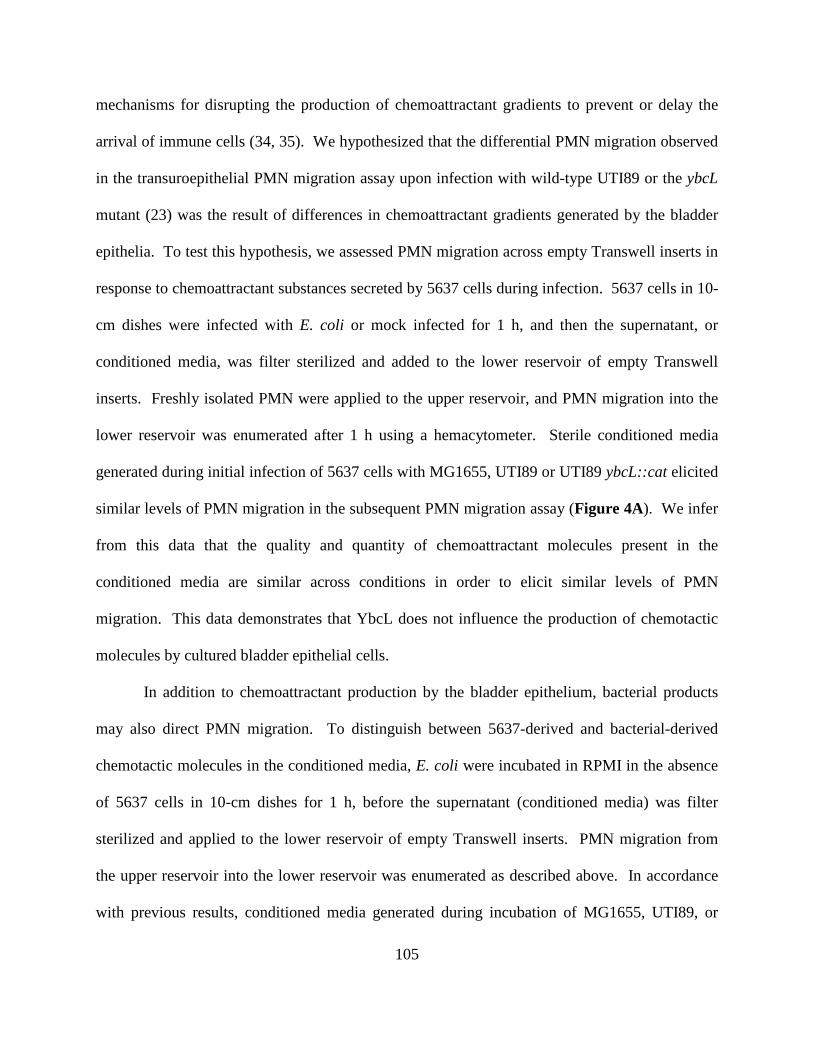
105
mechanisms for disrupting the production of chemoattractant gradients to prevent or delay the
arrival of immune cells (34, 35). We hypothesized that the differential PMN migration observed
in the transuroepithelial PMN migration assay upon infection with wild-type UTI89 or the ybcL
mutant (23) was the result of differences in chemoattractant gradients generated by the bladder
epithelia. To test this hypothesis, we assessed PMN migration across empty Transwell inserts in
response to chemoattractant substances secreted by 5637 cells during infection. 5637 cells in 10-
cm dishes were infected with E. coli or mock infected for 1 h, and then the supernatant, or
conditioned media, was filter sterilized and added to the lower reservoir of empty Transwell
inserts. Freshly isolated PMN were applied to the upper reservoir, and PMN migration into the
lower reservoir was enumerated after 1 h using a hemacytometer. Sterile conditioned media
generated during initial infection of 5637 cells with MG1655, UTI89 or UTI89 ybcL::cat elicited
similar levels of PMN migration in the subsequent PMN migration assay (Figure 4A). We infer
from this data that the quality and quantity of chemoattractant molecules present in the
conditioned media are similar across conditions in order to elicit similar levels of PMN
migration. This data demonstrates that YbcL does not influence the production of chemotactic
molecules by cultured bladder epithelial cells.
In addition to chemoattractant production by the bladder epithelium, bacterial products
may also direct PMN migration. To distinguish between 5637-derived and bacterial-derived
chemotactic molecules in the conditioned media, E. coli were incubated in RPMI in the absence
of 5637 cells in 10-cm dishes for 1 h, before the supernatant (conditioned media) was filter
sterilized and applied to the lower reservoir of empty Transwell inserts. PMN migration from
the upper reservoir into the lower reservoir was enumerated as described above. In accordance
with previous results, conditioned media generated during incubation of MG1655, UTI89, or
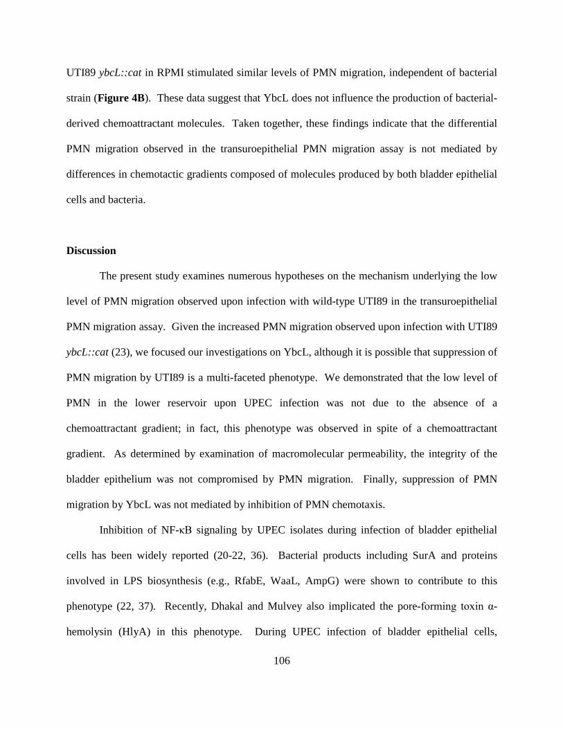
106
UTI89 ybcL::cat in RPMI stimulated similar levels of PMN migration, independent of bacterial
strain (Figure 4B). These data suggest that YbcL does not influence the production of bacterial-
derived chemoattractant molecules. Taken together, these findings indicate that the differential
PMN migration observed in the transuroepithelial PMN migration assay is not mediated by
differences in chemotactic gradients composed of molecules produced by both bladder epithelial
cells and bacteria.
Discussion
The present study examines numerous hypotheses on the mechanism underlying the low
level of PMN migration observed upon infection with wild-type UTI89 in the transuroepithelial
PMN migration assay. Given the increased PMN migration observed upon infection with UTI89
ybcL::cat (23), we focused our investigations on YbcL, although it is possible that suppression of
PMN migration by UTI89 is a multi-faceted phenotype. We demonstrated that the low level of
PMN in the lower reservoir upon UPEC infection was not due to the absence of a
chemoattractant gradient; in fact, this phenotype was observed in spite of a chemoattractant
gradient. As determined by examination of macromolecular permeability, the integrity of the
bladder epithelium was not compromised by PMN migration. Finally, suppression of PMN
migration by YbcL was not mediated by inhibition of PMN chemotaxis.
Inhibition of NF-κB signaling by UPEC isolates during infection of bladder epithelial
cells has been widely reported (20-22, 36). Bacterial products including SurA and proteins
involved in LPS biosynthesis (e.g., RfabE, WaaL, AmpG) were shown to contribute to this
phenotype (22, 37). Recently, Dhakal and Mulvey also implicated the pore-forming toxin α-
hemolysin (HlyA) in this phenotype. During UPEC infection of bladder epithelial cells,
107
activation of serine proteases, dependent upon HlyA, resulted in the degradation of proteins
involved in numerous signaling cascades, including the NF-κB pathway (38). In agreement with
these findings, infection with either wild-type UTI89 or UTI89 ybcL::cat resulted in lower levels
of both the phosphorylated and unphosphorylated forms of NF-κB p65 compared to mock
infection. Despite structural homology between YbcL and RKIP (28), a mammalian protein that
inhibits the NF-κB pathway (30), YbcL had no effect on NF-κB signaling. Indeed, the lower
levels of NF-κB p65 and phospho-NF-κB p65 upon UPEC infection were dependent upon HlyA,
as infection with UTI89 ΔhlyA restored NF-κB p65 levels (M. Lau and D. Hunstad, unpublished
data). From these data, we conclude that the differential PMN migration observed upon
infection with wild-type UTI89 or the ybcL mutant in the transuroepithelial PMN migration
assay is not the result of differences in the activation status of the NF-κB pathway.
As production of chemotactic molecules is only partly dependent upon NF-κB activation,
we took a more general approach to investigate potential differences in chemoattractants
produced during infection. We demonstrated that sterile conditioned media generated in the
presence or absence of bladder epithelial cells elicited similar levels of PMN migration across
empty Transwell inserts independent of E. coli strain. Given that nonpathogenic E. coli and
UPEC express functionally distinct YbcL variants (23), we conclude that YbcL does not
influence the production of chemotactic molecules by either bladder epithelial cells or bacteria.
In support of these data and the observations on NF-κB activation, quantification of IL-8, a PMN
chemoattractant, in the upper and lower reservoirs during the transuroepithelial PMN migration
assay revealed no significant differences upon infection with UTI89 versus the ybcL mutant (M.
Lau and D. Hunstad, unpublished data). These results suggest that the increased PMN migration
observed upon infection with UTI89 ybcL::cat compared to wild-type UTI89 in this assay is not
108
due to augmented production of chemotactic molecules. Although all of the chemoattractants in
these assays have not been identified, these results suggest that a chemoattractant gradient is
produced, possibly in an NF-κB-independent manner, across the bladder epithelium during
infection with UTI89 in the transuroepithelial PMN migration assay. It remains unclear how
UPEC utilizes YbcL to prevent PMN from transiting the epithelium in response to these
chemotactic signals.
In modifying the transuroepithelial PMN migration assay to remove the bladder epithelial
barrier, we were able to examine the direct effect of purified YbcL on PMN movement in
response to chemotactic molecules. In contrast to the suppressive effect observed in the presence
of a bladder epithelium, YbcL had no effect on PMN migration across empty Transwell inserts in
response to a peptide chemoattractant, fMLF. In agreement with these data, sterile conditioned
media generated during initial infection of bladder epithelial cells with wild-type UTI89
suppressed PMN migration when a bladder epithelium was present on the Transwell insert (23),
but not in the absence of a bladder epithelium. As YbcL was unable to influence PMN migration
in the absence of an epithelial barrier even at very high concentrations, we conclude that YbcL
does not inhibit PMN chemotaxis (e.g., polarization or cytoskeletal rearrangement) or the
perception of chemotactic signals by PMN. Therefore, the low level of PMN migration observed
upon infection with wild-type UTI89 in the transuroepithelial PMN migration assay is not the
result of inhibition of PMN movement or prevention of chemotactic signal detection by YbcL.
These data suggest that YbcL instead exerts its effect on the bladder epithelium, or on
interactions between PMN and epithelial cells. Experiments aimed at addressing the site of
action of YbcL have been largely unfruitful, likely because of the extremely low concentration of
109
YbcL required to suppress PMN migration. The development of assays with increased
sensitivity will facilitate further investigation.
Pathogenic E. coli that cause disease in the gastrointestinal tract produce bacterial
effectors that increase epithelial permeability through disruption of cell-cell contacts (32),
facilitating movement of immune cells into the lumen. In contrast, probiotic bacteria enhance
barrier function of the gastrointestinal epithelium, preventing pathogenic strains from invading
underlying tissue and inhibiting immune cell migration (39). Given these findings in the
gastrointestinal tract, we tested the hypothesis that the differential PMN migration observed in
the transuroepithelial PMN migration assay was the result of differences in barrier function of
the epithelium. We found that macromolecular permeability of the bladder epithelium did not
correlate with the level of PMN migration, demonstrating that increased PMN migration
observed upon infection with MG1655 or UTI89 ybcL::cat (23) was not due to increased
epithelial permeability. These results serve as a contrast to pathogenic E. coli in the
gastrointestinal tract. It is likely that precise rearrangement of cell-cell contacts during PMN
traversal of the cultured uroepithelium prevents macromolecular flux into the lower reservoir,
analogous to PMN migration across epithelial barriers in vivo. These observations further
validate this in vitro assay as a means to investigate PMN movement through intercellular
contacts of the bladder epithelium.
In this study, we investigated a role for YbcL in manipulating the processes required for
PMN to transit the bladder epithelium. We interrogated activation of the NF-κB pathway,
production of chemotactic molecules by bladder epithelial cells and bacteria, integrity of the
bladder epithelium and, finally, PMN chemotaxis. We conclude that YbcL does not influence
these processes. The transit of PMN through epithelia is a multi-step process that requires the
110
coordinated activity of many proteins and signaling cascades, many of which are incompletely
defined. Additional work, utilizing the in vitro transuroepithelial PMN migration assay, focused
on identifying these factors may illuminate additional steps in the cascade that are subject to
inhibition by bacterial proteins such as YbcL.
Acknowledgements
This work was supported by National Institutes of Health (NIH) grants R01-DK080752
and P50-DK064540. We thank J. Loughman and K. Tiemann for technical assistance.
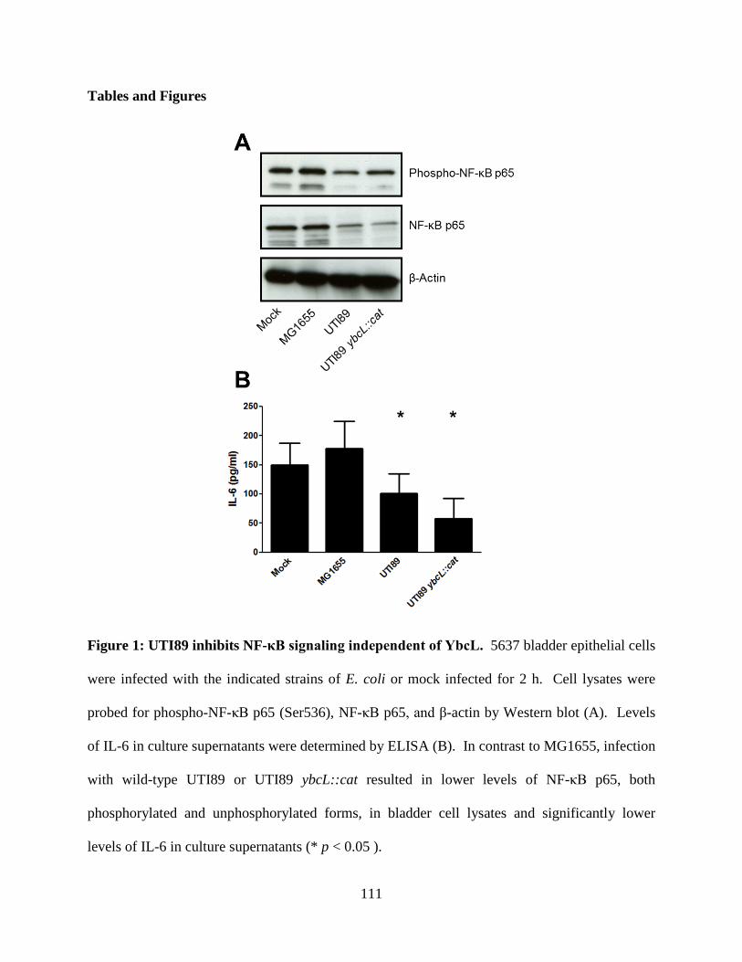
111
Tables and Figures
Figure 1: UTI89 inhibits NF-κB signaling independent of YbcL. 5637 bladder epithelial cells
were infected with the indicated strains of E. coli or mock infected for 2 h. Cell lysates were
probed for phospho-NF-κB p65 (Ser536), NF-κB p65, and β-actin by Western blot (A). Levels
of IL-6 in culture supernatants were determined by ELISA (B). In contrast to MG1655, infection
with wild-type UTI89 or UTI89 ybcL::cat resulted in lower levels of NF-κB p65, both
phosphorylated and unphosphorylated forms, in bladder cell lysates and significantly lower
levels of IL-6 in culture supernatants (* p < 0.05 ).
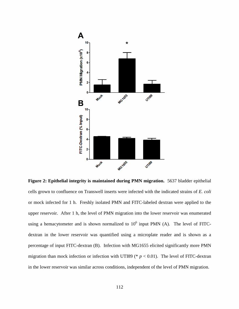
112
Figure 2: Epithelial integrity is maintained during PMN migration. 5637 bladder epithelial
cells grown to confluence on Transwell inserts were infected with the indicated strains of E. coli
or mock infected for 1 h. Freshly isolated PMN and FITC-labeled dextran were applied to the
upper reservoir. After 1 h, the level of PMN migration into the lower reservoir was enumerated
using a hemacytometer and is shown normalized to 106 input PMN (A). The level of FITC-
dextran in the lower reservoir was quantified using a microplate reader and is shown as a
percentage of input FITC-dextran (B). Infection with MG1655 elicited significantly more PMN
migration than mock infection or infection with UTI89 (* p < 0.01). The level of FITC-dextran
in the lower reservoir was similar across conditions, independent of the level of PMN migration.
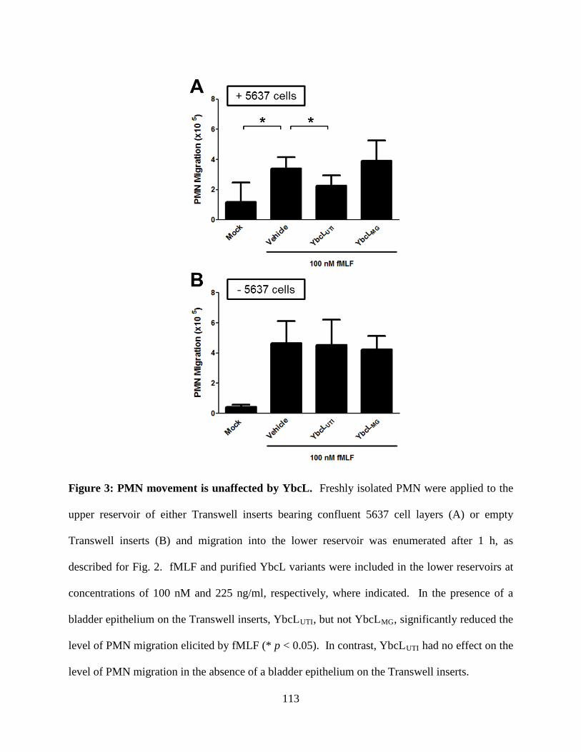
113
Figure 3: PMN movement is unaffected by YbcL. Freshly isolated PMN were applied to the
upper reservoir of either Transwell inserts bearing confluent 5637 cell layers (A) or empty
Transwell inserts (B) and migration into the lower reservoir was enumerated after 1 h, as
described for Fig. 2. fMLF and purified YbcL variants were included in the lower reservoirs at
concentrations of 100 nM and 225 ng/ml, respectively, where indicated. In the presence of a
bladder epithelium on the Transwell inserts, YbcLUTI, but not YbcLMG, significantly reduced the
level of PMN migration elicited by fMLF (* p < 0.05). In contrast, YbcLUTI had no effect on the
level of PMN migration in the absence of a bladder epithelium on the Transwell inserts.
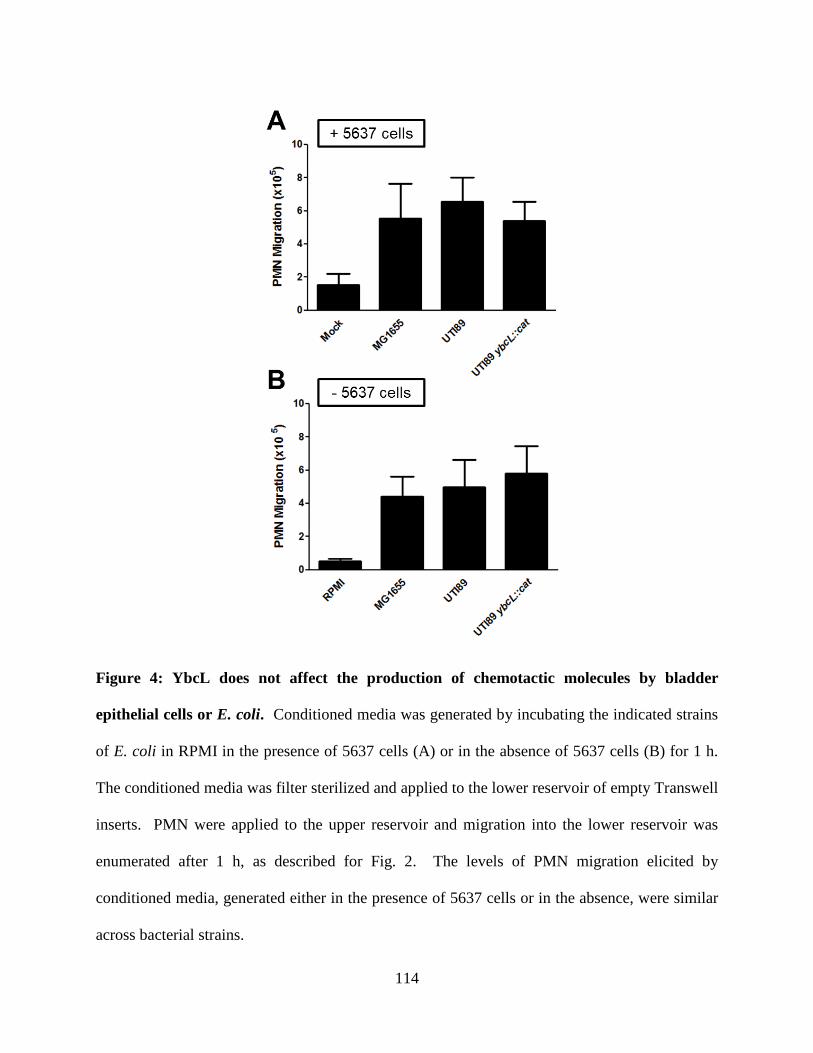
114
Figure 4: YbcL does not affect the production of chemotactic molecules by bladder
epithelial cells or E. coli. Conditioned media was generated by incubating the indicated strains
of E. coli in RPMI in the presence of 5637 cells (A) or in the absence of 5637 cells (B) for 1 h.
The conditioned media was filter sterilized and applied to the lower reservoir of empty Transwell
inserts. PMN were applied to the upper reservoir and migration into the lower reservoir was
enumerated after 1 h, as described for Fig. 2. The levels of PMN migration elicited by
conditioned media, generated either in the presence of 5637 cells or in the absence, were similar
across bacterial strains.
115
References
1. Hedges S, Anderson P, Lidin-Janson G, de Man P, Svanborg C. 1991. Interleukin-6
response to deliberate colonization of the human urinary tract with gram-negative
bacteria. Infect Immun 59:421-427.
2. Samuelsson P, Hang L, Wullt B, Irjala H, Svanborg C. 2004. Toll-like receptor 4
expression and cytokine responses in the human urinary tract mucosa. Infect Immun
72:3179-3186.
3. Agace WW, Hedges SR, Ceska M, Svanborg C. 1993. Interleukin-8 and the neutrophil
response to mucosal gram-negative infection. J Clin Invest 92:780-785.
4. Lundstedt AC, Leijonhufvud I, Ragnarsdottir B, Karpman D, Andersson B, Svanborg C.
2007. Inherited susceptibility to acute pyelonephritis: a family study of urinary tract
infection. J Infect Dis 195:1227-1234.
5. Lundstedt AC, McCarthy S, Gustafsson MC, Godaly G, Jodal U, Karpman D,
Leijonhufvud I, Linden C, Martinell J, Ragnarsdottir B, Samuelsson M, Truedsson L,
Andersson B, Svanborg C. 2007. A genetic basis of susceptibility to acute pyelonephritis.
PLoS One 2:e825.
6. Ragnarsdottir B, Samuelsson M, Gustafsson MC, Leijonhufvud I, Karpman D, Svanborg
C. 2007. Reduced toll-like receptor 4 expression in children with asymptomatic
bacteriuria. J Infect Dis 196:475-484.
7. Andersen-Nissen E, Hawn TR, Smith KD, Nachman A, Lampano AE, Uematsu S, Akira
S, Aderem A. 2007. Cutting edge: Tlr5-/- mice are more susceptible to Escherichia coli
urinary tract infection. J Immunol 178:4717-4720.
116
8. Ashkar AA, Mossman KL, Coombes BK, Gyles CL, Mackenzie R. 2008. FimH adhesin
of type 1 fimbriae is a potent inducer of innate antimicrobial responses which requires
TLR4 and type 1 interferon signalling. PLoS Pathog 4:e1000233.
9. Bergsten G, Wullt B, Schembri MA, Leijonhufvud I, Svanborg C. 2007. Do type 1
fimbriae promote inflammation in the human urinary tract? Cell Microbiol 9:1766-1781.
10. Fischer H, Ellstrom P, Ekstrom K, Gustafsson L, Gustafsson M, Svanborg C. 2007.
Ceramide as a TLR4 agonist; a putative signalling intermediate between sphingolipid
receptors for microbial ligands and TLR4. Cell Microbiol 9:1239-1251.
11. Schilling JD, Martin SM, Hunstad DA, Patel KP, Mulvey MA, Justice SS, Lorenz RG,
Hultgren SJ. 2003. CD14- and Toll-like receptor-dependent activation of bladder
epithelial cells by lipopolysaccharide and type 1 piliated Escherichia coli. Infect Immun
71:1470-1480.
12. Godaly G, Proudfoot AE, Offord RE, Svanborg C, Agace WW. 1997. Role of epithelial
interleukin-8 (IL-8) and neutrophil IL-8 receptor A in Escherichia coli-induced
transuroepithelial neutrophil migration. Infect Immun 65:3451-3456.
13. Hang L, Haraoka M, Agace WW, Leffler H, Burdick M, Strieter R, Svanborg C. 1999.
Macrophage inflammatory protein-2 is required for neutrophil passage across the
epithelial barrier of the infected urinary tract. J Immunol 162:3037-3044.
14. Chin AC, Parkos CA. 2007. Pathobiology of neutrophil transepithelial migration:
implications in mediating epithelial injury. Annu Rev Pathol 2:111-143.
15. Wang F. 2009. The signaling mechanisms underlying cell polarity and chemotaxis. Cold
Spring Harbor Perspect Biol 1:a002980.
117
16. Agace WW. 1996. The role of the epithelial cell in Escherichia coli induced neutrophil
migration into the urinary tract. Eur Respir J 9:1713-1728.
17. Zen K, Parkos CA. 2003. Leukocyte-epithelial interactions. Curr Opin Cell Biol 15:557-
564.
18. Thomas EL, Lehrer RI, Rest RF. 1988. Human neutrophil antimicrobial activity. Rev
Infect Dis 10 Suppl 2:S450-456.
19. Loughman JA, Hunstad DA. 2011. Attenuation of human neutrophil migration and
function by uropathogenic bacteria. Microbes Infect 13:555-565.
20. Billips BK, Forrestal SG, Rycyk MT, Johnson JR, Klumpp DJ, Schaeffer AJ. 2007.
Modulation of host innate immune response in the bladder by uropathogenic Escherichia
coli. Infect Immun 75:5353-5360.
21. Hilbert DW, Pascal KE, Libby EK, Mordechai E, Adelson ME, Trama JP. 2008.
Uropathogenic Escherichia coli dominantly suppress the innate immune response of
bladder epithelial cells by a lipopolysaccharide- and Toll-like receptor 4-independent
pathway. Microbes Infect 10:114-121.
22. Hunstad DA, Justice SS, Hung CS, Lauer SR, Hultgren SJ. 2005. Suppression of bladder
epithelial cytokine responses by uropathogenic Escherichia coli. Infect Immun 73:3999-
4006.
23. Lau ME, Loughman JA, Hunstad DA. 2012. YbcL of uropathogenic Escherichia coli
suppresses transepithelial neutrophil migration. Infect Immun 80:4123-4132.
24. Blattner FR, Plunkett G, 3rd, Bloch CA, Perna NT, Burland V, Riley M, Collado-Vides J,
Glasner JD, Rode CK, Mayhew GF, Gregor J, Davis NW, Kirkpatrick HA, Goeden MA,
118
Rose DJ, Mau B, Shao Y. 1997. The complete genome sequence of Escherichia coli K-
12. Science 277:1453-1474.
25. Chen SL, Hung CS, Xu J, Reigstad CS, Magrini V, Sabo A, Blasiar D, Bieri T, Meyer
RR, Ozersky P, Armstrong JR, Fulton RS, Latreille JP, Spieth J, Hooton TM, Mardis ER,
Hultgren SJ, Gordon JI. 2006. Identification of genes subject to positive selection in
uropathogenic strains of Escherichia coli: A comparative genomics approach. Proc Natl
Acad Sci U S A 103:5977-5982.
26. Murphy KC, Campellone KG. 2003. Lambda Red-mediated recombinogenic engineering
of enterohemorrhagic and enteropathogenic E. coli. BMC Mol Biol 4:11.
27. Datsenko KA, Wanner BL. 2000. One-step inactivation of chromosomal genes in
Escherichia coli K-12 using PCR products. Proc Natl Acad Sci U S A 97:6640-6645.
28. Serre L, Pereira de Jesus K, Zelwer C, Bureaud N, Schoentgen F, Benedetti H. 2001.
Crystal structures of YBHB and YBCL from Escherichia coli, two bacterial homologues
to a Raf kinase inhibitor protein. J Mol Biol 310:617-634.
29. Yeung K, Seitz T, Li S, Janosch P, McFerran B, Kaiser C, Fee F, Katsanakis KD, Rose
DW, Mischak H, Sedivy JM, Kolch W. 1999. Suppression of Raf-1 kinase activity and
MAP kinase signalling by RKIP. Nature 401:173-177.
30. Yeung KC, Rose DW, Dhillon AS, Yaros D, Gustafsson M, Chatterjee D, McFerran B,
Wyche J, Kolch W, Sedivy JM. 2001. Raf kinase inhibitor protein interacts with NF-
kappaB-inducing kinase and TAK1 and inhibits NF-kappaB activation. Mol Cell Biol
21:7207-7217.
119
31. Viatour P, Merville MP, Bours V, Chariot A. 2005. Phosphorylation of NF-kappaB and
IkappaB proteins: implications in cancer and inflammation. Trends Biochem Sci 30:43-
52.
32. Guttman JA, Finlay BB. 2009. Tight junctions as targets of infectious agents. Biochim
Biophys Acta 1788:832-841.
33. Lemonnier M, Landraud L, Lemichez E. 2007. Rho GTPase-activating bacterial toxins:
from bacterial virulence regulation to eukaryotic cell biology. FEMS Microbiol Rev
31:515-534.
34. Baxt LA, Garza-Mayers AC, Goldberg MB. 2013. Bacterial subversion of host innate
immune pathways. Science 340:697-701.
35. Alto NM, Orth K. 2012. Subversion of cell signaling by pathogens. Cold Spring Harbor
Perspect Biol 4:a006114.
36. Klumpp DJ, Weiser AC, Sengupta S, Forrestal SG, Batler RA, Schaeffer AJ. 2001.
Uropathogenic Escherichia coli Potentiates Type 1 Pilus-Induced Apoptosis by
Suppressing NF-kappa B. Infect. Immun. 69:6689-6695.
37. Billips BK, Schaeffer AJ, Klumpp DJ. 2008. Molecular basis of uropathogenic
Escherichia coli evasion of the innate immune response in the bladder. Infect Immun
76:3891-3900.
38. Dhakal BK, Mulvey MA. 2012. The UPEC pore-forming toxin alpha-hemolysin triggers
proteolysis of host proteins to disrupt cell adhesion, inflammatory, and survival
pathways. Cell Host Microbe 11:58-69.
120
39. Ulluwishewa D, Anderson RC, McNabb WC, Moughan PJ, Wells JM, Roy NC. 2011.
Regulation of tight junction permeability by intestinal bacteria and dietary components. J
Nutr 141:769-776.
121
CHAPTER 5
CONCLUDING REMARKS AND FUTURE DIRECTIONS
The overarching goal of this work was to further our understanding of uropathogenic
Escherichia coli (UPEC)-mediated modulation of the innate immune response in the bladder. A
transcriptional study identified ybcL as being up-regulated during UPEC infection of cultured
bladder epithelial cells and human polymorphonuclear leukocytes (PMN; neutrophils),
suggesting that the gene product was involved in adaptation of UPEC to these cell types. We
subsequently demonstrated that YbcL suppressed PMN migration across a cultured bladder
epithelium in an in vitro model of transuroepithelial PMN migration and an in vivo model of
murine cystitis (i.e., bladder infection) (1). The bulk of this thesis work focused on investigating
how a low abundance protein encoded by UPEC, YbcL, could have such a profound effect on the
innate immune response in the bladder.
Allelic Variation in ybcL Loci
In examining YbcL homologs encoded by pathogenic and nonpathogenic E. coli, amino
acid differences were identified. The presence of a threonine at position 78 in YbcL correlated
with suppression of PMN migration in the transuroepithelial PMN migration assay (1). The
majority of sequenced and clinical UPEC strains encoded a threonine-containing YbcL variant
(1), demonstrating that suppression of PMN migration by YbcL is a conserved mechanism for
modulating the innate immune response. In contrast, few nonpathogenic E. coli strains encoded
a threonine-containing YbcL variant (1). It is not clear if valine- and alanine-containing YbcL
variants confer an advantage to E. coli that warrants maintenance of these loci. Threonine at
122
position 78 in YbcL was highly conserved among clinical UPEC isolates associated with
pyelonephritis (i.e., kidney infection) and asymptomatic bacteriuria (ASB) (i.e., bacteria in the
urine in the absence of symptoms). In addition to influencing the innate immune response in the
bladder, YbcL may also suppresses PMN recruitment to the kidneys, though suppression of
PMN migration across kidney epithelial cells in vitro has not been assessed. The lack of a well-
developed animal model that is specific for pyelonephritis would limit extension of such in vitro
findings to an in vivo scenario. The conservation of T78 in YbcL among clinical isolates
associated with ASB is puzzling, as the absence of an inflammatory response in ASB has been
primarily attributed to colonization with less virulent bacteria rather than active suppression of
inflammatory pathways. Studies investigating the dynamics of ASB may uncover a role for
YbcL in this disease state.
YbcL is structurally homologous to Raf kinase inhibitory protein (RKIP) (2), a
mammalian protein that inhibits numerous signaling cascades including the MAPK and NF-κB
pathways (3, 4). Despite this structural homology, we demonstrated that YbcL does not inhibit
signaling through these pathways. Phosphorylation of RKIP at serine 153 influences its affinity
for binding partners (5, 6). Given the importance of T78 in YbcL and the structural homology to
RKIP, YbcL may be phosphorylated at this residue, likely by a eukaryotic kinase. Mutation of
the threonine at position 78 to a glutamic acid to mimic a phosphorylated threonine yielded a
YbcL variant (YbcLT78E) that was unable to suppress PMN migration (M. Lau and D. Hunstad,
unpublished data). As this result is difficult to interpret, additional tools such as anti-phospho-
threonine antibodies would be useful in further testing this hypothesis.
Recent findings have demonstrated that post-translational modification (i.e.,
phosphorylation at serine 153) of RKIP leads to homodimer formation, which in turn, affects
123
interactions with binding partners (7). The K12 YbcL variant crystallized as a homodimer (2).
In monomeric form, the presumed ligand binding pocket of YbcL appears to be incomplete. In
contrast, completion of the RKIP ligand binding pocket, which mediates interactions with
binding partners, does not require dimerization, as monomeric RKIP contains an intact pocket
(8). It is not clear if an intact binding pocket and/or dimerization are required for suppression of
PMN migration by YbcL. In addition to investigating post-translational modification of T78 in
YbcL, experiments addressing dimer formation by YbcL variants will be informative. As with
RKIP, an amino acid distal to the binding pocket (T78) may affect the activity of YbcL by
influencing its ability to oligomerize.
Threonine at position 78 was not required for release of YbcL from the bacterial
periplasm, as a valine-containing variant was detected in the supernatant at levels similar to a
threonine-containing variant. One hypothesis is that T78 is instead required for YbcL activity
following release from the periplasm; for example, T78 may influence protein-protein
interactions. RKIP has been shown to interact with phosphorylated protein targets, and we
hypothesize that YbcL functions similarly. As YbcL was detected in the supernatant as well as
in cell lysates prepared from UPEC-infected bladder epithelial cells and PMN, the exact site of
action of YbcL is unclear. This finding in addition to the low concentration of YbcL required to
suppress PMN migration complicates protein-protein interaction studies, discussed below.
Additional experiments are required to address questions such as site of action and subcellular
localization before protein interaction studies will yield fruitful results.
Release of YbcL from the Periplasm
124
Using a number of different approaches, we demonstrated that suppression of PMN
migration by UTI89 required release of YbcL from the bacterial periplasm (1). Levels of YbcL
and GroEL, a bacterial cytoplasmic protein, in the supernatant were similar independent of
bacterial strain, MG1655 or UTI89 ybcL::cat (M. Lau and D. Hunstad, unpublished data),
suggesting the mode of release is conserved between nonpathogenic and uropathogenic E. coli.
In support of these data and the finding that liberation of YbcL is required for suppression of
PMN migration, MG1655 episomally expressing the UTI89 YbcL variant elicited significantly
fewer PMN than wild-type MG1655. This commonality allowed us to eliminate the scenario in
which potential secretion machinery was not encoded by both strains.
Secretion of effectors or toxins by bacterial pathogens is a common strategy to influence
eukaryotic processes. Many bacterial pathogens encode type 3 secretion systems (T3SS) and
type 4 secretion systems (T4SS) to achieve these means. However, these complex machines are
not encoded by UPEC. Recently, type 2 secretion systems (T2SS), type 4 pilus systems (T4PS)
and outer membrane vesicles (OMVs) have been implicated in UPEC pathogenesis (9, 10).
Because YbcL was released into the supernatant in soluble form and did not appear to be
packaged in OMVs, we examined a role for the T2SS and T4PS found in both MG1655 and
UTI89. The level of YbcL detected in the supernatant was unchanged upon deletion of either
secretin, YheF or HofQ, demonstrating that neither secretion system is solely responsible for
liberation of YbcL. The generation of a strain containing mutations in both secretins would be
required to eliminate the possibility of functional redundancy between these systems. A second
putative T2SS gene cluster encoded by UTI89 shares homology with a cluster encoded by
enterotoxigenic E. coli (ETEC) strain H10407 (10). This gene cluster is absent from the clinical
UPEC strain CFT073, isolated from the blood of a patient with pyelonephritis, and has extremely
125
low homology to the T2SS encoded by MG1655. Although it is unlikely that this second T2SS
mediates release of YbcL, mutation of the secretin component, GspD, in UTI89 would address
this uncertainty. Taken together, these findings suggest that liberation of YbcL from the
bacterial cell is not mediated by the T2SS or T4PS.
Many integral membrane proteins found in the bacterial outer membrane are porins or
transporters that regulate the movement of ions and solutes in and out of the periplasm. The
pores or channels of these proteins are not large enough to allow passage of a fully folded
protein, even one as small as YbcL. However, there are proteins and protein complexes that
function to transport folded proteins across the bacterial outer membrane. For example, ushers
of the chaperone/usher secretion pathway serve as platforms for pilus biogenesis, assembling and
transporting pilus subunits through their large central channels (11). A number of
chaperone/usher systems are encoded by UPEC, although few are found in MG1655. When
pilus biogenesis is not active, the ushers are gated to prevent the unregulated flux of ions and
solutes (12, 13). In addition, the assembly of pilin subunits is a highly ordered process that
requires interactions between a cognate periplasmic chaperone and the N and C termini of the
usher (14). Chaperone/usher systems have not yet been implicated in the secretion of proteins
other than pili subunits. Given these observations, it is unlikely that ushers facilitate transport of
YbcL across the bacterial outer membrane, though this hypothesis could be tested through the
generation of usher mutants.
Similar to pili, the biogenesis of flagella requires a protein complex to move bacterial
proteins across the outer membrane (15). Evolutionarily related to T3SS, flagella have been
shown to secrete bacterial proteins into the supernatant, in addition to mediating motility (16).
Composed of FlgH, the L-ring situated in the outer membrane forms a pore large enough to
126
transport folded proteins such as flagellin (FliC). In UTI89, the ybcL locus is surrounded by
genes involved in flagella biosynthesis (17). In light of these observations, we examined the
hypothesis that YbcL was released from the periplasm via flagella biosynthesis machinery. As
the level of YbcL in the supernatant was unaffected by mutation of flgH (M. Lau and D.
Hunstad, unpublished data), YbcL is not transported across the bacterial outer membrane by the
flagellar L-ring. It is possible that additional outer membrane proteins, encoded by both
MG1655 and UTI89, exist that could transport YbcL across the outer membrane into the
supernatant.
Given the presence of cytoplasmic proteins GroEL and RNA Polymerase α subunit in the
supernatant during UPEC infection, we hypothesized that release of YbcL from the periplasm
was achieved through bacterial cell lysis. The genes encoding Shiga toxin (Stx1), stxA and stxB,
are often found within prophages of the λ bacteriophage family (18). Stx1 is produced upon
phage induction and is transported to the bacterial periplasm. The accumulation of Stx1 in the
periplasm is controlled by phage-mediated bacterial lysis, which is also the mechanism of Stx1
release (19). In MG1655, the ybcL locus is found within a lambdoid-like prophage, DLP12 (20).
This gene organization is not conserved in UTI89, although DLP12 prophage is present in the
UTI89 genome. Nonetheless, we tested the hypothesis that YbcL was delivered in a manner
analogous to Stx1 by examining a role for UTI89-encoded DLP12 prophage lysis proteins,
YbcR2, YbcS2 and C5133, homologs of the lambda proteins S, R, and Rz/Rz (1), respectively,
as mediators of bacterial lysis and release of YbcL from the periplasm. The level of YbcL
detected in the supernatant was unaltered by deletion of these lysis genes (M. Lau and D.
Hunstad, unpublished data), suggesting that DLP12 prophage-mediated bacterial lysis is not the
mechanism by which YbcL is liberated from the periplasm.
127
Prophages in addition to DLP12 are present in both MG1655 and UTI89, although no
genetic link exists between ybcL and these loci. Additionally, it is unclear if these prophages are
competent for induction. Treatment of UTI89 pYbcL::TEM-1 with mitomycin C or hydrogen
peroxide, DNA-damaging agents known to activate recA-dependent bacteriophage induction,
during growth in RPMI alone could implicate phage induction in lysis of UTI89. Increased
release of YbcL::TEM-1 under these conditions would suggest that one or more prophages are
competent for induction. Ultimately, implication of a prophage in YbcL release would require
mutation of the phage lysis genes and a corresponding decrease in YbcL::TEM-1 levels in the
supernatant during infection of bladder epithelial cells.
The development of a β-lactamase reporter assay facilitated precise investigation into
YbcL release from the periplasm. Assuming the dynamics of YbcL::TEM-1 release mimic the
dynamics of release of the wild-type YbcL variant, we conclude that release of YbcL from the
periplasm occurs in a dose-and time-dependent manner. In addition, the presence of YbcL in the
supernatant was also dependent upon cultured bladder epithelial cells. Dependency on dose,
time, and bladder epithelial cells suggests that this process may be regulated, although it is
currently unclear how this is achieved. These data are consistent with transcriptional data that
demonstrated that ybcL was up-regulated during UPEC infection of cultured bladder epithelial
cells and human PMN (1). Release of YbcL::TEM-1 from the periplasm has not yet been
assessed in the presence of PMN. Given the transcriptional data and the findings using bladder
epithelial cells, we hypothesize that release of YbcL::TEM-1 would exhibit similar dynamics in
the presence of PMN.
Bacterial cytoplasmic proteins GroEL and RNA Polymerase α subunit were detected in
filter sterilized, TCA-precipitated supernatant by Western blot after UPEC infection of cultured
128
bladder epithelial cells. However, the cytoplasmic fusion Gst::TEM-1 was not detected in the
supernatant under similar conditions. In an effort to clarify these observations, additional TEM-
1 fusions with cytoplasmic localization could be generated and their release into the supernatant
assessed. Initially, we hypothesized that the presence of cytoplasmic proteins in the supernatant
was the result of bacterial lysis. However, it is not clear how release of cytoplasmic proteins
could be selective during lysis. The presence of cytoplasmic proteins in the supernatant in the
absence of bacterial lysis would imply that these proteins crossed two bacterial membranes, a
phenomenon unlikely to occur incidentally. Given the well-characterized roles for RNA
Polymerase α subunit and GroEL in transcription and protein folding, respectively, both
cytoplasmic processes, it is unlikely that these proteins would be purposefully secreted into the
supernatant. It is possible that the cytoplasmic proteins in the supernatant represent
contamination from the overnight bacterial culture, despite extensive washing of the bacterial
inoculum. A working hypothesis is that the bacterial outer membrane increases in permeability
in the presence of bladder epithelial cells, nonspecifically releasing periplasmic contents.
Detection of Skp::TEM-1 in the supernatant is consistent with this hypothesis. Increased
permeability of the outer membrane in a nonselective manner could be mediated by bladder
epithelial cell products. Additional experiments are required to clarify these observations.
Through targeted deletion, we demonstrated that a T2SS, a T4PS, the flagella
biosynthesis machinery and the DLP12 phage lysis genes are not responsible for release of YbcL
from the periplasm during UPEC infection. In light of these findings, one hypothesis is that
release of YbcL is mediated by other UPEC-encoded proteins, possibly via bacterial lysis. To
take a more general approach to identify potential bacterial proteins involved in YbcL liberation,
we could conduct a transposon mutant screen using the β-lactamase reporter assay. Compared to
129
probing filter sterilized, TCA-precipitated supernatant by Western blot, the β-lactamase reporter
assay is amenable to high-throughput experimentation. YbcL is transcriptionally up-regulated
and then released into the supernatant during UPEC infection of cultured bladder epithelial cells
(1). Potential bacterial machinery responsible for YbcL release may be similarly regulated. In
addition to the transposon mutant screen, we could reexamine the data generated from the
transcriptional profiling experiment that identified YbcL, looking for genes that could mediate
YbcL release.
In contrast to other bacterial pathogens, the number of proteins shown thus far to be
secreted by UPEC is low, and, consequently, our understanding of UPEC secretion is
rudimentary. Two secreted effectors, α-hemolysin and Cnf1 are secreted by a T1SS and OMVs,
respectively (9, 21). Though T2SS and T4PS have been implicated in UPEC pathogenesis (10),
substrates of these systems have not been identified. Given the lack of involvement of traditional
secretion systems, the mode of release of YbcL from the periplasm may represent a novel
mechanism of bacterial secretion. Alternatively, liberation of YbcL may be mediated by
eukaryotic products rather than bacterial products. Additional experimentation is required to
address these hypotheses.
Suppression of Transuroepithelial PMN Migration by YbcL
UPEC YbcL suppresses PMN migration across a bladder epithelium in an in vitro model
of transepithelial PMN migration and an in vivo murine model of cystitis (1). The increased
PMN migration observed in the absence of YbcL compared to in the presence of YbcL was not
the result of increased chemokine production, increased epithelial permeability, or increased
130
PMN mobility. Though hypotheses have been rejected, the mechanism by which YbcL
suppresses PMN migration is still unclear.
During UPEC infection, YbcL was detected by Western blot in both bladder epithelial
cell and PMN lysates (1). Additionally, YbcL suppressed PMN migration elicited by fMLF in
the presence of bladder epithelial cells on the Transwell inserts, but had no effect in the absence
of bladder epithelial cells. These data might suggest that the site of action of YbcL is the bladder
epithelium rather than the PMN, despite detection of YbcL in PMN lysate. Previously, we
hypothesized that the YbcL signal in the PMN lysate originated from bacteria that had been
phagocytosed and killed. To test this hypothesis, we attempted to restrict YbcL to one cell type
in the transepithelial PMN migration assay. Bladder epithelial cells on Transwell inserts were
preincubated with purified YbcL before the epithelial cells were washed and the level of PMN
migration in response to fMLF was assessed. In a similar experiment, PMN, rather than bladder
epithelial cells, were preincubated with purified YbcL. In both cases, YbcL reduced the level of
PMN migration (M. Lau and D. Hunstad, unpublished data). It is likely that removal of excess
YbcL was incomplete. Given the low concentration of YbcL required to suppress PMN
migration (<10 pM) (1), it is not surprising that reduced PMN migration was observed upon pre-
exposure of either cell type if extracellular YbcL was still present.
An alternative approach to restrict YbcL localization to one cell type is ectopic
expression. While cultured bladder epithelial cells are amenable to transfection and transduction,
human PMN are not due to the short half-life observed after harvest from the bloodstream.
However, we could evaluate the ability of HL-60 cells, a cell line generated from human
promyelocytic leukemia cells which can be differentiated chemically into neutrophil-like cells, to
migrate across a cultured bladder epithelium in response to UPEC infection and peptide
131
chemoattractants (22). In contrast to PMN, HL-60 cells can be transfected. If HL-60 cells
exhibit phenotypes similar to human PMN in the transepithelial PMN migration assay, then these
cells would enable a number of additional experimental approaches that are unfeasible with
human PMN. If expression of YbcL in cultured bladder epithelial cells or HL-60 cells does not
result in low levels of PMN or HL-60 migration in the transepithelial migration assay, then YbcL
may function from the extracellular milieu.
In another approach to determine the site of action of YbcL, we developed a protocol for
imaging bladder epithelial cells and PMN on Transwell inserts by confocal fluorescence
microscopy. Our attempts to visualize YbcL localization in the transepithelial PMN migration
assay, where YbcL has been shown to have an effect, were unsuccessful. Again, the level of
YbcL required to suppress PMN migration may have been too low to detect by microscopy,
especially if the localization pattern was diffuse. Alternatively, YbcL may function primarily
from an extracellular location, which would require a different protocol for visualization.
As mentioned previously, YbcL was detected in bladder epithelial cell and PMN lysates
during UPEC infection (1). Given the protocol used to generate cell lysate, it is impossible to
distinguish between YbcL localization to the cellular membrane and YbcL localization to the
cytoplasm. Fractionation of infected cells could be conducted to distinguish between these
possibilities. It is unclear how YbcL might achieve cytoplasmic localization, although it may
depend in part on bacterial invasion. These uncertainties could be addressed using purified
YbcL. In addition to elucidating YbcL localization at the subcellular level, these experiments
would inform protein-protein interaction studies, as interaction between YbcL and a membrane
protein might be best detected using alternative protocols such as Far Western blot.
132
Traversal of an epithelial barrier by PMN is a multistep process that requires the
coordinated activity of multiple adhesins and signaling pathways (23, 24). Initially, PMN adhere
to the basolateral face of the epithelium. Then, they move between epithelial cells, causing the
rearrangement of cell-cell contacts without disrupting epithelial integrity. After transit through
tight junctions, PMN adhere to the apical face of the epithelial and are released into the lumen. It
is currently unclear at which step in this complex pathway UPEC inhibit PMN migration. In the
transepithelial PMN migration assay, the number of PMN in the lower reservoir represents PMN
released into the lumen. As the apical sides of the Transwell inserts are lightly scraped during
collection of PMN, it is unlikely that UPEC-mediated inhibition of PMN migration occurs by
preventing detachment of PMN from the apical face of the epithelium. The concentration of
PMN remaining in the upper reservoir is too low to reliably determine by hemacytometer. To
assess PMN levels in the upper reservoir and PMN associated with the bladder epithelium, a
more sensitive method for detecting PMN is required. An assay based on myeloperoxidase
(MPO) activity or MPO protein levels may enable more accurate quantitation of PMN.
Additionally, PMN association with the bladder epithelium could be investigated using confocal
fluorescence microscopy. Given the amorphous appearance of the epithelium, this endeavor may
be difficult. Understanding the point at which UPEC inhibit PMN migration may highlight
adhesins or signaling pathways that could be subject to inhibition by YbcL.
To further interrogate how YbcL suppresses PMN migration, understanding the
molecular requirements for traversal of a bladder epithelial barrier by PMN is required.
Significant work has been done in the gastrointestinal tract and lung to identify the adhesins
required for PMN to transit these epithelial barriers (23, 25). It is unclear if the same adhesins
play a role in the bladder. Agace and colleagues have demonstrated that expression of
133
intercellular adhesion molecule-1 (ICAM-1) by the bladder epithelial cell line J82 and expression
of the β–integrin CD11b/CD18 (Mac-1) by PMN is required for traversal of the epithelial layer
in their model (26). The involvement of other adhesins (e.g., SIRPα, CD47, CAR) that have
been implicated at other epithelial surfaces has not yet been examined. Expression of these
adhesins by cultured bladder epithelial cells and PMN could be validated by quantitative PCR
and Western blot. The requirement of these proteins for PMN migration in the transepithelial
PMN migration assay could be examined using blocking antibodies. These findings could be
extended to the murine model of cystitis through the use of genetically modified mice.
The movement of PMN across epithelial barriers is a highly complex process that can be
influenced by many factors. We demonstrated that the differential PMN migration observed
upon infection with wild-type UTI89 and UTI89 ybcL::cat in the transepithelial PMN migration
assay was not the result of differences in chemoattractant gradients, epithelial integrity, or PMN
mobility. The challenge moving forward is that the current understanding of PMN recruitment
to the bladder is incomplete. To interrogate a role for YbcL, there is a need to investigate the
basic processes required for PMN migration at this site, focusing on adhesion interactions and
signaling cascades. Findings at other epithelial surfaces will inform these studies.
Concluding Remarks
In summary, the goal of this thesis work was to contribute to the current understanding of
host-pathogen interactions in the urinary tract. UPEC employ multiple strategies to suppress and
delay the acute inflammatory response in the bladder, extending the period during which UPEC
can initiate an intracellular reservoir in the absence of immune pressure. As formation of this
intracellular reservoir is required to propagate infection, the events preceding bacterial invasion
134
have the potential to influence disease outcome. This study identified a UPEC protein YbcL that
contributes to modulation of the innate immune response by suppressing the migration of
neutrophils across bladder epithelia. YbcL is the first UPEC protein implicated in this
phenotype. Additionally, this work demonstrated that YbcL was released from the periplasm,
though this liberation was not mediated by a canonical secretion system. YbcL represents the
first UPEC protein shown to function outside the bacterial periplasm without an identified mode
of release. Finally, the mechanism by which YbcL influences PMN migration appears to be
unique, as hypotheses based on the functions of other bacterial effectors were disproved.
Although additional experimentation is required to clarify some of these findings, this thesis
extends our understanding of strategies used by uropathogens to manipulate the innate immune
response during infection.
135
References
1. Lau ME, Loughman JA, Hunstad DA. 2012. YbcL of uropathogenic Escherichia coli
suppresses transepithelial neutrophil migration. Infect Immun 80:4123-4132.
2. Serre L, Pereira de Jesus K, Zelwer C, Bureaud N, Schoentgen F, Benedetti H. 2001.
Crystal structures of YBHB and YBCL from Escherichia coli, two bacterial homologues
to a Raf kinase inhibitor protein. J Mol Biol 310:617-634.
3. Yeung K, Seitz T, Li S, Janosch P, McFerran B, Kaiser C, Fee F, Katsanakis KD, Rose
DW, Mischak H, Sedivy JM, Kolch W. 1999. Suppression of Raf-1 kinase activity and
MAP kinase signalling by RKIP. Nature 401:173-177.
4. Yeung KC, Rose DW, Dhillon AS, Yaros D, Gustafsson M, Chatterjee D, McFerran B,
Wyche J, Kolch W, Sedivy JM. 2001. Raf kinase inhibitor protein interacts with NF-
kappaB-inducing kinase and TAK1 and inhibits NF-kappaB activation. Mol Cell Biol
21:7207-7217.
5. Lorenz K, Lohse MJ, Quitterer U. 2003. Protein kinase C switches the Raf kinase
inhibitor from Raf-1 to GRK-2. Nature 426:574-579.
6. Corbit KC, Trakul N, Eves EM, Diaz B, Marshall M, Rosner MR. 2003. Activation of
Raf-1 signaling by protein kinase C through a mechanism involving Raf kinase inhibitory
protein. J Biol Chem 278:13061-13068.
7. Deiss K, Kisker C, Lohse MJ, Lorenz K. 2012. Raf kinase inhibitor protein (RKIP) dimer
formation controls its target switch from Raf1 to G protein-coupled receptor kinase
(GRK) 2. J Biol Chem 287:23407-23417.
136
8. Banfield MJ, Barker JJ, Perry AC, Brady RL. 1998. Function from structure? The crystal
structure of human phosphatidylethanolamine-binding protein suggests a role in
membrane signal transduction. Structure 6:1245-1254.
9. Davis JM, Carvalho HM, Rasmussen SB, O'Brien AD. 2006. Cytotoxic necrotizing factor
type 1 delivered by outer membrane vesicles of uropathogenic Escherichia coli attenuates
polymorphonuclear leukocyte antimicrobial activity and chemotaxis. Infect Immun
74:4401-4408.
10. Kulkarni R, Dhakal BK, Slechta ES, Kurtz Z, Mulvey MA, Thanassi DG. 2009. Roles of
putative type II secretion and type IV pilus systems in the virulence of uropathogenic
Escherichia coli. PLoS One 4:e4752.
11. Busch A, Waksman G. 2012. Chaperone-usher pathways: diversity and pilus assembly
mechanism. Philos Trans R Soc Lond B Biol Sci 367:1112-1122.
12. Remaut H, Tang C, Henderson NS, Pinkner JS, Wang T, Hultgren SJ, Thanassi DG,
Waksman G, Li H. 2008. Fiber formation across the bacterial outer membrane by the
chaperone/usher pathway. Cell 133:640-652.
13. Phan G, Remaut H, Wang T, Allen WJ, Pirker KF, Lebedev A, Henderson NS, Geibel S,
Volkan E, Yan J, Kunze MB, Pinkner JS, Ford B, Kay CW, Li H, Hultgren SJ, Thanassi
DG, Waksman G. 2011. Crystal structure of the FimD usher bound to its cognate FimC-
FimH substrate. Nature 474:49-53.
14. Volkan E, Ford BA, Pinkner JS, Dodson KW, Henderson NS, Thanassi DG, Waksman G,
Hultgren SJ. 2012. Domain activities of PapC usher reveal the mechanism of action of an
Escherichia coli molecular machine. Proc Natl Acad Sci U S A 109:9563-9568.
137
15. Apel D, Surette MG. 2008. Bringing order to a complex molecular machine: the
assembly of the bacterial flagella. Biochim Biophys Acta 1778:1851-1858.
16. Young GM, Schmiel DH, Miller VL. 1999. A new pathway for the secretion of virulence
factors by bacteria: the flagellar export apparatus functions as a protein-secretion system.
Proc Natl Acad Sci U S A 96:6456-6461.
17. Chen SL, Hung CS, Xu J, Reigstad CS, Magrini V, Sabo A, Blasiar D, Bieri T, Meyer
RR, Ozersky P, Armstrong JR, Fulton RS, Latreille JP, Spieth J, Hooton TM, Mardis ER,
Hultgren SJ, Gordon JI. 2006. Identification of genes subject to positive selection in
uropathogenic strains of Escherichia coli: A comparative genomics approach. Proc Natl
Acad Sci U S A 103:5977-5982.
18. Herold S, Karch H, Schmidt H. 2004. Shiga toxin-encoding bacteriophages--genomes in
motion. Int J Med Microbiol 294:115-121.
19. Wagner PL, Livny J, Neely MN, Acheson DW, Friedman DI, Waldor MK. 2002.
Bacteriophage control of Shiga toxin 1 production and release by Escherichia coli. Mol
Microbiol 44:957-970.
20. Blattner FR, Plunkett G, 3rd, Bloch CA, Perna NT, Burland V, Riley M, Collado-Vides J,
Glasner JD, Rode CK, Mayhew GF, Gregor J, Davis NW, Kirkpatrick HA, Goeden MA,
Rose DJ, Mau B, Shao Y. 1997. The complete genome sequence of Escherichia coli K-
12. Science 277:1453-1474.
21. Gentschev I, Dietrich G, Goebel W. 2002. The E. coli alpha-hemolysin secretion system
and its use in vaccine development. Trends Microbiol 10:39-45.
138
22. Hauert AB, Martinelli S, Marone C, Niggli V. 2002. Differentiated HL-60 cells are a
valid model system for the analysis of human neutrophil migration and chemotaxis. Int J
Biochem Cell Biol 34:838-854.
23. Chin AC, Parkos CA. 2007. Pathobiology of neutrophil transepithelial migration:
implications in mediating epithelial injury. Annu Rev Pathol 2:111-143.
24. Zen K, Parkos CA. 2003. Leukocyte-epithelial interactions. Curr Opin Cell Biol 15:557-
564.
25. Zemans RL, Colgan SP, Downey GP. 2009. Transepithelial migration of neutrophils:
mechanisms and implications for acute lung injury. Am J Respir Cell Mol Biol 40:519-
535.
26. Agace WW, Patarroyo M, Svensson M, Carlemalm E, Svanborg C. 1995. Escherichia
coli induces transuroepithelial neutrophil migration by an intercellular adhesion
molecule-1-dependent mechanism. Infect Immun 63:4054-4062.
139
APPENDIX I
QUANTITATIVE ASSESSMENT OF HUMAN NEUTROPHIL MIGRATION ACROSS A
CULTURED BLADDER EPITHELIUM
Megan E. Lau1 and David A. Hunstad1,2*
Departments of Pediatrics1 and Molecular Microbiology and Microbial Pathogenesis2,
Washington University School of Medicine, St. Louis, Missouri 63110
* For correspondence. US Mail: Department of Pediatrics, Washington University School of
Medicine, 660 S. Euclid Ave., Campus Box 8208, Saint Louis, MO 63110-1093.
Phone: (314) 286-2710. Fax: (314) 286-2895. E-mail: [email protected].
Modified from Lau and Hunstad (2012) J Vis Exp. In press. Short Abstract
We developed an in vitro model that mimics an important component of the acute inflammatory
response during infection of the bladder with uropathogenic Escherichia coli. The
transuroepithelial neutrophil migration assay enables quantitative assessment of human
neutrophil migration across bladder epithelia, cultured on permeable supports, in response to
bacterial infection or chemoattractant substances.
Long Abstract
140
The recruitment of immune cells from the periphery to the site of inflammation is an essential
step in the innate immune response at any mucosal surface. During infection of the urinary
bladder, polymorphonuclear leukocytes (PMN; neutrophils) migrate from the bloodstream and
traverse the bladder epithelium. Failure to resolve infection in the absence of a neutrophilic
response demonstrates the importance of PMN in bladder defense. To facilitate colonization of
the bladder epithelium, uropathogenic Escherichia coli (UPEC), the causative agent of the
majority of urinary tract infections (UTIs), dampen the acute inflammatory response using a
variety of partially defined mechanisms. To further investigate the interplay between host and
bacterial pathogen, we developed an in vitro model of this aspect of the innate immune response
to UPEC. In the transuroepithelial neutrophil migration assay, a variation on the Boyden
chamber, cultured bladder epithelial cells are grown to confluence on the underside of a
permeable support. PMN are isolated from human venous blood and are applied to the
basolateral side of the bladder epithelial cell layers. PMN migration representing the
physiologically relevant basolateral-to-apical direction in response to bacterial infection or
chemoattractant molecules is enumerated using a hemacytometer. This model can be used to
investigate interactions between UPEC and eukaryotic cells as well as to interrogate the
molecular requirements for the traversal of bladder epithelia by PMN. The transuroepithelial
neutrophil migration model will further our understanding of the initial inflammatory response to
UPEC in the bladder.
Introduction
The movement of cells throughout the body, often across long distances, is required for
growth and development, wound healing, and immune response. Cell migration is complex and
requires the coordination of many different processes, including signaling cascades and the
141
rearrangement of cytoskeletal components. Cells can move randomly (chemokinesis) as well as
toward defined chemical gradients (chemotaxis). Many techniques have been developed to study
cell migration in vitro. The oldest and most common technique, the Boyden chamber, consists of
a vertical two-chamber system where a chemoattractant substance is placed in the bottom
chamber and cells of interest are placed in the top chamber (1) . The movement of cells across
the permeable filter, with pores of defined size, separating the two chambers is monitored.
Additional techniques have been developed to investigate cell migration including the Zigmond
chamber (2) and the Dunn chamber (3). These collective approaches have yielded significant
insight into the movement of many different cell types.
In addition to interrogating the basic principles of chemokinesis and chemotaxis, two-
chamber assays have facilitated the investigation of cell migration through extracellular matrix
components and both endothelial and epithelial cell layers. An advantage of two-chamber
systems over other techniques is that the porous membrane can be coated with proteins such as
collagen or fibrinogen, and cell migration across an extracellular matrix-like barrier can be
assessed. Additionally, cultured cell lines can be grown and differentiated on the permeable
supports. To investigate the movement of cells across an endothelial barrier, cultured endothelial
cells are seeded and grown in the upper reservoir of the permeable supports. Motile cells, such
as immune cells, are added to the upper reservoir and migration into the lower reservoir across
the endothelial barrier in the physiologic apical-to-basolateral direction is observed. This model
has been invaluable in understanding extravasation of immune cells out of the blood stream. In
contrast to transendothelial migration, the movement of cells across an epithelial barrier typically
occurs in the basolateral-to-apical direction. In order to model these events in vitro, researchers
seed and grow cultured epithelial cells on the underside of the permeable supports. Motile cells
142
are added to the upper reservoir and migration across the epithelial barrier, representing the
basolateral-to-apical direction, is monitored. Such models of transepithelial migration have
significantly contributed to our understanding of inflammatory responses at mucosal surfaces,
particularly those of the lung and gut (4, 5).
In contrast, immune cell trafficking through the epithelium of the urinary tract has
received much less attention. To further our understanding of innate immune responses in the
urinary tract during infection with uropathogenic Escherichia coli (UPEC), we developed an in
vitro assay, the transuroepithelial neutrophil migration assay, that enables investigation of
polymorphonuclear leukocyte (PMN; neutrophil) movement across a bladder epithelial barrier
(6-8). As with other two-chamber models of transepithelial migration, cultured human bladder
epithelial cells are grown on the underside of a permeable support and form confluent epithelial
layers. Human neutrophils, isolated from venous blood, are applied to the basolateral side of the
epithelial layer, and migration across the epithelium in the physiologically relevant basolateral-
to-apical direction is quantified in response to infection with different strains of E. coli or the
presence of chemoattractant molecules. Much research had focused on neutrophil movement,
both chemokinesis and chemotaxis, in the absence of additional cell types. The
transuroepithelial neutrophil migration assay is advantageous as it takes into account complex
interactions between bladder epithelial cells and immune cells during infection. This tractable in
vitro model has the potential to permit the detailed investigation of immune responses at
uroepithelial surfaces.
Protocol
1) Culturing 5637 Bladder Epithelial Cells
143
Perform the following steps in a laminar flow tissue-culture hood prepared with UV irradiation
and wiped down with 70% ethanol.
1.1) Prepare RPMI-1640 medium containing 10% fetal bovine serum (FBS) [termed RPMI+],
filter sterilize using a 0.22-µm pore size filter, and warm to 37°C in a water bath.
1.2) Thaw a cryovial of 5637 cells (American Type Culture Collection HTB-9; derived from
bladder carcinoma) in a 37°C water bath. Quickly transfer the thawed cells to a 75 cm2 tissue
culture flask containing 20 ml RPMI+.
1.3) Incubate the flask at 37°C in a humidified atmosphere with 5% CO2 until the cells are
approximately 95% confluent (~6 × 10⁶ cells per flask), about 4 days.
1.4) To subculture the 5637 cells:
1.4.1) Remove the medium from the flask. Wash the cells with 10 ml Dulbecco’s Phosphate-
Buffered Saline (DPBS) at room temperature.
1.4.2) Add 6 ml warm (37°C) 0.05% trypsin, 0.02% EDTA solution to the flask, and incubate at
37°C with 5% CO2 for 15 min.
1.4.3) Transfer the cells to a 15 ml conical tube, and pellet by centrifugation at 300 × g for 5 min.
Remove the trypsin/EDTA solution.
1.4.4) Resuspend the cells in 6 ml RPMI+ (106 cells/ml), and transfer 1 ml (106 cells) to a new
75 cm2 flask containing 20 ml RPMI+.
1.5) Repeat step 1.4 to subculture cells every 4 days or when the cells reach 95% confluence.
2) Seeding and Growing 5637 Cells on Permeable Supports
Perform the following steps in a tissue culture hood using aseptic technique. For optimal results,
use 5637 cells that have undergone fewer than 10 subcultures.
144
2.1) Using sterile forceps, invert the permeable supports in a sterile 25 mm deep tissue culture
dish.
2.2) Trypsinize a 75 cm2 flask of 5637 cells at 95% confluence according to steps 1.4.1-1.4.3.
2.3) Using a 1000 μl pipet, gently resuspend the cells in ~ 2 ml RPMI+ to a concentration of 3 ×
106 cells/ml, determined by cell counting using a hemacytometer.
2.4) Apply 50 µl of the cell suspension (1.5 × 10⁵ cells) to each permeable support without
touching the membrane. Place the lid on the dish, and carefully place the dish at 37°C with 5%
CO2 for no more than 16 h.
2.5) Using sterile forceps, right the permeable supports into a 24-well plate containing 0.6 ml
RPMI+ per well. Add 0.1 ml RPMI+ to the upper reservoir of each permeable support. Incubate
at 37°C with 5% CO2.
2.6) Replace the medium every 2 days. First, aspirate medium from the upper reservoir followed
by the lower reservoir, and then apply fresh RPMI+ in the reverse order, to the lower reservoir
(0.6 ml) and then the upper reservoir (0.1 ml).
2.7) Seven days after seeding the permeable supports with 5637 cells, assess confluence of the
cells. Fill the upper reservoir with RPMI+ (~0.35 ml). The cells are sufficiently confluent when
the medium does not equilibrate between the upper and lower reservoirs.
3) Preparation of the Bacterial Inoculum
3.1) Using aseptic technique, add 20 ml of appropriate culture broth to a 250 ml flask with a cap.
Use Luria-Bertani (LB) broth for E. coli cultures, and add antibiotics to the broth where
appropriate.
145
3.2) Using a sterile inoculation loop, inoculate the broth with bacteria from a glycerol stock or a
streak plate. For UPEC strains, we incubate the bacterial culture at 37°C for approximately 16 h,
without shaking (to promote production of type 1 pili).
3.3) Transfer the bacterial culture to a centrifuge tube. Pellet the bacteria by centrifugation at
8000 × g for 10 min.
3.4) Decant the supernatant, and resuspend the bacteria in PBS to OD600 nm = 1, equivalent to
~109 CFU/ml.
3.4.1) To generate a heat-killed bacterial stimulus, incubate an aliquot (e.g., ~1.5 × 10⁸ CFU in
150 μl) of the resuspended bacteria at 55°C for 30 min. Plate an aliquot of the heat-killed
suspension to confirm bacterial death.
3.5) Immediately before use, dilute the resuspended bacteria (live or heat-killed) 10-fold to 108
CFU/ml in warm serum-free RPMI [termed RPMI-] in a microfuge tube.
4) Isolation of Human Neutrophils from Peripheral Blood
The collection of blood from adult volunteers requires advance review and approval from an
institutional review board. Wear appropriate personal protective equipment and properly dispose
of hazardous materials to avoid exposure to human blood.
4.1) Approximately 25 ml of venous blood from a healthy adult volunteer should be drawn into 3
sterile sodium heparin-containing blood collection tubes by staff trained in phlebotomy.
4.1.1) Tightly wrap a rubber tourniquet around the upper arm, above the elbow.
4.1.2) Disinfect the entry site, the antecubital fossa, using a sterile 70% alcohol wipe.
4.1.3) Attach a plastic tube holder to a winged butterfly needle system.
146
4.1.4) Insert the needle into an antecubital vein at a 30° angle or less, bevel facing up. The
needle has punctured the vein when a spurt of blood appears in the plastic tubing.
4.1.5) Insert a blood collection tube into the plastic tube holder. When the first collection tube is
full, replace with the second collection tube. Repeat with the third tube.
4.1.6) When the third collection tube is approximately half full, remove the tourniquet.
4.1.7) Cover the puncture site with a sterile cotton ball and slowly withdraw the needle from the
vein. Slide the protective shield over the needle and place in a biohazard sharps container.
4.1.8) Apply pressure to the puncture site. Cover the puncture site and cotton ball with an
adhesive bandage.
4.1.9) Gently invert the blood collection tubes to disperse the heparin.
Perform the following steps in a tissue culture hood using aseptic technique.
4.2) Using a 10 ml pipet, gently transfer the blood to a fresh, sterile 50-ml conical tube. Add an
equivalent volume of 3% (w/v) dextran in 0.9% NaCl and mix by inversion. Incubate the tube
upright at room temperature for 20 min.
4.3) Without disrupting the lower layer, carefully aspirate the upper layer and transfer it to a new
50 ml conical tube. Pellet the cells by centrifugation at 300 × g for 10 min. Discard the
supernatant.
4.4) Resuspend the cell pellet in a volume of 0.9% NaCl equivalent to the starting volume of
blood.
4.5) Layer 10 ml of density centrifugation solution under the cell suspension, preserving the
interface between the two phases. Centrifuge at 400 × g for 30 min with no brake. Discard the
supernatant.
147
4.6) To lyse remaining red blood cells, resuspend the cell pellet in 10 ml cold 0.2% NaCl.
Incubate for 30 sec, and then promptly add 10 ml cold 1.6% NaCl to restore isotonicity.
4.7) Pellet the cells by centrifugation at 300 × g for 6 min. Discard the supernatant.
4.8) Repeat steps 4.6-4.7 until the cell pellet appears to be free of red blood cells, typically 3
rounds of lysis.
4.9) Using a 1000 μl pipet, resuspend the cell pellet, primarily PMN, in warm (37°C) RPMI- to a
concentration of 107 cells/ml, determined by cell counting using a hemacytometer. Keep the
cells at 37°C until use. Typically, PMN viability and purity are > 99% as assessed by trypan
blue exclusion and visualization of nuclear morphology after staining, respectively.
5) Transuroepithelial Neutrophil Migration Assay
Perform the following steps in a tissue culture hood using aseptic technique. Use only permeable
supports bearing confluent 5637 cell layers, as determined in step 2.7. The 24-well plates
containing RPMI- can be prepared in advance and kept at 37°C with 5% CO2 until use.
5.1) Aliquot 1 ml warm RPMI- per well in a 24-well plate; prepare 3 wells per permeable
support.
5.2) Aspirate the medium from the upper and lower reservoirs of the permeable supports.
5.3) Using sterile forceps, transfer the permeable supports to the 24-well plate prepared in step
5.1. Wash the permeable supports three times by transferring the supports from well to well.
5.4) If a bacterial inoculum is to be used, invert the permeable supports in a sterile 25 mm deep
tissue culture dish. If a chemoattractant (e.g., N-Formyl-Met-Leu-Phe (fMLF) or IL-8) is to be
used, proceed to step 5.6.
148
5.5) For experiments with live or killed bacterial stimuli, inoculate the apical side of each
permeable support with 60 μl RPMI- (mock infection) or with the bacterial inoculum (6 × 10⁶
CFU) prepared in step 3.5. Place the lid on the dish and incubate at 37°C with 5% CO2 for 1 h.
5.6) Add 0.6 ml RPMI- per well to a 24-well low-attachment plate; prepare 1 well per permeable
support. Prepare 3 wells containing 0.5 ml RPMI- to enumerate PMN input.
5.6.1) If a chemoattractant is being used in place of bacteria, add the chemoattractant to 0.6 ml
RPMI- in the 24-well plate prepared in step 5.6.
5.7) Right the permeable supports into the 24-well low-attachment plate.
5.8) Add 0.1 ml PMN (106 PMN), prepared in step 4.9, to the upper reservoir (basolateral side)
of each permeable support. Add 0.1 ml PMN directly to wells containing 0.5 ml RPMI-.
Incubate at 37°C with 5% CO2 for 1 h.
5.9) Using sterile forceps, gently scrape the membrane of the permeable support against the edge
of the well to remove additional PMN from the apical side and then dispose of the support.
5.10) Collect PMN by gently scraping the bottom of each well with the 1000-μl pipet tip, and
transfer PMN suspensions to microfuge tubes.
5.11) Enumerate PMN using a hemacytometer.
5.12) To calculate the total number of PMN in the lower reservoir, multiply the number of PMN
in 1 mm2 (100 nl) by 6000. Data can be reported as a proportion of input PMN that have
migrated, or as absolute numbers of PMN.
Representative Results
The transuroepithelial neutrophil migration assay enables the quantitative assessment of
human PMN migration across cultured bladder epithelial cell layers in response to various
149
stimuli (Figure 1B). While the protocol is straightforward, there are a number of variables that
can influence PMN migration and consequently affect the reproducibility of this assay.
Measures should be taken while preparing the permeable supports and the PMN to reduce
variability between technical and biological replicates. For example, only permeable supports
containing sufficiently confluent 5637 cell layers should be used in an experiment. Confluence
of the 5637 cells is assessed using a functional assay that measures impermeability to liquid. If
medium added to the upper reservoir equilibrates across the permeable support, then the 5637
cells are not sufficiently confluent to conduct the experiment. If the volume in the upper
reservoir is maintained, then the permeable support can be used to assess PMN migration. We
have measured transepithelial electrical resistance in this system, which rises modestly upon
confluence of the cells; if this method is chosen, care should be taken not to contaminate the
otherwise sterile setup. Confluence of the 5637 cells 7 days after seeding can be influenced by
multiple factors, including the passage number of the cells and the number of cells seeded on the
permeable support. In addition, the amount of time that the 5637 cells are incubated on the
permeable support in the inverted position during seeding should not exceed 16 h (Figure 1A).
For optimal reproducibility, the protocol should be followed precisely. Finally, permeable
supports containing confluent 5637 cell layers should be used within 1-2 days, and the
membranes of the supports should never be touched during either growth of the 5637 cells or
during the transuroepithelial neutrophil migration assay.
In addition to the 5637 cells, variability can also be introduced during PMN preparation.
Using the protocol detailed above to isolate PMN, 1 ml of human blood typically yields about
106 PMN, although this number varies from individual to individual. Once the typical yield of
an individual donor’s blood is known, the isolation protocol can be scaled up or down
150
accordingly. PMN from unhealthy or ill individuals should be avoided, and different PMN
donors should be used for biological replicates to ensure that results observed are reproducible.
PMN should be handled gently and sterilely to avoid activation during isolation. Lastly, the
timing of the experimental procedures is crucial, as PMN do not survive for extended periods of
time once removed from the body. We utilize PMN within 1 h of completing the isolation
procedure. Given these considerations, at least 3 technical replicates should be included in each
biological replicate.
The number of PMN in the lower reservoir after 1 h is shown in Figure 2 normalized to
106 input PMN. Alternatively, PMN numbers can be compared to an internal control after
normalization to input PMN, which may reduce variation between biological replicates.
Adherence to the protocol outlined above with attention to detail enables the enumeration of
PMN migration in response to stimuli including bacteria (Figure 2A) and chemoattractant
substances (Figure 2B).
Discussion
Using a cultured bladder epithelial cell line and freshly isolated human PMN, we
established an in vitro model of transuroepithelial neutrophil migration. This model has been
instrumental in beginning to dissect the complexities of the innate immune response during
urinary tract infection (UTI), an extremely common bacterial infection typically caused by
UPEC (9). During infection of the bladder, or cystitis, recruitment of PMN to the bladder lumen
is essential for bacterial clearance (10). To establish a foothold in the face of an inflammatory
response, UPEC delay the arrival of PMN to the bladder, which prolongs the period during
which UPEC can invade the bladder epithelium in the absence of immune pressure (6, 11). This
151
phenotype, suppression of PMN migration by UPEC at early time points, is observed in our in
vitro model of transuroepithelial PMN migration. Infection with nonpathogenic E. coli MG1655
elicits significantly more PMN migration than infection with uropathogenic E. coli UTI89
(Figure 2A); co-infection with MG1655 and UTI89 yields the uropathogenic phenotype (i.e.,
low levels of PMN migration) (6). Furthermore, we identified a UPEC protein, YbcL, that
contributes to the suppressive phenotype, as deletion of ybcL resulted in significantly more PMN
in the lower reservoir compared to wild-type UTI89 (Figure 2A) (7). High levels of PMN
migration were also elicited by heat-killed MG1655, fMLF and IL-8 (Figure 2A and B). In
comparison to infection with a live bacterial stimulus, the use of chemoattractants may simplify
both the experimental protocol and the interpretation of results. It is likely that other
chemoattractants (e.g., bacterial products or chemokines) would also elicit PMN in this model.
Thus far, the transuroepithelial neutrophil migration assay has facilitated investigations into
suppression of the early inflammatory response by UPEC, revealing phenotypes that have been
verified in in vivo models (6-8), and will be an invaluable tool in future endeavors.
While this assay has the potential to address a number of questions, there are a few
limitations. Given the number of variables inherent to this assay, care must be taken while
preparing and conducting experiments to ensure reproducibility between replicates.
Enumerating PMN by counting is prone to error, as counting can be time intensive and PMN are
short-lived once removed from the body, especially in the presence of bacteria. Reducing the
time between PMN sample collection and PMN enumeration will result in more accurate data
collection. Researchers have described colorimetric assays that measure myeloperoxidase
(MPO) enzyme activity as a surrogate for PMN (12, 13). Assays utilizing colorimetric substrates
such as 3, 3’, 5, 5’,-tetramethylbenzidine (TMB) or 2, 2’-azino-bis (3-ethylbenzothiazoline-6-
152
sulphonic acid) (ABTS) are not sufficiently sensitive to detect the concentrations of PMN
present in the lower reservoirs using the transuroepithelial neutrophil migration protocol detailed
above (Lau and Hunstad, unpublished data). The parameters of the migration assay could be
manipulated to increase the PMN density in the lower reservoir samples. Alternatively, an MPO
assay with greater sensitivity, potentially utilizing a fluorescent substrate, could be established.
Measurement of MPO activity represents an alternative to PMN counting and may enable more
accurate enumeration of PMN in the lower reservoir samples. Additionally, such an assay may
also allow enumeration of PMN adherent to the epithelial layers and remaining in the upper
reservoir. A validated MPO-based protocol would represent a powerful tool that could expand
the amount and type of data that could be collected from the transuroepithelial neutrophil
migration assay.
Transepithelial neutrophil migration assays modeling the innate immune response in the
gastrointestinal tract and lung are widespread and are responsible for our current understanding
of the traversal of epithelial barriers by PMN (4, 5). In contrast, PMN movement across
uroepithelial barriers has received far less attention. Säve and colleagues have reported a model
that uses a polarized epithelium composed of differentiated UROtsa cells, an immortalized cell
line derived from ureter tissue, grown to confluence on permeable supports (14). Agace and
colleagues have reported the use of undifferentiated bladder (J82) and kidney (A498) epithelial
cells in a similar model (15). In the transuroepithelial neutrophil migration model detailed
herein, though the 5637 cell layers are not stratified and likely not formally polarized, tight
junctions are formed, assessed by impermeability to macromolecular flux and the expression and
localization of tight junction proteins (Lau and Hunstad, unpublished data). Of note, the
epithelial layer also maintains such impermeability during the infection conditions we have
153
described. The protocols reported by Säve and Agace model the inflammatory response after
infection with UPEC isolates for 24 h. In these models, UPEC elicit robust PMN migration. In
contrast, the epithelial layers in our model are exposed to UPEC for a relatively short period of
time, 1 h, in order to examine the initial interactions between host and pathogen. Furthermore,
the use of a bladder epithelial cell line and a cystitis-derived UPEC isolate enables potential
translation of in vitro findings to the murine cystitis model (16). Lastly, the studies mentioned
above utilized large permeable supports that require 6-well tissue culture dishes. The use of
smaller permeable supports, as in our model, reduces reagent use and increases the number of
inserts that can be manipulated per experiment. Although each of these model systems has
advantages and disadvantages, the collective potential of these models to define events required
for PMN migration into urinary tract tissues is substantial.
Although less well understood than migration across endothelial barriers, the passage of
PMN across gastrointestinal and pulmonary epithelial barriers has received much study (4, 5).
Transepithelial neutrophil migration models that employ permeable supports and cultured
epithelial cells have revealed some of the signaling events and adhesion molecules involved in
the movement of PMN through these epithelia. Preliminary studies using cultured urinary
epithelial cells suggest the involvement of intercellular adhesion molecule-1 (ICAM-1) and the
β–integrin CD11b/CD18 (Mac-1) in PMN migration across urinary tissues (15). It is unclear
which additional signaling pathways and adhesive molecules are involved in these complex
processes in the urinary tract. Using the transuroepithelial neutrophil migration model described
herein, perhaps augmented with microscopic examination, these basic questions and many others
can be interrogated. Additionally, in conjunction with bacterial genetics, this model can be used
to further evaluate pathogen-specific phenotypes such as suppression of PMN migration by
154
UPEC. It is unclear at which point in the multi-step process of PMN migration UPEC exerts its
suppressive effect. Also, the mechanism by which YbcL influences PMN migration has yet to
be elucidated. By first understanding the basic requirements for the passage of PMN across the
bladder epithelium, we can then begin to probe how UPEC manipulates these processes to
facilitate disease.
In summary, while numerous techniques exist to study the movement of cells, fewer
approaches are available to interrogate cell migration across cellular barriers. Modifications to
the Boyden chamber have been integral to investigating cell migration across endothelial and
epithelial barriers. A tractable in vitro model of the acute inflammatory response in the urinary
tract, such as the transuroepithelial neutrophil migration assay detailed herein, is a valuable tool
for interrogating these complex processes. Lastly, modifications to this assay could facilitate
investigations into other disease states of the urinary tract.
Acknowledgments
This work was supported by National Institutes of Health (NIH) grants R01-DK080752 and P50-
DK064540. We thank J. Loughman for her efforts in establishing this assay.
Disclosures
The authors have nothing to disclose.
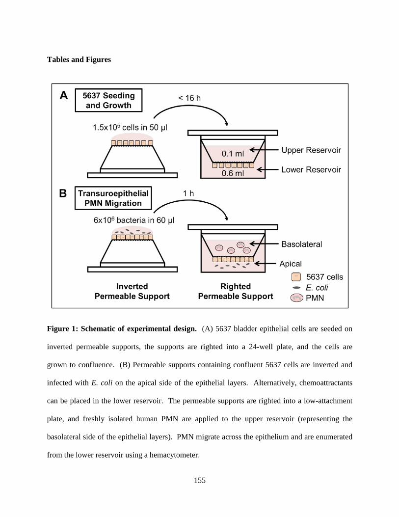
155
Tables and Figures
Figure 1: Schematic of experimental design. (A) 5637 bladder epithelial cells are seeded on
inverted permeable supports, the supports are righted into a 24-well plate, and the cells are
grown to confluence. (B) Permeable supports containing confluent 5637 cells are inverted and
infected with E. coli on the apical side of the epithelial layers. Alternatively, chemoattractants
can be placed in the lower reservoir. The permeable supports are righted into a low-attachment
plate, and freshly isolated human PMN are applied to the upper reservoir (representing the
basolateral side of the epithelial layers). PMN migrate across the epithelium and are enumerated
from the lower reservoir using a hemacytometer.
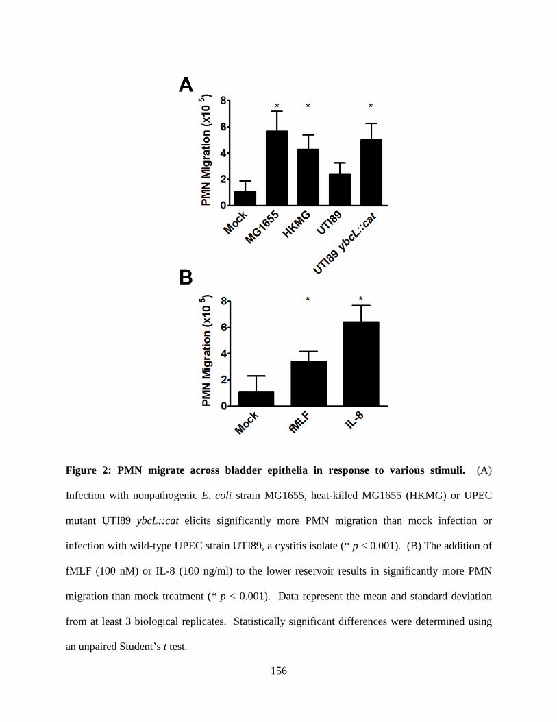
156
Figure 2: PMN migrate across bladder epithelia in response to various stimuli. (A)
Infection with nonpathogenic E. coli strain MG1655, heat-killed MG1655 (HKMG) or UPEC
mutant UTI89 ybcL::cat elicits significantly more PMN migration than mock infection or
infection with wild-type UPEC strain UTI89, a cystitis isolate (* p < 0.001). (B) The addition of
fMLF (100 nM) or IL-8 (100 ng/ml) to the lower reservoir results in significantly more PMN
migration than mock treatment (* p < 0.001). Data represent the mean and standard deviation
from at least 3 biological replicates. Statistically significant differences were determined using
an unpaired Student’s t test.
157
References
1. Boyden S. 1962. The chemotactic effect of mixtures of antibody and antigen on
polymorphonuclear leucocytes. J Exp Med 115:453-466.
2. Zigmond SH. 1988. Orientation chamber in chemotaxis. Methods Enzymol 162:65-72.
3. Zicha D, Dunn GA, Brown AF. 1991. A new direct-viewing chemotaxis chamber. J Cell
Sci 99 ( Pt 4):769-775.
4. Chin AC, Parkos CA. 2007. Pathobiology of neutrophil transepithelial migration:
implications in mediating epithelial injury. Annu Rev Pathol 2:111-143.
5. Zemans RL, Colgan SP, Downey GP. 2009. Transepithelial migration of neutrophils:
mechanisms and implications for acute lung injury. Am J Respir Cell Mol Biol 40:519-
535.
6. Loughman JA, Hunstad DA. 2011. Attenuation of human neutrophil migration and
function by uropathogenic bacteria. Microbes Infect 13:555-565.
7. Lau ME, Loughman JA, Hunstad DA. 2012. YbcL of uropathogenic Escherichia coli
suppresses transepithelial neutrophil migration. Infect Immun 80:4123-4132.
8. Loughman JA, Hunstad DA. 2012. Induction of indoleamine 2,3-dioxygenase by
uropathogenic bacteria attenuates innate responses to epithelial infection. J Infect Dis
205:1830-1839.
9. Foxman B. 2010. The epidemiology of urinary tract infection. Nat Rev Urol 7:653-660.
10. Haraoka M, Hang L, Frendéus B, Godaly G, Burdick M, Strieter R, Svanborg C. 1999.
Neutrophil Recruitment and Resistance to Urinary Tract Infection. J Infect Dis 180:1220-
1229.
158
11. Billips BK, Forrestal SG, Rycyk MT, Johnson JR, Klumpp DJ, Schaeffer AJ. 2007.
Modulation of host innate immune response in the bladder by uropathogenic Escherichia
coli. Infect Immun 75:5353-5360.
12. Bozeman PM, Learn DB, Thomas EL. 1990. Assay of the human leukocyte enzymes
myeloperoxidase and eosinophil peroxidase. J Immunol Methods 126:125-133.
13. Lee WY, Chin AC, Voss S, Parkos CA. 2006. In vitro neutrophil transepithelial
migration. Methods Mol Biol 341:205-215.
14. Save S, Mohlin C, Vumma R, Persson K. 2011. Activation of adenosine A2A receptors
inhibits neutrophil transuroepithelial migration. Infect Immun 79:3431-3437.
15. Agace WW, Patarroyo M, Svensson M, Carlemalm E, Svanborg C. 1995. Escherichia
coli induces transuroepithelial neutrophil migration by an intercellular adhesion
molecule-1-dependent mechanism. Infect Immun 63:4054-4062.
16. Hung CS, Dodson KW, Hultgren SJ. 2009. A murine model of urinary tract infection.
Nat Protoc 4:1230-1243.