Embed Size (px)
Citation preview
The upper motor neuron syndrome in amyotrophic lateral sclerosis
Article (Accepted Version)
http://sro.sussex.ac.uk
Swash, Michael, Burke, David, Turner, Martin R, Grosskreutz, Julian, Leigh, P Nigel, de Carvalho, Mamede and Kiernan, Matthew C (2019) The upper motor neuron syndrome in amyotrophic lateral sclerosis. Journal of Neurology, Neurosurgery and Psychiatry. ISSN 0022-3050 (Accepted)
This version is available from Sussex Research Online: http://sro.sussex.ac.uk/id/eprint/89267/
This document is made available in accordance with publisher policies and may differ from the published version or from the version of record. If you wish to cite this item you are advised to consult the publisher’s version. Please see the URL above for details on accessing the published version.
Copyright and reuse: Sussex Research Online is a digital repository of the research output of the University.
Copyright and all moral rights to the version of the paper presented here belong to the individual author(s) and/or other copyright owners. To the extent reasonable and practicable, the material made available in SRO has been checked for eligibility before being made available.
Copies of full text items generally can be reproduced, displayed or performed and given to third parties in any format or medium for personal research or study, educational, or not-for-profit purposes without prior permission or charge, provided that the authors, title and full bibliographic details are credited, a hyperlink and/or URL is given for the original metadata page and the content is not changed in any way.
brought to you by COREView metadata, citation and similar papers at core.ac.uk
provided by Sussex Research Online
1
The upper motor neuron syndrome in amyotrophic lateral sclerosis
Michael Swash, David Burke, Martin R Turner, Julian Grosskreutz, P Nigel Leigh, Mamede
de Carvalho and Matthew C Kiernan
Affiliations: MS: Barts and the London School of Medicine, QMUL, London UK; and Instituto de Medicina Molecular, Faculdade de Medicina, Univeridade de Lisboa DB: University of Sydney and Department of Neurology, Royal Prince Alfred Hospital, Sydney, NSW, Australia MRT: Nuffield Department of Clinical Neurosciences, University of Oxford, Oxford, UK MCK: University of Sydney and Department of Neurology, Royal Prince Alfred Hospital, Sydney, NSW, Australia PNL: Trafford Centre for Biomedical Research, Department of Neuroscience, Brighton and Sussex Medical School, University of Sussex, Brighton UK JG: Universitätsklinikum Jena, Friedrich-Schiller-University Jena, Jena, Germany MdeC: Instituto de Fisiologia, Instituto de Medicina Molecular, Faculdade de Medicina, Univeridade de Lisboa, and Department of Neurosciences and Mental Health, Hospital de Santa Maria, Centro Hospitalar Universitário de Lisboa Norte. Lisbon, Portugal Funding acknowledgements: MCK receives funding support from the National Health and Medical Research Council of Australia Program Grant (#1132524), Partnership Project (#1153439) and Practitioner Fellowship (#1156093). PNL is supported by funding from: The European Union H2020 Program (Grant No 633413); The MND Association; The Dunhill Trust; The Wellcome Trust. JG is supported by the Dt. Gesellschaft für Muskelkranke (DGM). Correspondence: [email protected] Word count: 4474 Abstract word count: 164 References: 153 Tables: 1 Illustrations: 1
2
Introduction
The diagnosis of amyotrophic lateral sclerosis (ALS) requires recognition of both lower
(LMN) and upper motor neuron (UMN) dysfunction.1 However, classical UMN signs are
frequently difficult to identify in ALS.2 LMN involvement is sensitively detected by
electromyography (EMG)3 but, as yet, there are no generally accepted markers for monitoring
UMN abnormalities,4 the neurobiology of ALS itself, and disease spread through the brain
and spinal cord,.5 Full clinical assessment is therefore necessary to exclude other diagnoses
and to monitor disease progression. In part, this difficulty regarding detection of UMN
involvement in ALS derives from the definition of ‘the UMN syndrome’. Abnormalities of
motor control in ALS require reformulation within an expanded concept of the UMN,
together with the neuropathological, neuro-imaging and neurophysiological abnormalities in
ALS. We review these issues here.
The Lower Motor Neuron
Sir Charles Sherrington (1857-1952) defined the LMN6,7 as the anterior horn cell and its
motor axon, constituting the final common pathway for reflex action.8 In 1906 Sherrington,7
following Hughlings Jackson’s insights, concluded that motor acts were initiated in the brain
by sensory input, thus building on activation of this simple reflex pathway, a view further
developed by Sir Francis Walshe (1885-1973).9 Merton et al10 likened the effect of reflex
action to a follow-up length servo, an influential hypothesis, that was subsequently modified
as servo assistance to emphasize that stretch reflexes support movement, generated centrally,
rather than drive it.11,12 Despite these ideas, the UMN syndrome is not well defined.7,13-15
The Upper Motor Neuron
The clinical criteria (Table 1) used by generations of neurologists to define the ‘corticospinal’
or ‘pyramidal’ syndrome, a term frequently but erroneously regarded as synonymous with
‘UMN syndrome’, rest on surprisingly uncertain pathophysiological underpinnings. The term
UMN was introduced by Sir William Gowers (1845-1915) in his Manual of Neurology13
published before Sherrington’s work.14 Later, the anatomist, Alf Brodal (1910-1988),
emphasized that the UMN16 consists not just of corticospinal fibres but of all those fibres
with motor functions that descend through the pyramids in the lower brainstem on each side.
The UMNs therefore include crossed and uncrossed corticospinal tracts (CST), corticobulbar,
3
tectospinal, rubrospinal, vestibulospinal and reticulospinal tracts, as well as various short
internuncials and cerebellar connexions.15-17 The CSTs constitute only 2-3% of fibres in the
pyramidal UMN pathway.18 They provide direct connexions between Betz ‘giant’ cells in
primary motor cortex and anterior horn cells in the anterior spinal grey matter and also,
through corticobulbar connexions, to neurons in the bulbar motor nuclei. This corticospinal
projection consists of large diameter (>10µm), thickly myelinated, monosynaptic, fast-
conducting motor efferents. However, most fibres passing caudally through the pyramids are
much smaller, <4µm in diameter.9,18 The majority of fibres in the medullary pyramids have
indirect, polysynaptic projections to spinal interneurons and motoneurons. In addition to the
well-known monosynaptic corticomotoneuronal projection, in cat, macaque and humans
corticospinal axons have disynaptic projections to upper-limb motoneuron pools through
propriospinal neurons located in the C3-C4 levels. This relay allows the corticospinal
command to be modulated before it reaches segmental level through a combination of
feedback from the moving limb and feedforward inhibition from supraspinal centres. Within
these diverse efferent motor projections there are additional descending fibres derived widely
from the cerebral cortex, including sensory cortex, that also project to interneurons and
primary motor neurons in the anterior horns of the cord, as well as to sensory neurons in the
dorsal horn. These descending projections modulate both sensory input to the cord and its
motor output.19 In summary, the grey matter of the spinal cord is a busy place and much of
what goes on there is not under direct voluntary control. This is consistent with the semi-
automatic nature of rapid object grasping. As Lemon19 summarised: ‘the descending
pathways function as part of a large network rather than as separate controllers of the spinal
cord’ and ‘the spinal cord functions as part of the brain not as its servant’. The clinical terms
“pyramidal syndrome” or “UMN syndrome” conceal a complex motor system.9,15
The clinician’s corticospinal syndrome
Hughlings Jackson20 made detailed studies of the clinical features of hemiplegia in stroke. He
drew attention not only to negative features, such as loss of strength and orienting responses,
but also to positive features, such as increased muscular tone, and a brisk knee jerk. The
Babinski response was incorporated later (Table 1).21-23 In hemiplegia Jackson recognized
residual, voluntary limb motor function and characteristic resting limb and body postures. For
these and other reasons, especially those related to his observation of the ‘march of focal
epilepsy’, he concluded that movements were represented in cerebral cortex and muscles in
spinal segments, a view that remains generally accepted.24 Modern descriptions of lesions
4
ascribed to the pyramidal pathway emphasize weakness, loss of dexterity, slowness and
poverty of hand movements, brisk tendon reflexes, a spastic increase in muscle tone and the
extensor plantar response (Table 1). Spasticity and weakness do not necessarily coexist, and
probably relate to dysfunction in different pathways. Denny-Brown and Botterell25 found that
ablation of Brodmann cortical area 4 in the macaque led to flaccid hemiparesis, followed in a
few days by increased tendon jerks and hypertonus of distal limb segments, whereas ablation
of Brodmann area 6 caused a more widespread hypertonus resembling the clinician’s
‘extrapyramidal rigidity’.25 However, in the macaque, Fulton described spasticity,
hemiparesis and apraxia after area 6 ablation.26 Much therefore depends on the site and extent
of any lesion in the motor system; and also on the ability of researchers to examine primates
as fully as human subjects. Walshe9 reviewed these and earlier experiments, including early
ablation studies in primates,27 and studies of electrical stimulation of the cerebral cortex in
humans.28 He drew the important conclusions that cortical electrical stimulation was likely to
be dependent on the characteristics of the stimulation technique, a factor difficult to
quantify.9
Tower29 found that section of the pyramid at the medullary level in monkey caused a ‘grave
and general poverty of movement’ and initial hypotonia. Fine, discrete movements were lost
and there was impairment of aim and precision of movement performance, i.e., poverty of
movement with loss of dexterity. In the chimpanzee, but not in the monkey, a Babinski reflex
could be elicited and there was increased proprioceptive grasping in the upper limb. In
searching for methods to alleviate Parkinsonian tremor, Bucy30,31 surgically sectioned the
human ipsilateral cerebral peduncle. There was less resultant paresis than anticipated and
remarkable recovery occurred, but with persistent impairment of fine manipulative finger and
hand movement. Electrical stimulation of the uninjured peduncle delineated a medial fronto-
pontine bundle, associated with hand and forearm movements, and a more lateral temporo-
pontine tract. Mid or upper cervical pyramidotomy, as reported by Lassek et al32 for surgical
alleviation of tremor, caused paralysis below the site of the lesion that gradually improved,
with considerable residual impairment of upper limb movements, weakness of foot
dorsiflexion, increased tendon reflexes and a Babinski response.32
The functions of the complex motor pathways at brainstem level were addressed by
Lawrence and Kuypers in their now-classic primate experiments.33-35 After bilateral
pyramidotomy at olivopontine level that interrupted the corticospinal pathway from cortical
5
area 4, climbing behaviour, as an example of whole body movement, was largely intact, but
there was impaired speed and fluency. There was loss of dexterity of hands and digits in
retrieving food rewards and isolated actions, such as reaching and grasping, were also
severely and permanently affected. Subsequent interruption of the ventromedial descending
motor pathway in the medial reticular formation in the floor of the fourth ventricle, consisting
of descending fibres from the tectum, the pontine and medullary medial reticular formation
and the vestibular complex, caused loss of righting responses, impaired unsupported sitting,
walking and climbing and of head, shoulder and trunk movement, but without loss of
automatic hand grasping. Lesion of the magnocellular rubrospinal fibres in the lateral
medullary brainstem pathway that project to the dorso-lateral zones of the spinal anterior
horns, caused loss of ipsilateral hand movements, with a persistent posture of flexion of the
arm and extension of the fingers. Bilateral pontine lesions caused similar abnormalities.
Lawrence and Kuypers’ work confirmed that the brain motor system consists of much more
than the CST and the primary motor cortex.33,34 They concluded that the ventromedial
brainstem pathways are the basic system by which the brain controls bodily movement,
maintenance of posture, and integration of body-limb movements and locomotion, while the
lateral brainstem pathway confers the ability to superimpose independent movements of the
extremities, especially the hand, and the corticospinal pathways facilitate further fractionation
of movement, especially finger movements. The lateral CSTs project to the intermediate
internuncial zone of ventral spinal grey matter, linked to motor neurons innervating muscles
of the distal extremities. Corticospinal neurons originating in M1 project directly to these
spinal motor neurons, and to the ventromedial intermediate zone controlling trunk and limb-
girdle muscles. In addition, some fibres in the CSTs originate in the primary somatosensory
cortex and terminate in the spinal dorsal horn.
Single corticomotoneurons and their pyramidal tract axons project to multiple muscles in the
primate upper-limb, though usually with a stronger projection to one muscle – stronger and
more widespread to extensor muscles than flexors, and stronger distally than proximally.36
There may be plasticity at the corticomotoneuronal synapse, since connectivity is altered by
movement in primates,,37 and segmental interneurons are active during voluntary movement38
As in the cat,39 and the macaque,40 in human subjects the CST projects to upper cervical
propriospinal neurons which then relay some of the corticospinal command to upper limb
6
motor neurons.41,42 This allows updating of the motor command by sensory feedback from the
moving limb.43 There seem to be no such projections to the intrinsic muscles of the hand.
The UMN deficit in ALS
The UMN features (Table 1) in ALS are not typical of the classic UMN syndrome (Table 1).
For example, the plantar responses may be downgoing, even in the presence of other classical
UMN features.2 UMN lesions cause loss of the local extensor reflexes, such as the plantar
reflex response, and also the abdominal and cremasteric reflexes, and disinhibition of the
flexion withdrawal response, manifested by activation of extensor hallucis longus and
therefore a dorsiflexor (extensor) Babinski toe response,44 but this will depend on the force
exerted by these opposing reflex systems, which may be disrupted by the motor network
disorder in ALS. In In ALS there is widespread involvement of the UMN2 beyond the
archetypal corticospinal lesion familiar from internal capsular infarction.16,20 Attribution of
components of the motor syndrome in ALS specifically to UMN or LMN dysfunction is
difficult since both are usually present. LMN features often predominate and spasticity and
increased reflexes may be subtle.2 The progressive pattern of LMN weakness and atrophy in
ALS suggests relatively orderly spread from a clinical site of origin,45 perhaps representing
spread by contiguity in spinal segments,46,47 but ‘skip lesion’ weakness and atrophy also
occur,48,49 and a CNS origin for these phenomena has been proposed.50
Kinnier Wilson51 taught that flexor muscles are earlier and more severely affected than
extensors, although long extensors of the forearm are weakened before long flexors. In the
hand the abductor pollicis brevis and first dorsal interroseous muscles are particularly
susceptible but the abductor digiti minimi is relatively spared. This ‘split hand’52 has been
linked to the dense corticospinal innervation of the more susceptible muscles53 associated
with their importance in thumb movement and grasping,50 but this pattern of wasting is
inconstant and other, perhaps related, explanations are possible.54 The motor syndrome in
ALS includes abnormalities of stance and balance, and of foot placement, sometimes with
features consistent with loss of orienting reflexes.55 Hand and finger movement is often
markedly affected, with loss of dexterity and slowness of movement, sometimes described as
clumsiness, in addition to objective weakness of grasp and other hand and finger movements.
The gait is also clumsy and unreactive to barriers, as in managing ambulation over a rough
surface. When there is bulbar involvement the normal precise coordination of respiratory
pattern, voice, speech, swallowing, saliva management, and facial movement is impaired
7
causing degradation and coarseness of all these functions. These deficits result from
degeneration of small-fibre propriospinal rather than corticospinal motor pathways and their
central network connections, as shown by the Lawrence and Kuypers experiments described
above.
Higher-order functional motor deficits in ALS
Loss of dexterity is a well-recognised feature of the “UMN syndrome” in stroke.56 When the
corticospinal tract is damaged, recovery of the function of intrinsic muscles of the hand is
less reliant on oligosynaptic corticospinal and other descending inputs, because they are the
only upper-limb muscles to receive an exclusively monosynaptic (and lateralised)
corticospinal input.42 In his textbook Kinnier Wilson commented on prominent
‘awkwardness of fine finger movements’ in the early stages of ALS, despite only slight
weakness and the absence of spasticity.51 This forgotten observation suggests a higher-order
motor defect, or apraxia, associated with frontotemporal cortical atrophy and the associated
tract degeneration that characterize the CNS disorder in ALS and ALS-FTLD. Higher-order
motor deficits are particularly evident in behavioural variant FTLD, manifest by motor
slowness and loss of intuitive, complex patterns of voluntary movement, and dominated by a
prominent frontal executive syndrome with frontal and prefrontal cortical atrophy, with or
without an associated ALS syndrome.
The term apraxia has not been applied to the motor disorder in ALS perhaps because this
extends the concept of apraxia beyond its classical definition as a higher-order motor disorder
in the absence of focal neurological signs, especially weakness or sensory loss.57,58 However,
in modern usage, apraxia, due to loss of specialised cortical function from focal lesion or
degeneration, has been termed ‘hodological apraxia’, and disconnexion syndromes due to
fibre tract degeneration, ‘topological apraxia’.59 Cortical and pathway lesions may induce
increased or decreased excitability in the damaged motor system.59 Recognition of higher-
level motor disturbances in ALS extends understanding of the UMN or central motor
dysfunction. ALS patients require marked effort to achieve adequate velocity and precision of
movement, but retain ability to visualize and describe motor components necessary to
perform fine graduated movements. Thus, the core features of ideomotor apraxia are absent,
in ALS57,58 although they may be recognisable in ALS-FTLD. In ALS, the cortical disorder
and secondary motor tract degeneration cause disconnexion of the cerebral motor systems
from the spinal cord motor systems, including propriospinal motor connections and
8
proprioceptive control mechanisms. Disruption and slowness of movement in ALS results
both from degeneration in descending motor pathways and loss of control mechanisms; for
example, connexions to basal ganglia and cerebellum that normally fine tune the motor drive.
Neuropathology of the UMN in ALS
Pathological studies of the CNS in ALS are inevitably limited to end-stage disease. The first
descriptions of cellular pathology in the motor cortex and subcortical motor pathways derive
from Marie who, with Charcot, described ‘atrophy of the large pyramidal cells of the cortex’,
loss of these cells, and ‘numerous granular bodies’ in the subcortical white matter, interpreted
as degenerating corticofugal fibres.60,61 Degeneration of corticofugal fibres was traced
through the internal capsule into the cerebral peduncles, the medullary pyramids and spinal
cord, but was not seen at a higher level,62-64 in contrast to the pattern of degeneration
following vascular lesions of the motor cortex in which the process progressed caudally - a
‘dying forward’ process.60 Marie therefore dismissed the notion that in ALS degeneration of
the CST proceeds caudally from the motor cortex to the spinal cord, in parallel with loss of
spinal motor neurons: “Unfortunately, gentlemen, this seductive theory very imperfectly
explains the morbid process which produces amyotrophic lateral sclerosis and serious
objections may be made to its adoption”.60 This puzzle remains unresolved’65 but is
consistent with emerging concepts of ALS as a network connectivity disorder.
There is variable loss of pyramidal neurons in ALS, particularly Betz cells, in the primary
motor cortex and surrounding areas.63,64,66-69 but cerebral pathology is not solely restricted to
the primary motor cortex.7,64 In ALS-FTLD syndromes there is marked frontal atrophy; with
neuronal loss in layers 2, 3 and 5, ‘status spongiosus’, astrogliosis, and microglial
proliferation as co-indicators of widespread pathology. At autopsy abnormalities in ALS are
widespread in central motor pathways. REF Loss of pyramidal neurons in layers 4 and 5 of
the primary motor cortex, and of cortical peptidergic and GABAergic interneurons70-71 is
controversial,72 but loss of pyramidal cells and interneurons extends to cortical areas 4, 9 and
24. Loss of cortical pyramidal neurons and interneurons in distant, indirectly connected
cortical areas is consistent with the notion that ALS and FTLD are related anterior brain
degenerations,. Selective susceptibility of long axons, as a concept,73 has been superseded by
the notion of vulnerability of functionally related neuronal and glial networks, associated
with TDP43 deposits in remaining neurons. It is difficult to correlate clinical phenotype with
motor or frontal cortical or CST pathology in ALS.63-66 Indeed, in progressive muscular
9
atrophy (PMA) despite little or no clinical evidence of UMN involvement there is almost
universal pathological evidence of CST degeneration,74,75 perhaps clinically undetectable due
to the extent of LMN loss and muscle atrophy.
Overall, therefore, the pathological evidence points towards a process of axonal degeneration.
Occasionally MRI reveals striking signal change in the cerebral CSTs76 but whether or not
this represents Wallerian degeneration, a progressive anterograde degeneration of axons in
reaction to injury, is unclear.77,782However, blocking the molecular pathways that contribute
to Wallerian degeneration does not modulate neurodegeneration in mouse ALS models.79
Neuronal cell bodies and axons in CNS motor pathways seem to be involved together..
Intracellular inclusions containing ubiquitin, p62, and abnormal TDP-43 are far less marked
in cortical motor neurons than in somatic motor neurons of the brain stem and spinal cord, or
in neurons in layers 2, 3 and 5 in the prefrontal and temporal regions in FTLD and ALS-
FTLD. Altered TDP-43 probably drives degeneration in the CST. Abnormalities in Betz cells
and pyramidal cells of the primary motor cortex in sporadic and familial ALS, and in ALS-
FTLD, include fragmentation, vacuolation, and atrophy of apical dendrites, loss of spines,
apical dendrite retraction and loss of postsynaptic densities.80-83 Studies in animal models
suggest that dendritic pathology is an early, indeed pre-symptomatic, feature of ALS84-86 and
that TDP-43 cytoplasmic mislocalisation is associated with reduction in dendritic spine
density.862How far these observations mirror the evolution of pathology in human ALS is
uncertain.87 In summary, there is incomplete understanding of the dynamics of UMN
degeneration in ALS.65 Genetic heterogeneity, and the wide variation in the distribution and
burden of UMN pathology across the ALS and ALS-FTLD syndromes, suggests marked
variability in the underlying dynamic processes, even in clinically similar ALS syndromes.88
Structural biochemistry of the UMN in ALS
Neurofilaments (NFs) are components of the neuronal cytoskeleton, classified by molecular
weight into light chain (NfL), heavy chain (NfH) and intermediate chain. Raised CSF and
blood levels have been demonstrated in many CNS diseases, correlated with the clinical
intensity and presumably reflecting the rate of neuronal and axonal loss.89 In the earliest
reports of raised CSF NF levels seen in ALS, it was noted that levels were highest in those
with UMN signs.90,91 This was replicated in larger patient series, in which a strong
relationship to rate of increasing disability was confirmed.92 The assumption that high CSF
10
levels in ALS reflect CST damage was tested using paired diffusion tensor imaging
measures, but the results showed limited5 or no apparent association.93 Additional CSF and
blood-based studies have reported only weak distinction between NF levels and clinical
UMN versus LMN involvement in ALS.94,95
Imaging in ALS: widespread UMN abnormalities
Macroscopic post mortem cerebral atrophy is strikingly limited in ALS. Localized, ‘knife-
edge’ atrophic pre-central gyri are seen in slowly progressive cases, especially in Primary
Lateral Sclerosis, a syndrome in which degeneration is clinically limited to the UMN.96
Automated volumetric MRI studies in ALS may detect diffuse frontal cerebral atrophy,
especially associated with cognitive impairment97-99 but there is currently only limited
evidence supporting somatotopic motor cortical atrophy in relation to regional motor
disability.99,100 In some patients there is hyperintensity in the CSTs in T2-weighted MRI,101
but with weak clinical correlation.102 However, T2-based MRI signal analysis, using
diffusion tensor imaging (DTI), has confirmed consistent loss of CST integrity more
consistently related to classic clinical UMN involvement.103Linkage of primary motor
cortical atrophy and clinical UMN signs is strengthened by MR spectroscopy, using reduced
N-acetylaspartate levels as a surrogate marker for neuronal loss, both in region-of-interest104
and whole-brain studies.105 Interhemispheric motor cortical fibres in the central corpus
callosum (Fig 1) are consistently involved in ALS,106,107,108 especially in PLS.109,110 DTI
changes correlate with clinical and transcortical magnetic stimulation studies of UMN
involvement.107,109,111 and Wallerian degeneration with microglial infiltration has been
suggested as correlates of these white matter tract MRI changes.112,113
White matter tract damage in ALS (Fig 1) is invariably bilateral in DTI studies and extends
far beyond the pyramidal tracts and the corpus callosum, even in patients studied soon after
the onset of focally restricted symptoms.114 Structural studies focused beyond the cortical
grey matter in ALS have shown associated changes in basal ganglia,99 particularly in the
thalami - points of integration with widespread frontotemporal cortical involvement in the
course of the disease.115 MRI has confirmed the neuropathological finding that in progressive
muscular atrophy (PMA) there is typically also sub-clinical degeneration of the pyramidal
pathway.74,75
11
Neuroimaging markers and disability in ALS are poorly correlated, reflecting dependence of
the ALS Functional Rating Scale on LMN loss.116 Functional brain imaging with positron
emission tomography (PET), using radiotracers sensitive to glucose metabolism and blood
flow has also demonstrated brain changes beyond primary motor regions.117,118 Blood flow
PET during performance of a focused upper limb task revealed cortical activation extending
to facial areas of the motor cortex, implying an alteration in local circuit neurophysiology,
whether compensatory or a primary pathological process.119 Subsequent ligand PET studies
using Flumazenil as a marker of GABAergic inhibitory receptors showed loss of binding in
motor and premotor regions in ALS, but with relative preservation in familial slowly-
progressive ALS.120 Combined DTI and functional MRI studies provide limited support for a
more direct role of inhibitory interneuron loss in the pathogenesis of ALS, rather than a
solely compensatory process121 and MR spectroscopy has provided limited evidence for
reduced GABA-ergic influence within the primary motor cortex.122
Functional MRI based on regional patterns of synchronously fluctuating blood oxygenation
level-dependent (BOLD) signal in the task-free, so-called resting state, has revealed a
network-based dysfunction underlying neurodegenerative disorders more broadly, 123 but also
to ALS.124 Resting state network abnormalities, in the form of increased functional
connectivity, are detectable in asymptomatic carriers of penetrant ALS-causing genetic
variants.125 Further, the unique temporal sensitivity of magnetoencephalography in
demonstrating differences in beta-band cortical oscillations associated with the preparation,
execution and recovery from motor activity126 promises to be potentially powerful for studies
of corticomuscular coherence in analysing broader motor system ‘connectome’ dysfunction
in ALS.127
Neurophysiological studies of the UMN in ALS
Early studies used transcranial electrical stimulation of the motor cortex.128 This induced
depolarization of large pyramidal neurons, and showed absent or delayed cortical responses,
confirming that the fast conducting UMN tract was damaged in this disorder.129-131 The
central conduction time was found to be more frequently delayed in patients with UMN
signs129 and this test was more sensitive than clinical assessment in the identification of UMN
dysfunction.129-131 Transcranial magnetic stimulation132 (TMS), which superseded electrical
brain stimulation, induces an intracortical current causing a transmembrane ionic flow that
induces preferential transsynaptic excitation of pyramidal cells.133 Motor cortical dysfunction,
12
related to clinical findings, is detectable in ~70% of ALS patients,134 and in ~30% of those
with pure LMN presentations.134,135 In addition, in early ALS the cortical motor threshold is
reduced in strong muscles, in particular in those with fasciculations.134-138 Short interval
intracortical inhibition (SICI), 1-4ms, measured using a paired stimulus technique is
mediated by GABA-ergic (GABA-A) interneuronal circuits, and is reduced in ALS.139,140 An
automated cortical threshold tracking technique, recording decreased motor amplitude in the
target muscle141 has shown that this is a consistent early marker of ALS, and that it precedes
clinical onset in SOD1 familial ALS. Furthermore, this abnormality is partially normalized
by riluzole.142 Peri-stimulus time histogram studies in early affected patients showed
increased magnitude of EPSPs.143 Fasciculations, a typical marker of LMN dysfunction in
ALS, can sometimes be evoked by TMS,144 probably representing LMN hyperexcitability.
Spasticity, a feature of the classical UMN syndrome, is a sign of alpha-corticomotoneuronal
hyperexcitability.39 This membrane change is associated with stable membrane potentials
(plateau potentials), that resist changes in response to peripheral inputs,145 shown by
analyzing the variability of the LMN firing rate in ALS and PLS.146 The cortical silent period
(CSP), mainly representing cortical inhibition,147 is a period of EMG silence during muscle
contraction following a motor response evoked by TMS. It tends to be shorter in ALS,
especially early in disease progression.148 The H-reflex, mirroring the monosynaptic tendon-
reflex, is abnormal consistent with clinical signs of UMN involvement, especially in analyses
of the slope angle of its earliest rising phase.149 These changes are consistent with coupled
UMN-LMN hyperexcitability. However, adapted interneuronal responses in the spinal cord
resulting from reduced corticospinal input, leading to increased compensatory alpha-
motoneuron hyperexcitability are also a possible mechanism.150 Hyperexcitability may be an
early feature of neuronal degeneration but, also, a transitory adaptive process to compensate
for neuronal loss, although the latter seems less likely.151,152 Current neurophysiological
methods do not address function in most of the ancillary UMN pathways, as reviewed above,
that have a critical role in the disease process. In addition, the role of spinal cord UMN
pathways, an integral component of the CNS, is not well defined.
Conclusions
ALS is a disorder characterised by anterior brain neurodegeneration, that seems to result
from interactions between genetic and potential environmental risk factors, with striking
clinical variability.88 Recognition of the UMN abnormality in ALS has always been difficult,
13
despite its importance for robust diagnosis.1,3,4 This reflects diagnostic emphasis on the
classical clinical signs of internal capsular lesions as the epitome of the UMN syndrome.
However, in ALS, frontal brain degeneration is widespread, with complex secondary efferent
and commissural tract degenerations diffusely involving the brain motor network and its
related connections. Involvement of other brain structures, including thalamic123 and
cerebellar changes,153 and the anterior horns in the spinal cord,2 and frontal cognitive
abnormalities, is consistent with this concept of anterior brain degeneration. In ALS and
ALS-FTLD classical UMN features, as seen in focal brain lesions, are overwhelmed by
anterior horn cell and interneuronal degeneration in the spinal cord,2 and by higher-order
functional motor deficits. The latter have been underestimated by ALS clinicians. Expansion
of the concept of the UMN deficit in the ALS syndrome, including structural and functional
brain imaging and neurophysiological assessment of cortical and deep white matter motor
systems will facilitate understanding of the functional deficits. Given the pathophysiological
complexity of the UMN syndrome it is not surprising that the full clinical syndrome is often
not present in ALS, underlying the need for surrogate markers of UMN dysfunction. A wider
concept of the UMN syndrome in ALS is required.
14
References
1. Brooks BR and World Federation of Neurology sub-Committee on Motor Neuron Diseases.
El Escorial World Federation of Neurology criteria for diagnosis of amyotrophic lateral
sclerosis. J Neurol Sci. 1994;124:965–1085
2. Swash M. Why are upper motor neuron signs so difficult to elicit in amyotrophic lateral
sclerosis? J Neurol Neurosurg Psychiatry 2012;83:659-66
3. de Carvalho M, Swash M. Awaji algorithm increases sensitivity of El Escorial diagnostic
criteria in ALS diagnosis. Amyotroph Lat Sclerosis 2008;10:53-7
4. de Carvalho M, Dengler R, Eisen A, England JD, Kaji R, Kimura J, Mills K, Mitsumoto H,
Nodera H, Shefner J, Swash M. Electrodiagnostic criteria for diagnosis of ALS: Consensus
of an International Symposium sponsored by IFCN, December 3-5 2006, Awaji-shima,
Japan. Clin Neurophysiol 2008;119:407-503
5. Menke RA, Gray E, Lu CH, Kuhle J, Talbot K, Malaspina A et al. CSF neurofilament light
chain reflects corticospinal tract degeneration in ALS. Ann Clin Transl Neurol 2015;2:748-
55
6. Liddell EGT, Sherrington CS. Recruitment and some other features of reflex inhibition. Proc
Roy Soc 1925;97B:488-518
7. Sherrington CS. Integrative Action of the Nervous System. New Haven. Yale University
Press. 1906:1-411
8. Sherrington CS. The correlation of reflexes and the principle of the common path. Brit Ass
Rep 1904;74:728-41
9. Walshe FMR. Critical Studies in Neurology. Edinburgh. E & S Livingstone. Chapters I-V.
1948:3-237
10. Merton PA (1953) Speculations on the servo‐control of movement, in The Spinal Cord, Ciba
Foundation Symposium, ed. Wolstenholme GEW, pp. 247-60, Churchill, London.
11. Marsden CD, Merton PA, Morton HB. (1972) Servo action in human voluntary movement.
Nature 238: 140–3.
12. Merton PA. Neurophysiology in man. J Neurol Neurosurg Psychiatry 1981;44:861-70
13. Gowers W. A Manual of Diseases of the Nervous System. London. J&A Churchill.1886
14. Sherrington CS. The mammalian spinal cord as an organ of reflex function. Philos Trans
Roy Soc 1898;190B:45-186 (see also abstract of Croonian Lecture in Proc Roy Soc
1897;61:220-1
15. Phillips CG, Landau WM. Upper and lower motor neuron; the little old synecdoche that
works. Neurology 1990;40:884-6
16. Brodal A. Neurological Anatomy in Relation to Clinical Medicine. Oxford. Oxford University
Press, 1981:180-293.
15
17. Holstege G. Somatic motoneurons and descending motor pathways: limbic and no-limbic
components. In PN Leigh and M Swash (Eds), Motor Neuron Disease; Biology and
management. London. Springer-Verlag. 1995:259-330
18. Lassek AM The human pyramidal tract. IV. A study of the mature, myelinated fibers of the
pyramid. J Comp Anat 1942;76:217-25
19. Lemon RN. Descending pathways in motor control. Ann Rev Neurosci 2008;31:195-218
20. Jackson JH. Two lectures on hemiplegia. Lond Hosp Reports 1865;1::297-312 and 313-32
21. Babinski MJ. Sur la reflexe cutanée plantaire dans certaines affections organiques de la
systéme nerveux centrale. CR Soc Biol 1896;48:207-8
22. Nathan PW, Smith MC. The Babinski response: a review and new observations. J Neurol
Neurosurg Psychiat 1955;18:250-9
23. Wilkins RH, Brody IA. Babinski’s sign. Arch Neurol 1967;17:441-5
24. Grillner S, Hallgren J, Menard A, Saitoh K, Wilkinson MA. Mechanisms for selection of
basic motor programs: roles for the striatum and pallidum. Trends Neurosci 2005;28:304-70
25. Denny-Brown D, Botterell EH. The motor functions of the agranularfrontal cortex. Proc
Assoc Res Nerv Ment Dis 1948; 27: 235–345.
26. Fulton JF. A note on the definition of the ‘frontal’ and ‘prefrontal’ motor areas. Brain
1935;58:311-16
27. Leyton ASF, Sherrington CS. Observations on the excitable motor cortex of the
chimpanzee, orang-utang and gorilla. Q J Physiol 1917;11:135-222
28. Penfield W, Boldrey E. Somatic motor and sensory representation in the cerebral cortex of
man as studied by electrical stimulation. Brain 1937;60:389-443
29. Tower SS. Pyramidal lesion in the monkey. Brain 1940;63:36–90
30. Bucy PC. Is there a pyramidal tract? Brain 1957;80:376-92
31. Bucy PC, Keplinger JE, Siqueira JA. Destruction of the ‘pyramidal tract’ in man. J
Neurosurg1964;21:385-98
32. Lassek AM, Woolsey CN, Walker AE, Boshes B. Symposium of Inquiry: The pyramidal tract
(moderator, Augustus Rose). Neurology 1957;7:496-509
33. Lawrence DG, Kuypers HGJM. The functional organization of the motor system in the
monkey. I. The effects of bilateral pyramidal lesions. Brain 1968;91:1-14
34. Lawrence DG, Kuypers HGJM. The functional organization of the motor system in the
monkey. II. The effects of lesions of the descending brain-stem pathway. Brain 1968;91:15-
36
35. Lemon RN, Landau W, Tutsell D, Lawrence DG. Lawrence and Kuypers (1968a,b)
revisited: copies of the original filmed material from their classic papers in Brain.
Brain 2012;135:2290-95
36. Fetz EE, Cheney PD. Postspike facilitation of forelimb muscle activity by primate
corticomotoneuronal cells. J Neurophysiol. 1980;44:751-72
16
37. Nishimura Y, Perlmutter SI, Eaton RW, Fetz EE. Spike-timing-dependent plasticity
in primate corticospinal connections induced during free behavior. Neuron.
2013;80:1301-9
38. Perlmutter SI, Maier MA, Fetz EE. Activity of spinal interneurons and their effects
on forearm muscles during voluntary wrist movements in the monkey. J
Neurophysiol. 1998;80:2475-94
39. Lundberg A. Descending control of forelimb movements in the cat. Brain Res
Bull1999;50:323-24
40. Kinoshita M, Matsui R, Kato S, Hasegawa T, Kasahara H, Isa K, Watakabe A, Yamamori T,
Nishimura Y, Alstermark B, Watanabe D, Kobayashi K, Isa T. Genetic dissection of the
circuit for hand dexterity in primates. Nature 2012;487:235-38.
41. Pierrot-Deseilligny E. Propriospinal transmission of part of the corticospinal excitation in
humans. Muscle Nerve 2002;26:155-72.
42. Pierrot-Deseilligny E, Burke D. The Circuitry of the Human Spinal Cord: Spinal and
Corticospinal Mechanisms of Movement. Cambridge University Press: New York,
2012;606pp.
43. Tohyama T, Kinoshita M, Kobayashi K, Isa K, Watanabe D, Kobayashi K, Liu M, Isa T.
(2017) Contribution of propriospinal neurons to recovery of hand dexterity after
corticospinal tract lesions in monkeys. Proc Natl Acad Sci USA 2017;114: 604-9
44. Kugelberg, E, Eklund, K, Grimby, L. (1960). An electromyographic study of the nociceptive
reflexes of the lower limb. Mechanism of the plantar responses.
45. Swash M. Vulnerability of lower brachial myotomes in motor neurone disease: clinical and
EMG studies, J Neurol Sci 1980;47:59-68
46. Swash M, Leader M, Brown A, Swettenham K. Focal loss of anterior horn cells in the
cervical cord in motor neurone disease. Brain 1986;109:939-52
47. Ravits JM, LaSpada AR. ALS phenotype heterogeneity, focality and spread: deconstructing
motor neuron disease. Neurology 2009;73:805-811
48. Sekiguchi T, Kanouchi T, Noto Y, Yagi Y, Inaba A, Abe K et al. Spreading of amyotrophic
lateral sclerosis – multifocal hits and local spread. J Neurol Neurosurg Psychiatry
2014;85;85-91
49. Swash M. How does ALS spread between neurons in the CNS. J Neurol Neurosurg
Psychiatry 2013;84;116-117
50. Eisen A, Kuwabara S. The split hand syndrome in ALS. J Neurol Neurosurg Psychiatry
2012;83:399-403
51. Wilson SAK. Amyotrophic lateral sclerosis. In Neurology. London, Edward Arnold. 1940;
Chapter 54:1007-1032
52. Wilbourn AJ. The ‘split hand’ syndrome. Muscle Nerve 2000;23:138
53. Kuwabara S, Sonoo M, Komori T, Shinnan T, Hirashima F, Inaba A et al. Dissociated small
hand muscle atrophy in amyotrophic lateral sclerosis: frequency, extent and specificity.
Muscle Nerve 2008;37:426-430
17
54. De Carvalho M, Swash M. The split hand in amyotrophic lateral sclerosis: a role for the
neuromuscular junction. Amyotroph Lat Scler Fronto Temporal Degen 2019;20:368-375
55. Desai J, Swash M. Extrapyramidal involvement in ALS: backward falls and retropulsion. J
Neurol Neurosurg Psychiatry 1999;67:214-216
56. Burke D, Wissel J, Donnan GA. Pathophysiology of spasticity in stroke. Neurology
2013;80(suppl 2):S20-S26
57. Leiguarda R. Apraxias as traditionally defined. In Freund H-J, Jeannerod M, Hallett M,
Leiguarda R (Eds) Higher-order motor disorders: from neuroanatomy and neurobiology to
clinical neurology. Oxford, Oxford University Press. 2005;Chapter 17:303-338
58. Liepmann H. Das Krankheitsbild der Apraxie (motorische Asymbolie) auf Grund eines
Falles von einseitiger Apraxie. Monatsschr Psychiat Neurol 1900;8:15-44, 102-132, 182-
197
59. Catani M, ffytche DH. The rises and falls of disconnection syndromes. Brain
2005;128:2224-2239
60. Marie, P. Lectures on Diseases of the Spinal Cord. New Sydenham Society, London,
Lecture XXXVIII. Amyotrophic Lateral Sclerosis. 1885. 467-474.
61. Charcot JM, Marie P. Deux Nouveau cas de la sclérose amyotrophique latérale suivis
d’autopsie. Arch Neurol 1885;101-135, 168-186.
62. Bertrand L, Van Bogaert L. La Sclérose Amyotrophique Latérale (anatomie pathologique).
Rev Neurol 1925;32:779-806.
63. Davison C. Amyotrophic Lateral Sclerosis: origin and extent of the upper motor neuron
lesion. Arch Neurol Psychiat 1941;46:1039-1056.
64. Brownell B, Oppenheimer DR, Hughes JT. The central nervous system in motor neuron
disease. J Neurol Neurosurg Psychiatry 1970;33:338-357.
65. Baker MR. ALS – dying forward, backward or outward? Nat Rev Neurol 2014;10:660
66. Lawyer T Jr, Netsky MG. Amyotrophic lateral sclerosis: a clinico-anatomic study of 53
cases. Arch Neurol 1953;69:171-193.
67. Smith M. Nerve fibre degeneration in the brain in amyotrophic lateral sclerosis. J Neurol
Neurosurg Psychiatry 1960;23:269-282.
68. Martin J, Swash M. Alternative Approaches to the Pathology of Motor Disease. In: Leigh
PN and Swash M. Motor Neuron Disease: Biology and Management . Springer–Verlag,
London. 1985;119-161.
69. Chou S. Pathology of Motor System Disorder. In: Leigh PN and Swash M. Motor Neuron
Disease: Biology and Management Springer–Verlag, London. 1995;pp 53-92.
70. Nihei K, McKee AC, Kowall NW. Patterns of neuronal degeneration in the motor cortex of
amyotrophic lateral sclerosis patients. Acta Neuropathol1993;86:55-64.
71. Maekawa S, Al-Sarraj S, Kibble M, Landau S, Parnavelas J, Cotter D, Everall I, Leigh PN.
Cortical selective vulnerability in motor neuron disease: a morphometric study. Brain.
2004;127:1237-51.
18
72. Gredal O, Pakkenberg H, Karlsborg M, Pakkenberg B. Unchanged total number of neurons
in motor cortex and neocortex in amyotrophic lateral sclerosis: a stereological study. J
Neurosci Methods 2000;95:171-6.
73. Cavanagh JB. The problems of neurons with long axons. Lancet 1984;1:1284-7.
74. Ince PG, Evans J, Knopp M, Forster G, Hamdalla HH, Wharton SB, Shaw PJ. Corticospinal
tract degeneration in the progressive muscular atrophy variant of ALS. Neurology
2003;60:1252-1258.
75. Rosenbohm A, Muller HP, Hubers A, Ludolph AC, Kassubek J. Corticoefferent pathways in
pure lower motor neuron disease: a diffusion tensor imaging study. J Neurol 2016;263:2430-
7
76. Ellis C, Simmons A, Dawson JM, Williams SC, Leigh PN. Distinct hyperintense, MRI signal
changes in the corticospinal tracts of a patient with motor neuron disease. Amyotroph Lateral
Scler Other Motor Neuron Disord 1999;1:41-4.
77. Adalbert R, Coleman MP. Review: Axon pathology in age-related neurodegenerative
disorders. Neuropathol Appl Neurobiol 2013;39:90-108.
78. Yaron A, Schuldiner O. Common and Divergent Mechanisms in Developmental Neuronal
Remodeling and Dying Back Neurodegeneration. Curr Biol 2016;26:R628-R639
79. Conforti L, Gilley J, Coleman MP. Wallerian degeneration: an emerging axon death pathway
linking injury and disease. Nat Rev Neurosci 2014;15:394-409
80. Hammer RP, Tomiyasu U, Scheibel AB (1979). Degeneration of the Human Betz Cell in
Amyotrophic Lateral Sclerosis. Experimental Neurology 63:336-346.
81. Horoupian DS, Thal L, Katzman R, Terry RD, Davies P, Hirano A, DeTeresa R, Fuld PA,
Petito C, Blass J, et al. (). Dementia and motor neuron disease: morphometric, biochemical,
and Golgi studies. Ann Neurol 1984;16:305-13
82. Ferrer I. Neurons and their Dendrites in Frontotemporal Dementia. Dement Geriatr Cogn
Disord 10(suppl) 1999;1:55-60
83. Genç B, Jara JH, Lagrimas AK, Pytel P, Roos RP, Mesulam MM, et al. Apical dendrite
degeneration, a novel cellular pathology for Betz cells in ALS. Sci Rep. 2017;7:srep41765.
84. Jara JH, Villa SR, Khan NA, Bohn MC, Ozdinler PH. AAV2 mediated retrograde transduction
of corticospinal motor neurons reveals initial and selective apical dendrite degeneration in
ALS. Neurobiol Dis 2012;47:174-83.
85. Fogarty MJ, Klenowski PM, Lee JD, Drieberg-Thompson JR, Bartlett SE, Ngo ST, Hilliard
MA, Bellingham MC, Noakes PG. Cortical synaptic and dendritic spine abnormalities in a
presymptomatic TDP-43 model of amyotrophic lateral sclerosis. Sci Rep 2016;9;6:37968
86. Handley EE, Pitman KA, Dawkins E, Young KM, Clark RM, Jiang TC, Turner BJ, Dickson
TC, Blizzard CA. Synapse Dysfunction of Layer V Pyramidal Neurons Precedes
Neurodegeneration in a Mouse Model of TDP-43 Proteinopathies. Cerebr Cortex
2017;27:3630-3647
87. Broad RJ, Gabel MC, Dowell NG, Schwartzman DJ, Seth AK, Zhang H, Alexander DC,
Cercignani M, Leigh PN. Neurite orientation and dispersion density imaging (NODDI) detects
19
cortical and corticospinal tract degeneration in ALS. J Neurol Neurosurg Psychiatry.
2019;90:404-11.
88. Brown RH Jr, Al-Chalabi A. Amyotrophic Lateral Sclerosis. N Engl J Med. 2017;377:162-172
and 1602(letter)
89. Olsson B, Portclius E, Cullen NC, Sandelius A, Zetterborg H, Andreasson U et al.
Association of cerebrospinal fluid neurofilament light protein levels with cognition in patients
with dementia, motor neuron disease and movement disorders. JAMA Neurol 2019;76:318-
25
90. Rosengren LE, Karlsson JE, Karlsson JP, Persson LI, Wikkelso C. Patients with amyotrophic
lateral sclerosis and other neurodegenerative diseases have increased levels of
neurofilament protein in cerebrospinal fluid. J Neurochem 1996;67:2013-18
91. Brettschneider J, Petzold A, Sussmuth SD, Ludolph AC, Tomani H. Axonal damage markers
are increased in amyotrophic lateral sclerosis. Neurology 2006;66:852-56
92. Lu CH, MacDonald-Wallis C, Gray E, Pearce N, Petzold A, Norgen A et al. Neurofilament
light chain: a prognostic biomarker of amyotrophic lateral sclerosis. Neurology 2015;84:2247-
57
93. Verde F, Steinacker P, Weishaupt JH, Kassubek J, Oecki P, Halbgebauer S et al..
Neurofilament light chain in serum for the diagnosis of amyotrophic lateral sclerosis. J Neurol
Neurosurg Psychiatry 2019;90:157-161
94. Feneberg E, Oecki P, Steinacker O, Verde F, Barro C, van Damme P et al. Multicenter
evaluation of neurofilaments in early symptomatic onset amyotrophic lateral sclerosis.
Neurology 2018;90:e22-230 PMID 29212830
95. Poesen K, de Schepdryver M, Stubendorff B, Gille B, Muckova P, Wendler S et al.
Neurofilament markers for amyotrophic lateral sclerosis correlate with extent of upper and
lower motor neuron disease. Neurology 2017;88:2302-2309
96. Pringle CE, Hudson AJ, Munoz DG, Kiernan JA, Brown WF, Ebers GC. Primary lateral
sclerosis. Clinical features, neuropathology and diagnostic criteria. Brain 1992;115:495-520
97. Chang JL, Lomen-Hoerth C, Murphy J, Henry RG, Kramer JH, Miller BL, et al. A voxel-based
morphometry study of patterns of brain atrophy in ALS and ALS/FTLD. Neurology.
2005;65:75-80
98. Grosskreutz J, Kaufmann J, Fradrich J, Dengler R, Heinze HJ, Peschel T. Widespread
sensorimotor and frontal cortical atrophy in Amyotrophic Lateral Sclerosis. BMC Neurol
2006;6:17
99. Schuster C, Kasper E, Dyrba M, Machts J, Bittner D, Kaufmann J, et al. Cortical thinning and
its relation to cognition in amyotrophic lateral sclerosis. Neurobiology of aging. 2014;35:240-6
100. Bede P, Bokde A, Elamin M, Byrne S, McLaughlin RL, Jordan N, et al. Grey matter
correlates of clinical variables in amyotrophic lateral sclerosis (ALS): a neuroimaging study of
ALS motor phenotype heterogeneity and cortical focality. J neurol Neurosurg Psychiatry.
2013;84:766-73
20
101. Goodin DS, Rowley HA, Olney RK. Magnetic resonance imaging in amyotrophic lateral
sclerosis. Ann Neurol. 1988;23:418-20
102. Fabes J, Matthews L, Filippini N, Talbot K, Jenkinson M, Turner MR. Quantitative FLAIR MRI
in Amyotrophic Lateral Sclerosis. Acad Radiol. 2017;24:1187-94
103. Ellis CM, Simmons A, Jones DK, Bland J, Dawson JM, Horsfield MA, et al. Diffusion tensor
MRI assesses corticospinal tract damage in ALS. Neurology. 1999;53:1051-8
104. Pioro EP, Antel JP, Cashman NR, Arnold DL. Detection of cortical neuron loss in motor
neuron disease by proton magnetic resonance spectroscopic imaging in vivo. Neurology.
1994;44:1933-8
105. Stagg CJ, Knight S, Talbot K, Jenkinson M, Maudsley AA, Turner MR. Whole-brain magnetic
resonance spectroscopic imaging measures are related to disability in ALS. Neurology.
2013;80:610-5
106. Kassubek J, Unrath A, Huppertz HJ, Lule D, Ethofer T, Sperfeld AD, et al for the World
Federation of Neurology Research Group on Motor Neuron Diseases. Global brain atrophy
and corticospinal tract alterations in ALS, as investigated by voxel-based morphometry of 3-
D MRI. Amyotroph Lat Scler other Motor Neuron Disord:. 2005;6:213-20
107. Filippini N, Douaud G, Mackay CE, Knight S, Talbot K, Turner MR. Corpus callosum
involvement is a consistent feature of amyotrophic lateral sclerosis. Neurology.
2010;75:1645-52
108. Probst M. Zur Kenntnis der amyotrophischen Lateralsklerose. S-B Akad Wiss Wien.
1903;112:683-824
109. Iwata NK, Aoki S, Okabe S, Arai N, Terao Y, Kwak S, et al. Evaluation of corticospinal tracts
in ALS with diffusion tensor MRI and brainstem stimulation. Neurology. 2008;70:528-32
110. Agosta F, Galantucci S, Riva N, Chio A, Messina S, Iannaccone S, et al. Intrahemispheric
and interhemispheric structural network abnormalities in PLS and ALS. Human brain
mapping. 2014;35:1710-22
111. Sach M, Winkler G, Glauche V, Liepert J, Heimbach B, Koch MA, et al. Diffusion tensor MRI
of early upper motor neuron involvement in amyotrophic lateral sclerosis. Brain
2004;127:340-50
112. Cardenas AM, Sarlls JE, Kwan JY, Bageac D, Gala ZS, Danielian LE, et al. Pathology of
callosal damage in ALS: An ex-vivo, 7 T diffusion tensor MRI study. NeuroImage Clinical.
2017;15:200-8
113. Pallebage-Gamarallage M, Foxley S, Menke RAL, Huszar IN, Jenkinson M, Tendler BC, et
al. Dissecting the pathobiology of altered MRI signal in amyotrophic lateral sclerosis: A post
mortem whole brain sampling strategy for the integration of ultra-high-field MRI and
quantitative neuropathology. BMC Neurosci 2018;19:11
114. Menke RA, Korner S, Filippini N, Douaud G, Knight S, Talbot K et al. Widespread grey
matter pathology dominates the longitudinal cerebraland clinical landscape of amyotropjhic
lateral sclerosis. Brain 2014;137:2546-55
21
115. Tu S, Elamin M, Byrne S, McLaughlin RL, Kenna K, Vajda A et al. Regional thalamic marker
of widespread cortical pathology and progressive frontotemporal involvement in amyotrophic
lateral sclerosis. J Neurol Neurosurg Psychiatry 2018;89:1250-58
116. Verstraete E, Turner MR, Grosskreutz J, Filippi M, Benatar M. Mind the gap: the mismatch
between clinical and imaging metrics in ALS. Amyotroph Lat Scler Frontotemporal Degen.
2015;16:524-9
117. Dalakas MC, Hatazawa J, Brooks RA, Di Chiro G. Lowered cerebral glucose utilization in
amyotrophic lateral sclerosis. Ann Neurol 1987;22:580-6
118. Van Laere K, Vanhee A, Verschueren J, De Coster L, Driesen A, Dupont P, et al. Value of
18fluorodeoxyglucose-positron-emission tomography in amyotrophic lateral sclerosis: a
prospective study. JAMA Neurol. 2014;71:553-61
119. Kew JJ, Leigh PN, Playford ED, Passingham RE, Goldstein LH, Frackowiak RS, et al.
Cortical function in amyotrophic lateral sclerosis. A positron emission tomography study.
Brain 1993;116:655-80
120. Turner MR, Hammers A, Al-Chalabi A, Shaw CE, Andersen PM, Brooks DJ, et al. Distinct
cerebral lesions in sporadic and 'D90A' SOD1 ALS: studies with [11C]flumazenil PET. Brain
2005;128:1323-9
121. Douaud G, Filippini N, Knight S, Talbot K, Turner MR. Integration of structural and functional
magnetic resonance imaging in amyotrophic lateral sclerosis. Brain . 2011;134:3470-9
122. Foerster BR, Callaghan BC, Petrou M, Edden RA, Chenevert TL, Feldman EL. Decreased
motor cortex gamma-aminobutyric acid in amyotrophic lateral sclerosis. Neurology
2012;78:1596-600
123. Seeley WW, Crawford RK, Zhou J, Miller BL, Gricius MD. Neurodegenerative diseases target
large-scale brain networks. Neuron 2009;62:42-52
124. Eisen A, Turner MR. Does variation in neurodegenerative disease susceptibility and
phenotype reflect cerebral differences at the network level? Amyotrophic Lat Scler
Frontotemporal Degener. 2013;14:487-93
125. Menke RA, Proudfoot M, Wuu J, Andersen PM, Talbot K, Benatar M, Turner MR. Increased
functional connectivity cpmmon to symptomatic amyotrophic lateral sclerosis and those at
genetic risk. J Neurol, Neurosurg Psychaitry 2016;87:580-8
126. Proudfoot M, Rohenkohl G, Quinn A, Colclough GL, Wuu J, Talbot K et al. Altered beta-band
oscillations reflect motor system degeneration in amyotrophic lateral sclerosis. Hum Brain
Mapp 2017;38:237-54
127. Proudfoot M, van Ede F, Quinn A, Colclough GL, Wuu F, Talbot K et al. Impaired
corticomuscular and interhemispheric cortical beta oscillation coupling in amyotrophic lateral
sclerosis. Clin Neurophysiol 2018;129:1479-89
128. Merton PA, Morton HB. Stimulation of the cerebral cortex in the intact human subject.
Nature 1980; 285:227
129. Ingram DA, Swash M. Central motor conduction is abnormal in motor neuron disease. J
Neurol Neurosurg Psychiatry. 1987; Feb 1;50:159-66
22
130. Berardelli A, Inghilleri M, Formisano R, Accornero N, Manfredi M. Stimulation of motor
tracts in motor neuron disease. Journal of Neurology, Neurosurgery & Psychiatry
1987;50:732-7;
131. Hugon J, Lubeau M, Tabaraud F, Chazot F, Vallat JM, Dumas M. Central motor conduction
in motor neuron disease. Ann Neurol. 1987;22:544-6.
132. Barker AT, Freeston IL, Jalinous R, Jarratt JA. Clinical evaluation of conduction time
measurements in central motor pathways using magnetic stimulation of human brain.
Lancet. 1986;327:1325-6.
133. Schriefer TN, Hess CW, Mills KR, Murray NM. Central motor conduction studies in motor
neurone disease using magnetic brain stimulation. Electroencephalogr Clin
Neurophysiol/Evoked Potentials Section. 1989;74:431-7
134. Eisen A, Shytbel W, Murphy K, Hoirch M. Cortical magnetic stimulation in amyotrophic
lateral sclerosis. Muscle & Nerve 1990;13:146-51
135. Triggs WJ, Menkes D, Onorato J, Yan RH, Young MS, Newell K, Sander HW, Soto O,
Chiappa KH, Cros D. Transcranial magnetic stimulation identifies upper motor neuron
involvement in motor neuron disease. Neurology 1999;53:605-11.
136. Caramia MD, Cicinelli P, Paradiso C, Mariorenzi R, Zarola F, Bernardi G, Rossini PM.
‘Excitability’changes of muscular responses to magnetic brain stimulation in patients with
central motor disorders. Electroencephalogr Clin Neurophysiol/Evoked Potentials Section.
1991;81:243-50
137. Desiato MT, Caramia MD. Towards a neurophysiological marker of amyotrophic lateral
sclerosis as revealed by changes in cortical excitability: Distinguishing characteristics of
amyotrophic lateral sclerosis by using cortical stimulation. Electroencephalogr Clin
Neurophysiol/Electromyography and Motor Control 1997;105:1-7
138. Eisen A, Pant B, Stewart H. Cortical excitability in amyotrophic lateral sclerosis: a clue to
pathogenesis. Can J Neurol Sci 1993;20:11-16.
139. Yokota T, Yoshino A, Inaba A, Saito Y. Double cortical stimulation in amyotrophic lateral
sclerosis. J Neurol Neurosurg Psychiatry 1996;61:596-600.
140. Ziemann U, Winter M, Reimers CD, Reimers K, Tergau F, Paulus W. Impaired motor cortex
inhibition in patients with ALS: Evidence from paired transcranial magnetic stimulation.
Neurology 1997;49:1292-8.
141. Vucic S, van den Bos M, Menon P, Howells J, Dharmadasa T, Kiernan MC. Utility of
threshold tracking transcranial magnetic stimulation in ALS. Clin Neurophysiol Pract.
2018;3:164-172
142. Vucic S, Lin CS, Cheah BC, Murray J, Menon P, Krishnan AV, Kiernan MC. Riluzole exerts
central and peripheral modulating effects in amyotrophic lateral sclerosis. Brain
2013;136:1361-70.
143. Kohara N, Kaji R, Kojima Y, Mills KR, Fujii H, Hamano T, Kimura J, Takamatsu N,
Uchiyama T. Abnormal excitability of the corticospinal pathway in patients with amyotrophic
lateral sclerosis: a single motor unit study using transcranial magnetic stimulation.
23
Electroencephalogr Clin Neurophysiol/Electromyography and Motor Control. 1996;101:32-
41.
144. de Carvalho M, Miranda PC, Luís MD, Ducla-Soares E. Neurophysiological features of
fasciculation potentials evoked by transcranial magnetic stimulation in amyotrophic lateral
sclerosis. J Neurol 2000;247:189-94.
145. Floeter MK, Zhai P, Saigal R, Kim Y, Statland J. Motor neuron firing dysfunction in spastic
patients with primary lateral sclerosis. J Neurophysiol 2005;94:919-27.
146. de Carvalho M, Turkman A, Swash M. Motor unit firing in amyotrophic lateral sclerosis and
other upper and lower motor neuron disorders. Clin Neurophysiol 2012;123:2312-8.
147. Chen R, Lozano AM, Ashby P. Mechanism of the silent period following transcranial
magnetic stimulation evidence from epidural recordings. Experim Brain Res 1999;128:539-
42.
148. Siciliano G, Manca ML, Sagliocco L, Pastorini E, Pellegrinetti A, Sartucci F, Sabatini A,
Murri L. Cortical silent period in patients with amyotrophic lateral sclerosis. J Neurol Sci
1999;169:93-7.
149. Simon NG, Lin CS, Lee M, Howells J, Vucic S, Burke D, Kiernan MC. Segmental
motoneuronal dysfunction is a feature of amyotrophic lateral sclerosis. Clin Neurophysiol.
2015;126:828-36.
150. Sangari S, Iglesias C, El Mendili MM, Benali H, Pradat PF, Marchand-Pauvert V.
Impairment of sensory-motor integration at spinal level in amyotrophic lateral sclerosis. Clin
Neurophysiol. 2016;127:1968-77.
151. Fisher KM, Zaaimi B, Williams TL, Baker SN, Baker MR. Beta-band intermuscular
coherence: a novel biomarker of upper motor neuron dysfunction in motor neuron disease.
Brain. 2012;135:2849-64.
152. Proudfoot M, van Ede F, Quinn A, Colclough GL, Wuu J, Talbot K, Benatar M, Woolrich
MW, Nobre AC, Turner MR. Impaired corticomuscular and interhemispheric cortical beta
oscillation coupling in amyotrophic lateral sclerosis. Clin Neurophysiol 2018;129:1479-89.
153. Schmahmann JD, Weilburg JB, Sherman JC. The neuropsychiatry of the cerebellum –
insights from the clinic. Cerebellum 2007;6:254-267
24
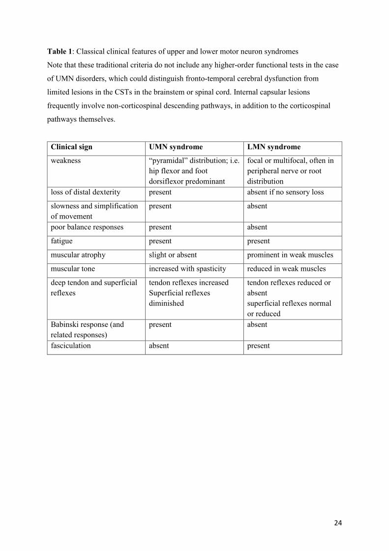
Table 1: Classical clinical features of upper and lower motor neuron syndromes
Note that these traditional criteria do not include any higher-order functional tests in the case
of UMN disorders, which could distinguish fronto-temporal cerebral dysfunction from
limited lesions in the CSTs in the brainstem or spinal cord. Internal capsular lesions
frequently involve non-corticospinal descending pathways, in addition to the corticospinal
pathways themselves.
Clinical sign UMN syndrome LMN syndrome
weakness “pyramidal” distribution; i.e. hip flexor and foot dorsiflexor predominant
focal or multifocal, often in peripheral nerve or root distribution
loss of distal dexterity present absent if no sensory loss
slowness and simplification of movement
present absent
poor balance responses present absent
fatigue present present
muscular atrophy slight or absent prominent in weak muscles
muscular tone increased with spasticity reduced in weak muscles
deep tendon and superficial reflexes
tendon reflexes increased Superficial reflexes diminished
tendon reflexes reduced or absent superficial reflexes normal or reduced
Babinski response (and related responses)
present absent
fasciculation absent present
25
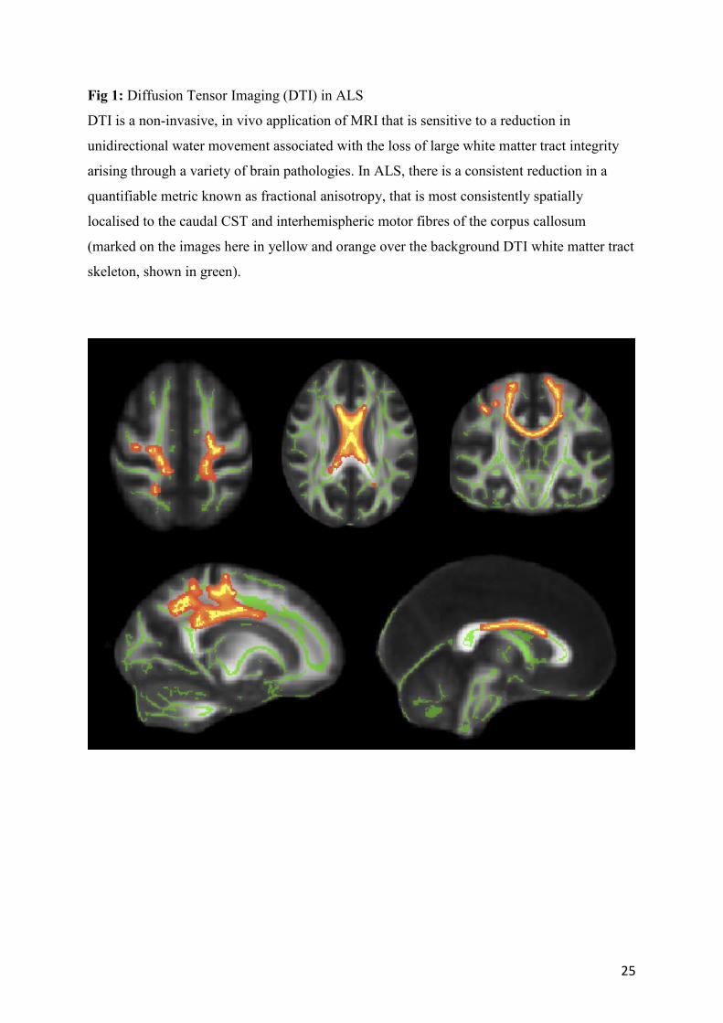
Fig 1: Diffusion Tensor Imaging (DTI) in ALS
DTI is a non-invasive, in vivo application of MRI that is sensitive to a reduction in
unidirectional water movement associated with the loss of large white matter tract integrity
arising through a variety of brain pathologies. In ALS, there is a consistent reduction in a
quantifiable metric known as fractional anisotropy, that is most consistently spatially
localised to the caudal CST and interhemispheric motor fibres of the corpus callosum
(marked on the images here in yellow and orange over the background DTI white matter tract
skeleton, shown in green).