Embed Size (px)
Citation preview
The Upper Limbs
Basic Anatomy The upper limb is a multijointed lever that is freely movable on the trunk at the
shoulder joint. At the distal end of the upper limb is the important organ, the hand.
Much of the importance of the hand depends on the pincer-like action of the
thumb, which enables one to grasp objects between the thumb and index finger.
The upper limb is divided into the shoulder (junction of the trunk with the arm),
arm, elbow, forearm, wrist, and hand.
Bones of the Shoulder Girdle and Arm
The shoulder girdle consists of the clavicle and the scapula, which articulate with
one another at the acromioclavicular joint.
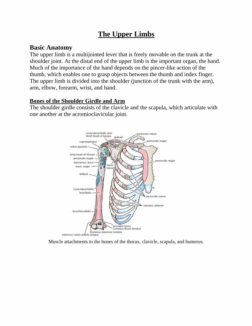
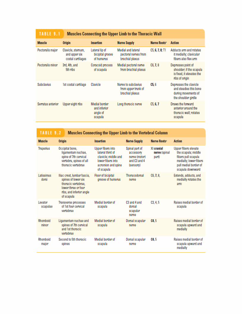
Muscle attachments to the bones of the thorax, clavicle, scapula, and humerus.
Clavicle
The clavicle is a long, slender bone that lies horizontally across the root of the neck
just beneath the skin. It articulates with the sternum and 1st costal cartilage
medially and with the acromion process of the scapula laterally. The clavicle acts
as a strut that holds the arm away from the trunk. It also transmits forces from the
upper limb to the axial skeleton and provides attachment for muscles.
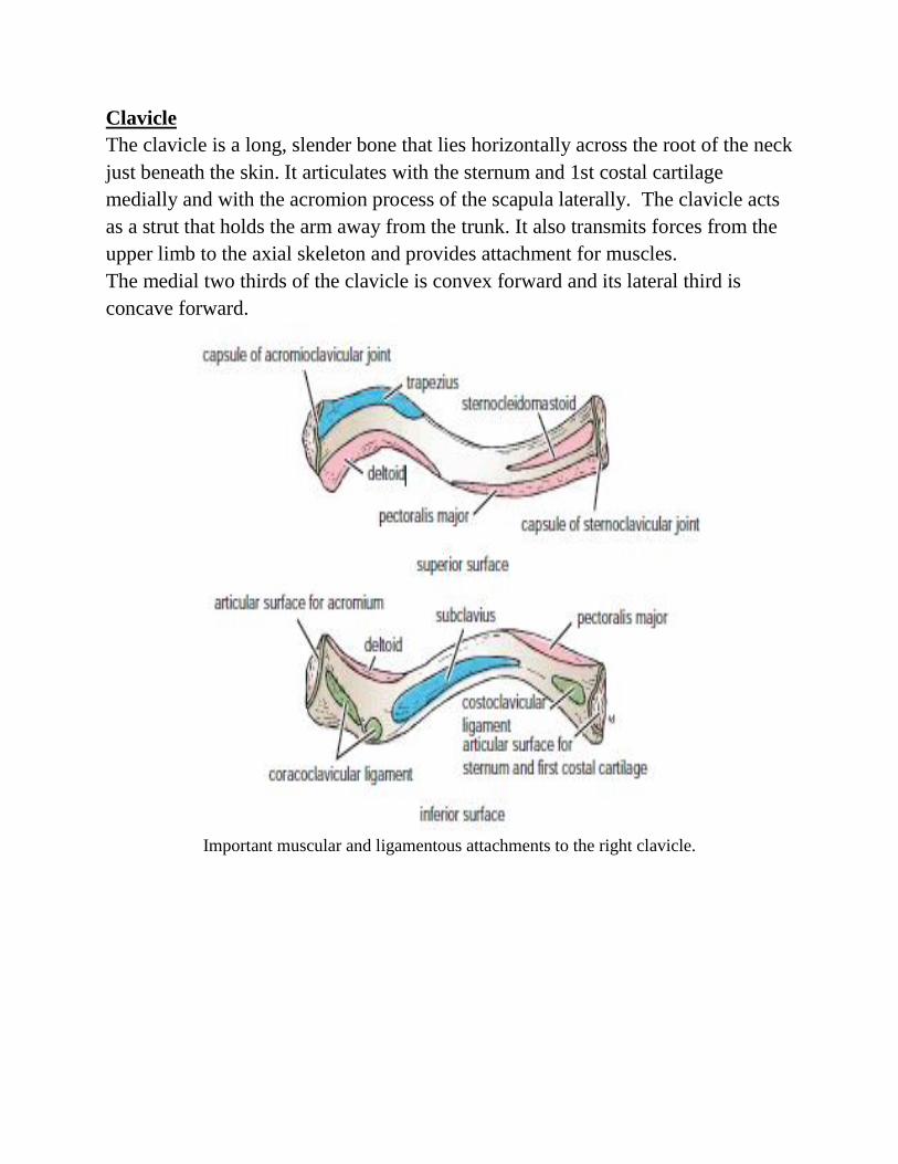
The medial two thirds of the clavicle is convex forward and its lateral third is
concave forward.
Important muscular and ligamentous attachments to the right clavicle.
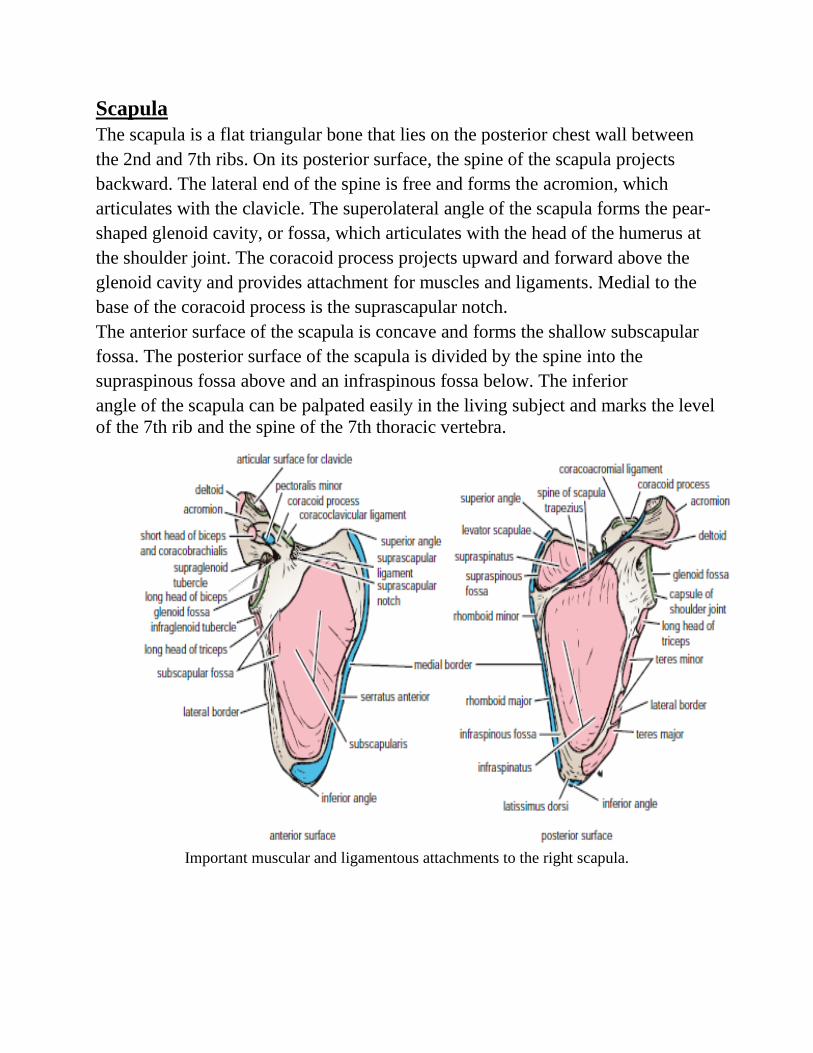
Scapula
The scapula is a flat triangular bone that lies on the posterior chest wall between
the 2nd and 7th ribs. On its posterior surface, the spine of the scapula projects
backward. The lateral end of the spine is free and forms the acromion, which
articulates with the clavicle. The superolateral angle of the scapula forms the pear-
shaped glenoid cavity, or fossa, which articulates with the head of the humerus at
the shoulder joint. The coracoid process projects upward and forward above the
glenoid cavity and provides attachment for muscles and ligaments. Medial to the
base of the coracoid process is the suprascapular notch.
The anterior surface of the scapula is concave and forms the shallow subscapular
fossa. The posterior surface of the scapula is divided by the spine into the
supraspinous fossa above and an infraspinous fossa below. The inferior
angle of the scapula can be palpated easily in the living subject and marks the level
of the 7th rib and the spine of the 7th thoracic vertebra.
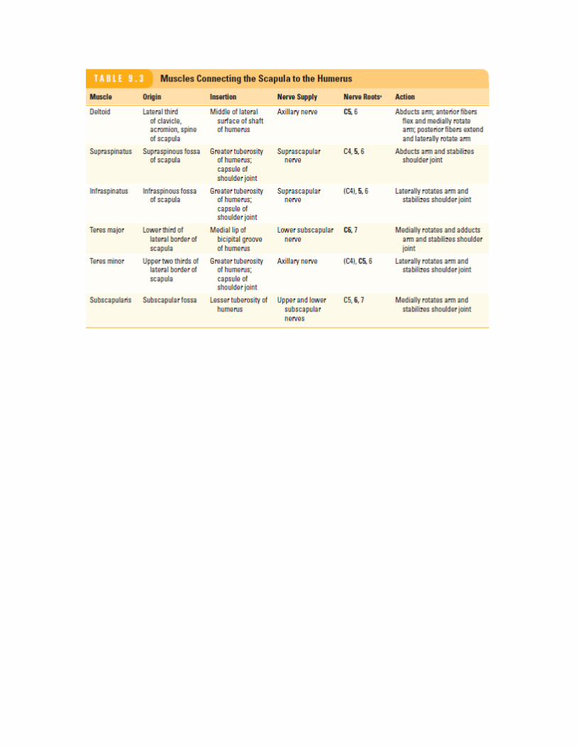
Important muscular and ligamentous attachments to the right scapula.
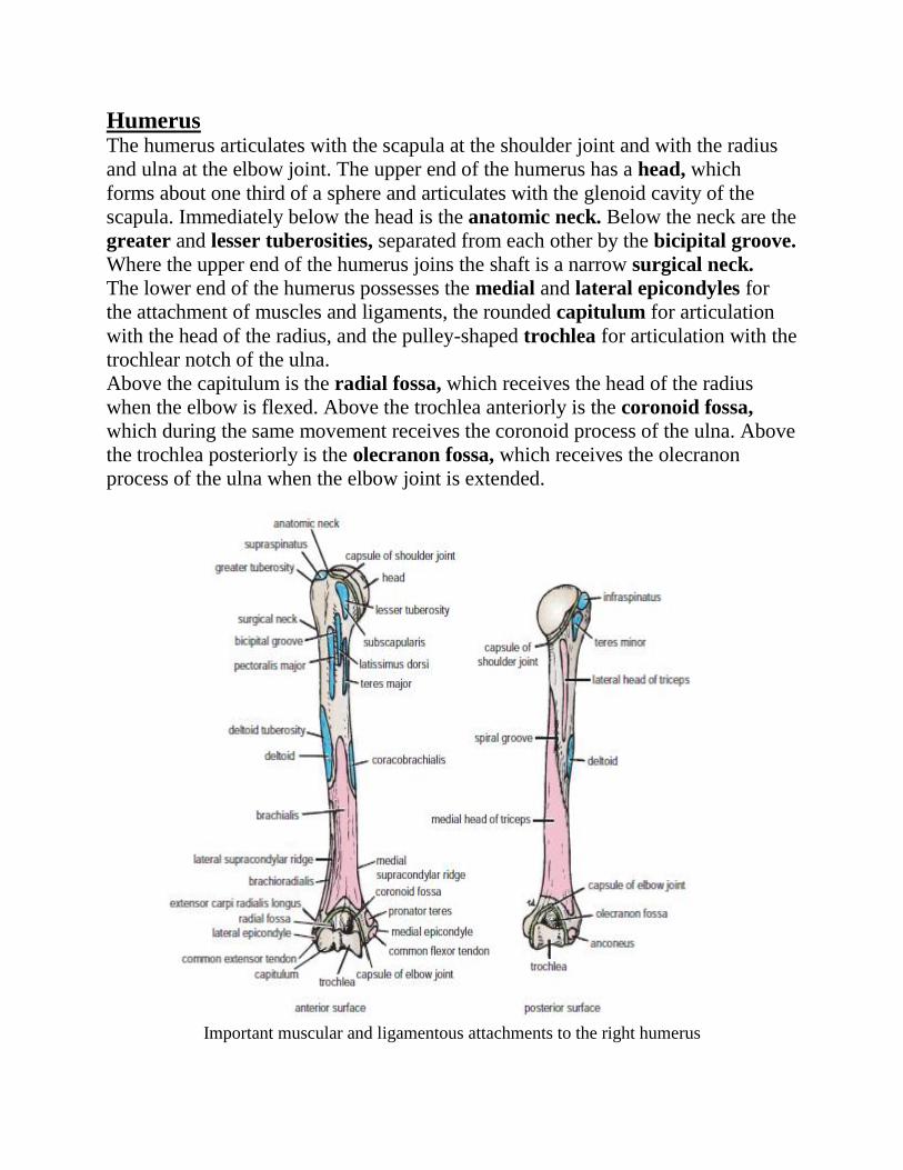
Humerus The humerus articulates with the scapula at the shoulder joint and with the radius
and ulna at the elbow joint. The upper end of the humerus has a head, which
forms about one third of a sphere and articulates with the glenoid cavity of the
scapula. Immediately below the head is the anatomic neck. Below the neck are the
greater and lesser tuberosities, separated from each other by the bicipital groove.
Where the upper end of the humerus joins the shaft is a narrow surgical neck.
The lower end of the humerus possesses the medial and lateral epicondyles for
the attachment of muscles and ligaments, the rounded capitulum for articulation
with the head of the radius, and the pulley-shaped trochlea for articulation with the
trochlear notch of the ulna.
Above the capitulum is the radial fossa, which receives the head of the radius
when the elbow is flexed. Above the trochlea anteriorly is the coronoid fossa,
which during the same movement receives the coronoid process of the ulna. Above
the trochlea posteriorly is the olecranon fossa, which receives the olecranon
process of the ulna when the elbow joint is extended.
Important muscular and ligamentous attachments to the right humerus
The Axilla
The axilla, or armpit, is a pyramid-shaped space between the upper part of the arm
and the side of the chest. It forms an important passage for nerves, blood, and
lymph vessels as they travel from the root of the neck to the upper limb. The upper
end of the axilla, or apex, is directed into the root of the neck and is bounded in
front by the clavicle, behind by the upper border of the scapula, and medially by
the outer border of the first rib. The lower end, or base, is bounded in front by
the anterior axillary fold (formed by the lower border of the pectoralis major
muscle), behind by the posterior axillary fold (formed by the tendon of latissimus
dorsi and the teres major muscle), and medially by the chest wall.
Walls of the Axilla
The walls of the axilla are made up as follows:
Anterior wall: By the pectoralis major, subclavius, and pectoralis minor
muscles
Posterior wall: By the subscapularis, latissimus dorsi, and teres major
muscles from above down
Medial wall: By the upper four or five ribs and the intercostal spaces
covered by the serratus anterior muscle.
Lateral wall: By the coracobrachialis and biceps muscles in the bicipital
groove of the humerus.
The base is formed by the skin stretching between the anterior and posterior walls
The axilla contains the principal vessels and nerves to the upper limb and many
lymph nodes.
Contents of the Axilla
The axilla contains the axillary artery and its branches, which supply blood to the
upper limb; the axillary vein and its tributaries, which drain blood from the upper
limb; and lymph vessels and lymph nodes, which drain lymph from the upper limb
and the breast and from the skin of the trunk, down as far as the level of the
umbilicus. Lying among these structures in the axilla is an important nerve plexus,
the brachial plexus, which innervates the upper limb. These structures are
embedded in fat.
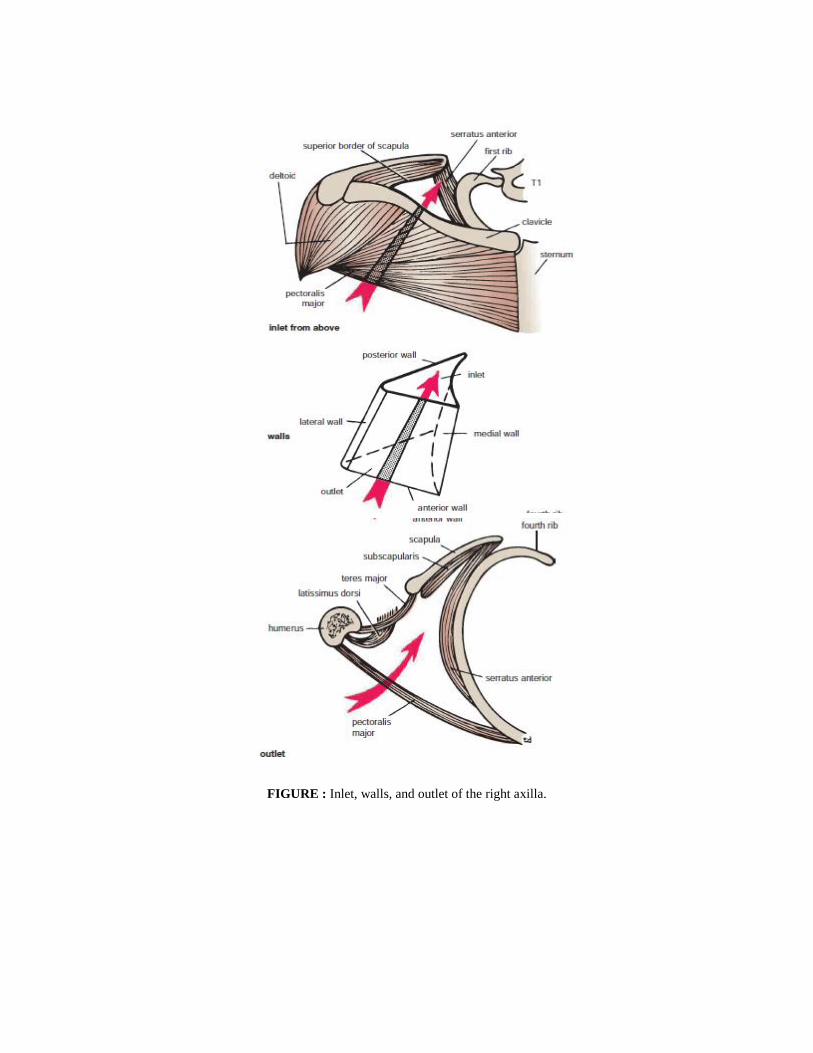
FIGURE : Inlet, walls, and outlet of the right axilla.
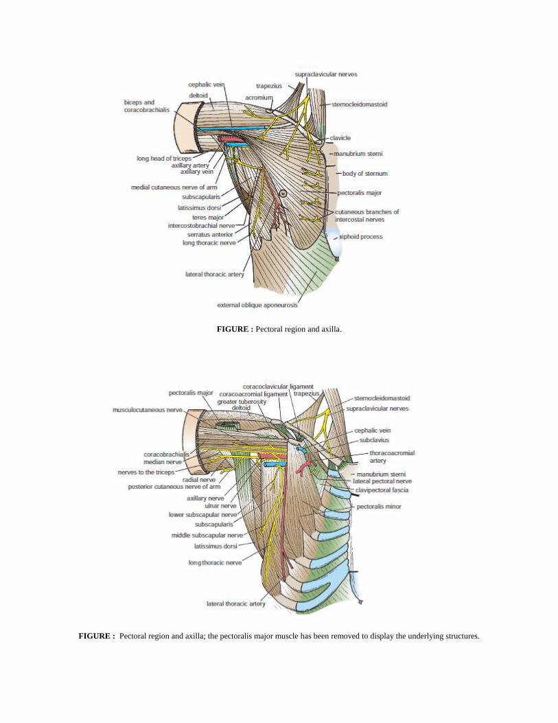
FIGURE : Pectoral region and axilla.
FIGURE : Pectoral region and axilla; the pectoralis major muscle has been removed to display the underlying structures.
Axillary Artery
The axillary artery begins at the lateral border of the 1st rib as a continuation of the
subclavian and ends at the lower border of the teres major muscle, where it
continues as the brachial artery. Throughout its course, the artery is closely related
to the cords of the brachial plexus and their branches and is enclosed with them in
a connective tissue sheath called the axillary sheath. If this sheath is traced
upward into the root of the neck, it is seen to be continuous with the prevertebral
fascia. The pectoralis minor muscle crosses in front of the axillary artery and
divides it into three parts .
First Part of the Axillary Artery This extends from the lateral border of the 1st
rib to the upper border of the pectoralis minor.
Relations
Anteriorly: The pectoralis major and the skin. The cephalic vein crosses the
artery.
Posteriorly: The long thoracic nerve (nerve to the serratus anterior).
Laterally: The three cords of the brachial plexus.
Medially: The axillary vein .
Second Part of the Axillary Artery This lies behind the pectoralis minor muscle
Relations
Anteriorly: The pectoralis minor, the pectoralis major, and the skin.
Posteriorly: The posterior cord of the brachial plexus, the subscapularis
muscle, and the shoulder joint.
Laterally: The lateral cord of the brachial plexus (Figs.
Medially: The medial cord of the brachial plexus and the axillary vein.
Third Part of the Axillary Artery This extends from the lower border of the
pectoralis minor to the lower border of the teres major.
Relations
Anteriorly: The pectoralis major for a short distance; lower down the artery,
it is crossed by the medial root of the median nerve.
Posteriorly: The subscapularis, the latissimus dorsi, and the teres major. The
axillary and radial nerves also lie behind the artery.
Laterally: The coracobrachialis, the biceps, and the humerus. The lateral
root of the median and the musculocutaneous nerves also lies on the lateral
side.
Medially: The ulnar nerve, the axillary vein, and the medial cutaneous nerve
of the arm.
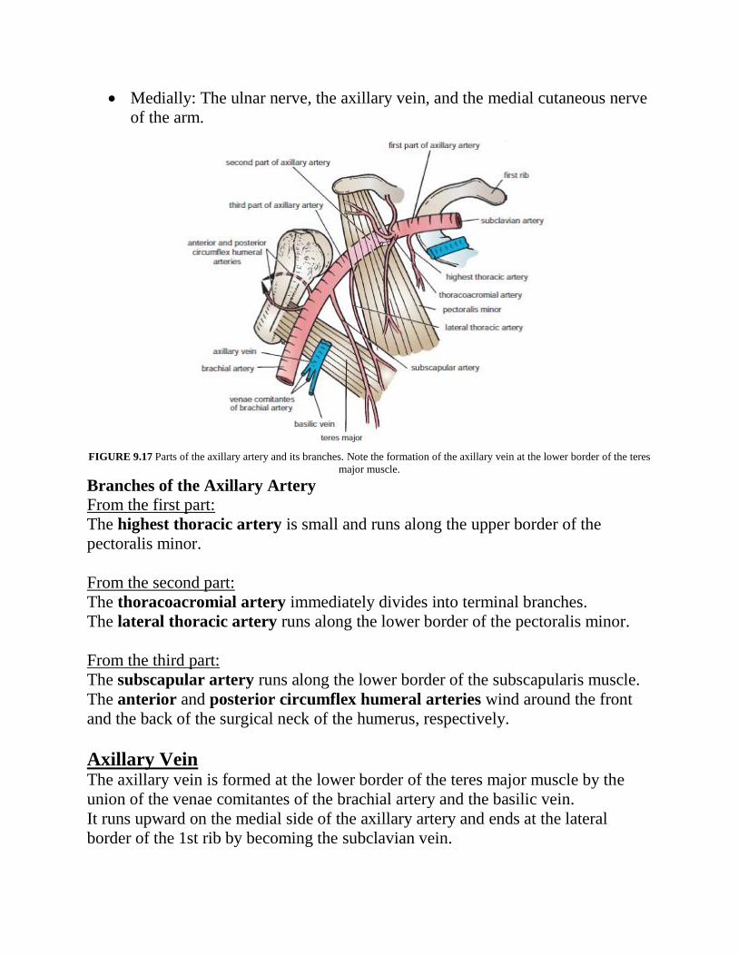
FIGURE 9.17 Parts of the axillary artery and its branches. Note the formation of the axillary vein at the lower border of the teres
major muscle.
Branches of the Axillary Artery
From the first part:
The highest thoracic artery is small and runs along the upper border of the
pectoralis minor.
From the second part:
The thoracoacromial artery immediately divides into terminal branches.
The lateral thoracic artery runs along the lower border of the pectoralis minor.
From the third part:
The subscapular artery runs along the lower border of the subscapularis muscle.
The anterior and posterior circumflex humeral arteries wind around the front
and the back of the surgical neck of the humerus, respectively.
Axillary Vein The axillary vein is formed at the lower border of the teres major muscle by the
union of the venae comitantes of the brachial artery and the basilic vein.
It runs upward on the medial side of the axillary artery and ends at the lateral
border of the 1st rib by becoming the subclavian vein.
The vein receives tributaries, which correspond to the branches of the axillary
artery, and the cephalic vein.
Brachial Plexus The nerves entering the upper limb provide the following important functions:
sensory innervation to the skin and deep structures, such as the joints; motor
innervation to the muscles; influence over the diameters of the blood vessels
by the sympathetic vasomotor nerves; and sympathetic secretomotor supply to the
sweat glands.
At the root of the neck, the nerves form a complicated plexus called the brachial
plexus. This allows the nerve fibers derived from different segments of the spinal
cord to be arranged and distributed efficiently in different nerve trunks to the
various parts of the upper limb. The brachial plexus is formed in the posterior
triangle of the neck by the union of the anterior rami of the 5th, 6th, 7th, and 8th
cervical and the 1st thoracic spinal nerves. The plexus can be divided into roots,
trunks, divisions, and cords. The roots of C5 and 6 unite to form the upper trunk,
the root of C7 continues as the middle trunk, and the roots of C8 and T1 unite to
form the lower trunk. Each trunk then divides into anterior and posterior divisions.
The anterior divisions of the upper and middle trunks unite to form the lateral cord,
the anterior division of the lower trunk continues as the medial cord, and the
posterior divisions of all three trunks join to form the posterior cord.
The cords become arranged around the axillary artery in the axilla.
Here, the brachial plexus and the axillary artery and vein are enclosed in the
axillary sheath. Cords of the Brachial Plexus All three cords of the brachial plexus
lie above and lateral to the first part of the axillary artery. The medial cord crosses
behind the artery to reach the medial side of the second part of the artery.The
posterior cord lies behind the second part of the artery, and the lateral cord lies on
the lateral side of the second part of the artery. Thus, the cords of the plexus have
the relationship to the second part of the axillary artery that is indicated by their
names. Most branches of the cords that form the main nerve trunks of the upper
limb continue this relationship to the artery in its third part.
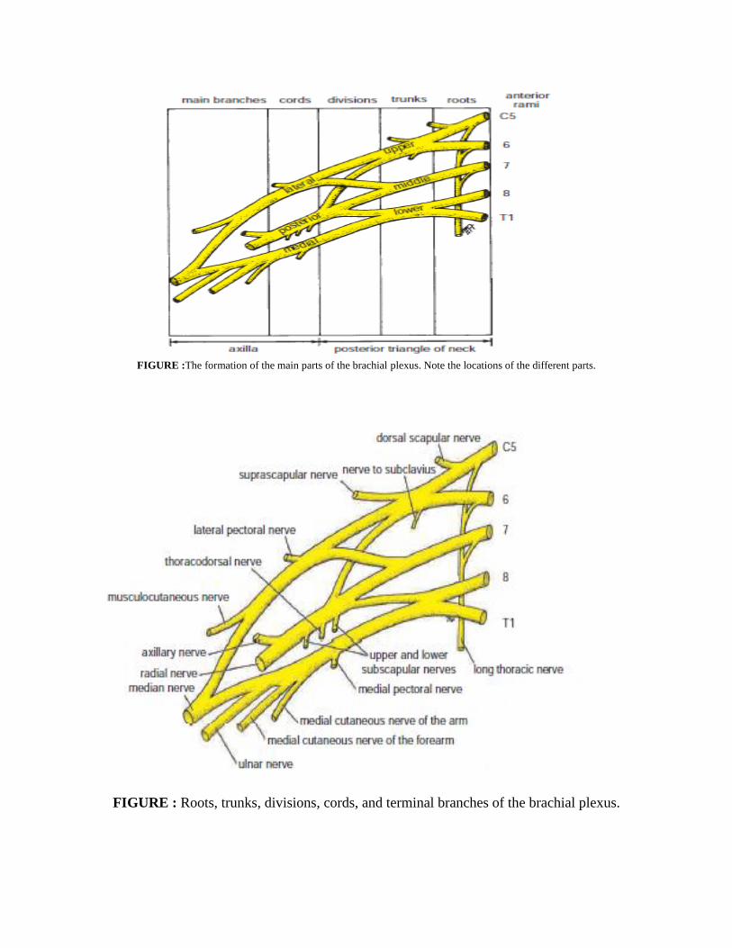
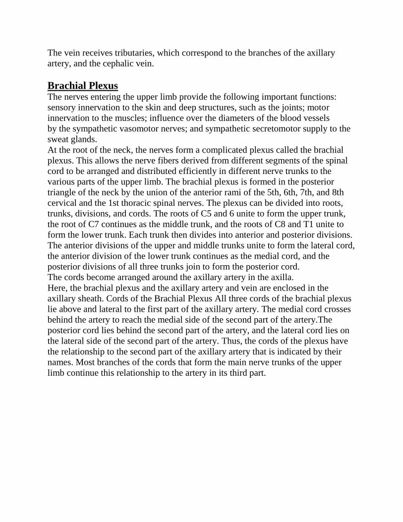
FIGURE :The formation of the main parts of the brachial plexus. Note the locations of the different parts.
FIGURE : Roots, trunks, divisions, cords, and terminal branches of the brachial plexus.
Lymph Nodes of the Axilla
The axillary lymph nodes (20 to 30 in number) drain lymph vessels from the lateral
quadrants of the breast, the superficial lymph vessels from the thoracoabdominal
walls above the level of the umbilicus, and the vessels from the upper limb.
The lymph nodes are arranged in six groups.
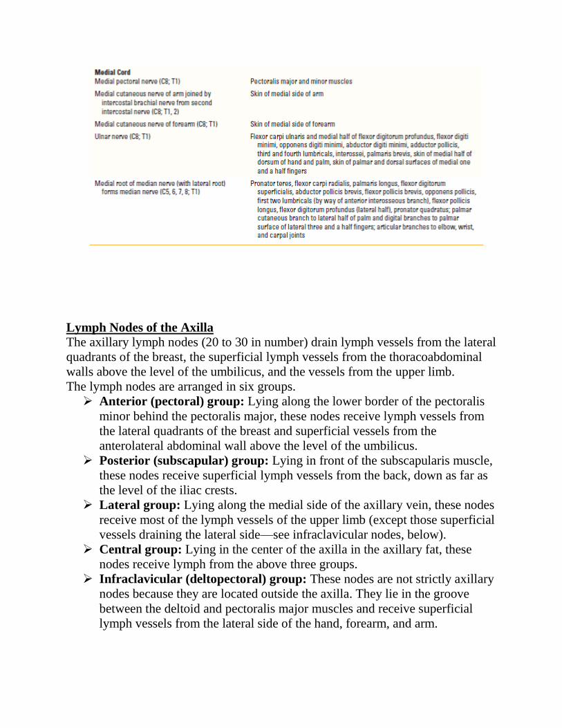
Anterior (pectoral) group: Lying along the lower border of the pectoralis
minor behind the pectoralis major, these nodes receive lymph vessels from
the lateral quadrants of the breast and superficial vessels from the
anterolateral abdominal wall above the level of the umbilicus.
Posterior (subscapular) group: Lying in front of the subscapularis muscle,
these nodes receive superficial lymph vessels from the back, down as far as
the level of the iliac crests.
Lateral group: Lying along the medial side of the axillary vein, these nodes
receive most of the lymph vessels of the upper limb (except those superficial
vessels draining the lateral side—see infraclavicular nodes, below).
Central group: Lying in the center of the axilla in the axillary fat, these
nodes receive lymph from the above three groups.
Infraclavicular (deltopectoral) group: These nodes are not strictly axillary
nodes because they are located outside the axilla. They lie in the groove
between the deltoid and pectoralis major muscles and receive superficial
lymph vessels from the lateral side of the hand, forearm, and arm.
Apical group: Lying at the apex of the axilla at the lateral border of the 1st
rib, these nodes receive the efferent lymph vessels from all the other axillary
nodes.
The apical nodes drain into the subclavian lymph trunk. On the left side,
this trunk drains into the thoracic duct; on the right side, it drains into the
right lymph trunk. Alternatively, the lymph trunks may drain directly into
one of the large veins at the root of the neck.
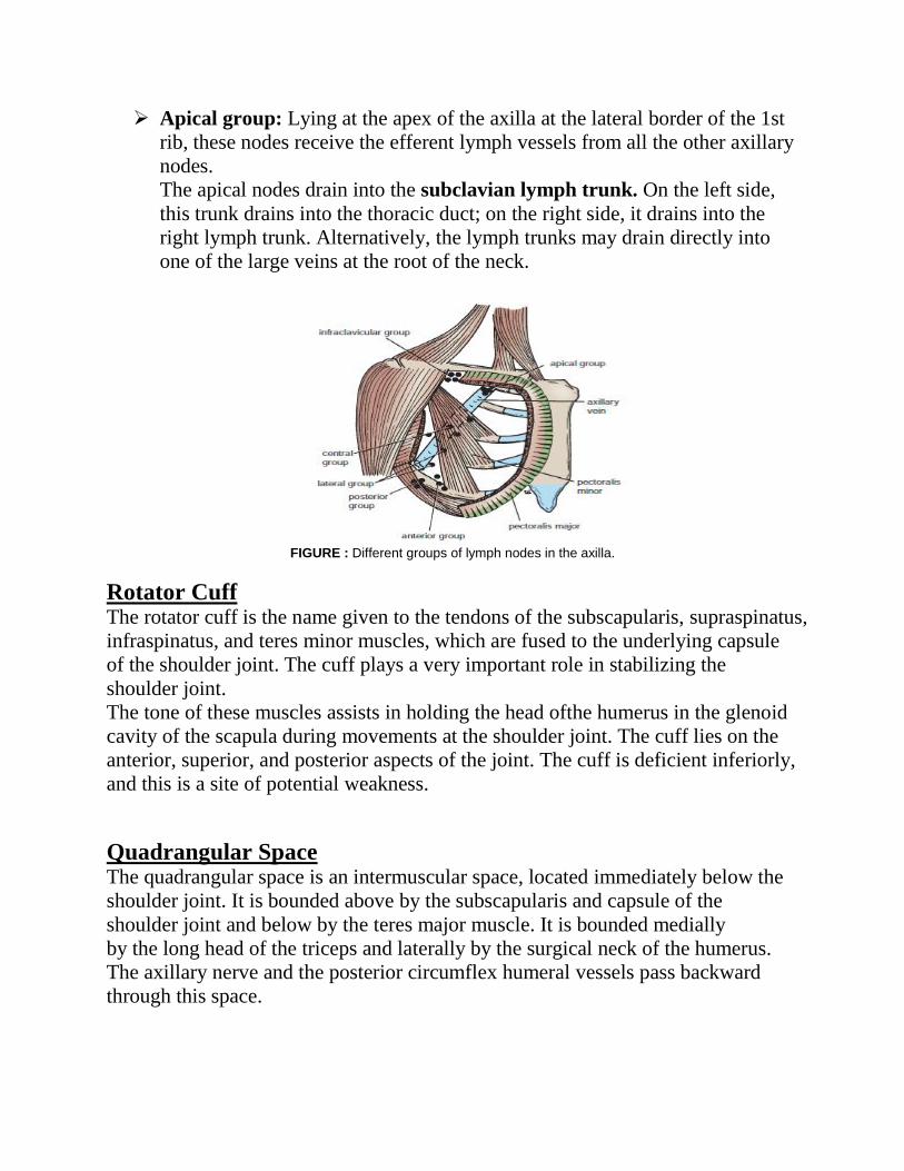
FIGURE : Different groups of lymph nodes in the axilla.
Rotator Cuff The rotator cuff is the name given to the tendons of the subscapularis, supraspinatus,
infraspinatus, and teres minor muscles, which are fused to the underlying capsule
of the shoulder joint. The cuff plays a very important role in stabilizing the
shoulder joint.
The tone of these muscles assists in holding the head ofthe humerus in the glenoid
cavity of the scapula during movements at the shoulder joint. The cuff lies on the
anterior, superior, and posterior aspects of the joint. The cuff is deficient inferiorly,
and this is a site of potential weakness.
Quadrangular Space The quadrangular space is an intermuscular space, located immediately below the
shoulder joint. It is bounded above by the subscapularis and capsule of the
shoulder joint and below by the teres major muscle. It is bounded medially
by the long head of the triceps and laterally by the surgical neck of the humerus.
The axillary nerve and the posterior circumflex humeral vessels pass backward
through this space.
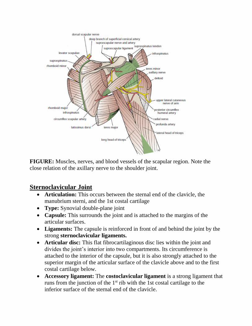
FIGURE: Muscles, nerves, and blood vessels of the scapular region. Note the
close relation of the axillary nerve to the shoulder joint.
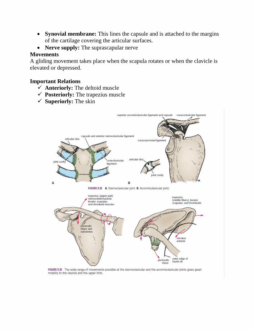
Sternoclavicular Joint Articulation: This occurs between the sternal end of the clavicle, the
manubrium sterni, and the 1st costal cartilage
Type: Synovial double-plane joint
Capsule: This surrounds the joint and is attached to the margins of the
articular surfaces.
Ligaments: The capsule is reinforced in front of and behind the joint by the
strong sternoclavicular ligaments.
Articular disc: This flat fibrocartilaginous disc lies within the joint and
divides the joint’s interior into two compartments. Its circumference is
attached to the interior of the capsule, but it is also strongly attached to the
superior margin of the articular surface of the clavicle above and to the first
costal cartilage below.
Accessory ligament: The costoclavicular ligament is a strong ligament that
runs from the junction of the 1st rib with the 1st costal cartilage to the
inferior surface of the sternal end of the clavicle.
Synovial membrane: This lines the capsule and is attached to the margins
of the cartilage covering the articular surfaces.
Nerve supply: The supraclavicular nerve and the nerve to the subclavius
muscle.
Movements
Forward and backward movement of the clavicle takes place in the medial compartment.
Elevation and depression of the clavicle take place in the lateral compartment.
Muscles Producing Movement
The forward movement of the clavicle is produced by the serratus anterior
muscle.
The backward movement is produced by the trapezius and rhomboid
muscles.
Elevation of the clavicle is produced by the trapezius, sternocleidomastoid,
levator scapulae, and rhomboid muscles.
Depression of the clavicle is produced by the pectoralis minor and the
subclavius muscles.
Important Relations
Anteriorly: The skin and some fibers of the sternocleidomastoid and
pectoralis major muscles
Posteriorly: The sternohyoid muscle; on the right, the brachiocephalic
artery; on the left, the left brachiocephalic vein and the left common carotid
artery
Acromioclavicular Joint Articulation: This occurs between the acromion of the scapula and the
lateral end of the clavicle.
Type: Synovial plane joint
Capsule: This surrounds the joint and is attached to the margins of the
articular surfaces.
Ligaments: Superior and inferior acromioclavicular ligaments reinforce
the capsule; from the capsule, awedge-shaped fibrocartilaginous disc
projects into the joint cavity from above.
Accessory ligament: The very strong coracoclavicular ligament extends
from the coracoid process to the undersurface of the clavicle. It is largely
responsible for suspending the weight of the scapula and the upper limb
from the clavicle.
Synovial membrane: This lines the capsule and is attached to the margins
of the cartilage covering the articular surfaces.
Nerve supply: The suprascapular nerve
Movements
A gliding movement takes place when the scapula rotates or when the clavicle is
elevated or depressed.
Important Relations
Anteriorly: The deltoid muscle
Posteriorly: The trapezius muscle
Superiorly: The skin
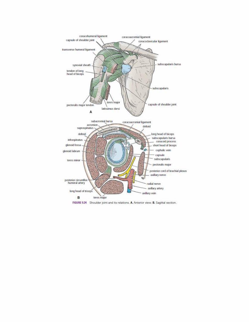
Shoulder Joint Articulation: This occurs between the rounded head of the humerus and the
shallow, pear-shaped glenoid cavity of the scapula. The articular surfaces are
covered by hyaline articular cartilage, and the glenoid cavity is deepened by
the presence of a fibrocartilaginous rim called the glenoid labrum.
Type: Synovial ball-and-socket joint
Capsule: This surrounds the joint and is attached medially to the margin of
the glenoid cavity outside the labrum; laterally, it is attached to the anatomic
neck of the humerus (Fig. 9.35). The capsule is thin and lax, allowing a wide
range of movement. It is strengthened by fibrous slips from the tendons of
the subscapularis, supraspinatus, infraspinatus, and teres minor muscles (the
rotator cuff muscles).
Ligaments: The glenohumeral ligaments are three weak bands of fibrous
tissue that strengthen the front of the capsule. The transverse humeral
ligament strengthens the capsule and bridges the gap between the two
tuberosities. The coracohumeral ligament strengthens the capsule above
and stretches from the root of the coracoid process to the greater tuberosity
of the humerus.
Accessory ligaments: The coracoacromial ligament extends between the
coracoid process and the acromion. Its function is to protect the superior
aspect of the joint.
Synovial membrane: This lines the capsule and is attached to the margins
of the cartilage covering the articular surfaces. It forms a tubular sheath
around the tendon of the long head of the biceps brachii. It extends through
the anterior wall of the capsule to form the subscapularis bursa beneath the
subscapularis muscle.
Nerve supply: The axillary and suprascapular nerves
Movements
The shoulder joint has a wide range of movement, and the stability of the joint has
been sacrificed to permit this. (Compare with the hip joint, which is stable but
limited in its movements.)
The following movements are possible:
Flexion: Normal flexion is about 90° and is performed by the anterior fibers
of the deltoid, pectoralis major, biceps, and coracobrachialis muscles.
Extension: Normal extension is about 45° and is performed by the posterior
fibers of the deltoid, latissimus dorsi, and teres major muscles.
Abduction: Abduction of the upper limb occurs both at the shoulder joint
and between the scapula and the thoracic wall. The middle fibers of the
deltoid, assisted by the supraspinatus, are involved. The supraspinatus
muscle initiates the movement of abduction and holds the head of the
humerus against the glenoid fossa of the scapula; this latter function allows
the deltoid muscle to contract and abduct the humerus at the shoulder joint.
Adduction: Normally, the upper limb can be swung 45° across the front of
the chest. This is performed by the pectoralis major, latissimus dorsi, teres
major, and teres minor muscles.
Lateral rotation: Normal lateral rotation is 40° to 45°. This is performed by
the infraspinatus, the teres minor, and the posterior fibers of the deltoid
muscle.
Medial rotation: Normal medial rotation is about 55°. This is performed by
the subscapularis, the latissimus dorsi, the teres major, and the anterior fibers
of the deltoid muscle.
Circumduction: This is a combination of the above movements.