Embed Size (px)
Citation preview
Surg Today Jpn J Surg (1997) 27:282-284 SURGERY TODAY
© Springer-Verlag 1997
The Unusual Imaging Appearance of Primary Retroperitoneal Teratoma: Report of a Case
MAYANK GOYAL, t RAJU SHARMA, 1 PYUSH SAWHNEY, 2 MEHAR CHAND SHARMA, 3 a n d MANORAMA BERRY l
Departments of a Radiodiagnosis, 2 Gastrointestinal Surgery, and 3 Pathology, All India Institute of Medical Sciences, New Delhi 110029 India
Abstract: Primary retroperitoneal (RP) teratoma is a rare entity which has a distinctive imaging appearance. We de- scribe herein the case of a 25-year-old man in whom a RP teratoma was found to have an extremely unusual imaging morphology by ultrasound and computed tomography (CT). The tumor was resected and histopathological examination confirmed the diagnosis of primary benign RP teratoma.
Key Words: retroperitoneal teratoma, ultrasound, computed tomography
Introduct ion
Primary retroperitoneal (RP) teratomas are extremely rare tumors representing less than 10% of all primary RP tumors? Their diagnosis may be suspected when the presence of calcification is evident on a plain radiograph of the abdomen; however, ultrasound and computed tomography (CT) scan are able to offer a more defini- tive diagnosis by the detection of fat and calcification. The present case illustrates a RP teratoma with an un- usual imaging appearance and distribution of fat.
Case R e p o r t
A 25-year-old man presented to our hospital with a 2- year history of progressive distention of the abdomen. It was not associated with any pain or weight loss. He had no bowel or urinary symptoms and his general physical examination was normal. Abdominal examination re- vealed a large mass of approximately 20 cm × 25 cm in
Reprint requests to: R. Sharma (Received for publication on Aug. 31,1995; accepted on July 4, 1996)
size in the epigastric and umbilical region. The mass was soft-to-firm and not significantly mobile in any direc- tion. There was no evidence of hepatosplenomegaly or ascites and routine laboratory investigations were unremarkable.
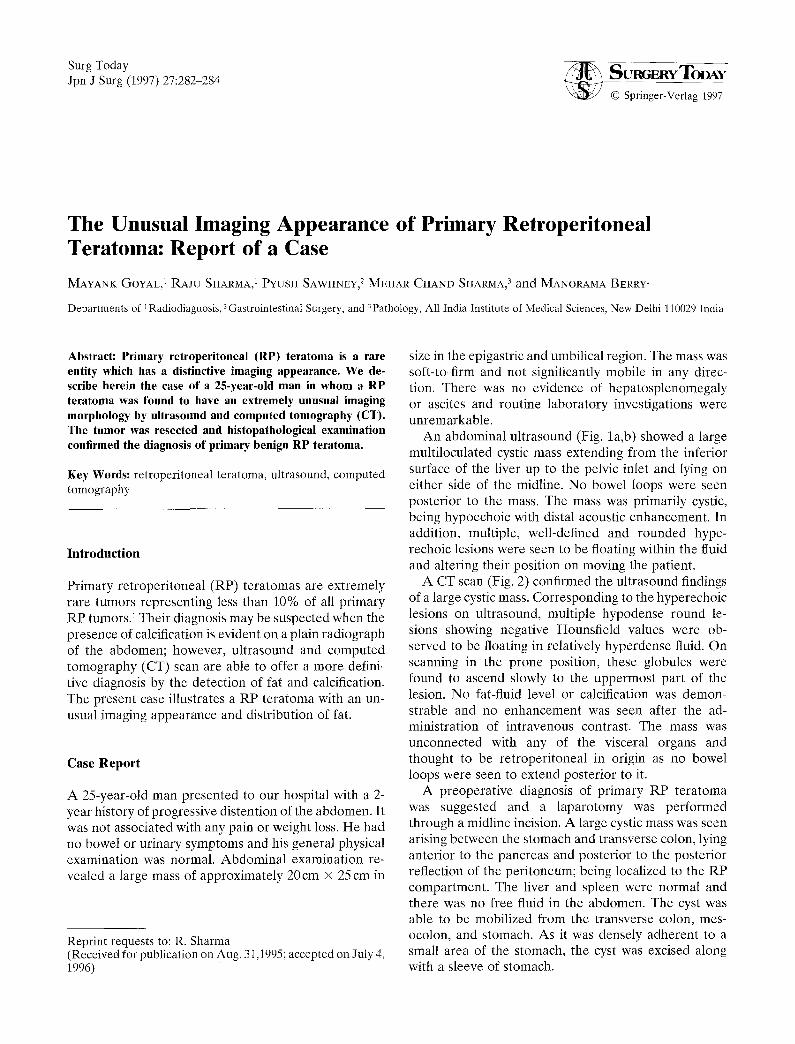
An abdominal ultrasound (Fig. la,b) showed a large multiloculated cystic mass extending from the inferior surface of the liver up to the pelvic inlet and lying on either side of the midline. No bowel loops were seen posterior to the mass. The mass was primarily cystic, being hypoechoic with distal acoustic enhancement. In addition, multiple, well-defined and rounded hype- rechoic lesions were seen to be floating within the fluid and altering their position on moving the patient.
A C T scan (Fig. 2) confirmed the ultrasound findings of a large cystic mass. Corresponding to the hyperechoic lesions on ultrasound, multiple hypodense round le- sions showing negative Hounsfield values were ob- served to be floating in relatively hyperdense fluid. On scanning in the prone position, these globules were found to ascend slowly to the uppermost part of the lesion. No fat-fluid level or calcification was demon- strable and no enhancement was seen after the ad- ministration of intravenous contrast. The mass was unconnected with any of the visceral organs and thought to be retroperitoneal in origin as no bowel loops were seen to extend posterior to it.
A preoperative diagnosis of primary RP teratoma was suggested and a laparotomy was performed through a midline incision. A large cystic mass was seen arising between the stomach and transverse colon, lying anterior to the pancreas and posterior to the posterior reflection of the peritoneum; being localized to the RP compartment. The liver and spleen were normal and there was no free fluid in the abdomen. The cyst was able to be mobilized from the transverse colon, mes- ocolon, and stomach. As it was densely adherent to a small area of the stomach, the cyst was excised along with a sleeve of stomach.

M. Goyal et al.: Retroperitoneal Teratoma 283
Fig. la,b. Ultrasound shows a large, multiloculated cystic mass with round, hyperechoic mobile lesions
Fig. 2. Computed tomography (CT) scan at the correspond- ing levels demonstrating hypodense floating lesions suggestive of fat ( -30 Hounsfield units)
Histopathology confirmed a diagnosis of teratoma (Fig. 3). Multiple sections examined from the specimen showed skin adnexal structures, mature cartilage, brain tissue, adipose tissue, and intestinal glands. No imma- ture tissue was identified and the floating lesions were confirmed to be comprised primarily of fat.
Discussion
Primary retroperi toneal (RP) teratomas are extremely rare and comprise less than 10% of all primary RP tumors? Approximately half the patients are diagnosed in the first decade of life and cases of teratomas being
detected in the fetus have been reported. 2 Less than 20% of cases occur in individuals over the age of 30 years. RP teratomas are significantly more common in females?, 3
Teratomas arise from totipotential germ cells un- dergoing variable differentiation and hence show components derived from ectoderm, mesoderm, and endoderm. TM On gross examination, cystic teratomas generally show the presence of sebaceous material and mature tissue. Teratomas having solid components are often malignant and contain immature embryonic tissue in addition to bony, fibrous, and fatty components. 2,3 RP teratomas are extremely variable in size, with tumors weighing more than 35 kg having been described in the literature. 2,5
RP teratomas are more frequently malignant in adults than in children, with incidences of 26% and 10%, respective. 46 Primary benign RP teratomas in adults are either asymptomatic or present with minimal symptoms such as vomiting, abdominal distention, back pain, and pedal edema. 1,2,4,7 However, malignant teratomas progress rapidly and pain is a prominent symptom. 7
Plain radiographs of the abdomen usually show a soft tissue mass displacing the bowel loops. The presence of bone or teeth, a pathognomonic finding in teratomas, is extremely rare in RP teratomas. 1,2 The presence of fat within the tumor is detected much less frequently on plain films as compared to CT scans? Calcification is also evident in 60%-80% of these lesions on CT scan, 3 and even though 74% of benign lesions show calcifica- tion, it also occurs in 25% of malignant RP tera tomasY Thus, calcification cannot be taken as a sign of benignity. Furthermore, the evaluation of size and the presence of solid components is not helpful in differ- entiating benign from malignant lesions, s

284 M. Goyal et al.: Retroperitoneal Teratoma
Fig. 3. Photomicrograph showing the ectodermal component in the form of hair follicles and mesodermal compo- nents, confirming the diagnosis of teratoma. (H&E × 100)
Ultrasound is an excellent modality to evaluate the cystic component of the mass as a hypoechoic region with distal acoustic enhancement. Fat has an extremely variable echogenicity on ultrasonography, ranging from echogenic to echolucent. 3,7 Occasionally, a fat-fluid level due to the presence of sebum, and distal acoustic shad- owing due to the presence of calcification, may be de- monstrable? ,9 The ultrasound image of our patient showed a cystic mass with hyperechoic, mobile globules and no calcification. However, as echogenicity depends not only on the presence of fat, but also on tissue inter- faces with different acoustic impedance, CT is thought to be better than ultrasound in the diagnosis of fat- containing tumors¢ ,9
The CT scan is able to demonstrate accurately the presence of fat as negative Hounsfield values and subtle calcifications. The presence of a fat-fluid level within a cystic lesion on CT scan is virtually pathognomonic of a t e ra tomaY ~9 CT is also better than ultrasonography in defining the extent, evaluation of involvement of the surrounding organs, and localization of the tumor to the RP compartment in the case of very large tumors. How- ever, the Hounsfield value of fat is variable, and teratomas without the presence of fat both on CT scan and histopathology have been reported?
According to the existing world literature, the diag- nosis of teratoma has been determined by the presence of fat, within a mass, with or without a fat-fluid level, and by loci of calcification. To our knowledge, the pat- tern of distribution of lipid globules demonstrated in the present case has never been described before. The fea- ture of low mobility of the fat globules to the uppermost
part of the lesion both on ultrasound and CT scan is also distinctly unusual. These well-defined rounded lesions on CT may simulate the appearance of cysts within a hydatid cyst; however, the presence of mobility of the globules, the hyperechoic appearance on ultrasound, and the negative Hounsfield value on CT help to differ- entiate the two conditions.
In conclusion, this case report illustrates an extremely unusual imaging morphology of primary RP teratoma by ultrasound and CT scanning.
References
1. Gschwend J, Burke TW, Woodward JE, Heller PB (1987) Retro- peritoneal teratoma presenting as an abdominopelvic mass. Obstet Gynecol 70:500-502
2. Pantoja E, Llobet R, Gonzalez-Flores B (1976) Retroperitoneal teratoma: historical review. J Urol 115:520-523
3. Davidson AJ, Hartman DS, Goldman SM (1989) Mature teratomas of the retroperitoneum: radiologic, pathologic and clini- cal correlation. Radiology 172:421-425
4. Engel RM, Elkins RC, Fletcher BD (1968) Retroperitoneal teratoma: review of literature and presentation of an unusual case. Cancer 22:1068-1073
5. Bruneton JN, Diard F, Drouillard JP~ Sabatier JC, Travernier JF (1980) Primary retroperitoneal teratoma in adults. Radiology 134:613-616
6. Lambrianides AL, Walker MM, Rosin RD (1987) Primary retro- peritoneal teratoma in adults. Urology 29:310-312
7. Panageas E (1991) Primary retroperitoneal teratoma. Am J Roentgenol 156:1292-1294
8. Billmire DF, Grosfeld JL (1986) Teratomas in childhood: analysis of 142 cases. J Pediatr Surg 21:548-551
9. Friedman AC, Pyatt RS, Hartman DS, Downey EF Jr, Olson WB (1982) CT of benign cystic teratomas. Am J Roentgenol 138:659- 665