Embed Size (px)
Citation preview
Biogeosciences, 4, 219–232, 2007www.biogeosciences.net/4/219/2007/© Author(s) 2007. This work is licensedunder a Creative Commons License.
Biogeosciences
The unique skeleton of siliceous sponges (Porifera; Hexactinellidaand Demospongiae) that evolved first from the Urmetazoa duringthe Proterozoic: a review
W. E. G. Muller1, Jinhe Li2, H. C. Schroder1, Li Qiao3, and Xiaohong Wang4
1Institut fur Physiologische Chemie, Abteilung Angewandte Molekularbiologie, Duesbergweg 6, 55099 Mainz, Germany2Institute of Oceanology, Chinese Academy of Sciences, 7 Nanhai Road, 266071 Qingdao, P. R. China3Department of Materials Science and Technology, Tsinghua University, 100084 Beijing, P. R. China4National Research Center for Geoanalysis, 26 Baiwanzhuang Dajie, 100037 Beijing, P. R. China
Received: 8 January 2007 – Published in Biogeosciences Discuss.: 6 February 2007Revised: 10 April 2007 – Accepted: 20 April 2007 – Published: 3 May 2007
Abstract. Sponges (phylum Porifera) had been consideredas an enigmatic phylum, prior to the analysis of their geneticrepertoire/tool kit. Already with the isolation of the first ad-hesion molecule, galectin, it became clear that the sequencesof sponge cell surface receptors and of molecules forming theintracellular signal transduction pathways triggered by them,share high similarity with those identified in other metazoanphyla. These studies demonstrated that all metazoan phyla,including Porifera, originate from one common ancestor, theUrmetazoa. The sponges evolved prior to the Ediacaran-Cambrian boundary (542 million years ago [myr]) duringtwo major “snowball earth events”, the Sturtian glaciation(710 to 680 myr) and the Varanger-Marinoan ice ages (605to 585 myr). During this period the ocean was richer in sil-ica due to the silicate weathering. The oldest sponge fossils(Hexactinellida) have been described from Australia, Chinaand Mongolia and are thought to have existed coeval withthe diverse Ediacara fauna. Only little younger are the fossilsdiscovered in the Sansha section in Hunan (Early Cambrian;China). It has been proposed that only the sponges possessedthe genetic repertoire to cope with the adverse conditions,e.g. temperature-protection molecules or proteins protectingthem against ultraviolet radiation.
The skeletal elements of the Hexactinellida (model organ-isms Monorhaphis chuniand Monorhaphis intermediaorHyalonema sieboldi) and Demospongiae (modelsSuberitesdomunculaandGeodia cydonium), the spicules, are formedenzymatically by the anabolic enzyme silicatein and thecatabolic enzyme silicase. Both, the spicules of Hexactinell-ida and of Demospongiae, comprise a central axial canal and
Correspondence to:W. E. G. Muller([email protected])
an axial filament which harbors the silicatein. After intracel-lular formation of the first lamella around the channel andthe subsequent extracellular apposition of further lamellaethe spicules are completed in a net formed of collagen fibers.
The data summarized here substantiate that with the find-ing of silicatein a new aera in the field of bio/inorganic chem-istry started. For the first time strategies could be formulatedand experimentally proven that allow the formation/synthesisof inorganic structures by organic molecules. These find-ings are not only of importance for the further understand-ing of basic pathways in the body plan formation of spongesbut also of eminent importance for applied/commercialprocesses in a sustainable use of biomolecules for novelbio/inorganic materials.
1 Introduction
The origin of the first ancestor of all metazoan phyla re-mained enigmatic until the identification of first sequencescoding for informative proteins from a sponge (phylumPorifera) by application of molecular biological techniques(Pfeifer et al., 1993). Before it had been only speculatedthat the sponges are true metazoans that are composed of in-dividually reacting and acting cells (see: Pechenik, 2000).With the isolation of the first sponge sequence, a galectin,it became overt that these animals have the genetic toolkit(reviewed in: Pilcher 2005) to allow their cells to dif-ferentiate from omnipotent via pluripotent to finally deter-mined somatic cells (reviewed in: Muller, 2006); most ofthe functional analyses were performed with the spongesSuberites domunculaandGeodia cydonium(see: Muller et
Published by Copernicus GmbH on behalf of the European Geosciences Union.
220 W. E. G. Muller et al.: The unique skeleton of siliceous sponges
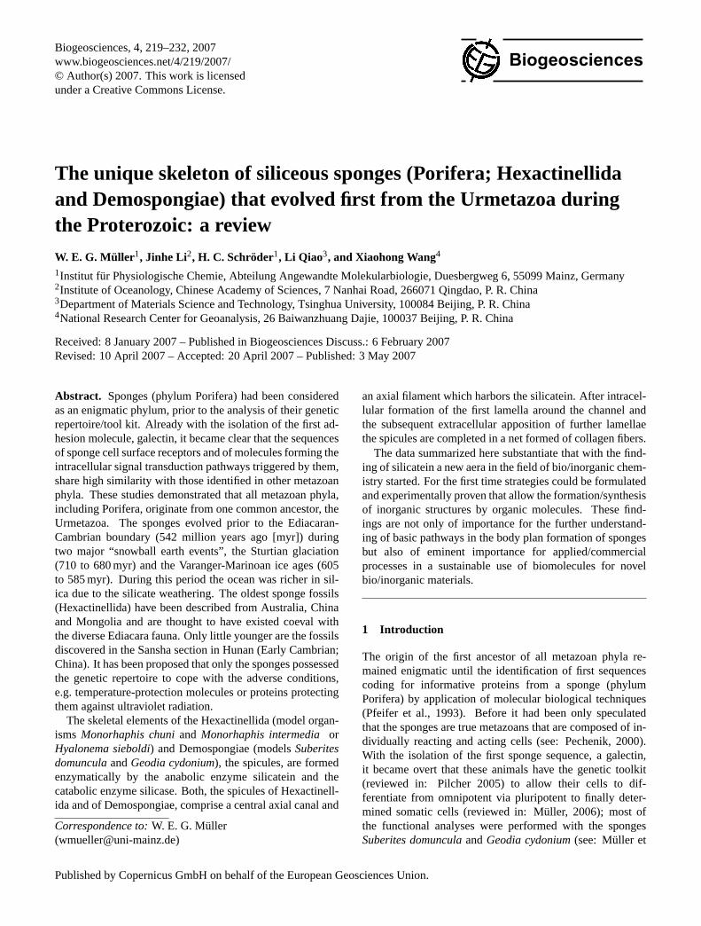
Fig. 1. Non-enzymatic precipitation of silica (A andB) and enzymatic formation of biosilica by sponges (C to E). (A) Roman vase (150years AC) showing opalescence [silicon-based inorganic films] on the surface of the glass (size: 7 cm). (B) Lithography from Haeckel (1899)showing the diverse forms of diatoms. (C) A specimen ofMonorhaphis chuniwith its giant spicule (height: 1.3 meters) is shown. (D) AHyalonemaspecies (Hexactinellida), collected by Doflein (1906) (height: 12 cm). (E) Lubomirskia baicalensis(Demospongiae), an endemicfreshwater sponge from Lake Baikal (height: 40 cm).
al., 2004a). With the first cell-matrix adhesion molecule, anintegrin (Pancer et al., 1997; reviewed in: Nichols et al.,2006), it could be substantiated that the sponges representorganisms that are composed of cells expressing cell surfacemolecules allowing their cross talks and in turn also divisionsand restrictions of their physiological functions (Muller andMuller, 2003). The individuality of a sponge specimen andthe morphogenetic arrangement of its cells according to a de-fined body plan was underscored by the discovery of apop-
totic as well as organizer-specific axis-forming molecules inS. domuncula(reviewed in: Wiens et al., 2004; Muller, 2005;Wiens and Muller, 2006). The next challenge was to under-stand the environmental factors which drove the evolution ofthe phylogenetically oldest animals, the sponges. One of thekey elements, promoting the emergence of these animals wassilicon, which displays both morphogenetic and also struc-tural properties (Krasko et al., 2002).
Biogeosciences, 4, 219–232, 2007 www.biogeosciences.net/4/219/2007/
W. E. G. Muller et al.: The unique skeleton of siliceous sponges 221
Urmetazoa
1x10 2x10 3x10
PalaeoproterozoicNeoproterozoic
Sno
wba
ll ev
ents
Bacteria
EukaryaMetazoa
Porifera
Years before present9 9 9
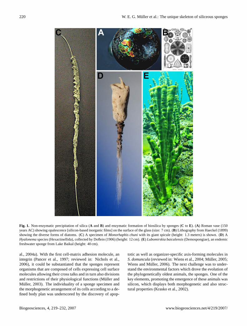
Fig. 2. Frequency of occurrence of major glacial periods (blue). The appearance of the different organismic groups [Bacteria; Eukarya;Metazoa and Porifera] are indicated. The Urmetazoa evolved during the inter-ice period between 600 MA and approximately 800 MA(modified after Hoffmann and Schrag 2002). The light blue bars indicate the major glacial periods, and the dark blue bars the occurrence of“banded iron formation”.
2 Role of silicon and silicate
Most skeletal structures in Metazoa are composed ofcalcium-salts, e.g. calcite or apatite. However, as has beenfound in vertebrates, silica deposition is seen prior to theossification process forming calcium-apatite bones (Carlisle,1986). The morphogenetic role of silicon is not restrictedto mammals, but has also been identified already in sponges(Krasko et al., 2000). InS. domunculathis element causes es-pecially the expression of genes that are required for the for-mation of the skeletal elements (Krasko et al., 2002; Mulleret al., 2004a; Muller, 2006). Silicon/silica is used in the twosponge classes Hexactinellida and Demospongiae, as start-ing substrate for the enzymatic synthesis of their silica-basedspicules which are the key structures, allowing the forma-tion/arrangement of differentiated cells within an individualaccording to a body plan (Wiens et al., 2006; Muller, 2006).
Silicon/silicate is usually precipitated passively onto in-organic matrices, e.g. Roman glasses (Fig. 1A), or on or-ganic matrices like in diatoms (Fig. 1B). In sponges, how-ever, the formation of “polymerized”/condensed silicate isgoverned by an enzyme termed silicatein (Cha et al., 1999).Even after this information is available, it remains to be stud-ied which morphogenetic factors allowed the formation ofup to two meters large and highly elaborated sponges, e.g.the hexactinellidsMonorhaphis chuni(Fig. 1C) or other hex-actinellids likeHyalonemaspecies (Fig. 1D) and also demo-sponges, e.g.Lubomirskia baicalensis(Fig. 1E). The ques-tion arises: When and in which environment did the spongesappear/evolve?
3 Evolution during the Proterozoic: evolution ofsponges in the silicon ocean
It is surprising that 542 million years ago [myr], near theEdiacaran-Cambrian boundary, a rapid appearance of differ-ent animal types occurred. Exciting examples of these emer-gences were the so-called Ediacaran biota, which are olderthan the Cambrian animals (Knoll and Caroll, 1999). Thedevelopment and divergence of the major animal clades weresurely driven by the genetic tool kits available at that time.The major, or perhaps even only, metazoan phylum with hardskeleton that existed at the border from Ediacaran to Cam-brian (approximately 543 MA) which did not become extinctare the sponges. Consequently, sponges were also termed“living fossils” (Muller, 1998); they represent the evolution-ary oldest, still extant taxon which testifies the developmen-tal level of the animals that lived in the Neo-Proterozoiceon (1000 to 520 MA); Figs. 2 and 3. This must be espe-cially mentioned since two major “snowball earth events”occurred, the Sturtian glaciation (710 to 680 myr) and theVaranger-Marinoan ice ages (605 to 585 myr), which verylikely caused the covering of the earth by a continuous icelayer (Hoffmann and Schrag, 2002). It was proposed that asa consequence of these ice ages most species went extinct,perhaps more than 85% (Hoffman et al., 1998).
The primordial earth surface comprised initially insolu-ble silicates and carbonates as well as, to a small extent,phosphates. During the silicate weathering-carbonate pre-cipitation cycle, prior or simultaneously with the glaciations,a dissolution of these surface rocks composed of insolu-ble silicates [CaSiO3] resulted in formation of soluble cal-cium carbonate [CaCO3] and soluble silica [SiO2], under
www.biogeosciences.net/4/219/2007/ Biogeosciences, 4, 219–232, 2007
222 W. E. G. Muller et al.: The unique skeleton of siliceous sponges
Urbilateria
Deuterostomia
CnidariaCtenophora
oral/aboral axis
dorsoventral polarity
biradial symmetry
radial symmetry
Hexactinellida
Demospongiae
Calcarea
PORIFERA
Urmetazoa
(silicic acid skeleton)
living fossils
Protostomia
(Ca-carbonate skeleton)
cell-cell- and cell-matrix molecules / immune molecules
apoptosis / morphogens
Archaeocyatha
extinct
Sturtian Glaciation (710-680 MYA)
Varanger-Marinoan Glaciation(605-585 MYA)
silicic acid
Ca-carbonate
Cacarbonate
apatite
?
?
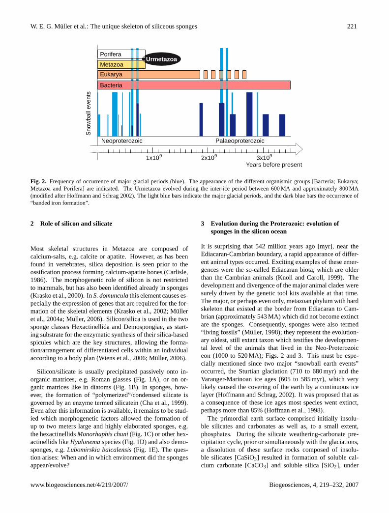
Fig. 3. Phylogenetic position of the Porifera between the Urmetazoa and the Urbilateria. The major evolutionary novelties which have to beattributed to the Urmetazoa are those molecules which mediate apoptosis and control morphogenesis, the immune molecules and primarilythe cell adhesion molecules. The siliceous sponges with the two classes Hexactinellida and Demospongiae emerged first and finally theCalcarea, which possess a calcareous skeleton, appeared. These three classes of Porifera are living fossils that provide a reservoir formolecular biological studies. The Archaeocyatha, sponge related animals with a calcareous skeleton, became extinct. The Calcarea are verylikely a sister group of the Cnidaria (Schutze et al., 1999). From the latter phylum the Ctenophora might have evolved which comprise notonly an oral/aboral polarity but also a biradial symmetry. Finally the Urbilateria emerged from which the Protostomia and the Deuterostomiaoriginated. Very likely the Urmetazoa emerged between the two major “snowball earth events”, the Sturtian glaciation (710 to 680 myr) andthe Varanger-Marinoan ice ages (605 to 585 myr) (Hoffmann and Schrag 2002); the ice ages are marked (green triangles). In the two poriferanclasses Hexactinellida and Demospongiae the skeleton is composed of amorphous and hydrated silica, while the spicules of Calcarea arecomposed of Ca-carbonate. The latter biomineral is also prevalent in Protostomia and Deuterostomia. In vertebrates the bones are composedof Ca-phosphate [apatite].
consumption of atmospheric CO2 (Walker, 2003). The re-sulting soluble minerals leached into the waters of the rivers,lakes and oceans. There, they were again re-precipitated intonew minerals, as part of the sedimentary rocks. Such pro-cesses depend upon temperature, pH and atmospheric carbondioxide. Passively, the minerals are transformed diageneti-cally to secondary structures.
In contrast to passive re-precipitation, biogenic depositionof minerals by metazoans is first seen in sponges. The old-est sponge fossils (Hexactinellida) have been described fromAustralia, China and Mongolia (from more than 540 MA)(Gehling and Rigby, 1996; Brasier et al., 1997; Li et al.,1998). Hence, the Hexactinellida are the oldest group ofsponges as documented there and later in fossil records ofthe Sansha section in Hunan (Early Cambrian; China; Steiner
et al., 1993; Steiner, 1994). In both lower and upper lev-els of the Niutitang Formation more or less completely pre-served sponge fossils, e.g.Solactiniella plumata(Fig. 4A-ato Fig. 4A-c), have been discovered (Steiner et al., 1993).The occurrence of almost intact sponge fossils also in thebasal part of this formation is real (Rigby and Guang, 1996)and is stratigraphically equivalent to the Chengjiang assem-blage in Yunnan (China). So far the base of the NiutitangFormation has been correlated with the Tommotian blackshales, “Badaowan” Member. However, the transgressionthat deposited the black shale of the Niutitang Formationand the “Badaowan” Member was a diacronous event, whichfirst became evident in the basin environment, Hunan/SE –Guizhon/S-Anhui, and progressed across the platform, Yun-nan/Sichuan (Steiner, 1994). Therefore, the base of the
Biogeosciences, 4, 219–232, 2007 www.biogeosciences.net/4/219/2007/
W. E. G. Muller et al.: The unique skeleton of siliceous sponges 223
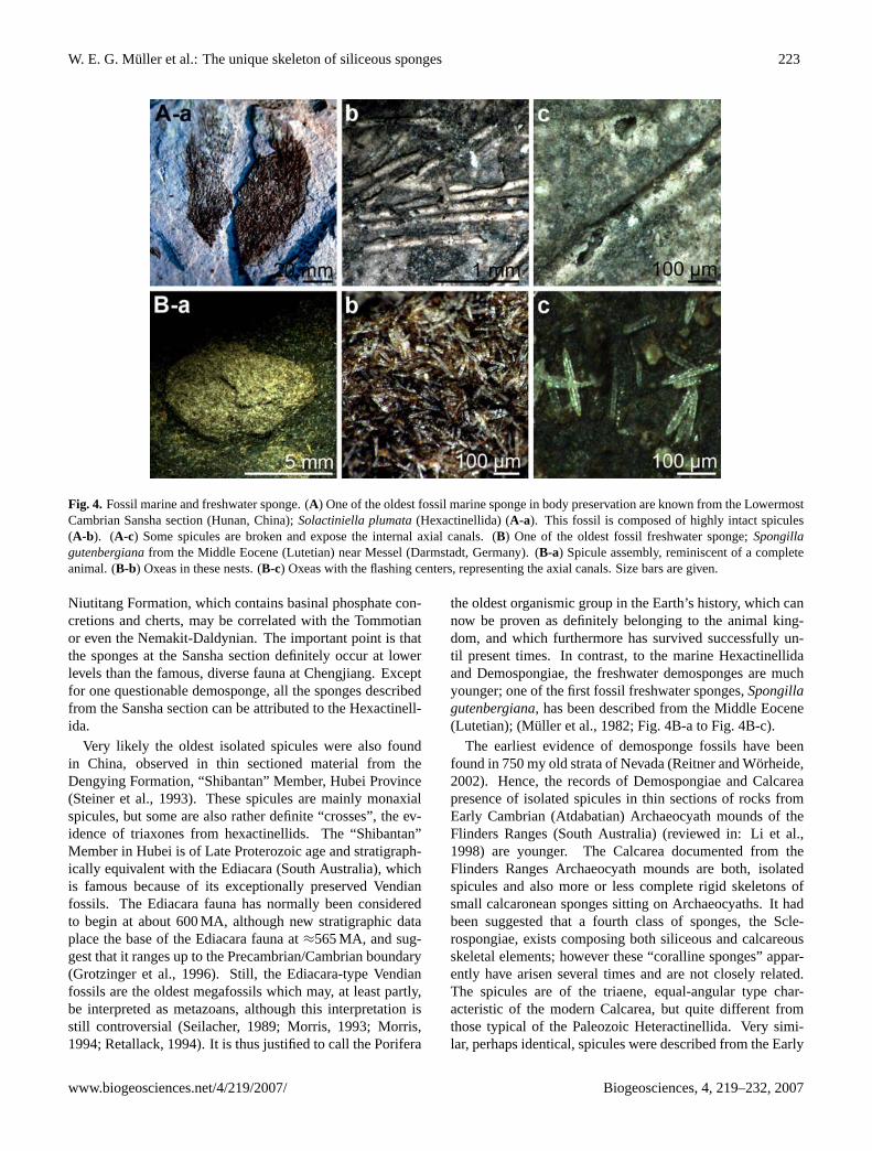
Fig. 4. Fossil marine and freshwater sponge. (A) One of the oldest fossil marine sponge in body preservation are known from the LowermostCambrian Sansha section (Hunan, China);Solactiniella plumata(Hexactinellida) (A-a). This fossil is composed of highly intact spicules(A-b). (A-c) Some spicules are broken and expose the internal axial canals. (B) One of the oldest fossil freshwater sponge;Spongillagutenbergianafrom the Middle Eocene (Lutetian) near Messel (Darmstadt, Germany). (B-a) Spicule assembly, reminiscent of a completeanimal. (B-b) Oxeas in these nests. (B-c) Oxeas with the flashing centers, representing the axial canals. Size bars are given.
Niutitang Formation, which contains basinal phosphate con-cretions and cherts, may be correlated with the Tommotianor even the Nemakit-Daldynian. The important point is thatthe sponges at the Sansha section definitely occur at lowerlevels than the famous, diverse fauna at Chengjiang. Exceptfor one questionable demosponge, all the sponges describedfrom the Sansha section can be attributed to the Hexactinell-ida.
Very likely the oldest isolated spicules were also foundin China, observed in thin sectioned material from theDengying Formation, “Shibantan” Member, Hubei Province(Steiner et al., 1993). These spicules are mainly monaxialspicules, but some are also rather definite “crosses”, the ev-idence of triaxones from hexactinellids. The “Shibantan”Member in Hubei is of Late Proterozoic age and stratigraph-ically equivalent with the Ediacara (South Australia), whichis famous because of its exceptionally preserved Vendianfossils. The Ediacara fauna has normally been consideredto begin at about 600 MA, although new stratigraphic dataplace the base of the Ediacara fauna at≈565 MA, and sug-gest that it ranges up to the Precambrian/Cambrian boundary(Grotzinger et al., 1996). Still, the Ediacara-type Vendianfossils are the oldest megafossils which may, at least partly,be interpreted as metazoans, although this interpretation isstill controversial (Seilacher, 1989; Morris, 1993; Morris,1994; Retallack, 1994). It is thus justified to call the Porifera
the oldest organismic group in the Earth’s history, which cannow be proven as definitely belonging to the animal king-dom, and which furthermore has survived successfully un-til present times. In contrast, to the marine Hexactinellidaand Demospongiae, the freshwater demosponges are muchyounger; one of the first fossil freshwater sponges,Spongillagutenbergiana, has been described from the Middle Eocene(Lutetian); (Muller et al., 1982; Fig. 4B-a to Fig. 4B-c).
The earliest evidence of demosponge fossils have beenfound in 750 my old strata of Nevada (Reitner and Worheide,2002). Hence, the records of Demospongiae and Calcareapresence of isolated spicules in thin sections of rocks fromEarly Cambrian (Atdabatian) Archaeocyath mounds of theFlinders Ranges (South Australia) (reviewed in: Li et al.,1998) are younger. The Calcarea documented from theFlinders Ranges Archaeocyath mounds are both, isolatedspicules and also more or less complete rigid skeletons ofsmall calcaronean sponges sitting on Archaeocyaths. It hadbeen suggested that a fourth class of sponges, the Scle-rospongiae, exists composing both siliceous and calcareousskeletal elements; however these “coralline sponges” appar-ently have arisen several times and are not closely related.The spicules are of the triaene, equal-angular type char-acteristic of the modern Calcarea, but quite different fromthose typical of the Paleozoic Heteractinellida. Very simi-lar, perhaps identical, spicules were described from the Early
www.biogeosciences.net/4/219/2007/ Biogeosciences, 4, 219–232, 2007
224 W. E. G. Muller et al.: The unique skeleton of siliceous sponges
Cambrian of Sardinia asSardospongia triradiataand at-tributed to the Heteractinellida (Mostler, 1985). The com-plete calcarean spongeGravestockia pharetronensis(Reit-ner, 1992) from the Flinders Ranges possessed a rigid cal-citic skeleton with affinity to the modern Pharetronida. Asone of the earliest families of demosponges the Geodiidaehave been described on the basis of their sterrasters (Reitnerand Mehl, 1995).
Those taxa that survived the “snowball earth” episodesmust have had the genetic repertoire to cope with the ad-verse conditions, meaning: (i) proteins to protect againstadverse temperature, (ii ) possibility to survive food restric-tion and (iii ) – not to forget – also the protection machin-ery against ultraviolet radiation. During the pre-Sturtian pe-riod, atmospheric CO2 had been consumed and removedfrom the atmosphere resulting in extreme temperature ampli-tudes. Sponges have cryoprotective proteins, e.g. galactose-specific lectins (Pfeifer et al., 1993; Wiens et al., 2003) whichdisplay cryoprotective protein-membrane activity, or, inter-estingly, alsoβγ -crystallins, proteins that contain only lowamounts of water and hence are resistant to adverse proteinfolding (Krasko et al., 1997). Food restriction was proba-bly compensated by the establishment of a symbiotic rela-tionship with microorganisms (Muller et al., 2004b), suchas gram-positive (Thakur et al., 2005) and gram-negativebacteria (Wiens et al., 2005) as well as fungi (Perovic-Ottstadt et al., 2004a). This eukaryotic-prokaryotic “labor-division” allowed the sponges a flexible and rapid adapta-tion to the changing environment. It is furthermore amaz-ing that sponges have an unexpected variety of protectionsystems against mutagens, including also ultraviolet radia-tion. Several protection systems against radiation have beendescribed; e.g. the (6–4) photolyase system (Krasko et al.,2003b) or the SOS-response-like mechanism (Krasko et al.,1998), and numerous is the literature on protection systemsagainst environmental stress (e.g. Efremova et al., 2002).Perhaps the greatest fortune for the sponges was their abil-ity to utilize silicic acid as substrate for their skeleton. Whensponges emerged, the insoluble silicate minerals were con-verted into monomeric, soluble silica providing the spongeswith an advantageous basis for survival and diversification,with respect to the number of the species and their abun-dance. In this context it should be mentioned that with thediversification and evolution of sponges (at least valid forendemic freshwater species) an increase in the gene num-ber for silicateins occurred (Muller et al., 2006d). Early as-sumptions postulated that those taxa which survived mass ex-tinction, e.g. the Foraminifera, became ecological and mor-phologically generalized species (Cifelli, 1969). Further-more, the number of species, the diversity of the biota, in-creased rapidly after each period of extinction (Futuyma,1986). Hence, the sponges (survivor taxon) became the ben-eficiaries of the glaciation crises and received the chance tocolonize those habitats which had been de-populated (seealso: Butterfield, 2007).
The urmetazoans/sponges (Muller et al., 2001) had al-ready the basic genetic toolkit for all derived metazoanswhich emerged during the “Cambrian Explosion” (Figs. 2and 3). This statement implies that the genetic repertoire ofthe sponges, which survived the glaciations, gave the frame,potentials/potentialities but also the limits of the body planconstruction seen in higher derived taxa and which exist inthe present day animal phyla. The derived taxa utilized thepre-existing molecules and pathways for their diversificationof patterning and for an increase in the genetic network com-plexity. It can be postulated that during the progress of evo-lution the degree of redundancy decreased on the expenseof an increase in complexity. This progressive “perfection”might be detrimental to the stability and survival of most ofthe species which are evolutionary younger.
The oxygen level in the atmosphere and in the water dur-ing the early Proterozoic was lower than at present (Towe,1970; Hayes, 1994). It had been postulated that with therise of oxygen level the synthesis of collagen became pos-sible (Towe, 1981); sponges contain and express collagengenes (Garrone, 1998). The ability to form these extracellu-lar fibrils had been considered to be a crucial prerequisite forthe origin of multicellular animals and the establishment of ametazoan body plan (Towe, 1981). Until now, no experimen-tal evidence for an existence of a primordial, blood oxygen-transporting system in sponges has been found. Therefore,we assume that most of the oxygen required by these ani-mals to allow intermediary metabolism is provided by diffu-sion. Perhaps oxygen is partly transported/generated via thetyrosinases (Muller et al., 2004b). However, this apparentdisadvantage was surely of benefit for the sponges to survivethe oxygen deficiency in waters because of an ice cover dur-ing the glaciations.
4 Unique formation and degradation of biosilica insponges: silicatein and silicase
Sponges are sessile filter-feeding organisms; their body iscomposed of an epithelial layer which surrounds a meso-hyl compartment; this is reticulated in a highly organizedmanner by a canal system. The main structural and func-tional novelties, which evolved during the major evolution-ary transitions to the Porifera, are summarized in Fig. 3.One characteristic trait of the demosponges and hexactinel-lids sponges are the spicules (the sclerocytes) which stabi-lize the sponge bodies and provide the platform on which thebody plan can develop (reviewed in: Muller et al., 2004a).In the center of the spicules is a hollow canal of varyingdiameter around which the silica is deposited under for-mation of concentric layers (Uriz et al., 2000; Uriz et al.,2006). An organic filament, called axial filament, aroundwhich the axial canal had been formed is composed of theenzyme silicatein (Fig. 5). Interestingly, the spicules withtheir characteristic features, the axial canals, can be identified
Biogeosciences, 4, 219–232, 2007 www.biogeosciences.net/4/219/2007/
W. E. G. Muller et al.: The unique skeleton of siliceous sponges 225
in fossil sponges (Fig. 4A-c). The inorganic silica phase ofthe siliceous spicules contains 6–13% water, yielding an ap-proximate formula of (SiO2)2−5·H2O (reviewed in: Sanford,2003). High resolution magnetic resonance microimagingstudies revealed that this water is largely present in a “mo-bile” form at least in certain freshwater sponges (Muller etal., 2006b). In addition, spicules contain trace amounts ofother elements, mainly S, Al, K, and Ca, but also Cl, Fe, Na,Zn, and Cu (reviewed in: Uriz et al., 2003; Sanford, 2003).
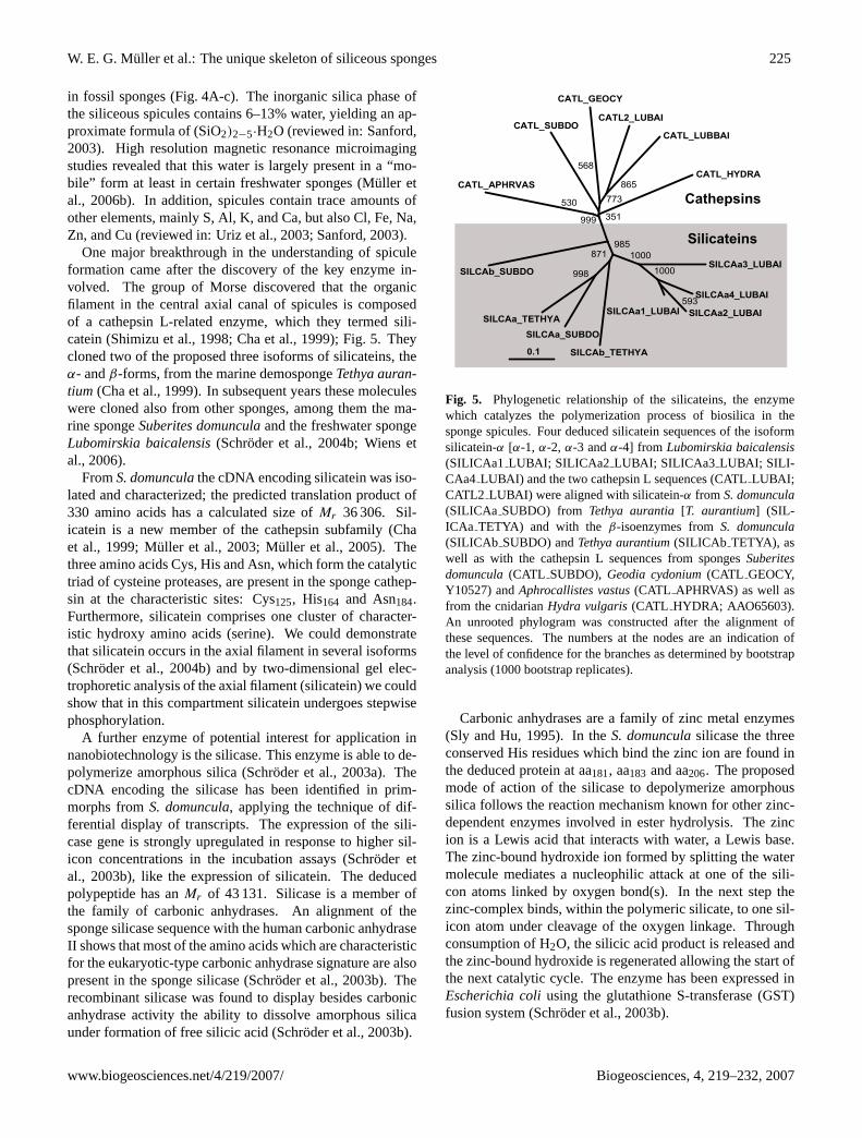
One major breakthrough in the understanding of spiculeformation came after the discovery of the key enzyme in-volved. The group of Morse discovered that the organicfilament in the central axial canal of spicules is composedof a cathepsin L-related enzyme, which they termed sili-catein (Shimizu et al., 1998; Cha et al., 1999); Fig. 5. Theycloned two of the proposed three isoforms of silicateins, theα- andβ-forms, from the marine demospongeTethya auran-tium (Cha et al., 1999). In subsequent years these moleculeswere cloned also from other sponges, among them the ma-rine spongeSuberites domunculaand the freshwater spongeLubomirskia baicalensis(Schroder et al., 2004b; Wiens etal., 2006).
FromS. domunculathe cDNA encoding silicatein was iso-lated and characterized; the predicted translation product of330 amino acids has a calculated size ofMr 36 306. Sil-icatein is a new member of the cathepsin subfamily (Chaet al., 1999; Muller et al., 2003; Muller et al., 2005). Thethree amino acids Cys, His and Asn, which form the catalytictriad of cysteine proteases, are present in the sponge cathep-sin at the characteristic sites: Cys125, His164 and Asn184.Furthermore, silicatein comprises one cluster of character-istic hydroxy amino acids (serine). We could demonstratethat silicatein occurs in the axial filament in several isoforms(Schroder et al., 2004b) and by two-dimensional gel elec-trophoretic analysis of the axial filament (silicatein) we couldshow that in this compartment silicatein undergoes stepwisephosphorylation.
A further enzyme of potential interest for application innanobiotechnology is the silicase. This enzyme is able to de-polymerize amorphous silica (Schroder et al., 2003a). ThecDNA encoding the silicase has been identified in prim-morphs fromS. domuncula, applying the technique of dif-ferential display of transcripts. The expression of the sili-case gene is strongly upregulated in response to higher sil-icon concentrations in the incubation assays (Schroder etal., 2003b), like the expression of silicatein. The deducedpolypeptide has anMr of 43 131. Silicase is a member ofthe family of carbonic anhydrases. An alignment of thesponge silicase sequence with the human carbonic anhydraseII shows that most of the amino acids which are characteristicfor the eukaryotic-type carbonic anhydrase signature are alsopresent in the sponge silicase (Schroder et al., 2003b). Therecombinant silicase was found to display besides carbonicanhydrase activity the ability to dissolve amorphous silicaunder formation of free silicic acid (Schroder et al., 2003b).
0.1
SILCAa3_LUBAI
SILCAa4_LUBAI
SILCAa2_LUBAISILCAa1_LUBAI593
1000
1000
SILCAb_TETHYA
SILCAa_SUBDOSILCAa_TETHYA
998
871985
SILCAb_SUBDO
CATL_APHRVAS
530
CATL_SUBDO
CATL_GEOCY
CATL2_LUBAI
CATL_LUBBAI
865773
568CATL_HYDRA
351999
Silicateins
Cathepsins
Fig. 5. Phylogenetic relationship of the silicateins, the enzymewhich catalyzes the polymerization process of biosilica in thesponge spicules. Four deduced silicatein sequences of the isoformsilicatein-α [α-1, α-2, α-3 andα-4] from Lubomirskia baicalensis(SILICAa1 LUBAI; SILICAa2 LUBAI; SILICAa3 LUBAI; SILI-CAa4 LUBAI) and the two cathepsin L sequences (CATLLUBAI;CATL2 LUBAI) were aligned with silicatein-α from S. domuncula(SILICAa SUBDO) from Tethya aurantia[T. aurantium] (SIL-ICAa TETYA) and with the β-isoenzymes fromS. domuncula(SILICAb SUBDO) andTethya aurantium(SILICAb TETYA), aswell as with the cathepsin L sequences from spongesSuberitesdomuncula(CATL SUBDO), Geodia cydonium(CATL GEOCY,Y10527) andAphrocallistes vastus(CATL APHRVAS) as well asfrom the cnidarianHydra vulgaris(CATL HYDRA; AAO65603).An unrooted phylogram was constructed after the alignment ofthese sequences. The numbers at the nodes are an indication ofthe level of confidence for the branches as determined by bootstrapanalysis (1000 bootstrap replicates).
Carbonic anhydrases are a family of zinc metal enzymes(Sly and Hu, 1995). In theS. domunculasilicase the threeconserved His residues which bind the zinc ion are found inthe deduced protein at aa181, aa183 and aa206. The proposedmode of action of the silicase to depolymerize amorphoussilica follows the reaction mechanism known for other zinc-dependent enzymes involved in ester hydrolysis. The zincion is a Lewis acid that interacts with water, a Lewis base.The zinc-bound hydroxide ion formed by splitting the watermolecule mediates a nucleophilic attack at one of the sili-con atoms linked by oxygen bond(s). In the next step thezinc-complex binds, within the polymeric silicate, to one sil-icon atom under cleavage of the oxygen linkage. Throughconsumption of H2O, the silicic acid product is released andthe zinc-bound hydroxide is regenerated allowing the start ofthe next catalytic cycle. The enzyme has been expressed inEscherichia coliusing the glutathione S-transferase (GST)fusion system (Schroder et al., 2003b).
www.biogeosciences.net/4/219/2007/ Biogeosciences, 4, 219–232, 2007
226 W. E. G. Muller et al.: The unique skeleton of siliceous sponges
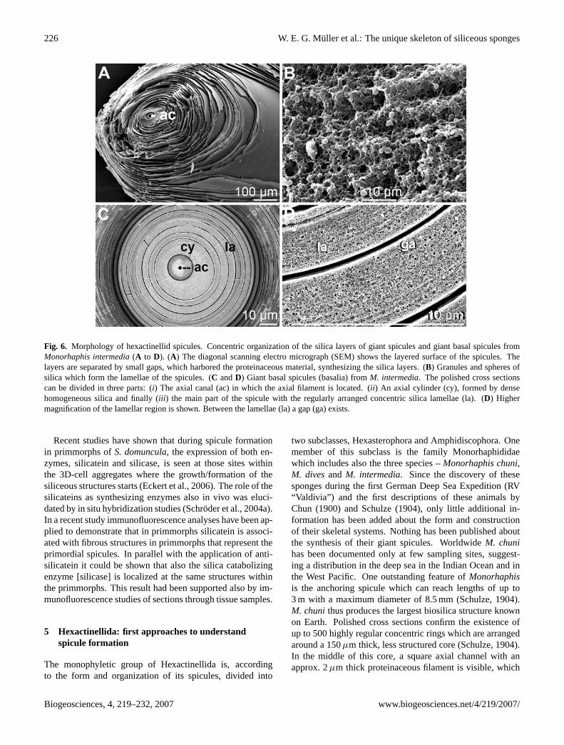
Fig. 6. Morphology of hexactinellid spicules. Concentric organization of the silica layers of giant spicules and giant basal spicules fromMonorhaphis intermedia(A to D). (A) The diagonal scanning electro micrograph (SEM) shows the layered surface of the spicules. Thelayers are separated by small gaps, which harbored the proteinaceous material, synthesizing the silica layers. (B) Granules and spheres ofsilica which form the lamellae of the spicules. (C andD) Giant basal spicules (basalia) fromM. intermedia. The polished cross sectionscan be divided in three parts: (i) The axial canal (ac) in which the axial filament is located. (ii ) An axial cylinder (cy), formed by densehomogeneous silica and finally (iii ) the main part of the spicule with the regularly arranged concentric silica lamellae (la). (D) Highermagnification of the lamellar region is shown. Between the lamellae (la) a gap (ga) exists.
Recent studies have shown that during spicule formationin primmorphs ofS. domuncula, the expression of both en-zymes, silicatein and silicase, is seen at those sites withinthe 3D-cell aggregates where the growth/formation of thesiliceous structures starts (Eckert et al., 2006). The role of thesilicateins as synthesizing enzymes also in vivo was eluci-dated by in situ hybridization studies (Schroder et al., 2004a).In a recent study immunofluorescence analyses have been ap-plied to demonstrate that in primmorphs silicatein is associ-ated with fibrous structures in primmorphs that represent theprimordial spicules. In parallel with the application of anti-silicatein it could be shown that also the silica catabolizingenzyme [silicase] is localized at the same structures withinthe primmorphs. This result had been supported also by im-munofluorescence studies of sections through tissue samples.
5 Hexactinellida: first approaches to understandspicule formation
The monophyletic group of Hexactinellida is, accordingto the form and organization of its spicules, divided into
two subclasses, Hexasterophora and Amphidiscophora. Onemember of this subclass is the family Monorhaphididaewhich includes also the three species –Monorhaphis chuni,M. divesand M. intermedia. Since the discovery of thesesponges during the first German Deep Sea Expedition (RV“Valdivia”) and the first descriptions of these animals byChun (1900) and Schulze (1904), only little additional in-formation has been added about the form and constructionof their skeletal systems. Nothing has been published aboutthe synthesis of their giant spicules. WorldwideM. chunihas been documented only at few sampling sites, suggest-ing a distribution in the deep sea in the Indian Ocean and inthe West Pacific. One outstanding feature ofMonorhaphisis the anchoring spicule which can reach lengths of up to3 m with a maximum diameter of 8.5 mm (Schulze, 1904).M. chunithus produces the largest biosilica structure knownon Earth. Polished cross sections confirm the existence ofup to 500 highly regular concentric rings which are arrangedaround a 150µm thick, less structured core (Schulze, 1904).In the middle of this core, a square axial channel with anapprox. 2µm thick proteinaceous filament is visible, which
Biogeosciences, 4, 219–232, 2007 www.biogeosciences.net/4/219/2007/
W. E. G. Muller et al.: The unique skeleton of siliceous sponges 227
runs through the entire spicule from one end to the other(Fig. 6A, C and D). The silica layers are composed of smallergranules and spheres within a network of (perhaps) proteina-ceous material (Fig. 5B).
An additional hexactinellidHyalonema sieboldilikewisepossesses long stalk spicules that attach them to the substra-tum; the animals live in a depth of more than 600 fathoms(1000 m); (Wyville Thomson, 1874 and also Muller et al.,2006c). The long stalk spicules are composed of distinctsiliceous layers which are also superposed in a stratified pat-tern around a central axial filament (Schulze, 1904). It couldbe demonstrated that these giant spicules from the root-tuft ofH. siebolditransmit light with high efficiency. Surprisingly,however, the blue light with a wavelength between 400 and600 nm is filtered out. Data elaborated in our group suggestthat the spicules fromH. sieboldiact as optical absorbent ina novel photoreception system (Muller et al., 2006c).
Very recently, Aizenberg et al. (2005) published a detailedstructural analysis of the spicule formation in the hexactinel-lid Euplectella. They demonstrated structural hierarchies ofthe spicule synthesis starting from the nanometer-sized par-ticles of silica to the final mature spicules. The compositionsof the proteins which are associated with and found in thespicules have not been described. In a recent study Ehrlichet al. (2005) dissolved the basal spicules of the hexactinellidH. sieboldiin alkaline solution for 14 days and showed thatthe abundant structural protein, associated with the spiculesmight be collagen.
We performed microscopic analyses of the spicules fromM. chuni with major emphasis of the large-sized giantspicules (giant basal spicules or basalia; size of 1 m) and alsothe large comitalia (small di- or tri-actine spicules of a sizeof around 60 mm). The focus of the study was put on theorganic components of these spicules, including the collagenfibrils which surround them. Electron microprobe analysisdata show a regionally different distribution of sodium andpotassium within the spicules. With respect to the organiccomponents, it should be highlighted that after dissolution ofthe spicules several proteins could been visualized; (i) one ofthem cross-reacted with antibodies raised against silicatein(Wang et al., 2007; Muller et al., 2007), while (ii ) others dis-played proteolytic activity (to be published).
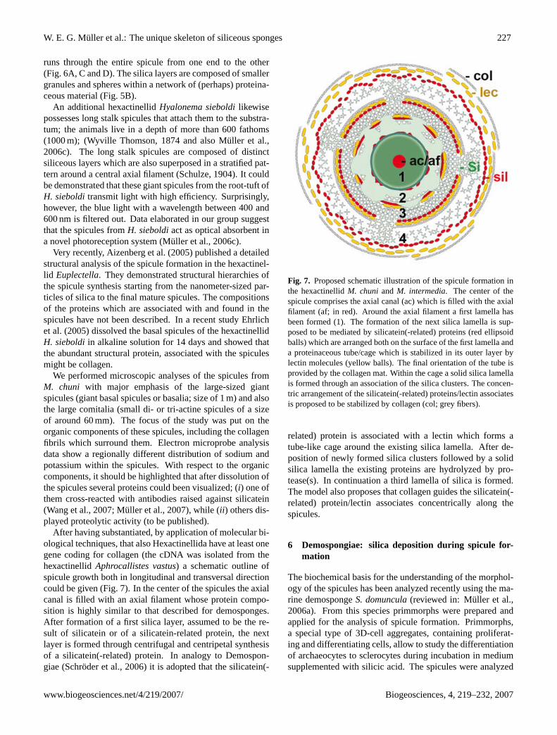
After having substantiated, by application of molecular bi-ological techniques, that also Hexactinellida have at least onegene coding for collagen (the cDNA was isolated from thehexactinellidAphrocallistes vastus) a schematic outline ofspicule growth both in longitudinal and transversal directioncould be given (Fig. 7). In the center of the spicules the axialcanal is filled with an axial filament whose protein compo-sition is highly similar to that described for demosponges.After formation of a first silica layer, assumed to be the re-sult of silicatein or of a silicatein-related protein, the nextlayer is formed through centrifugal and centripetal synthesisof a silicatein(-related) protein. In analogy to Demospon-giae (Schroder et al., 2006) it is adopted that the silicatein(-
Fig. 7. Proposed schematic illustration of the spicule formation inthe hexactinellidM. chuni and M. intermedia. The center of thespicule comprises the axial canal (ac) which is filled with the axialfilament (af; in red). Around the axial filament a first lamella hasbeen formed (1). The formation of the next silica lamella is sup-posed to be mediated by silicatein(-related) proteins (red ellipsoidballs) which are arranged both on the surface of the first lamella anda proteinaceous tube/cage which is stabilized in its outer layer bylectin molecules (yellow balls). The final orientation of the tube isprovided by the collagen mat. Within the cage a solid silica lamellais formed through an association of the silica clusters. The concen-tric arrangement of the silicatein(-related) proteins/lectin associatesis proposed to be stabilized by collagen (col; grey fibers).
related) protein is associated with a lectin which forms atube-like cage around the existing silica lamella. After de-position of newly formed silica clusters followed by a solidsilica lamella the existing proteins are hydrolyzed by pro-tease(s). In continuation a third lamella of silica is formed.The model also proposes that collagen guides the silicatein(-related) protein/lectin associates concentrically along thespicules.
6 Demospongiae: silica deposition during spicule for-mation
The biochemical basis for the understanding of the morphol-ogy of the spicules has been analyzed recently using the ma-rine demospongeS. domuncula(reviewed in: Muller et al.,2006a). From this species primmorphs were prepared andapplied for the analysis of spicule formation. Primmorphs,a special type of 3D-cell aggregates, containing proliferat-ing and differentiating cells, allow to study the differentiationof archaeocytes to sclerocytes during incubation in mediumsupplemented with silicic acid. The spicules were analyzed
www.biogeosciences.net/4/219/2007/ Biogeosciences, 4, 219–232, 2007
228 W. E. G. Muller et al.: The unique skeleton of siliceous sponges
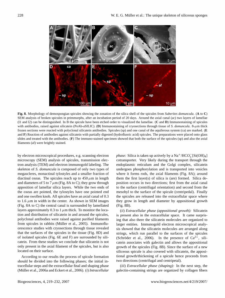
Fig. 8. Morphology of demospongian spicules showing the zonation of the silica shell of the spicules fromSuberites domuncula. (A to C)SEM analysis of broken spicules in primmorphs, after an incubation period of 20 days. Around the axial canal (ac) two layers of lamellae(l1 and l2) can be distinguished. In B the spicule have been etched order to visualized the lamellae. (C andD) Immunostaining of spiculeswith antibodies, raised against silicatein (PoAb-aSILIC). (D) Immunostaining of cryosections through tissue ofS. domuncula. 8-µm thickfrozen sections were reacted with polyclonal silicatein antibodies. Spicules (sp) and one canal of the aquiferous system (ca) are marked. (EandF) Reaction of antibodies against silicatein with partially digested (hydrofluoric acid) spicules. The preparations were placed onto glassslides and treated with the antibodies. (F) The immuno-stained specimen showed that both the surface of the spicules (sp) and also the axialfilaments (af) were brightly stained.
by electron microscopical procedures, e.g. scanning electronmicroscopy (SEM) analysis of spicules, transmission elec-tron analysis (TEM) and electron immunogold labeling. Theskeleton ofS. domunculais composed of only two types ofmegascleres, monactinal tylostyles and a smaller fraction ofdiactinal oxeas. The spicules reach up to 450µm in lengthand diameters of 5 to 7µm (Fig. 8A to C); they grow throughapposition of lamellar silica layers. While the two ends ofthe oxeas are pointed, the tylostyles have one pointed endand one swollen knob. All spicules have an axial canal of 0.3to 1.6µm in width in the center. As shown in SEM images(Fig. 8A to C) the central canal is surrounded by lamellatedlayers approximately 0.3 to 1µm thick. To monitor the loca-tion and distribution of silicatein in and around the spicules,polyclonal antibodies were raised against purified filamentsfrom spicules in rabbits (Muller et al., 2005). Immunoflu-orescence studies with cryosections through tissue revealedthat the surfaces of the spicules in the tissue (Fig. 8D) andof isolated spicules (Fig. 6E and F) are surrounded by sili-catein. From these studies we conclude that silicatein is notonly present in the axial filament of the spicules, but is alsolocated on their surface.
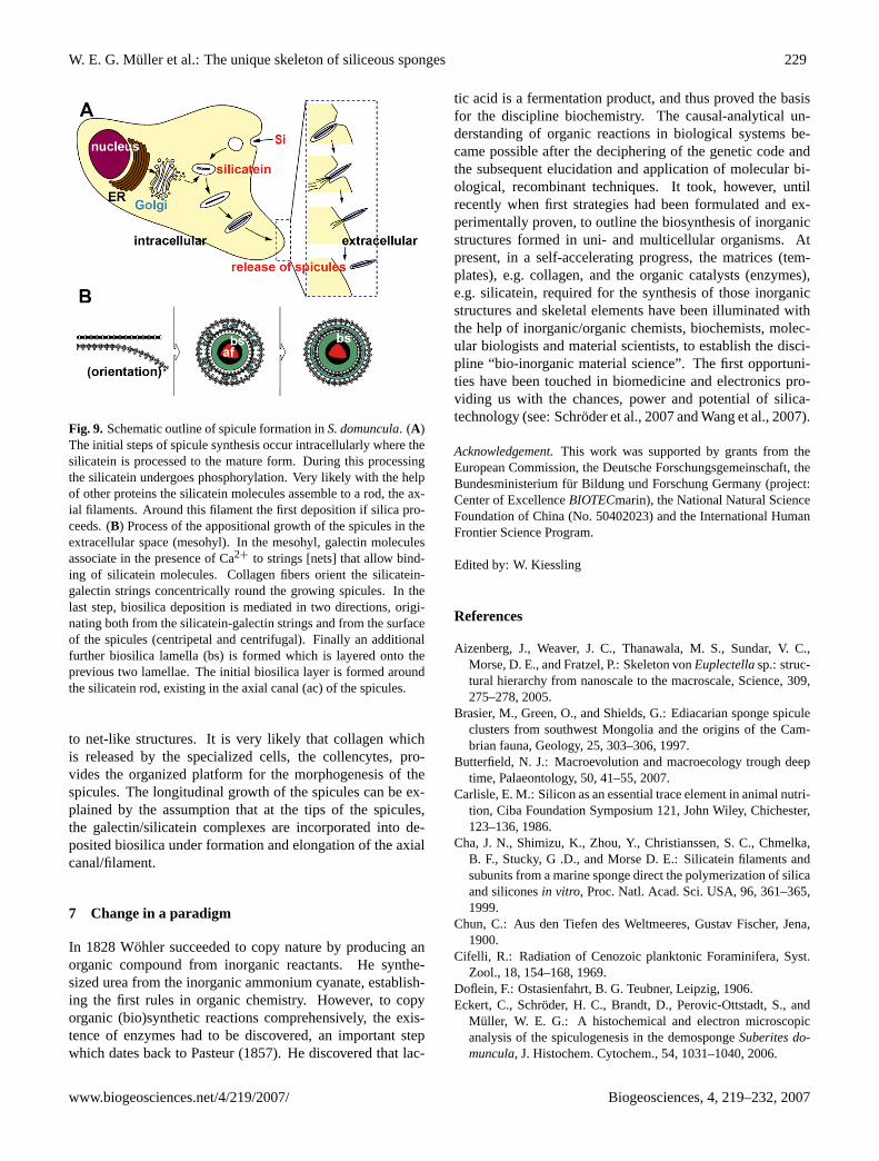
According to our results the process of spicule formationshould be divided into the following phases; the initial in-tracellular steps and the extracellular final and shaping phase(Muller et al., 2006a and Eckert et al., 2006). (i) Intracellular
phase:Silica is taken up actively by a Na+/HCO−
3 [Si(OH)4]cotransporter. Very likely during the transport through theendoplasmic reticulum and the Golgi complex, silicateinundergoes phosphorylation and is transported into vesicleswhere it forms rods, the axial filaments (Fig. 8A); aroundthem the first layer(s) of silica is (are) formed. Silica de-position occurs in two directions; first from the axial canalto the surface (centrifugal orientation) and second from themesohyl to the surface of the spicule (centripedal). Finallythe spicules are released into the extracellular space wherethey grow in length and diameter by appositional growth(Fig. 8B).
(ii ) Extracellular phase (appositional growth):Silicateinis present also in the extracellular space. It came surpris-ing that also there the silicatein molecules are organized tolarger entities. Immunogold electron microscopical analy-sis showed that the silicatein molecules are arranged alongstrings, which run parallel to the surfaces of the spicules(Schroder et al., 2006). In the presence of Ca2+, sili-catein associates with galectin and allows the appositionalgrowth of the spicules (Fig. 8B). Since the surface of a newsiliceous spicule is also covered with silicatein, the apposi-tional growth/thickening of a spicule hence proceeds fromtwo directions (centrifugal and centripetal).
(iii ) Extracellular phase (shaping):In the next step, thegalectin-containing strings are organized by collagen fibers
Biogeosciences, 4, 219–232, 2007 www.biogeosciences.net/4/219/2007/
W. E. G. Muller et al.: The unique skeleton of siliceous sponges 229
Fig. 9. Schematic outline of spicule formation inS. domuncula. (A)The initial steps of spicule synthesis occur intracellularly where thesilicatein is processed to the mature form. During this processingthe silicatein undergoes phosphorylation. Very likely with the helpof other proteins the silicatein molecules assemble to a rod, the ax-ial filaments. Around this filament the first deposition if silica pro-ceeds. (B) Process of the appositional growth of the spicules in theextracellular space (mesohyl). In the mesohyl, galectin moleculesassociate in the presence of Ca2+ to strings [nets] that allow bind-ing of silicatein molecules. Collagen fibers orient the silicatein-galectin strings concentrically round the growing spicules. In thelast step, biosilica deposition is mediated in two directions, origi-nating both from the silicatein-galectin strings and from the surfaceof the spicules (centripetal and centrifugal). Finally an additionalfurther biosilica lamella (bs) is formed which is layered onto theprevious two lamellae. The initial biosilica layer is formed aroundthe silicatein rod, existing in the axial canal (ac) of the spicules.
to net-like structures. It is very likely that collagen whichis released by the specialized cells, the collencytes, pro-vides the organized platform for the morphogenesis of thespicules. The longitudinal growth of the spicules can be ex-plained by the assumption that at the tips of the spicules,the galectin/silicatein complexes are incorporated into de-posited biosilica under formation and elongation of the axialcanal/filament.
7 Change in a paradigm
In 1828 Wohler succeeded to copy nature by producing anorganic compound from inorganic reactants. He synthe-sized urea from the inorganic ammonium cyanate, establish-ing the first rules in organic chemistry. However, to copyorganic (bio)synthetic reactions comprehensively, the exis-tence of enzymes had to be discovered, an important stepwhich dates back to Pasteur (1857). He discovered that lac-
tic acid is a fermentation product, and thus proved the basisfor the discipline biochemistry. The causal-analytical un-derstanding of organic reactions in biological systems be-came possible after the deciphering of the genetic code andthe subsequent elucidation and application of molecular bi-ological, recombinant techniques. It took, however, untilrecently when first strategies had been formulated and ex-perimentally proven, to outline the biosynthesis of inorganicstructures formed in uni- and multicellular organisms. Atpresent, in a self-accelerating progress, the matrices (tem-plates), e.g. collagen, and the organic catalysts (enzymes),e.g. silicatein, required for the synthesis of those inorganicstructures and skeletal elements have been illuminated withthe help of inorganic/organic chemists, biochemists, molec-ular biologists and material scientists, to establish the disci-pline “bio-inorganic material science”. The first opportuni-ties have been touched in biomedicine and electronics pro-viding us with the chances, power and potential of silica-technology (see: Schroder et al., 2007 and Wang et al., 2007).
Acknowledgement.This work was supported by grants from theEuropean Commission, the Deutsche Forschungsgemeinschaft, theBundesministerium fur Bildung und Forschung Germany (project:Center of ExcellenceBIOTECmarin), the National Natural ScienceFoundation of China (No. 50402023) and the International HumanFrontier Science Program.
Edited by: W. Kiessling
References
Aizenberg, J., Weaver, J. C., Thanawala, M. S., Sundar, V. C.,Morse, D. E., and Fratzel, P.: Skeleton vonEuplectellasp.: struc-tural hierarchy from nanoscale to the macroscale, Science, 309,275–278, 2005.
Brasier, M., Green, O., and Shields, G.: Ediacarian sponge spiculeclusters from southwest Mongolia and the origins of the Cam-brian fauna, Geology, 25, 303–306, 1997.
Butterfield, N. J.: Macroevolution and macroecology trough deeptime, Palaeontology, 50, 41–55, 2007.
Carlisle, E. M.: Silicon as an essential trace element in animal nutri-tion, Ciba Foundation Symposium 121, John Wiley, Chichester,123–136, 1986.
Cha, J. N., Shimizu, K., Zhou, Y., Christianssen, S. C., Chmelka,B. F., Stucky, G .D., and Morse D. E.: Silicatein filaments andsubunits from a marine sponge direct the polymerization of silicaand siliconesin vitro, Proc. Natl. Acad. Sci. USA, 96, 361–365,1999.
Chun, C.: Aus den Tiefen des Weltmeeres, Gustav Fischer, Jena,1900.
Cifelli, R.: Radiation of Cenozoic planktonic Foraminifera, Syst.Zool., 18, 154–168, 1969.
Doflein, F.: Ostasienfahrt, B. G. Teubner, Leipzig, 1906.Eckert, C., Schroder, H. C., Brandt, D., Perovic-Ottstadt, S., and
Muller, W. E. G.: A histochemical and electron microscopicanalysis of the spiculogenesis in the demospongeSuberites do-muncula, J. Histochem. Cytochem., 54, 1031–1040, 2006.
www.biogeosciences.net/4/219/2007/ Biogeosciences, 4, 219–232, 2007
230 W. E. G. Muller et al.: The unique skeleton of siliceous sponges
Efremova, S. M., Margulis, B. A., Guzhova, I. V., Itskovich, V. B.,Lauenroth, S., Muller, W. E. G., and Schroder, H. C.: Heat shockprotein Hsp70 expression and DNA damage in Baikalian spongesexposed to model pollutants and wastewater from Baikalsk Pulpand paper plant, Aquat. Toxicol., 57, 267–280, 2002.
Ehrlich, H., Hanke, T., Simon, P., Goebel, C., Heinmann, S., Born,R., and Worch, H.: Demineralisation von naturlichen Silikat-basierten Biomaterialien: neue Strategie zur Isolation organis-cher Geruststrukturen, BIOmaterialien, 6, 297–302, 2005.
Futuyma, D. J.: Evolutionary Biology, Sinauer Ass, Sunderland,1986.
Garrone, R.: Evolution of metazoan collagens, Progr. Molec. Sub-cell. Biol., 21, 191–139, 1998.
Gehling, J. G. and Rigby J. K.: Long expected sponges from theNeoproterozoic Ediacara Fauna of South Australia, J. Paleontol.,70, 185–195, 1996.
Grotzinger, J. P., Bowring, S. A., Saylor, B., and Kauffmann, A.J.: New biostratigraphic and geochronologic constraints on earlyanimal evolution, Science, 270, 598–604, 1995.
Haeckel, E.: Kunstformen der Natur, Bibliographisches Institut,Leipzig, 1899.
Hayes, J. M.: Global methanotrophy at the Archean-Proterozoictransition, in: Early Life on Earth, edited by Bengtson, S.,Columbia University Press, New York, 220–236, 1994.
Hoffman, P. F., Kaufman, A. J., Halverson, G. P., and Schrag, D.P.: A Neoproterozoic snowball earth, Science, 281, 1342–1346,1998.
Hoffmann, P. A. and Schrag, D. P.: The snowball Earth hypothesis:testing the limits of global change, Terra Nova, 129–155, 2002.
Knoll, A. H. and Carroll, S. B.: Early animal evolution: emerg-ing views from comparative biology and geology, Science, 284,2129–2137, 1999.
Krasko, A., Muller, I. M., and Muller, W. E. G.: Evolutionary rela-tionships of the metazoanßγ -crystallins, including the one fromthe marine spongeGeodia cydonium, Proc. Royal Soci. Lond. B,264, 1077–1084, 1997.
Krasko, A., Schroder, H. C., Hassanein, H. M. A., Batel, R., Muller,I. M., and Muller, W. E. G.: Identification and expression ofthe SOS-response,aidB-like, gene in the marine spongeGeo-dia cydonium: implication for the phylogenetic relationships ofmetazoan Acyl-CoA dehydrogenases and Acyl-CoA oxidases, J.Molec. Evol., 47, 343–352, 1998.
Krasko, A., Lorenz, B., Batel, R., Schroder, H. C., Muller, I. M., andMuller, W. E. G.: Expression of silicatein and collagen genes inthe marine spongeSuberites domunculais controlled by silicateand myotrophin, Europ. J. Biochem., 267, 4878–4887, 2000.
Krasko, A., Schroder, H. C., Batel, R., Grebenjuk, V. A., Steffen,R., Muller, I. M., and Muller, W. E. G.: Iron induces prolifera-tion and morphogenesis in primmorphs from the marine spongeSuberites domuncula, DNA & Cell Biol., 21, 67–80, 2002.
Krasko, A., Gundacker, D., Leys, S. P., Schroder, H. C., Muller, I.M., and Muller, W. E. G.: Molecular and functional analysis ofthe (6-4) photolyase from the hexactinellidAphrocallistes vastus,Biochim. Biophys. Acta., 1651, 41–49, 2003.
Li, C. W., Chen, J. Y., and Hua T. E.: Precambrian sponges withcellular structures, Science, 279, 879–882 1998.
Morris, C. S.: The fossil record and the early evolution of the Meta-zoa, Nature, 361, 219–225, 1993.
Morris, C. S.: Why molecular biology needs palaeontology, De-
velop. Suppl., 1–13, 1994.Mostler, H.: Neue heteractinide Spongien (Calcispongea) aus dem
Unter- und Mittelkambrium Sudwestsardiniens, Ber. nat. med.Ver. Innsbruck, 72, 7–32, 1985.
Muller, W. E. G.: Origin of Metazoa: sponges as living fossils,Naturwiss., 85, 11–25, 1998.
Muller, W. E. G.: Spatial and temporal expression patterns in ani-mals, in: Encyclopedia of Molecular Cell Biology and MolecularMedicine, edited by: Meyers, R. A., WILEY-VCH Press, Wein-heim, 13, 269–309, 2005.
Muller, W. E. G.: The stem cell concept in sponges (Porifera):metazoan traits, Seminars in Cell & Develop. Biol., 17, 481–491,2006.
Muller, W. E. G. and Muller, I. M.: Origin of the metazoan im-mune system: Identification of the molecules and their functionsin sponges, Integr. Comp. Biol., 43, 281–292, 2003.
Muller, W. E. G., Zahn, R. K., and Maidhof, A.:Spongilla guten-bergianan.sp., ein Sußwasserschwamm aus dem Mittel-Eozanvon Messel, Senckenbergiana lethaea, 63, 465–472, 1982.
Muller, W. E. G., Schroder, H. C., Skorokhod, A., Bunz, C., Muller,I. M., and Grebenjuk, V. A.: Contribution of sponge genesto unravel the genome of the hypothetical ancestor of metazoa(Urmetazoa), Gene, 276, 161–173, 2001.
Muller, W. E. G., Krasko, A., Le Pennec, G., Steffen, R., Ammar,M. S. A., Wiens, M., Muller, I. M., and Schroder, H. C.: Molecu-lar mechanism of spicule formation in the demospongeSuberitesdomuncula: Silicatein – collagen – myotrophin, Progress Molec.Subcell. Biol., 33, 195–221, 2003.
Muller, W. E. G., Wiens, M., Adell, T., Gamulin, V., Schroder, H.C., and Muller, I. M.: The Bauplan of the Urmetazoa: The basisof the genetic complexity of Metazoa using the siliceous sponges[Porifera] as living fossils, Int. Rev. Cytol., 235, 53–92, 2004a.
Muller, W. E. G., Grebenjuk, V. A., Thakur, N. L., Thakur, A. N.,Batel, R., Krasko, A., Muller, I. M., and Breter, H. J.: Oxygen-controlled bacterial growth in the spongeSuberites domuncula:Towards a molecular understanding of the symbiotic relation-ships between sponge and bacteria, Appl. Envir. Microbiol., 70,2332–2341, 2004b.
Muller, W. E. G., Rothenberger, M., Boreiko, A., Tremel, W.,Reiber, A., and Schroder, H. C.: Formation of siliceous spiculesin the marine demospongeSuberites domuncula, Cell & TissueRes., 321, 285–297, 2005.
Muller, W. E. G., Belikov, S. I., Tremel, W., Perry, C. C., Gieskes,W. W. C., Boreiko, A., and Schroder, H. C.: Siliceous spicules inmarine demosponges (exampleSuberites domuncula), Micron.,37, 107–120, 2006a.
Muller, W. E. G., Kaluzhnaya, O. V., Belikov, S. I., Rothenberger,M., Schroder, H. C., Reiber, A., Kaandorp, J. A., Manz, B., Mi-etchen, D., and Volke, F.: Magnetic resonance imaging of thesiliceous skeleton of the demospongeLubomirskia baicalensis,J. Struct. Biol., 153, 31–41, 2006b.
Muller, W. E. G., Wendt, K., Geppert, C., Wiens, M., Reiber, A.,and Schroder, H. C.: Novel photoreception system in sponges?Unique transmission properties of the stalk spicules from thehexactinellidHyalonema sieboldi, Biosensors and Bioelectron-ics, 21, 1149–1155, 2006c.
Muller, W. E. G., Schroder, H. C., Wrede, P., Kaluzhnaya, O. V.,and Belikov, S. I.: Speciation of sponges in Baikal-Tuva region(an outline), J. Zool. Syst. Evol. Res., 44, 105–117, 2006d.
Biogeosciences, 4, 219–232, 2007 www.biogeosciences.net/4/219/2007/
W. E. G. Muller et al.: The unique skeleton of siliceous sponges 231
Muller, W. E. G., Eckert, C., Kropf, K., Wang, X., Schloßmacher,U., Seckert, C., Wolf, S. E., Tremel, W., and Schroder, H. C.:Formation of the giant spicules of the deep sea hexactinellidMonorhaphis chuni(Schulze 1904): electron microscopic andbiochemical studies, Cell & Tissue Res., doi:10.1007/s00441-007-0402-x, 2007.
Nichols, S. A., Dirks, W., Pearse, J. S., and King, N.: Early evo-lution of animal cell signaling and adhesion genes. Proc. Natl.Acad. Sci. USA, 103, 12 451–12 456, 2006.
Pancer, Z., Kruse, M., Muller, I., and Muller, W. E. G.: On theorigin of adhesion receptors of metazoa: cloning of the integrinα subunit cDNA from the spongeGeodia cydonium, Molec. Biol.Evol., 14, 391–398, 1997.
Pasteur, L.: Memoire sur la fermentation appelee lactique, Mem,Soc. Sci. Agric et Arts, 5, 13–26, 1857.
Perovic-Ottstadt, S., Adell, T., Proksch, P., Wiens, M., Korzhev,M., Gamulin, V., Muller, I. M., and Muller, W. E. G.: A (1→3)-β-d-glucan recognition protein from the spongeSuberites do-muncula: mediated activation of fibrinogen-like protein and epi-dermal growth factor gene expression, Eur. J. Biochem., 271,1924–1937, 2004a.
Pechenik, J. A.: Biology of the Invertebrates, McGraw Hill, Boston,2000.
Pilcher, H.: Back to our roots, Nature, 435, 1022–1023, 2005.Pfeifer, K., Haasemann, M., Gamulin, V., Bretting, H., Fahrenholz,
F., and Muller, W. E. G.: S-type lectins occur also in inverte-brates: high conservation of the carbohydrate recognition do-main in the lectin genes from the marine spongeGeodia cydo-nium, Glycobiol., 3, 179–184, 1993.
Pilcher, H.: Back to our roots, Nature, 435, 1022–1023, 2005.Reitner, J.: Coralline Spongien. Der Versuch einer phylogenetisch-
taxonomischen Analyse, Berliner Geowiss. Abh. (E), 1, 1–352,1992.
Reitner, J. and Mehl, D.: Early Paleozoic deversification ofsponges: New data and evidences, Geol. Palaont. Mitt. Inns-bruck, 20, 335–347, 1995.
Reitner, J. and Worheide, G.: Non-Lithistid fossil Demospongiae –Origins of their Palaeobiodiversity and Highlights in History ofPreservation, in: Systema Porifera: A Guide to the Classifica-tion of Sponges, edited by: Hooper, J. N. A. and Van Soest, R.,Kluwer, New York, pp. 52–68, 2002.
Retallack, G. J.: Were the Ediacaran fossils lichens?, Paleobiology,20, 523–544, 1994.
Rigby, J. K. and Hou, Xian-Guang.: Lower Cambrian demospongesand hexactinellid sponges from Yunnan,China, J. Paleont., 69,1009–1019, 1995.
Sanford, F.: Physical and chemical analysis of the siliceous skele-ton in six sponges of two groups (Demospongiae and Hexactinel-lida), Micr. Res. Techn., 62, 336–355, 2003.
Schutze, J., Custodio, M. R., Efremova, S. M., Muller, I. M., andMuller, W. E. G. Evolutionary relationship of metazoa within theeukaryotes based on molecular data from Porifera, Proc. Roy.Soc. Lond. B, 266, 63–73, 1999.
Schroder, H. C., Krasko, A., Ushijima, H., Gamulin, V., Schutze, J.,Muller, I. M., and Muller, W. E. G.: Emergence and disappear-ance of an immune molecule, an antimicrobial lectin, in basalMetazoa: the tachylectin family, J. Biol. Chem., 278, 32 810–32 817, 2003a.
Schroder, H. C., Krasko, A., Le Pennec, G., Adell, T., Wiens, M.,
Hassanein, H., Muller, I. M., and Muller, W. E. G.: Silicase, anenzyme which degrades biogenous amorphous silica: Contribu-tion to the metabolism of silica deposition in the demospongeSuberites domuncula, Prog. Mol. Subcell. Biol., 33, 250–268,2003b.
Schroder, H. C., Perovic-Ottstadt, S., Rothenberger, M., Wiens,M., Schwertner, H., Batel, R., Korzhev, M., Muller, I. M., andMuller, W. E. G.: Silica transport in the demospongeSuberitesdomuncula: fluorescence emission analysis using the PDMPOprobe and cloning of a potential transporter, Biochem. J., 381,665==673, 2004a.
Schroder, H. C., Perovi-Ottstadt, S., Wiens, M., Batel, R., Muller, I.M., and Muller, W. E. G.: Differentiation capacity of the epithe-lial cells in the spongeSuberites domuncula, Cell & Tissue Res.,316, 271==280, 2004b.
Schroder, H. C., Boreiko, A., Korzhev, M., Tahir, M. N., Tremel,W., Eckert, C., Ushijima, H., Muller, I. M., and Muller, W. E.G.: Co-Expression and functional interaction of silicatein withgalectin: matrix-guided formation of siliceous spicules in themarine demospongeSuberites domuncula, J. Biol. Chem., 281,12 001–12 009, 2006.
Schroder, H. C., Brandt, D., Schloßmacher, U., Wang, X., Tahir, M.N., Tremel, W., Belikov, S. I., and Muller, W. E. G., Enzymaticproduction of biosilica-glass using enzymes from sponges: Basicaspects and application in nanobiotechnology (material sciencesand medicine), Naturwissenschaften, 94, 339–359, 2007.
Schulze, F. E.: Hexactinellida. Wissenschaftliche Ergebnisse derDeutschen Tiefsee-Expedition auf dem Dampfer “Valdivia”1898–1899, Gustav Fischer Verlag, Stuttgart, pp. 1–266, 1904.
Seilacher, A.: Vendozoa: Organismic construction in the protero-zoic biosphere, Lethaia, 22, 229–239, 1989.
Shimizu, K., Cha, J., Stucky, G. D., and Morse, D. E.: Silicateinalpha: cathepsin L-like protein in sponge biosilica, Proc. Natl.Acad. Sci. USA, 95, 6234–6238, 1998.
Sly, W. S. and Hu, P. Y.: Human carbonic anhydrases and car-bonic anhydrase deficiencies, Annu. Rev. Biochem., 64, 375–401, 1995.
Steiner, M., Mehl, D., Reitner, J., and Erdtmann, B. D.: Oldest en-tirely preserved sponges and other fossils from the LowermostCambrian and a new facies reconstruction of the Yangtze Plat-form (China), Berliner Geowiss. Abh. (E), 9, 293–329, 1993.
Steiner, M.: Die Neoproterozoischen Megaalgen Sudchinas,Berliner Geowiss. Abh., 15, 100146, 1994.
Thakur, N. L., Perovi-Ottstadt, S., Batel, R., Korzhev, M., Diehl-Seifert, B., Muller, I. M., and Muller, W. E. G.: Innate im-mune defense of the spongeSuberites domunculaagainst gram-positive bacteria: induction of lysozyme and AdaPTin, Mar.Biol., 146, 271–282, 2005.
Towe, K. M.: Environmental conditions surrounding the origin andearly evolution of life, Precambrian Res., 16, 10010, 1981.
Towe, K. M.: Oxygen-collagen priortiy and the metzoan fossilrecord, Proc. Natl. Acad. Sci. USA, 65, 78100788, 1970.
Uriz, M. J., Turon, X., and Becerro, M. A.: Silica deposition in De-mospongiae: spiculogenesis inCrambe crambe, Cell & TissueRes., 301, 299–309, 2000.
Uriz, M. J., Turon, X., and Beccero, M. A.: Silica deposition indemosponges, Progr. Molec. Subcell. Biol., 33, 163–193, 2003.
Uriz, M. J.: Mineral skeletogenesis in sponges, Canadian J. Zool.,84, 322–356, 2006.
www.biogeosciences.net/4/219/2007/ Biogeosciences, 4, 219–232, 2007
232 W. E. G. Muller et al.: The unique skeleton of siliceous sponges
Wang, X. and Wang, Y.: An introduction to the study on naturalcharacteristics of sponge spicules and bionic applications, Adv.Earth Sci., 21, 37–42, 2006.
Wang, X., Li, J., Qiao, L., Schroder, H. C., Eckert, C., Kropf, K.,and Muller, W. E. G.: The giant spicules of the deep sea hex-actinellidan sponges of the genusMonorhaphis(Hexactinellida:Amphidiscosida: Monorhaphididae), Acta Zoologica Sinica, inpress, 2007.
Walker, G.: Snowball Earth: The Story of the Great Global Catas-trophe that Spawned Life as we Know it, Crown Publishers, NewYork, 2003.
Wiens, M. and Muller, W. E. G.: Cell death in Porifera: molecularplayers in the game of apoptotic cell death in living fossils, Can.J. Zool./Rev. Can. Zool., 84, 307–321, 2006.
Wiens, M., Mangoni, A., D’Esposito, M., Fattorusso, E., Korchag-ina, N., Schroder, H. C., Grebenjuk, V. A., Krasko, A., Batel,R., Muller, I. M., and Muller, W. E. G.: The molecular basis forthe evolution of the metazoan bodyplan: extracellular matrix-mediated morphogenesis in marine demosponges, J. Mol. Evol.,57, 1–16, 2003.
Wiens, M., Perovi-Ottstadt, S., Muller, I. M., and Muller, W. E.G.: Allograft rejection in the mixed cell reaction system of thedemospongeSuberites domunculais controlled by differentialexpression of apoptotic genes, Immunogenetics, 56, 597–610,2004.
Wiens, M., Korzhev, M., Krasko, A., Thakur, N. L., Perovi-Ottstadt, S., Breter, H. J., Ushijima, H., Diehl-Seifert, B.,Muller, I. M., and Muller, W. E. G.: Innate immune defenseof the spongeSuberites domunculaagainst bacteria involves aMyD88-dependent signaling pathway: induction of a perforin-like molecule, J. Biol. Chem., 280, 27 949–27 959, 2005.
Wiens, M., Belikov, S. I., Kaluzhnaya, O. V., Krasko, A., Schroder,H. C., Perovic-Ottstadt, S., and Muller, W. E. G.: Molecular con-trol of serial module formation along the apical-basal axis in thespongeLubomirskia baicalensis: silicateins, mannose-bindinglectin and Mago Nashi, Develop. Genes Evol., 216, 229–242,2006.
Wohler, F.: Ueber kunstliche Bildung des Harnstoffs, Annalen derPhysik und Chemie, 12, 253–256, 1828.
Biogeosciences, 4, 219–232, 2007 www.biogeosciences.net/4/219/2007/