Embed Size (px)
Citation preview

The Truth About OcclusionGene D. McCoy, D.D.S.
A commentary on the controversies regarding
dentistry’s most important subject.
Presented at:
Yankee Dental Congress 32
January 24-28, 2007
Boston, Massachusetts
Copyright © 2006 by Gene D. McCoy, D.D.S.All Rights Reserved

November 13, 2006
Although occlusion principles permeate almost all of dentistry, the area is confounded by confusing theories, non-practical techniques, contradictory ‘beliefs,’ and practitioners unaware of the basic concepts of occlusion. As a result, most dental patients go without the benefi ts of dental therapy based on several occlusal principles.
Dr. McCoy’s paper presents some well proven, practical concepts in oc-clusion based on his long experience and sound engineering principles. It is thought provoking and useful.
Gordon J. Christensen DDS, MSD, PhD Diplomate American Board of Prosthodontics

CONTENTS
Introduction ......................................................................................................................... 1
What Is Occlusion?—Defi nitions as a Major Source of Confusion ................................... 2
The Human Stomatognathic System—How Should It Function Ideally? .......................... 3
The Morphology of Teeth ................................................................................................... 4Vertical Loading ............................................................................................................ 5Neutralization ................................................................................................................ 6Prevention of Off-Loading ............................................................................................ 7Proper Condylar Seating ............................................................................................... 7
What Happens When the System Dysfunctions? ................................................................ 8The Etiology of Dysfunction ........................................................................................ 9The Signs of Dysfunction ............................................................................................. 9
Deformations of the Dentition .............................................................................. 10Deformations of Restorative Materials ................................................................. 14Deformations of Bone (Exostosis) ........................................................................ 15
Epidemiology .............................................................................................................. 16The Management of Dysfunction ............................................................................... 16
Step 1: Education .................................................................................................. 17Step 2: Equilibration ............................................................................................. 17Step 3: Guard Therapy .......................................................................................... 19
What Is Being Taught Today, and Why Is It Incoherent? ................................................. 21Normal Function: Vertical vs. Horizontal ................................................................... 21Morphology and Occlusion of the Dentition .............................................................. 23
Discussion and Recommendations ................................................................................... 27Which Concept on Occlusion Is Correct? ................................................................... 27Problem-Solving for TMJDs ....................................................................................... 28DCS and Periodontal Disease ..................................................................................... 29DCS and the Oral Implant Patient .............................................................................. 29
Notes ................................................................................................................................. 31


Introduction
Everyone agrees that a good understanding of occlusion is essential to ensure
optimum dental health. Unfortunately, that seems to be the only point of consensus for
this ambiguous subject. One cannot read a text or take a course on occlusion and walk
away with an understanding of how the design of teeth and the way they touch each other
affect the effi ciency and function of the stomatognathic system. Consequently, there are
many unanswered questions: primarily, the role that occlusion plays in the etiology of
temporomandibular disorders (TMDs) and oral facial pain. The confusion is evident when
we hear phrases such as “no one occlusal scheme will serve all patients” and questions like
“which concept on occlusion is correct?” We are led to believe that, as general practitioners
(GPs), we do not have the expertise to manage problems associated with this subject and
are solicited to take specialized postgraduate courses at the Las Vegas Institute (LVI) or
the Pankey Institute. However, each of these courses is costly in both tuition (at least
$15,000) and time away from the offi ce (2–3 weeks), plus there is the cost of expensive
instrumentation.
Patients of dentists who follow such a stylized form of occlusal rehabilitation now have
to share in the expense of the learning program, which limits treatment to the affl uent. What
about the patients who cannot afford it? Most people have little or no dental restorative work
done each year. Whether the reason is fi nancial, lack of accessibility, fear, or indifference,
that’s the way it is. Those who do frequent their dentists on a regular basis usually restrict
their work to limitations set by their insurance. Subsequent work has to wait until next
year.
Considering the circumstances, how can the GP take care of occlusal-related problems
in a cost-effective way that will benefi t patients with limited resources? In order to answer
this question, we truly need to understand occlusion, and that is the problem. As it is being
taught today, occlusion is incoherent. Taylor explains that, “due to the empirical nature of
1

Gene D. McCoy, D.D.S.
the literature, the study of occlusion has been extremely complex and troublesome to both
pre- and post-doctoral students.”1 Simon likens searching for the truth in occlusion to Alice
looking for the right path in Wonderland2 There are so many diverse opinions regarding
this subject that it is not uncommon to witness discord among colleagues at professional
meetings. So, what is the truth?
What Is Occlusion?—Defi nitions as a Major Source of Confusion
It is appropriate to begin with the defi nition of occlusion because that is a major source
of confusion.
The original defi nition of occlusion was the act or process of occluding, from the Latin
occludere, “to shut up or close up.” Dental occlusion refers to the closure of teeth—nothing
more.
Today, however, the word has at least three different meanings. Taber’s Cyclopedic
Medical Dictionary limits the defi nition to: “relation of the teeth when the jaws are closed.”3
Dorland’s Pocket Medical Dictionary fi rst defi ned dental occlusion as “the closure of
teeth,”4 then expanded the defi nition to “the contact of the teeth of both jaws during those
excursive movements of the mandible essential to the function of mastication.”5 Jablonski’s
Illustrated Dictionary of Dentistry defi nes occlusion as:
The relationship between all the components of the masticatory system in normal function, dysfunction, and parafunction, including the morphological and functional features of contacting surfaces of opposing teeth and restorations, occlusal trauma and dysfunction, neuromuscular physiology, the temporomandibular joint and muscle function, swallowing and mastication, psychophysiological status, and the diagnosis, prevention, and treatment of functional disorders of the masticatory system.6
The different interpretations are a major distraction to understanding this subject. It
clouds the issues and makes questions such as “What has occlusion got to do with TMD?”
2

The Truth About Occlusion
impossible to answer. Are we asking if tooth contact during closure is causing a problem
with the TMJ, or do we want to know if the stomatognathic system is not functioning
correctly? Modern-day texts on occlusion are not on the simple touching of upper and
lower teeth, but rather present a detailed analysis of the whole stomatognathic system.
Occlusion and the stomatognathic system are two distinct entities and should be described
separately to remove the present ambiguity.
The Human Stomatognathic System—How Should It Function Ideally?
There are two principal parts to the stomatognathic system: the maxilla, a U-shaped
row of teeth supported by alveolar bone, which is fi xed to the skull; and the mandible,
which is movable. The mandible houses another U-shaped row of teeth that are embedded
in very dense alveolar bone and is attached to the skull by a hinge-gliding joint. Usually,
the maxillary teeth overlap the mandibular teeth, but there are many variations due to
genetics, such as crossbites and differences in the anterior length of either jaw.
The mandible has three primary functions: communication, mastication, and swallowing.
Although the mandible has the capability of three-dimensional movement, the direction of
the three primary functions is up and down. The vector of mastication is a vertical teardrop
with lateral movement of 5mm to 6mm during the fi rst phase of crushing; as the teeth
approach each other, the lateral displacement lessens to 3mm to 4mm from the starting
position.7
When the mandible is not performing a task and the muscles of mastication are relaxed,
the occlusal surfaces of the teeth are slightly apart, creating what is called the “freeway
space.” The position of the mandible changes anteriorly-posteriorly due to gravitational
forces as the position of the head deviates from the vertical. This is easy to demonstrate.
If you tilt your head back and swallow, you can feel your posterior teeth lightly touch and
then open. If you tilt your head forward and swallow, the contact shifts to the anterior
3

Gene D. McCoy, D.D.S.
teeth. The different positions of the mandible upon swallowing are insignifi cant due to
the light closure force. What is signifi cant is the position of the mandible/condyles during
full intercuspation while clenching. Since forces generated by clenching can exceed 300
pounds per square inch, it is important that centric occlusion (CO) coincide with centric
relation (CR), because it is in this position that the condyles are positively braced in the
anterior-superior portion of their fossæ to receive these strong forces.
The Morphology of Teeth
It is the role of teeth to cut food effi ciently, aid in speech, bite, and ensure proper
condylar positioning upon closure. What is the ideal design for those purposes within the
stomatognathic system?
We often hear debates on the question whether form follows function, or vice versa.
The question is never satisfactorily answered because it is not a good question. There
are, however, design principles that appear to govern the structure-function relationship
in organisms—that is, there is an interface between mechanical engineering and biology.
The idea is that biological materials and structures have functions for which they are
designed.
If we are looking for the ideal design of teeth to ensure healthy occlusion and function,
we have to look at nature as the architect of excellence (Figs. 1 and 2).
Figs. 1–2: Nature’s Biological Cutting Instruments
4

The Truth About Occlusion
Teeth are designed to cut food. If one examines a cross-section of molars and bicuspids
in occlusion before they are worn down, there are two noteworthy observations: (a) there
is minimal contact between the teeth, which is confi ned to the tips of the functional cusps;
and (b) there is a generous space between the incline planes of the cusps, which is called
the intra-incline space (Figs. 3–6).8
Figs. 3–6: Sections of Ideal Occlusion
From these observations, it is interesting to note that teeth do not require large areas
of contact in order to maintain their position, work effi ciently, and be comfortable. But
what was nature’s intention in providing such clearance between the incline planes? From
an engineering point of view, there are four advantages: vertical loading, neutralization,
prevention of off-loading, and proper condylar seating.
Vertical Loading
The intra-incline space ensures vertical loading. Misch and Bidez describe normal
vertical compression forces as those that are perpendicular to the alveolar bone and maintain
the integrity of that bone (Fig. 7).9
5
3 4 5 6

Gene D. McCoy, D.D.S.
Fig. 7: Normal Vertical Loading
Neutralization
Neutralization is the desired buccal-lingual position of the tooth by reciprocal action of
the muscles of the tongue and cheek. When the incline planes do not touch, the tooth is free
to assume a neutral position (Fig. 8).10
Fig. 8: Neutralization
6
7
8
Cheek Tongue

The Truth About Occlusion
Prevention of Off-Loading
When the incline planes of the cusps are in contact, bending or off-loading of the tooth
is likely during mastication and compression, resulting in destructive shearing forces,
which act parallel to the alveolar bone (see Fig. 9).11
Fig. 9: Off-Loading
Proper Condylar Seating
The intra-incline space plays a very important role in condylar seating. In 1899, Angle
stated: “The occlusion of the teeth is maintained fi rst by the occlusal inclined planes of
the cusps.”12 This is a valid statement, but is it what we want? Our objective is for centric
relation (CR) to equal centric occlusion (CO). What if, due to clenching and grinding,
the mandible has worked its way forward so that CO is anterior to CR, and that position
is locked in by the incline planes? If the incline planes of the cusps do not touch, there
would be no occlusal resistance when the contracting swallowing muscles reposition the
mandible up and back upon closure. If there is no resistance, there should be no impediment
to achieving CR (Fig. 10).
7
9

Gene D. McCoy, D.D.S.
Fig. 10: Correct Positioning of the Condyle
What Happens When the System Dysfunctions?
Regardless of the differences in anatomical confi guration between the maxilla,
mandible, and number of teeth, the primary reason any stomatognathic system is correct is
because it is not affected by any form of abnormal or impaired function. Dysfunction of the
stomatognathic system manifests itself by clenching and/or grinding of one’s teeth.
Since the late 1800s, this condition has had ten different formal names, such as Bruxism,
Neuralgia Traumatica, and Parafunction, to name a few. Some of the names referred to
grinding, whereas others implied clenching. The term Dental Compression Syndrome
(DCS) was coined to encompass all forms of traumatic compression and to achieve better
patient understanding.13
Why is dysfunction important? Because of the power (300 to 500 pounds per square
inch is not uncommon) and the damage that it infl icts upon the stomatognathic system
over a lifetime. DCS deserves recognition for its long successful reign from prehistoric to
8

The Truth About Occlusion
modern times. Newsweek reported that DCS is of epidemic proportions and that Americans
spend one billion dollars a year for mouth guards to seek relief.14
The Etiology of Dysfunction
The fl attened dentition of our ancestors tells us that DCS has been epidemic throughout
the ages. What causes it? There is no doubt that life stress causes a majority of people to
clench and grind their teeth, but there are other factors that have to be considered when
consulting with patients:
1. Exercise and Sports: boxing, motorcycle riding, rowing, water skiing, weightlifting, or any sport in which there is a bracing of the body.
2. Psychological Factors: aggression, anger, anxiety, dreaming, fear, pleasure, stress, tension.
3. Medical Factors: oral pain, pain in other parts of the body, sleep apnea.
4. Drugs: amphetamines, caffeine, cocaine, ecstasy, fl uvoxamine, fl uoxetine, haloperidol, paroxetine, sertraline, and venlafaxine.
5. Bio-Engineering Factors: horizontal distraction of the mandible upon closure, misalignment of the TMJ components, off-loading of teeth, and prematurities.
The Signs of Dysfunction
One reason DCS has been so successful over the centuries is that it works well within
one’s subconscious. Since few patients affected by DCS are aware of it, dentists must
recognize the visual signs of compression in order to address the problem. Beside the obvious
signs of fl attened dentition and hypertrophied muscles of mastication, there are certain
deformations caused by compression that many dentists misdiagnose or don’t understand.
Nevertheless, these deformations affect dentition, bone, and restorative materials.
9

Gene D. McCoy, D.D.S.
Deformations of the Dentition. Classifi ed as non-carious lesions (NCLs), these defects
typically are site-specifi c, in that they appear at the tips of functional cusps and the gingival
area of teeth where susceptibility to stress is high (Figs. 11 and 12) A fi nite element analysis
of a tooth model confi rms that stress is highest in these areas15 (Fig. 13).
Fig. 11: Compression NCLs—Tips of Functional Cusps
Fig. 12: Compression NCLs—Gingival Area
10

The Truth About Occlusion
Fig. 13: Finite Element Analysis of Tooth Model
There are two distinct mechanisms responsible for the loss of tooth structure during
compression: tensile forces16 and positive ion egress.17 Engineers tell us that these high
stresses may be responsible for the pain experienced by patients who have restorations
in the gingival area, where tensile forces are powerful enough to pull apart the enamel
prisms.18
Although NCLs can be caused by a variety of agents, such as low pH and mechanical
abrasion, compression NCLs are distinguished by a glassy sheen. Kornfeld wrote about this
phenomenon in 1932, when he observed that these defects were hard, smooth, and almost
glasslike in appearance.19 This glassy effect may be due to the exit of positive ions from
11
-1600
-1600
-1600 -1600
-1600
+1600
-800
-800
-800
-800
+800
+800
+800
-2400
-2400
-2400
-2400
-2400
-3200
-3200
-4000
-4000
-4000
-4000
-5600
0
0
0
+
+
Plane Section
Principal StressIn PSI
13

Gene D. McCoy, D.D.S.
these focal points of high stress.20 The ions are produced by the compression of collagen in
the dentition and alveolar bone — the piezoelectric effect.
It is to be noted that compression NCLs do not appear on all patients who clench
their teeth, not only because of variations in the intensity and frequency of DCS, but also
genetics. NCLs seem to be more prevalent and dramatic in patients with dense alveolar
bone than in patients with periodontally compromised teeth.21 Compression NCLs have
been the subject of controversy among dentists for decades. W. I. Ferrier once wrote that
“their etiology seems to be shrouded in mystery.”22 But NCLs are not such a mystery if we
understand the science of biomechanics. Subject to distracting labels such as “McCoy’s
notches”23 and “abfractions,”24 these defects require a more scientifi c identifi cation, which
is essential to understanding their signifi cance. What we are actually seeing are multi-
shaped examples of hard tissue fatigue (Figs. 14–22).
Figs. 14–19: Various Examples of Compression NCLs25
12

The Truth About Occlusion
Figs. 20–22: More Examples of Compress NCLs
Fatigue applies to changes in the properties of a material due to repeated applications
of stress or strain—in this case, compression failure from DCS. A professor of materials at
Reading University, J. E. Gordon, describes fatigue as “one of the most insidious causes
of loss of strength in a structure.”26
If an object, such as a tennis ball, rebounds to its original shape after repeated
compression, it is said to be elastic in nature. However, if an object exhibits residual defects
after repeated compression, it is said to be plastic in nature. Biological structures, such as
teeth and bone, are termed viscoelastic.
Compression fatigue also occurs in the spine (Fig. 23). In orthopedics, these sites of
destructive stress are termed compression or wedge fractures.27
Fig. 23: Vertebral Compression or Wedge Fracture
13

Gene D. McCoy, D.D.S.
The compression failure of an object occurs at its most vulnerable site. Teeth are most
susceptible at the gingival area (Fig. 24).
Fig. 24: Axisymmetric Finite Element Model
If alveolar bone recedes, the failure site will also be lowered. Figs. 25 and 26 demonstrate
defects that appear in tandem as the supporting bone atrophies, thus changing the fulcrum
point. Also note in Fig. 25 that the only occlusal contact is on the incline plane, forcing the
bicuspid to be fl exed toward the lingual when the patient clenches.
Figs. 25–26: Gingival Fatigue in Tandem
Deformations of Restorative Materials. Fatigue easily manifests itself in prostheses
and restorative materials such as amalgam and acrylic. In engineering, these wavy patterns
are called “Luder Lines,” or molecular slipbands. The explanation for the patterns is that
14
24
Axisymmetric Loading
A B
C DE FG HI J
LK
M N
1
3
45
6 7
88
96
107
251 255 257 12
105
119
123
56
55 18
54
53
95
8
9
10
12
1314
16
1517
22
1923
262457
58
5930
29
3331
3260
503561
267 271 273 135 137 139
6870
72
43
307304 60 121 175300
291
283 287 151 153 155
295 159 163
311 182 188186
45
39 41
345 348
239
240 242
238236344
341
338 231
235
332 335 222 225
340232
128
47
The Truth About Occlusion
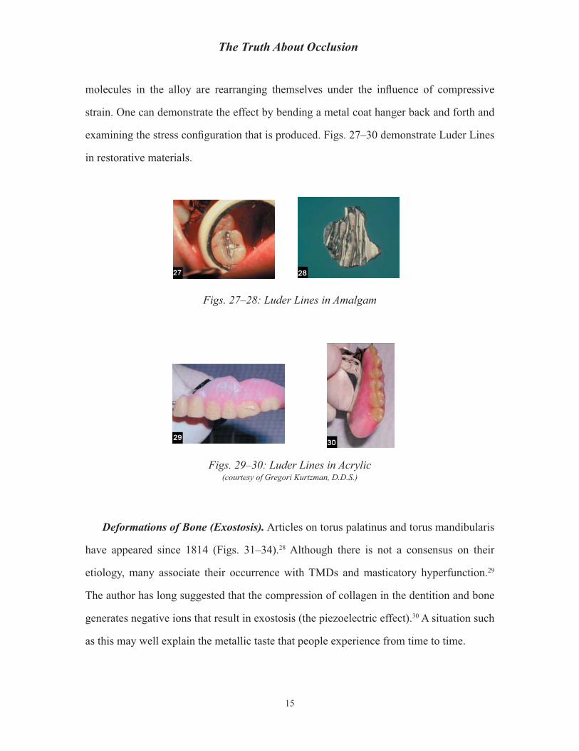
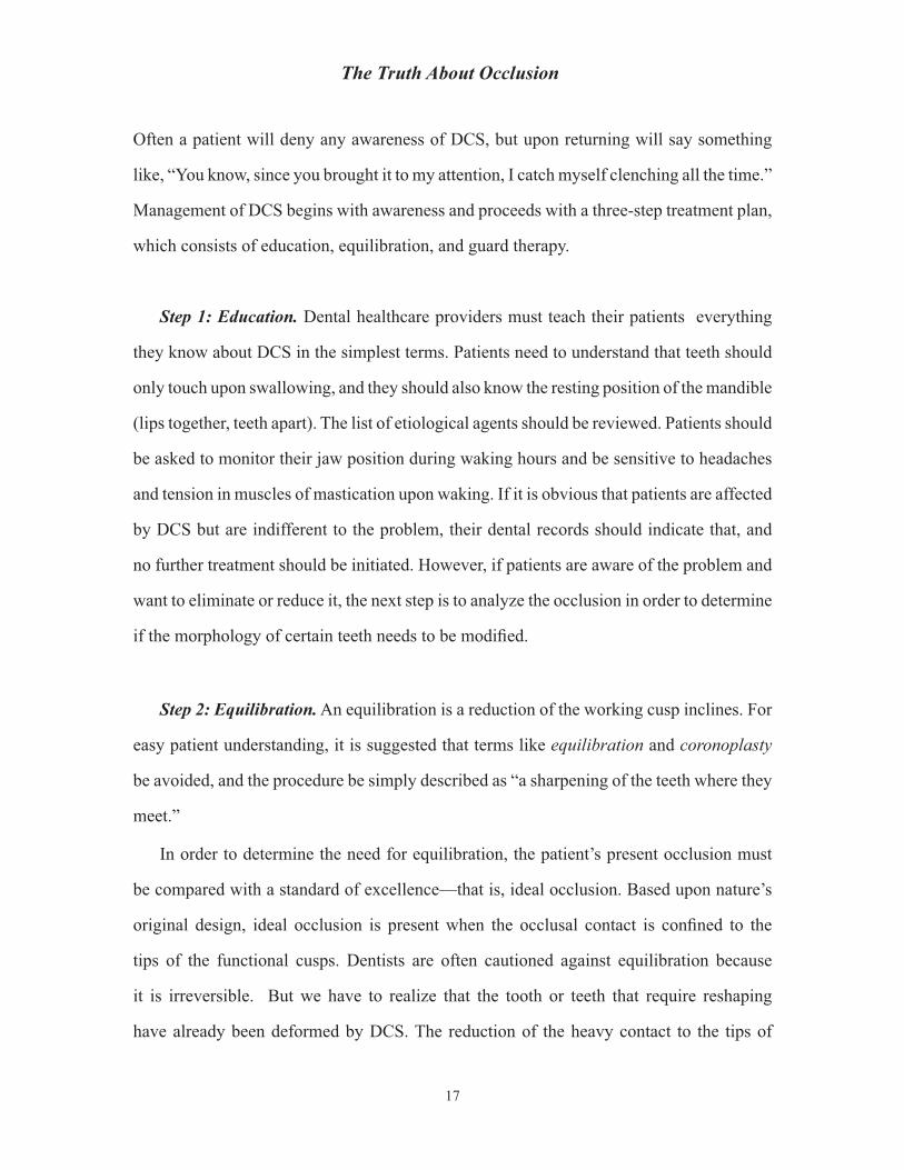
molecules in the alloy are rearranging themselves under the infl uence of compressive
strain. One can demonstrate the effect by bending a metal coat hanger back and forth and
examining the stress confi guration that is produced. Figs. 27–30 demonstrate Luder Lines
in restorative materials.
Figs. 27–28: Luder Lines in Amalgam
Figs. 29–30: Luder Lines in Acrylic(courtesy of Gregori Kurtzman, D.D.S.)
Deformations of Bone (Exostosis). Articles on torus palatinus and torus mandibularis
have appeared since 1814 (Figs. 31–34).28 Although there is not a consensus on their
etiology, many associate their occurrence with TMDs and masticatory hyperfunction.29
The author has long suggested that the compression of collagen in the dentition and bone
generates negative ions that result in exostosis (the piezoelectric effect).30 A situation such
as this may well explain the metallic taste that people experience from time to time.
15

Gene D. McCoy, D.D.S.
Figs. 31–34: Examples of Exostosis
Epidemiology
A survey was taken of 100 patients (50 female; 50 male; age range, 17–76) to determine
how many exhibited signs and symptoms of DCS and TMD (see Table).31
Table: Signs and Symptoms of DCS and TMD
Overall % Female % Male %Signs of DCS 95 96 94Awareness of DCS 61 66 56TMD 34 36 32Sensitivity to cold 54 62 46Muscle enlargement 12 10 14Flattened teeth 58 56 60Exostosis 54 48 60Gingival NCLs 58 54 62Tip of Cusp NCLs 67 68 66
The Management of Dysfunction
The presence of deformations in the oral environment should stimulate a dialogue
between the dentist and patient to determine if the patient is currently grinding and/or
clenching his or her teeth, or whether this damage occurred during a prior stressful period.
16
The Truth About Occlusion
Often a patient will deny any awareness of DCS, but upon returning will say something
like, “You know, since you brought it to my attention, I catch myself clenching all the time.”
Management of DCS begins with awareness and proceeds with a three-step treatment plan,
which consists of education, equilibration, and guard therapy.
Step 1: Education. Dental healthcare providers must teach their patients everything
they know about DCS in the simplest terms. Patients need to understand that teeth should
only touch upon swallowing, and they should also know the resting position of the mandible
(lips together, teeth apart). The list of etiological agents should be reviewed. Patients should
be asked to monitor their jaw position during waking hours and be sensitive to headaches
and tension in muscles of mastication upon waking. If it is obvious that patients are affected
by DCS but are indifferent to the problem, their dental records should indicate that, and
no further treatment should be initiated. However, if patients are aware of the problem and
want to eliminate or reduce it, the next step is to analyze the occlusion in order to determine
if the morphology of certain teeth needs to be modifi ed.
Step 2: Equilibration. An equilibration is a reduction of the working cusp inclines. For
easy patient understanding, it is suggested that terms like equilibration and coronoplasty
be avoided, and the procedure be simply described as “a sharpening of the teeth where they
meet.”
In order to determine the need for equilibration, the patient’s present occlusion must
be compared with a standard of excellence—that is, ideal occlusion. Based upon nature’s
original design, ideal occlusion is present when the occlusal contact is confi ned to the
tips of the functional cusps. Dentists are often cautioned against equilibration because
it is irreversible. But we have to realize that the tooth or teeth that require reshaping
have already been deformed by DCS. The reduction of the heavy contact to the tips of
17

Gene D. McCoy, D.D.S.
the functional cusps will reduce the forces in the gingival area by minimizing occlusal
contact during mastication. If the vertical height of the dentition is not reduced, there is no
downside. The procedure will improve the masticatory effi ciency and reduce the physical
stress on the dentition. After good engineering principles have been applied, it is time to
address the stress with a guard and/or counseling.
There are two methods of equilibration: indirect and direct. The indirect method involves
repositioning the condyles, mounting the models on a three-dimensional articulator,
adjusting the occlusion on the models, and reproducing this adjustment on the actual teeth.
The downside of this method is that it is time-consuming, expensive, and not as accurate
as the direct method.
The direct method involves utilizing the patient’s own stomatognathic system as a
biological articulator, employing occlusal indicator wax to demonstrate the contacts in
closure, analyzing the areas of displaced wax, and eliminating contacts on the incline
planes. The advantages of this method over the indirect one are that it is more accurate,
takes less time, is inexpensive, and is easy to perform.
Patients, who should sign a consent form, need to be told that their teeth will not be
shortened, and that the benefi ts (increased comfort and diminished DCS) will far outweigh
the conservative loss of enamel. The entire procedure should take no longer than fi fteen or
twenty minutes. Patients should be seen a week or two after the procedure for fi nal analysis
and polishing.
A review of fi fteen articles on occlusal equilibrations published in professional journals
reveal generalized agreement on the following points:
1. Occlusal adjustment is a misunderstood and under-utilized procedure.
2. Prophylactic adjustments in the absence of pathology are not acceptable.
18

The Truth About Occlusion
3. CR should equal CO.
4. There should be no interferences in lateral excursions.
5. The height of the buccal cusps should never be shortened except to eliminate interference in lateral excursions.
6. Traumatic occlusal relationships should be eliminated before restorative procedures.
7. Cusps should touch loosely in the opposing fossæ.
8. Inclined planes should not touch to ensure axial loading.
9. Occlusal indicator wax is the most effective way to demonstrate how teeth touch.
10. There should be no fl at plane occlusion in humans.
11. Cuspid-guided occlusion is preferred.32
A recent 17-year study evaluated the relationship between gingival fatigue due to DCS
and its relief by sharpening the functional cusps.33 The authors found that the hypersensitivity
from gingival fatigue in 246 teeth was relieved by equilibration. The study confi rmed that
the equilibration specifi cally involved reduction of the working cusp inclines.
Step 3: Guard Therapy. Although equilibration satisfi es the engineering requirements
of the problem, and education can help in stress management during waking hours, only a
guard can ensure protection during sleep.34 The question is, what kind of guard—hard or
soft and full arch or anterior?
Unfortunately, there are confl icting studies on this issue.35 Again, we have to evaluate
our objectives from an engineering point of view. If our goal is to diminish the force on
the TMJ and reduce muscle tension, the best design is a small, thin, hard acrylic appliance
19

Gene D. McCoy, D.D.S.
that covers the lingual surfaces of the maxillary anterior teeth. It is often referred to as a
deprogrammer or mandibular repositioner. A common question regarding this design is,
“Do the posterior teeth supererupt?” No, this appliance is not like the Hawley retainer,
which is worn virtually all the time. Posterior teeth do not supererupt overnight . If they
did, all mouth-breathers would have supererupted teeth.
Regarding hard or soft appliances, a recent study suggests that soft and hard splints
are equal in reducing masticatory muscle pain.36 Although that may be true, there is an
additional factor that the study did not include. In my own private practice, I had considerable
experience with soft guards and found that they were effective in reducing TMJ stress, but
patients often compressed against them simply because they were resilient.
Generally, studies agree that there is an overall reduction of oral-facial pain when DCS
is treated with any type of guard,37 but in my opinion the smaller anterior deprogrammer
seems to work best (Figs. 35 and 36).
If the intensity of the DCS is such that the three-step treatment therapy is not effective,
biofeedback, hypnotism, physical therapy, and/or drug therapy must be considered.
Figs. 35–36: Anterior Guard
20

The Truth About Occlusion
What Is Being Taught Today and Why Is It Incoherent?
The human stomatognathic system is healthiest when it functions vertically, has a
naturally sharp dentition, and is free from dysfunction. Is this what is being taught today?
Unfortunately, no. There are confl icting ideas about how the mandible moves and how
teeth touch each other when functioning normally.
Normal Function: Vertical vs. Horizontal
When one studies the work of the early investigators—Gibbs, Lundeen, Hildebrand,
Stallard, Stuart, Rugh, and Smith—it is clear that they all agreed that the mandible functions
vertically and that there are only slight lateral or protrusive excursions during mastication.
So, then, why are we focused on the way teeth touch each other horizontally, when in
fact vertical function is correct? Because dentists are concerned that compressive loads
in lateral excursions damage their patients’ dentition. Rather than focusing on prevention,
dentists developed an interest in the least harmful way that teeth should touch each other
during these lateral excursions in order to reduce the destructive shearing forces that act
parallel to the alveolar bone. In other words, they were searching for what they call the
optimum functional occlusion, which Okeson explained as follows:
The problem facing dentistry today is apparent when a patient with the signs and symptoms of occlusal pathosis comes to the dental offi ce for treatment. The dentist must determine which occlusal confi guration is most likely to eliminate this pathosis. What occlusion is least likely to create any pathologic effects for most people over the longest time? What is the optimum functional occlusion? Although many concepts exist, the study of occlusion is so complex that these questions have not been satisfactorily answered.38
This quest for the optimum functional occlusion resulted in a number of very different
concepts as to how the teeth should touch each other during lateral excursions, including:
21
Gene D. McCoy, D.D.S.
(a) mutually protected occlusion; (b) canine-protected occlusion; (c) group function
occlusion; (d) balanced occlusion; (e) theoretically ideal occlusion; (f) physiologic
occlusion; (g) non-physiologic occlusion; and (h) anterior guidance occlusion.
Of these eight concepts, the one that is most in vogue at the moment is anterior guidance
occlusion. Although this concept is taught in every dental school in the world today, I
believe that it is one of the biggest distractions to understanding how the stomatognathic
system should normally function. Why? Because it has nothing to do with normal function
(mastication) and everything to do with dysfunction (DCS).
The justifi cation for anterior guidance was to reduce parafunctional forces, but as Clark
observes:
The whole concept of canine guidance and canine-protected occlusion is actually a concept that is illogical if protection from parafunction is the subject of debate. That canines do not inherently protect the jaw and teeth from bruxism is clear because in the strong bruxer, the clinical observation of canine attrition is common.39
The concept of anterior guidance is also fl awed for several other reasons. The most
important is that people do not eat in lateral excursions with their teeth closed under heavy
compressive forces. This is the horizontal component of DCS. People eat vertically and
grind laterally. We are reminded that masticatory forces are approximately 60 pounds per
square inch, and that teeth rarely touch during this process, and only lightly when they
do.
The credibility of anterior guidance does not hold up under scrutiny. Dawson states that
the anterior teeth are the key factor in protecting the posterior teeth.40 But why do we need
to protect the formidable posterior teeth, since they are designed for high-force mastication?
Even if we concede that anterior guidance provides a modicum of relief during horizontal
DCS, it provides no relief at all from vertical DCS. Consider a patient with a prosthesis
22

The Truth About Occlusion
supported by endosteal implants in the anterior maxilla, and, while sleeping, that patient
thrusts his or her mandible up against the prosthesis. Only a guard, not anterior guidance,
will ensure protection for that patient.
One more point. Dawson also teaches that the functional relationship of the anterior
teeth is the principal determinant of posterior occlusal form.41 Others, however, believe that
occlusal form is determined not only by anterior teeth but also by the TMJs.42 But if we are
to be accurate and objective, it is genetics that determines the morphology of the dentition.
The original design of our body parts is always perfect (with the exception of anomalies)
when we fi rst get them. Teeth are beautifully designed to cut food, facilitate digestion, and
provide easy accommodation for the mandible during function and closure.
Finally, we have to consider that anterior guidance is not essential to the health and
well-being of the stomatognathic system. Patients with class II and III jaw relationships
have no anterior guidance, and they function just fi ne.
McNeill reminds us: “It must be emphasized that the teeth only come together
momentarily during swallowing and occasionally during mastication and that at all other
times the teeth should be apart in the resting range of the mandible.”43 If Dr. McNeill’s
statement correctly represents reality, why are we obsessed with lateral excursions? Why,
when a patient slides into a lateral excursion, do we call it the working side when it is not
doing any work at all?
There is no question that the reduction of lateral forces is desirable. There is a question,
however, as to the best method to reduce them. Rather than assigning certain teeth to be
shock absorbers, it is better to focus on prevention by educating patients and protecting
them with comfortable guards.
Morphology and Occlusion of the Dentition
Dentition best serves the stomatognathic system when the original, natural, sharp design
23

Gene D. McCoy, D.D.S.
is preserved, and the occlusion is confi ned to the tip of the functional cusps. But that is not
what is being taught today. Rather than a loose-fi tting occlusion, some dentists prefer a
strong bracing occlusion (tripodization), whose purpose is to solidly lock the mandible
in place so that the condyle maintains its position. Alternative thinking suggests that we
ask why the condyle has migrated and why it cannot return by itself upon swallowing. A
plausible explanation is that the condyle has come down and forward due to horizontal
DCS and cannot return due to the friction incurred by the incline planes of the teeth. A
common symptom of tripodization is clenching, which is a detriment. For dentures, which
have only 25% of the masticatory force of natural teeth, it is essential to have a sharp
design for optimum effi ciency in mastication. Unfortunately, we are offered a choice of
four different anatomical confi gurations for posterior denture teeth:
1. Anatomic: with cusp angles of 30 degrees or more
2. Semi-anatomic: with cusp angles of 20 degrees or more
3. Non-anatomic: with no cusp angles (0 degrees)
4. Lingual: with lingual cusp contact only
Since vertical forces used to penetrate a food bolus are minimized with sharp anatomical
tooth forms and maximized with fl at tooth forms, one has to come to the conclusion that
sharp teeth are superior to fl at ones for denture wearers. So why are fl at denture teeth even
considered? Because there is an idea in the profession that anatomical tooth forms, due to
their steep cusp angles, create more horizontal or lateral forces than fl at tooth forms. This
is even more signifi cant in patients with severely resorbed ridges that are less able to resist
horizontal forces than patients with fuller ridge contours. Ortman’s statement, “The fl atter
the ridge, the fl atter the cusp angles,” aptly summarizes this generally accepted concept.44
The question is, why is this a generally accepted concept? It doesn’t make sense. If we
24

The Truth About Occlusion
have a patient with a signifi cantly resorbed ridge, common sense dictates that we employ
anatomically sharp posterior teeth and instruct the patient to eat vertically, not horizontally.
So what is the origin of Ortman’s concept? For the most part, it was derived from research
that was done fi fty years ago.45 It is important to evaluate that work and to critique the
conclusions and recommendations derived from it. The objective of that research was to
determine how different occlusal morphologies affected the deformation of dentures during
mastication. The concern was that horizontal deformation of dentures could contribute to
resorption of the residual ridge.
Duplicate dentures were constructed with three different occlusal confi gurations: 33-
degree, 20-degree, and 0-degree posterior teeth. Two strain gauges, one above the other,
were embedded in the lingual fl ange of the lower denture at the midline, and patients
were asked to chew three things: raw carrots, salted peanuts, and artifi cial boli of latex
rubber and cotton rolls. The deformation—that is, the movement toward or away from the
midline—was measured across the posterior ends of the denture from the distal end of one
lingual fl ange to the distal end of the other.
The 33-degree posterior teeth caused the greatest horizontal deformation of the denture
base during mastication. The 20-degree posterior teeth had 10 percent less deformation,
and the 0-degree posterior teeth had 50 percent less deformation.
Deformation of the denture bases during the test procedures had a range of .0000 to
.0433 inches (039 inches = 1 mm).
The mean duration of the force during swallowing was 3.6 times greater than during
mastication.
There is only one valid conclusion that can be derived from this research: that vertical
compression of a denture results in a slight widening (1 mm) of the posterior portion. The
researchers theorized that this 1 mm distortion could contribute to lingual atrophy of the
alveolar ridge, but that theory has not been validated over the years, since the majority of
25

Gene D. McCoy, D.D.S.
horizontal atrophy of the alveolar ridge over time occurs on the buccal.
The conclusion derived from this research—that fl atter teeth reduce horizontal
distortion—is open to interpretation because, the amount of distortion exhibited by lingual
fl anges against the alveolar ridge is miniscule (1 mm divided bilaterally).
It is interesting to note that the researchers determined that swallowing contributes to
a greater transfer of energy to the underlying mucosa than does mastication. This is not
only because of the longer duration of the force application, but also because the forces
generated by swallowing, unlike those generated by chewing, are purely vertical.
It is common sense that vertical compression of a U-shaped denture would result in a
slight widening of the posterior portion due to the higher force application. When fl atter
teeth were used, the patients’ vector of function went from vertical to horizontal, not only
diminishing the distortion but also decreasing the effi ciency and power of the mastication.
This fi fty-year-old research leaves unanswered questions. First of all, why were the
stress gauges placed in the midline of the lingual fl ange and not in the lingual and buccal
fl anges of the posterior portion of the denture? Second, why were such hard substrates
as peanuts and raw carrots used for this experiment? We are told that denture wearers do
not yield related performances characterizing masticatory functions in either tough or soft
foods, but peanuts and raw carrots require much more effort than, say, cooked chicken or
steamed vegetables, which is what many denture wearers eat.
Finally, there is no description of how the patients chewed—vertically or horizontally.
Since the power of mastication for denture wearers is reduced by 75 percent, it is important
to maximize the chewing effi ciency by employing sharp posterior teeth and instructing
patients to eat vertically.
26

The Truth About Occlusion
Discussion and Recommendations
Which Concept on Occlusion Is Correct?
Gordon Christensen, in his annual review New Directions in Dentistry, stated: “There is
extreme controversy about which concept of occlusion is correct, and I do not see any relief
to that controversy.”46 In a subsequent article, he wrote that “the profession is in major chaos
relative to occlusion.”47 Frank Spear recently wrote that “a byproduct of increased interest
in occlusion has been a renewed debate about which occlusal philosophy is correct.”48
Why is this question so diffi cult to answer? Because of the different interpretations of
the word occlusion, we are not quite sure what the question is asking. If it is asking the best
way teeth should touch each other and when, the answer would be that the contact should
be confi ned to the tip of the cusps, and that contact should occur only during swallowing.
If it is asking the best way teeth should touch each other during mastication, the answer
would be that they shouldn’t. If it is asking for the most effi cient way the stomatognathic
system should function, the answer would be: without heavy compressive vertical and
lateral forces (DCS). But the question is not focused on any of these interpretations. What
Christensen and Spear are referring to are the two different philosophies taught at the
LVI and at the Pankey Institute: the neuromuscular methodology, on the one hand, and
the gnathological approach, on the other. But wait a minute! These are not concepts on
occlusion. These are two different methods of rehabilitation and/or reconstruction to be
used when patients are in trouble. So now the word occlusion has a fourth interpretation.
Is one method better than the other? That is not the important question. What we should
be asking is: What is the best way that general practitioners should be doing their work
in order to minimize the deleterious effects of DCS so that patients don’t have to go into
rehabilitation? Why general practitioners? Because it is the GPs who are doing the vast
majority of the work. Very few patients go into rehabilitation or reconstruction—probably
less than 1%. The majority of dental work that is performed each day throughout the
27

Gene D. McCoy, D.D.S.
world is by increments—a crown here, an amalgam restoration there, facial composites,
or some fi xed bridgework. Since this is reality, what is the best way GPs can perform this
incremental work, maintain the health and effi ciency of the stomatognathic system, and
prevent DCS? That is what we really want to know if we are going to interpret “concept on
occlusion” objectively.
What are the guidelines? GPs should:
1. Be alert to the signs and symptoms of DCS.
2. Thoroughly explain DCS to their patients.
3. Determine if an equilibration is necessary.
4. Determine if a guard is necessary.
5. Mimic the natural design of teeth when delivering dental prostheses to the mouth.
Problem-Solving for TMJDs
When patients present with oral-facial pain and/or discomfort in the TMJ, dentists
should consider that DCS might be the source of the problem. Traditionally, clenching and
grinding have been the most agreed-upon cause of TMJD.49 If this is confi rmed either by
the patient or by information gained by examining the dentition, the three-step management
therapy described earlier should be initiated to reduce the stress on the TMJ.
If a patient’s condyles have migrated down and forward, there are three traditional
methods of management: (a) manually reposition the condyles and then equilibrate the
dentition; (b) have the patient wear a splint (mandibular repositioner/deprogrammer) for
a period of time and then equilibrate; and (c) use neuromuscular instrumentation. For
GPs there is a fourth method that is simple and effective and produces immediate positive
results: equilibration. If occlusal indicator wax is used to diagnose the occlusal contacts, it
28

The Truth About Occlusion
is common to see that, due to DCS, the mandible has worked its way forward and cannot
return during swallowing or closure because the incline planes of the cusps are engaged. If
this is the case, recreation of the intra-incline space will allow the condyles to resume their
natural position.
DCS and Periodontal Disease
DCS can create periodontal disease through a disturbance of the physiology. Firestone
and Miller demonstrated how DCS can produce changes in salivary composition, blood
calcium levels, and extreme alveoloclasica.50
DCS and Oral Implant Patients
As with natural dentition, there has been an ongoing controversy about occlusion in
implant therapy. The primary concern is the durability and life span of the prosthesis.51
It is unfortunate that with implant therapy, the only question regarding occlusion that
implantologists seem to be concerned with is when the implant should be loaded. Instead,
they should be more concerned with the source, frequency, and power of that load. There
are two power sources of loading: mastication and DCS. During mastication, the loading
that is introduced through the bolus of food is not directed down the implant’s long axis but
rather distributed at various levels of the prosthesis, implant body, and surrounding bone
in the form of complex bending movements.52 However, this seems to be of little concern,
since the power source is only about 60 pounds per square inch. The loading we should
be concerned with results from the vertical and horizontal components of DCS, which can
exceed 300 pounds per square inch.
When planning implant reconstruction, the restorative dentist must consider that the
loss of the patient’s natural dentition may have been due to DCS, which will probably
jeopardize the integrity of the newly placed implants. Complications from DCS for
29

Gene D. McCoy, D.D.S.
implant-supported prostheses include acrylic and porcelain veneer fractures, abutment or
prosthetic screw loosening, fracture of prosthesis and implant body, and crestal bone loss.53
With these complications in mind, what guidelines should oral implantologists follow to
minimize heavy compressive forces? During the consultation phase, patients should be
questioned about their awareness of DCS. If patients are semi-edentulous, their remaining
dentition will reveal valuable information. Thus, their remaining teeth should be evaluated
to determine if a reduction of the working cusp inclines might be benefi cial.
Oral implantologists have to create the best defense against heavy compressive forces.
This is accomplished not only by establishing a strong implant foundation but also by
minimizing the effects of DCS by stress reduction and guards. Since at this time there is no
way the surface-to-bone interface of an implant can compare to its natural predecessor, it
is imperative that implantologists maximize the interface by using larger and/or additional
implants in high-quality bone sites.54 Sharp occlusal anatomy and vertical loading during
closure are mandatory. Finally, guard protection while sleeping is good insurance.
The subject of occlusion has been made more complicated than it has to be. It seems
that we are trying to explain everything by the way teeth come together, whereas the most
comfortable patients always have their teeth apart. The occlusion confusion has distracted
dentists from focusing on the more serious problem of patients grinding and clenching
their teeth. Would we have any problems related to occlusion if patients did not clench and
grind? I think very few. The fi rst line of defense for TMJD and oral facial pain is the GP’s
understanding of the relationship between occlusion, the stomatognathic system, and DCS
prevention.
30

The Truth About Occlusion
Notes1T. D. Taylor, J. Wiens, & A. Carr (2005), “Evidence-Based Considerations for Removable Prosthodontic
and Dental Implant Occlusion: A Literature Review,” Journal of Prosthetic Dentistry, 94: 555–560.2J. Simon (2006), “Dental Occlusion, What We Can Agree On,” Dentistry Today, 23 (11): 16–18.3C. W. Taber (1960), Taber’s Cyclopedic Medical Dictionary, 8th ed. (Philadelphia: F. A. Davis).4W. A. N. Dorland (1898), Dorland’s Pocket Medical Dictionary, 1st ed. (Philadelphia: W. B. Saunders).5W. A. N. Dorland (1959), Dorland’s Pocket Medical Dictionary, 20th ed. (Philadelphia: W. B.
Saunders).6S. Jablonski (1982), Illustrated Dictionary of Dentistry (Philadelphia: W. B. Saunders).7J. P. Okeson (1993), Management of Temporomandibular Disorders and Occlusion, 3rd ed. (St. Louis:
C. V. Mosby).8G. McCoy (1999), “Dental Compression Syndrome: A New Look at an Old Disease,” Journal of Oral
Implantology, 25: 35–49.9C. Misch & M. Bidez (1994), “Implant Protected Occlusion: A Biomechanical Rationale,” Compendium
of Continuing Education in Dentistry, 15: 1330–43.10P. E. Dawson (1989), Evaluation, Diagnosis, and Treatment of Occlusal Problems, 2nd. ed. (St. Louis:
C.V. Mosby).11C. Misch & M. Bidez (1994), “Implant Protected Occlusion: A Biomechanical Rationale,” Compendium
of Continuing Education in Dentistry, 15: 1330–43.12E. H. Angle (1899), “Classifi cation of Malocclusion,” Dental Cosmos, 41 (3).13G. McCoy (1993), “Dental Compression Syndrome,” The Quintessence, 13: 92–100.14J. Sedgwick (1995, December 4), “I Hear America Grinding,” Newsweek, p. 77.15L. G. Selna, H. T. Shillingburg, & P. A. Kerr (1975), “Finite Element Analysis of Dental Structures:
Axisymmetric and Plane Stress Idealizations,” Journal of Biomedical Matter, 9: 237–252.16A. L. Yettram, K. W. Wright, & H. M. Pickard (1976), “Finite Element Stress Analysis of the Crowns of
Normal and Restored Teeth,” Journal of Dental Research, 55: 1004–11.17G. McCoy (1995), “Examining the Role of Occlusion in the Function and Dysfunction of the Human
Mastication System,” Dental Focus (South Korea), 169: 10–15.18A. L. Yettram, K. W. Wright, & H. M. Pickard (1976), “Finite Element Stress Analysis of the Crowns of
Normal and Restored Teeth,” Journal of Dental Research, 55: 1004–11.19B. Kornfeld (1932), “Preliminary Report of Clinical Observations of Cervical Erosions: A Suggested
Analysis of the Cause and the Treatment for Its Relief,” Dental Items of Interest, 54: 905–909.20G. McCoy (1997), “Occlusion and Dental Compression Syndrome,” Nippon Dental Review, 659: 163–
183.21T. Kuroe, H. Itoh, A. A. Caputo, & H. Nakahara (1999). “Potential for Load-Induced Cervical Stress
Concentration as a Function of Periodontal Support,” Journal of Esthetic Dentistry, 1: 215–222.22W. I. Ferrier (1931, November–December), “Clinical Observations on Erosions and Their Restoration,”
Journal of the California State Dental Association.23S. E. Kennedy (1987), “Biodental Theory Examines Stress,” Dentistry Today, 6 (4); C. Misch (1993),
Contemporary Implant Dentistry (St. Louis: C. V. Mosby), pp. 161–162.24J. O. Grippo (1991), “Abfraction: A New Classifi cation of Hard Tissue Lesions of Teeth,” Journal of
Esthetic Dentistry, 3: 14–19.25Fig. 19 is courtesy of Reidan Sognnaes, D.M.D.26J. E. Gordon (1978), Structures or Why Things Don’t Fall Down (New York, Da Capo Press), pp. 333–
334.27J. L. Old & M. Calvert (2004), “Vertebral Compression Fractures in the Elderly,” American Family
Physician, 69: 111–116.28Y. H. Seah (1995), “Torus Palatinus and Torus Mandibularis: A Review of the Literature,” Australian
Dental Journal, 40: 318–321.29B. R. Pynn, N. S. Kurys-Kos, D. A. Walker, & J. T. Mayhall (1995), “Tori Mandibularis: A Case Report
31

Gene D. McCoy, D.D.S.
and Review of the Literature,” Journal of the Canadian Dental Association, 61: 1057–66; S. Sirirungrojying & D. K. H. Song Khln (1999), “Relationship Between Oral Tori and Temporomandibular Disorders,” International Dental Journal, 49: 101–104; K. E. Sonnier, G. M. Horning, & M. E. Cohen (1999), “Palatal Tubercles, Palatal Tori, and Mandibular Tori: Prevalence and Anatomical Features in a U.S. Population,” Journal of Periodontology, 70: 329–336.
30G. McCoy (1995), “Examining the Role of Occlusion in the Function and Dysfunction of the Human Mastication System,” Dental Focus (South Korea), 169: 10–15; G. McCoy (1997), “Occlusion and Dental Compression Syndrome,” Nippon Dental Review, 659: 163–183; G. McCoy (1999), “Dental Compression Syndrome: A New Look at an Old Disease,” Journal of Oral Implantology, 25: 35–49.
31G. McCoy (1999), “Dental Compression Syndrome: A New Look at an Old Disease,” Journal of Oral Implantology, 25: 35–49.
32S. O. Bartlett & R. W. Elliott (1973), “Training Devices for Group Instruction on Occlusal Equilibration,” Journal of Prosthetic Dentistry, 29: 517–523; F. M. Bush, J. H. Butler, & D. M. Abbott (1988), “Perspective on Occlusal Adjustment in the Treatment of Temporomandibular Joint Disorders,” Virginia Dental Journal, 65: 30–31; S. Cantor (1970), “Procedures in the Equilibration of the Natural Dentition Prior to Fixed Prosthetic Restoration,” Temple Dental Review, 40: 22–24; A. S. Freese (1966), “Occlusal Equilibration for the General Practitioner,” TIC, 25: 4–12; J. Harris (1971), “A Marking Device for Equilibration,” Journal of Clinical Orthodontics, 5: 111–113; W. C. Klein (1967), “A Simplifi ed Technique of Occlusal Equilibration,” Journal of the Ontario Dental Association, 44: 30; R. F. La Mug (1975), “Occlusal Equilibration,” Journal of the Philadelphia Dental Association, 26: 10–13; N. C. Murphy (1979), “Clinical Observations on Occlusal Adjustment,” Ohio Dental Journal, 53: 41–50; R. D. Oles (1990), “Occlusal Adjustment,” Journal of the University of Saskatchewan, 56: 527–531; C. H. Schuyler (1973), “Equilibration of Natural Dentition,” Journal of Prosthetic Dentistry, 30: 506–509; M. M. Silverman (1968), “Equilibration of the Natural Dentition Following Orthodontic Treatment to Prevent Movement of Teeth and Other Problems,” American Journal of Orthodontics, 54: 831–854; M. M. Silverman (1977, Winter), “Procedures in Equilibration of Natural Teeth,” Journal of District of Columbia Dental Society, 17: 17–27; R. B. Steadman (1988), “Occlusal Equilibration and TMJ Disorders,” Virginia Dental Journal, 65: 28–30; B. Wenneberg, T. Nystrum, & G. E. Carlson (1988), “Occlusal Equilibration and Other Stomatognathic Treatment in Patients with Mandibular Dysfunction and Headache,” Journal of Prosthetic Dentistry, 59: 478–483; D. J. Wilson (1973), “Occlusal Equilibration,” Journal of the Tennessee Dental Association, 53: 302–305.
33T. A. Coleman, J. O. Grippo, & K. E. Kinderknecht (2003), “Cervical Dentin Hypersensitivity, Part 3,” Quintessence International, 34: 427–434.
34G. T. Clark (1984), “A Critical Evaluation of Orthopedic Interocclusal Appliance Therapy: Design, Theory, and Overall Effectiveness,” Journal of the American Dental Association, 108: 359–364; A. Humsi, M. Naeje, & J. A. Hippe (1989), “The Immediate Effects of a Stabilization Splint on the Muscular Symmetry in the Masseter and Anterior Temporal Muscles of Patients with Craniomandibular Disorders,” Journal of Prosthetic Dentistry, 62: 339–343; Y. Kawazal, H. Kotani, & T. Hamada (1980), “Effect of Occlusal Splints on the Selectromyographic Activities of Masseter Muscles During Maximum Clenching in Patients with Myofacial Pain Dysfunction Syndrome,” Journal of Prosthetic Dentistry, 43: 578–580; S. C. Shan & W. H. Yum (1991), “Infl uence of an Occlusal Splint on Integrated Electromyography of the Masseter Muscle,” Journal of Oral Rehabilitation, 18: 253–256.
35E. Nevarro, N. Bargi, & R. Rey, R. (1985), “Clinical Evaluation of Maxillary Hard and Resilient Occlusal Splints,” Journal of Dental Research, 64: 313; J. P. Okeson (1987), “The Effects of Hard and Soft Occlusal Splints on Nocturnal Bruxism,” Journal of the American Dental Association, 114: 788–791.
36C. A. Pettengill, M. R. Growney, R. Schoof, & C. R. Kenworthy (1998), “A Pilot Study Comparing the Effi cacy of Hard and Soft Stabilizing Appliances in Treating Patients with Temporomandibular Disorders,” Journal of Prosthetic Dentistry, 79: 165–168.
37K. Holmgren, A. Sheikholeslam, & C. Riise (1993), “Effect of a Full-Arch Maxillary Occlusal Splint on Parafunctional Activity During Sleep in Patients with Nocturnal Bruxism and Signs and Symptoms of Craniomandibular Disorders,” Journal of Prosthetic Dentistry, 69: 293–297.
38J. P. Okeson (1993), Management of Temporomandibular Disorders and Occlusion, 3rd ed. (St. Louis:
32

The Truth About Occlusion
C. V. Mosby), p. 109.39G. T. Clark, Y. Tsukiyama, K. Baba, & T. Watanabe (1999), “Sixty-Eight Years of Experimental Occlusal
Interference Studies: What Have We learned,” Journal of Prosthetic Dentistry, 82: 704–713.40P. E. Dawson (1989), Evaluation, Diagnosis, and Treatment of Occlusal Problems, 2nd ed. (St. Louis:
C.V. Mosby), p. 275.41P. E. Dawson (1989), Evaluation, Diagnosis, and Treatment of Occlusal Problems, 2nd ed. (St. Louis:
C.V. Mosby), p. 274.42J. P. Okeson (1993), Management of Temporomandibular Disorders and Occlusion, 3rd ed. (St. Louis:
C. V. Mosby), p. 127.43C. McNeill (1997), “Selective Tooth Grinding and Equilibration,” in C. McNeill, ed., Science and
Practice of Occlusion (Carol Stream, IL: Quintessence), p. 412.44H. R. Ortman (1988), “Complete Denture Occlusion,” in S. Winkler, ed., Essentials of Complete Denture
Prosthodontics, 2nd ed. (St. Louis: C. V. Mosby), pp. 217–249.45W. L. Kydd (1956), “Complete Denture Base Deformation with Varied Occlusal Tooth Form,” Journal
of Prosthetic Dentistry, 6: 714–718; C. P. Regli & W. L. Kydd (1953), “A Preliminary Study of the Lateral Deformation of Metal Base Dentures in Relation to Plastic Base Dentures,” Journal of Prosthetic Dentistry, 3: 326–330; C. C. Swoope & W. L. Kydd (1966), “The Effect of Cusp Form and Occlusal Surface Area on Denture Base Deformation,” Journal of Prosthetic Dentistry, 16: 34–43.
46G. Christensen (2004, February), “New Directions in Dentistry,” Dentistry Today, 23: 82–84.47G. Christensen (2006, April), “Occlusion Confusion,” Dentaltown, 7 (4): 8–14.48F. M. Spear (2006), “The Business of Occlusion,” Journal of the American Dental Association, 137:
666–667.49A. G. Glaros, M. Forbes, J. Shanker, & E. G. Glass (2000), “Effect of Parafunctional Clenching on
Temporomandibular Disorder Pain and Proprioceptive Awareness,” Journal of Craniomandibular Practice, 18: 198–204; F. Lobbezoo & G. J. Lavigne (1997), “Do Bruxism and Temporomandibular Disorders Have a Cause and Effect Relationship?” Journal of Orofacial Pain, 11: 15–23; T. Magnusson, I. Egermark, & G. E. Carlsson (2000), “A Longitudinal Epidemiologic Study of Signs and Symptoms of Temporomandibular Disorders from 15 to 35 Years of Age,” Journal of Orofacial Pain, 14: 310–319; O. F. Molina, J. Dos Santos, S. J. Nelson, & T. Nowlin (2000), “Profi le of TMD and Bruxer Compared to TMD and Non-Bruxer Patients Regarding Chief Complaint, Previous Consultations, Modes of Therapy, and Chronicity,” Journal of Craniomandibular Practice, 18: 205–218; K. Nagahara, S. Murata, S. Nakamura, & T. Tsuchiya (1999), “Displacement and Stress Distribution in the Temporomandibular Joint During Clenching,” The Angle Orthodontist, 69: 372–397; A. R. Wright, R. J. Gatchel, L. Wildenstein, R. Riggs, P. Buschang, & E. Ellis (2004), “Biopsychosocial Differences Between High-Risk and Low-Risk Patients with Acute TMD-Related Pain,” Journal of the American Dental Association, 135: 374–382.
50J. M. Firestone & S. C. Miller (1947), “Psychosomatic Factors in the Etiology of Periodontal Disease,” American Journal of Periodontia, 33: 675–686.
51C. M. Stanford (2005), “Issues and Considerations in Dental Implant Occlusion: What Do We Know, and What Do We Need to Find Out?” Journal of the California Dental Association, 33: 329–336.
52C. M. Stanford (2005), “Issues and Considerations in Dental Implant Occlusion: What Do We Know, and What Do We Need to Find Out?” Journal of the California Dental Association, 33: 329–336.
53C. E. Misch (2006), “Consideration of Biomechanical Stress in Treatment with Dental Implants,” Dentistry Today, 25 (5): 80–85.
54C. E. Misch (2006), “Consideration of Biomechanical Stress in Treatment with Dental Implants,” Dentistry Today, 25 (5): 80–85.
33