Embed Size (px)
Citation preview
The TrematodesDR MONA BADR
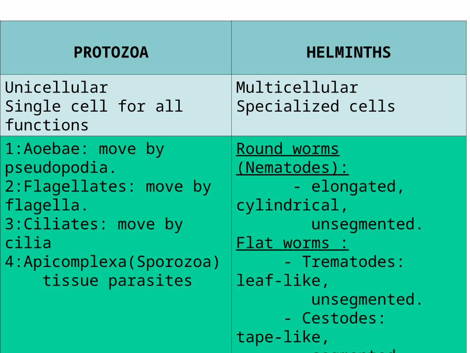
CLASSIFICATION OF PARASITESPROTOZOA HELMINTHS
UnicellularSingle cell for all functions
MulticellularSpecialized cells
1:Aoebae: move by pseudopodia.2:Flagellates: move by flagella.3:Ciliates: move by cilia 4:Apicomplexa(Sporozoa) tissue parasites
Round worms (Nematodes): - elongated, cylindrical, unsegmented.Flat worms : - Trematodes: leaf-like, unsegmented. - Cestodes: tape-like, segmented.
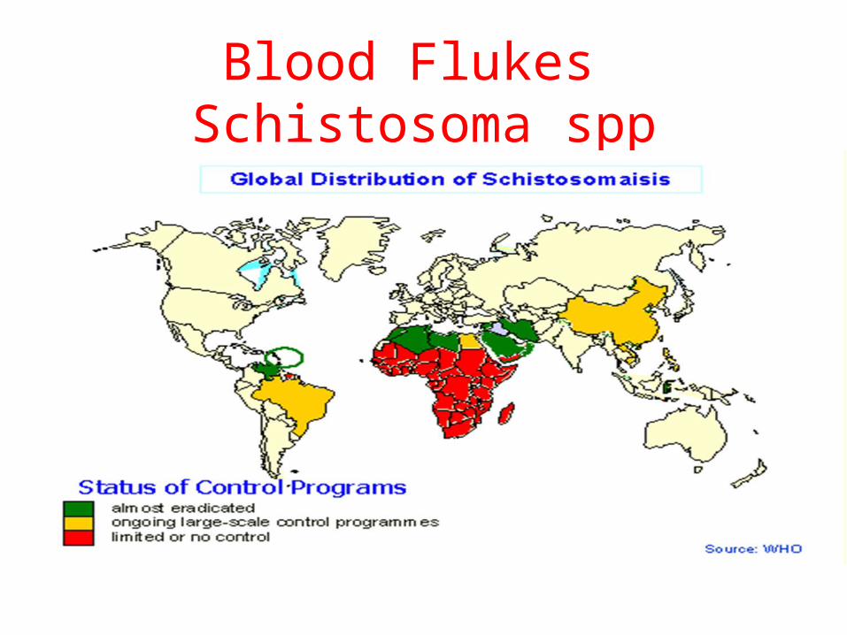
Blood Flukes Schistosoma spp
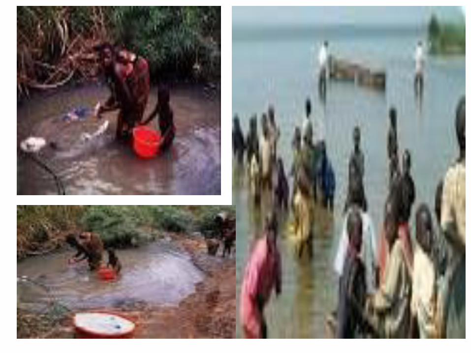
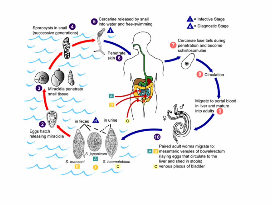
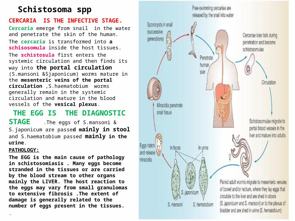
Schistosoma sppCERCARIA IS THE INFECTIVE STAGE.Cercaria emerge from snail in the water and penetrate the skin of the human.
The cercaria is transformed into a schisosomula inside the host tissues.
The schistosula first enters the systemic circulation and then finds its way into the portal circulation (S.mansoni &Sjaponicum) worms mature in the mesenteric veins of the portal circulation ,S.haematobium worms generally remain in the systemic circulation and mature in the blood vessels of the vesical plexus.
THE EGG IS THE DIAGNOSTIC STAGE .The eggs of
S.mansoni & S.japonicum are passed mainly in stool and S.haematobium passed mainly in the urine.
PATHOLOGY:
The EGG is the main cause of pathology in schistosomiasis . Many eggs become stranded in the tissues or are carried by the blood stream to other organs mainly the LIVER. The host reaction to the eggs may vary from small granulomas to extensive fibrosis .The extent of damage is generally related to the number of eggs present in the tissues.
.
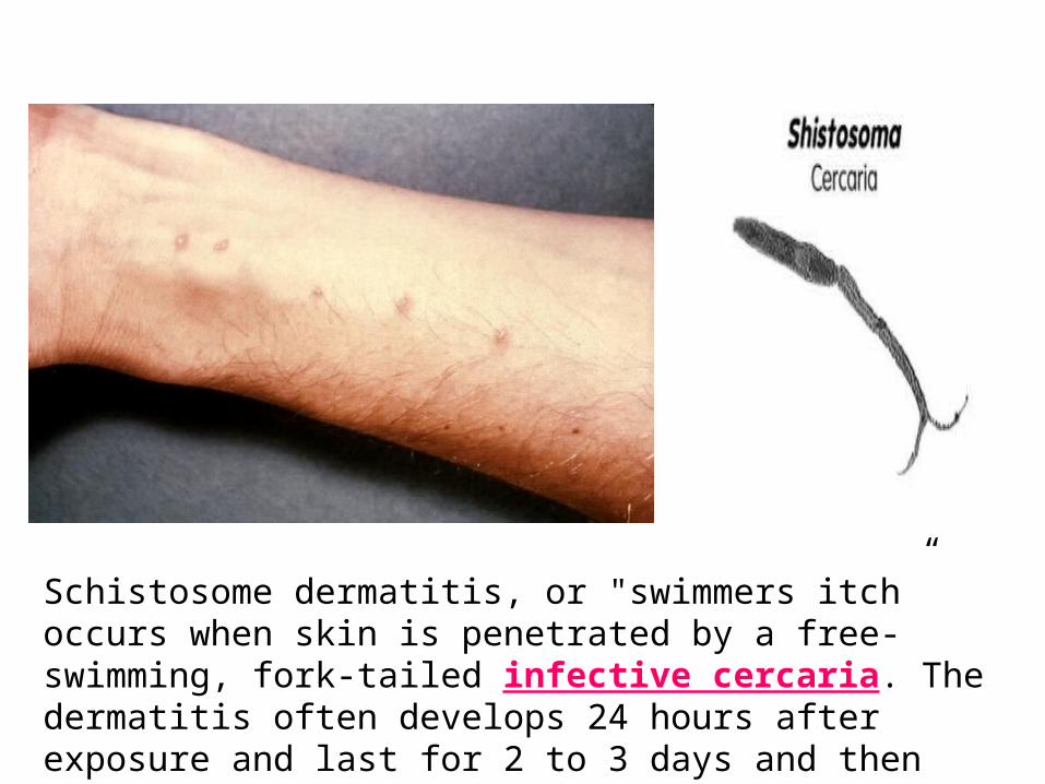
Schistosome dermatitis, or "swimmers itch” occurs when skin is penetrated by a free-swimming, fork-tailed infective cercaria. The dermatitis often develops 24 hours after exposure and last for 2 to 3 days and then spontaneously disappears. Source: WikiMedia.
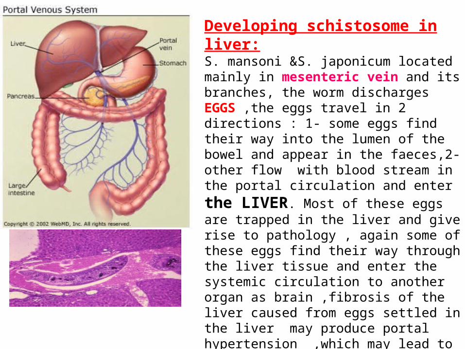
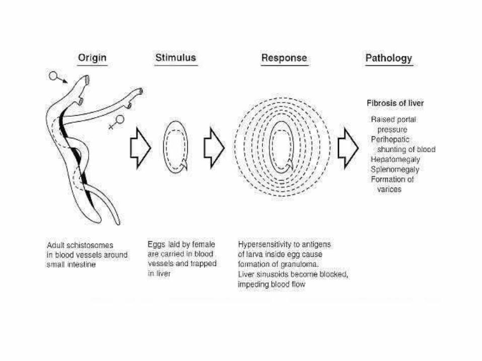
Developing schistosome in liver:S. mansoni &S. japonicum located mainly in mesenteric vein and its branches, the worm discharges EGGS ,the eggs travel in 2 directions : 1- some eggs find their way into the lumen of the bowel and appear in the faeces,2- other flow with blood stream in the portal circulation and
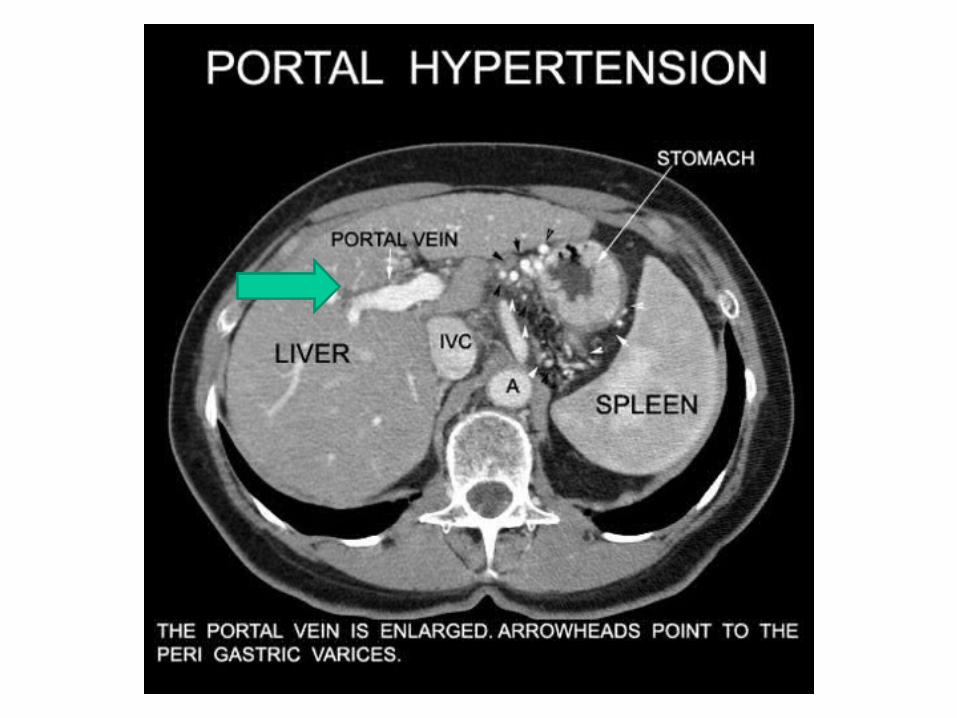
enter the LIVER. Most of these eggs are trapped in the liver and give rise to pathology , again some of these eggs find their way through the liver tissue and enter the systemic circulation to another organ as brain ,fibrosis of the liver caused from eggs settled in the liver may produce portal hypertension ,which may lead to hepatomegally ,splenomegally esophageal varices,haemorroids and ascites.
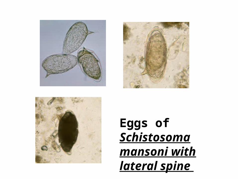
Eggs of Schistosoma mansoni with lateral spine
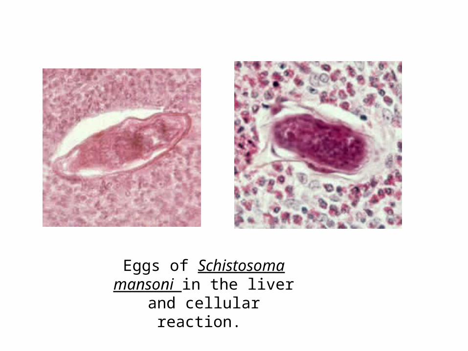
Eggs of Schistosoma mansoni in the liver and cellular
reaction.
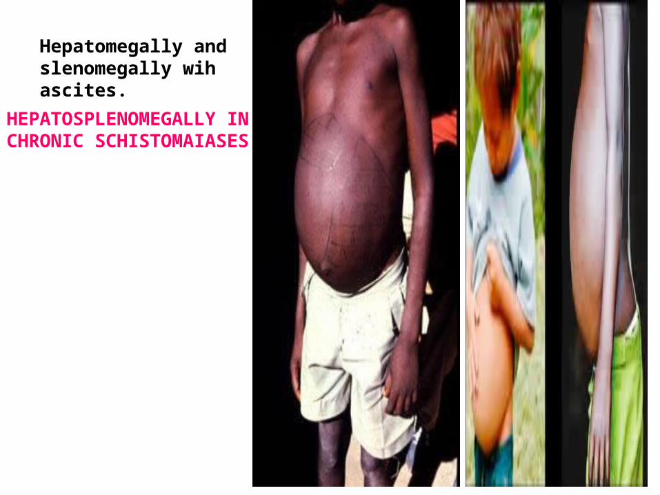
Hepatomegally and slenomegally wih ascites.
HEPATOSPLENOMEGALLY IN CHRONIC SCHISTOMAIASES.
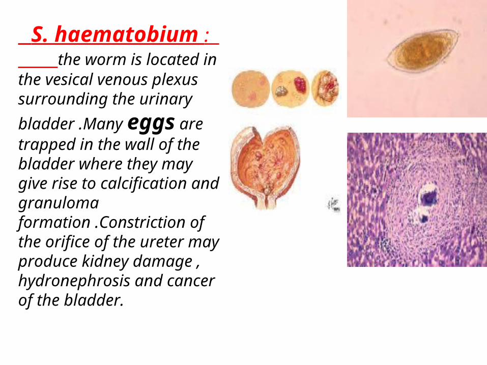
S. haematobium : the worm is located in the vesical venous plexus surrounding the urinary
bladder .Many eggs are trapped in the wall of the bladder where they may give rise to calcification and granuloma formation .Constriction of the orifice of the ureter may produce kidney damage , hydronephrosis and cancer of the bladder.
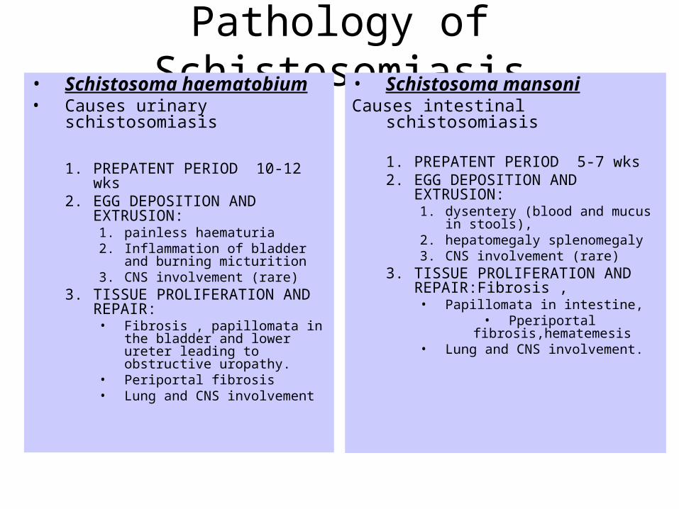
Pathology of Schistosomiasis• Schistosoma haematobium• Causes urinary schistosomiasis
1. PREPATENT PERIOD 10-12 wks2. EGG DEPOSITION AND
EXTRUSION: 1. painless haematuria2. Inflammation of bladder and burning
micturition3. CNS involvement (rare)
3. TISSUE PROLIFERATION AND REPAIR:• Fibrosis , papillomata in the bladder
and lower ureter leading to obstructive uropathy.
• Periportal fibrosis• Lung and CNS involvement
• Schistosoma mansoniCauses intestinal schistosomiasis
1. PREPATENT PERIOD 5-7 wks2. EGG DEPOSITION AND
EXTRUSION: 1. dysentery (blood and mucus in stools), 2. hepatomegaly splenomegaly3. CNS involvement (rare)
3. TISSUE PROLIFERATION AND REPAIR:Fibrosis , • Papillomata in intestine,
• Pperiportal fibrosis,hematemesis• Lung and CNS involvement.
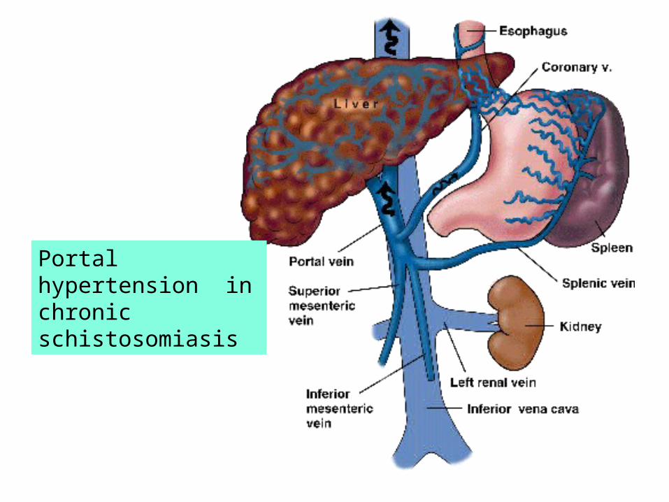
Portal hypertension in chronic schistosomiasis
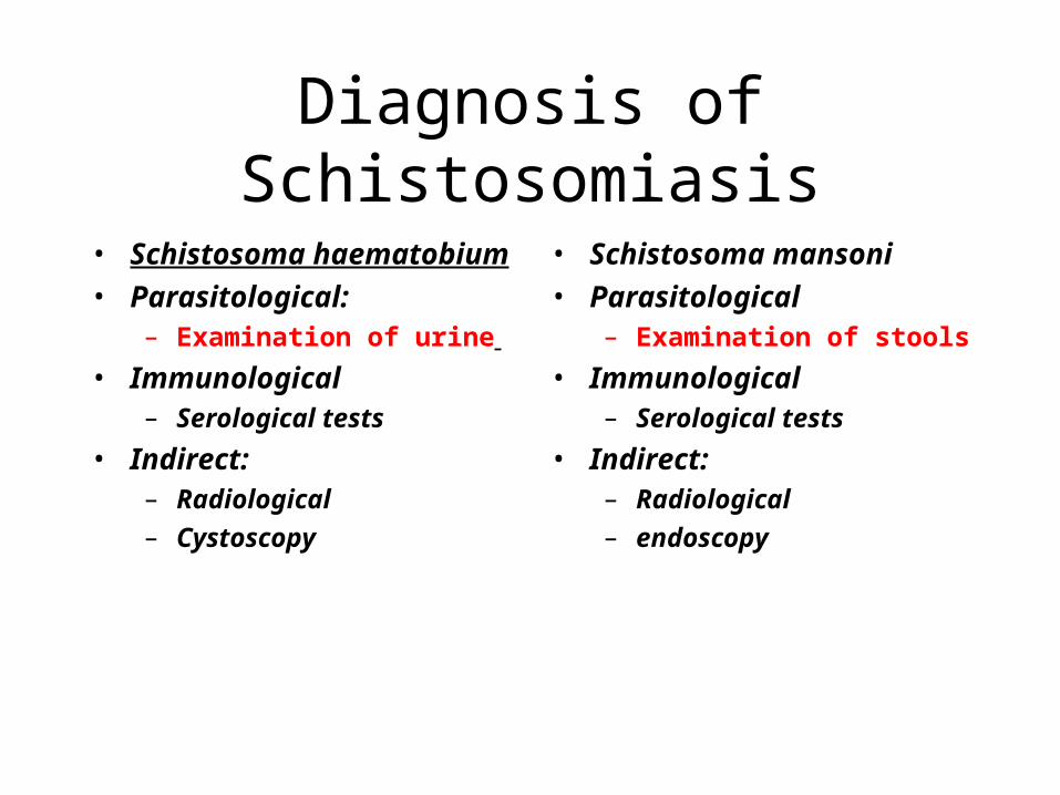
Diagnosis of Schistosomiasis
• Schistosoma haematobium
• Parasitological:– Examination of urine
• Immunological– Serological tests
• Indirect:– Radiological
– Cystoscopy
• Schistosoma mansoni
• Parasitological– Examination of stools
• Immunological– Serological tests
• Indirect:– Radiological
– endoscopy
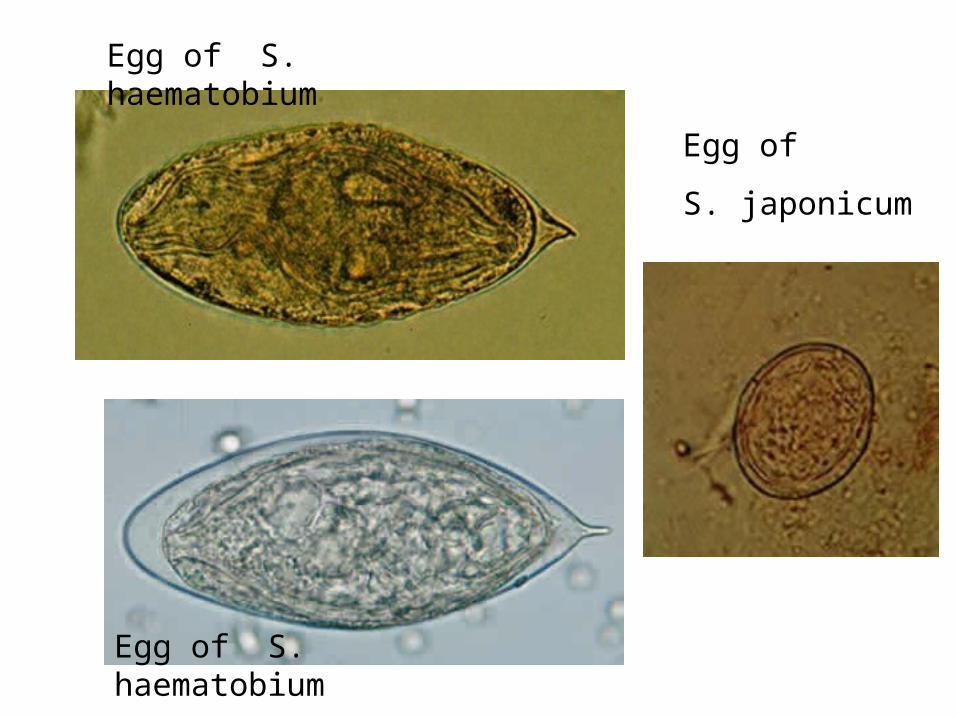
Egg of S. haematobium
Egg of S. haematobium
Egg of
S. japonicum
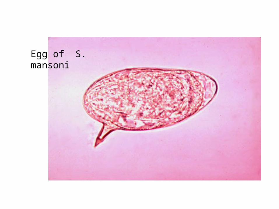
Egg of S. mansoni
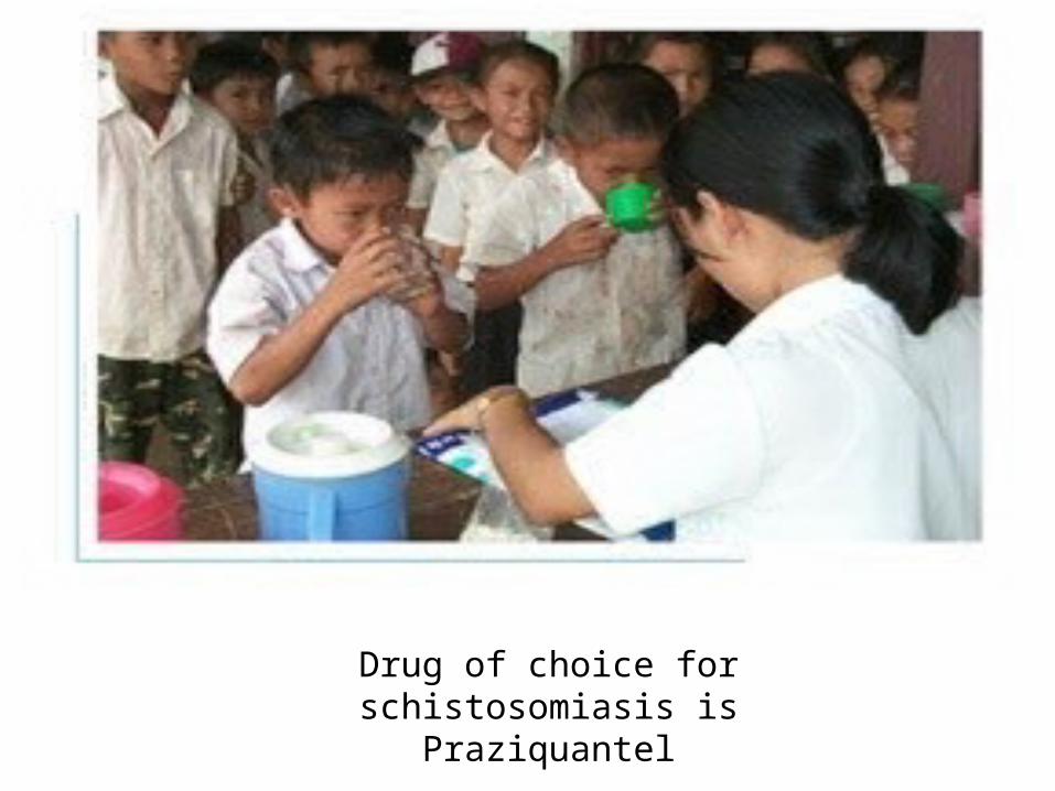
Drug of choice for schistosomiasis isPraziquantel
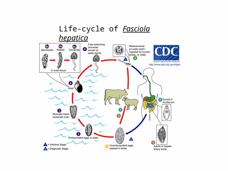
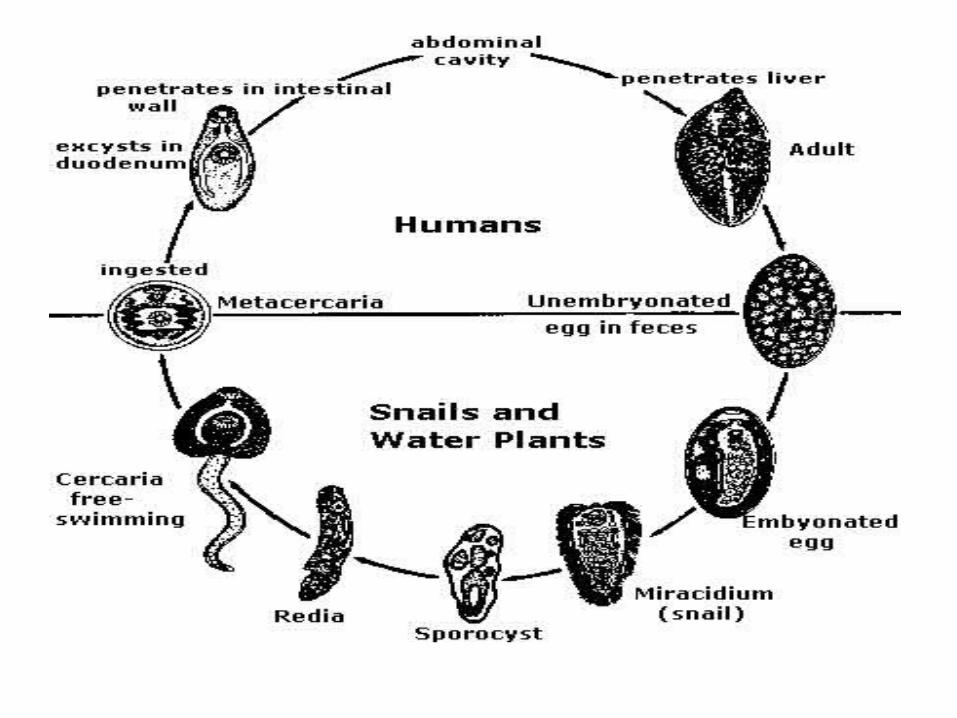
Life-cycle of Fasciola hepatica
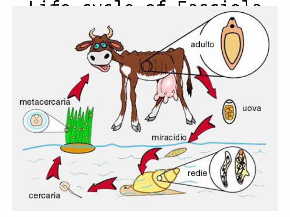
Life cycle of Fasciola hepatica
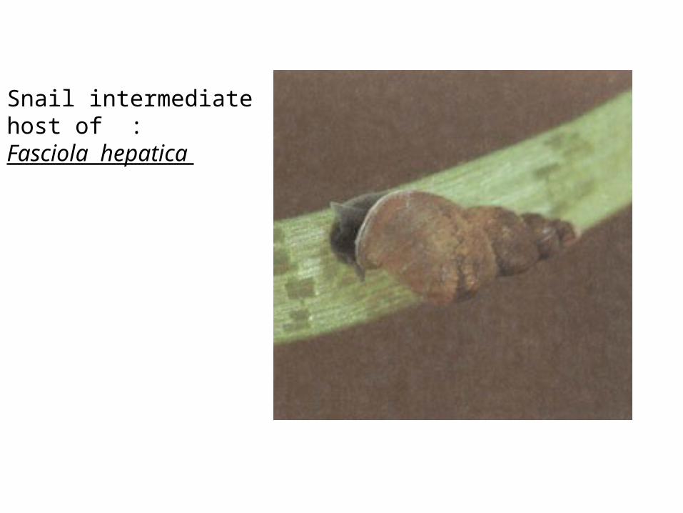
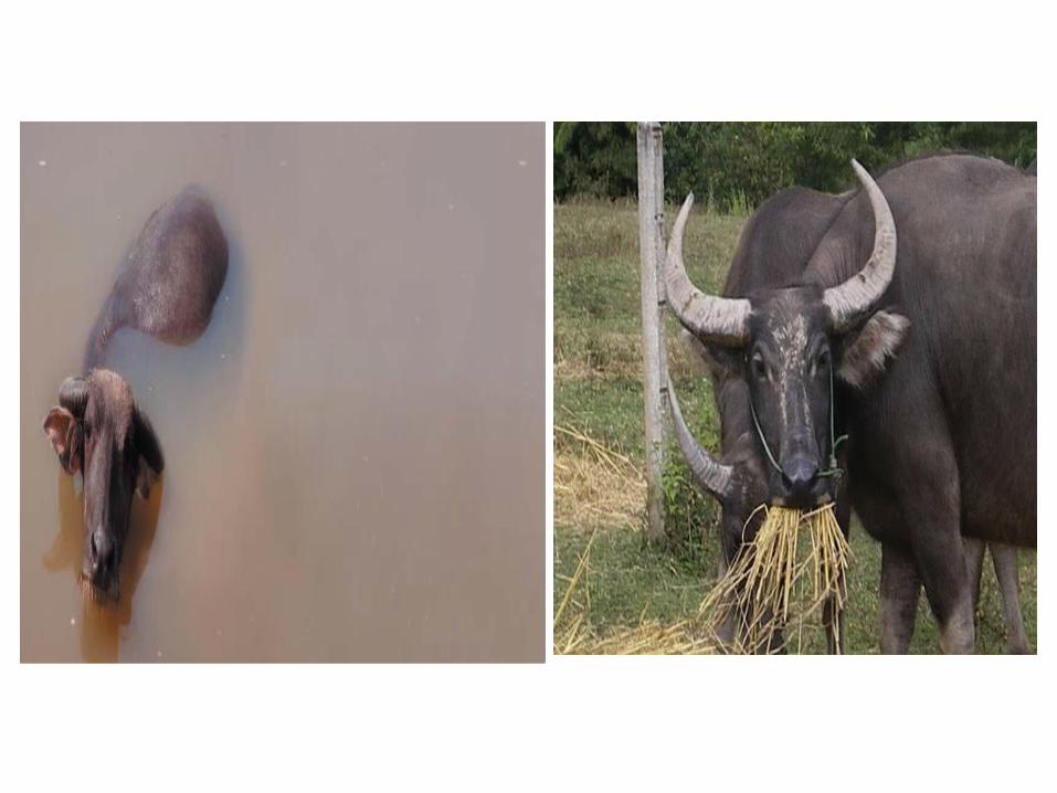
Snail intermediate host of :Fasciola hepatica
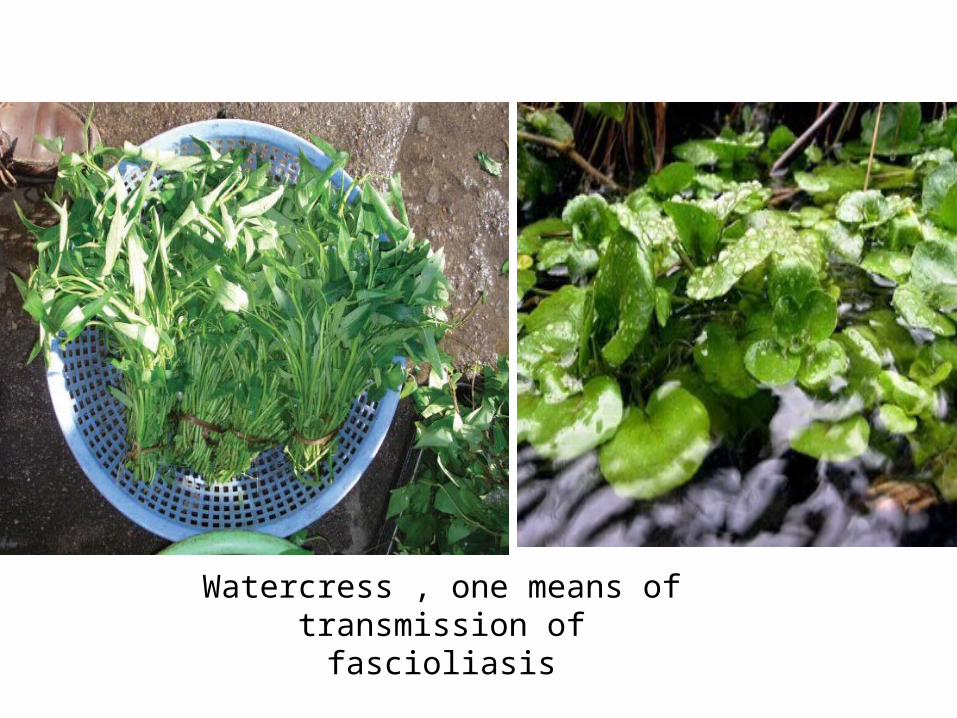
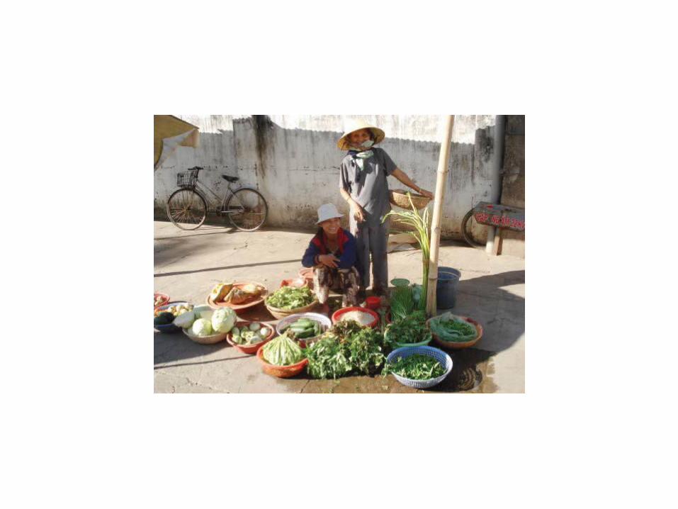
Watercress , one means of transmission of fascioliasis
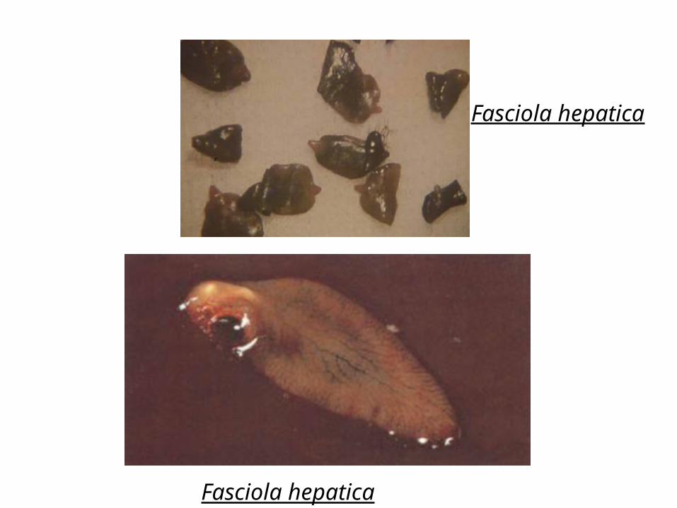
Fasciola hepatica
Fasciola hepatica
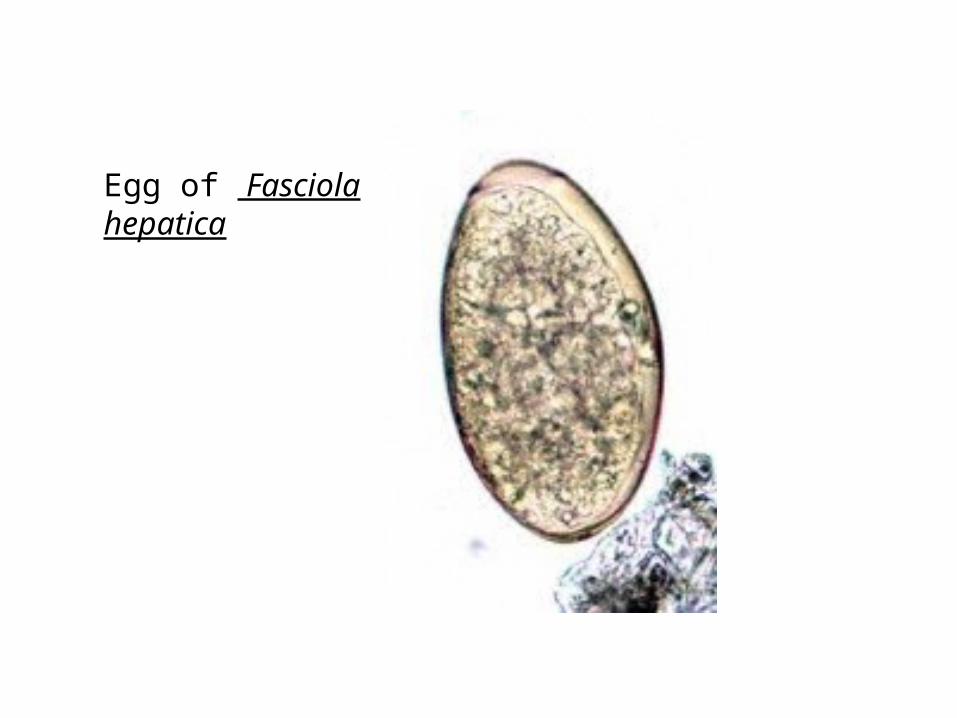
Egg of Fasciola hepatica
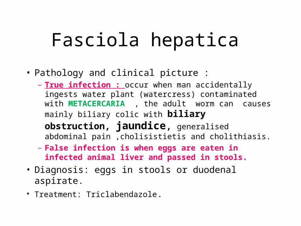
Fasciola hepatica
• Pathology and clinical picture : – True infection : occur when man accidentally ingests
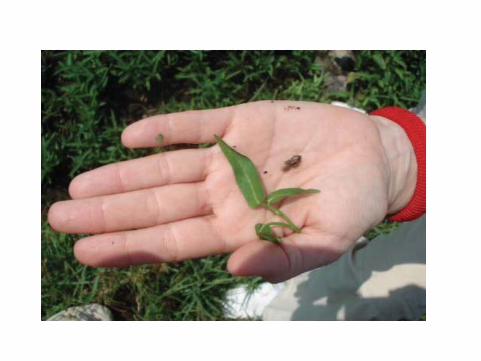
water plant (watercress) contaminated with METACERCARIA , the adult worm can causes mainly
biliary colic with biliary obstruction, jaundice, generalised abdominal pain ,cholisistietis and cholithiasis.
– False infection is when eggs are eaten in infected animal liver and passed in stools.
• Diagnosis: eggs in stools or duodenal aspirate.• Treatment: Triclabendazole.
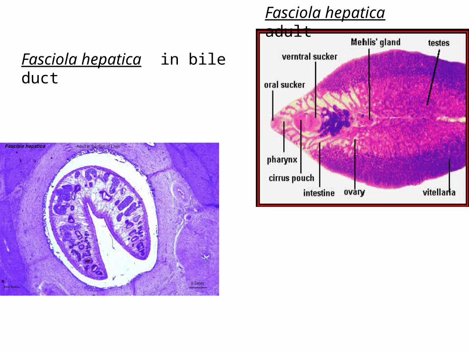
Fasciola hepatica in bile duct
Fasciola hepatica adult
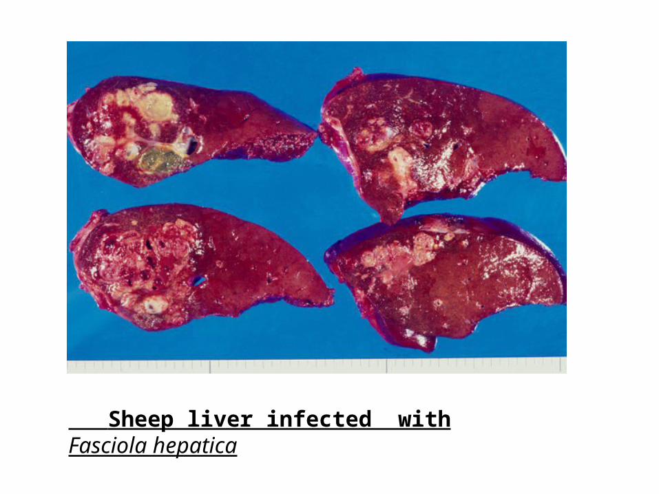
Sheep liver infected with Fasciola hepatica
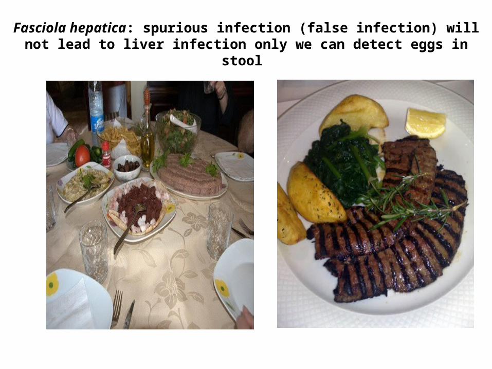
Fasciola hepatica: spurious infection (false infection) will not lead to liver infection only we can detect eggs in stool
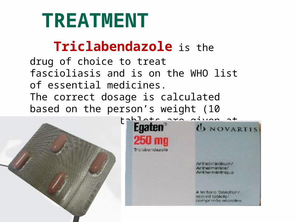
TREATMENT Triclabendazole is the drug of choice to treat fascioliasis and is on the WHO list of essential medicines.The correct dosage is calculated based on the person’s weight (10 mg/kg) and the tablets are given at one time.


![Lesson 96 Battle of Badr. [34] The Great Battle of Badr](https://img.pdfslide.us/doc/110x75/56649dc85503460f94abdc01/lesson-96-battle-of-badr-34-the-great-battle-of-badr.jpg)