Embed Size (px)
Citation preview
Biochimica et Biophysica Acta 1777 (2008) 1455–1462
Contents lists available at ScienceDirect
Biochimica et Biophysica Acta
j ourna l homepage: www.e lsev ie r.com/ locate /bbab io
The Toxoplasma gondii type-II NADH dehydrogenase TgNDH2-I is inhibited by1-hydroxy-2-alkyl-4(1H)quinolones
San San Lin a, Stefan Kerscher b, Ahmad Saleh a, Ulrich Brandt b, Uwe Groß a, Wolfgang Bohne a,⁎a Institute of Medical Microbiology, University of Göttingen, Kreuzbergring 57, Göttingen D-37075, Germanyb Johann Wolfgang Goethe-Universität, Fachbereich Medizin, Zentrum der Biologischen Chemie, Molekulare Bioenergetik, Centre of Excellence Frankfurt “Macromolecular Complexes”,Frankfurt am Main, Germany
Abbreviations: HFF, human foreskin fibroblasts; HDQQuinolone; HHQ, 1-Hydroxy-2-Hexyl-4(1H)Quinolone(1H)Quinolone; HTQ, 1-Hydroxy-2-Tetradecyl-4(1H)Qui⁎ Corresponding author. Tel.: +49 551 395869; fax: +4
E-mail address: [email protected] (W. Bohne).
0005-2728/$ – see front matter © 2008 Elsevier B.V. Aldoi:10.1016/j.bbabio.2008.08.006
a b s t r a c t
a r t i c l e i n f oArticle history:
The apicomplexan parasite Received 27 June 2008Received in revised form 11 August 2008Accepted 12 August 2008Available online 22 August 2008Keywords:Alternative (type-II) NADH dehydrogenase(NDH2)Toxoplasma gondiiInhibition kineticsPing-pong mechanism1-Hydroxy-2-alkyl-4(1)quinoloneHDQ
Toxoplasma gondii does not possess complex I of the mitochondrial respiratorychain, but has two genes encoding rotenone-insensitive, non-proton pumping type-II NADH dehydrogenases(NDH2s). The absence of such “alternative” NADH dehydrogenases in the human host defines these enzymesas potential drug targets. TgNDH2-I and TgNDH2-II are constitutively expressed in tachyzoites andbradyzoites and are localized to the mitochondrion as shown by epitope tagging. Functional expression ofTgNDH2-I in the yeast Yarrowia lipolytica as an internal enzyme, with the active site facing the mitochondrialmatrix, permitted growth in the presence of the complex I inhibitor DQA. Bisubstrate kinetics of TgNDH2-Imeasured within Y. lipolytica mitochondrial membrane preparations were in accordance with a ping-pongmechanism. Using inhibition kinetics we demonstrate here that 1-hydroxy-2-alkyl-4(1)quinolones with longalkyl chains of C12 (HDQ) and C14 are high affinity inhibitors for TgNDH2-I, while compounds with shorterside chains (C5 and C6) displayed significantly higher IC50 values. The efficiency of the various quinolonederivatives to inhibit TgNDH2-I enzyme activity mirrors their inhibitory potency in vivo, suggesting that along acyl site chain is critical for the inhibitory potential of these compounds.
© 2008 Elsevier B.V. All rights reserved.
1. Introduction
NADH:ubiquinone oxidoreductases, also known as NADH dehy-drogenases constitute one of the electron entry points into therespiratory chain, oxidizing NADH and generating ubiquinol. Ineukaryotes, this class of enzymes is divided into two majorsubfamilies, which can be discriminated on the basis of cofactorcontent and sensitivity towards rotenone into type-I NADH dehy-drogenases (complex I) and type-II NADH dehydrogenases (NDH2s)[1]. Proton-pumping complex I is a nearly ubiquitous enzyme thatcouples the rotenone-sensitive transfer of electrons from NADH toubiquinone with the active transport of protons across the innermitochondrial membrane [2]. Bacterial complex I typically consists offourteen subunits that are homologous to the seven mitochondriallycoded and the seven nuclear coded “central” subunits of theeukaryotic enzyme. Although eukaryotic complex I contains a variablenumber of so-called accessory subunits, with a total of 45 subunits and
, 1-Hydroxy-2-Dodecyl-4(1H); HPQ, 1-Hydroxy-2-Pentyl-4nolone9 551 395861.
l rights reserved.
a molecular mass of roughly 1 MDa in mammals, the bioenergeticfunction and the overall structure are conserved [3,4].
In contrast to complex I, type-II NADH dehydrogenases are non-proton-pumping, rotenone-insensitive, single polypeptides. Theiractive site can face either to the cytosol (external enzymes), therebyoxidizing cytosolic NADH, or to the mitochondrial matrix (internalenzymes), thereby oxidizing mitochondrial NADH. Seven NDH2isoforms are expressed in Arabidopsis, three of them are identifiedas internal enzymes, whereas the other four are external [5]. In Sac-charomyces cerevisiae mitochondria lacking complex I, one internaland two external enzymes have been described [6].
Type-II NADH dehydrogenases have been described in plants,fungi, protozoa and bacteria [1,7], but appear to be absent inmammals, which qualifies them as attractive drug targets. Theapicomplexan parasites Plasmodium falciparum and Toxoplasma gondii,which are the causative agents of malaria and toxoplasmosisrespectively, both lack complex I. Instead, the genome of P. falci-parum is predicted to encode a single NDH2 of unknown orientation,while the T. gondii genome encodes two NDH2 isoforms. Treatment ofP. falciparum with micromolar concentrations of diphenylene iodo-nium chloride, a low affinity inhibitor of NDH2, resulted in aninhibition of PfNDH2 activity, in a collapse of the parasite'smitochondrial membrane potential and finally in parasite death [8].

1456 S.S. Lin et al. / Biochimica et Biophysica Acta 1777 (2008) 1455–1462
The quinolone-like compound 1-hydroxy-2-dodecyl-4(1)quinolone(HDQ)was described as the first high affinity inhibitor of type-II NADHdehydrogenases that inhibits NDH2 activity in mitochondrial mem-branes of the yeast Yarrowia lipolytica with an IC50 of 200 nM [9]. Wehave recently shown that HDQ is highly effective against T. gondii andinhibits parasite replication with an IC50 in the nanomolar range [10].Moreover, a combined treatment of HDQ with the complex IIIinhibitor atovaquone resulted in synergism [10].
To further elucidate the suitability of type-II NADH dehydro-genases as drug targets it is crucial to obtain functional data frompathogen orthologs and to determine their interaction withputative inhibitors. We here report functional expression ofTgNDH2-I in the yeast Y. lipolytica. This allowed us to study thekinetics of this enzyme and to demonstrate that 1-hydroxy-2-alkyl-4(1)quinolones with long alkyl side chains are high affinityinhibitors for TgNDH2-I.
2. Materials and methods
2.1. Genome data mining and sequence analyses
Preliminary genomic and/or cDNA sequence data were accessed viahttp://ToxoDB.org (version 3.0) [11] and/or http://tigr.org/tdb/t_gondii/.Genomic datawere provided by the Institute for Genomic Research (NIHgrant #AI05093), and by the Sanger Center (Wellcome Trust). ESTsequences were generated by Washington University (NIH grant#1R01AI045806-01A1). Bioinformatics programs including MitoProt II3.0 (http://ihg.gsf.de/ihg/mitoprot.html) andSignalIP3.0 (http://ww.cgs.dtu.dk/services/SignalIP/) were used to predict subcellular location ofTgndh-I and Tgndh2-II.
2.2. Determination of the ATG initiation codons
An in-frame stop codon is present at 783 and 51 nt upstream ofthe presumed initiation ATG codons of tgndh2-I and tgndh2-IIrespectively. The deduced amino acid sequences coded by theseregions have no other in-frame methionine residues and no similarityto NDHs nor to other proteins when blasted using NCBI BLAST. Fortgndh2-I the next methionine residue is located 177 nt downstreamof the first one. However, only the amino acid sequence deducedfrom the first ATG has characteristics of a mitochondrial targetingsequence. For tgndh2-II, a putative second start ATG is located 6 ntdownstream of the first.
2.3. RNA extraction, RT-PCR and fusion-PCR
Total RNA was isolated using the GenElute Mammalian Total RNAKit (Sigma) and treated with DNase I (Sigma). Reverse transcription(RT) was done on 5 μg of total RNA, Oligo(dT) primer (Sigma) and M-MLV reverse transcriptase (RNase H minus, Sigma) according to themanufacturer's instructions. For PCR amplification, the reactionmixture was cycled in a thermal cycler. Fusion-PCR amplificationwas firstly performed in a volume of 50 μl containing 50 ng of eachpurified DNA fragment (QIAquick PCR purification kit, Qiagen) with 5cycles of PCR in the absence of primers. The PCR reaction wascontinued for another 7 cycles after adding the primer sets (BamHI-NUAM-Fusion+ with NDH2-I-FL− or NDH2-II-FL−).
2.4. Real-time PCR
Light cycler PCR (Roche) was performed to amplify cDNA oftgndh2-I, tgndh2-II and β-tubulin with the following primer sets RT-AND1/1+ and 2−, RT-AND2/1+ and 2−, and RT-Tub/3+ and 4−, as listedin Supplementary Table 1. A control sample without reversetranscriptase was incubated in parallel. The threshold crossing-pointvalues of tgndh2-I and tgndh2-II were normalized to that of β-tubulin.
2.5. Immunofluorescence microscopy
Samples were fixed with 4% paraformaldehyde/PBS for 10 min andpermeabilized with 0.25% Triton X-100/PBS for 15 min. After blockingfor 1 hwith 1% BSA/PBS, samples were incubatedwith a 1:250 dilutionof anti-myc mAb 9E10 (Sigma) followed by incubation with a 1:500dilution of Cy3-conjugated anti-mouse IgG (Dianova) in 1% BSA/PBSfor 1 h each.
2.6. T. gondii strains, cultivation and in vitro stage conversion
Parasites were propagated in human foreskin fibroblasts (HFF) aspreviously described [12]. A clonal isolate of the RH strainwas used forin vitro stage conversion and preparation of cDNA for real time RT-PCRanalysis. Transactivator expressing T. gondii of strain RH TATi-1 [13]were kindly provided by Dr. D. Soldati and Dr. M. Meissner and usedfor transfection experiments. Bradyzoites obtained by in vitro stageconversion were prepared as follows. T. gondii infected HFFs werefirstly cultivated in 1% FCS/DMEM for 3 h at 37 °C in a 5% CO2
humidified atmosphere. The mediumwas subsequently replaced withpH-shift medium (pH 8.3) to induce bradyzoite differentiation [14]and the cultures were incubated at 37 °C without CO2. The mediumwas changed daily with fresh pH-shift medium to remove theextracellular parasites and maintain a constant culture pH. After 4-day incubation, cells were detached and harvested for RNA isolation.
2.7. Generation of myc-tagged Tgndh2-I and Tgndh2-II for expression inparasites
The complete open reading frames (ORF) of Tgndh2-I and Tgndh2-IIwere amplified from cDNA of the RH strain using Pfu polymerase(Promega) with the primer sets AND1/ORF1+_AflII and AND1/ORF2−_AvrII, and AND2/ORF1+_NsiI and AND2/ORF2_AvrII, respectively. ThePCR fragments were cloned into pCR4.0-TOPO (Invitrogen) and DNAsequenced. The AflII/AvrII and NsiI/AvrII fragments were finallysubcloned into pTetO7Sag4-acyl carrier protein (ACP)-cmyc-DHFRvector (kindly provided by Dr. B. Striepen), thereby replacing the ACPORF with the Tgndh2-I and Tgndh2-II ORFs. The final constructspTetSag4-ndh2-I-cmyc-DHFR and pTet7Sag4-ndh2-II-cmyc-DHFRconsisted of the anhydrotetracycline (Atc)-regulable TetO7Sag4promoter element [15], which controls the expression of the completeTgndh2-I and Tgndh2-II ORFs with a C-terminal myc-tag, andadditionally includes a pyrimethamine resistance cassette for selec-tion [16]. Parasites (2×10⁷) were electroporated with 50 μg of NotI-linearized constructs as previously described [12]. Not I (25 U) wasadded to the cytomix before electroporation in order to increase thefrequency of stable transfectants [17]. Stably transfected parasiteswere selected with 1 μM pyrimethamine.
2.8. Plasmid construction and Y. lipolytica transformation
The NUAM-Tgndh2s fusion constructs were generated as transla-tional fusions comprising the Y. lipolytica mitochondrial targetingsequence of the complex I NUAM subunit [18] and the correspondingTgndh2-I or Tgndh2-II mature peptides. The NUAM DNA fragmentswere amplified by PCR from plasmid pUB38 [19] using proof-readingPhusion DNA polymerase (Finnzymes' Phusion high-fidelity DNApolymerase, NEB) with sense primer BamHI-NUAM-Fusion+ and thecorresponding antisense primers YL-NDH2-I-24−, YL-NDH2-I-51− andYL-NDH2-II-62− (Supplementary Table S1). For obtaining DNA frag-ments for the mature parts of the Tgndh2-I-(AA24), Tgndh2-I-(AA51)and Tgndh2-II-(AA62) constructs, cDNA isolated from the RH strainwas used as a template for PCR amplification with the followingprimer sets: YL-NDH2-I-24+ and NDH2-I-FL−, YL-NDH2-I-51+ andNDH2-I-FL, and YL-NDH2-II-62+ and NDH2-II-FL−, respectively. TheNUAM-Tgndh2 fusions were achieved by fusion-PCR. To create the
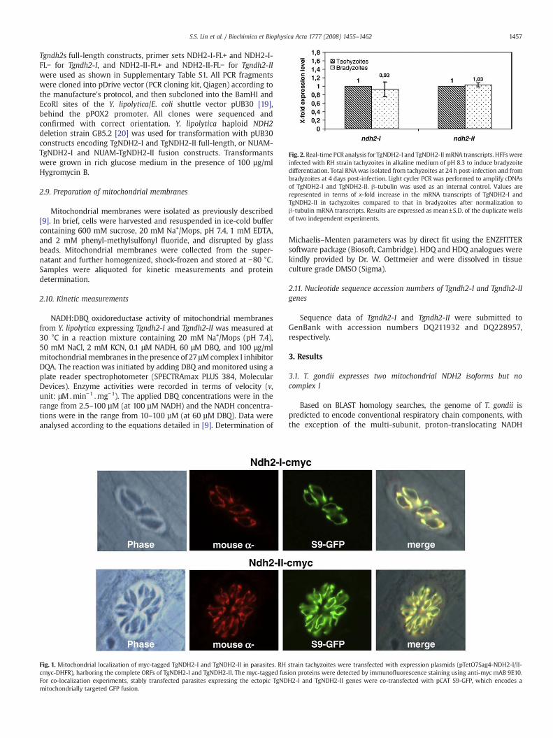
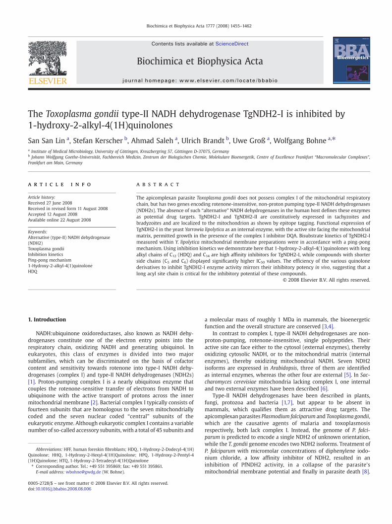
Fig. 2. Real-time PCR analysis for TgNDH2-I and TgNDH2-II mRNA transcripts. HFFswereinfected with RH strain tachyzoites in alkaline medium of pH 8.3 to induce bradyzoitedifferentiation. Total RNAwas isolated from tachyzoites at 24 h post-infection and frombradyzoites at 4 days post-infection. Light cycler PCR was performed to amplify cDNAsof TgNDH2-I and TgNDH2-II. β-tubulin was used as an internal control. Values arerepresented in terms of x-fold increase in the mRNA transcripts of TgNDH2-I andTgNDH2-II in tachyzoites compared to that in bradyzoites after normalization toβ-tubulin mRNA transcripts. Results are expressed as mean±S.D. of the duplicate wellsof two independent experiments.
1457S.S. Lin et al. / Biochimica et Biophysica Acta 1777 (2008) 1455–1462
Tgndh2s full-length constructs, primer sets NDH2-I-FL+ and NDH2-I-FL− for Tgndh2-I, and NDH2-II-FL+ and NDH2-II-FL− for Tgndh2-IIwere used as shown in Supplementary Table S1. All PCR fragmentswere cloned into pDrive vector (PCR cloning kit, Qiagen) according tothe manufacture's protocol, and then subcloned into the BamHI andEcoRI sites of the Y. lipolytica/E. coli shuttle vector pUB30 [19],behind the pPOX2 promoter. All clones were sequenced andconfirmed with correct orientation. Y. lipolytica haploid NDH2deletion strain GB5.2 [20] was used for transformation with pUB30constructs encoding TgNDH2-I and TgNDH2-II full-length, or NUAM-TgNDH2-I and NUAM-TgNDH2-II fusion constructs. Transformantswere grown in rich glucose medium in the presence of 100 µg/mlHygromycin B.
2.9. Preparation of mitochondrial membranes
Mitochondrial membranes were isolated as previously described[9]. In brief, cells were harvested and resuspended in ice-cold buffercontaining 600 mM sucrose, 20 mM Na+/Mops, pH 7.4, 1 mM EDTA,and 2 mM phenyl-methylsulfonyl fluoride, and disrupted by glassbeads. Mitochondrial membranes were collected from the super-natant and further homogenized, shock-frozen and stored at −80 °C.Samples were aliquoted for kinetic measurements and proteindetermination.
2.10. Kinetic measurements
NADH:DBQ oxidoreductase activity of mitochondrial membranesfrom Y. lipolytica expressing Tgndh2-I and Tgndh2-II was measured at30 °C in a reaction mixture containing 20 mM Na+/Mops (pH 7.4),50 mM NaCl, 2 mM KCN, 0.1 μM NADH, 60 μM DBQ, and 100 μg/mlmitochondrialmembranes in the presence of 27 μMcomplex I inhibitorDQA. The reaction was initiated by adding DBQ and monitored using aplate reader spectrophotometer (SPECTRAmax PLUS 384, MolecularDevices). Enzyme activities were recorded in terms of velocity (v,unit: μM Umin−1 Umg−1). The applied DBQ concentrations were in therange from 2.5–100 µM (at 100 µM NADH) and the NADH concentra-tions were in the range from 10–100 µM (at 60 µM DBQ). Data wereanalysed according to the equations detailed in [9]. Determination of
Fig. 1. Mitochondrial localization of myc-tagged TgNDH2-I and TgNDH2-II in parasites. RHcmyc-DHFR), harboring the complete ORFs of TgNDH2-I and TgNDH2-II. The myc-tagged fusFor co-localization experiments, stably transfected parasites expressing the ectopic TgNDmitochondrially targeted GFP fusion.
Michaelis–Menten parameters was by direct fit using the ENZFITTERsoftware package (Biosoft, Cambridge). HDQ and HDQ analogues werekindly provided by Dr. W. Oettmeier and were dissolved in tissueculture grade DMSO (Sigma).
2.11. Nucleotide sequence accession numbers of Tgndh2-I and Tgndh2-IIgenes
Sequence data of Tgndh2-I and Tgndh2-II were submitted toGenBank with accession numbers DQ211932 and DQ228957,respectively.
3. Results
3.1. T. gondii expresses two mitochondrial NDH2 isoforms but nocomplex I
Based on BLAST homology searches, the genome of T. gondii ispredicted to encode conventional respiratory chain components, withthe exception of the multi-subunit, proton-translocating NADH
strain tachyzoites were transfected with expression plasmids (pTetO7Sag4-NDH2-I/II-ion proteins were detected by immunofluorescence staining using anti-myc mAB 9E10.H2-I and TgNDH2-II genes were co-transfected with pCAT S9-GFP, which encodes a
1458 S.S. Lin et al. / Biochimica et Biophysica Acta 1777 (2008) 1455–1462
dehydrogenase known as complex I. Instead, two contigs (TGG_994254and TGG_994290) with high similarities to type-II NADH dehydro-genases were identified in the ToxoDB. The complete open readingframes of both genes were amplified, subcloned and sequenced from T.gondii RH strain cDNA. The two genes encoding the type-II NADHdehydrogenases were designated as tgndh2-I (accession#: DQ211932)and tgndh2-II (accession#: DQ228957). They encode precursor polypep-tides of 619 and 657 amino acids with predicted masses of 67.1 and72.1 kDa, respectively. Information on gene structures is given inSupplementary Fig. S1. The deduced primary structure of both proteinsincludes N-terminal mitochondrial targeting sequences as predicted by
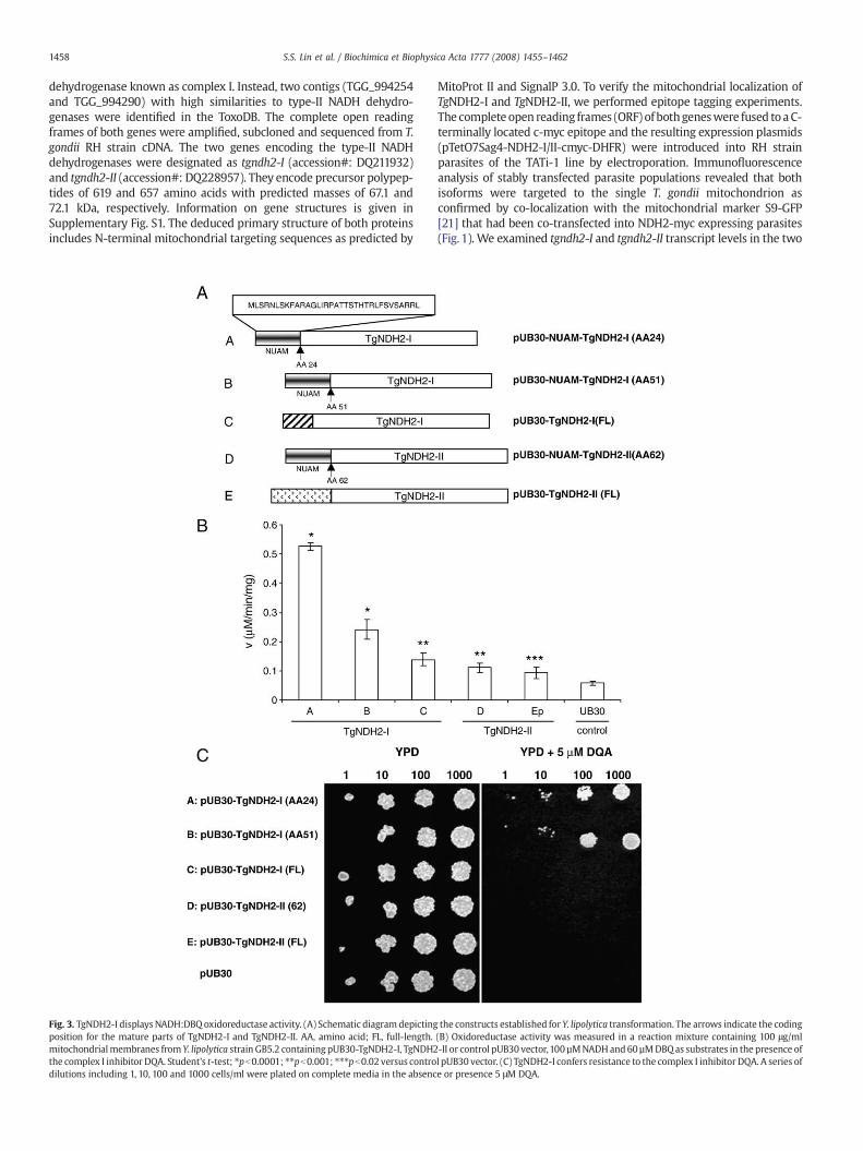
Fig. 3. TgNDH2-I displaysNADH:DBQ oxidoreductase activity. (A) Schematic diagram depictingposition for the mature parts of TgNDH2-I and TgNDH2-II. AA, amino acid; FL, full-length.mitochondrialmembranes from Y. lipolytica strain GB5.2 containing pUB30-TgNDH2-I, TgNDH2the complex I inhibitor DQA. Student's t-test; ⁎pb0.0001; ⁎⁎pb0.001; ⁎⁎⁎pb0.02 versus controdilutions including 1, 10, 100 and 1000 cells/ml were plated on complete media in the absenc
MitoProt II and SignalP 3.0. To verify the mitochondrial localization ofTgNDH2-I and TgNDH2-II, we performed epitope tagging experiments.The complete open reading frames (ORF) of bothgeneswere fused to aC-terminally located c-myc epitope and the resulting expression plasmids(pTetO7Sag4-NDH2-I/II-cmyc-DHFR) were introduced into RH strainparasites of the TATi-1 line by electroporation. Immunofluorescenceanalysis of stably transfected parasite populations revealed that bothisoforms were targeted to the single T. gondii mitochondrion asconfirmed by co-localization with the mitochondrial marker S9-GFP[21] that had been co-transfected into NDH2-myc expressing parasites(Fig. 1). We examined tgndh2-I and tgndh2-II transcript levels in the two
the constructs established for Y. lipolytica transformation. The arrows indicate the coding(B) Oxidoreductase activity was measured in a reaction mixture containing 100 μg/ml-II or control pUB30 vector,100 µMNADHand60 µMDBQas substrates in the presence ofl pUB30 vector. (C) TgNDH2-I confers resistance to the complex I inhibitor DQA. A series ofe or presence 5 μM DQA.

1459S.S. Lin et al. / Biochimica et Biophysica Acta 1777 (2008) 1455–1462
parasitic stages (tachyzoites and bradyzoites), which are present in thehuman host by quantitative real time RT-PCR. Both genes displayedcomparable mRNA levels in the analyzed stages (Fig. 2), suggesting thatthey were constitutively expressed rather than stage specificallyregulated.
3.2. Functional expression of TgNDH2-I in Y. lipolytica
The yeast Y. lipolytica expresses a single, external, non-essentialNDH2 and has been established as a model organism for studying thebiochemistry of alternative NADH dehydrogenases [22] and ofrespiratory chain complex I [23]. We used Y. lipolytica strain GB 5.2,in which the external NDH2 was deleted, for heterologous expressionof TgNDH2-I and TgNDH2-II. It is not knownwhether a mitochondrialimport sequence from a T. gondii protein is sufficient for accuratetargeting into Y. lipolytica mitochondria. Thus, in addition to full-length T. gondii constructs, we also employed fusions of the N-terminal part of the Y. lipolytica mitochondrial NUAM protein andmature versions of TgNDH2-I and TgNDH2-II. The predicted startposition for mature TgNDH2-I is at amino acid 24 and for matureTgNDH2-II at amino acid 62 (Fig. 3A). The predicted presequence forTgNDH2-I is relatively short and after a manual sequence alignmentwith TgNDH2-II, a second, although less likely start position formature TgNDH2-I was identified at position 51 and used for NUAMfusion. It has been demonstrated previously that the addition of theNUAM mitochondrial import signal to the external Y. lipolytica NDH2is sufficient to convert this enzyme into an internal, enzymaticallyactive isoform [18]. All constructs were placed under the control of thepPOX2 promotor in the replicative vector pUB30 [19]. Mitochondrialmembrane preparations of Y. lipolytica transformants were analyzed
Fig. 4. Inhibition of TgNDH2-I by HDQ derivatives differing in side chain length. The concentmitochondrial membranes was determined at a total protein concentration of 100 µg/ml in tside chains, respectively.
for NADH dehydrogenase activity in an enzymatic assay using DBQ aselectron acceptor. The NUAM-TgNDH2-I fusions displayed electrontransfer activities that were 10-fold (construct A: AA24) and 5-fold(construct B: AA51) higher than controls, demonstrating that thisisoform can be expressed as an active enzyme in Y. lipolytica (Fig. 3B).Both TgNDH2-II constructs displayed electron transfer activities thatwere less than 2-fold above controls, and the activity of full-lengthTgNDH2-I was less than 2.5-fold above controls.
Complex I, the proton-translocating multi-subunit NADH dehy-drogenase of the mitochondrial respiratory chain, is essential forgrowth of Y. lipolytica. The ability of the five expression constructs tocompensate for the loss of complex I activity was tested in a Y. lipo-lytica growth assay on YPD agar plates in the presence of the complex Iinhibitor DQA. Expression of TgNDH2-I as NUAM fusion proteinsconferred DQA resistance (Fig. 3C), in line with the results obtainedfrom the enzyme activity assay. This demonstrated that both NUAM-TgNDH2-I fusions were expressed as functional, internal enzymeswith their active site oriented towards the mitochondrial matrix.NUAM-TgNDH2-I expressed from construct A which displayed thehighest activity and was used in all kinetic assays and is termedTgNDH2i in the following.
3.3. TgNDH2i activity is effectively inhibited by 1-hydroxy-2-alkyl-4(1H)quinolones
The quinolone-like compound HDQ is a potent inhibitor of Y. lipoly-tica NDH2 [9] and our recent findings have shown that HDQ couldeffectively inhibit T. gondii replication [10]. We thus investigated theinhibitory effect of HDQ on the NADH dehydrogenase activity ofTgNDH2i in unsealed Y. lipolytica mitochondrial membranes. Using
ration of inhibitor required for half-maximal inhibition (IC50) of TgNDH2i in Y. lipolyticahe presence of 60 µM DBQ and 100 µM NADH. (A–D) HDQ with C12, C14, C5 and C6 alkyl

Fig. 6. Double reciprocal plots of HDQ inhibition kinetics of TgNDH2i. Inhibition kineticsof TgNDH2i in Y. lipolytica mitochondrial membranes were measured at a total proteinconcentration of 100 µg/ml for A, DBQ (at 100 µM NADH) and B, NADH (at 60 µM DBQ)in the absence (♦) and in the presence of 60 nM (□) and 300 nM (▴) C12-HDQ. The linesfor the diagrams were calculated using parameters obtained from direct fits to theMichaelis–Menten equation. In A, Vmax values were 0.92±0.06 (♦), 0.76±0.03 (□) and0.41±0.02 (▴) units/mg. Apparent Km values for DBQ were 13.8±2.4 (♦), 13.8±2.0 (□)and 12.9±2.5 µM (▴). In B, Vmax values were 1.16±0.07 (♦), 0.80±0.04 (□) and 0.44±0.03 (▴) U/mg. Apparent Km values for NADH were 76±7 (♦), 68±5 (□) and 61±8 µM(▴). Each data point represents the mean of five independent measurements.
1460 S.S. Lin et al. / Biochimica et Biophysica Acta 1777 (2008) 1455–1462
DBQ as a substrate, inhibition of NADH:ubiquinone oxidoreductaseactivity by HDQ was dose dependent with a concentration required forhalf-maximal inhibition (IC50) of about 300 nM (Fig. 4A). A 1-hydroxy-2-alkyl-4(1H)quinolone compound with a C14 alkyl side chain (HTQ,Fig. 4B) displayed a similar IC50 as HDQ (C12), while derivates withshorter alkyl side chains (C5=HPQ and C6=HHQ) were less effectivewith significantly higher IC50 values of about 3700 nM and 2300 nM,respectively (Fig. 4C, D).
3.4. TgNDH2i bisubstrate kinetics suggest a ping-pong reactionmechanism
Mitochondrial membrane preparations from Y. lipolytica expres-sing TgNDH2i were used to determine bisubstrate kinetics for NADHand DBQ. Km values of 76 µM for NADH and 14 µM for DBQ wereobtained. In a Hanes plot ([S]/v over [S]) of the kinetic data, theobtained lines crossed close to the y-axis (Fig. 5), which is inaccordance with a ping-pong reaction mechanism for TgNDH2i [9].A (random or ordered) sequential mechanism could be excluded sincein that case the lines would cross in the fourth quadrant. This resultsuggests that the enzyme forms binary complexes with each of thesubstrates, but is unable to form a ternary complex with bothsubstrates. Most likely, both substrates interact with a commonbinding site in a mutually exclusive fashion.
3.5. HDQ inhibition follows a non-competitive pattern for both substrates
To determine the mode of inhibition of HDQ on TgNDH2i inmitochondrialmembranepreparations from Y. lipolytica, we performedsteady-state inhibition kinetics for both NADH and DBQ. Doublereciprocal plots of the kinetic data obtained in the presence of 0, 60and 300 nM HDQ are depicted in Fig. 6. With increasing HDQconcentrations, the slopes increased, and the lines intersected closeto the y-axis. Similar as found with Y. lipolytica NDH2 [9], these resultsformally follow the pattern of non-competitive inhibition for bothsubstrates and similar values for the two inhibition constants (see
Fig. 5. TgNDH2-I employs a ping-pong reaction mechanism. Hanes plots of steady-statekinetics with TgNDH2i in Y. lipolytica mitochondrial membranes, assayed at a totalprotein concentration of 100 µg/ml. Vmax and apparent Km values for DBQ in thepresence of five different NADH concentrations were obtained from direct fits to theMichaelis–Menten equation and used to draw the lines. NADH concentrations were 10(□), 15 (♦), 30 (▵), 50 (▪) and 100 µM (w). Vmax values were 0.17±0.01 (□), 0.28±0.04(♦), 0.40±0.04 (▵), 0.57±0.06 (▪) and 0.77±0.07 (w) U/mg. Apparent Km values were3.5±0.5 (□), 5.3±2.9 (♦), 9.1±2.8 (▵),11.9±4.1 (▪) and 18.0±5.0 µM (w). Each data pointrepresents the mean of five independent measurements.
Supplementary Fig. S2). Using linear secondary plots of the slopes andy-axis intercepts of the lines in Fig. 6 against the HDQ concentrationsused, numeric values for the inhibition constants could be derived fromthe points of intersection with the x-axis (see Supplementary Fig. S3).Secondary plots of the slopes thus gave Ki=283 nM and Kii=292 nM,and secondary plots of the intercepts gave Ki=234 nM and Kii=198 nM.Within experimental error, these results are in excellent agreementwith the directly determined IC50 value for HDQ on TgNDH2i and withthe assumption that Ki equals Kii.
4. Discussion
In this study we have determined enzymatic parameters fromheterologously expressed, functionally active T. gondii NDH2-I andcould show that 1-hydroxy-2-alkyl-4(1)quinolones with long alkylchains are high affinity inhibitors for this enzyme. T. gondii lackscomplex I but possesses two mitochondrial type-II NADH dehydro-genases. Both are constitutively expressed, hampering direct deter-mination of individual enzymatic parameters in T. gondii lysates. Thus,for in vitro characterization, we have expressed TgNDH2-I in a strain ofthe yeast Y. lipolytica that lacks endogenous alternative NADHdehydrogenase activity. Functional expression of TgNDH2-I as aninternal NADH dehydrogenase, capable of electron transfer frommatrix NADH to membrane bound ubiquinone, was achieved byfusing the TgNDH2-I open reading frame without the endogenousmitochondrial targeting sequence to the N-terminal part of the NUAMsubunit of Y. lipolytica mitochondrial complex I. Direct evidence for invivo function of TgNDH2i was provided by the observation thatexpression of the transgene allowed cells of Y. lipolytica to survive in

1461S.S. Lin et al. / Biochimica et Biophysica Acta 1777 (2008) 1455–1462
the presence of the complex I inhibitor DQA. Thus we were able toachieve heterologous expression of a protozoan type-II NADHdehydrogenase in an active form. Functional expression of protozoantype-II NADH dehydrogenases in Y. lipolytica should facilitate futurecomparative studies of apicomplexan orthologs. In addition, expres-sion of protozoan type-II enzymes in Y. lipolytica could be used for thedevelopment of a screening assay to identify novel, specific inhibitorsfor this class of enzymes.
The steady-state inhibition kinetics of TgNDH2-I for the quinolonederivative HDQ were found to be very similar to those of Y. lipolyticaNDH2 [9] and formally follow the pattern of non-competitiveinhibition for both substrates, NADH and DBQ. In the case of a ping-pong reaction mechanism, this inhibition pattern is predicted whenthe inhibitor blocks both the enzyme, here TgNDH2i-FAD and theintermediate form, here TgNDH2i-FADH2 [24]. As with Y. lipolyticaNDH2 [9], our data thus support a model in which the inhibitor caninteract with a complex consisting of the enzyme and one of itssubstrates, presumably NADH, as depicted in Supplementary Fig. S2.
Bisubstrate kinetics revealed that the NADH:DBQ oxidoreductaseactivity of TgNDH2i followed a ping-pong reaction mechanism, a modeof action that was also shown for the Y. lipolytica ortholog [9] andproposed for the S. cerevisiae and T. brucei enzymes [25,26]. Thedetermined Km (NADH) of 76 µMwas significantly higher than the Km ofmost other eukaryotic enzymes, for example from S. tuberosum, N.crassa and S. cerevisiae, which were in the range of 11–32 µM [7]. Onlythe T. brucei enzyme with 120 µM displayed a higher Km than the T.gondii ortholog [26]. Also, the Km (DBQ) of 14 µM observed for TgNDH2iwas higher than the Km (DBQ) of 7.0 µM for Y. lipolytica NDH2 [9].However, differences in Km values have to be interpreted with caution,since different electron acceptorswere used in these studies and specificreaction conditions as the volume of the lipid phase could influenceactivities. Since the activity of TgNDH2i was about 4-fold lower than theactivity of NDH2, the external alternative NADH dehydrogenase of Y.lipolytica parental strains, we had to double the amount of totalmitochondrial protein used in the assays. Lower effective concentrationsof DBQ in the lipid phase would explain the elevated Km (DBQ) valuesobserved in the present study. In contrast, the Km (NADH) is notexpected to depend on the volume of the lipid phase.
The IC50 value of about 300 nMobtained for HDQ indicates that thiscompound is a high affinity inhibitor for TgNDH2-I. The only otherNDH2 enzyme for which the IC50 value for HDQ is known is the Yar-rowia orthologue, which with 200 nM is in the same range [9]. In thefuture, it will be important to determine IC50 values for furtherorthologues from pathogenic and non-pathogenic microorganisms, inorder to classify HDQ as a broad spectrum NDH2 inhibitor or as aninhibitor with a selected inhibitory potential. An example for arespiratory chain inhibitor with a selected inhibitory potential is theclinically approved antimalaria drug atovaquone. This drug is acomplex III inhibitor that interferes with the ubiquinol oxidation siteof the cytochrome bc1 complex by acting as a competitive inhibitor forubiquinol. Atovaquone possesses antimicrobial activity against certainapicomplexan parasites such as Plasmodium and Toxoplasma and theopportunistic fungal pathogen Pneumocystis carinii, whereas thehuman and bovine cytochrome bc1 complexes are insensitive towardsatovaquone [27].
For HDQ, the in vitro IC50 of ~300 nM for inhibition of TgNDH2iactivity is significantly higher than the in vivo IC50 of 2–4 nM for staticeffects on T. gondii parasites, determined in tissue culture after a 24 htreatment period [10]. Apparent differences in IC50 values should notbe overinterpreted since these are strongly dependent on thefractional volume of the lipid phase. Assuming that a highlyhydrophobic substance like HDQ will partition to the hydrophobicphase, the effective HDQ concentration for interaction with type-IINADH dehydrogenase may well differ by two orders of magnitudebetween the two assay systems. However, we can not exclude atpresent that HDQ inhibits other ubiquinone dependent NADH
oxidoreductases such as succinate dehydrogenase, dihydroorotatedehydrogenase, glycerol-3-phosphate dehydrogenase and the malate:quinone oxidoreductase, in addition to type-II NADH dehydrogenases.
A long alkyl side chain of C12 or C14 at position 2 is critical for theinhibition of NADH:DBQ oxidoreductase activity. Compounds withshort alkyl side chains of C5 (HPQ) and C6 (HHQ) with 3700 nM and2300 nM displayed significantly higher IC50 values as compoundswithlong alkyl side chains of C12 (HDQ, IC50=300 nM) and C14 (HTQ,IC50=300 nM). On the basis of structural similarities, it is tempting tospeculate that 1-hydroxy-2-alkyl-4(1H)quinolones are likely tocompete with ubiquinone for the same binding site. A long alkyl sitechain leads to a higher hydrophobicity and is likely to render thephysicochemical properties and the structure of 1-hydroxy-2-alkyl-4(1H)quinolones more similar to ubiquinone. Another aspect is thepartition of the compounds between aqueous and membrane lipidphase. Highly hydrophobic 1-hydroxy-2-alkyl-4(1H)quinolones asHDQ and HTQ, in contrast to those with smaller alkyl side chains,can be expected to partition almost quantitatively into the membranelipid phase. The correlation between IC50 values and length of thealkyl side chain mirrored the in vivo-efficiency of these drugs toinhibit parasite replication: HPQ (C5) did not show any inhibitoryeffect at 10 nM, while parasite growth was reduced to more than 50%by HDQ and HTQ at this concentration [10]. A reevaluation of theefficiency of HHQ (C6) to inhibit T. gondii replication yielded an IC50 of220 nM for HHQ (C6), which is more than 50-fold higher than the IC50for HDQ (data not shown). Together, these data reveal that only 1-hydroxy-2-alkyl-4(1H)quinolones with long alkyl site chains areeffective TgNDH2-I inhibitors and antiparasitic drugs.
Acknowledgments
This study has been supported by grants from the DeutscheForschungsgemeinschaft, DFG to WB (BO 1557/3-1) and UB (SFB472,Teilprojekt P2 and EXC115). S.S. Lin is supported by a scholarship fromthe Croucher foundation.
Appendix A. Supplementary data
Supplementary data associated with this article can be found, inthe online version, at doi:10.1016/j.bbabio.2008.08.006.
References
[1] S. Kerscher, V. Zickermann, U. Brandt, The three families of respiratory NADHdehydrogenases, Res. Probl. Cell Different 45 (2008) 185–222.
[2] U. Brandt, Energy converting NADH quinone oxidoreductases, Annu. Rev.Biochem. 75 (2006) 69–92.
[3] J. Carroll, I.M. Fearnley, R.J. Shannon, J. Hirst, J.E. Walker, Analysis of the subunitcomposition of complex I from bovine heart mitochondria, Mol. Cell. Proteomics 2(2003) 117–126.
[4] M. Radermacher, T. Ruiz, T. Clason, S. Benjamin, U. Brandt, V. Zickermann, Thethree-dimensional structure of complex I from Yarrowia lipolytica: a highlydynamic enzyme, J. Struct. Biol. 154 (2006) 269–279.
[5] D. Elhafez, M.W. Murcha, R. Clifton, K.L. Soole, D.A. Day, J. Whelan, Characterizationof mitochondrial alternative NAD(P)H dehydrogenases in Arabidopsis: intraorga-nelle location and expression, Plant Cell Physiol. 47 (2006) 43–54.
[6] K.M. Overkamp, B. Bakker, P. Kötter, A. van Tuijl, S. de Vries, J.P. van Dijken, J.T.Pronk, In vivo analysis of the mechanisms for oxidation of cytosolic NADH bySaccharomyces cerevisiae mitochondria, J. Bacteriol. 182 (2000) 2823–2830.
[7] A.M. Melo, T.M. Bandeiras, M. Teixeira, New insights into type II NAD(P)H:quinoneoxidoreductases, Microbiol. Mol. Biol. Rev. 68 (2004) 603–616.
[8] G.A. Biagini, P.O. Viriyavejakul, P.M. O'Neill, P.G. Bray, S.A. Ward, Functionalcharacterization and target validation of alternative complex I of Plasmodiumfalciparum mitochondria, Antimicrob. Agents Chemother. 50 (2006)1841–1851.
[9] A. Eschemann, A. Galkin, W. Oettmeier, U. Brandt, S. Kerscher, HDQ (1-hydroxy-2-dodecyl-4(1H)quinolone) a high affinity inhibitor for mitochondrial alternativeNADH dehydrogenase, J. Biol. Chem. 280 (2005) 3138–3142.
[10] A. Saleh, J. Friesen, S. Baumeister, U. Gross, W. Bohne, Growth inhibition ofToxoplasma gondii and Plasmodium falciparum by nanomolar concentrations of1-hydroxy-2-dodecyl-4(1H)quinolone a high-affinity inhibitor of alternative(type II) NADH dehydrogenases, Antimicrob. Agents Chemother. 51 (2007)1217–1222.

1462 S.S. Lin et al. / Biochimica et Biophysica Acta 1777 (2008) 1455–1462
[11] J.C. Kissinger, B. Gajria, I.T. Paulsen, D.S. Roos, ToxoDB: accessing the Toxoplasmagondii genome, Nucleic Acids Res. 31 (2003) 234–236.
[12] D.S. Roos, R.G. Donald, N.S. Morrissette, A.L. Moulton, Molecular tools for geneticdissection of the protozoan parasite Toxoplasma gondii, Methods Cell Biol. 45 (1994)27–63.
[13] M. Meissner, D. Schluter, D. Soldati, Role of Toxoplasma gondii myosin A inpowering parasite gliding and host cell invasion, Science 298 (2002) 837–840.
[14] M. Soete, D. Camus, J.F. Dubremetz, Experimental induction of bradyzoite-specificantigen expression and cyst formation by the RH strain of Toxoplasma gondii invitro, Exp. Parasitol. 78 (1994) 361–370.
[15] M. Meissner, S. Brecht, H. Bujard, D. Soldati, Modulation of myosin A expression bya newly established tetracycline repressor-based inducible system in Toxoplasmagondii, Nucleic Acids Res. 29 (2001) E115.
[16] R.G. Donald, D.S. Roos, Stable molecular transformation of Toxoplasma gondii:a selectable dihydrofolate reductase-thymidylate synthase marker based on drug-resistance mutations in malaria, Proc. Natl. Acad. Sci. U. S. A. 90 (1993)11703–11707.
[17] M.T. Black, F. Seeber, D. Soldati, K. Kim, J.C. Boothroyd, Restrictionenzyme-mediated integration elevates transformation frequency andenables co-transfection of Toxoplasma gondii, Mol. Biochem. Parasitol. 74(1995) 55–63.
[18] S. Kerscher, A. Eschemann, P.M. Okun, U. Brandt, External alternative NADH:ubiquinone oxidoreductase redirected to the internal face of the mitochondrialinner membrane rescues complex I deficiency in Yarrowia lipolytica, J. Cell. Sci. 114(2001) 3915–3921.
[19] A. Garofano, A. Eschemann, U. Brandt, S. Kerscher, Substrate-inducible versions ofinternal alternative NADH:ubiquinone oxidoreductase from Yarrowia lipolytica,Yeast 23 (2006) 1129–1136.
[20] S. Kerscher, J.G. Okun, U. Brandt, A single external enzyme confers alternativeNADH:ubiquinone oxidoreductase activity in Yarrowia lipolytica, J. Cell. Sci. 112(1999) 2347–2354.
[21] A. DeRocher, C.B. Hagen, J.E. Froehlich, J.E. Feagin, M. Parsons, Analysis of targetingsequences demonstrates that trafficking to the Toxoplasma gondii plastid branchesoff the secretory system, J. Cell. Sci. 113 (2000) 3969–3977.
[22] S. Kerscher, Diversity and origin of alternative NADH:ubiquinone oxidoreductases,Biochim. Biophys. Acta 1459 (2000) 274–283.
[23] S. Kerscher, S. Dröse, K. Zwicker, V. Zickermann, U. Brandt, Yarrowia lipolytica ayeast genetic system to study mitochondrial complex I, Biochim. Biophys. Acta1555 (2002) 83–91.
[24] W.W. Cleland, The kinetics of enzyme-catalyzed reactions with two or moresubstrates or products. III. Prediction of initial velocity and inhibition patterns byinspection, Biochim. Biophys. Acta 67 (1963) 188–196.
[25] I. Velazquez, J.P. Pardo, Kinetic characterization of the rotenone-insensitiveinternal NADH:ubiquinone oxidoreductase of mitochondria from Saccharomycescerevisiae, Arch. Biochem. Biophys. 389 (2001) 7–14.
[26] J. Fang, D.S. Beattie, Novel FMN-containing rotenone-insensitive NADH dehydro-genase from Trypanosoma brucei mitochondria: isolation and characterization,Biochemistry 41 (2002) 3065–3072.
[27] A.L. Baggish, D.R. Hill, Antiparasitic agent atovaquone, Antimicrob. AgentsChemother. 46 (2002) 1163–1173.