Embed Size (px)
Citation preview

The Journal of Neuroscience, February 1997, 7(2): 343356
The Toxic Effects of Ethylcholine Mustard Aziridinium Ion on Cholinergic Cells in the Chicken Retina
T. J. Millar,’ I. Ishimoto,lva M. Boelen,’ M. L. Epstein,* C. D. Johnson,3 and I. G. Morgan’
‘Department of Behavioural Biology, Research School of Biological Sciences, Australian National University, Canberra City ACT 2601, Australia, and 2Department of Anatomy, and 3Department of Zoology, University of Wisconsin, Madison, Wisconsin 53706
The chicken retina has been used to examine the toxicity of a highly reactive chemical analog of choline, ethylcholine mustard aziridinium ion (ECMA). Following a single intravit- real injection, retinas were analyzed biochemically for CAT and AChE activities, and GABA, glycine, and dopamine levels. Retinas were also examined using histofluorescence for dopamine histochemistry, for AChE, and immunohisto- chemistry with antibodies to CAT, tyrosine hydroxylase, GABA, SHT, Leu-enkephalin, and somatostatin. A dose of 50 nmol ECMA caused a prolonged 70% depletion of CAT activity and a 40% depletion of AChE activity. The other biochemical parameters were unchanged. This result cor- responds to the morphological finding that 2 populations of cholinergic cells were destroyed and that the AChE activity associated with their terminal arbors was lost. A third pop- ulation of cholinergic cells, located towards the middle of the inner nuclear layer, was resistant to the toxic effects of ECMA. The other cell types, except for somatostatin-im- munoreactive cells and photoreceptors, which showed tran- sient effects, were unaffected. ECMA therefore appears to be a highly specific toxin for cholinergic cells in the retina.
Aziridinium derivatives of choline mustard analogs have been developed to act as irreversible inhibitors of high-affinity choline uptake (Rylett and Colhoun, 1977; Fisher and Hanin, 1980). Their potential as selective cholinergic cytotoxins was popular- ized by Fisher and Hanin (1980), and initial reports indicated that one of them, monoethylcholine mustard aziridinium ion, (ECMA, often referred to as AF64A), caused prolonged cholin- ergic hypofunction (Mantione et al., 198 1).
Subsequent reports have called into question both the efficacy and selectivity of the toxin. Cholinergic hypofunction caused by ECMA has been reported in many cases, but variations in the efficacy of the toxin and the permanency of the effects have been reported in different preparations (Fisher et al., 1982; Han- in et al., 1983; Mantione et al., 1983a, b; Sandberg et al., 1984). This has led to uncertainty as to whether entire cholinergic cells are destroyed, whether the terminal arbors of cholinergic cells
Received Mar. 13, 1986; revised June 18, 1986; accepted June 26, 1986.
We would like to thank Ms. M. Canney and Ms. P. Miethke for their skilled technical assistance, and Ms. B. Piper for word processing.
Correspondence should be addressed to Dr. I. G. Morgan, Department of Be- havioural Biology, Research School of Biological Sciences, Australian National University, GPO Box 475, Canberra City ACT 2601, Australia.
* Present address: Department of Ophthalmology, Osaka University Medical School, l-l-56 Fukushima, Fukushimaku, Osaka 553, Japan.
Copyright 0 1987 Society for Neuroscience 0270-6474/87/020343-14$02.00/O
are damaged, or whether the cells down-regulate in response to exposure to the toxin. The specificity of the toxin has also been challenged (Levy et al., 1984). Complicating factors in many of these experiments have been the mode of administration of the toxin, the cellular complexity of the areas studied and the in- adequate characterization of cholinergic cells in histological studies.
In an attempt to resolve some of the uncertainties we have carried out an integrated biochemical and morphological study of the effects of intravitreal ECMA on the chicken retina. This tissue provides an ideal model system for testing the effects of neurotoxins because it is a highly organized structure composed of a limited number of neuronal classes (photoreceptors, and horizontal, bipolar, amacrine, and ganglion cells) having char- acteristic morphologies and locations (for a recent review, see Dowling and Dubin, 1984). In the chicken retina, cholinergic cells have been characterized using autoradiographic localiza- tion of choline uptake sites (Baughman and Bader, 1977) and CAT immunohistochemistry (Millar et al., 1985). Using these advantages of the retina as a model system, we have been able to demonstrate that, under appropriate conditions, ECMA kills cholinergic cells with great specificity and is thus potentially useful for unraveling the details of cholinergic function in the retina and in other parts of the nervous system.
Materials and Methods ECMA was prepared from acetylethylcholine mustard (AF64; Research Biochemicals, Maryland, MO), essentially as described by Fisher et al. (1982). Conversion rates of 40-50% were obtained, as determined by an iodine-thiosulfate titration method (Golumbic et al., 1946). No at- tempt was made to increase yields by varying the factors analyzed by Sandberg et al. (1985a) since satisfactory lesions were obtained, and variations to the preparative methods would have required recalibration of the dose-response curves.
One-week-old chickens, lightly anesthetized with ether, were given a single 10 ~1 intravitreal injection of ECMA into the left eye. Doses of OS-5000 nmol ECMA/eye were given, assuming complete conversion of the precursor. Ten microliters ofwater were injected into the control’s eyes. Control retinas showed no morphological or biochemical differ- ences from untreated retinas, so normal and control values were pooled. At the routine dose used, derived from 50 nmol of the precursor Ace- tylethyloholine mustard, the effective concentration of ECMA achieved is likely to be on the order of 12 PM, assuming 50% conversion and a dilution factor of around 200 based on comparative toxicities in vivo and in vitro using other neurotoxins (unpublished observations).
Chickens were killed from 1 hr to 60 d alter the injection, and their retinas were examined either biochemically or histologically. The pro- cessing of tissue for biochemical analysis was as previously described (Morgan and Ingham, 198 1; Ingham and Morgan, 1983). CAT activities were measured radioisotopically by the method of Glover and Green (1972) as modified by Haywood et al. (1975). AChE activities were
344 Millar et al. - Selective Destruction of Retinal Cholinergic Cells
12Or
0 choline ocetyltransferase x glutamine synthetase
l wet weight
0.5 5.0 50 500 5000
Amount of AF64A injected (nmol/eye)
Figure 1. Effects on the chicken retina of various doses of ECMA injected intravitreally. Various doses of ECMA were injected into the left eye of 7-d-old chickens; 7 d later, levels of CAT and glutamine synthetase activities and retinal wet weights were measured and com- pared with the levels in the right eye. Each point represents the mean and SD from at least 20 chickens injected with a particular dose of ECMA in the left eve. To uermit direct comuarison of the different parameters, the absolute values obtained are expressed as a percentage of the control eye. CAT activity gives an indication of the integrity of cholinergic cells in the retina, whereas glutamine synthetase activity and wet weight indicate the condition of the retina as a whole. A dose of 50 nmol depleted CAT levels by 70°h and caused minimal damage to the retina.
measured by the spectrophotometric method of Ellman et al. (196 1). Glutamine synthetase activities were measured by the glutamotrans- ferase method of Kirk and Moscona (1963). Levels of GABA and glycine were measured as previously described (Morgan and Ingham, 1981). Tyrosine hydroxylase activities were measured by a modification of the method of Karobath (197 1).
Histology
Chicks under deep ether anesthesia were perfused transcardially with 200 ml of either ice-cold Zamboni’s fixative for light microscopy (Zam- boni and DeMartino, 1967) or a modified Zamboni’s fixative for elec- tron microscopy (Somogyi and Takagi, 1982). Eyes with vitreous re- moved were placed for a further 1 hr in the same fixative at 4°C followed by 4 hr in several changes of 0.1 M sodium phosphate buffer at pH 7.4 before being processed for either light or electron microscopy.
Light microscopy
For light microscopy the retinas were removed and cryoprotected by washing in increasing concentrations of sucrose (10, 20, 30%) in 0.1 M
sodium phosphate buffer (pH 7.4). Tissue was then frozen in liquid nitrogen and 10-14 pm cryostat sections were cut and collected on chrom alum-coated glass slides. Sections were then air-dried before being processed for immunohistochemistry or AChE histochemistry.
Immunohistochemistty. Sections were incubated overnight in one of the following primary antisera diluted in 1% normal goat serum buffered at pH 7.4 in 0.1 M sodium phosphate buffer (pH 7.4):
(1) rabbit anti-chicken-CAT diluted 1:5000 (2) rabbit anti-tyrosine hydroxylase diluted 1:2000 (3) rabbit anti-GABA diluted 1:2000 (4) rabbit anti-S-HT diluted 1:300 (5) rabbit anti-Leu-enkephalin diluted 1:400 (6) rabbit anti-somatostatin diluted 1:400. Sections were then thoroughly washed in several changes of PBS and
then incubated at room temperature for 3 hr with fluorescein-labeled anti-rabbit immunoglobin (Wellcome) diluted 1:300. After a final wash, sections were mounted under a coverglass in phosphate-buffered glyc- erol and examined using a Leitz Orthoplan microscope equipped with epifluorescence.
01 , 048 1 2 4 i ‘WWk hours. days after injection
Figure 2. Study of the effects on the chicken retina at various times following a single 50 nmol intravitreal injection. ECMA was injected into the left eye of 7-d-old chickens and the effects on CAT, AChE, and glutamine synthetase activities and retinal wet weights were measured at various times afterwards, from 4 hr to 64 d. The absolute values obtained are expressed as a percentage of the control, right eye. Each point represents the mean and SD obtained from at least 12 birds, except for the 64 d point which is the mean and SD from 4 birds onlv. CAT activity fell very rapidly to 30% of control values and remained de- pressed. AChE activity also decreased but over a longer time course than for CAT. Glutamine synthetase and wet weights were not signif- icantly affected by the lesion.
Anti-CAT was prepared by Drs. M. L. Epstein and C. D. Johnson (University of Wisconsin, Madison). Its specificity is reported elsewhere (Johnson and Epstein, 1986). Anti-tyrosine hydroxylase was supplied by Drs. J. F. Powell and A. D. Smith (Oxford Universitv. U.K.). Its specificity has been reported previously (van den Pol et al., -1984). Anti- GABA was supplied by Dr. P. Somogyi (Oxford University, U.K.). Its specificity has been reported by Hodgson et al. (1985). Anti-somatostat- in and anti-Leu-enkephalin were supplied by Dr. J. Oliver, Flinders University Medical School, South Australia. Their specificities have been reported previously in Buckerfield et al. (198 1) and Millar et al. (1984). Anti-5-HT was purchased from Immuno-Nuclear Corporation.
AChE histochemistry. Sections were processed for AChE histochem- istry according to the method of Kamovsky and Roots (1964), with incubations of 45 min carried out at room temperature.
Normal histology. Some retinas were postfixed at room temperature in 2% osmium tetroxide in 0.1 M sodium phosphate buffer (PH 7.4) for 1 hr, and then dehydrated in alcohols and embedded in Araldite. Semi- thin (1 pm) sections were cut using a glass knife, collected on glass slides, and stained with toluidine blue.
Histofluorescence. For the measurement of the density of dopami- nergic cells, retinas were loaded with exogenous noradrenaline. Retinal flat mounts were then processed using a modified FAGLU technique (Fumess et al., 1977).
Electron microscopy After fixation, retinas were cryoprotected as described above, frozen in liquid nitrogen, and thawed in 0.1 M sodium phosphate buffer (pH 7.4). Vibratome sections (30 pm thickness) were processed for CAT im- munohistochemistry using a modification of a peroxidase-antiperoxi- dase technique, which has been described in detail previously (Ishimoto et al., 1986). Briefly, sections were incubated in primary antisera diluted 1:5000, washed, incubated in rabbit peroxidase+antiperoxidase com- plex, and then developed with diaminobenzidine. Sections were sub- sequently dehydrated in alcohols and embedded in Epon. Ultrathin sections were cut using a diamond knife and viewed under a Zeiss 109 EM.
Colchicine experiment At least 90% of the displaced amacrine cells in the chicken retina are cholinergic (Millar et al., in press). Since ganglion cells can be destroyed in newly hatched chickens by an intravitreal injection of colchicine (Morgan, 1981), newly hatched chickens under deep anesthesia were given an intravitreal injection of 0.5 pg colchicine in 10 ~1 of water in order to eliminate most of the ganglion cells, leaving predominantly displaced amacrine cells. To test whether these displaced cells were

The Journal of Neuroscience, February 1987, 7(2) 345
destroyed by ECMA, a test group of birds was given an intravitreal injection of a combination of 0.5 pg colchicine and 50 nmol ECMA in 10 111 of water. One week later the chickens were decapitated and the retinas, prepared as flat mounts, were stained with toluidine blue.
Results Biochemistry The effects of increasing doses of ECMA on CAT and glutamine synthetase activities and retinal wet weights 7 d after the injec- tion are shown in Figure 1. From these data, it appears that 50 nmol ECMA is close to the optimum dose for obtaining specific cholinergic neurotoxicity. Lower doses led to a smaller loss of CAT activity. Ten-fold higher doses, while slightly in- creasing the loss of CAT activity, caused total loss of glutamine synthetase, a marker of the major retinal glial cell population (Linser and Moscona, 1979; Riepe and Norenburg, 1977). A
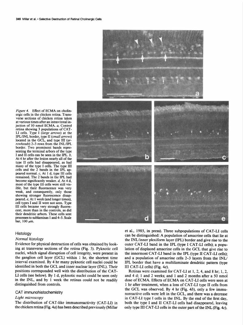
Figure 3. Toluidine blue-stained 1 pm semithin plastic sections of chicken ret- ina taken at different times after a 50 nmol intravitreal injection of ECMA. a, Control retina showing the various layers: outer nuclear layer, ON; outer plexiform layer, OP, inner nuclear lay- er, ZN; inner plexiform layer, ZP, gan- glion cell layer, G. b, Retina 4 hr after ECMA. Pyknotic cell nuclei can be seen in the GCL (small arrows), at the IPL/ INL border (large arrows), and 2-3 cell layers from this border (arrowheads). c, Retina 1 d after ECMA. Although pyk- notic cells could occasionally be ob- served in the GCL and at the IPL/INL border ([urge arrows), most pyknotic cell nuclei were deeper in the INL (arrow- heads). d, Retina 7 d following ECMA. The retina at this stage cannot be dis- tinguished from control retinas. Scale bar, 100 pm.
loo-fold higher amount caused total retinal degeneration. A time-course study using a 50 nmol dose of ECMA (Fig. 2)
showed that CAT activity was reduced to 55% of control levels within 4 hr of the injection. CAT activity had fallen to approx- imately 30% of control levels by 1 d after the injection. AChE also decreased, but this occurred more slowly than for CAT, with minimum levels (60% of control eye values) occurring 4 d after the lesion. At the optimum dose there did not seem to be gross effects on the retina for up to 64 d after the injection, since retinal wet weight and glutamine synthetase activity remained normal.
The other biochemical markers examined (tyrosine hydrox- ylase activity, GABA levels, and glycine levels) returned to nor- mal within 1 month of the injection of a 50 nmol dose of ECMA (Table l), although they were slightly depressed for some of this period.
346 Millar et al. l Selective Destruction of Retinal Cholinergic Cells
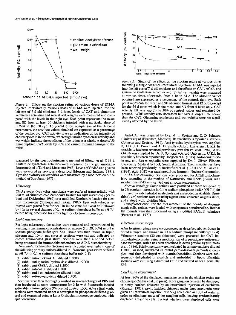
Figure 4. Effect of ECMA on cholin- ergic cells in the chicken retina. Trans- verse sections of chicken retina taken at various times after an intravitreal in- jection of 50 nmol ECMA. a, Control retina showing 3 populations of CAT- LI cells. Type I (large arrows) at the IPL/INL border, type II (small arrows) located in the CCL, and type III (ur- rowheads) 2-3 rows from the INL/IPL border. Two prominent bands repre- senting the terminal arbors of the type I and II cells can be seen in the IPL. b, At 4 hr after the lesion nearly all of the type II cells had disappeared, as had many of the type I cells. The type III cells and the 2 bands in the IPL ap- peared normal. c, At 1 d, type III cells remained. The 2 bands in the IPL had become significantly weaker. d, At 4 d, most of the type III cells were still vis- ible, but their fluorescence was very weak, and consequently, only those showing stronger fluorescence disap- peared. e, At 1 week (and longer times), cell types I and II were not seen. Type III cells became very strongly fluores- cent, more than in the controls, as did their dendritic arbors. These cells sent processes to sublaminas 1 and 4-5. Scale bar, 100 pm.
Histology Normal histology Evidence for physical destruction of cells was obtained by look- ing at transverse sections of the retina (Fig. 3). Pyknotic cell nuclei, which signal disruption of cell integrity, were present in the ganglion cell layer (GCL) within 1 hr, the shortest time interval examined. By 4 hr many pyknotic cell nuclei could be identified in both the GCL and inner nuclear layer (INL). Their positions corresponded well with the distribution of the CAT- LI cells (see below). By 1 d, pyknotic nuclei could be seen only in the INL, and by 1 week the retinas could not be readily distinguished from controls.
CAT immunohistochemistry Light microscopy The distribution of CAT-like immunoreactivity (CAT-LI) in the chicken retina (Fig. 4a) has been described previously (Millar
et al., 1985, in press). Three subpopulations of CAT-L1 cells can be distinguished: A population of amacrine cells that lie at the INWinner plexiform layer (IPL) border and give rise to the outer CAT-L1 band in the IPL (type I CAT-L1 cells); a popu- lation of displaced amacrine cells in the GCL that give rise to the innermost CAT-L1 band in the IPL (type II CAT-L1 cells); and a population of amacrine cells 2-3 layers from the INL/ IPL border that have a multilaminate dendritic pattern (type III CAT-L1 cells) (Fig. 4e).
Retinas were examined for CAT-L1 at 1,2,4, and 8 hr; 1,2, and 4 d, 1 and 2 weeks; and 1 and 2 months after a 50 nmol dose of ECMA. Effects of ECMA on CAT-L1 cells were seen at 1 hr after treatment, when a loss of CAT-L1 type II cells from the GCL was observed. By 4 hr (Fig. 4b), only a few immu- noreactive cells were left in the GCL, and there was a decrease in CAT-L1 type I cells in the INL. By the end of the first day, both the type I and II CAT-L1 cells had disappeared, leaving only type III CAT-L1 cells in the outer part of the INL (Fig. 4~).

The Journal of Neuroscience, February 1987, 7(2) 347
Figure 5. Ultrastructural appearance of CAT-L1 in the chicken retina at dif- ferent times following a 50 nmol injec- tion of ECMA. a, Type I CAT-L1 cell located at the INL/IPL border 8 hr after ECMA. The cytoplasm (Cyt) is severely disrupted and the nucleus (N) has be- come pyknotic. Nonimmunoreactive neighboring cells (A) appear normal. Scale bar, 2 pm. b, Type III CAT-L1 (III) cell located towards the center of the INL 8 hr after ECMA. This cell shows only weak immunoreactivity and is unaffected by the lesion. However, the nuclei of some adjacent cells (A) have darkened, but their cytoplasm ap- pears normal. Scale bar, 2 pm. c, Type II CAT-L1 cell located in the GCL 4 hr after an intravitreal injection of ECMA. The cytoplasmic matrix (Cyt) is swollen and severely disrupted and the nucleus (N) is pyknotic. Nearby ganglion cells (GC) are not affected by the lesion. Scale bar, 2 pm. d, Type III CAT-L1 cell (III) 7 d after ECMA. These cells appeared normal, as did neighboring cells. No other immunoreactive cells were seen at this stage or at later times after the lesion. Scale bar, 2 pm. e, CAT-L1 fibers in the IPL 8 hr after ECMA. Some fibers appear to be normal (I); others are swollen (2). Scale bar, 500 nm.f; CAT- LI fibers in the IPL 7 d after the ECMA lesion. These fibers are from the type III cholinergic cell. They generally have small profiles and terminate onto gan- glion (GC) and amacrine cell (A) pro- cesses. Scale bar, 500 nm.
At this stage, the 2 CAT-L1 bands in the IPL were only weakly type III cells were detectable (Fig. 4~). They were present in the immunoreactive. Four days after the lesion the type III cells, same numbers as in controls. The processes of the CAT-L1 type although present, showed only very weak immunoreactivity (Fig. III cells could be clearly seen after elimination of the 2 major 44. At the end of the first week and thereafter, only CAT-L1 CAT-L1 bands. These cells were multistratified, projecting to
Fi&.
ue
6.
Effe
ct o
f EC
MA
on d
ispla
ced
amac
rine
cells
. a,
Tol
uidi
ne
blue
-sta
ined
re
tinal
fla
t m
ount
fro
m
a 7-
d-ol
d ch
icke
n gi
ven
an i
ntra
vitre
al
inje
ctio
n of
10
~1 d
istill
ed
wat
er
shor
tly
afte
r ha
tch.
Not
e nu
mer
ous
gang
lion
cells
(la
rge
arro
ws)
an
d sm
alle
r di
spla
ced
amac
rine
cells
(sm
all
arro
ws)
. b,
Tol
uidi
ne
blue
-sta
ined
re
tinal
fla
t m
ount
fro
m a
7-d
-old
ch
icke
n gi
ven
an i
ntra
vitre
al
inje
ctio
n of
0.5
pg
colc
hici
ne
in 1
0 ~1
dis
tilled
w
ater
sh
ortly
af
ter
hatc
h. N
ote
the
exte
nsiv
e lo
ss o
f ce
lls.
Thre
e po
pula
tions
of
cel
ls a
re s
till
visi
ble:
su
rviv
ing
larg
e ga
nglio
n ce
lls (
larg
e ar
row
s),
smal
l di
spla
ced
amac
rine
cells
(sm
all
arro
ws)
, an
d fu
sifo
rm
supp
ortin
g ce
lls f
rom
th
e op
tic
nerv
e fib
er
laye
r. c,
Tol
uidi
ne
blue
-sta
ined
re
tinal
fla
t m
ount
fro
m
a 7-
d-ol
d ch
icke
n gi
ven
an
intra
vitre
al
inje
ctio
n of
0.5
pg
colc
hici
ne
and
50 n
mol
EC
MA
in
10 ~
1 di
stille
d w
ater
sh
ortly
af
ter
hatc
h.
Not
e th
e lo
ss o
f th
e sm
all
disp
lace
d am
acrin
e ce
lls a
nd
cont
inue
d su
rviv
al
of la
rge
colc
hici
ne-re
sist
ant
gang
lion
cells
(la
rge
arro
w).
Scal
e ba
r, 10
0 pm
.
The Journal of Neuroscience, February 1987, 7(2) 349
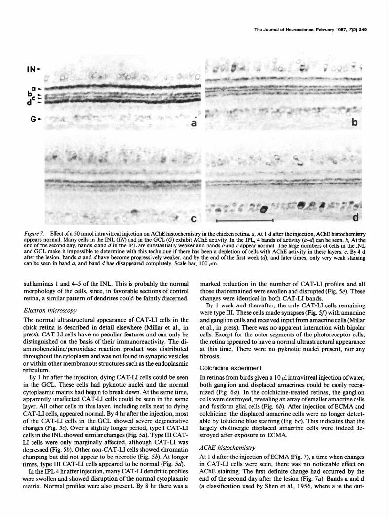
Figure 7. Effect of a 50 nmol intravitreal injection on AChE histochemistry in the chicken retina. u, At 1 d after the injection, AChE histochemistry appears normal. Many cells in the INL (IN) and in the GCL (@ exhibit AChE activity. In the IPL, 4 bands of activity (u-d) can be seen. b, At the end of the second day, bands a and din the IPL are substantially weaker and bands b and c appear normal. The large numbers of cells in the INL and GCL make it impossible to determine with this technique if there has been a depletion of cells with AChE activity in these layers. c, By 4 d after the lesion, bands a and d have become progressively weaker, and by the end of the first week (d), and later times, only very weak staining can be seen in band a, and band d has disappeared completely. Scale bar, 100 pm.
sublaminas 1 and 4-5 of the INL. This is probably the normal morphology of the cells, since, in favorable sections of control retina, a similar pattern of dendrites could be faintly discerned.
Electron microscopy The normal ultrastructural appearance of CAT-L1 cells in the chick retina is described in detail elsewhere (Millar et al., in press). CAT-L1 cells have no peculiar features and can only be distinguished on the basis of their immunoreactivity. The di- aminobenzidine/peroxidase reaction product was distributed throughout the cytoplasm and was not found in synaptic vesicles or within other membranous structures such as the endoplasmic reticulum.
By 1 hr after the injection, dying CAT-L1 cells could be seen in the GCL. These cells had pyknotic nuclei and the normal cytoplasmic matrix had begun to break down. At the same time, apparently unaffected CAT-L1 cells could be seen in the same layer. All other cells in this layer, including cells next to dying CAT-L1 cells, appeared normal. By 4 hr after the injection, most of the CAT-L1 cells in the GCL showed severe degenerative changes (Fig. 5~). Over a slightly longer period, type I CAT-L1 cells in the INL showed similar changes (Fig. 5~). Type III CAT- LI cells were only marginally affected, although CAT-L1 was depressed (Fig. 5b). Other non-CAT-L1 cells showed chromatin clumping but did not appear to be necrotic (Fig. 5b). At longer times, type III CAT-L1 cells appeared to be normal (Fig. 54.
In the IPL 4 hr after injection, many CAT-L1 dendritic profiles were swollen and showed disruption of the normal cytoplasmic matrix. Normal profiles were also present. By 8 hr there was a
marked reduction in the number of CAT-L1 profiles and all those that remained were swollen and disrupted (Fig. 5e). These changes were identical in both CAT-L1 bands.
By 1 week and thereafter, the only CAT-L1 cells remaining were type III. These cells made synapses (Fig. 5s) with amacrine and ganglion cells and received input from amacrine cells (Millar et al., in press). There was no apparent interaction with bipolar cells. Except for the outer segments of the photoreceptor cells, the retina appeared to have a normal ultrastructural appearance at this time. There were no pylcnotic nuclei present, nor any fibrosis.
Colchicine experiment
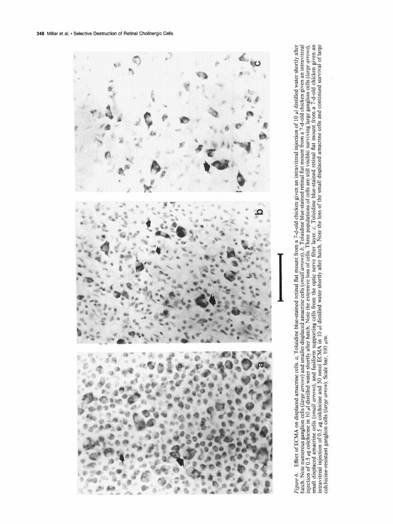
In retinas from birds given a 10 ~1 intravitreal injection of water, both ganglion and displaced amacrines could be easily recog- nized (Fig. 00). In the colchicine-treated retinas, the ganglion cells were destroyed, revealing an array of smaller amacrine cells and fusiform glial cells (Fig. 6b). After injection of ECMA and colchicine, the displaced amacrine cells were no longer detect- able by toluidine blue staining (Fig. 6~). This indicates that the largely cholinergic displaced amacrine cells were indeed de- stroyed after exposure to ECMA.
AChE histochemistry At 1 d after the injection of ECMA (Fig. 7), a time when changes in CAT-L1 cells were seen, there was no noticeable effect on AChE staining. The first definite change had occurred by the end of the second day after the lesion (Fig. 7~). Bands a and d (a classification used by Shen et al., 1956, where a is the out-

350 Millar et al. l Selective Destruction of Retinal Cholinergic Cells
Figure 8. Effects of ECMA on other immunologically identifiable cell types in the retina. a. 5-Ht-LI 7 d after the injection. At this time, and all others, immunoreactivity appeared normal. 5- Ht-LI can be seen in a population of amacrine cells towards the middle of the INL (large arrow) and in many bi- polar cells also located towards the middle of the INL (small arrows). b, GABA-LI 7 d after the injection. At this time, and at all others, immunoreactiv- ity appeared normal. GABA-LI can be seen in many amacrine cells in the INL (large arrows), a few cells in the GCL (arrowheads), and in some horizontal cells (small arrows). c, Tyrosine hy- droxylase-117 d after the injection. At this time, and all others, immunoreac- tivity appeared normal. Tyrosine hy- droxylase-11 can be seen in a few large amacrine cell bodies on the border be- tween the INL and the IPL. There is a fiber plexus on the outer margin of the IPL. d, Enkephalin-LI 7 d after the in- jection. At this time, and all others, im- munoreactivity appeared normal. En- kephalin-LI can be seen in a population of amacrine cell bodies located towards the middle of the INL, with faint fiber plexuses within the IPL. Scale bar, 100 pm.
ermost, then in order towards the GCL, b, c, and d) showed less AChE activity. The activity in these bands became pro- gressively weaker (Fig. 7, b, c), so that by the end of the first week virtually no activity was visible in band d and only min- imum activity in band a (Fig. 74. Bands b and c were unaffected by the ECMA. This change seemed permanent because no re- covery of activity was observed 2 months after the injection. Changes of AChE activity in cell soma were not obvious with this method, probably because of the large number of AChE- positive noncholinergic cells found in the retina (Millar et al., 1985).
Other cell types Dopaminergic cells In the retina these cells comprise a population of amacrine cells with a very different distribution from CAT-L1 cells. They are sparsely distributed at the INL/IPL border. These cells appeared to be unaffected by ECMA (Fig. 8~). Quantitative estimates of the number of dopaminergic cells were made on retinal flat mounts, pretreated with noradrenaline, and processed using the Faglu technique (Furness et al., 1977). In control retinas the dopaminergic cell density was 25.7 cells/mm*. One month after the ECMA injection, the density was effectively unchanged at 26.5 cells/mm* (Table 1).
GABA-LI cells
Several different populations of cells were labeled by the GABA antisera (Fig. 8b). In particular, many horizontal cells were la- beled and these were not affected by ECMA. Many cells in the INL and GCL also showed GABA immunoreactivity. Because of the large numbers of these cells in both layers it was impos- sible to determine with these techniques whether ECMA affected a small percentage of these cells. However, no effect on these cell populations was obvious any time after the lesion.
5-HT-LI cells
A large population of bipolar cells and a relatively sparse pop- ulation of amacrine cells were stained using the antiserum to 5-HT. Neither population of cells was affected by ECMA (Fig. 8a).
Enkephalin-LI cells
As reported previously (Millar et al., 1984), a population of amacrine cells located 2-3 rows away from the IPL showed enkephalin-11. These cells were unaffected by ECMA (Fig. 84.
Somatostatin-LI cells
Somatostatin-LI cells comprise a subpopulation of amacrine cells with dendritic projections to sublaminas 1 and 3-4 in the
The Journal of Neuroscience, February 1987. 7(2) 351
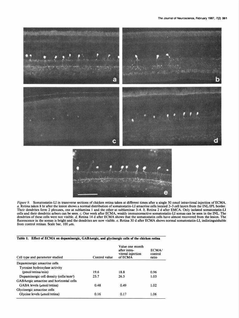
F&we 9. Somatostatin-LI in transverse sections of chicken retina taken at different times after a single 50 nmol intravitreal injection of ECMA, a, Retina taken 8 hr after the lesion shows a normal distribution of somatostatin-11 amacrine cells located 2-3 cell layers from the INIJIPL border. Their dendrites form 2 plexuses, one at sublamina 1 and the other at sublaminae 3-4. b, Retina 2 d after EMCA. Only isolated somatostatin-11 cells and their dendritic arbors can be seen. c, One week after ECMA, weakly immunoreactive somatostatin-11 somas can be seen in the INL. The dendrites of these cells were not visible. d, Retina 14 d after ECMA shows that the somatostatin cells have almost recovered from the lesion. The fluorescence in the somas is bright and the dendrites are now visible. e, Retina 30 d after ECMA shows normal somatostatin-11, indistinguishable from control retinas. Scale bar, 100 pm.
Table 1. Effect of ECMA on dopaminergic, GABAergic, and glycinergic cells of the chicken retina
Cell tvne and narameter studied Control value
Value one month after intra- v&real injection of ECMA
ECMA/ control ratio
Dopaminergic amacrine cells Tyrosine hydroxylase activity
(pmol/retina/min) Dopaminergic cell density (cells/mm2)
GABAergic amacrine and horizontal cells GABA levels @mol/retina)
Glycinergic amacrine cells Glycine levels &mol/retina)
19.6 18.8 0.96 25.7 26.5 1.03
0.48 0.49 1.02
0.16 0.17 1.06
352 Millar et al. l Selective Destruction of Retinal Cholinergic Cells
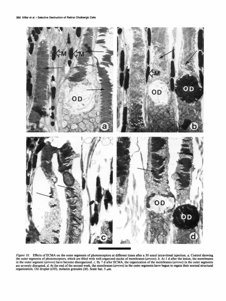
Figure 10. Effects of ECMA on the outer segments of photoreceptors at different times after a 50 nmol intravitreal injection. a, Control showing the outer segments of photoreceptors, which are filled with well-organized stacks of membranes (arrows). b, At 1 d after the lesion, the membranes in the outer segment (arrows) have become disorganized. c, By 7 d after ECMA, the organization of the membranes (arrows) in the outer segments are severely disrupted. d, At the end of the second week, the membranes (arrows) in the outer segments have begun to regain their normal structural organization. Oil droplet (OD), melanin granules (M). Scale bar, 5 pm.
The Journal of Neuroscience, February 1987. 7(2) 353
IPL (Fig. 9u). These cells were affected by the ECMA lesion. Retinas examined up to the end of the first day after the lesion appeared normal. In the period between 1 and 2 d, only isolated somatostatin-11 cells with normal dendritic projections could be seen (Fig. 9b). This pattern continued until the end of the first week, when only very weakly somatostatin-11 immuno- reactive cell bodies were visible (Fig. 9c). By the end of the second week the cell bodies were intensely fluorescent and the dendritic arbor of these cells became visible (Fig. 94. One month after the lesion, somatostatin-11 cells appeared normal in all respects: intensity of immunofluorescence, cell numbers, and distribution of the dendritic trees (Fig. 9e).
Photoreceptors With both light- and electron-microscopic techniques it was clear that the outer segments of the photoreceptors were dam- aged after exposure to ECMA. The normal well-organized stack- ing of the membranes was severely disrupted by the end of the first day (cf. Fig. lob with lOa). Disruption was more pro- nounced after 1 week (Fig. lOc), but by 1 month the process appeared to be reversing (Fig. 104.
Discussion Our data indicate that ECMA caused a profound and essentially irreversible loss of enzymatically detectable CAT from the chicken retina. In parallel with the loss of enzymatically de- tectable CAT, it became increasingly difficult to detect CAT- LI, indicating that the enzyme was eliminated, not simply in- activated, by the neurotoxin. The immunohistochemical studies of CAT-L1 also clarify some other aspects of the ECMA lesion. Both choline& cell bodies and terminal arbors disappeared after exposure to ECMA, indicating that under these conditions the terminals were not damaged selectively. Rather, both cho- linergic cell bodies and processes were affected. This contrasts with the reported lack of destruction of choline& cell bodies in the striatum after microinjection of ECMA (Sandberg et al., 1984). Unfortunately, in the latter study the less reliable crite- rion of AChE histochemistry was used to identify cholinergic cells.
The immunohistochemical studies were also used to dem- onstrate that the cholinergic cells were destroyed by ECMA. Electron-microscopic immunohistochemistry showed that grossly necrotic CAT-L1 cells were visible in the retina, both in the INL and GCL. The degree of disruption of the cholinergic cells virtually ruled out the possibility of recovery, and the fre- quency of occurrence of grossly necrotic cholinergic cells was consistent with the degree of loss of enzymatically and immu- nohistochemically detectable CAT activity observed. That the type II cholinergic cells (in the GCL) had been destroyed by ECMA was demonstrated directly, since after elimination of the ganglion cells with colchicine, the characteristic array of dis- placed amacrine cells (Ehrlich and Morgan, 1980; Morgan, 198 I), which are known to be CAT-L1 (Millar et al., in press), was no longer visible using Nissl staining-a technique that does not depend upon the cholinergic characteristics of the cells.
The data provide the first unequivocal demonstration of the ability of ECMA to kill, rather than simply to damage or down- regulate, choline@ cells in vivo. Our results are therefore con- sistent with the observation (Sandberg et al., 1985b) that ECMA kills in vitro a hybridoma cell line exhibiting cholinergic char-
acteristics. However, whether the mechanism of toxicity is the same in vivo and in vitro is unclear, since many cholinergic cells were clearly necrotic in vivo within 1 hr of the intravitreal in- jection of ECMA, whereas 18 hr was required for clear signs of cell destruction in vitro.
The only limitation to the efficacy of ECMA in our experi- ments concerns its apparent failure to kill, damage the terminal arbors, or permanently down-regulate the type III choline& amacrine cells, although CAT-L1 immunofluorescence was de- pressed in these cells for several days. Possible reasons for the selectivity of ECMA for different classes of choline&c cells will be discussed later in relation to the mechanism of action of ECMA, but it is important to note here that it was not possible to eliminate the type III cells by increasing the amount of ECMA injected intravitreally without causing first patchy and then total retinal degeneration.
Specificity of ECMA for cholinergic cells The dose-response curves for a general measure of retinal in- tegrity such as retinal wet weight and glutamine synthetase (as a measure of glial cell integrity) demonstrate that there is a narrow range of amounts of ECMA injected that will produce a large depletion in CAT without gross effects on retinal integ- rity.
Further indications of specificity came from the observations that necrotic cells were observed only in positions occupied by cholinergic cells and that at the electron-microscope level only CAT-L1 cells were grossly necrotic, in both the INL and GCL. In particular, necrotic cells were not observed in the outer part of the INL, where the horizontal and bipolar cells are located, or the outer nuclear layer. Attention was therefore focused on the effects of ECMA on noncholinergic amacrine cells and gan- glion cells, which are mixed in with the displaced amacrine cells of the GCL.
Nonspecific effects of ECMA on dopaminergic cells of the striatum have been reported, based on a decrease in dopamine levels 2 weeks after the injection and a very indirect behavioral assay 2 d after the injection (Levy et al., 1984). A detailed study of the dopaminergic cells of the chicken retina (dopamine and tyrosine hydroxylase levels, and histofluorescence and immu- nohistochemical analysis of cell integrity, number, and distri- bution) indicates that such effects were not seen in the chicken retina. Our observations therefore strengthen the claims of Sandberg et al. (1984) that striatal dopamine cells are only min- imally affected by ECMA at appropriate doses.
Two sorts of peptide-immunoreactive amacrine cells were examined, enkephalin-immunoreactive cells were apparently unaffected at any stage by ECMA, whereas some of the somato- statin-immunoreactive cells were disrupted by exposure to ECMA for a period of days. However, they were not destroyed by the toxin, since cells with normal morphologies and distri- butions were observed from 2 to 3 weeks after the injection. Some noncholinergic amacrine cells, in positions similar to those occupied by the somatostatin-immunoreactive amacrine cells, were affected at the electron-microscope level. However, they were not necrotic, unlike the type I and type II choline@ amacrine cells. Why the somatostatin-11 cells would be so mark- edly, albeit not lethally, affected by ECMA is not clear. Although the somatostatin-11 cells and the type III cholinergic cells oc- cupy similar positions, this is not a case of coexistence of ACh and somatostatin-11 (T. J. Millar, unpublished observations).

354 Millar et al. - Selective Destruction of Retinal Cholinergic Cells
SHT-immunoreactive cells were also examined. Neither the numerous bipolar cells nor the far less numerous amacrine cells seemed to be affected by exposure to ECMA. Horizontal and many amacrine cells were detected with GABA-LI. None of these seemed to be affected by ECMA, although so many ama- crine cells were revealed by GABA-LI that a slight loss of GABA- LI amacrine cells could not have been detected. However, levels of GABA were also followed. Despite a transient depression for 2-3 weeks following the injection of ECMA, GABA levels re- turned to normal by 4 weeks, suggesting that GABAergic ama- crine cells were not permanently affected by ECMA. The fate of glycinergic cells was also followed by monitoring glycine levels. These did not significantly change after exposure to ECMA.
The fate of the ganglion cells after exposure to ECMA alone was also examined in retinal flat mounts (results not shown). Apart from the absence of the small cells previously identified as displaced amacrine cells (Ehrlich and Morgan, 1980; Ehrlich, 198 1; Morgan, 198 l), no changes in morphologies and numbers of cells in the ganglion cell layer were observed.
These results taken together indicate that ECMA is very spe- cific in its effects on choline& cells, which in the chicken retina are exclusively amacrine cells (Bat&man and Bader, 1977; Mil- lar et al., 1985, in press). Short-term effects on the somatostatin- immunoreactive and GABA- and glycine-containing amacrine cells have been observed, but these cells appear to return to normal a few weeks after exposure to ECMA. It is not clear to what extent these transient effects represent lack of specificity of ECMA or whether the cells are transiently affected as a trans- synaptic response to removal of cholinergic cells that are either pre- or postsynaptic to them. In the longer term experiments, no permanent change in any other cell type has been observed to indicate preferential synaptic interactions with the destroyed cholinergic cells. The transient effects on the cells are not readily explicable in these terms, since the evidence in chicken (Morgan et al., 1981) and rabbit retina (Cunningham et al., 1983) is against interactions between cholinergic and somatostatin-11 cells. However, Cunningham and Neal (1983) suggest that cho- linergic cells are closely connected to GABAergic and glycinergic cells, which might explain the transient changes observed in these systems.
Efects of ECMA on AChE We have previously shown that the distributions of CAT-L1 and AChE in the chicken retina overlap but are far from identical (Millar et al., 1985), which underlines the importance of using CAT-L1 rather than AChE to characterize morphologically cho- line@ neurons. The CAT-L1 bands in the IPL correspond to the major AChE-positive bands, but there are 2 other AChE- positive bands that do not have associated CAT-L1 and thus do not appear to be associated with cholinergic transmission sites. At the cell-body level, choline& cell bodies are AChE-positive, but so are many others, and the most intensely AChE-positive cell bodies are not choline&.
It is therefore not surprising that effects of ECMA on cell- body AChE could not readily be seen. By contrast, in parallel with the decrease in enzymatically detectable AChE, AChE in the 2 cholinergic dendritric plexuses of the IPL disappeared. The 2 noncholinergic AChE-positive bands were not affected, which supports the idea that ECMA shows considerable spec- ificity for cholinergic cells and processes.
Effects of ECMA on photoreceptors The most obvious effects on cells other than the choline& amacrine cells were those seen on the photoreceptors, involving transient shortening of the outer segments of rods and cones. The sequence of events observed was almost identical to those reported by Pu and Masland (1984) for rod photoreceptors of rabbit retina exposed to hemicholinium-3, with vacuolization at the base of the outer segment followed by disorganization of the lamellae and shortening and often loss of the outer segments followed by partial to complete regeneration. Although the chicken retina is a cone-dominant retina, the rapid total dis- organization of outer segments followed by total cell degener- ation reported to occur for the rabbit cones was not observed in the chicken retina. Whether this difference is due to the dif- ferent agents or the different species used (ECMA on the chicken retina; hemicholinium-3 on the rabbit retina) is not clear, but it is interesting that in the chicken retina both type I and type II cells (the probable homologs of the cholinergic cells in the rabbit retina) are destroyed, while the photoreceptors survive, whereas in the rabbit the starburst a cells as well as the cones are destroyed, while the starburst b cells survive. These differ- ences point either to interesting species differences in the met- abolic states of apparently homologous retinal cell types or to unanticipated differences in the sites and modes of action of ECMA and hemicholinium-3.
Mode of action of ECMA Two distinct mechanisms of action of ECMA have now been proposed. One postulate is that ECMA would deprive cells of choline by irreversibly inactivating the high-affinity choline up- take mechanisms (Fisher and Hanin, 1980). In choline& cells, which seem to use choline preferentially for ACh synthesis, this might lead to a pronounced decrease in the synthesis of phos- phatidyl choline, perturbation of membrane synthesis, and ul- timately, perhaps, cell death. In general this might also lead to effects of ECMA on cells with a great demand for choline for membrane turnover, such as photoreceptors (Masland and Mills, 1980) and milder effects on all cell types. Such effects have been observed. Other choline uptake blockers such as hemicholin- ium-3 might also be expected to produce analogous effects (Pu and Masland, 1984).
The failure of ECMA to kill, or even markedly affect, the type III cholinergic amacrine cells at lower doses could reflect a lower rate of ACh and/or membrane phosphatidylcholine turnover in this cell type, or it could reflect a molecular heterogeneity of high-affinity choline uptake sites, as has been postulated to ex- plain the differing sensitivity of cholinergic markers in different brain regions after intraventricular injection of ECMA (Hanin et al., 1983). The effects on the somatostatin-11 amacrine cells could also be explained if they have a particularly high demand for choline for membrane synthesis.
A more trivial explanation for the survival of the type III CAT-L1 cells is that they are further from the vitreal site of injection of ECMA and that a marked concentration gradient of the highly reactive aziridinium ions might result in sparing of these cells. Although the later onset of effects on the type I as compared to the type II cells indicates that there may be dilfirsion barriers for ECMA, the distance between the cell bod- ies of the type I and type III cells is small (l&20 pm) compared

The Journal of Neuroscience, February 1997, 7(P) 355
to the distance ECMA has to diffuse to reach the type I cell bodies from the vitreous (- 100 pm), yet the difference in sen- sitivity of the 2 cell types seems almost absolute. Moreover, the dendrites of the type III cells in fact laminate closer to the vitreal injection sites than the dendrites of the type I and II cells. A final point against this idea is that retinal thickness varies from center to periphery, yet there were no obvious regional varia- tions in effects on any of the cell types.
The major alternative, but not necessarily conflicting, hy- pothesis is that ECMA might be taken up by the cholinergic cells as a “substrate” of the choline carrier and that it might inhibit enzymes that use choline or structurally related com- pounds as substrates (Sandberg et al., 1985b). Pu and Masland (1984) have proposed a similar hypothesis to account for some features of the effects of hemicholinium-3 on the retina. ECMA and related compounds inhibit CAT, AChE, choline dehydro- genase, and choline kinase in vitro (Rylett and Colhoun, 1979; Barlow and Marchbanks, 1984; Sandberg et al., 1985b), but there is considerable divergence in the concentrations required to produce appreciable inhibition. For all enzymes at least micromolar concentrations of ECMA may be required. We es- timate the extracellular concentration of ECMA achieved to be of the order of 12 PM. There is no way of determining what degree of concentration within choline& cells might be achieved, but Rylett and Colhoun (1984) have estimated from the relative kinetics of choline mustard aziridinium as a sub- strate and inhibitor ofhigh-affinity choline uptake that very little uptake would take place. Nevertheless, it is conceivable that the rapid decline in CAT activity may in part be due to ECMA acting as an inhibitor rather than a neurotoxin. Indeed, the loss of enzymatically detected CAT seems more rapid than the loss of CAT-LI. But with the exception of inhibition of choline ki- nase, one of the least susceptible enzymes, it is not clear why ECMA acting as a long-term inhibitor would ultimately destroy cells.
For these reasons we suggest that the major effects of ECMA in our system are likely to be due to its ability to deprive cells of choline by inhibiting high-affinity choline uptake, and less effectively low-affinity choline uptake, although some short-term effects may be due to uptake of ECMA and direct intracellular inhibition of enzymes. This mechanism of action, which is supported by the similar effects of ECMA and hemicholinium-3 (Pu and Masland, 1984) is, however, unproven, and it should be noted that the analogous noradrenergic neurotoxins DSP4 and xylamine may be able to cause degeneration of noradrenergic neurons by interacting with the plasma membranes of the cells (Ross, 1976; Jaim-Etcheverry and Zieher, 1983). Thus, general perturbation of membrane function after the toxins have been concentrated on carrier molecules may also have a role in this form of neurotoxicity.
ECMA as a cholinergic neurotoxin Most of the features of the retinal lesions produced by ECMA can be explained on the assumption that it acts by blocking high-affinity choline uptake, thus depriving cells of choline, per- turbing phospholipid and membrane synthesis, and ultimately producing a range of effects on different cell types depending on their demand for choline for membrane renewal and ACh syn- thesis. At higher concentrations, ECMA also inhibits low-affin- ity choline uptake (Rylett and Colhoun, 1984), and this, together
with the high general reactivity of aziridinium ions, might lead to general cell destruction. The dose-toxicity curves for ECMA seem to be quite steep, with only a narrow dose range separating maximum depletion of cholinergic cells with minimum side effects from signs of more general cell destruction.
These properties of ECMA as a cholinergic neurotoxin may explain, in part, why results using ECMA to affect cholinergic cells in other parts of the nervous system have been less clear- cut than those we have obtained on the retina. The concentra- tions of ECMA injected into the striatum, hippocampus, and intracerebroventricular fluid are high (in the low millimolar range) in very small volumes, and this might be expected to lead to focal necrosis. Moreover, if diffusion barriers are sig- nificant, it may be that the area of selective cholinergic toxicity is not wide. The solution to these difficulties may involve mul- tiple-site injections of lower concentrations of ECMA, prefer- ably keeping the volume low to minimize mechanical damage at the injection site. Such a procedure may be laborious, but our results demonstrate that it is possible to define conditions under which ECMA acts as a potent and remarkably selective cholinergic neurotoxin, which destroys cholinergic cells in vivo. ECMA therefore has the potential to be extremely useful for sorting out the details of the anatomy and functions of cholin- ergic pathways in the CNS, provided the problems of delivery involved in obtaining an effective and selective lesion can be solved.
References Barlow, P., and R. M. Marchbanks (1984) Effect of ethylcholine mus-
tard on choline dehydrogenase and other enzymes of choline metab- olism. J. Neurochem. 43: 1568-1573.
Baughman, R. W., and C. R. Bader (1977) Biochemical characteriza- tion and cellular localization of the cholinergic system in the chicken retina. Brain Res. 138: 469-485.
Buckerheld, M., J. 0. Oliver, I. W. Chubb, and I. G. Morgan (198 1) Somatostatin-like immunoreactivity in amacrine cells of the chick retina. Neuroscience 6: 686-689.
Cunningham, J. R., and M. J. Neal (1983) Effect of -y-aminobutyric acid agonists, glycine, tam-me and neuropeptides on acetylcholine release from the rabbit retina. J. Physiol. (Lond.) 336: 563-577.
Cunningham, J. R., C. Dawson, and M. J. Neal (1983) Evidence for a cholinergic inhibitory feed-back mechanism in the rabbit retina. J. Physiol. (Lond.) 340: 455-468.
Dowling, J. E., and M. W. Dubin (1984) The vertebrate retina. In Handbook of Physiology, Sec. I, Vol. 3, Sensory Processes, Part 1, I. Darian-Smith, ed., pp. 3 17-339, American Physiological Society, Be- thesda, MD.
Ehrlich, D. (198 1) Regional specialization of the chick retina as re- vealed by the size and density of neurons in the ganglion cell layer. J. Comp. Neurol. 195: 643-657.
Ehrlich, D., and I. G. Morgan (1980) Kainic acid destroys displaced amacrine cells in post-hatch chicken retina. Neurosci. Lett. 17: 43- 48.
Ellman, G. L., K. D. Courtney, V. Andres, and R. M. Featherstone (196 1) A new and rapid calorimetric determination of acetylcholin- esterase activity. Biochem. Pharmacol. 7: 88-95.
Fisher, A., and I. Hanin (1980) Choline analogs as potential tools in developing selective animal models of central cholinergic hypofunc- tion. Life Sci. 27: 1615-1634.
Fisher, A., C. R. Mantione, D. J. Abraham, and I. Hanin (1982) Long- term central choline& hypoftmction induced in mice by ethylcholine aziridinium ion (AF64A) in vivo. J. Pharmacol. Exp. Ther. 222: 140- 145.
Fumess, J. B., M. Costa, and A. L. Wilson (1977) Water-stable fluo- rophores produced by reaction with aldehyde solutions, for the his- tochemical localization of catechol- and indole-ethylamines. Histo- chemistry 52: 159-l 70.
356 Millar et al. - Selective Destruction of Retinal Cholinergic Cells
Glover, V., and D. P. L. Green (1972) A simple, quick microassay for choline acetyltransferase. J. Neurochem. 19: 2465-2466.
Golumbic, C., J. S. Fruton, and M. Bergmann (1946) Chemical re- actions of the nitrogen mustard gases. I. The transformations of meth- yl-bis(beta-chloroethyl) amine in water. J. Org. Chem. 1 I: 5 18-535.
Hanin, I., W. D. DeGroat, C. R. Mantione, J. T. Coyle, and A. Fisher (1983) Chemically-induced cholinotoxicity in vivo: Studies utilizing ethylcholine aziridinium ion (AF64A). Banbury Biol. Rep. 15: 243- 253.
Haywood, J., J. W. Hambley, and S. P. R. Rose (1975) Effects of exposure to an imprinting stimulus on the activity of enzymes in- volved in acetylcholine metabolism in chick brain. Brain Res. 92: 219-225.
Hodgson, A. J., B. Penke, A. Erdei, I. W. Chubb, and P. Somogyi (1985) Antisera to y-aminobutyric acid. 1. Production and characterization using a new model system. Histochem. Cytochem. 33: 229-239.
Ingham, C. A., and I. G. Morgan (1983) Dose-dependent effects of intravitreal kainic acid on specific cell types in chicken retina. Neu-
- _.
roscience 9: 165-l 8 1. Ishimoto. I.. T. .I. Millar. I. W. Chubb. and I. G. Moraan (1986) So-
matostatin immunoreactive amacrine cells of chicken retina: Retinal mosaic, ultrastructural features and light driven variations in peptide metabolism. Neuroscience 17: 12 17-l 233.
Jaim-Etcheverry, G., and L. M. Zieher (1983) 2-Chloroethylamines: New chemical tools for the study of noradrenergic neurons. Trends Pharmacol. Sci. 4: 473-475.
Johnson, C. D., and M. L. Epstein (1986) Monoclonal antibodies and polyvalent antiserum to chick choline acetyltransferase. J. Neuro- them. 46: 968-976.
Karnovsky, M. J., and L. Roots (1964) A “direct-coloring” thiocholine method for cholinesterase. J. Histochem. Cytochem. 12: 2 19-22 1.
Karobath, M. (197 1) Catecholamines and the hydroxylation of tyro- sine in synaptosomes isolated from rat brain. Proc. Natl. Acad. Sci. USA 68: 2370-2373.
Kirk, D. L., and A. A. Moscona (1963) Synthesis of experimentally- induced glutamine synthetase (glutamotransferase activity) in em- bryonic chick retina in vitro. Dev. Biol. 8: 341-357.
Levy, A., G. J. Kent, J. L. Meyerhoff, and L. E. Jarrard (1984) Non- cholinergic neurotoxic effects of AF64A in the substantia nigra. Brain Res. 305: 169-172.
Linser, P., and A. A. Moscona (1979) Induction of glutamine synthe- tase in embryonic neural retina: Localization in Mtiller cell fibres and dependence on cell interactions. Proc. Natl. Acad. Sci. USA 76: 6476- 6480.
Mantione, C. R., A. Fisher, and I. Hanin (198 1) The AF64A-treated mouse: Possible model for central choline& hypofunction. Science 213: 579-580.
Mantione, C. R., W. DeGroat, A. Fisher, and I. Hanin (1983a) Se- lective inhibition of peripheral cholinergic transmission in cat pro- duced by AF64A. J. Phartnacol. Exp. Ther. 225: 616-622.
Mantione. C. R.. M. J. Ziamond. A. Fisher. and I. Hanin (1983b) Selective presynaptic chilinet& neurotoxicity following idtrahip: pocampal AF64A injection in rats. J. Neurochem. 41: 251-255.
Masland, R. H., and J. W. Mills (1980) Choline accumulation by photoreceptor cells of the rabbit retina. Proc. Natl. Acad. Sci. USA 77: 1671-1675.
Millar, T. J., N. Salipan, J. 0. Oliver, I. G. Morgan, and I. W. Chubb (1984) The concentration of enkephalin-like material in the chick retina is light dependent. Neuroscience 13: 221-226.
Millar, T. J., I. Ishimoto, C. Johnson, M. Epstein, I. W. Chubb, and I. G. Morgan (1985) Cholinergic and acetylcholinesterase-containing neuronsof the chicken retina: Neurosci. I.&t. 61: 3 1 l-3 16.
Millar. T. J.. I. Ishimoto. I. W. Chubb, M. L. Epstein. C. D. Johnson, , ~, and I. G. Morgan (in press) Choline&c amac&e cells ofthe chicken retina: A light and electron microscope immunocytochemical study. Neuroscience.
Morgan. I. G. (198 1) Intraocular colchicine selectively destroys im- mature ganglion cells in chicken retina. Neurosci. Lett. 24: 255-260.
Morean. I. G.. and C. A. Ineham (1981) Kainic acid affects both pl~xif&m layers of chicken ietina. Neurosci. Lett. 21: 275-280.
Morgan, I. G., J. 0. Oliver, and I. W. Chubb (1981) Discrete distri- butions of putative cholinergic and somatostatinergic amacrine cell dendrites in the chicken retina. Neurosci. Lett. 27: 55-60.
Pu, G. A.-W., and R. H. Masland (1984) Biochemical interruption of membrane phospholipid renewal in retinal photoreceptor cells. J. Neurosci. 4: 1559-1576.
Riepe, R. E., and M. D. Norenburg (1977) Mtlller cell localization of glutamine synthetase in the rat retina. Nature 268: 654-655.
Ross, S. B. (1976) Long-term effects of N-2-chloroethyl-N-ethyl-2- bromobenzylamine hydrochloride on noradrenergic neurons in the rat brain and heart. Br. J. Pharmacol. 5: 521-527.
Rylett, B. J., and E. H. Colhoun (1977) Effects ofacetylcholine mustard aziridinium ion and its choline analogue on choline transport into synaptosomes. Can. J. Physiol. Pharmacol. 55: 769-772.
Rylett, B. J., and E. H. Colhoun (1979) The interactions of choline mustard aziridinium ion with choline acetyltransferase (EC 2.3.1.6). J. Neurochem. 32: 553-558.
Rylett, B. J., and E. H. Colhoun (1984) An evaluation of irreversible inhibition of synaptosomal high-affinity choline transport by choline mustard aziridinium ion. J: Neurochem. 43: 787-794.
Sandbera. K.. I. Hanin. A. Fisher. and J. T. Covle 11984) Selective choli&gic’neurotoxh: AF64A’s effects in rat*striatum.‘Brain Res. 293: 49-55.
Sandberg, K., R. L. Schnaar, and J. T. Coyle (1985a) Method for the quantitation and characterization of the cholinergic neurotoxin, mon- oethylcholine mustard aziridinium ion (AF64A). J. Neurosci. Meth- ods 14: 143-148.
Sandberg, K., R. L. Schnaar, M. McKinney, I. Hanin, A. Fisher, and J. T. Coyle (1985b) AF64A: An active site-directed irreversible inhibitor of choline acetyltransferase. J. Neurochem. 44: 439-445.
Shen, S.-C., P. Greenfield, and E. J. Boelle (1956) Localization of acetylcholinesterase in chick retina during histogenesis. J. Comp. Neurol. 106: 433-46 1.
Somogyi, P., and H. Takagi (1982) A note on the use of pi& acid- paraformaldehyde-glutaraldehyde fixative for correlated light and electron microscopic immunocytochemistry. Neuroscience 7: 1779- 1784.
van den Pol, A. N., R. S. Herbsi, and J. F. Powell (1984) Tyrosine hydroxylase-immunoreactive neurons of the hypothalamus: A light and electron microscopic study. Neuroscience 13: 1117-l 156.
Zamboni, L., and C. DeMartino (1967) Buffered picric acid-formal- dehyde: A new fixative for electron microscopy. J. Cell Biol. 35: 148A.
![Journal of Inflammation BioMed Central...mustard (2,2'-dichlorodiethyl sulfide; SM) has been investigated for nearly a century [1,2]. This toxic gas can damage the eyes and respiratory](https://img.pdfslide.us/doc/110x75/60b8329f6015e241d73f0d93/journal-of-inflammation-biomed-central-mustard-22-dichlorodiethyl-sulfide.jpg)