Embed Size (px)
Citation preview
The Third Window: Superior Semicircular Canal Dehiscence
Murtaza Kharodawala, MD
Faculty Advisor: Tomoko Makishima, MD, PhD
The University of Texas Medical Branch
Department of Otolaryngology – Head and Neck Surgery
Grand Rounds Presentation
November 8, 2006
Outline
Review Anatomy of Inner Ear
Review Physiology of Vestibular System
Superior Semicircular Canal Dehiscence
Description
Vestibular and Auditory Manifestations
Testing
Surgical Repair
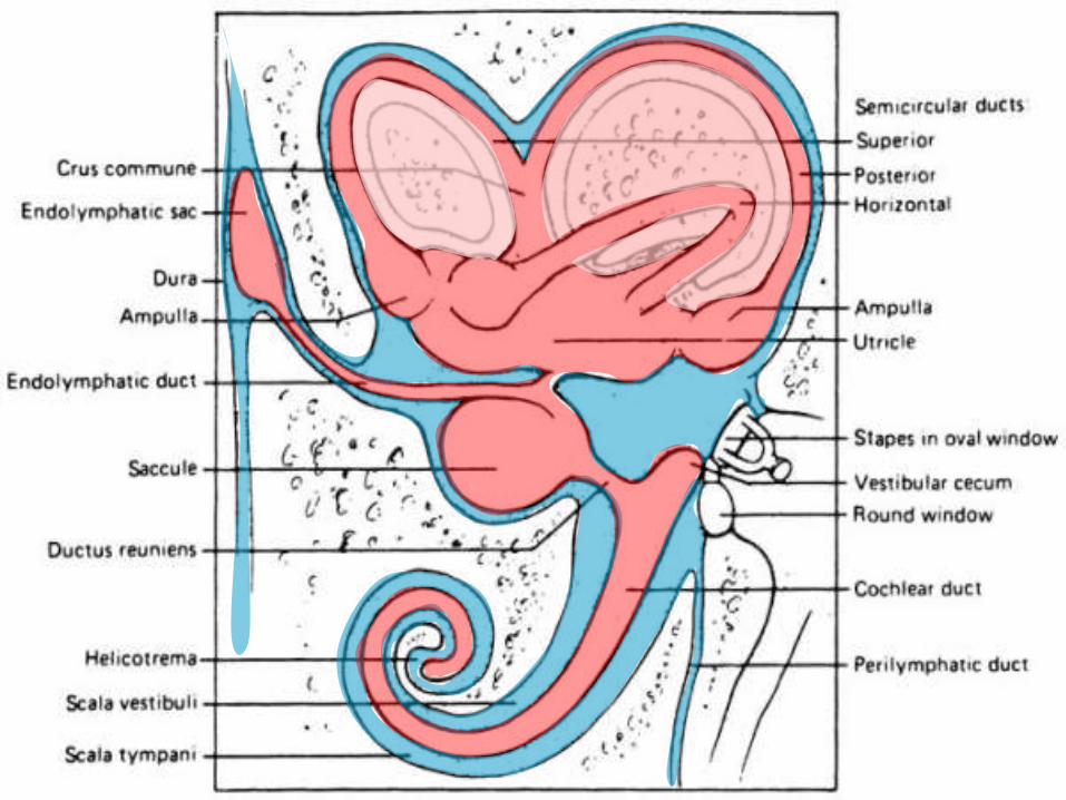
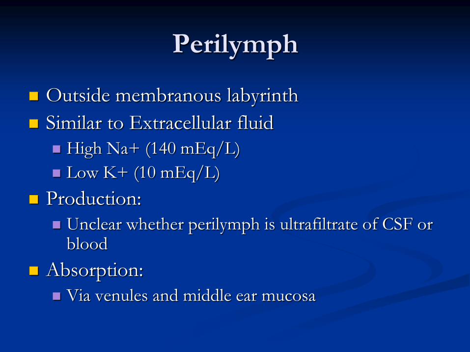
Perilymph
Outside membranous labyrinth
Similar to Extracellular fluid
High Na+ (140 mEq/L)
Low K+ (10 mEq/L)
Production:
Unclear whether perilymph is ultrafiltrate of CSF or blood
Absorption:
Via venules and middle ear mucosa
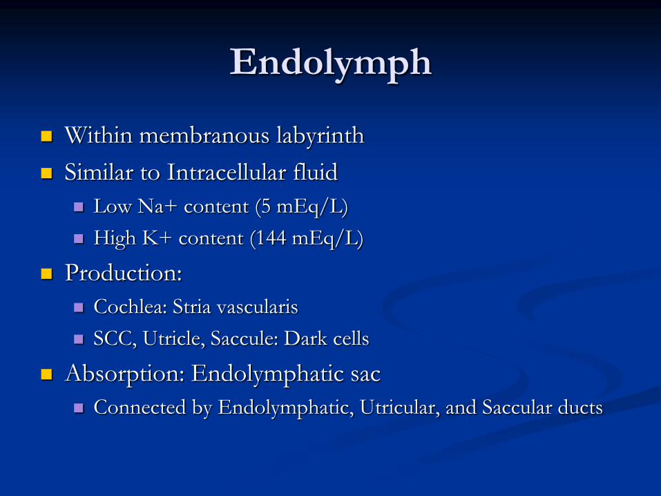
Endolymph
Within membranous labyrinth
Similar to Intracellular fluid
Low Na+ content (5 mEq/L)
High K+ content (144 mEq/L)
Production:
Cochlea: Stria vascularis
SCC, Utricle, Saccule: Dark cells
Absorption: Endolymphatic sac
Connected by Endolymphatic, Utricular, and Saccular ducts
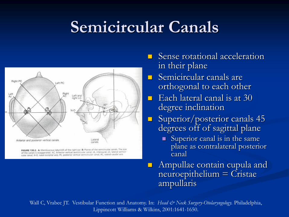
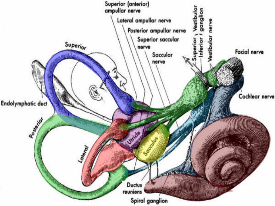
Semicircular Canals
Sense rotational acceleration in their plane
Semicircular canals are orthogonal to each other
Each lateral canal is at 30 degree inclination
Superior/posterior canals 45 degrees off of sagittal plane Superior canal is in the same
plane as contralateral posterior canal
Ampullae contain cupula and neuroepithelium = Cristae ampullaris
Wall C, Vrabec JT. Vestibular Function and Anatomy. In: Head & Neck Surgery-Otolaryngology. Philadelphia,
Lippincott Williams & Wilkins, 2001:1641-1650.
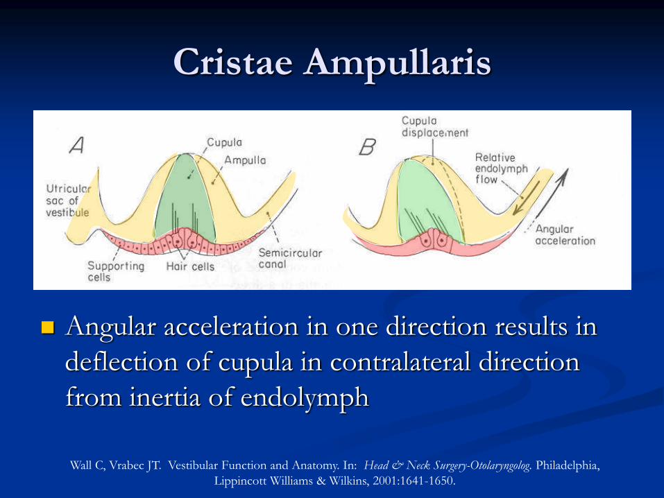
Cristae Ampullaris
Angular acceleration in one direction results in
deflection of cupula in contralateral direction
from inertia of endolymph
Wall C, Vrabec JT. Vestibular Function and Anatomy. In: Head & Neck Surgery-Otolaryngolog. Philadelphia,
Lippincott Williams & Wilkins, 2001:1641-1650.

Lysakowski, A. Anatomy of Vestibular End Organs and Neural Pathways. In Cummings: Otololaryngology: Head and
Surgery, Mosby, 2005.
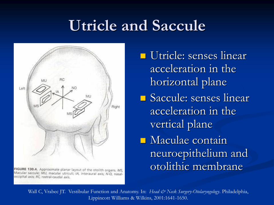
Utricle and Saccule
Utricle: senses linear acceleration in the horizontal plane
Saccule: senses linear acceleration in the vertical plane
Maculae contain neuroepithelium and otolithic membrane
Wall C, Vrabec JT. Vestibular Function and Anatomy. In: Head & Neck Surgery-Otolaryngology. Philadelphia,
Lippincott Williams & Wilkins, 2001:1641-1650.
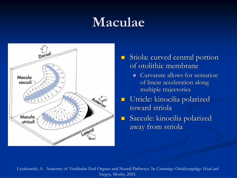
Maculae
Stiola: curved central portion of otolithic membrane Curvature allows for sensation
of linear acceleration along multiple trajectories
Utricle: kinocilia polarized toward striola
Saccule: kinocilia polarized away from striola
Lysakowski, A. Anatomy of Vestibular End Organs and Neural Pathways. In Cummings: Otololaryngology: Head and
Surgery, Mosby, 2005.
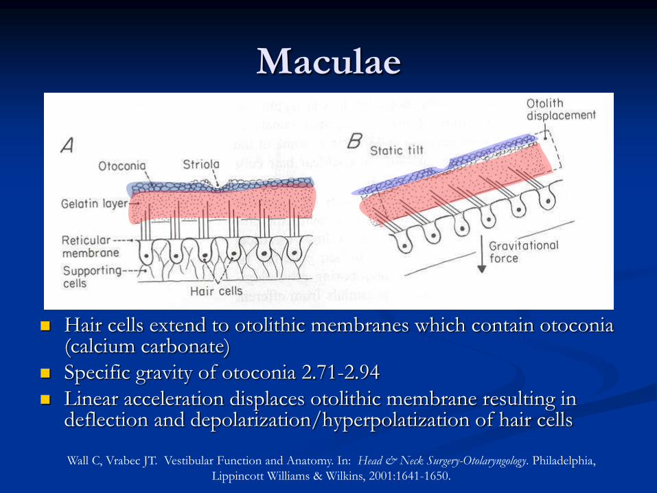
Maculae
Hair cells extend to otolithic membranes which contain otoconia (calcium carbonate)
Specific gravity of otoconia 2.71-2.94
Linear acceleration displaces otolithic membrane resulting in deflection and depolarization/hyperpolatization of hair cells
Wall C, Vrabec JT. Vestibular Function and Anatomy. In: Head & Neck Surgery-Otolaryngology. Philadelphia,
Lippincott Williams & Wilkins, 2001:1641-1650.
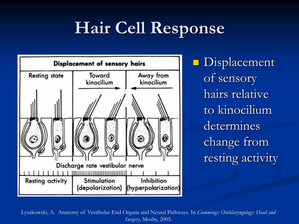
Hair Cell Response
Displacement
of sensory
hairs relative
to kinocilium
determines
change from
resting activity
Lysakowski, A. Anatomy of Vestibular End Organs and Neural Pathways. In Cummings: Otololaryngology: Head and
Surgery, Mosby, 2005.
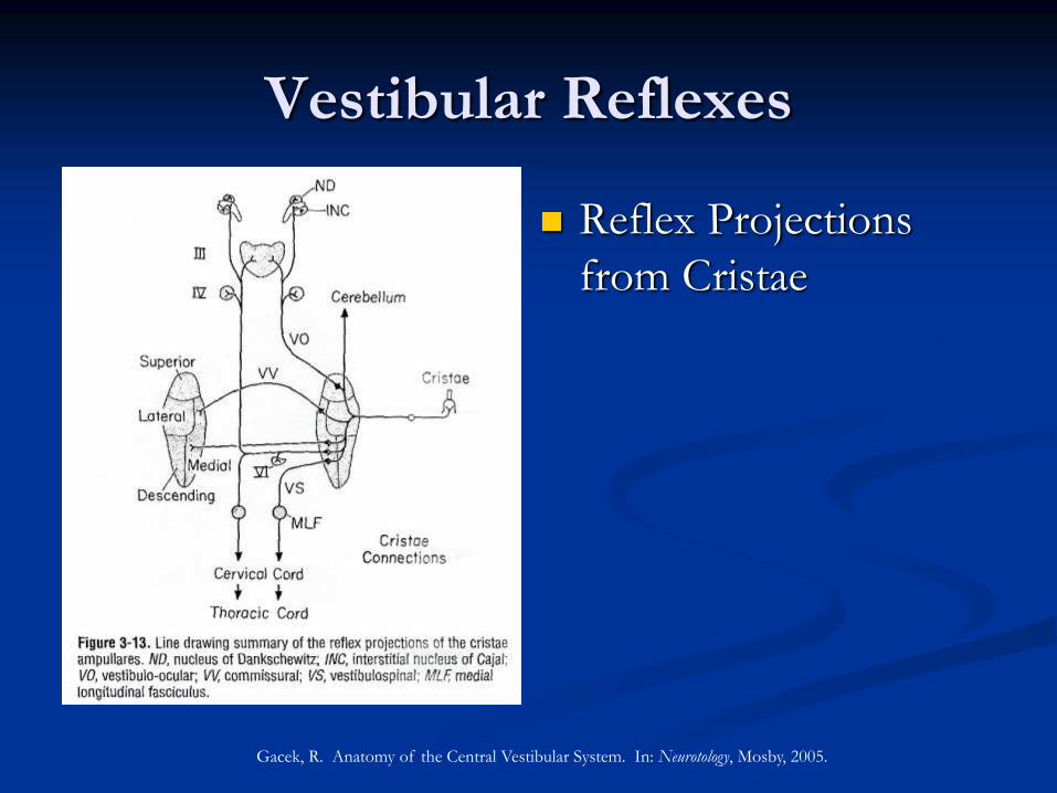
Vestibular Reflexes
Reflex Projections
from Cristae
Gacek, R. Anatomy of the Central Vestibular System. In: Neurotology, Mosby, 2005.
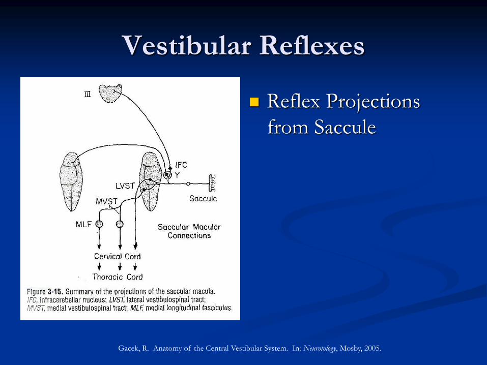
Vestibular Reflexes
Reflex Projections
from Saccule
Gacek, R. Anatomy of the Central Vestibular System. In: Neurotology, Mosby, 2005.
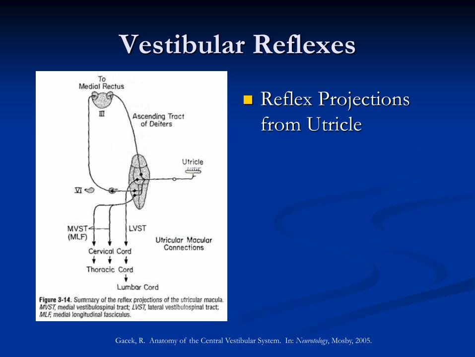
Vestibular Reflexes
Reflex Projections
from Utricle
Gacek, R. Anatomy of the Central Vestibular System. In: Neurotology, Mosby, 2005.
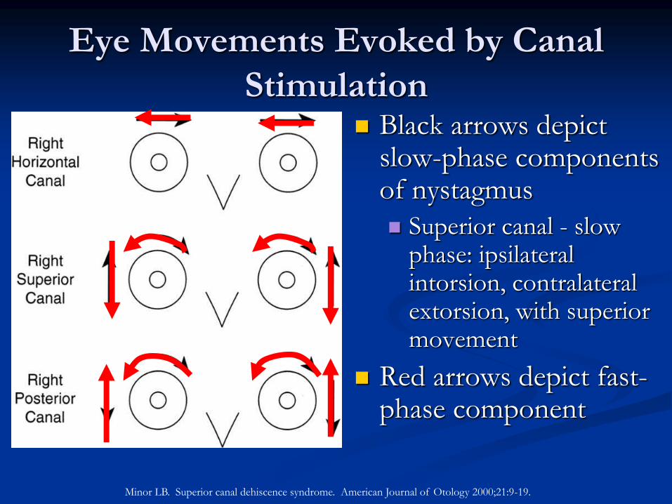
Eye Movements Evoked by Canal
Stimulation Black arrows depict
slow-phase components of nystagmus
Superior canal - slow phase: ipsilateral intorsion, contralateral extorsion, with superior movement
Red arrows depict fast-phase component
Minor LB. Superior canal dehiscence syndrome. American Journal of Otology 2000;21:9-19.
Superior Canal Dehiscence
Absence of bone overlying the superior
semicircular canal resulting in a third window to
the membranous labyrinth that may result in a
syndrome of vestibular and/or auditory
symptoms
Syndrome first described by Minor in 1998
Sound- and/or Pressure-Induced Vertigo Due
to Bone Dehiscence of the Superior Semicircular
Canal (Arch Otolaryngol Head Neck Surg)
8 patients
Vertigo
Sound-induced disequilibrium and/or oscillopsia
Pressure-induced disequilibrium and/or oscillopsia
Middle ear (pneumotoscopy, Valsalva with nose pinched)
Intracranial (Valsalva against closed glottis)
Superior Canal Dehiscence
Minor 1998
7 of 8 patients with vertical-torsional eye movements induced by sound and/or pressure indicating stimulation of superior canal
Video-oculography or Magnetic Field Search-Coil Recordings
Axial and Coronal CT with 1 mm slice thickness
2 of 8 patients with debilitating symptoms underwent plugging of superior semicircular canal via middle cranial fossa approach
Both had improvement of symptoms postoperatively
1 developed recurrent symptoms and underwent additional plugging with subsequent SNHL
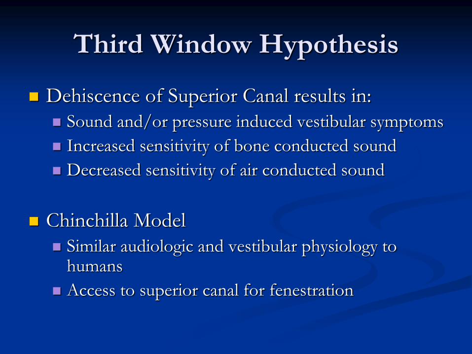
Third Window Hypothesis
Dehiscence of Superior Canal results in:
Sound and/or pressure induced vestibular symptoms
Increased sensitivity of bone conducted sound
Decreased sensitivity of air conducted sound
Chinchilla Model
Similar audiologic and vestibular physiology to humans
Access to superior canal for fenestration
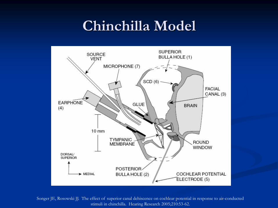
Songer JE, Rosowski JJ. The effect of superior canal dehiscence on cochlear potential in response to air-conducted
stimuli in chinchilla. Hearing Research 2005;210:53-62.
Chinchilla Model
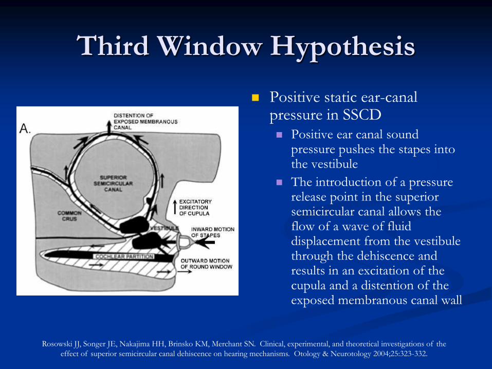
Third Window Hypothesis
Positive static ear-canal pressure in SSCD Positive ear canal sound
pressure pushes the stapes into the vestibule
The introduction of a pressure release point in the superior semicircular canal allows the flow of a wave of fluid displacement from the vestibule through the dehiscence and results in an excitation of the cupula and a distention of the exposed membranous canal wall
Rosowski JJ, Songer JE, Nakajima HH, Brinsko KM, Merchant SN. Clinical, experimental, and theoretical investigations of the
effect of superior semicircular canal dehiscence on hearing mechanisms. Otology & Neurotology 2004;25:323-332.
Chinchilla Model – Pressure Induced
Superior Canal Excitation
Vestibular nerve afferent measured in
chinchilla before and after fenestration of
superior canal with stimulation by EAC
pressure changes
100% specimens had excitation of superior
canal afferents with EAC pressure changes
after fenestration
Reversed with repair of fenestration
Hirvonen TP, Carey JP, Liang CJ, Minor LB. Superior canal dehiscence. Mechanisms of pressure sensitivity in a
chinchilla model. Arch Otolaryngol Head Neck Surg 2001;127:1331-1336.
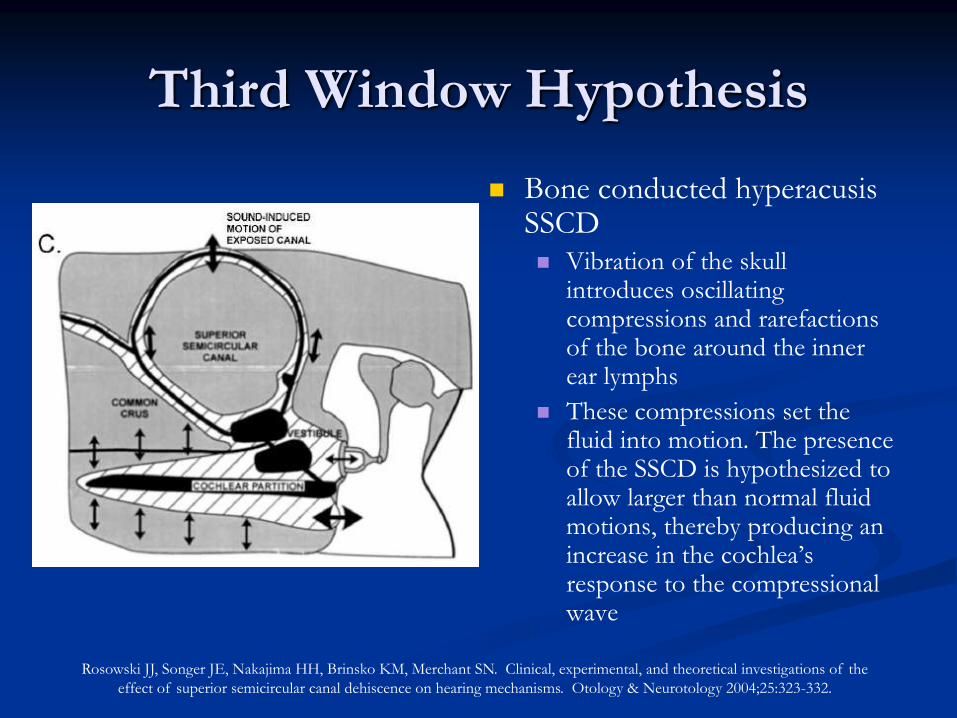
Bone conducted hyperacusis SSCD Vibration of the skull
introduces oscillating compressions and rarefactions of the bone around the inner ear lymphs
These compressions set the fluid into motion. The presence of the SSCD is hypothesized to allow larger than normal fluid motions, thereby producing an increase in the cochlea’s response to the compressional wave
Rosowski JJ, Songer JE, Nakajima HH, Brinsko KM, Merchant SN. Clinical, experimental, and theoretical investigations of the
effect of superior semicircular canal dehiscence on hearing mechanisms. Otology & Neurotology 2004;25:323-332.
Third Window Hypothesis
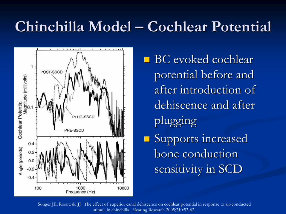
BC evoked cochlear
potential before and
after introduction of
dehiscence and after
plugging
Supports increased
bone conduction
sensitivity in SCD
Chinchilla Model – Cochlear Potential
Songer JE, Rosowski JJ. The effect of superior canal dehiscence on cochlear potential in response to air-conducted
stimuli in chinchilla. Hearing Research 2005;210:53-62.
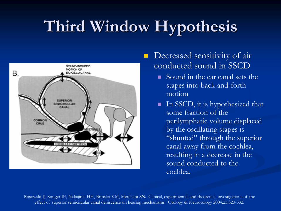
Decreased sensitivity of air conducted sound in SSCD Sound in the ear canal sets the
stapes into back-and-forth motion
In SSCD, it is hypothesized that some fraction of the perilymphatic volume displaced by the oscillating stapes is “shunted” through the superior canal away from the cochlea, resulting in a decrease in the sound conducted to the cochlea.
Rosowski JJ, Songer JE, Nakajima HH, Brinsko KM, Merchant SN. Clinical, experimental, and theoretical investigations of the
effect of superior semicircular canal dehiscence on hearing mechanisms. Otology & Neurotology 2004;25:323-332.
Third Window Hypothesis
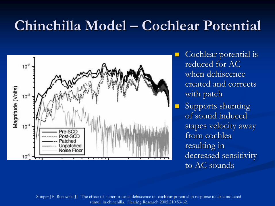
Cochlear potential is reduced for AC when dehiscence created and corrects with patch
Supports shunting of sound induced stapes velocity away from cochlea resulting in decreased sensitivity to AC sounds
Songer JE, Rosowski JJ. The effect of superior canal dehiscence on cochlear potential in response to air-conducted
stimuli in chinchilla. Hearing Research 2005;210:53-62.
Chinchilla Model – Cochlear Potential
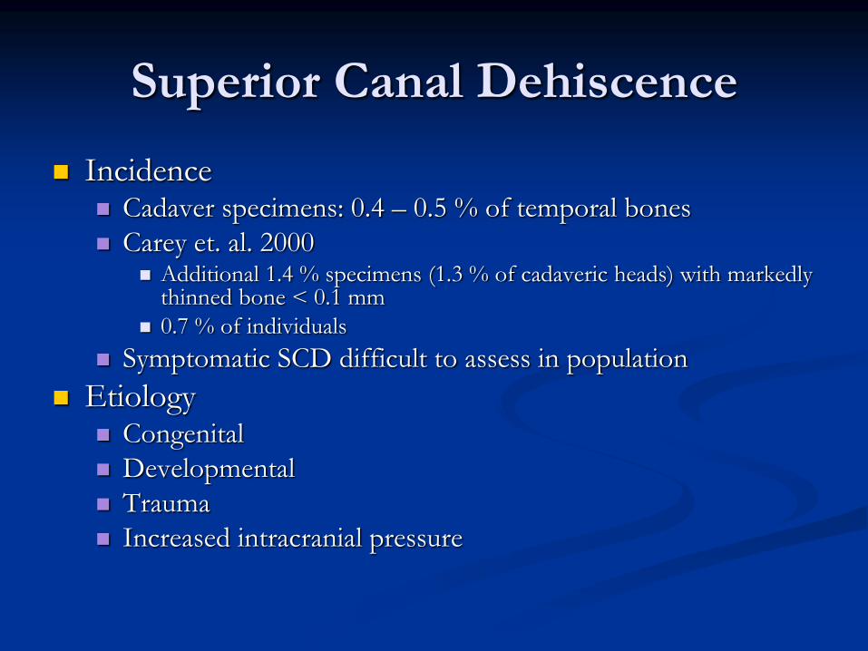
Incidence Cadaver specimens: 0.4 – 0.5 % of temporal bones
Carey et. al. 2000 Additional 1.4 % specimens (1.3 % of cadaveric heads) with markedly
thinned bone < 0.1 mm
0.7 % of individuals
Symptomatic SCD difficult to assess in population
Etiology Congenital
Developmental
Trauma
Increased intracranial pressure
Superior Canal Dehiscence
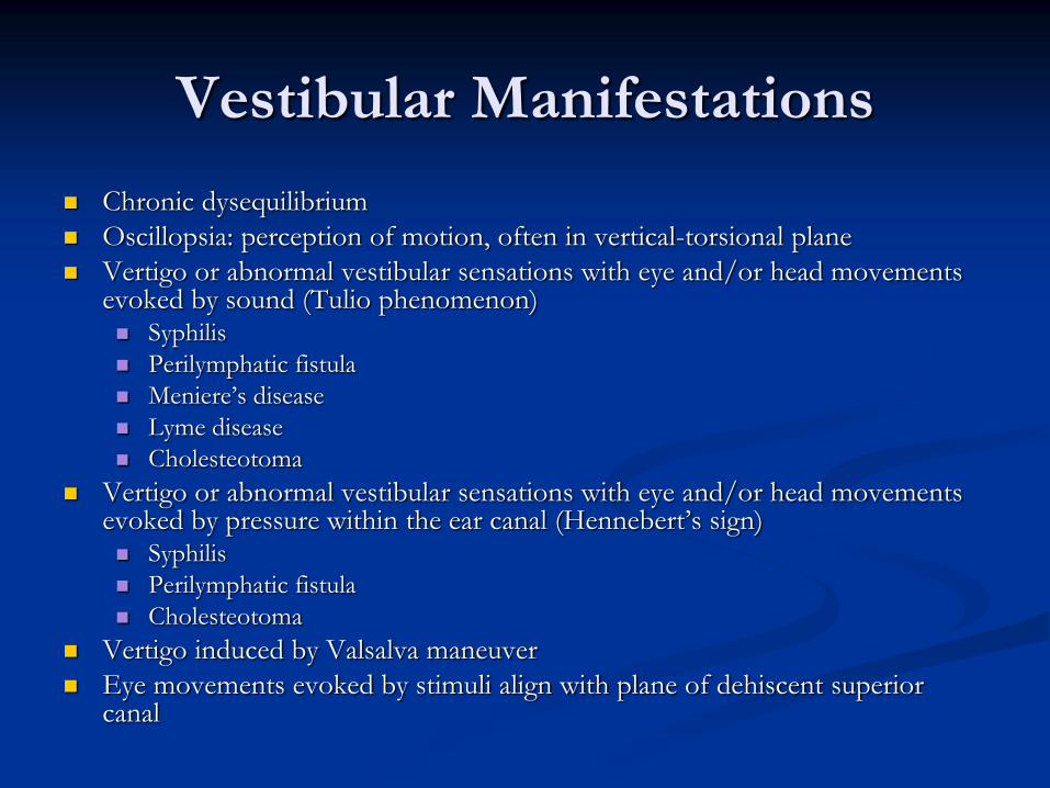
Vestibular Manifestations
Chronic dysequilibrium
Oscillopsia: perception of motion, often in vertical-torsional plane
Vertigo or abnormal vestibular sensations with eye and/or head movements evoked by sound (Tulio phenomenon) Syphilis
Perilymphatic fistula
Meniere’s disease
Lyme disease
Cholesteotoma
Vertigo or abnormal vestibular sensations with eye and/or head movements evoked by pressure within the ear canal (Hennebert’s sign) Syphilis
Perilymphatic fistula
Cholesteotoma
Vertigo induced by Valsalva maneuver
Eye movements evoked by stimuli align with plane of dehiscent superior canal
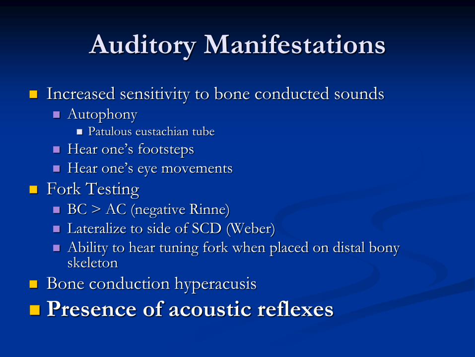
Auditory Manifestations
Increased sensitivity to bone conducted sounds Autophony
Patulous eustachian tube
Hear one’s footsteps
Hear one’s eye movements
Fork Testing BC > AC (negative Rinne)
Lateralize to side of SCD (Weber)
Ability to hear tuning fork when placed on distal bony skeleton
Bone conduction hyperacusis
Presence of acoustic reflexes
Diagnostic Studies
Audiometry with acoustic reflex
Oculography with sound and pressure
VEMP
Laser Doppler Vibrometry
CT
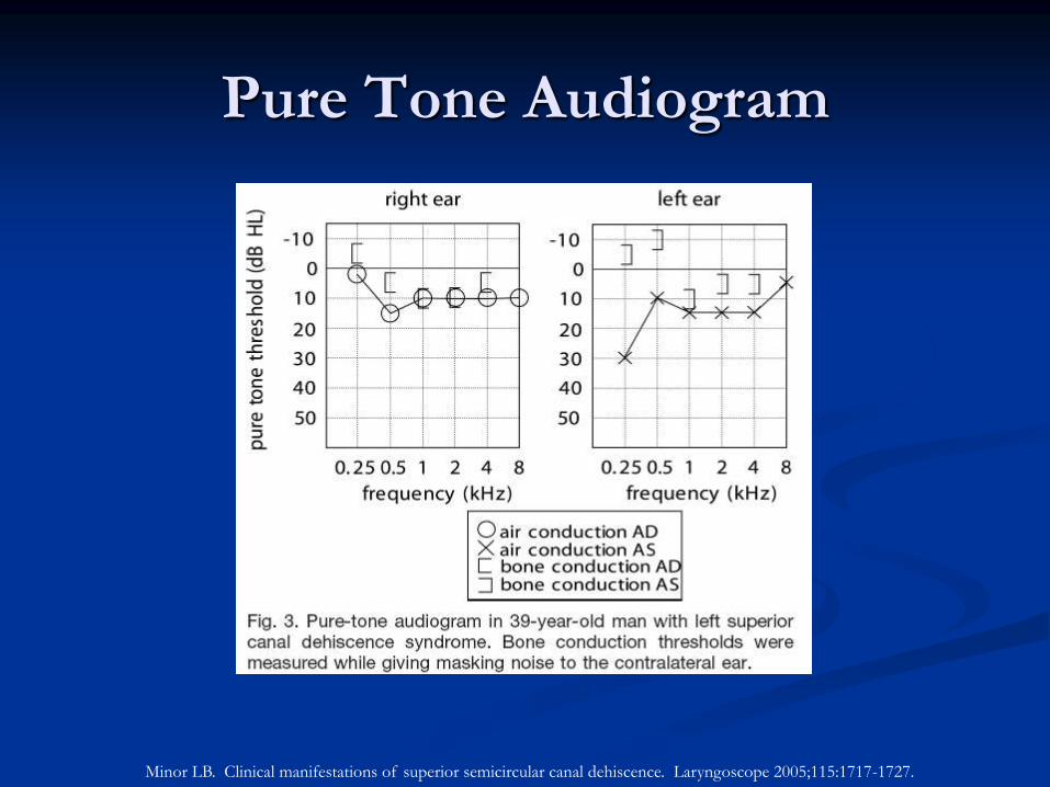
Pure Tone Audiogram
Minor LB. Clinical manifestations of superior semicircular canal dehiscence. Laryngoscope 2005;115:1717-1727.
VEMP
Vestibular Evoked Myogenic Potential
Brief (0.1 ms) loud (SPL > 90 dB) monaural clicks or
tone bursts or skull taps (1/s) presented in ear
evoking large (60 – 300 µV) short latency (8 ms)
inhibitory potential in the tonically contracted
ipsilateral SCM
Increased threshold or absent response
Middle ear pathology or ossicular chain abnormality
Decreased threshold
Perilymphatic fistula
Superior canal dehiscence
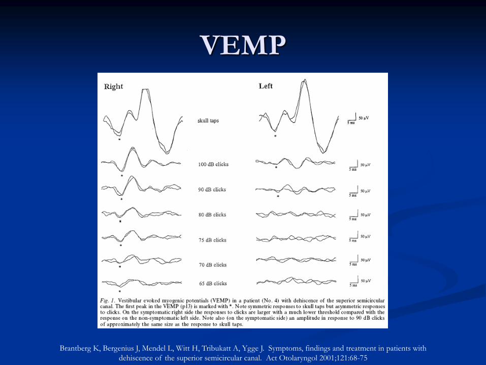
VEMP
Brantberg K, Bergenius J, Mendel L, Witt H, Tribukatt A, Ygge J. Symptoms, findings and treatment in patients with
dehiscence of the superior semicircular canal. Act Otolaryngol 2001;121:68-75
Used to measure sound-induced velocity of tympanic membrane
High velocity magnitude
Ossicular discontinuity
Superior canal dehiscence
Low velocity magnitude
Otosclerosis
Fixation of ossicular chain
Middle ear effusion
Laser Doppler Vibrometry
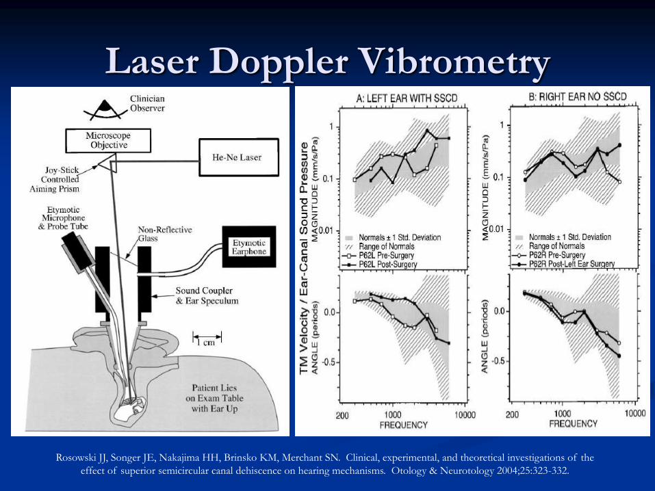
Laser Doppler Vibrometry
Rosowski JJ, Songer JE, Nakajima HH, Brinsko KM, Merchant SN. Clinical, experimental, and theoretical investigations of the
effect of superior semicircular canal dehiscence on hearing mechanisms. Otology & Neurotology 2004;25:323-332.
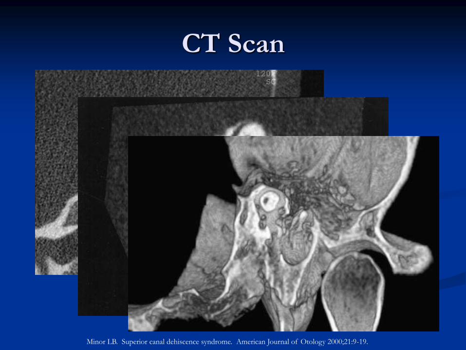
CT Scan
Minor LB. Superior canal dehiscence syndrome. American Journal of Otology 2000;21:9-19.
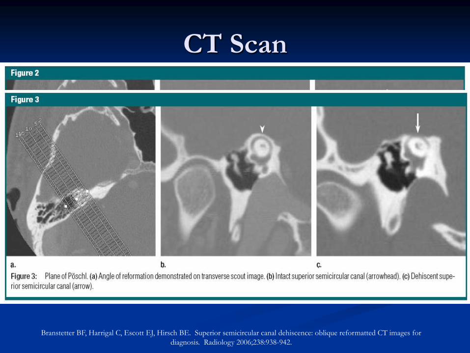
CT Scan
Branstetter BF, Harrigal C, Escott EJ, Hirsch BE. Superior semicircular canal dehiscence: oblique reformatted CT images for
diagnosis. Radiology 2006;238:938-942.
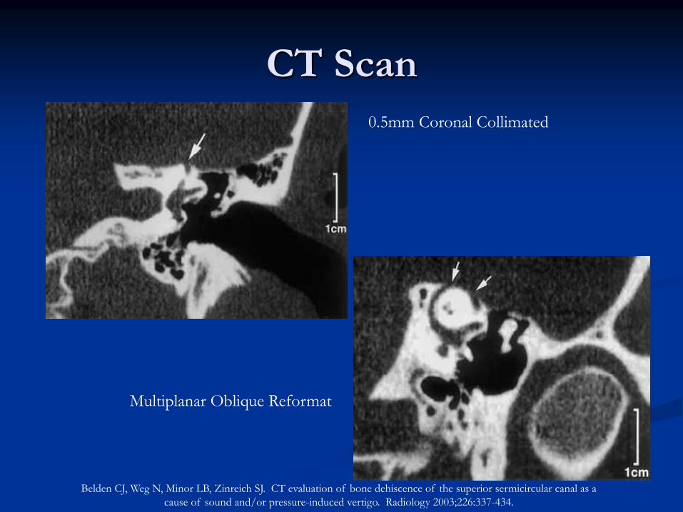
CT Scan
0.5mm Coronal Collimated
Multiplanar Oblique Reformat
Belden CJ, Weg N, Minor LB, Zinreich SJ. CT evaluation of bone dehiscence of the superior sermicircular canal as a
cause of sound and/or pressure-induced vertigo. Radiology 2003;226:337-434.
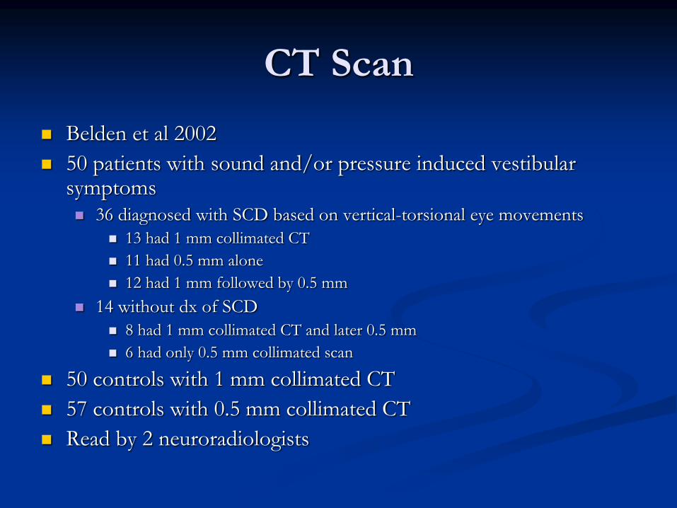
Belden et al 2002
50 patients with sound and/or pressure induced vestibular symptoms 36 diagnosed with SCD based on vertical-torsional eye movements
13 had 1 mm collimated CT
11 had 0.5 mm alone
12 had 1 mm followed by 0.5 mm
14 without dx of SCD
8 had 1 mm collimated CT and later 0.5 mm
6 had only 0.5 mm collimated scan
50 controls with 1 mm collimated CT
57 controls with 0.5 mm collimated CT
Read by 2 neuroradiologists
CT Scan
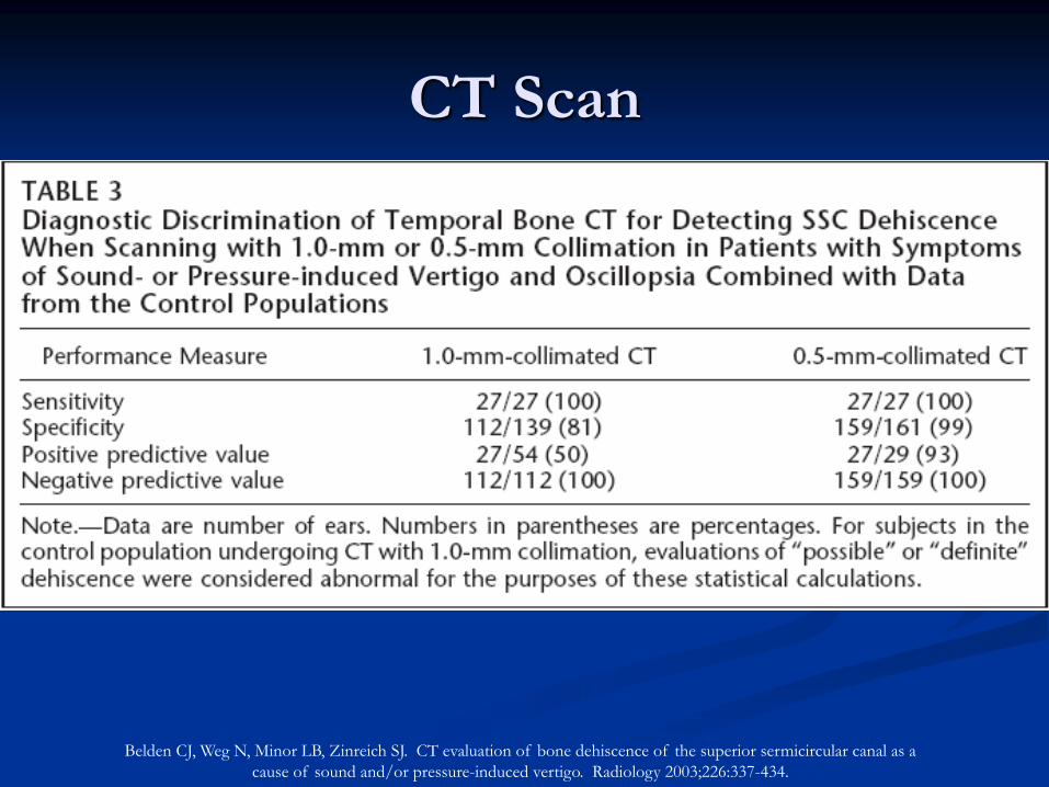
CT Scan
Belden CJ, Weg N, Minor LB, Zinreich SJ. CT evaluation of bone dehiscence of the superior sermicircular canal as a
cause of sound and/or pressure-induced vertigo. Radiology 2003;226:337-434.
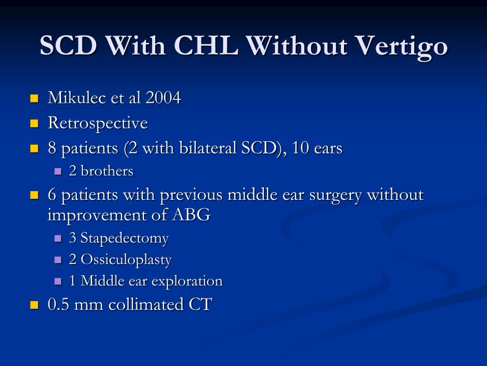
SCD With CHL Without Vertigo
Mikulec et al 2004
Retrospective
8 patients (2 with bilateral SCD), 10 ears
2 brothers
6 patients with previous middle ear surgery without improvement of ABG
3 Stapedectomy
2 Ossiculoplasty
1 Middle ear exploration
0.5 mm collimated CT
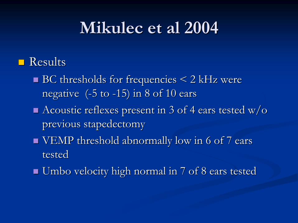
Mikulec et al 2004
Results
BC thresholds for frequencies < 2 kHz were
negative (-5 to -15) in 8 of 10 ears
Acoustic reflexes present in 3 of 4 ears tested w/o
previous stapedectomy
VEMP threshold abnormally low in 6 of 7 ears
tested
Umbo velocity high normal in 7 of 8 ears tested
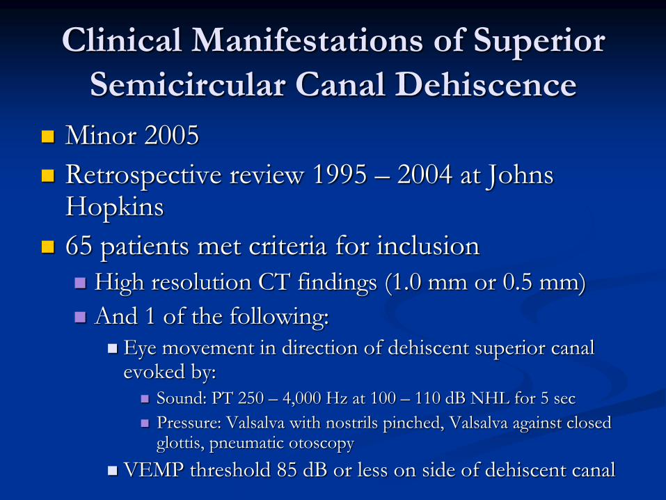
Clinical Manifestations of Superior
Semicircular Canal Dehiscence
Minor 2005
Retrospective review 1995 – 2004 at Johns Hopkins
65 patients met criteria for inclusion
High resolution CT findings (1.0 mm or 0.5 mm)
And 1 of the following:
Eye movement in direction of dehiscent superior canal evoked by:
Sound: PT 250 – 4,000 Hz at 100 – 110 dB NHL for 5 sec
Pressure: Valsalva with nostrils pinched, Valsalva against closed glottis, pneumatic otoscopy
VEMP threshold 85 dB or less on side of dehiscent canal
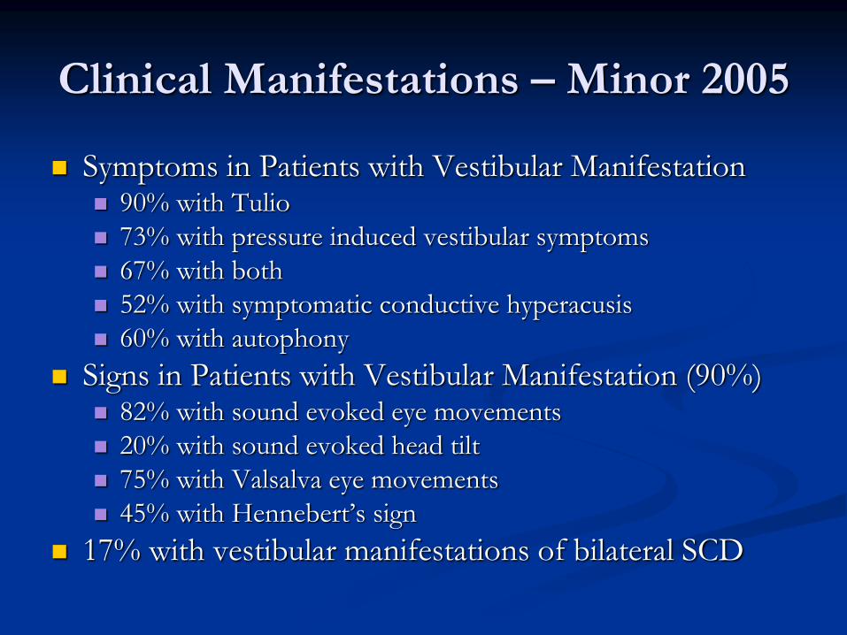
Symptoms in Patients with Vestibular Manifestation 90% with Tulio
73% with pressure induced vestibular symptoms
67% with both
52% with symptomatic conductive hyperacusis
60% with autophony
Signs in Patients with Vestibular Manifestation (90%) 82% with sound evoked eye movements
20% with sound evoked head tilt
75% with Valsalva eye movements
45% with Hennebert’s sign
17% with vestibular manifestations of bilateral SCD
Clinical Manifestations – Minor 2005
Clinical Manifestations – Minor 2005
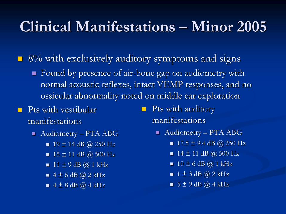
8% with exclusively auditory symptoms and signs
Found by presence of air-bone gap on audiometry with
normal acoustic reflexes, intact VEMP responses, and no
ossicular abnormality noted on middle ear exploration
Pts with vestibular
manifestations
Audiometry – PTA ABG
19 ± 14 dB @ 250 Hz
15 ± 11 dB @ 500 Hz
11 ± 9 dB @ 1 kHz
4 ± 6 dB @ 2 kHz
4 ± 8 dB @ 4 kHz
Pts with auditory
manifestations
Audiometry – PTA ABG
17.5 ± 9.4 dB @ 250 Hz
14 ± 11 dB @ 500 Hz
10 ± 6 dB @ 1 kHz
1 ± 3 dB @ 2 kHz
5 ± 9 dB @ 4 kHz
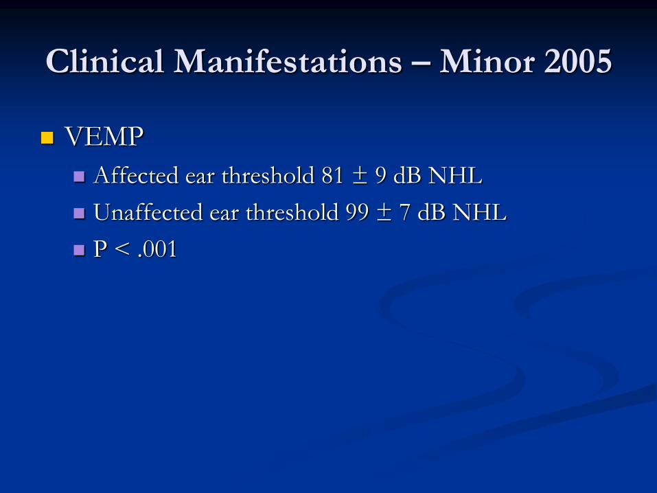
VEMP
Affected ear threshold 81 ± 9 dB NHL
Unaffected ear threshold 99 ± 7 dB NHL
P < .001
Clinical Manifestations – Minor 2005
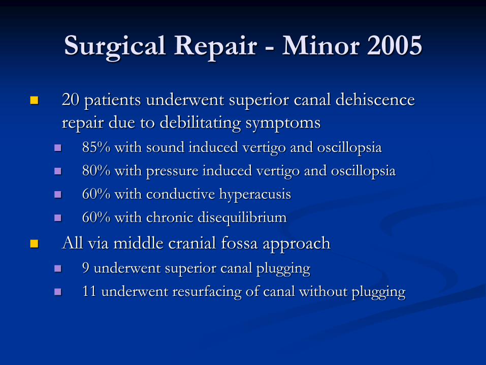
Surgical Repair - Minor 2005
20 patients underwent superior canal dehiscence
repair due to debilitating symptoms
85% with sound induced vertigo and oscillopsia
80% with pressure induced vertigo and oscillopsia
60% with conductive hyperacusis
60% with chronic disequilibrium
All via middle cranial fossa approach
9 underwent superior canal plugging
11 underwent resurfacing of canal without plugging
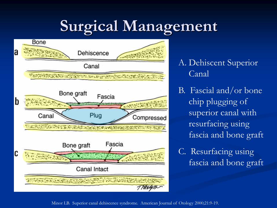
Surgical Management
Minor LB. Superior canal dehiscence syndrome. American Journal of Otology 2000;21:9-19.
A. Dehiscent Superior
Canal
B. Fascial and/or bone
chip plugging of
superior canal with
resurfacing using
fascia and bone graft
C. Resurfacing using
fascia and bone graft
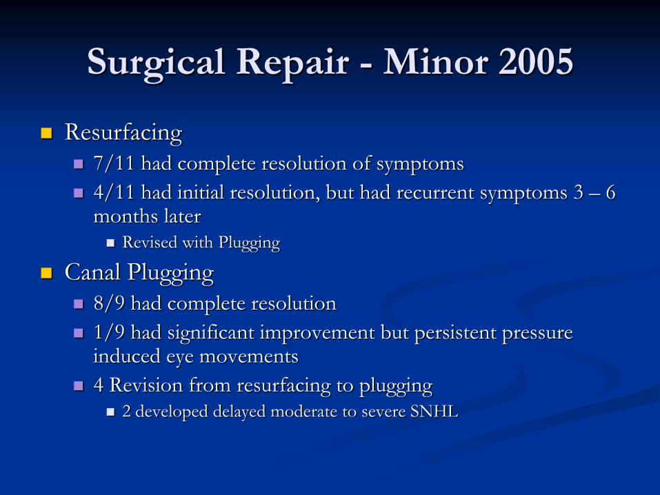
Resurfacing
7/11 had complete resolution of symptoms
4/11 had initial resolution, but had recurrent symptoms 3 – 6 months later Revised with Plugging
Canal Plugging
8/9 had complete resolution
1/9 had significant improvement but persistent pressure induced eye movements
4 Revision from resurfacing to plugging 2 developed delayed moderate to severe SNHL
Surgical Repair - Minor 2005
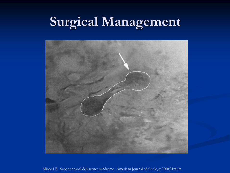
Surgical Management
Minor LB. Superior canal dehiscence syndrome. American Journal of Otology 2000;21:9-19.
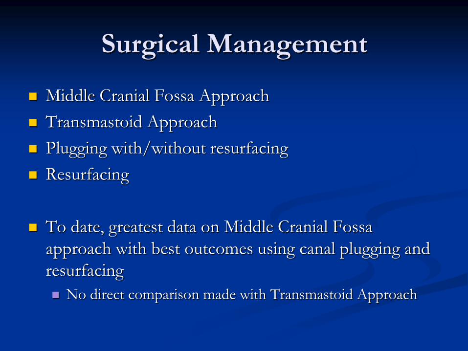
Middle Cranial Fossa Approach
Transmastoid Approach
Plugging with/without resurfacing
Resurfacing
To date, greatest data on Middle Cranial Fossa
approach with best outcomes using canal plugging and
resurfacing
No direct comparison made with Transmastoid Approach
Surgical Management
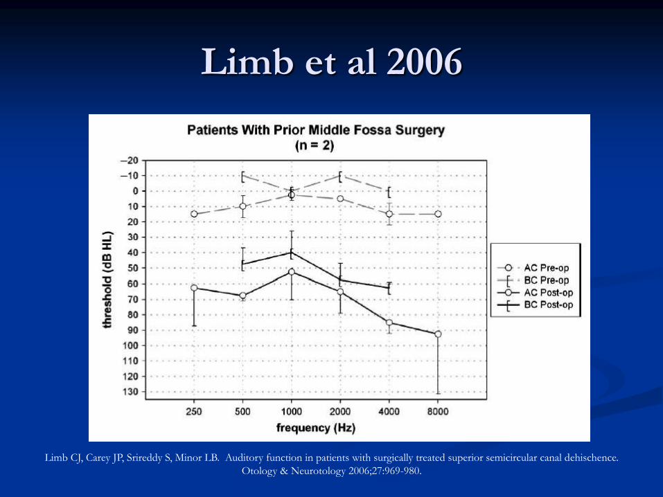
Auditory Function After Surgery
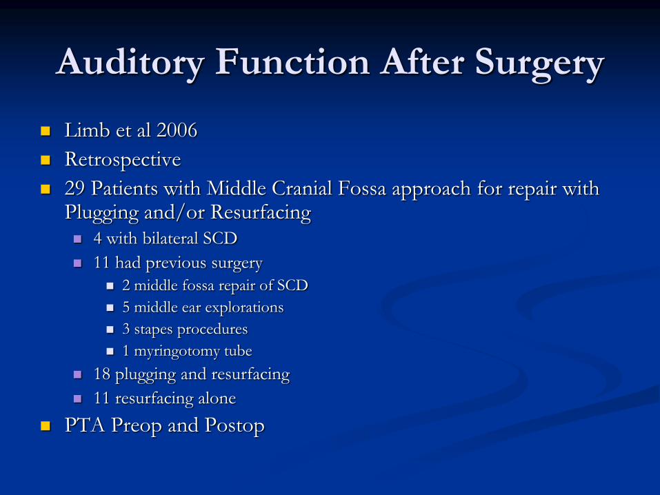
Limb et al 2006
Retrospective
29 Patients with Middle Cranial Fossa approach for repair with Plugging and/or Resurfacing 4 with bilateral SCD
11 had previous surgery
2 middle fossa repair of SCD
5 middle ear explorations
3 stapes procedures
1 myringotomy tube
18 plugging and resurfacing
11 resurfacing alone
PTA Preop and Postop

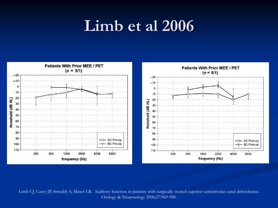
Limb et al 2006
Limb CJ, Carey JP, Srireddy S, Minor LB. Auditory function in patients with surgically treated superior semicircular canal dehischence.
Otology & Neurotology 2006;27:969-980.
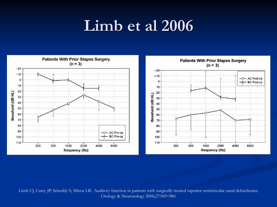
Limb et al 2006
Limb CJ, Carey JP, Srireddy S, Minor LB. Auditory function in patients with surgically treated superior semicircular canal dehischence.
Otology & Neurotology 2006;27:969-980.
Limb et al 2006
Limb CJ, Carey JP, Srireddy S, Minor LB. Auditory function in patients with surgically treated superior semicircular canal dehischence.
Otology & Neurotology 2006;27:969-980.
Limb et al 2006
Limb CJ, Carey JP, Srireddy S, Minor LB. Auditory function in patients with surgically treated superior semicircular canal dehischence.
Otology & Neurotology 2006;27:969-980.
Why did I choose this topic?
Superior Canal Dehiscence is a relatively new
diagnosis
Mimics many other middle ear/inner ear
disorders
Diagnostic studies
Decision-making
WHY IS IT IMPORTANT TO
YOU?
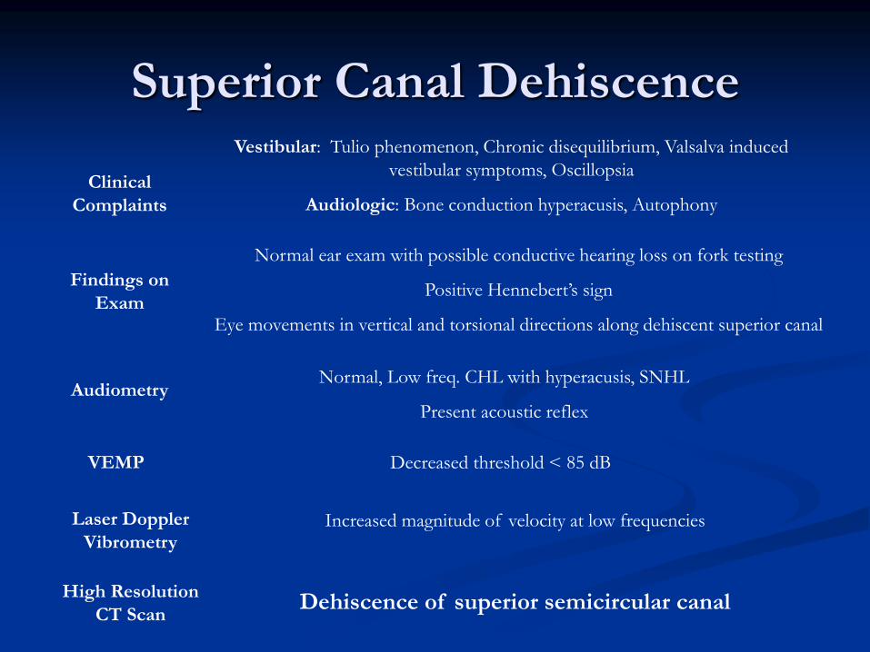
Superior Canal Dehiscence Vestibular: Tulio phenomenon, Chronic disequilibrium, Valsalva induced
vestibular symptoms, Oscillopsia
Audiologic: Bone conduction hyperacusis, Autophony
Clinical
Complaints
Findings on
Exam
Audiometry
VEMP
Normal ear exam with possible conductive hearing loss on fork testing
Positive Hennebert’s sign
Eye movements in vertical and torsional directions along dehiscent superior canal
Normal, Low freq. CHL with hyperacusis, SNHL
Present acoustic reflex
Decreased threshold < 85 dB
Laser Doppler
Vibrometry Increased magnitude of velocity at low frequencies
High Resolution
CT Scan Dehiscence of superior semicircular canal
Sources Banerjee A, Whyte A, Atlas MD. Superior canal dehiscence: a review of a new condition. Clinical Otolaryngology 2005;30:9-5. Belden CJ, Weg N, Minor LB, Zinreich SJ. CT evaluation of bone dehiscence of the superior sermicircular canal as a cause of sound
and/or pressure-induced vertigo. Radiology 2003;226:337-434. Branstetter BF, Harrigal C, Escott EJ, Hirsch BE. Superior semicircular canal dehiscence: oblique reformatted CT images for diagnosis.
Radiology 2006;238:938-942. Brantberg K, Bergenius J, Mendel L, Witt H, Tribukatt A, Ygge J. Symptoms, findings and treatment in patients with dehiscence of the
superior semicircular canal. Act Otolaryngol 2001;121:68-75. Gacek, R. Anatomy of the Central Vestibular System. In: Neurotology. Philadelphia, Mosby, 2005. Hillman TA, Kertesz TR, Hadley K, Shelton C. Reversible peripheral vestibulopathy: the treatment of superior canal dehiscence.
Otolaryngol Head Neck Surg 2006;134:431-436. Hirvonen TP, Carey JP, Liang CJ, Minor LB. Superior canal dehiscence. Mechanisms of pressure sensitivity in a chinchilla model.
Arch Otolaryngol Head Neck Surg 2001;127:1331-1336. Limb CJ, Carey JP, Srireddy S, Minor LB. Auditory function in patients with surgically treated superior semicircular canal dehischence.
Otology & Neurotology 2006;27:969-980. Lysakowski, A. Anatomy of Vestibular End Organs and Neural Pathways. In Cummings: Otololaryngology: Head and Surgery. Philadelphia,
Mosby, 2005. Mikulec AA, McKenna MJ, Ramsey MJ, Rosowski JJ, Hermann BS, Rauch SD, Curtin HD, Merchant SN. Superior semicircular canal
dehiscence presenting as conductive hearing loss without vertigo. Otology and Neurotology 2004;25:121-129. Minor LB, Solomon, D, Zinreich JS, Zee DS. Sound- and/or pressure-induced vertigo due to bone dehiscence of the superior
semicircular canal. Arch Otolaryngol Head Neck Surg 1998;124:249-258. Minor LB. Superior canal dehiscence syndrome. American Journal of Otology 2000;21:9-19. Minor LB. Clinical manifestations of superior semicircular canal dehiscence. Laryngoscope 2005;115:1717-1727. Minor LB, Carey JP, Cremer PD, Lustig LR, Streubel S. Dehiscence of bone overlying the superior canal as a cause of apparent
conductive hearing loss. Ostrowski VB, Byskosh A, Hain TC. Tulio phenomenon with dehiscence of the superior semicircular canal. Otology and Neurotology
2001;22:61-65. Rosowski JJ, Songer JE, Nakajima HH, Brinsko KM, Merchant SN. Clinical, experimental, and theoretical investigations of the effect
of superior semicircular canal dehiscence on hearing mechanisms. Otology & Neurotology 2004;25:323-332. Songer JE, Rosowski JJ. The effect of superior canal dehiscence on cochlear potential in response to air-conducted stimuli in chinchilla.
Hearing Research 2005;210:53-62. Wall C, Vrabec JT. Vestibular Function and Anatomy. In: Head & Neck Surgery-Otolaryngology. Philadelphia, Lippincott Williams &
Wilkins, 2001:1641-1650. Williamson RA, Vrabec JT, Coker NJ, Sandlin M. Coronal computed tomography prevalence of superior semicircular canal dehiscence.
Otolayngol Head Neck Surg 2003;481-489.