Embed Size (px)
Citation preview
Wayne State University
Wayne State University Dissertations
1-1-2015
The Therapeutic Targeting Of Folate ReceptorAlpha Positive Tumors Via Folate ReceptorSelective Novel 5- And 6- Substituted Pyrrolo[2,3-D]pyrimidine Antifolates"Shermaine Kimberly Mitchell-RyanWayne State University,
Follow this and additional works at: http://digitalcommons.wayne.edu/oa_dissertations
Part of the Molecular Biology Commons, Oncology Commons, and the PharmacologyCommons
This Open Access Dissertation is brought to you for free and open access by DigitalCommons@WayneState. It has been accepted for inclusion inWayne State University Dissertations by an authorized administrator of DigitalCommons@WayneState.
Recommended CitationMitchell-Ryan, Shermaine Kimberly, "The Therapeutic Targeting Of Folate Receptor Alpha Positive Tumors Via Folate ReceptorSelective Novel 5- And 6- Substituted Pyrrolo [2,3-D]pyrimidine Antifolates"" (2015). Wayne State University Dissertations. Paper1155.

THE THERAPEUTIC TARGETING OF FOLATE RECPTOR ALPHA POSITIVE
TUMORS VIA FOLATE RECEPTOR-SELECTIVE NOVEL 5-AND 6- SUBSTITUTED
PYRROLO [2,3-D] PYRIMIDINE ANTIFOLATES
by
SHERMAINE KIMBERLY MITCHELL-RYAN
DISSERTATION
Submitted to the Graduate School
of Wayne State University,
Detroit, Michigan
In partial fulfilment of the requirements
for the degree of
DOCTOR OF PHILOSOPHY
2015
Major: CANCER BIOLOGY
Approved By:
________________________________________
Advisor Date
________________________________________
________________________________________
________________________________________
________________________________________

ii
DEDICATION
This dissertation is dedicated to Seamus Isaiah Ryan, my sun (son), my world and my very
reason to press forward and claim every reward life has to offer.
My sojourn through this process would not have been possible without the love, support and
encouragement of my husband, Paul Seamus Ryan. Thank you for your steadfast devotion,
patience and understanding. You have been my guiding light, offering important insight in some
of my darkest moments. You are the quintessential professional and the lessons that you have
imparted upon me about maintaining my integrity and professionalism in every situation are
some of greatest lessons learned.
My strong sense of self, determination and integrity were instilled by the late Sherman Mitchell
(father) and Agnes Bryant (grandmother). You taught me to have faith and confidence in myself
and my abilities, which in turn helped me to gauge my self-worth. This self-esteem has served
as yard stick to help me measure up to the person I envision myself to be and to accept nothing
less while being intolerant of disrespect and unjust injury to my person.
My quest to have an impact on the world was strengthened by my life-long mentor, Dr. Frazier
O’Leary. You are the mentor I want to be. I hope to influence the lives of my students the way
you have and continue to influence mine.
Finally, just as I have stood firmly on the shoulder of giants, the trials I blaze are meant to
uncover a path for every girl of color who has dreams that drift them to careers in science. Do
not allow discouraging words to drown your curiosity. You and you alone are the ruler of your
destiny.

iii
ACKNOWLEDGEMENTS
I like to acknowledge and thank both past and present members of the Matherly lab for
your instruction, support and laughs throughout my stay.
I would like to give special thanks to Dr. Mark Stout, Dr. Eric Hales, Dr. Zhangjun Hou
and Dr. Lisa Polin (and staff) for being instrumental in the completion, training and analysis of
many of the experiments performed in this body of work. Your rigorous examination of questions
and pursuit of quality data have taught me to think as a scientist and to produce and present work
that I can proudly defend.
I would like to acknowledge Dr. Larry Matherly for the use of space and tools to explore
the questions offered in this dissertation.
Thank you to Dr. Aleem Gangjee, our collaborating medicinal chemistry for sharing some
of the most fascinating compounds. You made the chemistry of your compounds palatable and
easy to process.
The National Institute of Health Ruth L. Kirschstein National Research Service Awards
for Individual Predoctoral Fellowships to Promote Diversity in Health-Related Research (F31),
The Cancer Biology Graduate Program’s T32 training grant, and the Wayne State Graduate
Program’s Dean’s Diversity fellowship provided financial support for my graduate education.
I would also like to extend my gratitude to my surrogate mentor, Dr. Stanley Terlecky, who
has always been a huge source of encouragement and support. You have played an instrumental
role in my growth as a scientist from my start as first year student to the present.
Finally a word of thanks to of the brilliant women scientists I encountered during my time at
Wayne State (Dr. Malathy Shekhar, Dr. Karin List, Dr. Ellen Tisdale, Dr. Julie Boerner, Dr. Izabela
Podgroski) who were excellent role models and instructors.
iv
TABLE OF CONTENTS
Dedication ...................................................................................................................................... ii
Acknowledgements ....................................................................................................................... iii
List of Tables ................................................................................................................................ vi
List of Figures .............................................................................................................................. vii
Abbreviations ................................................................................................................................ ix
Chapter 1: Introduction: Folates in human health.......................................................................... 1
1.1 The identification and isolation of folate ..................................................................... 1
1.2 Folate biology and chemistry ....................................................................................... 3
1.3 Folate tissue absorption.............................................................................................. 21
1.4 Folate based targeted therapy..................................................................................... 43
1.5 Ovarian cancer ........................................................................................................... 57
Chapter 2: Discovery of 5-Substituted Pyrrolo[2,3-d]pyrimidine Antifolates as Dual
Acting Inhibitors of Glycinamide Ribonucleotide Formyltransferase and 5-
Aminoimidazole-4- Carboxamide Ribonucleotide Formyltransferase in de novo
Purine Nucleotide Biosynthesis: Implications of inhibiting 5-aminoimidazole-4-
carboxamide ribonucleotide formyltransferase to AMPK activation and anti-tumor
activity.................................................................................................................................... 69
2.1 Introduction ................................................................................................................ 69
2.2 Biological evaluation ................................................................................................. 75
2.3 Discussion .................................................................................................................. 97
2.4 Materials and methods ............................................................................................. 100
Chapter 3: Discussion ................................................................................................................ 107
3.1 Targeting the AMPK/mTOR pathway ..................................................................... 108
3.2 Targeting AICARFTase ........................................................................................... 110
References .................................................................................................................................. 114
Abstract ...................................................................................................................................... 152

v
Autobiographical Statement....................................................................................................... 154

vi
LIST OF TABLES
Table 1.1: A commonly used FRα positive cell line model with commonly used ovarian
cell line models ................................................................................................................ 64
Table 2.1: IC50s (in nM) for 5- and 6-substituted pyrrrolo[2,3-d]pyrimidine antifolates
and classical antifolates in RFC-, PCFT-, and FR-expressing cell lines .................... 81-82
vii
LIST OF FIGURES
Figure 1.1: The reduction of folic acid .......................................................................................... 5
Figure 1.2: Enzymatically active folate metabolites ...................................................................... 6
Figure 1.3: De novo purine biosynthesis...................................................................................... 16
Figure 1.4: The purine salvage pathway and cellular products ................................................... 18
Figure 1.5: The compartmentalization of folate metabolism ....................................................... 20
Figure 1.6: Predicted topology map of human reduced folate carrier (hRFC) ............................ 25
Figure 1.7: Homology model and predicted membrane topology for hPCFT ............................. 28
Figure 1.8: Folate receptors gene organization ............................................................................ 38
Figure 1.9: Ribbon and charge distribution surface model of FRα complex with folic acid
substrate ........................................................................................................................... 42
Figure 1.10: Pyrrolo[2,3-d]pyrimidine scaffold molecule ........................................................... 53
Figure 1.11: Structure of lead compounds from three distinct series of novel antifolates .......... 56
Figure 2.1: Structures of classical antifolates including MTX, PMX, raltitrexed (RTX)
and LMTX ....................................................................................................................... 73
Figure 2.2: Design of 5-substituted pyrrolo[2,3-d]pyrimidines based on structures of 6-
substituted analogs 1-4 and pemetrexed .......................................................................... 74
Figure 2.3: Inhibition of RFC-mediated transport and relative FRα-binding affinities by
6- (compound 2) and 5- (compound 8) substituted pyrrolo[2,3-d]pyrimidine
antifolates ......................................................................................................................... 79
Figure 2.4: Cell proliferation assays with protection by nucleosides including thymidine
and adenosine and 5-amino-imidazole-4-carboxamide (AICA) to identify
intracellular targets of compounds AG127 and AG136 .................................................. 86
Figure 2.5: Compounds AG23, AG71 AG127and AG136 deplete ATP pools in KB cells ........ 88
viii
Figure 2.6: Inhibition of de novo purine biosynthesis by compound AG127 results in
cytotoxicity and apoptosis................................................................................................ 91
Figure 2.7: In situ GARFT assay ................................................................................................. 94
Figure 2.8: Accumulation of ZMP by 5-substituted pyrrolo[2,3-d]pyrimidines PMX, and
compound AG127 ............................................................................................................ 96
Figure 3.1: “Big picture” schematic........................................................................................... 113
ix
Abbreviations
Ade: adenosine
AICA: 5-aminoimidizole-4-carboxamide
AICAR: 5-aminoimidazole-4-carboxamide ribonucleotide
AICARFTase: 5-aminoimidazole-4-carboxamide ribonucleotide formyltransferase
AIRS: 5’-Aminoimidazole ribonucleotide synthase
AK: adenosine kinase
AMP: adenosine monophosphate
AMPK: activated monophosphate kinase
AMT: aminopterin
APRT: adenine phosphoribosyl transferase
ASL: adenylosuccinate lyase
ATP: adenosine triphosphate
ATTC: AICARFT/IMP cyclohydrolase
CAIRS: carboxyaminoimidozole ribonucleotide synthase
CFD: cerebral folate deficiency
CHO: Chinese hamster ovary cells
DHFR: dihydrofolate reductase
DPBS: delbecco’s phosphate buffered saline
EOC: epithelial ovarian cancer
FA: folic acid added
FAICAR: formyl 5-aminoimidazole-4-carboxamide ribonucleotide
FBP1: folate binding protein 1
x
FGAM: formyl glycinamide ribonucleotide synthase
FGAR: formyl glycinamide ribonucleotide
FPGS: folylpoly gamma glutamate synthetase
FR: folate receptor
GAR: glycinamide ribonucleotide
GARFTase: glycinamide ribonucleotide formyltransferase
GMP: Guanosine monophosphate
GPI: glycosylphosphatidylinositol
GPAT: glutamine phosphoribosyl pyrophosphate amidotransferase
HBOB: hereditary breast and ovarian cancer
HGSC: high grade serous carcinoma
HNPCC: hereditary nonpolyposis colorectal cancer
IMP: inosine monophosphate
MFT: mitochondrial folate transporter
MFS: major facilitator superfamiliy
MS: methionine synthase
MTA: methylthioadenosine
MTAP: methylthioadenosine phosphorylase
MTHFC: methylene tetrahydrofolate cyclohadrase
MTHFR: methylene tetrahydrofolate reductase
mTOR: mammalian target of rapamycin
MTX: methotrexate
NA: no additions
xi
NTC: non-targeted control
NTD: neural tube defects
OEI: ovarian epithelial inclusion cyst
ORF: open reading frame
OSE: ovarian surface epithelium
PAICS: phosphoribosyl aminoimidazole carboxylase synthase
PCFT: proton-coupled folate transporter
PLP: pyridoxal-5- phosphate
PMX: pemetrexed
PRPP: phosphoribosyl pyrophosphate
PteGlu: pteroyl monoglutamate
RA: rheumatoid arthritis
RFC: reduced folate carrier
SAICARS: phosphoribosylaminoimidazolesuccinocarboxamide synthase
SAM: S-adenosyl methionine
SAR: structure activity relationship
SHMT: serine hydroxymethyltransferase
STIC: serous tubal intraepithelial carcinoma
TAM: tumor associated macrophage
Thyd: thymidine
THF: tetrahydrofolate
TS: thymidylate synthase
UTR: untranslated region

xii
ZMP: AICAR monophosphate (see AICAR)
1
CHAPTER 1: INTRODUCTION: FOLATES IN HUMAN HEALTH
1.1 The identification and isolation of folate
The functional importance of many essential vitamins and minerals, including Vitamin B9,
folicin or folic acid, was unearthed by rigorous studies that examined associated pathologies that
resulted from nutrient deficiencies. The history of folates in human health originates in
hematology. At this time, investigating the impact of diet on human disease was emerging, with
much emphasis being placed on what we now know as nutritional anemias.1,2 Macrocytic anemia,
a subclass of megaloblastic anemia, was prevalent among pregnant women in impoverished
communities whose access to proteins, vegetables and fruits was limited.1 This specific type of
anemia is characterized by enlarged erythrocytes which is thought to be a product of impaired
DNA synthesis in red blood cells; a cell type with a high rate of turnover.3,4 In observing the
effects of diet in this condition on albino rats and monkeys, Dr. Lucy Wills and colleagues
discovered that the administration of yeast, yeast extract ( Marmite™) or crude liver extract to
individuals subjected to a diet deficient in B vitamins, ameliorated the anemic condition.1,2,5 This
extrinsic factor contained in yeast was later called “Wills’ factors” by Drs. Janet Watson and
William B. Castle. Through the examination of multiple case studies of macrocytic anemia treated
with different fractions of liver extract and beef muscle, Watson and Castle concluded that there
was an additional component/ factor present that was responsible for assuagement of marocytic
anemia that was different from that which cured Addisonian anemia (pernicious anemia).5,6
Unbeknownst to the scientists of the time, yeast, plants and other microorganism are cable of de
novo folate biosynthesis and the liver is the primary site of the body’s folate storage.7,8 Based on
the empirical clinical data, there was an eruption of critical studies that examined the treatment
potential of liver extract (given orally or parentally) in anemia, which in turn gave rise to multiple
2
names for folate, based solely on the animal used in the experiment. These included M factor
(monkeys), vitamin Bc (chickens), Lactobacillus casei factor, and anti-anemia factor, to name a
few. The nomenclature used to describe folic acid or folates has undergone many iterations with
it being named pteroylglutamic acid due to the presence of a pteridine. The current name, folic
acid or folates, was not used until the 1940’s, upon isolation and identification of the compound
in green leafy vegetation.1,5,9
Folate was first isolated from spinach leaves in 1941 by Herschel Mitchell and colleagues and
was eponymously named folic acid after the Latin word folium that translates to leaf.1,10,11 In
1943, E. L. Robert Stokstad and colleagues isolated this very same “factor” from the liver that
promoted the growth of Lactobacillus casei.1,12 Stokstad synthesized the first pure crystal form of
the unconjugated compound, and from that, he was able to reveal important structural information
about the molecule. During his tenure at Berkley’s Nutritional Science department, Stokstad began
seminal research on folic acid metabolism. Stokstad’s research serves as the foundation for
antifolate drug development as he identified many of the enzymes involved in the folate metabolic
cycle nearly 34 years ago (1979).5 This early work was credited for leading to the development of
the first antifolate, aminopterin (4-aminopteroic acid) (AMT).
Today, folates are well recognized for the role they play in human health, growth and
development.13 One of many well documented examples of the impact of folates on human health
is the indisputable role that the vitamin plays in the full development and closure of the neural tube
during days 21-28 post-conception or embryogenesis.14 Adequate folate supplementation in
pregnant women has been thought to lead to the prevention of congenital malformations of the
central nervous system and consequently a decrease in infant mortality and morbidity as neural
tube defects (NTDs) are only second to cardiac defects as the most common congenital
3
malformations.15 The abundance of landmark studies that supported this notion lead to a
mandatory implementation of folic acid fortification in grain products in the United States.15 The
resulting rapid decline in infants born with spina bifida or anencephaly, as a consequence of neural
tubes defects, demonstrated the importance of folate in early development.14,15 Moreover,
deficiencies in this vitamin and mutations in genes associated with folate absorption and/or
metabolism have been linked to other disease states and disorders such as vascular disease, certain
subtypes of cancer and Alzheimer’s disease.4 This provided concrete evidence that folates were
vital to the preservation of normal cellular physiology and were a critical component to human
health.
1.2 Folate biology and chemistry
Many anabolic pathways require the addition or modification of carbon groups. Folates are
coenzymes that participate in the metabolism of amino acids and nucleotides by donating or
transferring the required carbon units, which is referred to as one-carbon metabolism.9,13 Because
folate metabolism plays an integral part in the movement of carbon, it has been described by many
as the cornerstone of one-carbon metabolism.13,16,17 One-carbon metabolism is thought to be
comprised of three distinct but interwoven metabolic cycles, the folate metabolic cycle, the
methionine cycle and the trans-sulfuration pathway.18-20 The unifying feature of these three
pathways is the transfer of active one carbon units in a number of enzymatic reactions.
Folate is a generic term that describes any compound that participates in similar cellular
activities as the vitamin that results in the production of amino acids, purine nucleotides and
thymidylate.21,22 This includes naturally occurring folates in foodstuffs, enzymatically converted
species and the synthetic derivative folic acid.21,22 The chemical name of the non-conjugated
vitamin (containing a single γ glutamate residue) is pteroylmonoglutamate (PteGlu).17 This
4
molecule is comprised of three groups, including (1) a pterin moiety, which has a methylene group
linage at the 6th position to a (2) para amino benzoyl group and (3) L-glutamatic acid.17 The
pteridine ring is of great functional importance in the metabolism of this vitamin. The two nitrogen
atoms placed on the ring favors reduction product that leads to di and/or tetra hydrogenation of the
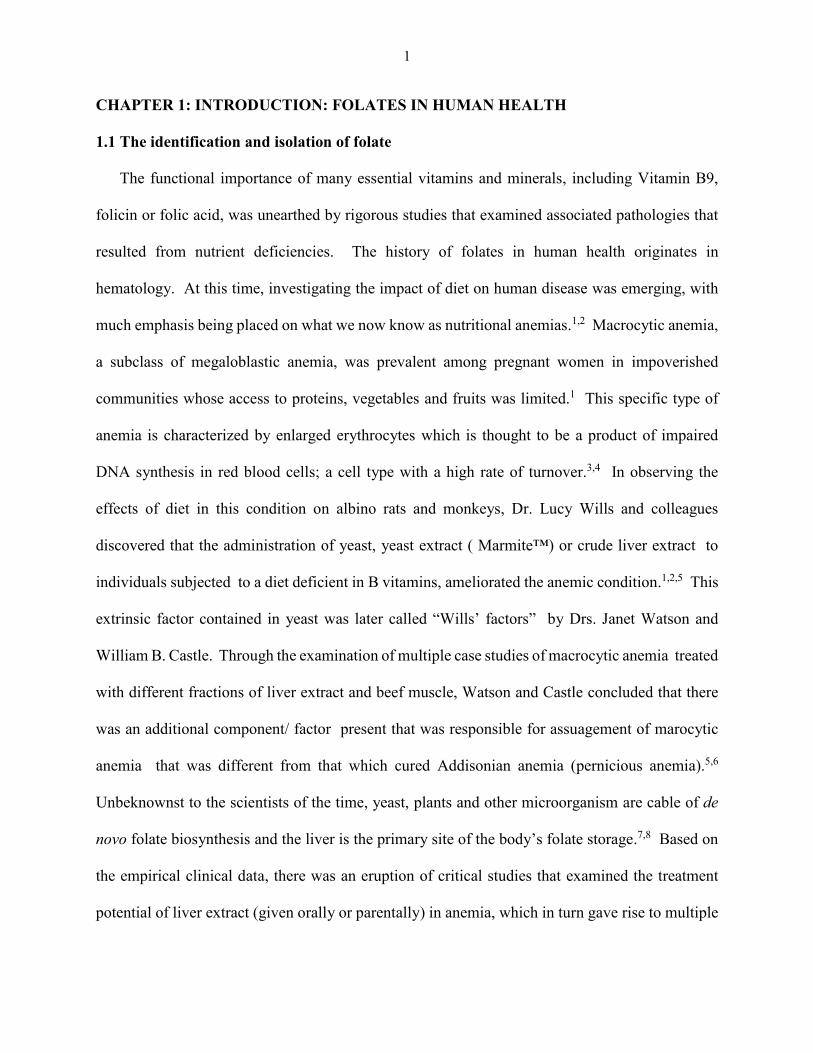
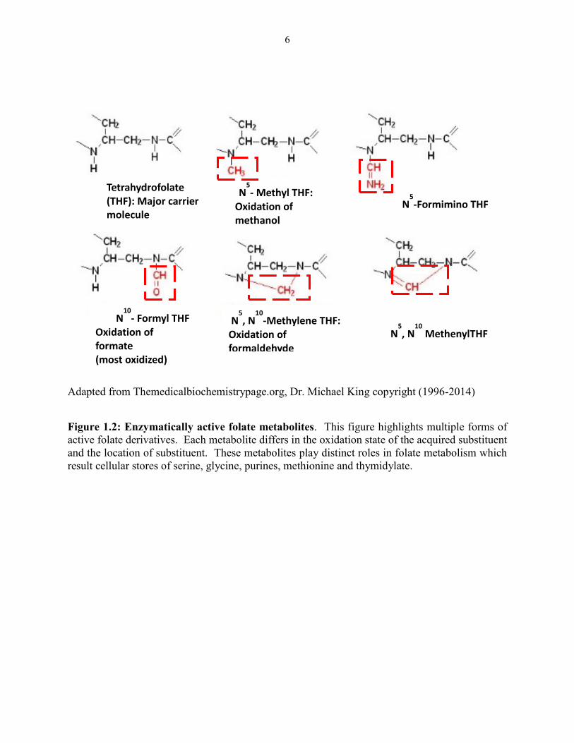
ring (Figure 1.1).
Folates destined for intracellular accumulation undergo extensive metabolism to assume their
role as cofactors in physiological reactions.8,17,19 This metabolism is thought to principally involve
the reduction of the pteridine ring (Figure 1.1) (which introduces an asymmetric center at the C6
position), polyglutamylation (the successive addition of glutamate moieties to the γ-glutamyl of
the parent molecule) and the transition to oxidative or reductive states.9,19,23,24 These reactions
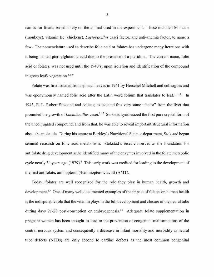
give rise to a number of active tetrahydrofolate (THF) forms that are distinguishable by the
substituents they carry at the N5 or N10 positions.8 They can include methenyl (CH+), methylene
(CH2), methyl (CH3), formyl groups (CHO) and formimino (CH=NH) (Figure 1.2).8 The active
tetrahydro forms are the only species that can readily donate or accept single carbons units.8,19
Naturally occurring folates exist as reduced folylpolyglutamates when within the cell, in which
there are multiple glutamate moieties attached to the γ-carboxyl end of the folate molecule.19,23
The most prevalent form of folate identified in mammals is reduced 5-methyl tetrahydrofolate.25,26
In contrast, the synthetic vitamin used in food fortification, folic acid, is fully oxidized and tends
to be less labile than naturally occurring folates, which become less stable with oxidation.9
Because of this, naturally occurring folates and folic acid have a different point of entry in the
metabolic cycle.
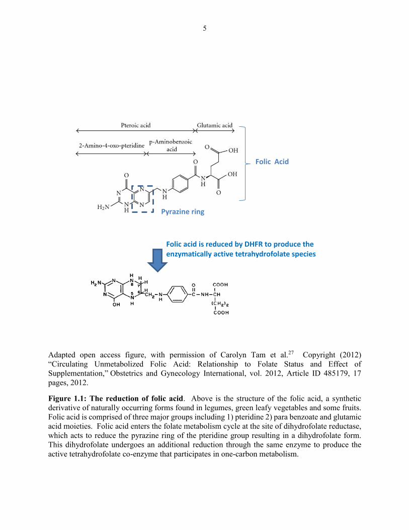
5
Adapted open access figure, with permission of Carolyn Tam et al.27 Copyright (2012) “Circulating Unmetabolized Folic Acid: Relationship to Folate Status and Effect of
Supplementation,” Obstetrics and Gynecology International, vol. 2012, Article ID 485179, 17
pages, 2012.
Figure 1.1: The reduction of folic acid. Above is the structure of the folic acid, a synthetic
derivative of naturally occurring forms found in legumes, green leafy vegetables and some fruits.
Folic acid is comprised of three major groups including 1) pteridine 2) para benzoate and glutamic
acid moieties. Folic acid enters the folate metabolism cycle at the site of dihydrofolate reductase,
which acts to reduce the pyrazine ring of the pteridine group resulting in a dihydrofolate form.
This dihydrofolate undergoes an additional reduction through the same enzyme to produce the
active tetrahydrofolate co-enzyme that participates in one-carbon metabolism.
Folic acid is reduced by DHFR to produce the enzymatically active tetrahydrofolate species
active tetrahydrofolate
Pyrazine ring
Folic Acid
6
Adapted from Themedicalbiochemistrypage.org, Dr. Michael King copyright (1996-2014)
Figure 1.2: Enzymatically active folate metabolites. This figure highlights multiple forms of
active folate derivatives. Each metabolite differs in the oxidation state of the acquired substituent
and the location of substituent. These metabolites play distinct roles in folate metabolism which
result cellular stores of serine, glycine, purines, methionine and thymidylate.
Tetrahydrofolate (THF): Major carrier molecule
N5, N
10-Methylene THF:
Oxidation of formaldehyde
N5-Formimino THF
N5- Methyl THF:
Oxidation of methanol (least reduced)
N10
- Formyl THF
Oxidation of formate
(most oxidized)
N
5, N
10 MethenylTHF
7
There are multiple enzymes and binding proteins involved in folate metabolism that act to
regulate folate homeostasis in tissues through a complex system of feedback loops, substrate
specificities and allosteric regulation.19 These reactions have a global impact on the cell by
affecting both genomic and physiological aspects of function.15,28 The epicenters of folate
mediated one-carbon metabolism reside in the cytoplasm, the mitochondria and the nucleus
(Figure 1.5).17,29 Cytoplasmic one-carbon metabolism has been studied extensively with well-
documented and detailed studies exploring the mechanics of folate metabolism in this
compartment.8,17,19 While our knowledge remains limited for both mitochondrial and nuclear
folate metabolism, there is a growing surge of attention directed towards these two compartments
with the hope of gaining a complete picture of the role of folates in cellular physiology. The
primary source for carbon units used in cytoplasmic one-carbon metabolism is believed to
originate from the one-carbon metabolism that occurs in the mitochondria which is estimated to
contain 40% of the total intracellular folates.20,25 While knowledge is limited on nuclear folate
metabolism, studies with a radio-labeled folate precursor suggest that 10% of total cellular folates
reside in the nucleus.20
Upon cellular entry, naturally occurring folates derived from leafy materials in foodstuff
exist as 5-methyl THF polyglutamates, but begin their journey across the cell membrane in the
monoglutamate transport form.19,26 Mammalian serum/plasma contains folate conjugases/γ-
glutamyl hydrolases, which strip the once polyglutamylated vitamin of additional glutamates at
the gamma carboxyl end of the molecule, resulting in mono-glutamylated folates in the blood and
urine.19 Folates carry a slight negative charge due to ionization of the dicarboxcyclic acid tail,
thereby requiring some form of transmembrane transport to gain intracellular entry.19,25,26
Following intracellular translocation through a specified mechanism of folate transport, folates are
8
then subjected to polyglutamylation performed by the cytosolic enzyme folylpoly-gamma-
glutamate synthetase (FPGS).19 Polyglutamylation via FPGS was validated as an essential step in
folate metabolism through the examination of the Chinese hamster ovary (CHO) cell lines that
were auxotrophic for glycine, thymidine and purines, (CHO AUX B1 cells). 19,23 These cells were
discovered to lack polyglutamylated folates due to the absence of FPGS activity, consequently
resulting in a low intracellular folate pool.19,23 This provided evidence of a dependency or
requirement for these cells to obtain products of folate metabolism/one carbon metabolism from
exogenous sources.19,23 More recently, studies that investigated the anti-tumor activity of
antifolates in cells with augmented human FPGS, reported that cells with increased expression of
the FPGS protein showed enhanced sensitivities to antifolates.30,31 Alternatively, the loss of FPGS
expression and/ or activity has been elucidated as a probable mechanism in antifolate resistance.32
In addition to serving as a mechanism of cellular retention, polyglutamylation also aids in
folate homeostasis by providing a layer of regulation by controlling substrate specificity.17 The
step-wise addition of glutamates at the tail of intracellular folates by FPGS has been well
documented.18,19 The extent of polyglutamylation that can differ among intracellular folates is
thought to create competition with the lower or monoglutamylated species for which the affinity
is higher when compared to extensively glutamylated species for which the Km has increased.18,19
In general, increasing the length of the polyglutamate tail is associated with an increased affinity
to enzymes involved in these pathways with the exception of DHFR.17 There is a clear correlation
between affinity and polyglutamylation. However there is a plateau or change in kinetics for the
heptaglutamylated species, which is the predominant form in humans.19,33 Curiously, some of
these highly glutamylated folate metabolic products can act as inhibitors towards other metabolic

9
enzymes within this pathway.17 This highlights the product-driven regulation of one-carbon
metabolism that occurs within the folate cycle.
Another important feature of polyglutamate tail is the role it plays in the “channeling” of
substrate.17,34,35 Channeling is described as the sequential movement of substrate from one enzyme
to another without exposure or release into the surrounding solvent.29,36 Channeling is thought to
add an additional layer of regulation in one-carbon metabolism due to the formation of enzyme
complexes necessary to produce the end product.20,34 Many investigators studying folate
metabolism, more specifically the enzymology associated with this complex and sophisticated
system, have proposed and supported the theory of the formation of a “metabolon” commonly
referred to as the “purinosome”.29 The purinosome has been described as a dynamic multi–enzyme
complex that forms in the cytoplasm during the G1 phase of the cell cycle that facilitates de novo
purine synthesis.29 While metabolic compartmentalization in organelles has been suggested to
serve as a regulatory mechanism, the temporal and spatial assembly of such multi-enzyme
complexes plays a role in directing the activation these pathways.20 Folates with extended
polyglutamyl tails are believed to participate in this distinct type of metabolic partitioning.19,34,37,38
Experts postulate that such channeling evolved not only to increase the efficiency of this metabolic
pathway but to also protect labile intermediates that would be rendered inactive due to extensive
oxidation.19,20,34,36 With this in mind, when surveying enzyme activity within this pathway, in situ
methods are more likely to accurately recapitulate the physiological picture as the channeling
process will be uninterrupted and the natural abundance of multiple polyglutamylated species
would be present.
Intracellular polyglutamylated folate species are the primary molecules that are prepared
to engage in cellular interactions with enzymes in one-carbon metabolism. 17 Many reviews on
10
one-carbon metabolism have separated these enzymes by their involvement in assorted pathways
based on their functions and the organelles that house these reactions. Fox and Stover created four
distinct functional categories for these enzymes which include (i) one carbon generating enzymes,
(ii) THF interconverting enzymes, (iii) THF-dependent biosynthetic enzymes and (iv) the non-
catalytic THF binding proteins.17 These enzymes can be further distinguished based on the
organelle in which they performed their specialized metabolic function. In general, the metabolic
products formed in these reactions are not exchanged with other cellular compartments, which
offers an explanation for the seemingly redundant existence of metabolic enzymes (or isozymes)
in the mitochondria, cytoplasm and the nucleus (Figure 1.5).8
There are a number of enzymes involved in the folate-dependent one-carbon metabolic
cycle. In this cycle, folate is used as a very versatile carrier molecule that can transfer activated
one-carbon units of varying oxidation states from one metabolic reaction to the other. These
enzymes may be present in multiple isoforms with very specific roles that are determined by where
they are compartmentalized and when they are expressed; this may vary with the stage of cell
cycle, cell type and most interestingly, in normal versus cancer cells.17,34 The first enzyme that
the naturally occurring folylpolyglutamate, 5-methyl THF, encounters is the cytoplasmic enzyme
methionine synthase (MS). This enzyme is characterized as a THF biosynthetic enzyme and acts
to transfer the methyl group from 5-methyl THF to participate in the remethylation of
homocysteine and the production of methionine. The latter, upon adenylation, becomes the
universal methyl donor, S-adenosylmethionine (SAM), which is responsible for a number of
cellular methylation events. In addition, this reaction also leads to the regeneration of the THF
cofactor, which can now act as a carrier molecule and serve as a substrate for cytoplasmic serine
hydroxymethyltransferase (SHMT). Cytoplasmic SHMT is a one-carbon generating enzyme that
11
participates in the pyridoxal-5 phosphate- (PLP also known as vitamin B6) dependent reversible
conversion of serine to glycine.17,39 In this reaction, the C3 of serine is transferred to THF, forming
N5,N10 methylene THF and the resulting glycine product. 19,23 SHMT is appropriately regarded as
the most critical enzyme in folate metabolism due to the importance of the metabolic product it
yields, N5, N10-methylene THF, which serves as a substrate for multiple enzymes in this pathway.
In the cell, SHMT exists as three isoforms with some overlapping functions. The function
of each isoform appears to be dictated by the organelle that houses the enzyme. However, one
feature that is common among the isoforms is the irreversible inhibition by 5-formyl THF
(leucovorin) which acts to negatively regulate all isoforms, albeit to varying degrees.40 Studies
have shown that the primary role for the cytoplasmic isoform, SHMT1, is to direct the cycle in the
direction of glycine, resulting in the production of N5, N10-methylene THF for the production of
thymidylate or thymidine monophosphate.41-43 The monophosphate is later converted into the tri-
phosphate form that serves to support DNA replication and repair as the pyrimidine nucleotide,
thymidine triphosphate (dTTP). However, depending on the cellular environment, N5,N10-
methylene THF can be converted into other metabolic products that could lead either to the
remethylation of homocysteine or the de novo production of purine nucleotides.
Limiting concentrations of N5,N10-methylene THF, in conjunction with the demand for this
folate derivative in feeding multiple outputs in the metabolic pathways, creates competition and a
requirement for intricate feedback regulation to prevent accumulation of metabolic products. The
interconnectedness of three metabolic cycles is clearly exemplified by the fate of N5,N10-
methylene THF and the importance of the SHMT enzyme. As previously mentioned, the folate
derivative 5-methyl THF serves as the immediate substrate leading to the remethylation of
homocysteine, resulting in methionine and consequently SAM production. Elevated SAM
12
concentrations can lead to the inhibition of 5,10-methylene tetrahydrofolate reductase (MTHR),
the enzyme responsible for the reduction of N5,N10-methylene THF to 5-methyl THF.8,17,19 In
doing so, SAM regulates the folate cycle to allow cellular provisions for de novo nucleotide
biosynthesis while maintaining homeostasis for the methionine cycle.42 Cytoplasmic SHMT has
also been implicated in the regulation of the flux of this pathway that preferentially supplies N5,
N10-methylene THF to TS by two proposed mechanisms: (1) acting as a folate binding protein and
sequestering 5-methyl THF from the enzymatic activity of MS; and, (2) as the SHMT reaction is
reversible, it can act to deplete the N5,N10-methylene THF pool to synthesize serine with the use
of glycine, therefore preventing its conversion to 5-methyl THF by MTHR.17,42
Additional validation of the important role of cytoplasmic SHMT role in the production of
thymidylate is suggested by the binding of 5-formyl THF to cytosolic SHMT1. Studies have
suggested that this binding acts a regulatory switch to drive the pathway towards thymidylate
synthesis instead of the remethylation of homocysteine.40,42 The mitochondrial SHMT2 isoform
is believed to be involved in the conversion of serine to glycine and is ubiquitously expressed
(thought to be responsible for the majority of the glycine content in cells). The cytoplasmic
isoform is predominately found in the kidney and liver where it is not as active in the serine to
glycine conversion. It facilitates the reversible interconversion of serine and glycine while forming
5,10 methylene THF.44
Serving as one of the most critical components of this pathway, 5, 10-methylene THF is
principally involved in the methylation of dUMP to dTMP in thymidylate formation catalyzed by
TS.26 Dihydrofolate is the by-product of this reaction, which is converted to THF by dihydrofolate
reductase (DHFR). THF can be metabolized to promote an additional revolution in this cycle.
13
Synthetic folic acid supplements also act to generate THF when reduced to the di- then
tetrahydrofolate cofactor form by DHFR.
In addition to participating in thymidylate formation, 5, 10-methylene THF can be reduced
to 5-methylTHF by MTHR. THF can be converted into 10-formyl THF by mitochondria-derived
formate and cytosolic ATP. This enzyme is also responsible for the irreversible conversion of 5,
10-methenyl THF to 5- formyl-THF.
Both the mitochondrial isozyme SHMT2, and cytoplasmic enzyme SHMT1 play a critical
roles in one-carbon metabolism through the production of active one-carbon units and the
production of serine, glycine and formate that can also serve as carbon donors for other reactions.
Interestingly, compartmentalization of these enzymes determines the fate of the newly formed
product, 5,10-methyleneTHF (Figure 1.5).20 Radioactive isotope tracing experiments provided
evidence that cytoplasmic SHMT (SHMT1) produces this substrate for the transmethylation of
dUMP, performed by the cytoplasmic and nuclear TS.45
Both in vivo and in vitro studies have identified the SHMT conversion of THF to 5, 10-
methylene THF as the rate-limiting step in thymidylate synthesis. In vitro studies with SHMT -/-
null mice show a near 75% reduction in TS activity in isolated nuclei.46,47 The abundance of
NADPH/NADP+ supports a reductive cytoplasmic space that makes the conversion of 5,10-
methylene THF to 10-formylTHF highly unlikely; therefore the cytoplasmic supply of 5,10-
methylene THF does not support the purine biosynthetic pathway.17 Rather, as demonstrated by
previous observations of SHMT1, the cytoplasmic isozyme is principally involved in the
production of 5,10-methylene THF to support thymidylate synthesis.42 There is research that
demonstrates the unique role of mitochondrial SHMT2 function, which is distinguishable from its
cytoplasmic counter parts, SHMT1 and SHMT2α.17,40
14
Mitochodrial folate metabolism
Conjugated folates cannot be transported into the mitochondria, as polyglutamylated
species are not substrates for the mitochondrial folate transporter (MFT), solute carrier family
member SLC25A32.19,20 However, the shared affinity for monoglutamylated folate forms between
MFT and folypolyglutamate synthase creates competition among these two proteins in the
cytoplasm.19 Monoglutamate forms of folates that successfully enter the mitochondria generate a
distinct folate pool that is maintained within the organelle.20 The mitochondria serves as an
important source of cytoplasmic carbon, glycine and formate.48 Mitochondrial formate is a product
of serine, glycine, dimethylglycine, sarcosine and choline catabolism.8 SHMT2 provides carbon,
glycine and formate to participate in cytoplasmic biosynthetic reactions offered by the
mitochondrial metabolism of serine.8 The glycine autotrophic CHO cell line clearly illustrates the
independence of function of the cytoplasmic and mitochondrial SHMT isozymes.19
Additional sources of 5, 10-methylene THF include the glycine cleavage system, which is
a multi-enzyme system that participates in the oxidation of glycine into 5, 10-methylene THF, CO2
and ammonia.8 This system is thought to produce 40% of glycine flux, with the one carbon units
produced from this system contributing to purine and thymidylate biosyntheses that occur in the
cytoplasm.8
De novo purine biosythesis
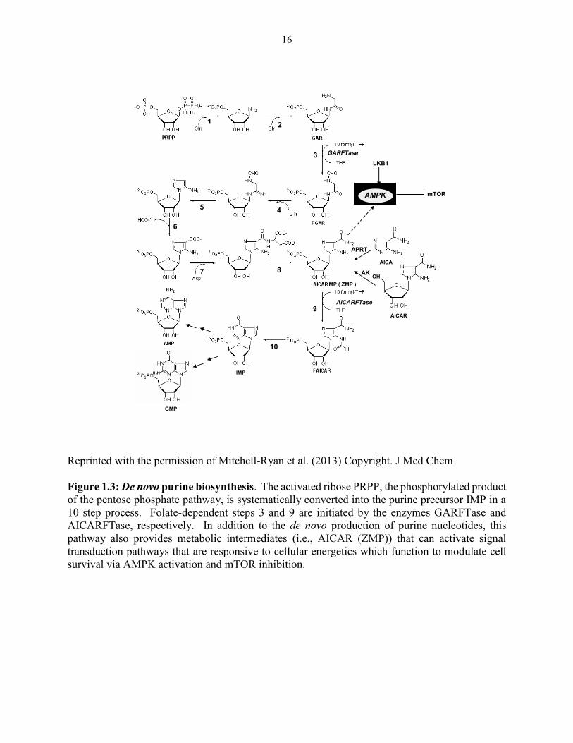
The de novo purine nucleotide biosynthetic pathway is a multi-step enzymatic process that
yields IMP from the initial starting product phosphoribosyl pyrophosphate (PRPP). The numbered
reactions shown in Figure 1.3 are catalyzed by the following monofunctional enzymes: 1,
15
glutamine phosphoribosylpyrophosphate amidotransferase (GPAT); 4, formylglycinamide
ribonucleotide synthase (FGAM synthetase); and 8, adenylosuccinate lyase (ASL). Reactions 2,
3 and 5 are catalyzed by the trifunctional glycinamide ribonucleotide (GAR) formyltransferase
(GARFTase) which contains GAR synthase (GARS; reaction 2), GAR formyltransferase
(GARFTase; reaction 3) and 5-aminoimidazole ribonucleotide synthase (AIRS; reaction 5)
activities. Reactions 6 and 7 are catalyzed by the bifunctional phosphoribosylaminoimidazole
carboxylase/phosphoribosylaminoimidazole succinocarboxamide synthetase (PAICS) enzyme,
which contains carboxyaminoimidazole ribonucleotide synthase (CAIRS; reaction 6) and 5-
aminoimidazole-4-(N-succinylocarboxamide ribonucleotide synthase (SAICARS; reaction 7)
activities. Reactions 9 and 10 are catalyzed by a bifunctional enzyme, 5-aminoimidazole-4-
carboxamide (AICA) ribonucleotide (AICAR) formyltransferase (AICARFTase)/IMP
cyclohydrolase (ATIC) that catalyzes the last two steps in the pathway for de novo synthesis of
IMP. Folate-dependent reactions (reactions 3 and 9) in which 10-formyl THF serves as the one-
carbon donor are catalyzed by GARFTase and AICARFTase. AICA and AICAR can be
metabolized to AICAR monophosphate (ZMP) by either adenine phosphoribosyl transferase
(APRT) or adenosine kinase (AK), thus circumventing the reaction catalyzed by GARFTase. The
activation of AMPK that results in inhibition of mTOR is also depicted.
16
Reprinted with the permission of Mitchell-Ryan et al. (2013) Copyright. J Med Chem
Figure 1.3: De novo purine biosynthesis. The activated ribose PRPP, the phosphorylated product
of the pentose phosphate pathway, is systematically converted into the purine precursor IMP in a
10 step process. Folate-dependent steps 3 and 9 are initiated by the enzymes GARFTase and
AICARFTase, respectively. In addition to the de novo production of purine nucleotides, this
pathway also provides metabolic intermediates (i.e., AICAR (ZMP)) that can activate signal
transduction pathways that are responsive to cellular energetics which function to modulate cell
survival via AMPK activation and mTOR inhibition.
GMP
IMP
MP ( ZMP )
OH
AICA
AICAR
APRT
AK
1
4
8
2
3
5
6
7
9
10
GARFTase
AICARFTase
LKB1
mTORAMPK
17
GARFTase and AICARFTase are two cytoplasmic enzymes involved in de novo purine
synthesis. These folate-dependent enzymes work to generate inosine monophosphate (IMP), a
purine precursor that can be further metabolized to guanosine and adenosine nucleotides. There
are two pathways responsible for intracellular purines, the de novo purine pathway and salvage
pathways (Figures 1.3 and 1.4). Most often, normal cells that possess intact machinery for the
salvage pathway will undergo this energy efficient method that uses preformed bases from
degraded and metabolized purines that are recycled into nucleotides for the use in DNA
synthesis.49 The process of cellular transformation introduces chromosomal abnormalities that
include deletions in chromosomal segments that house tumor suppressor genes. One common
deletion involves chromosome 9, and is reported to include the chromosome 9p21 locus of
multifunctional tumors suppressors, p14ink4a, p15ink4b and p16ARF.50,51 Methylthioadenosine
phosphorylase (MTAP), an essential enzyme in the purine salvage pathway that is often co-deleted
with other well-established aforementioned tumor suppressors located on chromosome 9.46,47
Tumors that harbor this deletion lack the capacity to use this purine salvage pathway as a source
of purines and are heavily dependent upon the de novo purine biosynthetic pathway to support the
high demand for purine nucleotides and carbon backbones in aberrant cell division.46,47
There is some debate over the folate metabolite(s) that acts as a coenzyme for purine
biosynthetic reactions. Earlier studies proposed 10-formyl THF as the sole cofactor associated
with the conversion of GAR and AICAR to their respective formylated species formyl glycinamide
ribonucleotide (FGAR) and formyl 5-aminoimidazole-4-carboxamide ribonucleotide of
(FAICAR), driven by GARFTase and AICARFTase.8,17 However, more recent studies suggest
that 10-formyl dihydrofolate not only plays a part in the activation of these enzymes but is the
preferred substrate for AICARFTase.52,53
18
Adapted open access figure with the permission of Lubin M, Lubin A (2009) Selective Killing of
Tumors Deficient in Methylthioadenosine Phosphorylase: A Novel Strategy. PLoS ONE 4(5):
e5735
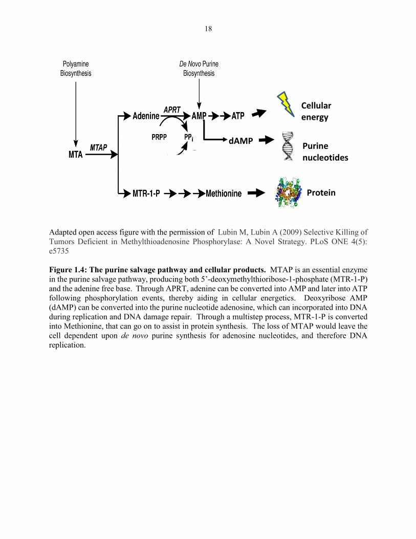
Figure 1.4: The purine salvage pathway and cellular products. MTAP is an essential enzyme
in the purine salvage pathway, producing both 5’-deoxymethylthioribose-1-phosphate (MTR-1-P)
and the adenine free base. Through APRT, adenine can be converted into AMP and later into ATP
following phosphorylation events, thereby aiding in cellular energetics. Deoxyribose AMP
(dAMP) can be converted into the purine nucleotide adenosine, which can incorporated into DNA
during replication and DNA damage repair. Through a multistep process, MTR-1-P is converted
into Methionine, that can go on to assist in protein synthesis. The loss of MTAP would leave the
cell dependent upon de novo purine synthesis for adenosine nucleotides, and therefore DNA
replication.
dAMP
Cellular energy
Purine nucleotides
Protein synthesis
19
Nuclear folate metabolism
An increasing number of studies have described a type of folate metabolism that occurs in
the nucleus. This is thought to arise from the nuclear translocation of SHMT2α (which has a
redundant role in both the cytoplasm and the nucleus), TS and DHFR, which form a multi-enzyme
complex to facilitate nuclear thymidylate synthesis.47 The purported significance of this
physiological event is to maintain genomic integrity in DNA replication and repair (as this has
been cited to occur during S, G2/M and during damage to DNA from UV exposure), in addition
to preventing the mis-incorporation of uracil.45,47 This also holds particular significance in the
prevention of uracil accumulation, which also plays a part in compromising genomic integrity.45
Further, in vitro studies have substantiated these observations by indicating that nuclear membrane
disruption (via sonication) resulted in nuclei that failed to possess de novo TS activity.45

20
Adapted with permission of Fox et al.17 Copyright (2008) Vitamins and Hormones. Ch. 1
Figure 1.5: The compartmentalization of folate metabolism. Folate metabolism can occur in
three compartments with the cell, the mitochondria, the nucleus and the cytoplasm. Not much is
known about nuclear folate metabolism, however, it is thought to play an important role in
thymidylate synthesis. Folate pools in the cytoplasm and the mitochondrial are not
interchangeable. The mitochondrial formate combines with cytoplasmic THF to produce the folate
cofactor essential for de novo purine synthesis.
Folate
Receptors
Reduced Folate Carrier Proton Coupled Folate
Transporter
21
1.3 Folate tissue absorption
Mammals must acquire preformed folates from the diet due to an inability to synthesize
these water–soluble vitamins de novo. Dark green leafy vegetables and select fruits are notable
high sources of the vitamin. Intracellular accumulation of folates is predominately performed by
transmembrane carriers/transporters and receptors. Tissue accumulation of folate is dictated by
the expression of transporters/carriers that permit the influx of folates, polyglutamylation at the γ
glutamate on the carboxyl end of the parent molecule and expression or ability for exporter proteins
to bind folate and contribute to their efflux from cells.
The mechanisms of folate transport have been evolutionary conserved over many millions
of years. The existence of the two solute carrier (SLC) family members that participate in folate
transport, SLC19A1 (reduced folate carrier or RFC) and SLC46A1 (proton coupled folate
transporter or PCFT) date back to the Vendian period (approximately 523-543 million years ago).15
Genes of both transporters have been identified in Pseudocoelomata, more specifically
C.elegans.15 Folate receptor genes evolved much later and made their first appearance in
chordates.15 Salbaum and colleagues speculate that folate receptors (FRs), with their superior
binding affinities, evolved to ensure genomic stability and to preserve DNA methylation patterns
even when folate supplies were limited.15 Folate availability is an important factor in modulating
the expression levels for each of the three proteins. A number of studies have confirmed that low
concentrations of folates lead to increased expression of intestinal RFC and PFCT.54-57 Much like
RFC and PCFT, FR alpha mRNA transcripts and protein expression is increased under folate-
deficient conditions.58-63 Cancer is one of many disease states that can result in a decrease in serum
folate levels due to the increased demand for folate cofactors to participate in DNA replication for
rapidly dividing cells.64 Interestingly, patients with FRα positive tumors, like ovarian cancer, have
22
increased levels of FRα protein in their serum.65-67 Due to this elevation, serum FRα levels have
been considered as a plausible biomarker to detect epithelial ovarian cancer.65,66
The Reduced Folate Carrier (RFC;SLC19A1)
The RFC, as its name suggests, is a facilitative transmembrane transporter with a very high
affinity for reduced folates (Km: 1-3µM), e.g 5-methyl-THF.25 The RFC protein is comprised of
591 amino acids and has a molecular mass of approximately 65 kDa (Figure 1.6).25,68 Due to
pronounced glycosylation at residue Asn 58, the molecular weight of the modified protein is
~85kDa, a ~20 kDa difference from the original molecular weight of ~65 kDa.25,69 The human
RFC gene is located on chromosome 21q22.2 and is reported to be regulated by a host of regulatory
proteins and transcriptional start sites.70 The very complex transcriptional regulation of RFC is
carried out by six non-coding exons with multiple promoters and with the aid of multiple
transcription factors from various families.71,72 RFC has been described by Whetstine and
colleagues as a ubiquitously, but differentially expressed protein.73 In this study they speculated
that the differential expression and the utilization of multiple promoters and transcription factors
could be the product of varying folate demands in various tissues.73 The expansive expression of
this protein, along with its ability to act as a “high capacity” carrier of reduced folates has led to
the belief that RFC is responsible for the bulk influx of reduced folates into mammalian cells,
thereby having the greatest impact on the intracellular folate pools.69,74 As the major transporter
of reduced folate derivatives, it is conceivable that both normal and malignant tissues would find
expression of this protein advantageous for successful DNA replication and for the preservation
of genomic integrity. In fact, quantitative real time PCR (RT-PCR) and multiple methods of
protein detection have validated RFC expression in various human tumors and tissues. Human
RFC mRNA transcripts have been identified in cells from various origins including the central
23
nervous, leukocytes, liver, placenta and the intestine.73,75 The presence of RFC expression in
normal tissues has also implicated the protein as the source of dose-limiting toxicities seen in
patients treated with antifolates that are accumulated into normal tissues through this mechanism.69
The bidirectional transport of RFC is driven by the extrusion of organic phosphate which
allows for the exchangeable influx of reduced folate derivatives and a select number of antifolates
that are recognized by this protein.25,69 While the exact mechanism of exchange is not fully
understood, some speculate that it involves a trans-stimulation phenomenon whereby the
movement of a molecule in one direction enhances or “stimulates” the movement of another
molecule in the opposite direction.76,77 Optimal transport generally occurs at a neutral pH (7.4),
although some studies have demonstrated residual RFC transport at a low pH.78,79 Interestingly,
prior to the discovery of PCFT, RFC was thought to be responsible for low pH folate transport in
the intestine where the protein is expressed.80 However, more recent experiments have confirmed
that RFC transport of leucovorin, a reduced folate derivative, was severely impaired at pH 6.5.69,81
While it has been demonstrated that RFC plays little to no role in the intestinal absorption of dietary
folates, folate depletion increases the intestinal expression of RFC and the role of RFC in this
context has yet to be determined.26,55,56 Studies suggest that RFC does not play a major role in the
tissue absorption and or transport of folic acid (Km~200µM) which may partly explain the reduced
transport efficiency seen with novel antifolates that closely resemble the chemical structure of folic
acid.25,75
The principal route of cellular entry for many “classical” antifolates (i.e., AMT,
methotrexate (MTX) and pemetrexed (PMX)) is through RFC. The pharmacological impact of
RFC expression on the activity of a number of antifolates has been extensively studied. Many of
these studies have demonstrated that the functional loss of RFC modulates drug sensitivity.74,82 In

24
the case where drug influx is completely RFC-dependent, cells exhibit resistance. Alternatively
and contrary to what one may expect, the loss of RFC increases drug sensitivity with agents that
can undergo non-RFC-mediated cell transport, thought to be a consequence of a contracted
intracellular folate pool, which diminishes competing substrates for targeted enzymes, therefore
increasing drug potency.82-84
25
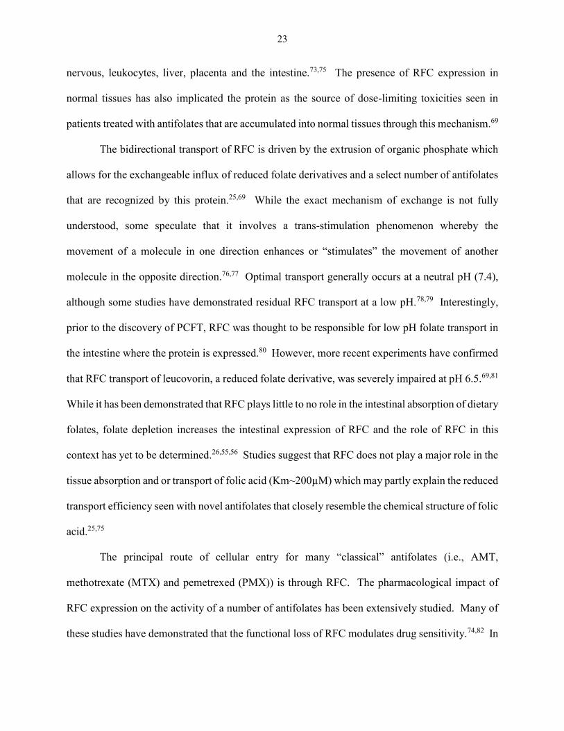
Reprinted with permission of Hou, et al. 85 Copyright (2006) American Society for Biochemistry
and Molecular Biology.
Figure 1.6: Predicted topology map of human reduced folate carrier (hRFC). hRFC is an
integral membrane protein consisting of 12 transmembrane domains and both intracellular and
extracellular loops. The diagram attempts to designate specific amino acid sequences within the
cell membrane, as well as those exposed to the extracellular and cytosolic space. Topology
modeling allows for a greater understanding of how hRFC functions and encourages the generation
of hypotheses surrounding the identification of critical amino acid residues involved in the activity
of the protein.
26
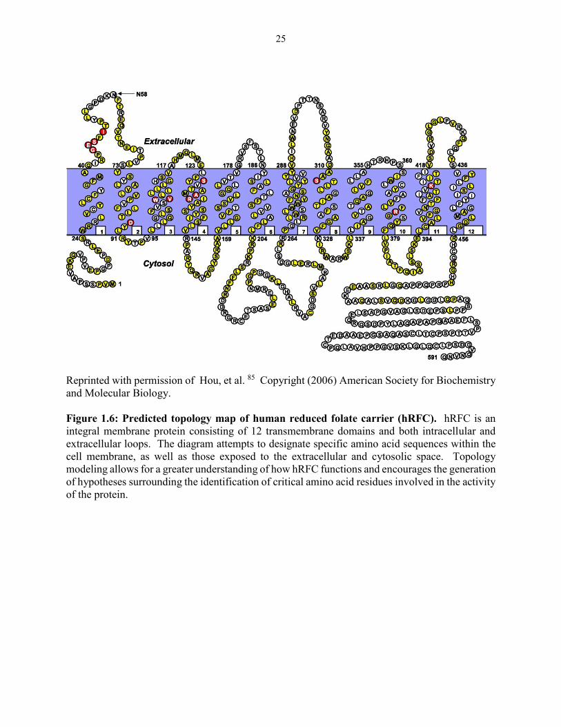
The Proton-Coupled Folate Transporter (PCFT; SLC46A1)
The proton couple folate transporter (PCFT) is the most recently identified of the
predominant mechanisms of folate transport.86 Prior to receiving its current designation as a folate
uptake mechanism, it was originally named the heme transporter protein-1, which describes its
ability to effectively transport heme in the intestine (Km:125µM).25,87 The dietary acquisition of
folates begins in the acidic micro-environment of the duodenum and upper jejunum of the
intestines.88 The existence of a low pH folate transporter became evident as accumulating data
ruled out RFC despite its expression in the intestinal brush boarder membrane. More specifically,
early experiments using a RFC-null HeLa model validated the existence of a non-RFC low pH
transport mechanism that was later determined to be PCFT.89 The cloning of PCFT by Qui et al
formally identified PCFT as the low-pH folate transporter.86 This soon led to structural and
biological studies to obtain more information about the protein and how it participates in folate
homeostasis and more importantly what role if any it plays on the pharmacological activity of
antifolates in clinical use.
Like RFC, PCFT is an integral membrane protein that participates in the facilitative
transport of (anti)folates and is also a member of the major facilitator super family (MFS).
However the transport profile and mechanism of transport is distinct from that of RFC.73 The
unidirectional transport of folates has been described as electrogenic and proton driven and occurs
optimally at a low pH (5.5), hence the naming “proton coupled”.73 The charge carried by the
proton creates an electrochemical gradient which classifies this proton driven mechanism as
electrogenic.80,90
The PCFT chromosomal location is 17q11.2 which encodes a 55 kDa protein made up of
459 amino acids (Figure 1.7).25,91,92 PCFT is encoded from 5 exons with a minimal transcriptional
27
regulatory region located -42 and +96 bases from the transcriptional start site.75,91-93. Transcription
of this protein is thought to be governed by a number of factors. Kruppel like factor -4 (KLF-4),
hepatocyte nuclear factor (HNF-1) and nuclear respiratory factor -1 (NRF-1) have all been reported
to enhance PCFT promoter activity resulting in an increase in PCFT transcripts.94,95 Vitamin D
was also shown to increase PCFT expression via the transactivation of the PCFT promoter by the
vitamin D receptor (VDR) when heterodimerized with retinoid X receptor –α (RXRα).96 Promoter
hypermethylation results in decreased transcriptional activity and therefore gene product.93 While
little is known about the intrinsic transcriptional regulation of PCFT, studies have shown that
xenobiotics, more specifically, proton pump inhibitors, can decrease intestinal PCFT expression
in patients administered this type of therapy.97
Compared to RFC, PCFT has restricted tissue expression that is limited to the duodenum,
jejunum, liver, kidneys, and choroid plexus and comparably much lower expression in the bone
marrow and colon.88 A wide range of malignant cells express PCFT, including but not limited to
tumors of the colon/rectum, lungs, liver and ovaries.98 The expression of PCFT may be especially
advantageous in tumors with high glycolytic activity or what had been described as “The Warburg
effect”, creating a proton-rich acidic microenvironment. In theory, the creation of such a
microclimate would increase the activity of PCFT and provide a context in which RFC activity is
reduced. This very observation is the basis of PCFT-targeted anti-tumor therapy.75 A better
understanding of the transcriptional regulation of PCFT will undoubtedly result in novel
approaches and creative strategies to improve drug efficacy in PCFT-expressing tumors.
28
Adapted with the permission of Shin, et al. 99Copyright (2012) The American Physiological
Society and Unal, et al. 100Copyright (2009) American Journal of Physiology. Cell Physiology
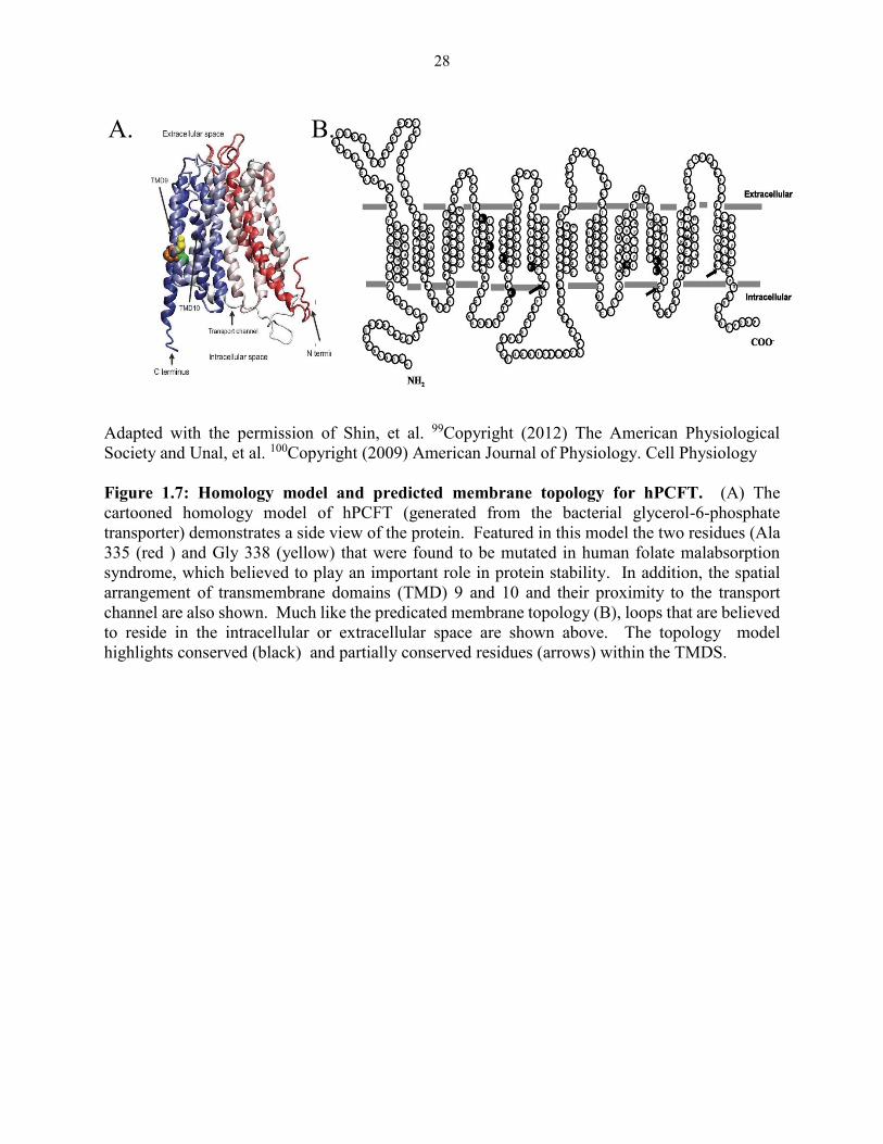
Figure 1.7: Homology model and predicted membrane topology for hPCFT. (A) The
cartooned homology model of hPCFT (generated from the bacterial glycerol-6-phosphate
transporter) demonstrates a side view of the protein. Featured in this model the two residues (Ala
335 (red ) and Gly 338 (yellow) that were found to be mutated in human folate malabsorption
syndrome, which believed to play an important role in protein stability. In addition, the spatial
arrangement of transmembrane domains (TMD) 9 and 10 and their proximity to the transport
channel are also shown. Much like the predicated membrane topology (B), loops that are believed
to reside in the intracellular or extracellular space are shown above. The topology model
highlights conserved (black) and partially conserved residues (arrows) within the TMDS.
A. B.
29
Folate Receptor alpha (FRα)
FRs are an extensively studied family of proteins that are recognized for their role in
embryonic and fetal development where they act as an essential mechanism of folate transport,
supporting rapid cell division during gestation. FRs, also known as folate binding proteins (FBP
or folbp for murine homologs) were first identified in bovine milk in 1972.101,102 Since the
discovery of these high affinity/low capacity folate binding proteins, four isoforms and pseudo-
genes have been identified. While each isoform shares some sequence homology (70-80%), they
each possess distinct structures, patterns of tissue expression (with some overlap), binding affinity
and stereospecificities.103,104 Collectively, FR’s have the highest affinities toward folic acid,
physiological reduced folates and formylated THF (5-formylTHF) compared to PCFT and RFC.25
These features allow FRs to successfully engage in cellular uptake when folate concentrations are
low. These genes are all a part of a larger superfamily that includes the riboflavin binding protein
and the retinbindin, a retinal binding protein.103 Prior to the solving of the crystal structure of FRα,
homology modeling using bovine riboflavin binding protein was employed to elucidate or to gather
more insight on the structural characteristics of this protein and possibly other identified isoforms
due to known similarities of each protein.105
FR genes, Folr 1-4, are localized to chromosome 11q13.3-14 and are responsible for the
transcription for FRα, β, γ/γ’ and δ.62,103,104 Of the four isoforms, FRδ is the most recently
discovered. While the mouse ortholog, folrbp3, is expressed in the mouse thymus and spleen and
is thought to play a role in the proliferation of immune cells, expression of this isoform has not
been detected in human adult or fetal cells.104 The failure to detect FRδ in human tissues has been
thought to arise from the temporal/spatial regulation and expression of the protein.104 Others
suggest that like FR pseudogene1 (Folr1p) identified by Raggoussis et al., the delta isoform is a
30
pseudogene in the human genome.104,106 Due to an elusive pattern of expression in human tissues,
the capacity of this isoform to bind folate is undetermined, as well as its putative role in folate
metabolism and perhaps cell signaling.
Much like the delta isoform, little is known about FRγ, when compared to the more
prominent and widely studied β and α isoforms. Unlike FRδ, the gamma isoform has been
identified in human tissue. The protein was first discovered in 1993 and was identified as an
overexpressed high affinity folate binding protein in chronic myelogenous leukemia (CML) and
acute myelogenous leukemia (AML) patient samples.107 Since the discovery of the γ isoform, it
has been identified in normal hematopoetic cells, bone marrow, spleen and thymus.103 Of the three
functional forms of FR expressed in human tissues, FRγ is to date, the only identified polymorphic
isoform of this family. A genetic polymorphism results in a mutation that codes for a premature
stop codon leading to the translation of a truncated non-functional protein, FRγ’.108 Despite
multiple N-glycosylation sites (3) which are shared among all isoforms and are thought to
contribute to the cell surface expression of the protein, there is an additional anomaly that
distinguishes FRγ from its counterparts- this protein is constitutively secreted due to a lack a
hydrophobic amino acid sequence that serves as a signal for glycosylphosphatidylinositol (GPI)
modification that anchors the protein to the cell surface.103,109
FRs are known to exist in membrane bound and soluble states. Membrane bound receptors
(α and β) are anchored to the membrane by glycolipids known as GPI anchors.103,110 This GPI
modification is signaled by the hydrophobic carboxyl terminal segments in the α and β isoforms
which allow for a post-translational transamidase reaction, resulting in the cleavage of the present
C-terminus while creating an amide linkage to the GPI and a newly formed C-terminus.103,110
Soluble folate binding proteins (sFBP) arise from the membrane-bound forms that undergo well-

31
defined cleavage events. They are generally present in extracellular fluids, which include milk,
cord blood, urine, and cerebrospinal and amniotic fluids.103 The soluble FR α isoforms are thought
to be the result of (1.) the phospholipase cleavage of the GPI anchor or (2.) proteolysis by a Mg+2
dependent protease.103 These mechanisms are thought to be responsible for soluble α isoforms
detected in KB nasopharyngeal carcinoma cells in culture and those detected from placental cells.
In contrast, the soluble β isoform is the product of two independent pathways that either result in
GPI anchor attachment or secretion.103 All soluble forms of the protein have been shown to bind
and stabilize folates; therefore these soluble proteins may have a functional role in normal
physiology.103 For instance, the sFBP in milk are thought to play a role in the intestinal absorption
of folates.103 These soluble proteins may also serve a diagnostic purpose in detecting disease as
many pathological conditions, including cancer, results in the aberrant shedding of these
membrane proteins into the circulation where they are not generally detected in healthy
individuals.103,111
The FR β has increasingly become the subject of interest due to its role in a number of
human pathologies, most notably cancer and inflammation.103,112-116 The expression of this
isoform is confined to hematopoietic cells of the myelomonocytic lineage and the protein is
routinely used as a differentiation marker in normal hematopoiesis.103 Protein expression extends
to the placenta and to mature neutrophils with elevated expression seen in activated blood
monocytes and the expression status remains consistent during their transformation to
macrophages.103 Macrophages are known to play a critical role in the initiation, maintenance and
resolution of the inflammatory process.113 Rheumatoid arthritis (RA) is a disease characterized by
the infiltration of lymphocytes and macrophages to synovial joints, causing proliferation of local
fibroblasts and severe joint damage. Activated macrophages in synovial tissues from RA patients
32
were shown to have significant expression of the β isoform.116 The selective expression of FR β
on this population of macrophages makes this isoform an excellent target for therapies specific to
this protein isoform. Presently, the antifolate MTX is the preferred treatment option in patients
suffering from RA.117 However, MTX substrate promiscuity among other mechanisms of folate
transport has created a demand for novel therapeutics that have selective uptake by FR β.117-119
These therapeutics may not only prove useful in the treatment in RA, but they may also show
efficacy in FR β-expressing malignancies such as leukemia (CML and AML). Tumor-associated
macrophages (TAM’s), which also express FRβ, have been implicated in the initiation and
progression of a number of malignancies.120-122 Folate-based therapies targeted to FR β may
extend the survival of patients diagnosed with difficult to treat tumors like pancreatic cancer which
are reported to have significant TAM infiltration.123,124 An additional and perhaps more practical
application in exploiting the population of TAMs in cancer is the use of fluorescent conjugates
linked to FR β, which may serve as an optical tool that can aid in detection of relapse (in the case
of hematopoietic disease), primary tumors and their metastasis.122
FRα is, by far, the most extensively studied of the four FR isoforms. While this receptor
has been well documented as a mechanism of folate transport, additional physiological roles of
FRα continue to be the subject of many ongoing studies. Much intrigue surrounds the functional
role of FRα especially when co-expressed with other high capacity folate uptake mechanisms such
as PCFT or RFC, which has led to speculation on alternative roles for this receptor. Undoubtedly,
FRα has a clear role in the reabsorption of folates from kidney proximal tubules and in embryonic
development, including the development of the neural tube and transplacental folate transport from
mother to fetus. What remains unclear is the utility of this receptor when expressed in other
epithelial tissues with limited or no access to circulating folates. One popular line of reasoning
33
suggests that FRα, when expressed in tissues with limited access to circulation, acts as a folate
scavenger and participates in the transcellular transport of folates, as described in the transport of
folates across the retinal pigment epithelia, the luminal uterine epithelia during pregnancy and in
lung epithelia to prevent bacterial growth.125,126 It has been proposed that FRα, like other GPI
anchored proteins clustered by lipid rafts, participates in signal transduction pathways that are
involved in regulation, survival and growth.127,128 More studies are required to determine and
validate the exact signaling pathways that are involved with ligand-bound FRα.
There are a number of disorders associated with mutations or the functional loss of FRα.
The homocysteinylation of FRα, a consequence of excess/elevated homocysteine, may lead to the
development of FRα autoantibodies which are thought to contribute a significant decrease in folate
uptake, resulting in defects in the developing embryo.129,130 Such deficiencies that arise from
deleted, dysfunctional or blocked FRα have been linked to NTDs, orofacial cleft, congenital heart
defects, autism spectrum disorders and cerebral folate deficiency (CFD).130-132 CFD is thought to
arise from the presence of FRα autoantibodies or a loss of function mutation in the folr1 gene,
causing a neurological syndrome that presents in children as young as 4 months with cognitive and
neuromuscular deficits.133,134 Studies have shown that maternal FR autoantibody concentrations
are linked to NTDs, further highlighting the important role of FRα in embryonic
development.130,131,135 An incomplete understanding of FRα’s role in signal transduction makes it
difficult to delineate if folate deficiency or a disruption in signal transduction pathways play a
causal role in the development of such malformations. More work in this area may lead to a greater
understanding of the tissue distribution of this protein and the global importance of its expression,
in addition its acknowledged role in folate metabolism.
34
As previously mentioned, FRα has a unique and distinct pattern of tissue expression. In
adult tissues, FRα is confined to the apical surface (luminal) of polarized (non-transformed) cells
of epithelial origin, including the choroid plexus, proximal renal tubules, fallopian tubes, uterus,
epididymus, acinar cells of the breast, submandibular salivary and bronchial glands, type 1 and 2
pnuemocytes in the lung and placental trophoblasts.103 The only known basolateral surface
expression of this protein has been identified in the epithelia of retinal pigment cells.103 The
luminal expression of FRα, by virtue of its physical location, makes this receptor inaccessible to
circulating folates in the blood.136-138 The role that FRα plays in the reabsorption of folates from
the kidneys creates an opportunity for renal FR to encounter serum folates.103 Interestingly, it has
been reported that glomerular filtration allows for the separation of low molecular weight FRα-
targeted drugs that are transcytosed, thereby preventing renal accumulation of cytotoxic agents
and circumventing neurotoxicity.136
Many epithelial based tumors also express FRα, however, ovarian and endometrial cancers
are among the most recognized partly due to their consistent and elevated expression of the protein.
One classic feature of the transformed cell is the absence of cellular organization (loss of polarity)
and a disruption of cellular architecture. These structural changes that occur during tumorigenesis
lead to the altered spatial orientation of the receptor such that it is expressed on the basolateral
surface. The newly acquired basolateral surface expression of the receptor on tumors permits the
sequestration of circulating (anti)folates due to a change in orientation that leads to accessibility.
We argue that this feature, in addition to the very narrow and restricted tissue expression exhibited
on normal tissues, makes FRα a rational and attractive target for drug delivery to promote the
selective killing of FRα-positive tumors.
35
There are many theories surrounding the elevated expression of FRα in select epithelial
malignancies. Many of these encouraged thorough investigations of the regulation of folr1 gene
and gene products in both normal and transformed cells. The complex regulation of FR genes is
governed by a variety of factors which include serum folate concentrations, nuclear receptors and
their respective ligands, and post transcriptional/translational modifications. Although FR
isoforms share significant sequence homology, the organization of the gene, the use of alternative
promoters and alternative splicing distinguishes each isoform from the other while also accounting
for the differences observed in tissue distribution. 96
The folr1 gene has a reported length between 6.7 139 and 7.5 kb 140 and is composed of 7
exons and 6 introns that encodes a 257 residue polypeptide and a 38-42 kDa protein.141,142
Multiple transcripts of folr1 have been identified and appear to be the product the use of alternative
promoters, as well as alternative splicing that occurs at exons1-4.141-143 The promoters responsible
for the diverse FRα mRNAs are TATA-less and designated P1 and P4, located upstream of exons
1 and exon 4, respectively (Figure 1.8).103,140 Although they produce divergent transcripts, the
protein product derived from each promoter is identical.142 The regulation of transcription, the
efficiency of translation and the length and sequence of the resulting transcripts are where these
two promoters diverge.82,111,142 Despite these differences, the open reading frame (ORF), 3’
untranslated region (UTR) and mRNA stabilities are the same for transcripts derived from both
promoters in FRα-expressing cells.142 The P1 and P4 promoters are reported to be under tissue-
specific regulation which is thought to account for the abundance of specific transcripts in specific
tissues. 82,142
Relative to P4, not much is known P1 promoter, but this promoter is understood to exhibit
functional and structural differences from the P4 promoter.141 Transcripts produced by the P1
36
promoter have been identified in the normal human kidney, the testis and the cerebellum of the
brain.103,141 The multiple initiation sites combined with the alternative splicing of upstream exons
results in heterogeneity of the 5’ UTR in the transcripts generated and explains the varying lengths
and sequences of the 5’ UTR among the transcripts.142 The regulation of the P1 promoter in certain
ovarian cancer cells differs from that which is seen in normal human cells. Bagnoli and colleagues
described an inverse relationship between caveolin-1 protein, known to negatively regulate
intermediates in signal transduction involved with cell proliferation, and FRα expression in human
ovarian cancer cells.139,144 The result of their efforts determined that caveolin-1 negatively
regulated FRα expression through repression of the P1 promoter.144 Additional studies examining
P1 regulation in ovarian carcinomas concluded that the promoter was under the control of a variant
hepatocyte nuclear protein that enhanced promoter activity via the interaction of a DNA-binding
site located in the untranslated region of exon1.139 Exon 1 is believed to include the elements
required for optimal transcriptional activity for the P1-derived FRα transcript in ovarian cancer
cells.139 For reasons not fully understood, P1 transcripts display a lower rate of translation
efficiency when compared transcripts generated from the P4 promoter.142 Whether or not
translation efficiency corresponds to surface expression density, is yet to be determined.
Additional research is required to validate if similar factors modulate P1 promoter activity in
normal cells and to determine if there is a direct relationship between promoter activity and overall
protein expression.
A number of FRα expressing cells transcribe FRα under the control of the P4 promoter
including the KB nasopharyngeal carcinoma cells and normal epithelial tissues of the lung,
placenta, salivary gland, uterus breast and stomach.103 This promoter’s activity is influenced by
three GC rich sequences that allow for non-canonical SP1 binding, with each of the three sites
37
making a significant contribution to promoter activity.103,143 Multiple sites of potential
transcription factor binding were reported to be contained within this promoter, including sequence
motifs for AP2, E-box and the aforementioned SP1 (Figure 1.8).101,140
mRNA stabilities in FRα-expressing cells have been discussed; irrespective of the
promoter of origin, all transcripts are reported to have similar stabilities.103,140,142 However a novel
nuclear post transcriptional mechanism of FRα regulation has been described. Residing in the
ORF is a discrete 60 base pair mRNA element that is reported to control the stability of FRα
mRNA.140 This structure ultimately results in the selective nuclear degradation of FRα transcripts
in FRα null cells and may be responsible for the restricted tissue expression seen with this
protein.140
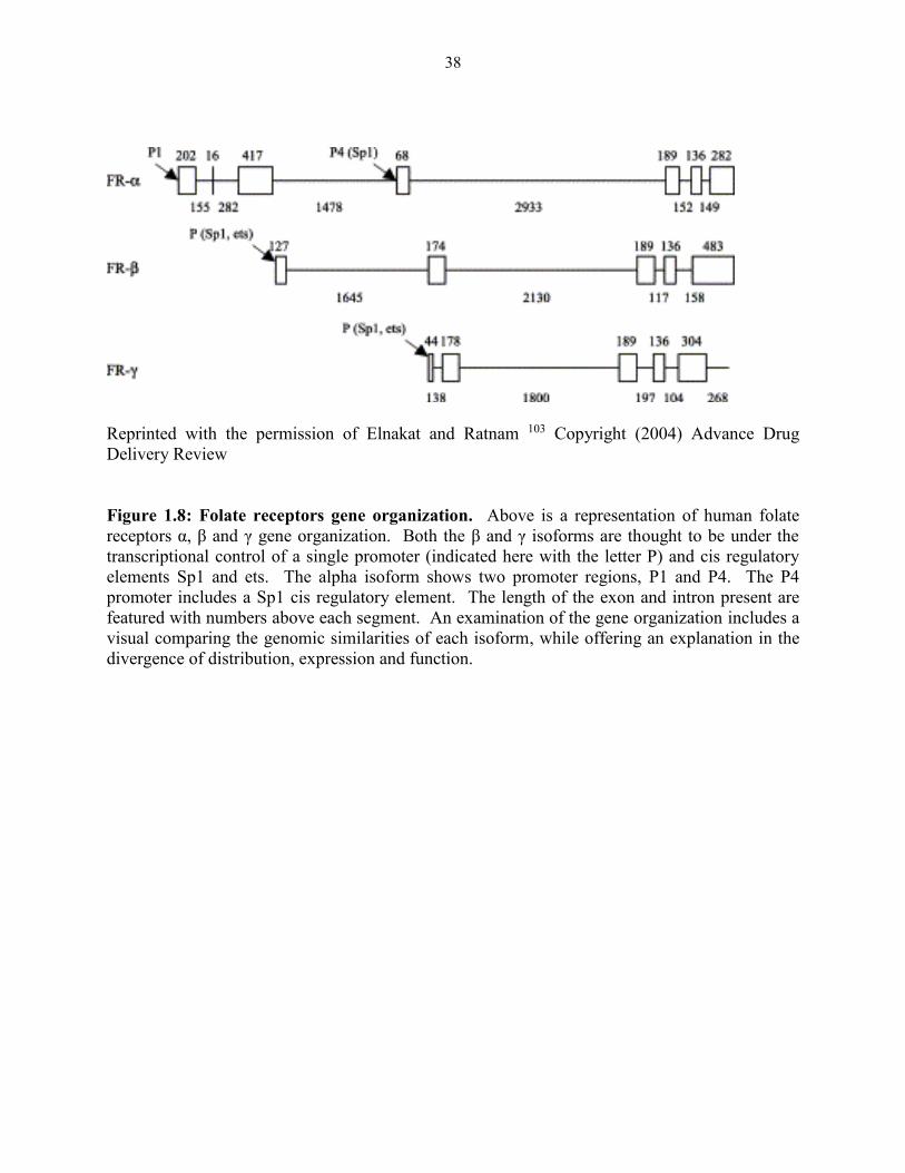
38
Reprinted with the permission of Elnakat and Ratnam 103 Copyright (2004) Advance Drug
Delivery Review
Figure 1.8: Folate receptors gene organization. Above is a representation of human folate
receptors α, β and γ gene organization. Both the β and γ isoforms are thought to be under the
transcriptional control of a single promoter (indicated here with the letter P) and cis regulatory
elements Sp1 and ets. The alpha isoform shows two promoter regions, P1 and P4. The P4
promoter includes a Sp1 cis regulatory element. The length of the exon and intron present are
featured with numbers above each segment. An examination of the gene organization includes a
visual comparing the genomic similarities of each isoform, while offering an explanation in the
divergence of distribution, expression and function.
39
The inverse relationship between FRα expression levels and serum folate concentrations
has been well documented.59,145-147 Both in vivo and in vitro models have validated the regulation
of this isoform by serum and media folate levels, using both folate-deplete and folate-replete
conditions, the latter including multiple folate metabolites.58,59,148-152 In each instance, restricted
or reduced folate levels accompanied an increase in FRα expression levels, while increasing folate
levels reversed the phenotype resulting in reduced expression levels of the protein. In support of
these studies, an examination of serum folate levels in patients with FRα-positive tumors
recapitulated these experimental results, suggesting that this regulation by folate is a naturally
occurring phenomenon.64,153-156 A consequence of reduced or depleted serum folates is a rise in
homocysteine levels due to inactivity of MS, which is responsible for the conversion of
homocysteine to methionine. Elevated homocysteine levels were shown to encourage or increase
the interaction between the heterogeneous nuclear ribonucleoprotein E1 (hnRNP E1) and the cis-
element in FRα transcripts, which is believed to result in unregulated translation.157 Other studies
have reported an alteration in the level of mRNA transcripts or amplification of the FR gene.103
The use of antifolates, more specifically MTX, has also been shown to increase FRα
expression.81 MTX resistance is thought to be a result of a number of factors including the
functional loss of RFC or a reduction or silencing of protein expression via gene hypermethylation
or other mechanisms.25,158,159 Experts suggest that MTX resistant cells experience an increase of
FRα expression as an attempt to meet the folate requirements of a malignant cell through the use
of a transport mechanism not affected by drug exposure.81 The exact transcriptional mechanism
is unknown; however, it has been postulated that DHFR inhibition by MTX may mimic the
conditions of folate depletion and may trigger the same regulatory pathways that lead to the
enhancement of FRα expression.81
40
Steroid receptors have also been implicated in the regulation of FRα. Activation of
androgen160, glucocorticoid161, and progesterone162 receptors have been associated with increased
transcriptional activity of the P4 FRα promoter and an increase in cell surface FRα protein
expression. Steroid hormone regulation of the FRα P4 promoter by the progesterone and
glucocorticoid receptors is indirect but it is direct by androgen receptors. In contrast, the estrogen
receptor has been identified to negatively regulate FRα through transcriptional repression.163
Estrogen exerts FRα transcriptional repression by interacting with GC-rich Sp1 binding site(s) of
the P4 promoter and recruiting the co-repressor SMRT which prevents the cluster of transcriptional
machinery.163 This finding was thought to help explain the lack of FRα in ER-positive tumors and
the increased expression in anti-estrogen (tamoxifen) treated tumors.
Many suspect the physiological role of FRα extends beyond the role it plays in folate
transport and metabolism.164,165 Studies have shown that cells that are transfected with FRα
undergo a rapid increase in cellular proliferation and alternatively, cells treated with antibodies,
sense/antisense particles or oligonucelotides directed to this receptor showed a delay in growth
and in some cases reversion of the malignant phenotype.127,164-166 Folate metabolism alone cannot
account for a robust proliferative response but rather alterations in signaling pathways may be
involved. Notch and Hes-1 were both implicated in FRα directed cell growth in alpha T3-1 cells
transfected with FRα.164 In addition to cellular proliferation, FRα has also been linked to cell
motility, resistance to apoptosis, migration and invasion.167,168 This is somewhat consistent with
the correlation made between FRα overexpression and a poor prognosis/aggressive phenotype and
decreased survival in ovarian cancer patients.169,170 FRα expression has been linked to many
changes seen in malignant transformation which has led some investigators to think the protein
plays a significant role in normal to malignant transition.168
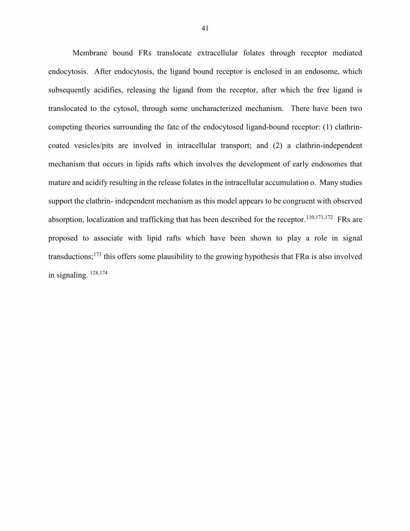
41
Membrane bound FRs translocate extracellular folates through receptor mediated
endocytosis. After endocytosis, the ligand bound receptor is enclosed in an endosome, which
subsequently acidifies, releasing the ligand from the receptor, after which the free ligand is
translocated to the cytosol, through some uncharacterized mechanism. There have been two
competing theories surrounding the fate of the endocytosed ligand-bound receptor: (1) clathrin-
coated vesicles/pits are involved in intracellular transport; and (2) a clathrin-independent
mechanism that occurs in lipids rafts which involves the development of early endosomes that
mature and acidify resulting in the release folates in the intracellular accumulation o. Many studies
support the clathrin- independent mechanism as this model appears to be congruent with observed
absorption, localization and trafficking that has been described for the receptor.110,171,172 FRs are
proposed to associate with lipid rafts which have been shown to play a role in signal
transductions;173 this offers some plausibility to the growing hypothesis that FRα is also involved
in signaling. 128,174
42
Reprinted (adapted) with the permission of Chen et al.175 Copyright (2013) Nature letters.
Figure 1.9: Ribbon and charge distribution surface model of FRα complex with folic acid
substrate. (Above) The alpha and beta helices (shown in green) demonstrate how the protein
interacts with the endogenous ligand, folic acid. (Top-Right) A 90 degree rotation of the protein
offers additional information about the protein-substrate interaction and the key amino acids
involved in the folic acid recognition/binding process. (Bottom) The charge distribution surface
model illustrates folic acid contained within the binding pocket and the charge of the amino acids
surrounding the binding surface.
43
1.4 Folate based targeted therapy
Shortly following the discovery and first synthetic synthesis of folic acid, folate analogs
termed antifolates were the subject of heightened attention resulting from their roles as effective
agents against hematopoietic malignancies.176,177 More specifically, the development of the first
clinically used antifolate, AMT, fundamentally changed the treatment of pediatric leukemia and
ultimately served as a platform for the development of many novel antifolates.176,178,179 The vast
and rapidly accumulating knowledge of folate metabolism also played an integral role in
understanding the pharmacology of these analogs in addition to providing direction in the
development of less toxic congeners.
Antifolates describe a class of agents that are structural analogs of folate and exert their
antiproliferative actions through inhibition of enzymes in folate metabolism, resulting in the
depletion of nucleotides required for DNA replication and DNA repair, and RNA synthesis.180
Today antifolates remain the most well-studied chemotherapy agents with well-defined and well-
characterized mechanisms of action. The recent discovery of the molecular structure of FRα and
growing structural information about the β isoform will encourage the synthesis and development
of more sophisticated agents with the promise of achieving successful and precise tumor
targeting.175,181 The identification of malignancies that over-express ideal targetable folate
transport systems (FR’s and PCFT) are steadily growing. Recent studies that examined patient
tumor samples reveal that in vitro tumor models may inaccurately reflect the amount of folate
transporter proteins that are actually expressed in in vivo patient tumors (personal communication;
Folate Receptor Conference, Couzumel (2013)). This overexpression not only increases target
area and points of entry for FR targeted drugs, but this may also have dramatic impact on tumor
response and the concentration of drug required for efficacy.
44
There has been tremendous progress in the area of antifolate development since the
introduction AMT and MTX. While these agents were the first ground breaking therapies of their
kind, there are currently a number of novel agents under investigation that target different folate
pathway enzyme and inhibit growth of neoplastic cells through the disruption of DNA synthesis.
This diverse array of compounds not only includes new and improved DHFR inhibitors that differ
from the first generation agents, AMT and MTX, such as pralatrexate, but also PMX which inhibits
TS.25,182,183 GARFTase inhibitors are under development as well.25 These novel compounds can
be grouped based on their intended enzyme target, their chemical structure and their ability to
undergo polyglutamylation.
GARFTase has become a metabolic target of increasing relevance since the inception of
lometrexol (LMTX) by the Eli Lilly Corporation in 1985.91,184 Much of the interest surrounding
LMTX and subsequent GARFTase inhibitors reflects their ability to disable the rate-limiting step
in the de novo biosynthetic pathway of purines which involves the transfer of the formyl group
from 10-formyl-THF to the primary amine of β GAR which provides the C8 carbon in the purine
skeleton.185,186 The disruption of purine production in tumor cells is believed to be a favorable
feature of a chemotherapeutic drug, as these drugs not only interfere with DNA replication but
they also disturb the cellular energy balance via ATP depletion, thereby interfering with energy
requiring processes necessary for cell viability.187 Another interesting and advantageous aspect of
many of the prototypical GARFTase inhibitors is that they were designed based on the crystal
structure of either human or E. coli GARFTase, which share homology at the folate co-factor
binding site.185 The structure-based drug design approach used for these inhibitors may account
for their characterization as “clean” drugs, in that they have a clear mechanism of action unlike
nucleoside analogs or purine based drugs that may have some influence on purine synthesis but
45
also possess the capacity to be incorporated into nucleic acids, making their primary function
indeterminate.185
LMTX was the first folate-based antipurine that possessed potent inhibitory actions against
GARFTase.184 Remarkable preclinical data in xenografts of human tumors and murine models of
solid tumors aided in making LMTX the first GARFTase inhibitor that was clinically
investigated.184,188 Further development of LMTX was halted during clinical trials due to delayed
and cumulative side effects, which include severe and unpredictable myelosupression and
neuropathy that was thought to be a function of a long terminal half-life and liver
accumulation.91,184,189 RFC-directed transport of LMTX may have been an additional contributing
factor to toxicity seen in patients.189 Folic acid supplementation was shown to curtail these
adverse side effects and allowed patients to tolerate higher concentrations of drug.184 In light of
this, new dosing schedules with co-administration of folic acid were implemented in additional
studies using this drug.190,191 However, further clinical development of LMTX was brought to a
standstill, reportedly for economic reasons.192
The LMTX studies validated GARFTase as an important target in de novo purine
biosynthesis. In efforts to create novel GARFTase inhibitors that maintained the potent biological
activity of LMTX, but with reduced untoward toxicities, computer assisted drug design was used
to create AG2034 and AG2037. The GRID program, a computer generated method for
systematically investigating an array of chemical groups and how they interact with suggested
protein binding sites, revealed two hotspots at the GARFTase binding site that possessed an
affinity for a thioester probe.185 The structure of human GARFTase was the model for drug
design.193 The sulfur containing AG2034 was designed to complement the two identified hotspots
at the GARFTase active site identified by the GRID program.185 AG2034 possessed promising
46
characteristics of an ideal GARFTase inhibitor, with a Ki of 28 nM against the human enzyme,185
and the ability to undergo polyglutamylation.185,193 Together, these attributes are thought to be
responsible for the potent antiproliferative activity seen in L1210 cells (IC50:4 nM) and CCRF-
CEM cells (IC50:2.9 nM).193 Cells with a mutant p53 and/or those lacking a functional G1
checkpoint were particularly sensitive to purine deprivation elicited by this drug (A549 and MCF-
7).194 After being selected for preclinical trials and tested against a range of murine tumors and
human xenograft models, AG2034 was taken to clinical trials. These initial trials were suspended
to investigate the newer analog AG2037. AG2037, like AG2034, is a sulfur containing GARFTase
inhibitor. However unlike, AG2034 and LMTX, which can achieve cellular entry through both
RFC and FR, AG2037 is transported by RFC exclusively.185 The selective transport by RFC was
thought to be ideal at the time. This was supported by a belief that efficacy of drugs that undergo
FR-mediated transport would be modified based on the nutritional status (folate intake) of the
patients.185 A clinical trial with AG2037 in colorectal cancer patients that failed treatment was
completed in 2004, however this study has no posted or published results to date.
Attempts to develop improved GARFTase inhibitors continued with the creation of Eli
Lilly’s GARFT II, also referred to as LY309887. When compared to LMTX, LY309887 had a 9-
fold greater potency but produced metabolites with reduced polyglutamylation.188 To address the
issue of polyglutamylation, two monoglutamate GARFTase inhibitors were introduced,
LY254155 and LY222306. LY254155 and LY222306 are 2’, 5 thiophene and 2’, 5 furan
derivatives of LMTX, respectively. These compounds exhibited greater inhibition against
GARFTase when compared to the parental compound LMTX and both possessed reduced affinity
towards FRα (LY254155: 6 fold, LY222306: 350 fold).195 These agents would presumably be
valuable in the treatment of tumors with inherently low FPGS activity or tumors that have acquired
47
resistance to antifolates through reduced expression and or function of FPGS or increased gamma
glutamyl hydrolase activity.195 The search for an efficacious but well tolerated GARFTase
inhibitor is ongoing and recent developments in folate transport proteins may produce a generation
of equally as effective but targeted agents.
Currently, there is no antifolate that uses AICARFTase as its primary target. While the
multi-targeted antifolate, PMX, inhibits AICARFTase as a secondary target, the extent of enzyme
inhibition by this drug has not been directly quantified.196,197 The rising and alarming acquired
resistance to current antifolate therapies through multiple mechanisms, including overexpression
of enzyme targets and decreased expression of essential transport proteins. The need to circumvent
antifolate resistance encouraged the design of novel AICARFTase and GARFTase inhibitors
(among the most infrequently targeted enzymes in folate metabolism) that use an alternative
mechanism of cellular entry.158
FR targeted drugs
Experimental evidence from the Matherly lab and a host of others have shown that classical
antifolates like MTX, LMTX and PMX bind, at varying degrees, to FRα and elicit a cytotoxic
effect in FRα-expressing cells, albeit at different concentrations.136,198 Increasing concentrations
of these agents may introduce off target effects, making it difficult to ascertain if the toxic effects
were FRα-mediated or otherwise. Most of the drugs in this class are transported by the
ubiquitously-expressed RFC, which is thought to lead to intestinal and bone marrow toxicities as
these are highly proliferative tissues with an increased dependence on the very enzymes these
drugs inhibit.136
The adverse effects of these drugs were thought to arise, at least in part, from their RFC
transport. This created demand for agents that would reduce undesirable toxicities in normal cells.
48
Ovarian and endometrial cancers were recognized for their expression of FRα and were considered
difficult to treat with available therapies. These factors may have led to the quest for FRα-targeted
therapies. In the 1980s, efforts to design a FR-based antifolate culminated in the development of
the TS inhibitor CB3717.199 This agent showed preferential transport by FRα over RFC and could
be metabolized to polyglutamates, but further development of this compound was halted due to
nephrotoxicity (thought to be a product of insolubility associated with high dose administration
rather than FRα transport) and myelosupression.200
The cyclopenta(g)quinazoline based drug, CB300638, became the first validated FRα
targeted TS inhibitor.136 This agent showed high selectivity for FRα when compared to related
analogs, and showed efficacy in a physiological folate concentration (20 nM) and inhibited TS in
KB cells but did not compromise normal tissue.201 BCG 945, like CB300638, also a
cycoplenta(g)quinazoline based compound, showed inhibitory effects against TS in FRα-
expressing cells.202 Due to its in vitro and in vivo efficacy and FRα selectivity, BCG 945 was
selected as a candidate for phase I clinical trials that are currently on going.202
De novo purine synthesis has become a rather attractive target in non-RFC based therapies
with the development of novel GARFTase inhibitors that possess selective membrane transport
for FRs and PCFT over RFC. The circumvention of RFC membrane transport by a collection of
agents within this category can be seen as an improvement upon earlier agents such as LMT which
was reported to induce severe myelosuppression in clinical trials, likely due to RFC transport.
Both FR- and PCFT- targeting have distinct advantages that include narrow tissue expression
(PCFT and FR), optimal performance in tumor acidic microenvironment (PCFT) and basolateral
expression (circulation accessible) expression only seen in tumor cells (FRα). These pyrrolo[2,3-
d]pyrimidine compounds are currently undergoing development, but show extraordinary potency
49
toward PCFT- and FRα- positive engineered cell lines, as well as towards PCFT- and FRα-
expressing tumors in both in vivo and in vitro models.198,203-207
Monoclonal antibodies
FRα-specific monoclonal antibodies have been developed for both therapeutic and
diagnostic applications.208 One of the first antibodies directed to FRα was the murine LK26 whose
further development was impeded by the inability to create a humanized version that maintained
a suitable affinity towards FRα.208 Farletuzumab (MORab -003), the improved humanized
“offspring” of LK26, is a composite of murine complementary determining regions into the human
IG-1 Kappa backbone.125,208,209 This improved congener retained its high affinity to FRα with
limited reactivity to normal tissue. Farletuzumab’s proposed mechanism of action is through
complement-dependent cytotoxicity (CDC) and antibody-dependent cell mediated cytotoxicity
(ADCC).125 The antibody is also thought to inhibit cell proliferation through suggested
interactions with Lyn kinase and membrane signaling complexes that act to inhibit growth.208,209
The binding of farletuzumab does not interfere with FRα binding to its ligands and was reported
to marginally reduce the rate of folate delivery, which may offer opportunities for use in
combination treatment with other FR directed therapies.
Farletuzumab is reported to be a well-tolerated drug with adverse effects that are well
managed with antihistamine treatment.208 When used in combination with carboplatin/taxane
treatment, there was reported clinical activity and improved duration of response when treatment
was followed with the single agent maintenance with farletuzumab.125 No safety concerns or dose-
limiting toxicities have been reported for farletuzumab and a maximum tolerated dose has not yet
been identified.208
50
Folic acid-conjugated therapies
Folic acid conjugated therapies present a platform to repurpose potent but non- specific
cytotoxins for delivery in a context that allows for target specificity. The γ-carboxylic group on
the folic acid permits linkage of a “warhead” (potent cytotoxin) without disturbing the affinity of
the ligand to the target.208 Like monocolonal antibodies, folic acid conjugates can be used for
imaging/diagnostic purposes or therapy, all which is dictated by the conjugate compound. An
excellent example of this is seen in vintafolide (EC145) and etarfolatide (EC20) developed by
Endoycote. Vintafolide is a folic acid conjugate comprised of a potent vinca alkaloid,
desaceytlvinblastine hydrazine “warhead” and etarfolatide is a folic acid conjugate containing the
medical radioisotope technetinum 99m.210-212 Vintafolide was shown to be most effective (~80%
overall response rate) in ovarian cancer patients who showed high detection of FRα positive tumors
by etarfolatide.213 While 90% of ovarian cancers overexpress FRα, there is much variation in the
level of expression among individuals and subtypes of the disease. Imaging with etarfolatide will
aid in the identification of patients who would most benefit from this form of FR-targeted
treatment. While these agents performed well in phase I and phase II clinical trials, a phase III
clinical trial in platinum resistant ovarian cancer was halted due to the failure to produce proper
efficacy (an improvement in progression free survival).
Cytotoxic warheads are not limited to the vinca alkaloids. Promising pre-clinical data were
obtained from EC0746, a modified folic acid ligand linked to a γ hydrazide analog of AMT. The
compound was directed towards FRβ expressing activated macrophages in murine models of
autoimmune uveitis and encephalomyelitis and showed significant activity through macrophage
modulation.214 The concept of re-directing RFC-transported cytotoxic antifolates to alternative
routes for uptake, like FRα, is a very attractive idea. FRα-mediated uptake by a multi-targeted
51
antifolate, like PMX, may allow ovarian cancer patients to benefit from the activity of this drug
without the consequence of its debilitating side effects.
Novel pyrrolo[2,3-d]pyrimidine antifolates
Pyrrolo[2,3-d]pyrimidines define a collection of heterocyclic compounds that share a
common heterocyclic fused-ring structure. These compounds may vary in respect to their
physiochemical properties as many of them participate in a wide range of biological activities, that
include functioning as kinase inhibitors215-217, metabolic inhibitors204,205,207,218 and modulators of
efflux pumps that are often referred to as multi-drug resistance proteins, MRP1s.219 The diverse
actions of pyrrolo[2,3-d]pyrimidine compounds are thought to be dictated by the addition of
functional groups (R-group or substituents) at the C4-C7 positions on the molecule218 (Figure
1.10a). Studies performed in this body of work as well as earlier studies published by Matherly
and colleagues203-205,207,220,221 suggest that the chemical and structural properties of the R-groups
have a tremendous impact on pharmacology of these molecules.
Pyrrolo[2,3-d]pyrimidines are naturally occurring compounds that can be synthesized by
variety of organisms including marine and terrestrial bacteria222. The pyrrolo[2,3-d]pyrimidine
compounds synthesized by these organisms (also referred to 7-deazapurines) are described as
secondary metabolites because of their non-essential role in primary metabolism; however these
compounds are thought to provide an advantage to the organism when present in a host.222 7-
deazapurines are found to be involved in the disruption of cellular process that involve adenine
nucleosides and nucleotides and were also discovered to perturb cellular metabolism. These
compounds are suitable substrates for phosphorylation and can be converted into di-, tri-
phosphorylated forms which can then be incorporated into DNA and or RNA as modified bases.222
Together, these features have made these compounds useful tools against bacterial223, fungal and

52
viral infections as well as neoplasms.224,225 Toyocamycin, tubercidine and sangivamycin are all
soil derived 7-deazapurines which contain the pyrrolopyrimidine scaffold with different
substituents making the action of each compound distinct from the other (Figure 1.10b).
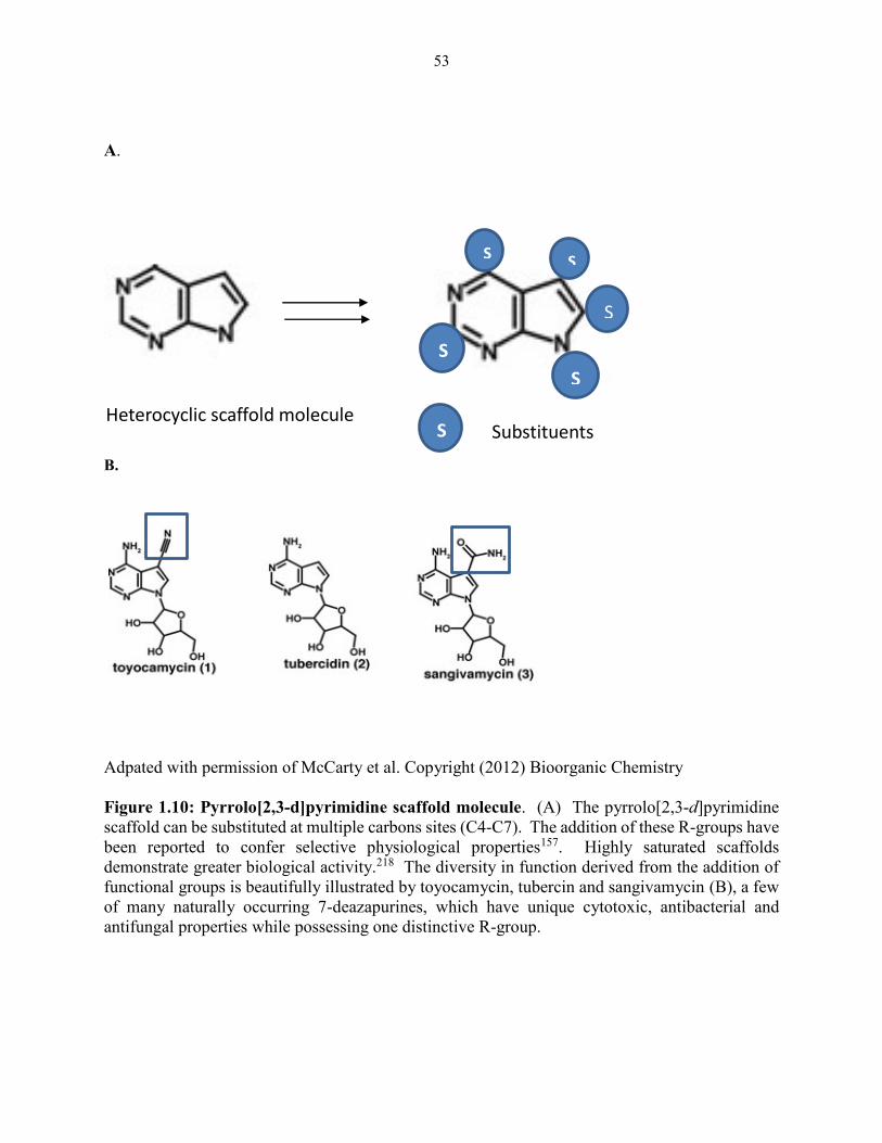
53
A.
B.
Adpated with permission of McCarty et al. Copyright (2012) Bioorganic Chemistry
Figure 1.10: Pyrrolo[2,3-d]pyrimidine scaffold molecule. (A) The pyrrolo[2,3-d]pyrimidine
scaffold can be substituted at multiple carbons sites (C4-C7). The addition of these R-groups have
been reported to confer selective physiological properties157. Highly saturated scaffolds
demonstrate greater biological activity.218 The diversity in function derived from the addition of
functional groups is beautifully illustrated by toyocamycin, tubercin and sangivamycin (B), a few
of many naturally occurring 7-deazapurines, which have unique cytotoxic, antibacterial and
antifungal properties while possessing one distinctive R-group.
Substituents Heterocyclic scaffold molecule
S
S
S
S
S
S
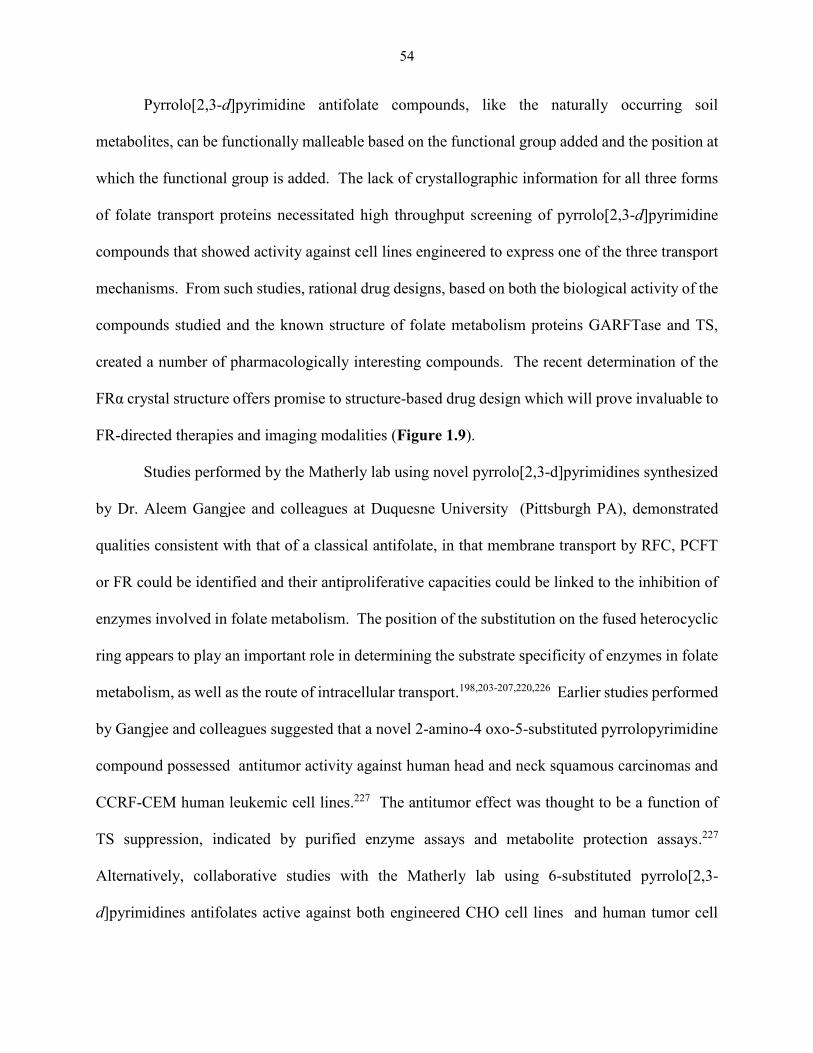
54
Pyrrolo[2,3-d]pyrimidine antifolate compounds, like the naturally occurring soil
metabolites, can be functionally malleable based on the functional group added and the position at
which the functional group is added. The lack of crystallographic information for all three forms
of folate transport proteins necessitated high throughput screening of pyrrolo[2,3-d]pyrimidine
compounds that showed activity against cell lines engineered to express one of the three transport
mechanisms. From such studies, rational drug designs, based on both the biological activity of the
compounds studied and the known structure of folate metabolism proteins GARFTase and TS,
created a number of pharmacologically interesting compounds. The recent determination of the
FRα crystal structure offers promise to structure-based drug design which will prove invaluable to
FR-directed therapies and imaging modalities (Figure 1.9).
Studies performed by the Matherly lab using novel pyrrolo[2,3-d]pyrimidines synthesized
by Dr. Aleem Gangjee and colleagues at Duquesne University (Pittsburgh PA), demonstrated
qualities consistent with that of a classical antifolate, in that membrane transport by RFC, PCFT
or FR could be identified and their antiproliferative capacities could be linked to the inhibition of
enzymes involved in folate metabolism. The position of the substitution on the fused heterocyclic
ring appears to play an important role in determining the substrate specificity of enzymes in folate
metabolism, as well as the route of intracellular transport.198,203-207,220,226 Earlier studies performed
by Gangjee and colleagues suggested that a novel 2-amino-4 oxo-5-substituted pyrrolopyrimidine
compound possessed antitumor activity against human head and neck squamous carcinomas and
CCRF-CEM human leukemic cell lines.227 The antitumor effect was thought to be a function of
TS suppression, indicated by purified enzyme assays and metabolite protection assays.227
Alternatively, collaborative studies with the Matherly lab using 6-substituted pyrrolo[2,3-
d]pyrimidines antifolates active against both engineered CHO cell lines and human tumor cell
55
lines expressing folate transporters, demonstrated inhibition towards GARFTase. GARFTase
suppression was demonstrated via metabolic nucleoside protection assays as well as radioactive
metabolic incorporation assays and isolated enzyme assays.198,205,220 Further, these studies also
revealed that drug activity can be dictated by the length of the carbon bridge region which connects
to either a thienoyl or benzoyl ring system. These studies provided greater insight into the
structural components required for folate-based targeted drug activity.
The most potent, tumor-targeted analogs from these studies were lead compounds AG23,
AG71, and AG94. These three compounds represent three distinct series of novel pyrrolo[2,3-
d]pyrimidine antifolates. While structurally unique, each of the lead compounds was found to
have profound antitumor activity against FRα and PCFT expressing cell lines
(nanomaolar/subnanomolar range) with minimal or greatly reduced activity towards RFC
expressing cell lines.198,203-206 These antipurines are effective inhibitors of de novo purine
synthesis as the studies cited above demonstrated remarkable dose-dependent inhibition of
GARFTase. Mindful of the impressive targeted antitumor activity of these leads, congeners of
these compounds are thoroughly investigated in this body of work to determine how structural
modifications impact membrane transport, inhibition of targeted enzymes and ultimately cell
death.
56
Reprinted with the permission of Mitchell-Ryan et al. (2013). Copyright. J Med Chem.
Figure 1.11: Structure of lead compounds from three distinct series of novel antifolates. Each
compound depicted in this figure contains the 2-amino-4-oxo fused hetercyclic ring structure
(pyrrolo [2,3-d]pyrimidine). Both AG71 and AG94 conclude their carbon bridge with thienoyl
ring system unlike AG23 that contains a benzoyl ring in its second ring system. Each compound
is substituted at the 6 position. All novel pyrrolo[2,3-d]pyrimidine compounds presented in this
work were synthesized by the lab of collaborating medicinal chemist Dr. Aleem Gangjee.
57
1.5 Ovarian cancer
Gonadal tissues in the female reproductive tract originate from mesoderm layer of
embryonic tissue. A vast majority of the female reproductive tracts is developed from the
paramesonephric duct, also known as Mullierian ducts, which is responsible for the formation for
approximately two thirds of the female reproductive tract.228 The ovaries, however, are
descendants from the genital ridge and are involved in the production of eggs and the hormones
that help to nurture and prepare eggs for ovulation.229 During ovulation, the eggs travel through
the Fallopian tube to the uterus where they await fertilization. There are a host of hormones,
cytokines and cells that are involved in the cyclical transition of ovulation and menstruation, some
of which are thought to be influential in ovarian pathogenesis.230-232
Ovarian cancer is a rare but very lethal gynecological pathology in the United States. While
the lifetime risk of developing ovarian cancer is 1.6%, the majority of the women diagnosed will
ultimately die from the disease, as it is the fifth leading cause of cancer death in American
women.232,233 The American Cancer Society 2014 statistics estimates that there will 21,980 new
cases of ovarian which is a negligible decrease from the 2013 estimate of 22,240 cases.233,234 While
there has been a slight decline in the estimated rate of incidence, the mortality rates remain
staggering with the 2014 estimate of 14,270, which is a slight increase from the 2013 estimate of
14,030 deaths.233,234 A vast majority of women diagnosed present at late stage with aggressive
disease that is often inclined to recur as a fatal treatment resistant disease. There are a number of
factors that contribute to late stage (stage III/IV) diagnosis including the lack of appropriate and
reliable screening modalities and the mismanagement of primary disease. The mismanagement of
ovarian cancer, which generally entails the failure to direct patients to experienced gynecological
oncologists for optimal cytoreduction strategies, has a direct impact on the mortality disparities

58
that exist between populations within this disease.235,236 The molecular profiles of ovarian cancer
are identical among African-American and Caucasian women, suggesting that there are no genetic
factors leading to a more aggressive phenotype for African-American women.236,237 However, the
mortality rates are higher among African-American women, which some believe is a function of
mismanagement of disease, unequal application of existing treatments and the perceived socio-
economic status of the patient.235,236,238
Late stage ovarian cancer presents a host of treatment challenges. An overwhelming
majority of patients are diagnosed with late-stage ovarian cancer which is no longer confined to
the ovary. At this time, treatment options are limited and are generally ineffective with gross
toxicities to the patients. Depending on the extent of tumor burden, surgical reduction no longer
exists as an option.236 An additional determinant in choosing a candidate for debulking surgery is
the presence of comorbidities that are contraindications for the procedure.239,240 Independent of
these conundrums, approximately 90% of patients that show initial response to first-line
platinum/taxane therapy will experience a recurrence of treatment resistant disease.240
The term ovarian cancer serves an umbrella to describe a collection of malignant tissues
that are distinguishable by cell type and more recently, by specific molecular events. These
subtypes correspond to different types of epithelia and organs within the female reproductive
system. Traditionally, ovarian cancers were separated in to three major subtypes based on their
histology, including: (1) surface epithelium which includes serous, mucinous, endometroid, clear
cell and Brenner tumors; (2) sex cord-stroma which includes granulosa cell, sertoli cell , sertoli
leydig , thecoma, fibroma and steroid tumors; and (3) germ cell tumors that include dysgerminoma,
yolk sac, embryonal carcinoma, choriocarcinoma and teratomas.241 In line with the initial system
59
of categorizing these tumors, they were further characterized based on clinical behavior as benign,
malignant and intermediate.242
Congruent with the general principles of carcinogenesis, ovarian cancer is typically a
disease of aging with the median age of diagnosis at 63 years.243 Epithelial ovarian cancer (EOC)
is the most common form of this disease that accounts for 90% of ovarian cancers.213 Historically
this subtype of ovarian cancer was thought to arise from ovarian surface epithelium (OSE).244-247
This early perspective on ovarian cancer suggests that OSE is capable of phenotypic plasticity
which is derived from the numerous histological transitions that begin with a stromal ovarian
epithelial inclusion cyst (OEI).247 Through metaplasia, this cyst acquires a Mullerian phenotype
which eventually evolves into a malignant transformation resulting in the multiple subtypes of
EOC.247 Critics of this theory express skepticism due to (1) the failure to identify an ovarian
precursor lesion and (2) there are no reported observations of the transition of an OEI to multiple
EOC subtypes.247 Further, OSE shows no resemblance to EOC and is distinct from Mullerian
epithelia.247 However proponents of this theory offer a theory based on experimental models and
histopathology that suggests that tumor promoting agents have the ability to transform OSE to
malignant tumors.246
A growing number of reports suggest that the ovaries serve only as a secondary site and
that the premalignant lesion is formed at a primary site adjacent to the ovaries.232,247-256 The
“Secondary Mullerian System Theory” was the first suggestion that ovarian cancer originates from
an alternate tissue of origin.257 This theory suggest that pelvic tumors with a mullerian phenotype
(like many ovarian cancers) originate or are derived from mullerian type epithelium (uterus, cervix,
Fallopian tubes) directly by a metaplastic process.247 This theory fostered the hypothesis that most
EOC begin as precursor lesion in the distal Fallopian tube.
60
High grade serous carcinoma (HGSC) is the most common and lethal subtype of EOC,
accounting for 70-80% of the cancers within this category (clear cell carcinoma and endometroid
carcinoma accounts for 10 %).213,232,247 HGSC frequently displays p53 mutations and genomic
instability.213,247 This is in stark contrast to type I low grade carcinomas, which include low grade
serous carcinoma, low grade endometroid carcinoma, clear cell carcinoma, mucinous carcinoma
and Brenner tumors. 211 Type I tumors are described as a clinically non-aggressive disease that
generally present at early stage. 209,243 Type I tumors rarely contain p53 mutations and are typified
by mutations involving KRAS, BRAF, ERBB2, PTEN, CTNNB1, PIK3CA, ARID1A and
PPP2R1A.213,247 Type I and Type II tumors are distinguishable not only by molecular markers,
but they differ in presentation, metastatic potential, response to therapy and prognosis.232
There have been a number of observations that support Fallopian tube involvement in
ovarian cancer, many of which include the identification of putative precursor lesions. BRCA1/2
mutant carriers have a 30-60% increased risk of EOC.232 In one study that examined the Fallopian
tubes of asymptomatic BRCA1/2 mutation carriers along with women from the general population
diagnosed with pelvic serous carcinoma, identified at least one of three putative precancerous
lesions; they include: (1) serous tubal intraepithelial carcinoma (STIC) in the Fallopian tubal
frimbria; (2) p53 signatures; or (3) frimbrial secretory outgrowth.247 STIC are generally located
at the distal end of the Fallopian tube and are characterized by frequent p53 mutations, increased
cell proliferation, DNA damage and a secretory phenotype.252,253,258,259 The multiple shared
characteristics of STIC to HGSC are believed to be suggestive of a histological relation between
the two. Similarities in copy number changes, tubal involvement in HGSC cases, a shared
secretory phenotype of STIC and HGSC all serve to support the hypothesis that HGSC is the clonal
outgrowth of STIC lesions.247,260,261 Further, there is a dramatic 98% reduction of ovarian,

61
Fallopian tube and primary peritoneal cancer risk in BRCA1/2 carriers that underwent prophylactic
bilateral salphingo-oophorectomy which involves the removal of both the ovaries and the
Fallopian tubes.232 The Sectioning and Extensively Examining the FIMbria protocol (SEE-FIM)
which was put in place to maximize the detection of precursor lesions in early Fallopian and
ovarian cancer, demonstrated that women diagnosed with ovarian or primary peritoneal HGSC
present with STIC (40-60%) that have identical mutational status observed in tumors and also
show evidence of tubal involvement (70%).232 Altogether, these studies provide a convincing link
to HGSC and the Fallopian tubes, and point to STIC as the initiating lesion that leads to this
aggressive cancer.
The ovaries are believed to be the secondary tumor site for disease that originates in the
Fallopian tubes. The proposed mechanism by which this occurs is thought to involve the shedding
of STIC cells from the distal Fallopian tubes onto the nearby ovaries during ovulation.247 While
trapped in the ovary, these pre-malignant Fallopian tube-derived cells thrive in the ovulatory
environment where they are exposed to hormones, pro-inflammatory cytokines and reactive
oxygen species (ROS) which all may aid in the clonal expansion or re-initiation of cell proliferation
in the ovary.230,247 This proposed mechanism has helped to construct a new model for the
development of HGSC which differs from the step-wise development observed in type I low grade
carcinomas.247
An additional feature of EOC (including 90% of HGSC) is the overexpression of FRα. In
1991, amino acid sequencing analysis determined that the protein enriched on the surface of non-
mucinous ovarian cancer was FRα.213 This identification led to the emergence of the FR protein
as a tumor marker for this cancer.213 The surface expression of the FRα protein corresponded with
grade and stage of the tumor.170,262,263 Interestingly, tumors (with some degree of FRα expression)

62
treated with chemotherapy (i.e., MTX, tamoxifen and cisplatin/carboplatin) experienced increased
(MTX, Tamoxfien) or stable (cisplatin/carboplatin) expression of FRα.264 Knowing this, drugs
that selectively target FRα can be used as adjuvant therapy in situations where first-line therapies
have failed.
Currently, there is no widely accepted comprehensive tumor progression model for ovarian
cancer, which presents a myriad of challenges in studying this cancer.265 Cell line models (Table
1.1) and mouse models of ovarian cancer, which are the existing experimental tools, have their
limitations and in some cases fail to accurately represent the in vivo (patient) tumor. Molecular
profiling of ovarian cancers has allowed research to distinguish between high grade (type II) and
low grade (type I) tumors.266,267 This categorization of tumors based on molecular markers has
called into question the true histopathology of many of the human ovarian cancer cell lines
currently used in research.268 Cell line contamination may also be a factor contributing to the
conflicting reports and misidentification concerning the histopathology of these experimental cell
lines.269,270 Mouse models of human ovarian cancer make the assumption that the ovary is the
primary site of disease. The current models attempt to create tumors in the ovary through the use
of promoters, many of which are described as leaky.271 Ex vivo implants and use of transgenes
arguably do not mimic the natural evolution of human EOC and are not representative disease
models.271 The limitations inherent in both in vivo and in vitro models present an opportunity for
the development or the investigation of a new model that incorporates an identified and validated
precursor lesion.
While there may be continued debate over the etiology of ovarian cancer and the utility of
the models currently used to study the disease, reproductive, genetic and environmental factors
continue to be implicated as risk factors in this multifactorial malignancy. Like most other cancers,

63
increasing age, inherited genetic cancer syndromes and family history remain important
determinants of ovarian cancer risk.272
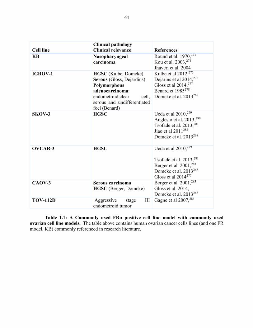
64
Cell line
Clinical pathology
Clinical relevance
References
KB Nasopharyngeal
carcinoma
Round et al. 1970,273
Kou et al. 2003,274
Jhaveri et al. 2004
IGROV-1 HGSC (Kulbe, Domcke)
Serous (Gloss, Dejardins)
Polymorphous
adenocarcinoma:
endometroid,clear cell,
serous and undifferentiated
foci (Benard)
Kulbe et al 2012,275
Dejarins et al 2014,276
Gloss et al 2014,277
Benard et 1985278
Domcke et al. 2013268
SKOV-3 HGSC Ueda et al 2010,279
Anglesio et al. 2013,280
Tsofade et al. 2013,281
Jiao et al 2011282
Domcke et al. 2013268
OVCAR-3 HGSC Ueda et al 2010,279
Tsofade et al. 2013,281
Berger et al. 2001,283
Domcke et al. 2013268
Gloss et al 2014277
CAOV-3 Serous carcinoma
HGSC (Berger, Domcke)
Berger et al. 2001,283
Gloss et al. 2014,
Domcke et al. 2013268
TOV-112D Aggressive stage III
endometroid tumor
Gagne et al 2007,284
Table 1.1: A Commonly used FRα positive cell line model with commonly used
ovarian cell line models. The table above contains human ovarian cancer cells lines (and one FR
model, KB) commonly referenced in research literature.
65
Reproductive risk factors
Ovulation has been thought to contribute to malignant transformation of the epithelium due
to proliferation that occurs during this process. This proliferation is believed to promote
carcinogenesis via the accumulation of genetic mutations in a repetitive “trauma-and-repair”
cycle.265,272 Therefore, agents that reduce ovulation should have a protective effect against the
development of disease.285-287 Indeed, oral contraceptive use has been shown to have an inverse
relationship with ovarian cancer risk in a number of epidemiological studies.285-287 Gestation also
temporarily suppresses ovulation; therefore parity, number of pregnancies and long term breast
feeding have all been reported to have protective effects against the development of ovarian
cancer.265,272,288
Genetic risk factors
Women with a family history of ovarian cancer have an increased risk of cancer
development. Women with one or more first-degree relatives with ovarian cancer have an
increased life-time risk of ovarian cancer development of 5-9%.272 Hereditary cancer syndromes,
like BRCA1/2 mutations, also dramatically increases the risk of developing ovarian cancer;
however, this only accounts for approximately 5-12% of cases.289 Most ovarian cancer cases arise
from sporadic mutations that may not be associated with genes found in hereditary breast and
ovarian cancer (HBOC) and hereditary nonpolyposis colorectal cancer (HNPCC) syndromes.265,272
Aberrations in TP53, HER2, EGFR, DAB2, AKT, RAS, MYC, PI3K and AR have been suggested
to play some role in sporadic cases129,130 , yet mechanistic details of their involvement in
carcinogenesis have not been thoroughly investigated.
Environmental risk factors
66
Obesity is a common modifier of cancer risk in nearly all types of cancer. Obesity has
been linked to inflammation and there is mounting evidence that links both obesity and
inflammation to cancer risk. 129,130 Pelvic inflammatory disease and endometriosis exposes the
ovaries and other reproductive organs to prolonged inflammation that may increase ovarian cancer
risk.290-294
There have been conflicting reports on the role that genital talcum powder exposure plays
in the development of ovarian cancer.295 The source surrounding much of this controversy lies in
the fact that prior to 1976 talc was contaminated with asbestos and much of the data was thought
to be confounded due to the presence of cancer causing substance.295 However, it has been
postulated that ovarian cancers can arise from exposure to hydrous magnesium silicates (e.g.,
talcum powder) and recent findings are not tainted by the asbestos association.295,296 Cancerous
ovaries removed from patients have been shown to contain traces of talc.272
Early detection and prevention
To date, there is no reliable screening method to detect early stage ovarian cancers.
Transvaginal ultrasounds, CA (Cancer Antigen)-125 serum levels and pelvic exams are the
primary detection/screening tools.129,130,297 Together, these methods are deemed inefficient or
inadequate due to their inability to capture subtle changes that may occur during tumor initiation
and early cancer progression in ovarian cancer.297 These modalities also fail to possess the
specificity and sensitivity required for practical positive predictive values.297 The absence of a
defined and validated precursor lesion only adds to the challenge in developing a proper screening
modality.129,130 There are currently efforts to identify new biomarkers to replace or to use in
conjunction with tools that are currently available.298
Current treatments for ovarian cancer

67
Ideally, patients who are declared good surgical candidates are recommended for
cytoreductive surgery followed by adjuvant chemotherapy.129,138,139 Unfortunately, at stages I-II
of disease, patients with EOC are asymptomatic or symptoms are easily mistaken for innocuous
conditions.299 This, in conjunction with the lack of adequate early detection screening, results in
infrequent early stage-diagnosis.129, 297,139 Vaginal bleeding/discharge, frequent urination and
constipation are typical symptoms of early stage ovarian cancer but are often ignored because they
are common symptoms of less severe minor gynecological abnormalities.129 Approximately 1.5%
of patients present with early stage disease for whom complete surgical removal of the tumor can
be successful and survival is greater than 90% (when cancer is confined to the ovaries); therefore,
chemotherapy is the primary treatment option for a vast majority of cases.129, 130, 297,300 Over the
past 40 years, standard therapies used to treat EOC have not significantly extended the median
overall survival beyond 25-30% in women diagnosed with advanced disease.130,137 Approximately
75-80% of ovarian cancers exhibit initial chemotherapy sensitivity to first line taxane- and
platinum-based agents (paclitaxel/docetaxel and cisplatin/carboplatin). However, most of these
patients will experience relapse within a year.258,301 Due to the chronic recurring nature of EOC,
treatment strategies should allow for prolonged treatment by using agents with low toxicity
profiles, to ensure reasonable patient comfort and minimal disruption to the quality of life for the
patient.
There are well recognized cumulative and sometimes irreversible toxicities associated with
these standard agents (regardless of method of delivery) which makes additional treatment with
these classes of agents intolerable for patients, thereby reducing the available options in treatment
and creating a precipitous decline in patients’ quality of life.300-302 Hematological and neurological
toxicities are of greatest concern as they may impede the patient’s ability to carry out everyday

68
activities. With this in mind, there is growing support to include “improved quality of life” along
with “duration of survival” as major considerations in the therapy of advanced cancers.300
69
CHAPTER 2: DISCOVERY OF 5-SUBSTITUTED PYRROLO[2,3-D]PYRIMIDINE
ANTIFOLATES AS DUAL ACTING INHIBITORS OF GARFTASE AND AICARFTASE
IN DE NOVO PURINE NUCLEOTIDE BIOSYNTHESIS: IMPLICATIONS OF
INHIBITING AICARFTASE TO AMPK ACTIVATION AND ANTI-TUMOR ACTIVITY
2.1 Introduction
The antifolates are widely recognized for their inhibition of folate metabolism that leads to
growth suppression, tumor reduction and remission in a number of malignancies and the reduction
of inflammation in pro-inflammatory conditions.38,303,304 Major enzyme targets of this particular
class of agents include TS and DHFR. Inhibition of these enzymes suppress de novo nucleotide
biosynthesis, resulting in an imbalance of purine and pyrimidine precursors, rendering cells
incapable of undergoing accurate DNA replication, ultimately resulting in cell death. Clinically
relevant TS and DHFR inhibitors, typified by PMX, and MTX (Figure 2.1) and pralatrexate,
respectively, continue to play important roles in treating hematologic malignancies and solid
tumors.25,303
Antifolates targeting de novo purine nucleotide biosynthesis were also described and
include LMTX [(6R)5,10-dideazatetrahydrofolate] (Figure 2.1), (2S)-2-[[5-[2-[(6R)-2-amino-4-
oxo-5,6,7,8-tetrahydro-1H-pyrido[2,3-d]pyrimidin-6-yl]ethyl]thiophene-2-
carbonyl]amino]pentanedioic acid (LY309887)188,305 and (2S)-2-[[5-[2-[(6S)-2-amino-4-oxo-
1,6,7,8-tetrahydropyrimido[5,4-bate][1,4]thiazin-6-yl]ethyl]thiophene-2-
carbonyl]amino]pentanedioic acid (AG2034).183 These drugs inhibit the first folate-dependent
step in purine biosynthesis, catalyzed by GARFTase, and have progressed to clinical trials.69,306,307
However, their toxicities were dose-limiting, most likely a consequence of their cellular uptake
and metabolism to polyglutamates in normal tissues.
The RFC is one of three principal mechanisms of (anti)folate uptake into mammalian
cells.308 Cellular requirements for folate cofactors for DNA replication provide a plausible
70
explanation for the high levels of RFC in most tumors. However, demands for folates, and
consequently RFC, are also shared by normal tissues, such that RFC may not be the optimal
mechanism for tumor-selective uptake of cytotoxic folate analogs. Other cellular uptake
mechanisms, notably the PCFT and FRs α and β, are also expressed in tumors, while showing
more restricted expression in normal tissues.92 Furthermore, PCFT is a proton symporter such that
in the acidic microenvironment generated by glycolytic tumors, membrane transport and selective
tumor targeting by this mechanism is enhanced.75,88 For FRα, the apical spatial orientation in
normal epithelial tissues is often disrupted in tumors such that its basolateral membrane expression
in tumors results in exposure to the circulation.88,92 These features provide compelling rationale
for developing folate-based therapeutics which target PCFT and FRα for cancer therapy.
Examples of FRα-targeted therapies include a monoclonal antibody [Farletuzumab
(Morphotech)]138, a cytotoxic folate conjugate, N-[4-[[(2-amino-1, 4-dihydro-4-oxo-
6pteridinyl)methyl]amino]benzoyl]-L-(-glutamyl-L-α-aspartyl-L-α-aspartyl-L-cysteine [e.g.,
Vintafolide (EC145; Endocyte)],211 and a classical antifolate which is selectively transported into
cells by FRs over RFC and inhibits de novo thymidylate biosynthesis, [(2R)-2-((4R)-4-carboxy-5-
(4-((2-(hydroxymethyl)-4-oxo-4,6,7,8-tetrahydro-3H-cyclopenta[g]quinazolin-6-yl)(prop-2-yn-
1-yl)amino)phenyl)-5-oxopentanamido)pentanedioic acid] (ONX0801).202
PMX is a 5-substituted pyrrolo[2,3-d]pyrimidine antifolate with a 2 carbon bridge linked
to a p-aminobenzoyl glutamate. PMX is a good transport substrate for RFC and among the best
PCFT substrates,69,75,92 yet it is a modest substrate for FRs.198,205-207,220 We previously described
6-substituted pyrrolo[2,3-d]pyrimidine benzoyl antifolates related to PMX (compounds AG17,
AG23, AG71 and AG94 (Figure 2.2).198,204 Whereas the 6-regioisomer of PMX was
pharmacologically inert, the 3- and 4-bridge carbon analogs of this series (compounds AG17 and
71
AG23, respectively) were selectively transported by FRα- and PCFT over RFC and were potently
inhibitory toward FRα- and PCFT-expressing tumor cells.113,117 Subsequent generation 6-
substituted pyrrolo[2,3-d]pyrimidine thienonyl antifolates (compounds AG71 and AG94) with 3-
and 4- carbon bridge lengths, respectively, were even more potent as inhibitors of cell proliferation
then that of the corresponding benzoyl analogs203,207,226. Compounds AG17, AG23, AG71 and
AG94 all showed substantial PCFT membrane transport activity but not for RFC.
PMX was originally described as a “multi-targeted” antifolate with a primary inhibition at
TS and secondarily at GARFTase and DHFR.309 However, recent studies suggested that although
TS was the primary cellular target of PMX, AICARFTase could be a pharmacologically important
target in the presence of excess thymidine (circumvents TS) at sufficiently high PMX
concentrations.196,197 Hence, in the presence of thymidine, inhibition of proliferation of CCRF-
CEM leukemia cells and solid tumor cell lines was circumvented by hypoxanthine but only
partially by AICA which is metabolized to ZMP, the AICARFTase substrate.170,171 Further, PMX
resulted in marked accumulations of ZMP. By contrast, the 6-substituted pyrrolo[2,3-d]pyrimidine
analogs AG17, AG23 AG71 and AG94 are inhibitors of GARFTase whose inhibitory effects are
completely circumvented by AICA.113,117,207,226
The accumulation of ZMP in PMX-treated cells is intriguing as ZMP is an AMP mimetic
that activates AMP-activated protein kinase (AMPK).310 AMPK negatively regulates mTOR, a
critical pro-survival pathway that is activated in many tumor cells along with PI3K/AKT,
secondary to loss or mutation of PTEN.310-312 This may provide a possible explanation for the
inhibitory effects of PMX in the presence of thymidine, as purine nucleotides are not
depleted.170,171 However, this has not been directly tested, as no AICARFTase-targeted drugs
without TS inhibition have been described.

72
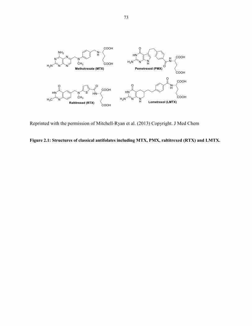
73
Reprinted with the permission of Mitchell-Ryan et al. (2013) Copyright. J Med Chem
Figure 2.1: Structures of classical antifolates including MTX, PMX, raltitrexed (RTX) and LMTX.
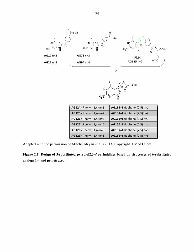
74
Adapted with the permission of Mitchell-Ryan et al. (2013) Copyright. J Med Chem
Figure 2.2: Design of 5-substituted pyrrolo[2,3-d]pyrimidines based on structures of 6-substituted
analogs 1-4 and pemetrexed.
75
In this chapter, we describe, synthesize and perform a systematical characterization of the
in vitro anti-proliferative activities and cellular mechanisms of novel 5-substituted pyrrolo[2,3-
d]pyrimidine antifolates (including PMX) with a benzoyl (AG124-AG129) or thienoyl (AG133-
AG138) rings in the side chain and one-to-six bridge carbons closely related to the 6-substituted
pyrrolo[2,3-d]pyrimidine analogs AG17, AG23, AG71 and AG94113,117207,226 (Figure 2.2). We
reasoned that the 5-substituted analogs might inhibit AICARFTase, analogous to PMX, albeit
without TS inhibition. Further, if this was accompanied by FRα and/or PCFT transport selectivity
over RFC, this would be especially appealing for targeting tumors that express high levels of these
systems.
We describe in this chapter the impact of these structural modifications on (i) transporter
specificity and (ii) the targeted pathway (TS versus de novo purine nucleotide biosynthesis),
including (iii) the extent of cellular GARFTase and AICARFTase inhibition. Our results
document an emerging structure–activity-relationship (SAR) for the pyrrolo[2,3-d]pyrimidine
antifolates, accompanying translocation of the 6-pyrrolo[2,3-d]pyrimidine substituent to the regio
5-position, including sustained FRα cellular uptake accompanying restoration of RFC and loss of
PCFT transport activities. The 4 carbon bridge analog of the benzoyl series (compound AG127)
is unique in that it provides the strongest evidence of dual inhibition of both AICARFTase and
GARFTase exclusive of TS in tumor cells, resulting in depletion of purine nucleotides. Finally,
our results directly examine the possibility of whether AMPK activation secondary to
AICARFTase inhibition contributes to decreased proliferation of KB human tumor cells.
2.2 Biological evaluation
Transporter-selectivity for inhibition of cell proliferation by 5- and 6-substituted
pyrrolo[2,3-d]pyrimidine regioisomers. Our initial goal was to better define the SARs for
76
membrane transport of pyrrolo[2,3-d]pyrimidine antifolates relating to the impact of 5- versus 6-
substitutions on the pyrrole ring, based on the established antitumor activities of the 6-pyrrolo[2,3-
d]pyrimidine-substituted compounds AG17, AG23, AG71 and AG94 with 3- and 4-bridge
carbons, respectively, 113,117,156,228 in addition to the preclinical and clinical efficacy of PMX,196 a
5-substituted pyrrolo[2,3-d]pyrimidine with a 2-carbon bridge (Figure 2.2). In our previous
studies,113,117,207,226 compounds AG17, AG23, AG71 and AG94 were efficiently internalized by
tumor cells via FRα and PCFT but not by RFC, whereas PMX was a substrate by all three
systems.113,117
Transporter specificities are exemplified by patterns of inhibition of cell proliferation
toward isogenic Chinese hamster ovary (CHO) cell models, engineered to express human RFC
(PC43-10), PCFT (R2/PCFT4), or FRα (RT16),198,205-207,220 treated with compounds AG17,
AG23, AG71 and AG94 (Table 2.1). Results are compared to those for RFC-, PCFT-, and FR-
null MTXRIIOuaR2-4 (R2) CHO cells. As previously reported, 113,117,207,226 compounds AG17,
AG23, AG71 and AG94 are potent inhibitors of FR-expressing RT16 and D4 cells, and to a
lesser (albeit still potent) extent, R2/PCFT4 cells. However, compounds AG17, AG23, AG71
and AG94 were poorly inhibitory toward RFC-expressing PC43-10 and R2 cells. Compounds
AG17, AG23, AG71 and AG94 were also highly active toward KB nasopharengeal carcinoma
cells and IGROV-1 ovarian carcinoma cells, both of which express elevated FRα, along with
RFC and PCFT.220 Complete reversal of antiproliferative effects of compounds AG17, AG23,
AG71 and AG94 toward the FRα-expressing cell lines was seen in the presence of excess (200
nM) folic acid, further demonstrating that cellular uptake of these analogs are mediated primarily
via FRα, as previously reported (Table 2.1).198,207
77
5-Substituted pyrrolo[2,3-d]pyrimidine analogs with a side-chain benzoyl group (analogs
of compounds AG17and AG23) and thienoyl group (analogs of compounds AG71 and AG94) with
bridge lengths from 1 to 6 carbons (compounds AG124-129 and AG133-138) were synthesized
and tested for their antiproliferative activities toward the CHO and human tumor models (Table
2.1). For the benzoyl series, the 2 carbon 5-pyrrolo[2,3-d]pyrimidine analog (compound AG125)
is structurally identical to PMX. 5-Substitutions restored RFC-directed anti-proliferative activity,
as reflected in dramatically increased anti-proliferative effects of this series toward PC43-10 cells
compared to RFC-, PCFT-, and FR-null R2 cells, in the order (numbers of bridge carbons noted in
parentheses), AG125 (2C) > AG127 (4C) ~AG126 (3C) > AG128 (5C) >> AG124 (1C) ~AG129
(6C) (benzoyl series), and AG134 (2C) ~AG136 (4C) > AG135 (3C) > AG137 (5C) >> AG133
(1C) ~ AG138 (6C) (thiophene series). Conversely, for PCFT-expressing R2/PCFT4 CHO cells,
with the exception of compound AG125 and (to a lesser extent) the thiophene analogs AG134 and
AG137, the antiproliferative effects for the 5-substituted analogs were attenuated compared to the
6-substituted analogs (e.g., for direct comparisons between paired 5- and 6-substituted analogs,
compare IC50s for compounds AG17 and AG126 and for compounds AG71 and AG135,
respectively in Table 1.1).
With the FRα-expressing sublines, potencies were also reduced for the 5-substituted
compounds, compared to 6-substituted analogs. These differences range from slight (compare
AG23 to AG127, and AG71 to AG28) to substantial (compare AG17 to AG126, AG71 to AG135,
and AG94 to AG136) (Table 2.1). In contrast to results for PCFT- and RFC-expressing cells, the
impacts of bridge lengths were distinctly different between the benzoyl and thienoyl series, with
anti-proliferative activities toward FRα-expressing RT16 CHO cells in rank order, AG127 (4C) >
AG125 (2C) > AG128 (5C) > AG126 (3C) > > AG124 (1C) ~ AG129 (6C) (benzoyl series), and
78
AG136 (4C) ~ AG137 (5C) > AG135 (3C) > AG134 (2C) > AG133 (1C) ~ AG138 (6C) (thioenoyl
series). An analogous pattern of sensitivities was obtained with KB human tumor cells, and for
both the KB and RT16 cell lines, inhibitory effects were substantially reversed by 200 nM folic
acid, confirming FRα as the major uptake mechanism. While PCFT and RFC likely mediate
cellular uptake of certain analogs (e.g., AG127 and AG136), uptake by FRα appears to
predominate.
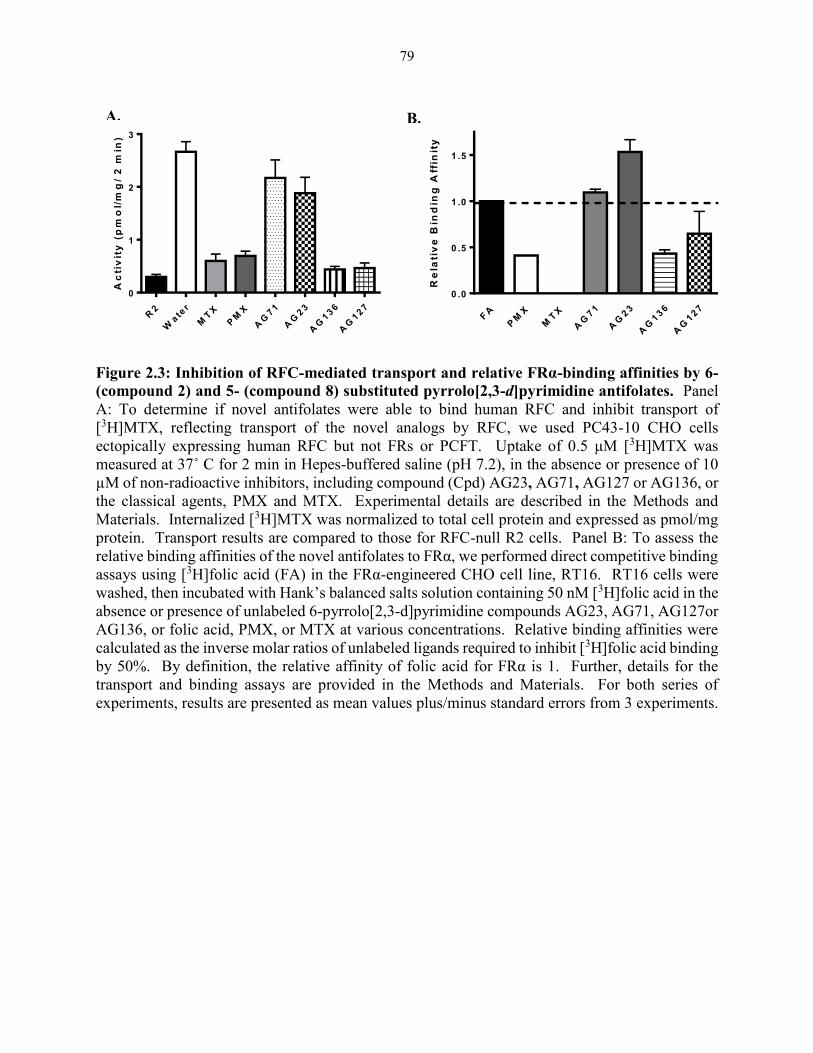
79
Figure 2.3: Inhibition of RFC-mediated transport and relative FRα-binding affinities by 6-
(compound 2) and 5- (compound 8) substituted pyrrolo[2,3-d]pyrimidine antifolates. Panel
A: To determine if novel antifolates were able to bind human RFC and inhibit transport of
[3H]MTX, reflecting transport of the novel analogs by RFC, we used PC43-10 CHO cells
ectopically expressing human RFC but not FRs or PCFT. Uptake of 0.5 μM [3H]MTX was
measured at 37˚ C for 2 min in Hepes-buffered saline (pH 7.2), in the absence or presence of 10
µM of non-radioactive inhibitors, including compound (Cpd) AG23, AG71, AG127 or AG136, or
the classical agents, PMX and MTX. Experimental details are described in the Methods and
Materials. Internalized [3H]MTX was normalized to total cell protein and expressed as pmol/mg
protein. Transport results are compared to those for RFC-null R2 cells. Panel B: To assess the
relative binding affinities of the novel antifolates to FRα, we performed direct competitive binding
assays using [3H]folic acid (FA) in the FRα-engineered CHO cell line, RT16. RT16 cells were
washed, then incubated with Hank’s balanced salts solution containing 50 nM [3H]folic acid in the
absence or presence of unlabeled 6-pyrrolo[2,3-d]pyrimidine compounds AG23, AG71, AG127or
AG136, or folic acid, PMX, or MTX at various concentrations. Relative binding affinities were
calculated as the inverse molar ratios of unlabeled ligands required to inhibit [3H]folic acid binding
by 50%. By definition, the relative affinity of folic acid for FRα is 1. Further, details for the
transport and binding assays are provided in the Methods and Materials. For both series of
experiments, results are presented as mean values plus/minus standard errors from 3 experiments.
R2
Wate
r
MT
X
PM
X
AG
71
AG
23
AG
136
AG
127
0
1
2
3
Ac
tiv
ity
(p
mo
l/m
g/
2 m
in)
FA
PM
X
MT
X
AG
71
AG
23
AG
136
AG
127
0 .0
0 .5
1 .0
1 .5
Re
lati
ve
Bin
din
g A
ffin
ity
A. B.
80
To complement results showing anti-proliferative effects of the most potent 5-substituted
analogs, AG127 and AG136, toward PC43-10 CHO cells expressing human RFC, we measured
competitive inhibitions of [3H]MTX (0.5 µM) transport in this model over 2 minutes at 37° C
(Figure 2.3A). MTX is a well-characterized surrogate substrate for RFC and the extent of
[3H]MTX transport inhibition by competitive unlabeled (anti)folate ligands correlates with their
membrane transport by RFC.9 For our study, we compared inhibitory effects of 10 μM of
compound AG127 and AG136, to those for the corresponding 6-substituted compound AG23 and
AG71, and to PMX and MTX. Transporter-null R2 cells were used to as a measure of baseline
[3H]MTX uptake. Whereas AG23 and AG71 were poor inhibitors of [3H]MTX uptake (< 25%
inhibition), compound AG127 and AG136 were a reasonably good inhibitors (>90%), with a
inhibitory potency approximating that for PMX and net transport only slightly greater than the
very low levels recorded for R2 cells. Clearly, unlike the 6-substituted pyrrolo[2,3-d]pyrimidine
analogs, the 5-substituted compounds lack specificity for FRα and/or PCFT over RFC.
Compounds AG127 and AG136 showed anti-proliferative effects toward FRα-expressing
RT16 CHO cells and KB human tumor cells (Table 2.1). To directly demonstrate drug binding to
FRα, a prerequisite of cellular uptake by this endocytotic mechanism, we used competitive binding
assays with [3H]folic acid with RT16 cells, treated with unlabeled ligands which compete for FRα
binding16-20 (Figure 2.3B). Relative FRα binding affinities for the 5- (compounds AG127 and
AG136) versus 6-substituted (compounds AG23 and AG71) analogs paralleled their anti-
proliferative activities. However, this correlation did not quite extend to PMX (compound AG125)
or to MTX, indicating that multiple factors in addition to FRα binding affinities contribute to net
growth inhibition of FRα-expressing cells for these antifolates.
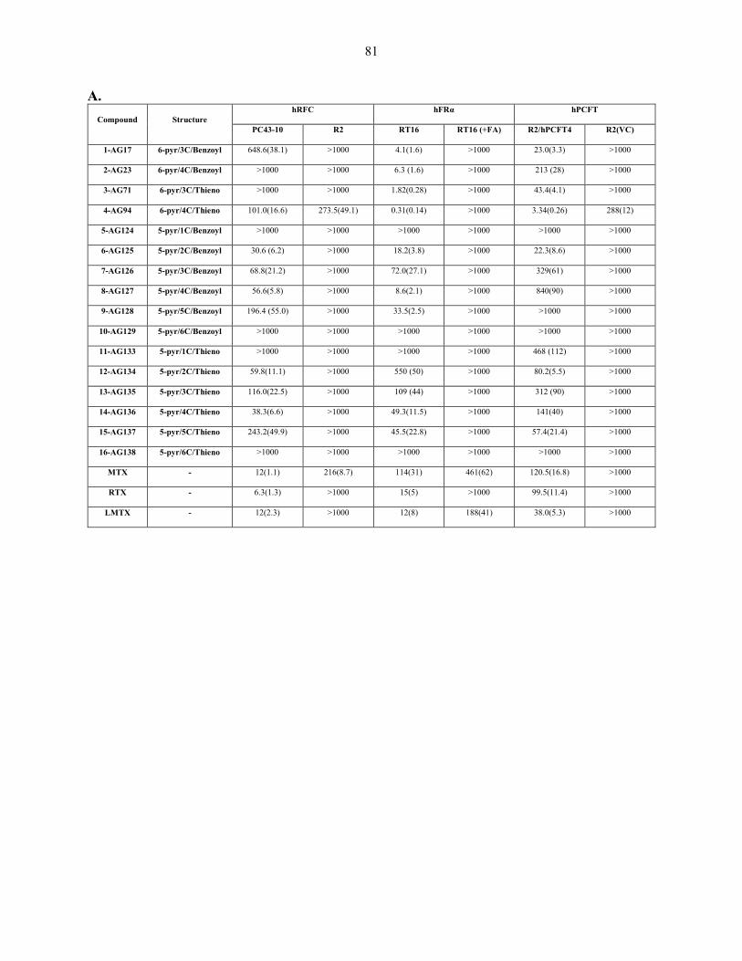
81
A.
Compound Structure hRFC hFRα hPCFT
PC43-10 R2 RT16 RT16 (+FA) R2/hPCFT4 R2(VC)
1-AG17 6-pyr/3C/Benzoyl 648.6(38.1) >1000 4.1(1.6) >1000 23.0(3.3) >1000
2-AG23 6-pyr/4C/Benzoyl >1000 >1000 6.3 (1.6) >1000 213 (28) >1000
3-AG71 6-pyr/3C/Thieno >1000 >1000 1.82(0.28) >1000 43.4(4.1) >1000
4-AG94 6-pyr/4C/Thieno 101.0(16.6) 273.5(49.1) 0.31(0.14) >1000 3.34(0.26) 288(12)
5-AG124 5-pyr/1C/Benzoyl >1000 >1000 >1000 >1000 >1000 >1000
6-AG125 5-pyr/2C/Benzoyl 30.6 (6.2) >1000 18.2(3.8) >1000 22.3(8.6) >1000
7-AG126 5-pyr/3C/Benzoyl 68.8(21.2) >1000 72.0(27.1) >1000 329(61) >1000
8-AG127 5-pyr/4C/Benzoyl 56.6(5.8) >1000 8.6(2.1) >1000 840(90) >1000
9-AG128 5-pyr/5C/Benzoyl 196.4 (55.0) >1000 33.5(2.5) >1000 >1000 >1000
10-AG129 5-pyr/6C/Benzoyl >1000 >1000 >1000 >1000 >1000 >1000
11-AG133 5-pyr/1C/Thieno >1000 >1000 >1000 >1000 468 (112) >1000
12-AG134 5-pyr/2C/Thieno 59.8(11.1) >1000 550 (50) >1000 80.2(5.5) >1000
13-AG135 5-pyr/3C/Thieno 116.0(22.5) >1000 109 (44) >1000 312 (90) >1000
14-AG136 5-pyr/4C/Thieno 38.3(6.6) >1000 49.3(11.5) >1000 141(40) >1000
15-AG137 5-pyr/5C/Thieno 243.2(49.9) >1000 45.5(22.8) >1000 57.4(21.4) >1000
16-AG138 5-pyr/6C/Thieno >1000 >1000 >1000 >1000 >1000 >1000
MTX - 12(1.1) 216(8.7) 114(31) 461(62) 120.5(16.8) >1000
RTX - 6.3(1.3) >1000 15(5) >1000 99.5(11.4) >1000
LMTX - 12(2.3) >1000 12(8) 188(41) 38.0(5.3) >1000
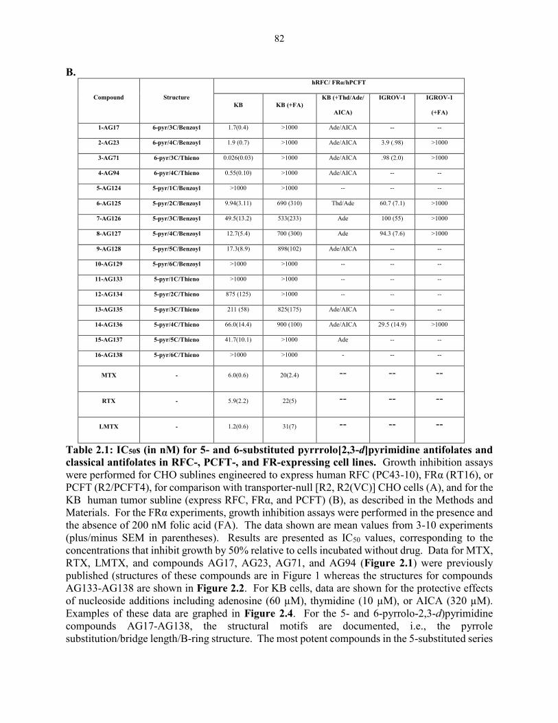
82
B.
Compound Structure
hRFC/ FRα/hPCFT
KB KB (+FA) KB (+Thd/Ade/
AICA)
IGROV-1 IGROV-1
(+FA)
1-AG17 6-pyr/3C/Benzoyl 1.7(0.4) >1000 Ade/AICA -- --
2-AG23 6-pyr/4C/Benzoyl 1.9 (0.7) >1000 Ade/AICA 3.9 (.98) >1000
3-AG71 6-pyr/3C/Thieno 0.026(0.03) >1000 Ade/AICA .98 (2.0) >1000
4-AG94 6-pyr/4C/Thieno 0.55(0.10) >1000 Ade/AICA -- --
5-AG124 5-pyr/1C/Benzoyl >1000 >1000 -- -- --
6-AG125 5-pyr/2C/Benzoyl 9.94(3.11) 690 (310) Thd/Ade 60.7 (7.1) >1000
7-AG126 5-pyr/3C/Benzoyl 49.5(13.2) 533(233) Ade 100 (55) >1000
8-AG127 5-pyr/4C/Benzoyl 12.7(5.4) 700 (300) Ade 94.3 (7.6) >1000
9-AG128 5-pyr/5C/Benzoyl 17.3(8.9) 898(102) Ade/AICA -- --
10-AG129 5-pyr/6C/Benzoyl >1000 >1000 -- -- --
11-AG133 5-pyr/1C/Thieno >1000 >1000 -- -- --
12-AG134 5-pyr/2C/Thieno 875 (125) >1000 -- -- --
13-AG135 5-pyr/3C/Thieno 211 (58) 825(175) Ade/AICA -- --
14-AG136 5-pyr/4C/Thieno 66.0(14.4) 900 (100) Ade/AICA 29.5 (14.9) >1000
15-AG137 5-pyr/5C/Thieno 41.7(10.1) >1000 Ade -- --
16-AG138 5-pyr/6C/Thieno >1000 >1000 - -- --
MTX - 6.0(0.6) 20(2.4) -- -- --
RTX - 5.9(2.2) 22(5) -- -- --
LMTX - 1.2(0.6) 31(7) -- -- --
Table 2.1: IC50s (in nM) for 5- and 6-substituted pyrrrolo[2,3-d]pyrimidine antifolates and
classical antifolates in RFC-, PCFT-, and FR-expressing cell lines. Growth inhibition assays
were performed for CHO sublines engineered to express human RFC (PC43-10), FRα (RT16), or
PCFT (R2/PCFT4), for comparison with transporter-null [R2, R2(VC)] CHO cells (A), and for the
KB human tumor subline (express RFC, FRα, and PCFT) (B), as described in the Methods and
Materials. For the FRα experiments, growth inhibition assays were performed in the presence and
the absence of 200 nM folic acid (FA). The data shown are mean values from 3-10 experiments
(plus/minus SEM in parentheses). Results are presented as IC50 values, corresponding to the
concentrations that inhibit growth by 50% relative to cells incubated without drug. Data for MTX,
RTX, LMTX, and compounds AG17, AG23, AG71, and AG94 (Figure 2.1) were previously
published (structures of these compounds are in Figure 1 whereas the structures for compounds
AG133-AG138 are shown in Figure 2.2. For KB cells, data are shown for the protective effects
of nucleoside additions including adenosine (60 µM), thymidine (10 µM), or AICA (320 µM).
Examples of these data are graphed in Figure 2.4. For the 5- and 6-pyrrolo-2,3-d)pyrimidine
compounds AG17-AG138, the structural motifs are documented, i.e., the pyrrole
substitution/bridge length/B-ring structure. The most potent compounds in the 5-substituted series

83
(AG127 and AG136), along with their 6- substituted analogs (AG23 and AG71) and PMX
(AG125) were evaluated for anti-tumor activity in the human ovarian cancer cell line IGROV-1.
Undefined abbreviations: Cpd, compound; ND, not determined. The results with compounds
AG17, AG23, AG71 and AG94 were previously reported.198,204,207,226
84
Inhibition of de novo purine nucleotide biosynthesis by both 5- and 6-pyrrolo[2,3-
d]pyrimidine compounds leads to a cytotoxic response via purine nucleotide depletion. De
novo purine nucleotide biosynthesis includes 10 reactions by which phosphoribosyl pyrophosphate
(PRPP) is converted into inosine monophosphate (IMP), the precursor of AMP and GMP (refer to
Figure 1.3 in chapter 1). There are two folate-dependent enzymes in the pathway which are
possible targets for folate-based therapies i.e., GARFTase (catalyzes steps 2, 3 and 5) and
AICARFTase (catalyzes steps 9 and 10). Previous studies established that GARFTase was the
intracellular enzyme target for LMTX183,313 and for compounds AG17, AG23, AG71 and
AG94.198,207 For PMX, TS is the primary intracellular target, although modest inhibitions of
GARFTase and DHFR were also reported.309 Most recently, AICARFTase was implicated as a
potentially important secondary enzyme target for PMX (in the presence of excess thymidine to
circumvent TS inhibition) by nucleoside protection experiments and metabolic assays.196,197
To identify the targeted pathway for 6-substituted compounds AG23 and AG71, we
previously used nucleoside protection experiments with adenosine (60 µM) and thymidine (10
µM), to distinguish de novo purine nucleotide from thymidylate biosynthesis, respectively.198,204-
207,220,313 To further identify the likely folate metabolizing enzyme targets in purine nucleotide
biosynthesis (GARFTase versus AICARFTase), cells were treated with the antifolates in the
presence of AICA (320 µM), which is metabolized to AICA ribonucleotide (ZMP), the substrate
for AICARFTase substrate, thus bypassing the step catalyzed by GARFTase198,204-207,220,313
(Figure 1.3).
We used this approach for KB cells treated with compounds AG125-128 and AG135-
AG137, with results compared to those for compounds AG23 and AG71, and to PMX (compound
AG125) (Table 2.1; Figure 2.4 shows the nucleoside protection results for PMX and compounds
85
AG23 and AG71 compared to those for compounds AG127 and AG136). With compounds AG23
and AG71, both adenosine and AICA were completely protective, establishing de novo purine
biosynthesis and GARFTase as the principal targets.198,204,207 With PMX, thymidine, adenosine,
and AICA were all partially protective, albeit to different extents. Combined thymidine and
adenosine completely protected KB cells from the growth inhibitory effects of PMX (not shown).
The growth inhibitory effects of the 5-substituted compounds AG126-AG128, AG135 and 137
with KB cells were unaffected by excess thymidine but were completely reversed in the presence
of adenosine alone, indicating that exclusively de novo purine nucleotide synthesis was being
targeted (rather than combined thymidylate and purine nucleotide synthesis). Whereas AICA
alone completely protected KB cells from the inhibitory effects of compound AG128 andAG135,
AICA alone was only partially protective with compounds AG126, AG127 and AG137.
Thus, inhibition of purine nucleotide biosynthesis by compounds AG126, AG127 and
AG136 is likely a composite effect of dual inhibitions involving both GARFTase and
AICARFTase. These results strongly imply that shifting the substituent from the 6- to the 5-
position on the pyrrolo[2,3-d]pyrimidine ring system results in an altered inhibition of the targeted
enzymes.
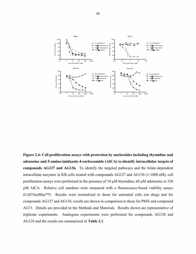
86
P M X
0 .1 1 1 0 1 0 0 1 0 0 0 1 0 0 0 0
0
2 5
5 0
7 5
1 0 0
1 2 5
N o A d d it io n s
6 0 M A d e n o s in e
1 0 M T h y m id in e
3 2 0 M A IC A
C o n c e n tra t io n (n M )
Pe
rc
en
t G
ro
wth
A G 7 1
0 .0 1 0 .1 1 1 0 1 0 0
0
2 5
5 0
7 5
1 0 0
1 2 5
N o A d d itio n s
60 M A d e n o s in e
10 M T h y m id in e
3 2 0 M A IC A
C o n c e n tra t io n (n M )
Pe
rc
en
t G
ro
wth
0 .1 1 1 0 1 0 0 1 0 0 0 1 0 0 0 0
0
2 5
5 0
7 5
1 0 0
1 2 5
C o n c e n tra t io n (n M )
Pe
rc
en
t G
ro
wth 6 0 M A d e n o s in e
3 2 0 A IC A
N o A d d itio n s
10 T h y m id in e
A G 1 3 6
0 .1 1 1 0 1 0 0 1 0 0 0 1 0 0 0 0
0
2 5
5 0
7 5
1 0 0
1 2 5
C o n c e n tra t io n (n M )
Pe
rc
en
t G
ro
wth 6 0 M A d e n o s in e
3 2 0 A IC A
N o A d d itio n s
10 T h y m id in e
A G 1 2 7
Figure 2.4: Cell proliferation assays with protection by nucleosides including thymidine and
adenosine and 5-amino-imidazole-4-carboxamide (AICA) to identify intracellular targets of
compounds AG127 and AG136. To identify the targeted pathways and the folate-dependent
intracellular enzymes in KB cells treated with compounds AG127 and AG136 (1-1000 nM), cell
proliferation assays were performed in the presence of 10 μM thymidine, 60 μM adenosine or 320
μM AICA. Relative cell numbers were measured with a fluorescence-based viability assays
(CellTiterBlue™). Results were normalized to those for untreated cells (no drug) and for
compounds AG127 and AG136, results are shown in comparison to those for PMX and compound
AG71. Details are provided in the Methods and Materials. Results shown are representative of
triplicate experiments. Analogous experiments were performed for compounds AG126 and
AG128 and the results are summarized in Table 2.1.
87
Decreased purine nucleotide biosynthesis resulting from inhibition of folate-dependent
enzymes can be monitored by following changes in ATP pools, a surrogate measure of total purine
nucleotides (Figure 2.5). KB tumor cells were treated for 24 h with the novel 5-substituted analog
AG127 and AG136, or the corresponding 6-subsituted regioisomers, compounds AG23 and AG71,
respectively. A parallel incubation was performed with PMX. ATP pools were extracted and
measured by an HPLC ion-pairing method.204,314 In KB cells, following a 24 h incubation,
compounds AG127 and AG136, like AG23 and AG71, potently depleted ATP pools (>90%),
whereas PMX was much less effective (~60% inhibition).
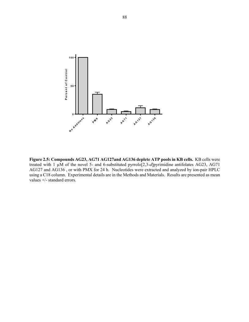
88
No
Ad
dit
ion
s
PM
X
AG
23
AG
71
AG
127
AG
136
0
5 0
1 0 0
Pe
rc
en
t o
f C
on
tro
l
Figure 2.5: Compounds AG23, AG71 AG127and AG136 deplete ATP pools in KB cells. KB cells were
treated with 1 µM of the novel 5- and 6-substituted pyrrolo[2,3-d]pyrimidine antifolates AG23, AG71
AG127 and AG136 , or with PMX for 24 h. Nucleotides were extracted and analyzed by ion-pair HPLC
using a C18 column. Experimental details are in the Methods and Materials. Results are presented as mean
values +/- standard errors.
89
Inhibition of de novo purine biosynthesis by compound 8 results in cytotoxicity and
apoptosis. To confirm that depletion of purine nucleotides by the most potent 5-substituted
analog, compound AG127, results in cytotoxicity toward KB cells, we performed colony-forming
assays. In these experiments, KB tumor cells were treated with various concentrations (1-10 µM)
for 48 h, then washed and plated without drug. Colonies were enumerated after 10 days (Figure
2.6A). Following drug exposures, inhibitory effects of compound AG127 were irreversible with
complete loss of colony formation at the highest drug concentrations, thereby establishing
compound AG127 as cytotoxic.
Programmed cell death type I (mitochondrial pathway) requires ATP to form the death
complex with cytochrome C and caspase 9 and to carry out other energy-requiring processes for
enzymatic degradation and modification of cellular components.315 Previous studies suggested
that the 6-substituted pyrrolo[2,3-d]pyrimidine antipurine antifolate AG17 was capable of
inducing apoptosis, albeit less than that indicated by daunorubicin over 24 h.198 Additional
experiments were performed with KB cells treated with compound AG127 to assess its effects on
cell cycle progression and induction of apoptosis, with comparisons to PMX and compound AG23.
Cells were exposed to 1 µM drugs for 48 or 96 h. Cultures were then divided, with one fraction
fixed and stained with propidium iodide (PI) for flow cytometry analysis of cell cycle distribution
(Figure 2.6B). An additional fraction was assayed for apoptosis by flow cytometry with annexin
V/fluorescein isothiocyanate (FITC) /7-amino actinomycin D (AAD) staining (Figure 2.6C).
At 48 h, minimal effects on the cell cycle were observed, except for PMX, which induced
a G1/Go-phase arrest relative to the untreated (DMSO) control (data not shown). At 96 h, all
compounds tested increased the percentages of cells in S-phase (albeit to different extents), while
reducing those in the G1/Go- and G2/M-phases of the cell cycle. The histograms display a change

90
in DNA content in cells treated with AG23 and AG127, suggesting aneuploidy. A prominent
increase in the sub-G1 population at 96 h was also seen (Figure 2.6B; most notable for compounds
AG23 and AG127), indicating apoptosis was induced.
Annexin V-FITC staining was used to further monitor induction of apoptosis. For
compounds AG23 and AG127, as well as PMX, positive annexin V-FITC staining increased over
time (results for 96 h are shown in Figure 2.6C, with distinct increases in both early (annexin-V-
FITChigh/7-AADlow) and late apoptotic fractions (annexin-V-FITChigh/7-AADhigh), compared to
those for the untreated (DMSO) control (i.e., the fractions of cells in early apoptosis were 33.8%
and 0.49% for compound AG127-treated and control cells, respectively, whereas the fractions in
late apoptosis at 96 h were 26.7% and 2.1%, respectively). Nearly identical results were obtained
with compound AG71, although for PMX, the distribution between the early and late apoptotic
fractions was somewhat different. Thus, the 5- and 6-substituted pyrrolo[2,3-d]pyrimidines AG23
and AG127 can induce apoptosis, resulting in cell death in spite of depleted ATP pools.
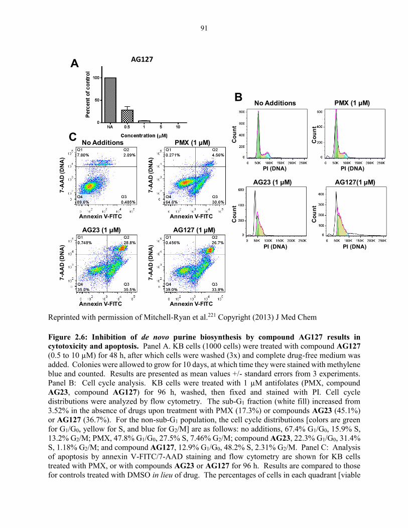
91
Reprinted with permission of Mitchell-Ryan et al.221 Copyright (2013) J Med Chem
Figure 2.6: Inhibition of de novo purine biosynthesis by compound AG127 results in
cytotoxicity and apoptosis. Panel A. KB cells (1000 cells) were treated with compound AG127
(0.5 to 10 µM) for 48 h, after which cells were washed (3x) and complete drug-free medium was
added. Colonies were allowed to grow for 10 days, at which time they were stained with methylene
blue and counted. Results are presented as mean values +/- standard errors from 3 experiments.
Panel B: Cell cycle analysis. KB cells were treated with 1 µM antifolates (PMX, compound
AG23, compound AG127) for 96 h, washed, then fixed and stained with PI. Cell cycle
distributions were analyzed by flow cytometry. The sub-G1 fraction (white fill) increased from
3.52% in the absence of drugs upon treatment with PMX (17.3%) or compounds AG23 (45.1%)
or AG127 (36.7%). For the non-sub-G1 population, the cell cycle distributions [colors are green
for G1/G0, yellow for S, and blue for G2/M] are as follows: no additions, 67.4% G1/G0, 15.9% S,
13.2% G2/M; PMX, 47.8% G1/G0, 27.5% S, 7.46% G2/M; compound AG23, 22.3% G1/G0, 31.4%
S, 1.18% G2/M; and compound AG127, 12.9% G1/G0, 48.2% S, 2.31% G2/M. Panel C: Analysis
of apoptosis by annexin V-FITC/7-AAD staining and flow cytometry are shown for KB cells
treated with PMX, or with compounds AG23 or AG127 for 96 h. Results are compared to those
for controls treated with DMSO in lieu of drug. The percentages of cells in each quadrant [viable
A
B
C Co
un
tC
ou
nt
Co
un
tC
ou
nt
PI (DNA) PI (DNA)
PI (DNA)PI (DNA)
AG127(1 µM)AG23 (1 µM)
No Additions PMX (1 µM)
AG127 (1 µM)AG23 (1 µM)
No Additions PMX (1 µM)
Annexin V-FITC Annexin V-FITC
Annexin V-FITC Annexin V-FITC
7-A
AD
(D
NA
)
7-A
AD
(D
NA
)
7-A
AD
(D
NA
)
7-A
AD
(D
NA
)
AG127
7

92
cells (annexin V-FITClow/7-AADlow;Q4), early apoptosis (annexin V-FITChigh/7-AADlow;Q3), and
late apoptosis/necrosis (annexin V-FITChigh/7-AADhigh;Q2) are summarized. These experiments
were ran with the assistance of Dr Eric Hales who performed the data analysis.

93
Dual Inhibition of GARFTase and AICARFTase by 5-substituted pyrrolo[2,3-
d]pyrimidine antifolates and downstream effects of accumulated ZMP on AMPK as
mechanisms resulting in cytotoxicity. Experiments were performed to measure cellular
GARFTase activity in KB tumor cells treated with the 6- (compounds AG23 and AG71) and 5-
substituted (compounds AG127 and AG136, and PMX) pyrrolo[2,3-d]pyrimidines. For this
purpose, we used an in situ activity assay for GARFTase which measures incorporation of
[14C]glycine into [14C]formyl GAR in the presence of azaserine over 16 h.147,153-156,168,233
Cells were harvested, washed with PBS and treated with trichloroacetic acid (TCA). Following
deproteinization, acid-soluble metabolites were ether-extracted and fractionated by ion-exchange
chromatography, permitting isolation and quantitation of [14C]formyl GAR. Both 5- and 6-
substituted analogs exhibited a dose-dependent decrease in [14C]formyl GAR. Compound AG71
was the most potent of the analogs tested in contrast to its 5-substituted analog, compound AG136,
which was the least. However, compound AG23 was more potent than either PMX or compound
AG127, with an IC50 for this compound exceeding those for 5-substituted compounds by 2- to 5-
fold (Figure 2.7).
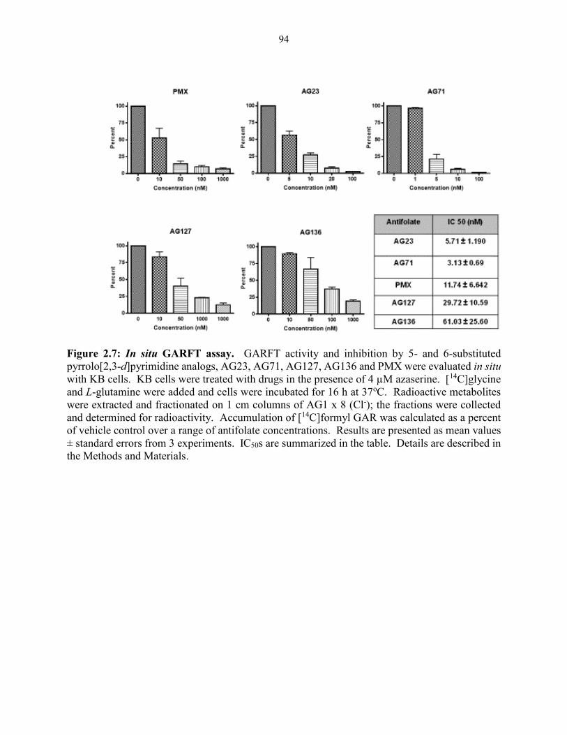
94
Figure 2.7: In situ GARFT assay. GARFT activity and inhibition by 5- and 6-substituted
pyrrolo[2,3-d]pyrimidine analogs, AG23, AG71, AG127, AG136 and PMX were evaluated in situ
with KB cells. KB cells were treated with drugs in the presence of 4 µM azaserine. [14C]glycine
and L-glutamine were added and cells were incubated for 16 h at 37oC. Radioactive metabolites
were extracted and fractionated on 1 cm columns of AG1 x 8 (Cl-); the fractions were collected
and determined for radioactivity. Accumulation of [14C]formyl GAR was calculated as a percent
of vehicle control over a range of antifolate concentrations. Results are presented as mean values
± standard errors from 3 experiments. IC50s are summarized in the table. Details are described in
the Methods and Materials.
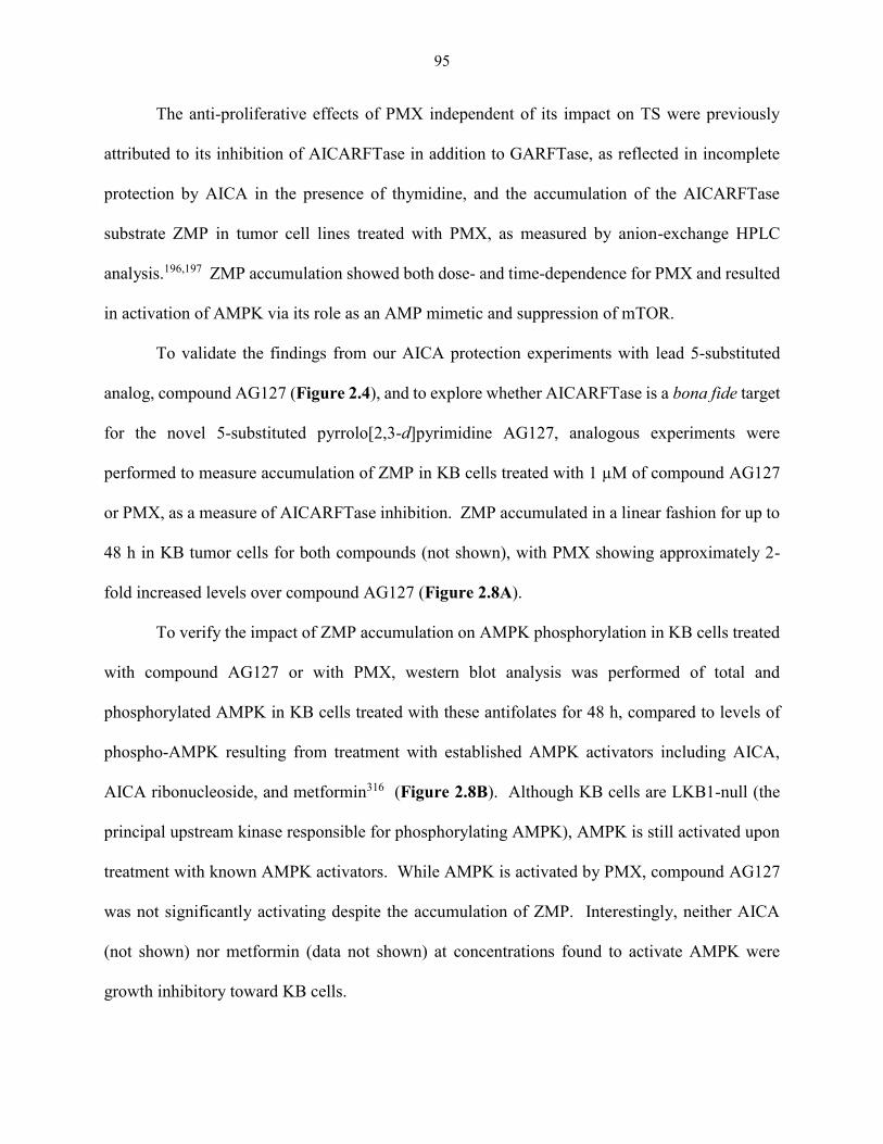
95
The anti-proliferative effects of PMX independent of its impact on TS were previously
attributed to its inhibition of AICARFTase in addition to GARFTase, as reflected in incomplete
protection by AICA in the presence of thymidine, and the accumulation of the AICARFTase
substrate ZMP in tumor cell lines treated with PMX, as measured by anion-exchange HPLC
analysis.196,197 ZMP accumulation showed both dose- and time-dependence for PMX and resulted
in activation of AMPK via its role as an AMP mimetic and suppression of mTOR.
To validate the findings from our AICA protection experiments with lead 5-substituted
analog, compound AG127 (Figure 2.4), and to explore whether AICARFTase is a bona fide target
for the novel 5-substituted pyrrolo[2,3-d]pyrimidine AG127, analogous experiments were
performed to measure accumulation of ZMP in KB cells treated with 1 µM of compound AG127
or PMX, as a measure of AICARFTase inhibition. ZMP accumulated in a linear fashion for up to
48 h in KB tumor cells for both compounds (not shown), with PMX showing approximately 2-
fold increased levels over compound AG127 (Figure 2.8A).
To verify the impact of ZMP accumulation on AMPK phosphorylation in KB cells treated
with compound AG127 or with PMX, western blot analysis was performed of total and
phosphorylated AMPK in KB cells treated with these antifolates for 48 h, compared to levels of
phospho-AMPK resulting from treatment with established AMPK activators including AICA,
AICA ribonucleoside, and metformin316 (Figure 2.8B). Although KB cells are LKB1-null (the
principal upstream kinase responsible for phosphorylating AMPK), AMPK is still activated upon
treatment with known AMPK activators. While AMPK is activated by PMX, compound AG127
was not significantly activating despite the accumulation of ZMP. Interestingly, neither AICA
(not shown) nor metformin (data not shown) at concentrations found to activate AMPK were
growth inhibitory toward KB cells.
96
Reprinted with permission of Mitchell-Ryan et al.221 Copyright (2013) J Med Chem
Figure 2.8: Accumulation of ZMP by 5-substituted pyrrolo[2,3-d]pyrimidines PMX, and
compound AG127. Panel A: ZMP (AICA ribonucleotide) accumulation was measured in KB
cells treated for 48 h with the 5-substituted pyrrolo[2,3-d]pyrimidine antifolate AG127, or PMX.
Methods for extracting nucleotide metabolites and for measuring ZMP levels by ion-exchange
HPLC are detailed in the Methods and Materials. Panel B: KB cells were treated at indicated for
48 h, followed by western analysis of phosphorylated AMPK on a 4-20% gradient gel. Blots were
probed with antibodies to phospho- and total AMPK, and to β-actin.
97
2.3 Discussion
The development of rationally designed cytotoxic antifolates remains an area of significant
interest to anticancer drug development, as exemplified by the relatively recent approval of
PMX317 and pralatrexate318 for cancer, more than 50 years since introduction of MTX.319
Unfortunately, the ubiquitous presence of RFC may contribute to unwanted toxicities of these
classical antifolates toward normal tissues. Based on this, growing attention has focused on
developing novel folate-based compounds with tumor targeting premised on transport selectivity
for FRs or PCFT over RFC,75,198,204-207,220 a pursuit that until recently175 continued to be challenged
by the lack of structural information on the major folate transport systems.
The antitumor efficacy for the 5-substituted 2-carbon bridge pyrrolo[2,3-d]pyrimidine
PMX reflects its cellular uptake by all the major folate transport systems.147,153-156 While the
corresponding 6-substituted analog of PMX was pharmacologically inert, with increasing bridge
lengths, 6-substituted pyrrolo[2,3-d]pyrimidines were surprisingly potent inhibitors of cell
proliferation, although membrane transport by RFC was lost.204,320 The 6-pyrrolo[2,3-
d]pyrimidine benzoyl analogs with 3 and 4 bridge carbons, compounds AG17 and AG23,
respectively, were the most active of this series, reflecting their FR and PCFT cellular uptake, with
little to no transport by RFC, and their potent inhibitions of GARFTase, the first folate-dependent
step leading to the synthesis of IMP.153,237 Inhibition of de novo synthesis of purine nucleotides
resulted in ATP depletion, leading to cytotoxicity and apoptosis.153,237
In this report, our goal was to further establish a comprehensive SAR for the 5- versus 6-
substituted pyrrolo[2,3-d]pyrimidine ring system in relation to folate transporter specificity and
inhibition of intracellular targets, based in part on findings of transporter promiscuity for
PMX,198,204-207 and reports that PMX may in part derive its antitumor effects by inhibiting
98
AICARTase, resulting in accumulation of ZMP and activation of AMPK in addition to inhibiting
TS. 145,146 Compared to the 6-substituted analogs, we found that the 5-substituted compounds in
general: (i) were better substrates for RFC with the highest antiproliferative activities for
compounds with 2-4 bridge carbons independent of the nature of the side chain ring system; (ii)
were poorer substrates for PCFT with the exception of the 2-carbon bridge compound, PMX
(AG125); and (iii) preserved FR cellular uptake, with the 4-carbon bridge analog AG127 showing
the greatest FRα selectivity and potency in this expanded 5-substituted series of analogs without
TS inhibitory activity.
Compound AG127 was cytotoxic, as reflected in loss of clonogenicity in KB cells, and
effected S-phase accumulation and apoptosis over 96 h. Like its 6-substituted counterpart
compound AG71,204 compound AG127 resulted in ATP depletion, although this appeared to be
only partly due to inhibition of GARFTase. Rather, a unique feature of the 5-substituted analog
AG127 relates to its dual inhibition of both the GARFTase and AICARFTase reactions in de novo
purine biosynthesis. While PMX also inhibits GARFTase, the finding that the AICARFTase
substrate ZMP accumulates in the presence of this drug suggested that GARFTase inhibition must
be incomplete and that AICARFTase must also be a target as previously described, 145,146 although
for PMX, total purine nucleotides, as reflected in ATP pools, were significantly preserved.
Compound AG127, also inhibited AICARFTase, as reflected in incomplete protection in cell
proliferation assays by AICA and demonstrated accumulation of the AICARFTase substrate ZMP,
albeit less than for a comparable concentration of PMX.
Previous studies suggested that AMPK activation secondary to ZMP accumulation and
AICARFTase inhibition may result in inhibition of mTOR and contribute to the antitumor effects
of high levels of PMX in the presence of thymidine.196,197 In the present study, we used KB human
99
tumor cells treated with PMX or with compound AG127. In this LKB1-deficient tumor cell line,
ZMP accumulation was accompanied by AMPK activation in the presence of PMX but not
compound AG127. Robust AMPK activation was also seen upon treatment with AICAR, or
metformin. However, ZMP accumulation and AMPK activation did not appear likely to contribute
to the anti-proliferative effects of PMX in KB cells, as neither AICA nor metformin inhibited cell
proliferation. The antiproliferative and cytotoxic effects of compound AG127 toward KB cells
must derive from a depletion of ATP pools, secondary to inhibition of both GARFTase and
AICARFTase, rather than from activation of AMPK.
In summary, a series of classical 5-substituted pyrrolo[2,3-d]pyrimidine antifolates
AG124-AG129 were designed and synthesized as a hybrid of PMX and 6-substituted pyrrolo[2,3-
d]pyrimidines AG17, AG23, AG71 and AG94. Compound AG127, the lead compound of this
series, has an IC50 =12.7 nM against KB tumor cells in culture. Rather than only targeting
GARFTase, as with AG17 and AG23, both GARFTase and AICARFTase were identified as the
intracellular targets of AG127. The unique dual GARFTase and AICARFTase inhibition of
compound AG127 makes it an excellent lead compound for further study including the design of
additional antitumor analogs to optimize cellular folate uptake selectivity by FRs and inhibition of
folate metabolizing enzymes, leading to a new generation of potent tumor-targeted agents. The
dual inhibition of both GARFTase and AICARFTase results in potent inhibition of purine
biosynthesis and could preserve antitumor activity in the event that tumors become resistant due
to alterations in one or the other targeted enzyme. Such dual targeted inhibitors would seem to
have substantial advantages over currently used agents that principally target only one intracellular
enzyme target.
100
2.4 Materials and methods
Reagents for biological studies. [3’, 5’, 7-3H]MTX (20 Ci/mmol), [3’, 5’, 7, 9-3H] folic
acid (25 Ci/mmol), and [14C(U)]-glycine (87mCi/mmol) were purchased from Moravek
Biochemicals (Brea, CA). Unlabeled folic acid was purchased from the Sigma Chemical Co. (St.
Louis, MO). LCV [(6R,S) 5-formyl tetrahydrofolate] was provided by the Drug Development
Branch, National Cancer Institute, Bethesda, MD. The sources of the antifolate drugs were as
follows: MTX, Drug Development Branch, National Cancer Institute (Bethesda, MD); and PMX
(Alimta) (LC Laboratories, Woburn, MA). Other chemicals were obtained from commercial
sources in the highest available purity. Novel antifolates (denoted by AG17-138 (see Table
2.1))221 were synthesized by Dr. Aleem Gangjee and colleuges at Duequesne University, Pittsburg
Pennsylvania.
Cell culture. MTXRIIOuaR2-4 (referred to as R2) is a RFC, PCFT and FRα null Chinese
Hamster (CHO) cell line that was a gift from Dr. Wayne Flintoff (University of Western
Ontario).321 From this parental cell line, RFC, PCFT and FRα were transfected in to give rise to
isogenic CHO cell lines designated PC43-10 (expresses RFC but not PCFT or FRα), R2/PCFT4
(expresses PCFT) and RT16 (expresses FRα).198,204-207,220,322 The CHO sublines were all
maintained in α-minimal essential medium (α-MEM) supplemented with 5% penicillin-
streptomycin solution, 2 mM L-glutamate and 10% heat-treated bovine calf serum (BCS)
(Invitrogen). Transfected cell lines were cultured in the presence of 1.5 mg/ml G418. CHO cell
lines were cultured prior to cell viability experiments in folate-free RPMI (FF-RPMI) (Invitrogen)
with dialyzed fetal bovine serum (DFBS) (Invitrogen) supplemented with 5% penicillin-
streptomycin and L-glutamate. Human FRα-expressing KB (nasopharyngeal) and IGROV-1
101
(ovarian) carcinoma cells were continuously maintained in FF-RPMI complete with 10% fetal
bovine serum (FBS) and 5% penicillin-streptomycin and L-glutamate.
Cell viability assays were performed exactly as previously described.198, 193-196 Cell lines
were treated with a range of concentrations of standard or novel antifolates (0-1 μM), metformin
(0-10 mM) or AICA (0-2 mM) in a 96 well plate with in FF-RPMI complete with DFBS, and 2
nM (RT-16, KB, IGROV1) or 25 nM LCV (R2/PCFT4), penicillin –streptomycin and L-glutamate
over a 96 h incubation period at 37◦C with 5% CO2. To confirm FRα-mediated drug uptake and
growth inhibition, 200 nM folic acid was added to the incubations with drug. For proliferation
assays with PC43-10 cells, cells were cultured in standard RPMI1640 with DFBS, penicillin-
streptomycin and L-glutamate for 96 h. To quantitative viable cells, media was removed and Cell
Titer-blue™ fluorescent reagent (Promega) was added. Relative cell numbers were determined by
relative fluorescence units measured with a fluorescence plate reader at 590 nm emission and 560
nm excitation with Softmax Pro plate reader software. IC50 values, corresponding to the drug
concentrations that resulted in 50% loss of cell growth, were calculated from dose-response curves
using Sigma Plot (v.12) software.
In order to identify the targeted pathways/folate-dependent enzyme by the classic and
targeted antifolates, proliferation assays were performed in the presence of adenosine (60 μM),
thymidine (10 μM), or AICA hydrochloride (320 μM) and results compared to incubations in
parallel without drug additions.198,205,220
Colony forming assays were performed with compound AG127 to verify a cytotoxic (as
opposed to cytostatic) response. KB cells (~500) were plated in 60 mm dishes in FFRPMI/DFBS
supplemented with 2 nM LCV for a 24 h incubation period at 37◦C in the presence of 5% CO2.
Concentrations ranging from 0-10 μM of compound AG127 were added to media for an additional
102
24 h, after which the media was aspirated and replaced with FFRPMI/DFBS supplemented with 2
nM LCV for 8-10 days. To visualize colonies, the cells were washed (2x) with room temperature
Dulbecco’s phosphate-buffered saline (PBS) (Sigma), followed by a wash of 5% trichloroactetic
acid (TCA). The TCA was removed, 10 mM borate buffer (pH 8.8) was added, followed by a 30
min incubation with 1% methylene blue in borate buffer at room temperature. The cells were was
three times with borate buffer and the stained colonies were counted.
Competitive MTX transport assays. To determine if novel antifolates bound to RFC,
reflecting membrane transport by this mechanism, competition with [3H]MTX for cellular uptake
was measured in PC43-10 CHO cells.198,207 PC43-10 cells (~1.5x10⁶ cells) were seeded in 60mm
dishes. After 48 h, cells were washed with PBS and uptake of [3H]MTX (0.5 μM) was measured
in Hank’s Balanced Salts Solution for 2 minutes at 37◦C. Cold (~4◦C) PBS was used to quench
the reactions. Cells were washed with ice-cold PBS (3X), then solubilized with 0.5N NaOH. The
cellular homogenates were assayed for radioactivity with a scintillation counter; protein
concentrations were measured with Folin-phenol reagent.203 Levels of drug uptake were expressed
as pmol/mg cell protein.
FRα affinity binding assay. To determine the binding affinity of novel antifolates to
FRα,198 FRα-expressing RT16 CHO cells (~1.5 x10⁶) were seeded in a monolayer with complete
α-MEM media. Cells were washed with ice-cold PBS, followed by an cold acetate buffer (10 mM
sodium acetate, 150 mM NaCl, pH 3.5) to release FRα-bound folates, then neutralized with ice-
cold Hepes-buffered saline (HBS) (20 mM Hepes, 140 mM NaCl, 5 mM KCl, 2 mM MgCl2 , 5
mM glucose, pH 7.4). Cells were then incubated at 0◦C for 15 min in a cocktail containing [3’, 5’,
7’,9- 3H]folic acid, in the absence or presence of unlabled folic acid or novel antifolate compounds
(0-1 μM). Cells were washed with cold HBS and solubilized with 0.5 N NaOH. The cell
103
homogenates were collected and analyzed for radioactivity using a liquid scintillation counter.
Protein concentrations were measured with the Folin-phenol reagent. Bound [3H]folic acid was
expressed as pmol/mg protein. Using Sigma plot (v.12) software, dose-response curves were
created and IC50’s were calculated. Relative binding affinities were calculated as the inverse molar
ratios of unlabeled ligands require to inhibit [3H]folic acid by 50% as determined graphically with
Graphpad (v. 6.0) software. By definition, the relative affinity of folic acid is 1.
HPLC detection of ATP. Intracellular ATP concentrations were determined using
nucleotide extractions and an HPLC method, exactly as previously described.204,314 Briefly, KB
cells were seeded in a T75 flask with FF-RPMI with 10% DFBS, 5% penicillin-streptomycin, 2
mM L-glutamine and 2nM LCV. Cells were treated with 1 µM of PMX, or compounds AG23,
AG71, AG127 or AG136 for 24 h. After the incubations cells were trypsinized and washed with
cold PBS. Nucleotide pools were fractionated on by reversed-phase HPLC on a C18 column for
determination of ATP pools.
Apoptosis and cell cycle analysis. KB cells were treated with 1 µM of PMX of
compounds AG23 or AG127 24, 48, 72 and 96 h in FF-RPMI media complete with 10% DFBS
and 5% penicillin-streptomycin and 2 nM LCV. Harvested cells were trypsinized and washed with
cold PBS. Samples were equally divided so cells could be stained with PI for cell cycle analysis
and with Annexin V/7-AAD (Beckman) for apoptosis assays. For cell cycle analysis, cells were
fixed with cold ethanol (added drop-wise), then resuspended and stained in PBS containing 50
µg/mL PI and 100 µg/mL RNAse (type I-A) (Sigma). Cell populations were analyzed by flow
cytometry and Flowjo software. Flow cytometry and Flowjo software were also used to
discriminate positively stained Annexin V/7-AAD populations including viable (annexin V and 7-
104
AAD negative), early apoptotic (annexin V positive/ PI negative or 7-AAD low), and late
apoptotic/necrotic (annexin V positive/PI positive or 7-AAD high).
In situ GARFT inhibition assays. To measure the extent of intracellular GARFT
inhibition in the presence of antifolate inhibitors, we examined the metabolic incorporation of
[14C(U)] glycine into [14C]formyl GAR (Figure 2.4) in the presence of azaserine. Using
previously published methods, 147,153-156,168,233 KB cells were seeded in complete media (above)
and allowed to adhere to substratum for 24 h. Cell were then washed with FF-RPMI, L- glutamine
free with 10% DFBS, 5% penicillin-streptomycin and 2 nM LCV. Cells were incubated at 37⁰C
with 5% CO2 for 1 h with folate- and L-glutamine-depleted media containing 2 nM LCV, with and
without PMX and compounds AG23, AG71, AG127, or AG136 (in DMSO) over a range of
concentrations. Control cells were treated with DMSO of equal volume. Azaserine (4 µM final
concentration) was added to the cells and allowed to incubate for 30 minutes. This was followed
by the addition of L-glutamine (2 mM) and [14C(U)]glycine (final specific activity 0.1 mCi/L).
Cells were incubated 16 h. Cells were washed with ice-cold PBS, trypsinized and collected. Cells
were washed with cold PBS, then treated with 5% TCA at 0⁰C. Samples were centrifuged (4oC,
14,000 rpm) and the protein precipitants were solubilized with 0.5 N NaOH for quantitation of
protein concentrations with the Folin-phenol protein method.323 The supernatants were collected
and extracted with cold ether. After evaporation of ether, the remaining aqueous layer was gravity
filtered through a 1 cm polypropylene chromatography column (Bio-rad) of AG1x8 (chloride
form). The columns were washed with 10 mL 0.5 N formic acid, followed by 4 N formic acid (10
ml), and finally eluted with 8 ml of 1 N HCL collected in 8 fractions. The eluate was measured
for radioactivity and based on the percentages of radioactivity in the [14C]formyl GAR and non-
105
specific radioactivity, the results calculated as pmol/mg protein. Data were graphed using
Sigmaplot (v.12) and IC50 were calculated.
HPLC detection of ZMP pools. Methods for detecting ZMP in KB cells treated with
drugs were based on those described by Rascanelli et al.196 Briefly, KB cells were treated with
assorted drugs (PMX, compound AG127 or drug free) for 48 h in FF-RPMI with 10% DFBS, 5%
penicillin-streptomycin, 2 mM L-glutamine, and 2 nM LCV. Cells were washed with PBS, treated
with ice-cold 5% TCA and proteins were precipitated by centrifugation (1600 rpm, 10 min). The
soluble fractions were extracted with ether (2x) and fractionated by ion-exchange HPLC on a SAX
250 x 2 mm column (Phenomenex) with a linear gradient from 5 mM NH4H2PO4 (pH 2.8) to 750
mM NH4H2PO4 (pH 3.9) over 25 min at a flow rate of 0.2 mL/min. Absorbance was monitored at
280 nm and the intracellular ZMP concentrations were established by fitting the peak absorbances
to a standard curve with commercial ZMP. AraCMP was used as an internal control.
Western blot analysis. KB cells were treated for 48 h with PMX or compound AG127 in
the presence of thymidine (5.6 µM). Controls include vehicle control (water or DMS0; results
were identical), or known activators of AMPK including metformin (10 mM), AICA (320 µM or
1 mM), or AICA ribonucleoside (1 mM). Cells were washed, suspended in 10 mM Tris-HCl (pH
7.0) plus proteolytic and phosphatase (Phosphostop) inhibitors (Roche Applied Science) and
disrupted by sonication. Samples were centrifuged (14,000 rpm, 10 min) and 90 µg protein
fractionated by SDS polyacrylamide gel electrophoresis on Laemmli gradient gels (4-20%).324
Proteins were electrotransferred to polyvinylidene difluoride (PVDF) membranes (Pierce).325
Detection of immunoreactive AMPK was with rabbit total and phospho-AMPK antibodies (Cell
Signaling) and IRDye800-conjugated secondary anti-rabbit antibody (LI-COR, Lincoln, NE)

106
using an Odyssey® infrared imaging system (LI-COR, Lincoln, NE). β actin was detected with
anti-β actin mouse antibody and IRDye800-conjugated secondary anti-mouse antibody (LI-COR).
107
CHAPTER 3: DISCUSSION
We are upon a major paradigm shift that has altered our view of ovarian cancer. With this
shift in thinking, changing the strategies in how we treat this disease is not only appropriate but
the next logical step in disease intervention. Currently, the treatment of this disease remains
unaltered from the standard agents, irrespective of the diverse molecular biology of the tumor. The
identified molecular markers should be used to determine appropriate targeted therapy to address
the driving oncogenic mechanism of the tumor. Armed with the knowledge of the precise
molecular pathways involved in the 2 categories of EOC (type I and type II), we can customize
and design novel therapeutic strategies that may prove to be more beneficial than the non-specific
chemotherapy that is currently in use.
Combination therapy has proven to be effective in the treatment of many malignancies.326
Mathematical models predict that combination therapy will be more successful at disease
eradication than single agent or adjuvant treatment because combination treatment is more likely
to prevent drug resistance.327 A treatment with a reduced propensity to drug resistance is highly
desirable in ovarian cancer, as the recurrent drug resistant tumors are difficult to treat and are
principally responsible for the patient’s demise.328 A recent study examined the effectiveness of
combining PMX with simvastatin ( a statin) in mesothelioma and lung cancers and reported an
enhanced apoptotic response with combination treatment than with single agent therapy.329 There
is a heighten interest in the use of statins as anticancer agents which stems from their ability to
disrupt the mevalonate pathway.329-335 This pathway has been associated with cell signaling, cell-
cycle progression, protein synthesis and membrane integrity.330 This study may serve as a platform
to encourage more studies examining the effectiveness of antifolates in conjunction with other well
established anticancer therapies (in addition to statins which are currently in their experimental
108
infancy as antineoplastics) that possess a different mechanism of action. Combination therapy
may have great significance in malignancies like ovarian cancers that harbor a number of mutations
in kinases, GTPases and receptors for which there are experimental and well establish targeted
therapies. For example a FRα targeted therapy may pair with inhibitors of AKT, K-RAS, or HER2,
as many ovarian tumors have activating mutations or overexpression of these kinases GTPases and
receptors.336
Overwhelming evidence suggests that FRα confers a growth advantage to malignant
cells.60,62,128,174 There is speculation that the use of DNA damaging agents, like platinum-based
therapies, may enhance FRα expression to increase intracellular folate levels to encourage an up
regulation of nucleotide production to assist in DNA repair to address drug induced damage.263
These identified molecular events in ovarian cancer offer an opportunity for the continued growth
of the next generation of FRα-targeted treatments. To date, classic antifolates have not made a
notable appearance in the treatment of ovarian cancer. In light of the enhanced expression of FRα
driven by the use DNA damaging agents, like those used in ovarian cancer, FRα targeted
antifolates can be useful as a second line therapeutic in this disease. PMX, which shows some
affinity to FR is currently being evaluated as a treatment option in platinum resistant ovarian
cancer.337,338
3.1 Targeting the AMPK/mTOR pathway
We have characterized a novel class of agents that are effective against a cell culture model
of HGSC, endometroid carcinoma and clear cell carcinoma. Prior to obtaining crystallographic
information pertaining to FRα, rational drug design aided in determining structural elements
required for FRα-mediated drug uptake. One unique feature of the agents under investigation is
their ability to act as dual inhibitors of GARFTase and AICARFTase. This ability to inhibit
109
AICARFTase, led to an accumulation of a purine intermediate (ZMP) but not AMPK activation.
Therefore this study also allowed us to review the potential significance of AMPK activation in
ovarian tumors and the potential of indirectly targeting AMPK and mTOR signaling for therapeutic
advantage. In recent years, AMPK-targeting in cancer treatment has become en vogue. Much of
its popularity can be attributed to the antidiabetics metformin and phenformin and their association
with antitumor activity and reports suggesting that type 2 diabetics taking metformin had better
cancer treatment outcomes than those taking other antidiabetics.339 While there has been much
excitement surrounding the antitumor effects metformin has displayed in breast and ovarian cancer
studies, the mechanism of action of this drug remains unclear.339-346 Most of this uncertainty arises
from metformin’s ability to induce these antiproliferative and anti-tumor effects in an AMPK-
dependent or -independent manner.345,347 This leaves one to question if the effect on the cellular
energy balance (AMPK/mTOR) has greater impact on cell viability than the antidiabetic is
affecting the insulin metabolism of the tumor which is causing growth suppression and or
apoptosis.
There are a number of studies that imply that AMPK activation may assist in the survival
and growth of select tumors (e.g., prostate cancer) through the activation of macroautophagy,
which in certain cellular contexts can lead to cell death or cell survival.348 Autophagy is a common
pathway for cell survival in many malignancies. The modulation of macroautophagy in autophagy
competent cells may result in no significant consequence for such cells. A recent study showed
that an apoptotic response to pemetrexed in combination with simvastatin can be enhanced in
MPM and lung cancer cells when autophagy is inhibited.332 This demonstrates that autophagy
inhibition can be an effective strategy to further potentiate cell death. In the case where FRα is the
sole means of drug transport, the method in which autophagy is inhibited becomes critical. Folate
110
receptors require acidification of the lysosome to release substrate. Many pharmacologic tools
used to inhibit autophagy involve agents that prevent acidification of lysosomes and similar
organelles, thus also making FRα-mediated uptake ineffective. ATG siRNAs are useful molecular
tools in an experimental setting; however, the use of such tools in a clinical setting, to date, may
be impractical. With the growing number of chemotherapeutics that are now being identified as
AMPK activators, a closer examination of the effects of this activation should be conducted on the
tumor of question prior to the selection of therapy.
3.2 Targeting AICARFTase
This study sought to describe the significance of AICARFTase inhibition in de novo purine
synthesis in FRα-positive tumor cells. Eukaryotic AICARFTase is a bifunctional protein that
participates in formylation and cyclization to produce the purine precursor molecule IMP.349-352
Like GARFTase, AICARFTase is a critical player in the de novo production of purine nucleotides
and therefore has been suggested as a viable target to disrupt the purine biosynthetic pathway.
Although both GARFTase and AICARFTase are transformylases in the same pathway and can use
the same folate co-factor to perform their activities, they share no sequence homology.351,353 This
divergence in sequence would make a drug directed to target one enzyme highly unlikely to behave
as a substrate for the other.351 For this reason, MTAs like MTX (primary target DHFR)and PMX
(primary target TS) are rather unique because they can inhibit both enzymes.349
There are no known clinically investigated antifolates that primarily target AICARFTase.
Burroughs and Wellcome designed and produced two antifolates, BW1540 and BW2315, to
selectively target AICARFTase. These two novel agents are sulfamido bridged 5,8 dideazafolate
analogs, that were designed to create a stronger interaction between the protein and the small
molecule.351 Both analogs were reported to exhibit strong inhibition towards AICARFTase
111
(BW1540, 8nM; BW2315, 6nM) as well as antitumor activity against human colon cancer cells (
BW1540 0.7-3.0µM; BW2315 1.0-5.0µM).351 Other non-folate based AICARFTase inhibitors,
326203-A (2, -[5 hydroxy-3-methyl-1-1(2 methyl-4-sulfophenyl0-H-pyrazol)-4 sulfo-benzoic
acid) have been explored, however none of these novel analog have progressed to clinical
studies.353
Experiments performed by the Moran lab with PMX prompted our own investigation using
5-substituted pyrrolo[2,3-d]pyrimidines that indirectly suggested inhibition of the enzyme. Direct
quantitation of enzyme inhibition may help to elucidate a role in AICARFTase targeting for the
disruption of de novo purine synthesis. Using the structural information for both FRα and the
AICARFTase enzyme may bring us closer to designing an FR directed inhibitor.
This body of work includes the characterization of novel potent 5- and 6-substituted pyrrolo
[2,3-d] pyrimidines that are effective against in vitro model of HGSC, endometroid carcinoma and
clear cell carcinoma (IGROV-1). Approximately 90% of advanced stage ovarian cancers are
HGSC as this subtype generally presents at an advanced stage.232,247 Patients who present in stages
III-IV have a 5 year survival rate of 15-30% and generally succumb to complications associated
with this disease. The grim statistics of ovarian cancer prompt the clinical development of highly
potent compounds with reduced toxicity to allow for treatment at longer intervals to achieve
maximum efficacy. We can learn from the failure of the folic acid-conjugated toxin, vintafolide,
and design improved folate-based cytotoxins akin to the novel compounds outlined in the study.
The novel compounds examined in these studies are excellent candidates for further
development. Pharmacokinetic data and orthotropic mouse models of EOC may enhance and aid
in greater support of these analogs as lead compound for more extensive pre-clinical and clinical
investigations. All together, we have developed agents with tumor selectivity based on an inherent
112
loss of purine salvage abilities due to the deletion of MTAP and their overexpression of FRα. FRα-
positive tumors treated with novel 6- substituted pyrrolo[2,3-d]pyrimidines will undergo cell death
due to purine nucleotide depletion via GARFTase inhibition. Such depletion results in a reduction
of cellular ATP pools, an induction of cell cycle arrest and eventually apoptosis. The 5- substituted
pyrrolo[2,3-d]pyrimidines share the unique characteristic of acting as a dual inhibitor of both
AICARFtase and GARFTase, albeit the potency of inhibition towards GARFTase is reduced.
While the 5-substituted analogs were able to induce ATP depletion, cell cycle aberrations and
apoptosis, despite ZMP accumulation (likely resulting from AICARFTase inhibition) they were
unable to activate AMPK. The accumulation of ZMP alone, independent of AMPK activation
may have been sufficient to initiate a cytotoxic response. There are a number of studies that
suggest that ZMP is a toxic metabolite with many cellular targets in addition to AMPK.354-356 ZMP
accumulation has been linked to mitochondrial distress, increased apoptosis in malignant cells in
an AMPK-independent manner and a depletion in pyrimidine nucleotide pools.356 While
substitution at the 5th position allows for the addition of an enzyme target, selective FRα-directed
membrane transport is compromised and RFC-transport is acquired (Figure 3.1). These studies
highlight the SAR of novel pyrrolo[2,3-d]pyrimidine antifolates . The success of PMX and the
expansion of its therapeutic range beyond lung cancer, warrants further investigation of novel
pyrrolo[2,3-d]pyrimidine antifolates. Using structure based drug design that incorporates the
recently discovered crystal structure of FRα along with the structural information of AICARFTase,
we will perhaps come closer to designing a potent FRα targeted AICARFTase inhibitor to provide
a less toxic, yet efficacious treatment option for ovarian cancer.
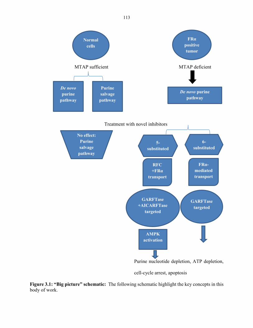
113
MTAP sufficient MTAP deficient
Treatment with novel inhibitors
Purine nucleotide depletion, ATP depletion,
cell-cycle arrest, apoptosis
Figure 3.1: “Big picture” schematic: The following schematic highlight the key concepts in this
body of work.
Normal
cells
FRα
positive
tumor
De novo
purine
pathway
Purine
salvage
pathway
De novo purine
pathway
No effect:
Purine
salvage
pathway
intact
5-
substituted
6-
substituted
RFC
+FRα
transport
FRα-
mediated
transport
GARFTase
+AICARFTase
targeted
GARFTase
targeted
AMPK
activation

114
REFERENCES
1. Hoffbrand, A.V. & Weir, D.G. The history of folic acid. British journal of haematology 113, 579-
589 (2001).
2. Bastian, H. Lucy Wills (1888-1964): the life and research of an adventurous independent woman.
The journal of the Royal College of Physicians of Edinburgh 38, 89-91 (2008).
3. Beck, W.S. Diagnosis of megaloblastic anemia. Annual review of medicine 42, 311-322 (1991).
4. McNulty, H., Pentieva, K., Hoey, L., Strain, J. & Ward, M. Nutrition throughout life: folate.
International journal for vitamin and nutrition research. Internationale Zeitschrift fur Vitamin-
und Ernahrungsforschung. Journal international de vitaminologie et de nutrition 82, 348-354
(2012).
5. Rosenberg, I.H. A History of the Isolation and Identification of Folic Acid (Folate). Annals of
Nutrition and Metabolism 61, 231-235 (2012).
6. Watson, J. & Castle, W.B. NUTRITIONAL MACROCYTIC ANEMIA, ESPECIALLY IN PREGNANCY:
Response to a Substance in Liver Other than that Effective in Pernicious Anemia*. The American
Journal of the Medical Sciences 211, 513&hyhen;530 (1946).
7. Medici, V. & Halsted, C.H. Folate, alcohol, and liver disease. Molecular nutrition & food research
57, 596-606 (2013).
8. Bailey, L.B. Folate in health and disease, (Taylor & Francis, Boca Raton, 2010).
9. Stokstad, E.L. & Koch, J. Folic acid metabolism. Physiological reviews 47, 83-116 (1967).
10. Mitchell, H.K., Snell, E.E. & Williams, R.J. THE CONCENTRATION OF “FOLIC ACID”. Nutrition
Reviews 46, 324-325 (1988).
11. Combs, G.F. Vitamins : Fundamental Aspects in Nutrition and Health (3rd Edition), (Academic
Press, Burlington, MA, USA, 2007).
115
12. Stokstad, E.L.R., Hutchings, B.L. & SubbaRow, Y. The Isolation of the Lactobacillus casei Factor
from Liver. Journal of the American Chemical Society 70, 3-5 (1948).
13. Bailey, L.B. Folate in Health and Disease, (Taylor & Francis, 1994).
14. Pitkin, R.M. Folate and neural tube defects. The American Journal of Clinical Nutrition 85, 285S-
288S (2007).
15. Salbaum, J.M. & Kappen, C. Genetic and Epigenomic Footprints of Folate. in Progress in
Molecular Biology and Translational Science, Vol. Volume 108 (eds. Bouchard, C. & Ordovas,
J.M.) 129-158 (Academic Press, 2012).
16. Anderson, O.S., Sant, K.E. & Dolinoy, D.C. Nutrition and epigenetics: an interplay of dietary
methyl donors, one-carbon metabolism and DNA methylation. The Journal of Nutritional
Biochemistry 23, 853-859 (2012).
17. Fox, J.T. & Stover, P.J. Folate-mediated one-carbon metabolism. Vitamins and hormones 79, 1-
44 (2008).
18. Brody, T., Shane, B. & Stokstad, E.L. Separation and identification of pteroylpolyglutamates by
polyacrylamide gel chromatography. Analytical biochemistry 92, 501-509 (1979).
19. Shane, B. Folylpolyglutamate synthesis and role in the regulation of one-carbon metabolism.
Vitamins and hormones 45, 263-335 (1989).
20. Stover, P.J. & Field, M.S. Trafficking of Intracellular Folates. Advances in Nutrition: An
International Review Journal 2, 325-331 (2011).
21. Winkels, R.M., Brouwer, I.A., Siebelink, E., Katan, M.B. & Verhoef, P. Bioavailability of food
folates is 80% of that of folic acid. The American Journal of Clinical Nutrition 85, 465-473 (2007).
22. Moat, S.J., et al. Folate, homocysteine, endothelial function and cardiovascular disease. The
Journal of Nutritional Biochemistry 15, 64-79 (2004).
116
23. McGuire, J.J. & Bertino, J.R. Enzymatic synthesis and function of folylpolyglutamates. Molecular
and cellular biochemistry 38 Spec No, 19-48 (1981).
24. Kisliuk, R.L. Pteroylpolyglutamates. Molecular and cellular biochemistry 39, 331-345 (1981).
25. Gonen, N. & Assaraf, Y.G. Antifolates in cancer therapy: Structure, activity and mechanisms of
drug resistance. Drug Resistance Updates 15, 183-210 (2012).
26. Zhao, R., Matherly, L.H. & Goldman, I.D. Membrane transporters and folate homeostasis:
intestinal absorption and transport into systemic compartments and tissues. Expert reviews in
molecular medicine 11, e4 (2009).
27. Tam, C., O'Connor, D. & Koren, G. Circulating Unmetabolized Folic Acid: Relationship to Folate
Status and Effect of Supplementation. Obstetrics and Gynecology International 2012, 17 (2012).
28. Locasale, J.W. Serine, glycine and one-carbon units: cancer metabolism in full circle. Nature
reviews. Cancer 13, 572-583 (2013).
29. Scotti, M., Stella, L., Shearer, E.J. & Stover, P.J. Modeling cellular compartmentation in one-
carbon metabolism. Wiley interdisciplinary reviews. Systems biology and medicine 5, 343-365
(2013).
30. Bienemann, K., et al. Targeted expression of human folylpolyglutamate synthase for selective
enhancement of methotrexate chemotherapy in osteosarcoma cells. Cancer gene therapy 20,
514-520 (2013).
31. Aghi, M., Kramm, C.M. & Breakefield, X.O. Folylpolyglutamyl Synthetase Gene Transfer and
Glioma Antifolate Sensitivity in Culture and In Vivo. Journal of the National Cancer Institute 91,
1233-1241 (1999).
32. Assaraf, Y.G. The role of multidrug resistance efflux transporters in antifolate resistance and
folate homeostasis. Drug Resistance Updates 9, 227-246 (2006).
117
33. Foo, S.K., McSloy, R.M., Rousseau, C. & Shane, B. Folate derivatives in human cells: studies on
normal and 5,10-methylenetetrahydrofolate reductase-deficient fibroblasts. The Journal of
nutrition 112, 1600-1608 (1982).
34. Appling, D.R. Compartmentation of folate-mediated one-carbon metabolism in eukaryotes.
FASEB journal : official publication of the Federation of American Societies for Experimental
Biology 5, 2645-2651 (1991).
35. Suh, J.R., Herbig, A.K. & Stover, P.J. New perspectives on folate catabolism. Annual review of
nutrition 21, 255-282 (2001).
36. Miles, E.W., Rhee, S. & Davies, D.R. The Molecular Basis of Substrate Channeling. Journal of
Biological Chemistry 274, 12193-12196 (1999).
37. Schirch, V. Interaction of folylpolyglutamates with enzymes in one-carbon metabolism. Archives
of biochemistry and biophysics 269, 371-380 (1989).
38. Wu, K., Cossins, E.A. & King, J. Folate Metabolism in Datura innoxia (In Vivo and in Vitro
Folylpolyglutamate Synthesis in Wild-Type and Methotrexate-Resistant Cells). Plant physiology
104, 373-380 (1994).
39. Lamers, Y., et al. Moderate dietary vitamin B-6 restriction raises plasma glycine and
cystathionine concentrations while minimally affecting the rates of glycine turnover and glycine
cleavage in healthy men and women. The Journal of nutrition 139, 452-460 (2009).
40. Chang, W.N., Tsai, J.N., Chen, B.H., Huang, H.S. & Fu, T.F. Serine hydroxymethyltransferase
isoforms are differentially inhibited by leucovorin: characterization and comparison of
recombinant zebrafish serine hydroxymethyltransferases. Drug metabolism and disposition: the
biological fate of chemicals 35, 2127-2137 (2007).
41. Anderson, D.D. & Stover, P.J. SHMT1 and SHMT2 are functionally redundant in nuclear de novo
thymidylate biosynthesis. PloS one 4, e5839 (2009).
118
42. Herbig, K., et al. Cytoplasmic Serine Hydroxymethyltransferase Mediates Competition between
Folate-dependent Deoxyribonucleotide andS-Adenosylmethionine Biosyntheses. Journal of
Biological Chemistry 277, 38381-38389 (2002).
43. MacFarlane, A.J., et al. Cytoplasmic serine hydroxymethyltransferase regulates the metabolic
partitioning of methylenetetrahydrofolate but is not essential in mice. The Journal of biological
chemistry 283, 25846-25853 (2008).
44. Wang, W., et al. Glycine metabolism in animals and humans: implications for nutrition and
health. Amino acids 45, 463-477 (2013).
45. MacFarlane, A.J., et al. Nuclear localization of de novo thymidylate biosynthesis pathway is
required to prevent uracil accumulation in DNA. The Journal of biological chemistry 286, 44015-
44022 (2011).
46. Anderson, D.D. & Stover, P.J. SHMT1 and SHMT2 Are Functionally Redundant in Nuclear
<italic>De novo</italic> Thymidylate Biosynthesis. PloS one 4, e5839 (2009).
47. Anderson, D.D., Woeller, C.F., Chiang, E.P., Shane, B. & Stover, P.J. Serine
hydroxymethyltransferase anchors de novo thymidylate synthesis pathway to nuclear lamina for
DNA synthesis. The Journal of biological chemistry 287, 7051-7062 (2012).
48. Tibbetts, A.S. Compartmentalization of Mammalian folate-mediated one-carbon metabolism.
Annual review of nutrition 30, 57-81 (2010).
49. de Brouwer, A.P.M., et al. PRPS1 Mutations: Four Distinct Syndromes and Potential Treatment.
The American Journal of Human Genetics 86, 506-518 (2010).
50. Bertino, J.R., Waud, W.R., Parker, W.B. & Lubin, M. Targeting tumors that lack
methylthioadenosine phosphorylase (MTAP) activity: current strategies. Cancer biology &
therapy 11, 627-632 (2011).
119
51. Lubin, M. & Lubin, A. Selective killing of tumors deficient in methylthioadenosine phosphorylase:
a novel strategy. PloS one 4, e5735 (2009).
52. Baggott, J.E. & Tamura, T. Evidence for the hypothesis that 10-formyldihydrofolate is the in vivo
substrate for aminoimidazolecarboxamide ribotide transformylase. Experimental biology and
medicine (Maywood, N.J.) 235, 271-277 (2010).
53. Baggott, J.E. & Tamura, T. Metabolism of 10-formyldihydrofolate in humans. Biomedicine &
pharmacotherapy = Biomedecine & pharmacotherapie 55, 454-457 (2001).
54. Zhao, R., Matherly, L.H. & Goldman, I.D. Membrane transporters and folate homeostasis:
intestinal absorption and transport into systemic compartments and tissues. Expert reviews in
molecular medicine 11, null-null (2009).
55. Said, H.M., et al. Adaptive regulation of intestinal folate uptake: effect of dietary folate
deficiency. American journal of physiology. Cell physiology 279, C1889-1895 (2000).
56. Subramanian, V.S., Chatterjee, N. & Said, H.M. Folate uptake in the human intestine: promoter
activity and effect of folate deficiency. Journal of cellular physiology 196, 403-408 (2003).
57. Wani, N.A. Mechanism of intestinal folate transport during folate deficiency in rodent model.
Indian journal of medical research (New Delhi, India : 1994) 136, 758-765 (2012).
58. Zhu, W.Y., Alliegro, M.A. & Melera, P.W. The rate of folate receptor alpha (FR alpha) synthesis in
folate depleted CHL cells is regulated by a translational mechanism sensitive to media folate
levels, while stable overexpression of its mRNA is mediated by gene amplification and an
increase in transcript half-life. Journal of cellular biochemistry 81, 205-219 (2001).
59. Hsueh, C.T. & Dolnick, B.J. Altered folate-binding protein mRNA stability in KB cells grown in
folate-deficient medium. Biochemical pharmacology 45, 2537-2545 (1993).
60. Kane, M.A., et al. Influence on immunoreactive folate-binding proteins of extracellular folate
concentration in cultured human cells. The Journal of clinical investigation 81, 1398-1406 (1988).
120
61. Kane, M.A. & Waxman, S. Role of folate binding proteins in folate metabolism. Lab Invest 60,
737-746 (1989).
62. Kelemen, L.E. The role of folate receptor alpha in cancer development, progression and
treatment: cause, consequence or innocent bystander? Int J Cancer 119, 243-250 (2006).
63. Luhrs, C.A., et al. Transfection of a glycosylated phosphatidylinositol-anchored folate-binding
protein complementary DNA provides cells with the ability to survive in low folate medium. The
Journal of clinical investigation 90, 840-847 (1992).
64. Magnus, E.M. Folate activity in serum and red cells of patients with cancer. Cancer research 27,
490-497 (1967).
65. Leung, F., Dimitromanolakis, A., Kobayashi, H., Diamandis, E.P. & Kulasingam, V. Folate-receptor
1 (FOLR1) protein is elevated in the serum of ovarian cancer patients. Clinical biochemistry
(2013).
66. O'Shannessy, D., et al. Serum folate receptor alpha, mesothelin and megakaryocyte potentiating
factor in ovarian cancer: association to disease stage and grade and comparison to CA125 and
HE4. Journal of ovarian research 6, 29 (2013).
67. O'Shannessy, D.J., Somers, E.B., Smale, R. & Fu, Y.S. Expression of folate receptor-alpha (FRA) in
gynecologic malignancies and its relationship to the tumor type. International journal of
gynecological pathology : official journal of the International Society of Gynecological
Pathologists 32, 258-268 (2013).
68. Moscow, J.A., et al. Isolation of a gene encoding a human reduced folate carrier (RFC1) and
analysis of its expression in transport-deficient, methotrexate-resistant human breast cancer
cells. Cancer research 55, 3790-3794 (1995).
69. Matherly, L.H., Hou, Z. & Deng, Y. Human reduced folate carrier: translation of basic biology to
cancer etiology and therapy. Cancer metastasis reviews 26, 111-128 (2007).
121
70. Litwack, G. Vitamins and hormones advances in research and applications. Volume 66 Volume
66. (Academic, San Diego, Calif.; London, 2003).
71. Payton, S.G., Liu, M., Ge, Y. & Matherly, L.H. Transcriptional regulation of the human reduced
folate carrier A1/A2 promoter: Identification of critical roles for the USF and GATA families of
transcription factors. Biochimica et biophysica acta 1731, 115-124 (2005).
72. Liu, M., et al. Transcriptional regulation of the human reduced folate carrier in childhood acute
lymphoblastic leukemia cells. Clinical cancer research : an official journal of the American
Association for Cancer Research 12, 608-616 (2006).
73. Whetstine, J.R., Flatley, R.M. & Matherly, L.H. The human reduced folate carrier gene is
ubiquitously and differentially expressed in normal human tissues: identification of seven non-
coding exons and characterization of a novel promoter. The Biochemical journal 367, 629-640
(2002).
74. Desmoulin, S.K., et al. Functional loss of the reduced folate carrier enhances the antitumor
activities of novel antifolates with selective uptake by the proton-coupled folate transporter.
Molecular pharmacology 82, 591-600 (2012).
75. Desmoulin, S.K., Hou, Z., Gangjee, A. & Matherly, L.H. The human proton-coupled folate
transporter: Biology and therapeutic applications to cancer. Cancer biology & therapy 13, 1355-
1373 (2012).
76. Brigle, K.E., Spinella, M.J., Sierra, E.E. & Goldman, I.D. Characterization of a Mutation in the
Reduced Folate Carrier in a Transport Defective L1210 Murine Leukemia Cell Line. Journal of
Biological Chemistry 270, 22974-22979 (1995).
77. Sanchez, C.P., et al. Differences in trans-stimulated chloroquine efflux kinetics are linked to
PfCRT in Plasmodium falciparum. Molecular Microbiology 64, 407-420 (2007).
122
78. Chiao, J.H., Roy, K., Tolner, B., Yang, C.H. & Sirotnak, F.M. RFC-1 gene expression regulates folate
absorption in mouse small intestine. The Journal of biological chemistry 272, 11165-11170
(1997).
79. Kumar, C.K., Nguyen, T.T., Gonzales, F.B. & Said, H.M. Comparison of intestinal folate carrier
clone expressed in IEC-6 cells and in Xenopus oocytes. The American journal of physiology 274,
C289-294 (1998).
80. Zhao, R. The molecular identity and characterization of a Proton-coupled Folate Transporter--
PCFT; biological ramifications and impact on the activity of pemetrexed. Cancer and metastasis
reviews 26, 129-139 (2007).
81. Matherly, L.H. & Goldman, D.I. Membrane transport of folates. Vitamins and hormones 66, 403-
456 (2003).
82. Chattopadhyay, S., Zhao, R., Krupenko, S.A., Krupenko, N. & Goldman, I.D. The inverse
relationship between reduced folate carrier function and Pemetrexed activity in a human colon
cancer cell line. Molecular Cancer Therapeutics 5, 438-449 (2006).
83. Zhao, R., Chattopadhyay, S., Hanscom, M. & Goldman, I.D. Antifolate resistance in a HeLa cell
line associated with impaired transport independent of the reduced folate carrier. Clinical
cancer research : an official journal of the American Association for Cancer Research 10, 8735-
8742 (2004).
84. Zhao, R., Hanscom, M., Chattopadhyay, S. & Goldman, I.D. Selective preservation of pemetrexed
pharmacological activity in HeLa cells lacking the reduced folate carrier: association with the
presence of a secondary transport pathway. Cancer research 64, 3313-3319 (2004).
85. Hou, Z., Ye, J., Haska, C.L. & Matherly, L.H. Transmembrane domains 4, 5, 7, 8, and 10 of the
human reduced folate carrier are important structural or functional components of the
123
transmembrane channel for folate substrates. The Journal of biological chemistry 281, 33588-
33596 (2006).
86. Qiu, A., et al. Identification of an Intestinal Folate Transporter and the Molecular Basis for
Hereditary Folate Malabsorption. Cell 127, 917-928 (2006).
87. Le Blanc, S., Garrick, M.D. & Arredondo, M. Heme carrier protein 1 transports heme and is
involved in heme-Fe metabolism. American journal of physiology. Cell physiology 302, C1780-
1785 (2012).
88. Qiu, A., et al. Identification of an intestinal folate transporter and the molecular basis for
hereditary folate malabsorption. Cell 127, 917-928 (2006).
89. Zhao, R., et al. The Proton-Coupled Folate Transporter: Impact on Pemetrexed Transport and on
Antifolates Activities Compared with the Reduced Folate Carrier. Molecular Pharmacology 74,
854-862 (2008).
90. Wolf, G. Identification of Proton-Coupled High-Affinity Human Intestinal Folate Transporter
Mutated in Human Hereditary Familial Folate Malabsorption. Nutrition Reviews 65, 554-557
(2007).
91. Jackman, A.L.L.C.P. Targeted drug strategies for cancer and inflammation. (Springer, New York,
2011).
92. Zhao, R. & Goldman, I.D. The molecular identity and characterization of a Proton-coupled Folate
Transporter--PCFT; biological ramifications and impact on the activity of pemetrexed. Cancer
metastasis reviews 26, 129-139 (2007).
93. Diop-Bove, N.K., Wu, J., Zhao, R., Locker, J. & Goldman, I.D. Hypermethylation of the human
proton-coupled folate transporter (SLC46A1) minimal transcriptional regulatory region in an
antifolate-resistant HeLa cell line. Mol Cancer Ther 8, 2424-2431 (2009).
124
94. Furumiya, M., Inoue, K., Ohta, K., Hayashi, Y. & Yuasa, H. Transcriptional regulation of PCFT by
KLF4, HNF4alpha, CDX2 and C/EBPalpha: implication in its site-specific expression in the small
intestine. Biochemical and biophysical research communications 431, 158-163 (2013).
95. Gonen, N. & Assaraf, Y.G. The Obligatory Intestinal Folate Transporter PCFT (SLC46A1) Is
Regulated by Nuclear Respiratory Factor 1. Journal of Biological Chemistry 285, 33602-33613
(2010).
96. Eloranta, J.J., et al. Vitamin D3 and Its Nuclear Receptor Increase the Expression and Activity of
the Human Proton-Coupled Folate Transporter. Molecular Pharmacology 76, 1062-1071 (2009).
97. Urquhart, B.L., et al. The human proton-coupled folate transporter (hPCFT): modulation of
intestinal expression and function by drugs. American journal of physiology. Gastrointestinal and
liver physiology 298, G248-254 (2010).
98. Q. Ashton Acton, P. Advances in Tumor Cell Line Research and Treatment: 2012 Edition
ScholarlyBrief. (ScholarlyEditions, 2012).
99. Shin, D.S., Zhao, R., Fiser, A. & Goldman, D.I. Functional roles of the A335 and G338 residues of
the proton-coupled folate transporter (PCFT-SLC46A1) mutated in hereditary folate
malabsorption, (2012).
100. Unal, E.S., Zhao, R. & Goldman, I.D. Role of the glutamate 185 residue in proton translocation
mediated by the proton-coupled folate transporter SLC46A1. American journal of physiology.
Cell physiology 297, C66-74 (2009).
101. Henderson, G.B. Folate-binding proteins. Annual review of nutrition 10, 319-335 (1990).
102. Salter, D.N., Ford, J.E., Scott, K.J. & Andrews, P. Isolation of the folate-binding protein from
cow's milk by the use of affinity chromatography. FEBS Lett 20, 302-306 (1972).
125
103. Elnakat, H. & Ratnam, M. Distribution, functionality and gene regulation of folate receptor
isoforms: implications in targeted therapy. Advanced drug delivery reviews 56, 1067-1084
(2004).
104. Spiegelstein, O., Eudy, J.D. & Finnell, R.H. Identification of two putative novel folate receptor
genes in humans and mouse. Gene 258, 117-125 (2000).
105. Della-Longa, S. & Arcovito, A. Structural and functional insights on folate receptor alpha
(FRalpha) by homology modeling, ligand docking and molecular dynamics. Journal of molecular
graphics & modelling 44, 197-207 (2013).
106. Ragoussis, J., Senger, G., Trowsdale, J. & Campbell, I.G. Genomic organization of the human
folate receptor genes on chromosome 11q13. Genomics 14, 423-430 (1992).
107. Shen, F., Ross, J.F., Wang, X. & Ratnam, M. Identification of a novel folate receptor, a truncated
receptor, and receptor type beta in hematopoietic cells: cDNA cloning, expression,
immunoreactivity, and tissue specificity. Biochemistry 33, 1209-1215 (1994).
108. Wang, H., Ross, J.F. & Ratnam, M. Structure and regulation of a polymorphic gene encoding
folate receptor type gamma/gamma'. Nucleic acids research 26, 2132-2142 (1998).
109. Shen, F., Wu, M., Ross, J.F., Miller, D. & Ratnam, M. Folate receptor type gamma is primarily a
secretory protein due to lack of an efficient signal for glycosylphosphatidylinositol modification:
protein characterization and cell type specificity. Biochemistry 34, 5660-5665 (1995).
110. Sabharanjak, S. & Mayor, S. Folate receptor endocytosis and trafficking. Advanced Drug Delivery
Reviews 56, 1099-1109 (2004).
111. Basal, E., et al. Functional folate receptor alpha is elevated in the blood of ovarian cancer
patients. PloS one 4, e6292 (2009).
126
112. Antony, A.C., et al. Megaloblastic hematopoiesis in vitro. Interaction of anti-folate receptor
antibodies with hematopoietic progenitor cells leads to a proliferative response independent of
megaloblastic changes. The Journal of clinical investigation 87, 313-325 (1991).
113. Fujiwara, N. & Kobayashi, K. Macrophages in inflammation. Current drug targets. Inflammation
and allergy 4, 281-286 (2005).
114. Hilgendorf, I. & Swirski, F.K. Folate Receptor: A Macrophage “Achilles' Heel”? Journal of the
American Heart Association 1(2012).
115. Jager, N.A., et al. Targeted folate receptor beta fluorescence imaging as a measure of
inflammation to estimate vulnerability within human atherosclerotic carotid plaque. Journal of
nuclear medicine : official publication, Society of Nuclear Medicine 53, 1222-1229 (2012).
116. van der Heijden, J.W., et al. Folate receptor beta as a potential delivery route for novel folate
antagonists to macrophages in the synovial tissue of rheumatoid arthritis patients. Arthritis and
rheumatism 60, 12-21 (2009).
117. Cipriani, P., Ruscitti, P., Carubbi, F., Liakouli, V. & Giacomelli, R. Methotrexate in Rheumatoid
Arthritis: Optimizing Therapy Among Different Formulations. Current and Emerging Paradigms.
Clinical therapeutics (2014).
118. Cronstein, B.N. Therapeutic cocktails for rheumatoid arthritis: the mixmaster's guide. Arthritis
and rheumatism 50, 2041-2043 (2004).
119. Rau, R. & Herborn, G. Benefit and risk of methotrexate treatment in rheumatoid arthritis.
Clinical and experimental rheumatology 22, S83-94 (2004).
120. Kurahara, H., et al. Clinical significance of folate receptor beta-expressing tumor-associated
macrophages in pancreatic cancer. Annals of surgical oncology 19, 2264-2271 (2012).
121. Fang, W., et al. Tumor-associated Macrophages Promote the Metastatic Potential of Thyroid
Papillary Cancer by Releasing CXCL8. Carcinogenesis (2014).
127
122. Sun, J.Y., et al. In vivo optical imaging of folate receptor-beta in head and neck squamous cell
carcinoma. The Laryngoscope (2014).
123. Kurahara, H., et al. Significance of M2-polarized tumor-associated macrophage in pancreatic
cancer. The Journal of surgical research 167, e211-219 (2011).
124. Liu, C.-Y., et al. M2-polarized tumor-associated macrophages promoted epithelial-mesenchymal
transition in pancreatic cancer cells, partially through TLR4/IL-10 signaling pathway. Lab Invest
93, 844-854 (2013).
125. Armstrong, D.K., White, A.J., Weil, S.C., Phillips, M. & Coleman, R.L. Farletuzumab (a monoclonal
antibody against folate receptor alpha) in relapsed platinum-sensitive ovarian cancer.
Gynecologic oncology 129, 452-458 (2013).
126. Bridges, C.C., El-Sherbeny, A., Ola, M.S., Ganapathy, V. & Smith, S.B. Transcellular transfer of
folate across the retinal pigment epithelium. Current eye research 24, 129-138 (2002).
127. Jhaveri, M.S., Rait, A.S., Chung, K.-N., Trepel, J.B. & Chang, E.H. Antisense oligonucleotides
targeted to the human α folate receptor inhibit breast cancer cell growth and sensitize the cells
to doxorubicin treatment. Molecular Cancer Therapeutics 3, 1505-1512 (2004).
128. Miotti, S., Bagnoli, M., Tomassetti, A., Colnaghi, M.I. & Canevari, S. Interaction of folate receptor
with signaling molecules lyn and G(alpha)(i-3) in detergent-resistant complexes from the ovary
carcinoma cell line IGROV1. Journal of cell science 113 Pt 2, 349-357 (2000).
129. Jakubowski, H. Pathophysiological Consequences of Homocysteine Excess. The Journal of
nutrition 136, 1741S-1749S (2006).
130. Taparia, S., Gelineau-van Waes, J., Rosenquist, T.H. & Finnell, R.H. Importance of folate-
homocysteine homeostasis during early embryonic development. Clinical chemistry and
laboratory medicine : CCLM / FESCC 45, 1717-1727 (2007).
128
131. Frye, R.E., Sequeira, J.M., Quadros, E.V., James, S.J. & Rossignol, D.A. Cerebral folate receptor
autoantibodies in autism spectrum disorder. Molecular psychiatry 18, 369-381 (2013).
132. Sequeira Jeffrey, M., Ramaekers Vincent, T. & Quadros Edward, V. The diagnostic utility of folate
receptor autoantibodies in blood. in Clinical Chemistry and Laboratory Medicine, Vol. 51 545
(2013).
133. Hyland, K., Shoffner, J. & Heales, S.J. Cerebral folate deficiency. Journal of inherited metabolic
disease 33, 563-570 (2010).
134. Steinfeld, R., et al. Folate receptor alpha defect causes cerebral folate transport deficiency: a
treatable neurodegenerative disorder associated with disturbed myelin metabolism. American
journal of human genetics 85, 354-363 (2009).
135. Cabrera, R.M., et al. Autoantibodies to folate receptor during pregnancy and neural tube defect
risk. Journal of reproductive immunology 79, 85-92 (2008).
136. Jackman, A.L., Theti, D.S. & Gibbs, D.D. Antifolates targeted specifically to the folate receptor.
Advanced Drug Delivery Reviews 56, 1111-1125 (2004).
137. Sudimack, J. & Lee, R.J. Targeted drug delivery via the folate receptor. Advanced Drug Delivery
Reviews 41, 147-162 (2000).
138. Xia, W. & Low, P.S. Folate-targeted therapies for cancer. Journal of medicinal chemistry 53,
6811-6824 (2010).
139. Tomassetti, A., et al. The variant hepatocyte nuclear factor 1 activates the P1 promoter of the
human alpha-folate receptor gene in ovarian carcinoma. Cancer research 63, 696-704 (2003).
140. Zheng, X., et al. mRNA Instability in the Nucleus Due to a Novel Open Reading Frame Element Is
a Major Determinant of the Narrow Tissue Specificity of Folate Receptor α. Molecular and
Cellular Biology 23, 2202-2212 (2003).
129
141. Elwood, P.C. The divergent 5' termini of the alpha human folate receptor (hFR) mRNAs originate
from two tissue-specific promoters and alternative splicing: characterization of the alpha hFR
gene structure. Biochemistry (Easton) 36, 1467-1478 (1997).
142. Roberts, S.J., Chung, K.N., Nachmanoff, K. & Elwood, P.C. Tissue-specific promoters of the alpha
human folate receptor gene yield transcripts with divergent 5' leader sequences and different
translational efficiencies. The Biochemical journal 326 ( Pt 2), 439-447 (1997).
143. Saikawa, Y., Price, K., Hance, K.W., Chen, T.Y. & Elwood, P.C. Structural and functional analysis of
the human KB cell folate receptor gene P4 promoter: cooperation of three clustered Sp1-binding
sites with initiator region for basal promoter activity. Biochemistry 34, 9951-9961 (1995).
144. Bagnoli, M., et al. Downmodulation of caveolin-1 expression in human ovarian carcinoma is
directly related to alpha-folate receptor overexpression. Oncogene 19, 4754-4763 (2000).
145. Crott, J.W., et al. Moderate folate depletion modulates the expression of selected genes
involved in cell cycle, intracellular signaling and folate uptake in human colonic epithelial cell
lines. J Nutr Biochem 19, 328-335 (2008).
146. Jansen, G., et al. Expression of a folate binding protein in L1210 cells grown in low folate
medium. Cancer research 49, 1959-1963 (1989).
147. Sadasivan, E., Regec, A. & Rothenberg, S.P. The half-life of the transcript encoding the folate
receptor alpha in KB cells is reduced by cytosolic proteins expressed in folate-replete and not in
folate-depleted cells. Gene 291, 149-158 (2002).
148. Doucette, M.M. & Stevens, V.L. Folate Receptor Function Is Regulated in Response to Different
Cellular Growth Rates in Cultured Mammalian Cells. The Journal of nutrition 131, 2819-2825
(2001).
130
149. Kamen, B.A. & Capdevila, A. Receptor-mediated folate accumulation is regulated by the cellular
folate content. Proceedings of the National Academy of Sciences of the United States of America
83, 5983-5987 (1986).
150. Miotti, S., et al. Growth of ovarian-carcinoma cell lines at physiological folate concentration:
effect on folate-binding protein expression in vitro and in vivo. Int J Cancer 63, 395-401 (1995).
151. Ross, J.F. Differential regulation of folate receptor isoforms in normal and malignant tissues in
vivo and in established cell lines. Physiologic and clinical implications. Cancer 73, 2432-2443
(1994).
152. Zhu, W.Y., et al. Severe folate restriction results in depletion of and alteration in the
composition of the intracellular folate pool, moderate sensitization to methotrexate and
trimetrexate, upregulation of endogenous DHFR activity, and overexpression of metallothionein
II and folate receptor alpha that, upon folate repletion, confer drug resistance to CHL cells.
Journal of experimental therapeutics & oncology 2, 264-277 (2002).
153. Corrocher, R. Serum folate binding capacity in leukemias, liver diseases and pregnancy. Acta
haematologica 61, 203-208 (1979).
154. Gruner, B.A. The folate receptor as a potential therapeutic anticancer target. Investigational
new drugs 16, 205-219 (1999).
155. Wani, N.A., Hamid, A. & Kaur, J. Folate status in various pathophysiological conditions. IUBMB
Life 60, 834-842 (2008).
156. Magnus, E. LOW SERUM-FOLIC-ACID IN MALIGNANCY. The Lancet 282, 302 (1963).
157. Antony, A., et al. Translational upregulation of folate receptors is mediated by homocysteine via
RNA-heterogeneous nuclear ribonucleoprotein E1 interactions. The Journal of clinical
investigation 113, 285-301 (2004).
131
158. Assaraf, Y.G. Molecular basis of antifolate resistance. Cancer metastasis reviews 26, 153-181
(2007).
159. Mauritz, R., et al. Multiple mechanisms of resistance to methotrexate and novel antifolates in
human CCRF-CEM leukemia cells and their implications for folate homeostasis. Biochemical
Pharmacology 63, 105-115 (2002).
160. Sivakumaran, S., et al. Androgen activation of the folate receptor alpha gene through partial
tethering of the androgen receptor by C/EBPalpha. The Journal of steroid biochemistry and
molecular biology 122, 333-340 (2010).
161. Tran, T., Shatnawi, A., Zheng, X., Kelley, K.M. & Ratnam, M. Enhancement of folate receptor
alpha expression in tumor cells through the glucocorticoid receptor: a promising means to
improved tumor detection and targeting. Cancer research 65, 4431-4441 (2005).
162. Shatnawi, A., Tran, T. & Ratnam, M. R5020 and RU486 act as progesterone receptor agonists to
enhance Sp1/Sp4-dependent gene transcription by an indirect mechanism. Molecular
endocrinology (Baltimore, Md.) 21, 635-650 (2007).
163. Kelley, K.M., Rowan, B.G. & Ratnam, M. Modulation of the folate receptor alpha gene by the
estrogen receptor: mechanism and implications in tumor targeting. Cancer research 63, 2820-
2828 (2003).
164. Yao, C., Evans, C.O., Stevens, V.L., Owens, T.R. & Oyesiku, N.M. Folate receptor alpha regulates
cell proliferation in mouse gonadotroph alphaT3-1 cells. Exp Cell Res 315, 3125-3132 (2009).
165. Sun, X.L., et al. Transduction of folate receptor cDNA into cervical carcinoma cells using
recombinant adeno-associated virions delays cell proliferation in vitro and in vivo. The Journal of
clinical investigation 96, 1535-1547 (1995).
166. Figini, M., et al. Reversion of transformed phenotype in ovarian cancer cells by intracellular
expression of anti folate receptor antibodies. Gene therapy 10, 1018-1025 (2003).
132
167. Boshnjaku, V., et al. Nuclear localization of folate receptor alpha: a new role as a transcription
factor. Scientific reports 2, 980 (2012).
168. Siu, M.K., et al. Paradoxical impact of two folate receptors, FRalpha and RFC, in ovarian cancer:
effect on cell proliferation, invasion and clinical outcome. PloS one 7, e47201 (2012).
169. Chen, Y.L., et al. Serous ovarian carcinoma patients with high alpha-folate receptor had reducing
survival and cytotoxic chemo-response. Molecular oncology 6, 360-369 (2012).
170. Toffoli, G., et al. Overexpression of folate binding protein in ovarian cancers. Int J Cancer 74,
193-198 (1997).
171. Ritter, T.E., Fajardo, O., Matsue, H., Anderson, R.G. & Lacey, S.W. Folate receptors targeted to
clathrin-coated pits cannot regulate vitamin uptake. Proceedings of the National Academy of
Sciences of the United States of America 92, 3824-3828 (1995).
172. Rothberg, K.G., Ying, Y.S., Kolhouse, J.F., Kamen, B.A. & Anderson, R.G. The glycophospholipid-
linked folate receptor internalizes folate without entering the clathrin-coated pit endocytic
pathway. The Journal of cell biology 110, 637-649 (1990).
173. Simons, K. & Toomre, D. Lipid rafts and signal transduction. Nature reviews. Molecular cell
biology 1, 31-39 (2000).
174. Antony, A.C. Folate receptors. Annual review of nutrition 16, 501-521 (1996).
175. Chen, C., et al. Structural basis for molecular recognition of folic acid by folate receptors. Nature
(2013).
176. McGuire, J.J. Anticancer antifolates: current status and future directions. Current
pharmaceutical design 9, 2593-2613 (2003).
177. Ratliff, A.F., et al. Phase I and pharmacokinetic trial of aminopterin in patients with refractory
malignancies. Journal of Clinical Oncology 16, 1458-1464 (1998).
133
178. Farber, S. & Diamond, L.K. Temporary remissions in acute leukemia in children produced by folic
acid antagonist, 4-aminopteroyl-glutamic acid. N Engl J Med 238, 787-793 (1948).
179. Jersild, T. Aminopterin therapy in leukemia in childhood. Acta pædiatrica 40, 127-142 (1951).
180. Walling, J. From methotrexate to pemetrexed and beyond. A review of the pharmacodynamic
and clinical properties of antifolates. Investigational new drugs 24, 37-77 (2006).
181. Wibowo, A.S., et al. Structures of human folate receptors reveal biological trafficking states and
diversity in folate and antifolate recognition. Proceedings of the National Academy of Sciences
(2013).
182. Jackman, A.L. Folate-based thymidylate synthase inhibitors as anticancer drugs. Annals of
oncology 6, 871-881 (1995).
183. Jackman, A.L. Antifolate drugs in cancer therapy, (Humana Press, Totowa, N.J., 1999).
184. Pearce, H.L. & Alice Miller, M. The evolution of cancer research and drug discovery at Lilly
Research Laboratories. Advances in enzyme regulation 45, 229-255 (2005).
185. Baguley, B.C.K.D.J. Anticancer drug development. (Academic Press, San Diego, 2002).
186. DeMartino, J.K., Hwang, I., Connelly, S., Wilson, I.A. & Boger, D.L. Asymmetric synthesis of
inhibitors of glycinamide ribonucleotide transformylase. Journal of medicinal chemistry 51,
5441-5448 (2008).
187. Obajimi, O. & Melera, P.W. The depletion of cellular ATP by AG2034 mediates cell death or
cytostasis in a hypoxanthine-dependent manner in human prostate cancer cells. Cancer
chemotherapy and pharmacology 62, 215-226 (2008).
188. Mendelsohn, L.G., Shih, C., Schultz, R.M. & Worzalla, J.F. Biochemistry and pharmacology of
glycinamide ribonucleotide formyltransferase inhibitors: LY309887 and lometrexol.
Investigational new drugs 14, 287-294 (1996).
189. Adams, J. & Elliott, P.J. New agents in cancer clinical trials. Oncogene 19, 6687-6692 (2000).
134
190. Laohavinij, S., et al. A phase I clinical study of the antipurine antifolate lometrexol (DDATHF)
given with oral folic acid. Investigational new drugs 14, 325-335 (1996).
191. Sessa, C., et al. Phase I study of the antipurine antifolate lometrexol (DDATHF) with folinic acid
rescue. Clinical cancer research : an official journal of the American Association for Cancer
Research 2, 1123-1127 (1996).
192. Connelly, S., DeMartino, J.K., Boger, D.L. & Wilson, I.A. Biological and structural evaluation of
10R- and 10S-methylthio-DDACTHF reveals a new role for sulfur in inhibition of glycinamide
ribonucleotide transformylase. Biochemistry 52, 5133-5144 (2013).
193. Boritzki, T.J., Barlett, C.A., Zhang, C. & Howland, E.F. AG2034: a novel inhibitor of glycinamide
ribonucleotide formyltransferase. Investigational new drugs 14, 295-303 (1996).
194. Zhang, C.C., Boritzki, T.J. & Jackson, R.C. An inhibitor of glycinamide ribonucleotide
formyltransferase is selectively cytotoxic to cells that lack a functional G1 checkpoint. Cancer
chemotherapy and pharmacology 41, 223-228 (1998).
195. Habeck, L.L., et al. A novel class of monoglutamated antifolates exhibits tight-binding inhibition
of human glycinamide ribonucleotide formyltransferase and potent activity against solid tumors.
Cancer research 54, 1021-1026 (1994).
196. Racanelli, A.C., Rothbart, S.B., Heyer, C.L. & Moran, R.G. Therapeutics by cytotoxic metabolite
accumulation: pemetrexed causes ZMP accumulation, AMPK activation, and mammalian target
of rapamycin inhibition. Cancer research 69, 5467-5474 (2009).
197. Rothbart, S.B., Racanelli, A.C. & Moran, R.G. Pemetrexed indirectly activates the metabolic
kinase AMPK in human carcinomas. Cancer research 70, 10299-10309 (2010).
198. Deng, Y., et al. Synthesis and Discovery of High Affinity Folate Receptor-Specific Glycinamide
Ribonucleotide Formyltransferase Inhibitors with Antitumor Activity. Journal of Medicinal
Chemistry 51, 5052-5063 (2008).
135
199. Jackman, A.L., Calvert, A.H., Hart, L.I. & Harrap, K.R. Inhibition of Thymidylate Synthetase by the
New Quinazoline Antifolate, CB 3717; Enzyme Purification and Kinetics. in Purine Metabolism in
Man-IV, Vol. 165 (eds. De Bruyn, C.M.M., Simmonds, H.A. & Müller, M.) 375-378 (Springer US,
1984).
200. Takemura, Y. & Jackman, A.L. Folate-based thymidylate synthase inhibitors in cancer
chemotherapy. Anticancer Drugs 8, 3-16 (1997).
201. Theti, D.S., et al. Selective delivery of CB300638, a cyclopenta[g]quinazoline-based thymidylate
synthase inhibitor into human tumor cell lines overexpressing the alpha-isoform of the folate
receptor. Cancer research 63, 3612-3618 (2003).
202. Gibbs, D.D., et al. BGC 945, a novel tumor-selective thymidylate synthase inhibitor targeted to
alpha-folate receptor-overexpressing tumors. Cancer research 65, 11721-11728 (2005).
203. Cherian, C., et al. Therapeutic targeting malignant mesothelioma with a novel 6-substituted
pyrrolo[2,3-d]pyrimidine thienoyl antifolate via its selective uptake by the proton-coupled folate
transporter. Cancer chemotherapy and pharmacology (2013).
204. Desmoulin, S.K., et al. Targeting the proton-coupled folate transporter for selective delivery of 6-
substituted pyrrolo[2,3-d]pyrimidine antifolate inhibitors of de novo purine biosynthesis in the
chemotherapy of solid tumors. Molecular pharmacology 78, 577-587 (2010).
205. Wang, L., et al. Synthesis and antitumor activity of a novel series of 6-substituted pyrrolo[2,3-
d]pyrimidine thienoyl antifolate inhibitors of purine biosynthesis with selectivity for high affinity
folate receptors and the proton-coupled folate transporter over the reduced folate carrier for
cellular entry. Journal of medicinal chemistry 53, 1306-1318 (2010).
206. Wang, L., et al. Synthesis and biological activity of 6-substituted pyrrolo[2,3-d]pyrimidine
thienoyl regioisomers as inhibitors of de novo purine biosynthesis with selectivity for cellular
136
uptake by high affinity folate receptors and the proton-coupled folate transporter over the
reduced folate carrier. Journal of medicinal chemistry 55, 1758-1770 (2012).
207. Wang, L., et al. Synthesis, biological, and antitumor activity of a highly potent 6-substituted
pyrrolo[2,3-d]pyrimidine thienoyl antifolate inhibitor with proton-coupled folate transporter
and folate receptor selectivity over the reduced folate carrier that inhibits beta-glycinamide
ribonucleotide formyltransferase. Journal of medicinal chemistry 54, 7150-7164 (2011).
208. Walters, C.L., Arend, R.C., Armstrong, D.K., Naumann, R.W. & Alvarez, R.D. Folate and folate
receptor alpha antagonists mechanism of action in ovarian cancer. Gynecologic oncology (2013).
209. Ebel, W., et al. Preclinical evaluation of MORAb-003, a humanized monoclonal antibody
antagonizing folate receptor-alpha. Cancer immunity 7, 6 (2007).
210. Li, J., et al. Clinical Pharmacokinetics and Exposure-Toxicity Relationship of a Folate-Vinca
Alkaloid Conjugate EC145 in Cancer Patients. The Journal of Clinical Pharmacology 49, 1467-
1476 (2009).
211. Lorusso, P.M., et al. Phase I study of folate conjugate EC145 (Vintafolide) in patients with
refractory solid tumors. Journal of clinical oncology : official journal of the American Society of
Clinical Oncology 30, 4011-4016 (2012).
212. Maurer, A.H., et al. Imaging the folate receptor on cancer cells with 99mTc-etarfolatide:
properties, clinical use, and future potential of folate receptor imaging. Journal of nuclear
medicine : official publication, Society of Nuclear Medicine 55, 701-704 (2014).
213. Serpe, L., Gallicchio, M., Canaparo, R. & Dosio, F. Targeted treatment of folate receptor-positive
platinum-resistant ovarian cancer and companion diagnostics, with specific focus on vintafolide
and etarfolatide. Pharmacogenomics and personalized medicine 7, 31-42 (2014).
137
214. Lu, Y., et al. Folate receptor-targeted aminopterin therapy is highly effective and specific in
experimental models of autoimmune uveitis and autoimmune encephalomyelitis. Clinical
immunology (Orlando, Fla.) 150, 64-77 (2014).
215. Clark, M.P. Development of new pyrrolopyrimidine-based inhibitors of Janus kinase 3 (JAK3).
Bioorganic & medicinal chemistry letters 17, 1250-1253 (2007).
216. Blake, J.F. Discovery of pyrrolopyrimidine inhibitors of Akt. Bioorganic & medicinal chemistry
letters 20, 5607-5612 (2010).
217. Shen, M., Zhou, S., Li, Y., Li, D. & Hou, T. Theoretical study on the interaction of
pyrrolopyrimidine derivatives as LIMK2 inhibitors: insight into structure-based inhibitor design.
Molecular BioSystems 9, 2435-2446 (2013).
218. Khalaf, A.I., et al. Structure based design and synthesis of antiparasitic pyrrolopyrimidines
targeting pteridine reductase 1. Journal of Medicinal Chemistry (2014).
219. Wang, S., et al. Design and synthesis of new templates derived from pyrrolopyrimidine as
selective multidrug-resistance-associated protein inhibitors in multidrug resistance. Journal of
medicinal chemistry 47, 1339-1350 (2004).
220. Deng, Y., et al. Synthesis and biological activity of a novel series of 6-substituted thieno[2,3-
d]pyrimidine antifolate inhibitors of purine biosynthesis with selectivity for high affinity folate
receptors over the reduced folate carrier and proton-coupled folate transporter for cellular
entry. Journal of medicinal chemistry 52, 2940-2951 (2009).
221. Mitchell-Ryan, S., et al. Discovery of 5-substituted pyrrolo[2,3-d]pyrimidine antifolates as dual-
acting inhibitors of glycinamide ribonucleotide formyltransferase and 5-aminoimidazole-4-
carboxamide ribonucleotide formyltransferase in de novo purine nucleotide biosynthesis:
implications of inhibiting 5-aminoimidazole-4-carboxamide ribonucleotide formyltransferase to
ampk activation and antitumor activity. Journal of medicinal chemistry 56, 10016-10032 (2013).
138
222. McCarty, R.M. & Bandarian, V. Biosynthesis of pyrrolopyrimidines. Bioorganic chemistry 43, 15-
25 (2012).
223. Renau, T.E., et al. Relationship between cytotoxicity and conversion of thiosangivamycin analogs
to toyocamycin analogs in cell culture medium. Biochemical pharmacology 48, 801-807 (1994).
224. Ri, M., et al. Identification of Toyocamycin, an agent cytotoxic for multiple myeloma cells, as a
potent inhibitor of ER stress-induced XBP1 mRNA splicing. Blood cancer journal 2, e79 (2012).
225. Bisel, H.F., Ansfield, F.J., Mason, J.H. & Wilson, W.L. Clinical studies with tubercidin administered
by direct intravenous injection. Cancer research 30, 76-78 (1970).
226. Kugel Desmoulin, S., et al. Therapeutic targeting of a novel 6-substituted pyrrolo [2,3-
d]pyrimidine thienoyl antifolate to human solid tumors based on selective uptake by the proton-
coupled folate transporter. Molecular pharmacology 80, 1096-1107 (2011).
227. Gangjee, A., et al. 2,4-Diamino-5-methyl-6-substituted arylthio-furo[2,3-d]pyrimidines as novel
classical and nonclassical antifolates as potential dual thymidylate synthase and dihydrofolate
reductase inhibitors. Bioorganic & Medicinal Chemistry 18, 953-961 (2010).
228. Mauch, R.B., Thiedemann, K.U. & Drews, U. The vagina is formed by downgrowth of Wolffian
and Mullerian ducts. Graphical reconstructions from normal and Tfm mouse embryos. Anatomy
and embryology 172, 75-87 (1985).
229. Allen, J.W., Cardall, S., Kittijarukhajorn, M. & Siegel, C.L. Incidence of ovarian maldescent in
women with mullerian duct anomalies: evaluation by MRI. AJR. American journal of
roentgenology 198, W381-385 (2012).
230. Nash, M.A., Ferrandina, G., Gordinier, M., Loercher, A. & Freedman, R.S. The role of cytokines in
both the normal and malignant ovary. Endocrine-related cancer 6, 93-107 (1999).
231. Piek, J.M., et al. BRCA1/2-related ovarian cancers are of tubal origin: a hypothesis. Gynecologic
oncology 90, 491 (2003).
139
232. Tone, A.A. The role of the fallopian tube in ovarian cancer. Clinical advances in hematology &
oncology 10, 296-306 (2012).
233. Siegel, R., Ma, J., Zou, Z. & Jemal, A. Cancer statistics, 2014. CA: A Cancer Journal for Clinicians
64, 9-29 (2014).
234. Siegel, R., Naishadham, D. & Jemal, A. Cancer statistics, 2013. CA: A Cancer Journal for Clinicians
63, 11-30 (2013).
235. Peterson, C.E. The association between neighborhood socioeconomic status and ovarian cancer
tumor characteristics. Cancer causes & control (2014).
236. Terplan, M., Smith, E.J. & Temkin, S.M. Race in ovarian cancer treatment and survival: a
systematic review with meta-analysis. Cancer causes & control : CCC 20, 1139-1150 (2009).
237. Barnholtz-Sloan, J.S., et al. Ethnic differences in survival among women with ovarian carcinoma.
Cancer 94, 1886-1893 (2002).
238. Chan, J.K. Racial disparities in surgical treatment and survival of epithelial ovarian cancer in
United States. Journal of surgical oncology 97, 103-107 (2008).
239. Bristow, R.E., Eisenhauer, E.L., Santillan, A. & Chi, D.S. Delaying the primary surgical effort for
advanced ovarian cancer: a systematic review of neoadjuvant chemotherapy and interval
cytoreduction. Gynecologic oncology 104, 480-490 (2007).
240. Schorge, J.O., McCann, C. & Del Carmen, M.G. Surgical debulking of ovarian cancer: what
difference does it make? Reviews in obstetrics & gynecology 3, 111-117 (2010).
241. Köbel, M., et al. Ovarian carcinoma subtypes are different diseases: implications for biomarker
studies. PLoS medicine 5, e232 (2008).
242. Shih, I.-M. & Kurman, R.J. Molecular Pathogenesis of Ovarian Borderline Tumors: New Insights
and Old Challenges. Clinical Cancer Research 11, 7273-7279 (2005).
140
243. Jelovac, D. & Armstrong, D.K. Recent progress in the diagnosis and treatment of ovarian cancer.
CA: A Cancer Journal for Clinicians 61, 183-203 (2011).
244. Abdallah, B.Y., et al. Ovarian cancer evolution through stochastic genome alterations: defining
the genomic role in ovarian cancer. Systems biology in reproductive medicine (2013).
245. Auersperg, N. Ovarian surface epithelium as a source of ovarian cancers: Unwarranted
speculation or evidence-based hypothesis? Gynecologic Oncology 130, 246-251 (2013).
246. Auersperg, N., Woo, M.M.M. & Gilks, C.B. The origin of ovarian carcinomas: A developmental
view. Gynecologic Oncology 110, 452-454 (2008).
247. Li, J., Fadare, O., Xiang, L., Kong, B. & Zheng, W. Ovarian serous carcinoma: recent concepts on
its origin and carcinogenesis. Journal of hematology & oncology 5, 8 (2012).
248. Hong, M.-K., Chu, T.-Y. & Ding, D.-C. The fallopian tube is the culprit and an accomplice in type II
ovarian cancer: A review. Tzu Chi Medical Journal.
249. Inoue, K., Tsubamoto, H., Hao, H., Tamura, K. & Hashimoto-Tamaoki, T. Ovarian carcinoma in
situ of presumable fallopian tube origin in a patient with Lynch syndrome: A case report.
Gynecologic Oncology Case Reports 5, 61-63 (2013).
250. Levanon, K., Crum, C. & Drapkin, R. New insights into the pathogenesis of serous ovarian cancer
and its clinical impact. Journal of clinical oncology : official journal of the American Society of
Clinical Oncology 26, 5284-5293 (2008).
251. Nik, N.N., Vang, R., Shih, I.M. & Kurman, R.J. Origin and Pathogenesis of Pelvic (Ovarian, Tubal,
and Primary Peritoneal) Serous Carcinoma. Annual review of pathology (2013).
252. Reade, C.J., et al. The fallopian tube as the origin of high grade serous ovarian cancer: review of
a paradigm shift. Journal of obstetrics and gynaecology Canada : JOGC = Journal d'obstetrique et
gynecologie du Canada : JOGC 36, 133-140 (2014).
141
253. Vang, R., Shih Ie, M. & Kurman, R.J. Fallopian tube precursors of ovarian low- and high-grade
serous neoplasms. Histopathology 62, 44-58 (2013).
254. Vang, R., Shih Ie, M. & Kurman, R.J. Ovarian low-grade and high-grade serous carcinoma:
pathogenesis, clinicopathologic and molecular biologic features, and diagnostic problems. Adv
Anat Pathol 16, 267-282 (2009).
255. Wen, J., Shi, J.L., Shen, D.H., Chen, Y.X. & Song, Q.J. [Morphologic changes of fallopian tubal
epithelium in ovarian serous tumors]. Zhonghua bing li xue za zhi Chinese journal of pathology
41, 433-437 (2012).
256. Nik, N.N. Origin and Pathogenesis of Pelvic (Ovarian, Tubal, and Primary Peritoneal) Serous
Carcinoma. Annual review of pathology 9, 130812142747006 (2013).
257. Kurman, R.J. The origin and pathogenesis of epithelial ovarian cancer: a proposed unifying
theory. The American journal of surgical pathology 34, 433-443 (2010).
258. Liu, J. & Matulonis, U.A. New advances in ovarian cancer. Oncology (Williston Park, N.Y.) 24,
721-728 (2010).
259. Shih Ie, M. & Davidson, B. Pathogenesis of ovarian cancer: clues from selected overexpressed
genes. Future oncology (London, England) 5, 1641-1657 (2009).
260. Kuhn, E., et al. TP53 mutations in serous tubal intraepithelial carcinoma and concurrent pelvic
high-grade serous carcinoma--evidence supporting the clonal relationship of the two lesions. J
Pathol 226, 421-426 (2012).
261. Li, H.X., et al. Advances in serous tubal intraepithelial carcinoma: correlation with high grade
serous carcinoma and ovarian carcinogenesis. International journal of clinical and experimental
pathology 7, 848-857 (2014).
262. O'Shannessy, D.J., et al. Folate receptor alpha expression in lung cancer: diagnostic and
prognostic significance. Oncotarget 3, 414-425 (2012).
142
263. Toffoli, G., et al. Expression of folate binding protein as a prognostic factor for response to
platinum-containing chemotherapy and survival in human ovarian cancer. International Journal
of Cancer 79, 121-126 (1998).
264. Despierre, E., et al. Folate receptor alpha (FRA) expression remains unchanged in epithelial
ovarian and endometrial cancer after chemotherapy. Gynecologic Oncology 130, 192-199
(2013).
265. Axilbund, J.E.B.R.E.M.D.G.A.L.V.K. Early Diagnosis and Treatment of Cancer Series: Ovarian
Cancer. (Elsevier).
266. Feigenberg, T., et al. Molecular Profiling and Clinical Outcome of High-Grade Serous Ovarian
Cancer Presenting with Low- versus High-Volume Ascites. BioMed research international 2014, 9
(2014).
267. Gershenson, D.M. The life and times of low-grade serous carcinoma of the ovary. American
Society of Clinical Oncology educational book / ASCO. American Society of Clinical Oncology.
Meeting (2013).
268. Domcke, S., Sinha, R., Levine, D.A., Sander, C. & Schultz, N. Evaluating cell lines as tumour
models by comparison of genomic profiles. Nat Commun 4(2013).
269. Korch, C., et al. DNA profiling analysis of endometrial and ovarian cell lines reveals
misidentification, redundancy and contamination. Gynecologic oncology 127, 241-248 (2012).
270. Jacob, F., Nixdorf, S., Hacker, N. & Heinzelmann-Schwarz, V. Reliable in vitro studies require
appropriate ovarian cancer cell lines. Journal of ovarian research 7, 60 (2014).
271. Fong, M.Y. & Kakar, S.S. Ovarian cancer mouse models: a summary of current models and their
limitations. Journal of ovarian research 2, 12 (2009).
272. Dow, K.H. Nursing care of women with cancer, (Mosby Elsevier, St. Louis, Mo., 2006).
143
273. Rounds, D.E. A growth-modifying factor from cell lines of human malignant origin. Cancer
research 30, 2847-2851 (1970).
274. Kuo, M.L., Hwang, H.S., Sosnay, P.R., Kunugi, K.A. & Kinsella, T.J. Overexpression of the R2
subunit of ribonucleotide reductase in human nasopharyngeal cancer cells reduces
radiosensitivity. Cancer journal (Sudbury, Mass.) 9, 277-285 (2003).
275. Kulbe, H., et al. A dynamic inflammatory cytokine network in the human ovarian cancer
microenvironment. Cancer research 72, 66-75 (2012).
276. Desjardins, M., et al. Versican regulates metastasis of epithelial ovarian carcinoma cells and
spheroids. Journal of ovarian research 7, 70 (2014).
277. Gloss, B., et al. ZNF300P1 Encodes a lincRNA that regulates cell polarity and is epigenetically
silenced in type II epithelial ovarian cancer. Molecular Cancer 13, 3 (2014).
278. Benard, J., et al. Characterization of a human ovarian adenocarcinoma line, IGROV1, in tissue
culture and in nude mice. Cancer research 45, 4970-4979 (1985).
279. Ueda, S.M., et al. Expression of Fatty Acid Synthase Depends on NAC1 and Is Associated with
Recurrent Ovarian Serous Carcinomas. Journal of Oncology 2010(2010).
280. Anglesio, M.S., et al. Type-specific cell line models for type-specific ovarian cancer research. PloS
one 8, e72162 (2013).
281. Tsofack, S., et al. Low expression of the X-linked ribosomal protein S4 in human serous epithelial
ovarian cancer is associated with a poor prognosis. BMC Cancer 13, 303 (2013).
282. Garand, C., et al. An integrative approach to identify YB-1-interacting proteins required for
cisplatin resistance in MCF7 and MDA-MB-231 breast cancer cells. Cancer Sci 102, 1410 - 1417
(2011).
283. Berger, S. Interleukin-10 in serous ovarian carcinoma cell lines. Cancer immunology,
immunotherapy 50, 328-333 (2001).
144
284. Gagne, J.-P., et al. Comparative proteome analysis of human epithelial ovarian cancer. Proteome
Science 5, 16 (2007).
285. Casagrande, J.T., et al. "Incessant ovulation" and ovarian cancer. Lancet 2, 170-173 (1979).
286. Weiss, N.S., Lyon, J.L., Liff, J.M., Vollmer, W.M. & Daling, J.R. Incidence of ovarian cancer in
relation to the use of oral contraceptives. Int J Cancer 28, 669-671 (1981).
287. Vessey, M.P. & Painter, R. Endometrial and ovarian cancer and oral contraceptives--findings in a
large cohort study. British journal of cancer 71, 1340-1342 (1995).
288. Vachon, C.M., et al. Association of parity and ovarian cancer risk by family history of breast or
ovarian cancer in a population-based study of postmenopausal women. Epidemiology
(Cambridge, Mass.) 13, 66-71 (2002).
289. Pal, T., et al. BRCA1 and BRCA2 mutations account for a large proportion of ovarian carcinoma
cases. Cancer 104, 2807-2816 (2005).
290. Munksgaard, P.S. & Blaakaer, J. The association between endometriosis and ovarian cancer: a
review of histological, genetic and molecular alterations. Gynecologic oncology 124, 164-169
(2012).
291. Nezhat, F.R., Pejovic, T., Reis, F.M. & Guo, S.W. The link between endometriosis and ovarian
cancer: clinical implications. International journal of gynecological cancer : official journal of the
International Gynecological Cancer Society 24, 623-628 (2014).
292. Rasmussen, C.B., et al. Pelvic inflammatory disease and risk of invasive ovarian cancer and
ovarian borderline tumors. Cancer causes & control : CCC 24, 1459-1464 (2013).
293. Risch, H.A. & Howe, G.R. Pelvic inflammatory disease and the risk of epithelial ovarian cancer.
Cancer epidemiology, biomarkers & prevention : a publication of the American Association for
Cancer Research, cosponsored by the American Society of Preventive Oncology 4, 447-451
(1995).
145
294. Vlahos, N.F., Kalampokas, T. & Fotiou, S. Endometriosis and ovarian cancer: a review.
Gynecological endocrinology : the official journal of the International Society of Gynecological
Endocrinology 26, 213-219 (2010).
295. Cramer, D.W., et al. Genital talc exposure and risk of ovarian cancer. Int J Cancer 81, 351-356
(1999).
296. Cramer, D.W., Welch, W.R., Scully, R.E. & Wojciechowski, C.A. Ovarian cancer and talc: a case-
control study. Cancer 50, 372-376 (1982).
297. Dutta, S., Wang, F.Q., Phalen, A. & Fishman, D.A. Biomarkers for ovarian cancer detection and
therapy. Cancer biology & therapy 9, 668-677 (2010).
298. Goldman, I.D. The antifolates: evolution, new agents in the clinic, and how targeting delivery via
specific membrane transporters is driving the development of a next generation of folate
analogs. Current opinion in investigational drugs (London, England : 2000) 11, 1409-1423 (2010).
299. Blewitt, K. Ovarian cancer: Listen for the disease that whispers. Nursing 40, 24-31; quiz 31-22
(2010).
300. Guastalla, J.P., 3rd & Dieras, V. The taxanes: toxicity and quality of life considerations in
advanced ovarian cancer. British journal of cancer 89 Suppl 3, S16-22 (2003).
301. Dunton, C.J. Management of Treatment-Related Toxicity in Advanced Ovarian Cancer.
Oncologist 7, 11-19 (2002).
302. Calhoun, E.A., et al. Perceptions of cisplatin-related toxicity among ovarian cancer patients and
gynecologic oncologists. Gynecologic oncology 71, 369-375 (1998).
303. Visentin, M., Zhao, R. & Goldman, I.D. The antifolates. Hematol Oncol Clin North Am 26, 629-
648, ix (2012).
146
304. Urakawa, K., et al. Polyglutamation of antifolates is not required for induction of extracellular
release of adenosine or expression of their anti-inflammatory effects. Immunopharmacology 48,
137-144 (2000).
305. Budman, D.R., et al. Phase I and pharmacokinetic study of LY309887: a specific inhibitor of
purine biosynthesis. Cancer chemotherapy and pharmacology 47, 525-531 (2001).
306. Ray, M.S., et al. Phase I study of (6R)-5,10-dideazatetrahydrofolate: a folate antimetabolite
inhibitory to de novo purine synthesis. J Natl Cancer Inst 85, 1154-1159 (1993).
307. Bissett, D., et al. Phase I dose-escalation and pharmacokinetic study of a novel folate analogue
AG2034. British journal of cancer 84, 308-312 (2001).
308. Elnakat, H. & Ratnam, M. Distribution, functionality and gene regulation of folate receptor
isoforms: implications in targeted therapy. Advanced Drug Delivery Reviews 56, 1067-1084
(2004).
309. Chattopadhyay, S., Moran, R.G. & Goldman, I.D. Pemetrexed: biochemical and cellular
pharmacology, mechanisms, and clinical applications. Molecular Cancer Therapeutics 6, 404-417
(2007).
310. Gwinn, D.M., et al. AMPK phosphorylation of raptor mediates a metabolic checkpoint. Molecular
cell 30, 214-226 (2008).
311. Inoki, K., Zhu, T. & Guan, K.L. TSC2 mediates cellular energy response to control cell growth and
survival. Cell 115, 577-590 (2003).
312. Ma, X.M. & Blenis, J. Molecular mechanisms of mTOR-mediated translational control. Nature
reviews. Molecular cell biology 10, 307-318 (2009).
313. Beardsley, G.P., Moroson, B.A., Taylor, E.C. & Moran, R.G. A new folate antimetabolite, 5,10-
dideaza-5,6,7,8-tetrahydrofolate is a potent inhibitor of de novo purine synthesis. The Journal of
biological chemistry 264, 328-333 (1989).
147
314. Huang, D., Zhang, Y. & Chen, X. Analysis of intracellular nucleoside triphosphate levels in normal
and tumor cell lines by high-performance liquid chromatography. Journal of chromatography. B,
Analytical technologies in the biomedical and life sciences 784, 101-109 (2003).
315. Brown, J.M. & Attardi, L.D. The role of apoptosis in cancer development and treatment
response. Nature reviews. Cancer 5, 231-237 (2005).
316. Mihaylova, M.M. & Shaw, R.J. The AMPK signalling pathway coordinates cell growth, autophagy
and metabolism. Nature cell biology 13, 1016-1023 (2011).
317. Hazarika, M., White, R.M., Johnson, J.R. & Pazdur, R. FDA drug approval summaries: pemetrexed
(Alimta). Oncologist 9, 482-488 (2004).
318. Thompson, C.A. FDA approves pralatrexate for treatment of rare lymphoma. American journal of
health-system pharmacy : AJHP : official journal of the American Society of Health-System
Pharmacists 66, 1890 (2009).
319. Visentin, M., Zhao, R. & Goldman, I.D. The Antifolates. Hematology/Oncology Clinics of North
America 26, 629-648 (2012).
320. Deng, Y., et al. Synthesis and discovery of high affinity folate receptor-specific glycinamide
ribonucleotide formyltransferase inhibitors with antitumor activity. Journal of medicinal
chemistry 51, 5052-5063 (2008).
321. Flintoff, W.F., Davidson, S.V. & Siminovitch, L. Isolation and partial characterization of three
methotrexate-resistant phenotypes from Chinese hamster ovary cells. Somatic cell genetics 2,
245-261 (1976).
322. Wong, S.C., Proefke, S.A., Bhushan, A. & Matherly, L.H. Isolation of human cDNAs that restore
methotrexate sensitivity and reduced folate carrier activity in methotrexate transport-defective
Chinese hamster ovary cells. The Journal of biological chemistry 270, 17468-17475 (1995).
148
323. Lowry, O.H., Rosebrough, N.J., Farr, A.L. & Randall, R.J. Protein measurement with the Folin
phenol reagent. The Journal of biological chemistry 193, 265-275 (1951).
324. Laemmli, U.K. Cleavage of structural proteins during the assembly of the head of bacteriophage
T4. Nature 227, 680-685 (1970).
325. Matsudaira, P. Sequence from picomole quantities of proteins electroblotted onto
polyvinylidene difluoride membranes. The Journal of biological chemistry 262, 10035-10038
(1987).
326. Kummar, S. Utilizing targeted cancer therapeutic agents in combination: novel approaches and
urgent requirements. Nature reviews. Drug discovery 9, 843-856 (2010).
327. Komarova, N.L. & Boland, C.R. Cancer: Calculated treatment. Nature 499, 291-292 (2013).
328. Luvero, D., Milani, A. & Ledermann, J.A. Treatment options in recurrent ovarian cancer: latest
evidence and clinical potential. Therapeutic advances in medical oncology 6, 229-239 (2014).
329. Hwang, K.-E. Enhanced apoptosis by pemetrexed and simvastatin in malignant mesothelioma
and lung cancer cells by reactive oxygen species-dependent mitochondrial dysfunction and Bim
induction. International journal of oncology 45, 1769-1777 (2014).
330. Chan, K.K., Oza, A.M. & Siu, L.L. The statins as anticancer agents. Clinical cancer research : an
official journal of the American Association for Cancer Research 9, 10-19 (2003).
331. Graaf, M.R., Richel, D.J., van Noorden, C.J. & Guchelaar, H.J. Effects of statins and
farnesyltransferase inhibitors on the development and progression of cancer. Cancer Treat Rev
30, 609-641 (2004).
332. Hwang, K.E., Kim, H.-R. & Jeong, E.-T. Inhibition of Autophagy Enhances Apoptotic Potential of
Pemetrexed and Simvastatin in Malignant Mesothelioma and Lung Cancer Cells. Chest 146, 584A
(2014).
149
333. Jakobisiak, M. & Golab, J. Potential antitumor effects of statins (Review). Int J Oncol 23, 1055-
1069 (2003).
334. Kochuparambil, S.T., Al-Husein, B., Goc, A., Soliman, S. & Somanath, P.R. Anticancer efficacy of
simvastatin on prostate cancer cells and tumor xenografts is associated with inhibition of Akt
and reduced prostate-specific antigen expression. The Journal of pharmacology and
experimental therapeutics 336, 496-505 (2011).
335. Slawinska, A. & Kandefer-Szerszen, M. [The anticancer properties of statins]. Postepy higieny i
medycyny doswiadczalnej (Online) 62, 393-404 (2008).
336. Koshiyama, M. & Matsumura, N. Recent concepts of ovarian carcinogenesis: type I and type II.
2014, 934261 (2014).
337. Miller, R.E. & Banerjee, S. The current state of pemetrexed in ovarian cancer. Expert opinion on
investigational drugs 22, 1201-1210 (2013).
338. Morotti, M., Valenzano Menada, M., Venturini, P.L., Mammoliti, S. & Ferrero, S. Pemetrexed
disodium in ovarian cancer treatment. Expert opinion on investigational drugs 21, 437-449
(2012).
339. Shank, J.J., et al. Metformin targets ovarian cancer stem cells in vitro and in vivo. Gynecologic
oncology 127, 390-397 (2012).
340. Hadad, S., et al. Evidence for biological effects of metformin in operable breast cancer: a pre-
operative, window-of-opportunity, randomized trial. Breast Cancer Res Treat 128, 783 - 794
(2011).
341. Hadad, S.M., Fleming, S. & Thompson, A.M. Targeting AMPK: a new therapeutic opportunity in
breast cancer. Crit Rev Oncol Hematol 67, 1-7 (2008).
150
342. Sarfstein, R., et al. Metformin downregulates the insulin/IGF-I signaling pathway and inhibits
different uterine serous carcinoma (USC) cells proliferation and migration in p53-dependent or -
independent manners. PloS one 8, e61537 (2013).
343. Hu, T., et al. Reprogramming ovarian and breast cancer cells into non-cancerous cells by low-
dose metformin or SN-38 through FOXO3 activation. Scientific reports 4, 5810 (2014).
344. Kumar, S., et al. Metformin intake is associated with better survival in ovarian cancer: a case-
control study. Cancer 119, 555-562 (2013).
345. Rattan, R., Graham, R.P., Maguire, J.L., Giri, S. & Shridhar, V. Metformin suppresses ovarian
cancer growth and metastasis with enhancement of cisplatin cytotoxicity in vivo. Neoplasia 13,
483-491 (2011).
346. Tebbe, C., et al. Metformin limits the adipocyte tumor-promoting effect on ovarian cancer.
Oncotarget 5, 4746-4764 (2014).
347. Rattan, R., Giri, S., Hartmann, L.C. & Shridhar, V. Metformin attenuates ovarian cancer cell
growth in an AMP-kinase dispensable manner. Journal of cellular and molecular medicine 15,
166-178 (2011).
348. Park, H.U., et al. AMP-activated protein kinase promotes human prostate cancer cell growth and
survival. Mol Cancer Ther 8, 733-741 (2009).
349. Rayl, E.A., Moroson, B.A. & Beardsley, G.P. The Human purH Gene Product, 5-Aminoimidazole-4-
carboxamide Ribonucleotide Formyltransferase/IMP Cyclohydrolase: CLONING, SEQUENCING,
EXPRESSION, PURIFICATION, KINETIC ANALYSIS, AND DOMAIN MAPPING. Journal of Biological
Chemistry 271, 2225-2233 (1996).
350. Wall, M., Shim, J.H. & Benkovic, S.J. Human AICAR transformylase: role of the 4-carboxamide of
AICAR in binding and catalysis. Biochemistry 39, 11303-11311 (2000).
151
351. Cheong, C.G., et al. Crystal structures of human bifunctional enzyme aminoimidazole-4-
carboxamide ribonucleotide transformylase/IMP cyclohydrolase in complex with potent
sulfonyl-containing antifolates. The Journal of biological chemistry 279, 18034-18045 (2004).
352. Vergis, J.M., Bulock, K.G., Fleming, K.G. & Beardsley, G.P. Human 5-aminoimidazole-4-
carboxamide ribonucleotide transformylase/inosine 5'-monophosphate cyclohydrolase. A
bifunctional protein requiring dimerization for transformylase activity but not for cyclohydrolase
activity. The Journal of biological chemistry 276, 7727-7733 (2001).
353. Xu, L., Li, C., Olson, A.J. & Wilson, I.A. Crystal structure of avian aminoimidazole-4-carboxamide
ribonucleotide transformylase in complex with a novel non-folate inhibitor identified by virtual
ligand screening. The Journal of biological chemistry 279, 50555-50565 (2004).
354. Funk, R.S., van Haandel, L., Becker, M.L. & Leeder, J.S. Low-dose methotrexate results in the
selective accumulation of aminoimidazole carboxamide ribotide in an erythroblastoid cell line.
The Journal of pharmacology and experimental therapeutics 347, 154-163 (2013).
355. Lopez, J.M. Is ZMP the toxic metabolite in Lesch-Nyhan disease? Medical hypotheses 71, 657-
663 (2008).
356. Daignan-Fornier, B. & Pinson, B. 5-Aminoimidazole-4-carboxamide-1-beta-D-ribofuranosyl 5'-
Monophosphate (AICAR), a Highly Conserved Purine Intermediate with Multiple Effects.
Metabolites 2, 292-302 (2012).
152
ABSTRACT
THE THERAPEUTIC TARGETING OF FOLATE RECPTOR ALPHA POSITIVE
TUMORS VIA FOLATE RECEPTOR-SELECTIVE NOVEL 5-AND 6- SUBSTITUTED
PYRROLO [2,3-D] PYRIMIDINE ANTIFOLATES
by
SHERMAINE KIMBERLY MITCHELL-RYAN
May 2015
Advisor: Dr. Larry H Matherly
Major: Cancer Biology (Pharmacology)
Degree: Doctor of Philosophy
Ovarian Cancer is the fifth leading cause of cancer-related death of women in the United
States. Epithelial Ovarian Cancer (EOC) constitutes 85-90% of malignancies within the ovary,
with an alarming majority of these cases diagnosed at advanced stage. While most patients are
initially highly responsive to the current treatment standard, there is a very high probability that
they will recur with a drug resistant fatal disease. Currently there is no validated comprehensive
model of disease progression for ovarian cancer, although tremendous progress has been made in
understanding the origin of this disease and a putative precursor lesion has been identified via
molecular profiling. This progress has led to the identification of molecular signatures that not
only distinguish high grade from low grade tumors, but it also highlights mutations that are unique
to each histological type. Even though we are armed with this information and are well within the
age of molecular targeting, the treatment of ovarian cancer has remained the same for over 40
years, when the use of platinum-based therapies and taxanes were introduced.
Approximately 90 percent of EOC are folate receptor alpha (FRα) positive with the extent
of receptor over-expression corresponding with stage and grade of disease. FRα based therapies
153
are a subject of increasing interest warranted by a growing number malignancies, from various
tissue types, showing FRα expression. Due to this increase of receptor expression in advanced
disease in EOC, designing FRα-targeted agents will enhance the therapeutic window in a
population of patients where most treatments fail. The clinically approved antifolates,
methotrexate (MTX) and pemetrexed (PMX), have greatly improved treatment outcomes in a
number of malignancies, however they have been implicated in dose-limiting toxicities that are
thought to arise from uptake mediated by the ubiquitously express protein, the reduced folate
carrier (RFC). This encourages the development of novel antifolates that utilize other available
folate transport mechanisms over RFC.
In this study we explored the impact of the addition of various structural components on a
pyrrolo[2,3-d]pyrimidine scaffold and demonstrated: (1) the significance of a 5 vs 6 position
substitution on the scaffold and how it impacts the pharmacology of the compound; (2) that novel
6-substituted pyrrolo[2,3-d]pyrimidines show remarkable potencies that are mediated via FRα
membrane transport; and (3) the impact of purine salvage on the efficacy of novel antifolates.
154
AUTOBIOGRAPHICAL STATEMENT
EDUCATION
Wayne State University, Detroit, MI......................................................................... 2009-14
Doctor of Philosophy: Cancer Biology (Minor in Pharmacology)
Georgetown University/University of the District of Columbia, WDC ................. 2007-09
Masters of Science: Cancer Biology, Prevention and Control
St. Mary’s Public Honor’s College, St. Mary’s, MD ............................................... 2002-06
Bachelor of Art: Biological Sciences (Minor in Theater Arts)
GRANTS/ SUPPORT
Deans Diversity Fellowship .......................................................................................... 2009-10
NIH T32 Training Grant ............................................................................................... 2010-11
NIH Ruth L Kirstein F31 Diversity Grant .................................................................... 2011-14
SELECTED PUBLICATIONS
Shermaine Mitchell-Ryan, Yiqiang Wang, Sudhir Raghavan, Manasa Punaha Ravindra, Eric
Hales, Steven Orr, Christina Cherian, Zhanjun Hou, Larry H. Matherly, and Aleem Gangjee.
Discovery of 5-Substituted Pyrrolo[2,3-d]pyrimidine Antifolates as Dual-Acting Inhibitors of
Glycinamide Ribonucleotide Formyltransferase and 5-Aminoimidazole-4-carboxamide
Ribonucleotide Formyltransferase in De Novo Purine Nucleotide Biosynthesis: Implications
of Inhibiting 5-Aminoimidazole-4-carboxamide Ribonucleotide Formyltransferase to AMPK
Activation and Antitumor Activity. Journal of Medicinal Chemistry 2013 56 (24), 10016-
10032.
Yiqiang Wang, Christina Cherian, Steven Orr, Shermaine Mitchell-Ryan, Zhanjun Hou, Sudhir
Raghavan, Larry H. Matherly, and Aleem Gangjee. Tumor-Targeting with Novel Non-
Benzoyl 6-Substituted Straight Chain Pyrrolo[2,3-d]pyrimidine Antifolates via Cellular
Uptake by Folate Receptor α and Inhibition of de Novo Purine Nucleotide Biosynthesis.
Journal of Medicinal Chemistry 2013 56 (21), 8684-8695.
Lei Wang, Christina Cherian, Sita Kugel Desmoulin, Shermaine Mitchell-Ryan, Zhanjun Hou,
Larry H. Matherly, and Aleem Gangjee. Synthesis and Biological Activity of 6-Substituted
Pyrrolo[2,3-d]pyrimidine Thienoyl Regioisomers as Inhibitors of de Novo Purine Biosynthesis
with Selectivity for Cellular Uptake by High Affinity Folate Receptors and the Proton-Coupled
Folate Transporter over the Reduced Folate Carrier. Journal of Medicinal Chemistry 2012 55
(4), 1758-1770.
Lei Wang, Sita Kugel Desmoulin, Christina Cherian, Lisa Polin, Kathryn White, Juiwanna
Kushner, Andreas Fulterer, Min-Hwang Chang, Shermaine Mitchell-Ryan, Mark Stout,
Michael F. Romero, Zhanjun Hou, Larry H. Matherly, and Aleem Gangjee. Synthesis,
Biological, and Antitumor Activity of a Highly Potent 6-Substituted Pyrrolo[2,3-d]pyrimidine
Thienoyl Antifolate Inhibitor with Proton-Coupled Folate Transporter and Folate Receptor
Selectivity over the Reduced Folate Carrier That Inhibits β-Glycinamide Ribonucleotide
Formyltransferase. Journal of Medicinal Chemistry 2011 54 (20), 7150-7164.