Embed Size (px)
Citation preview
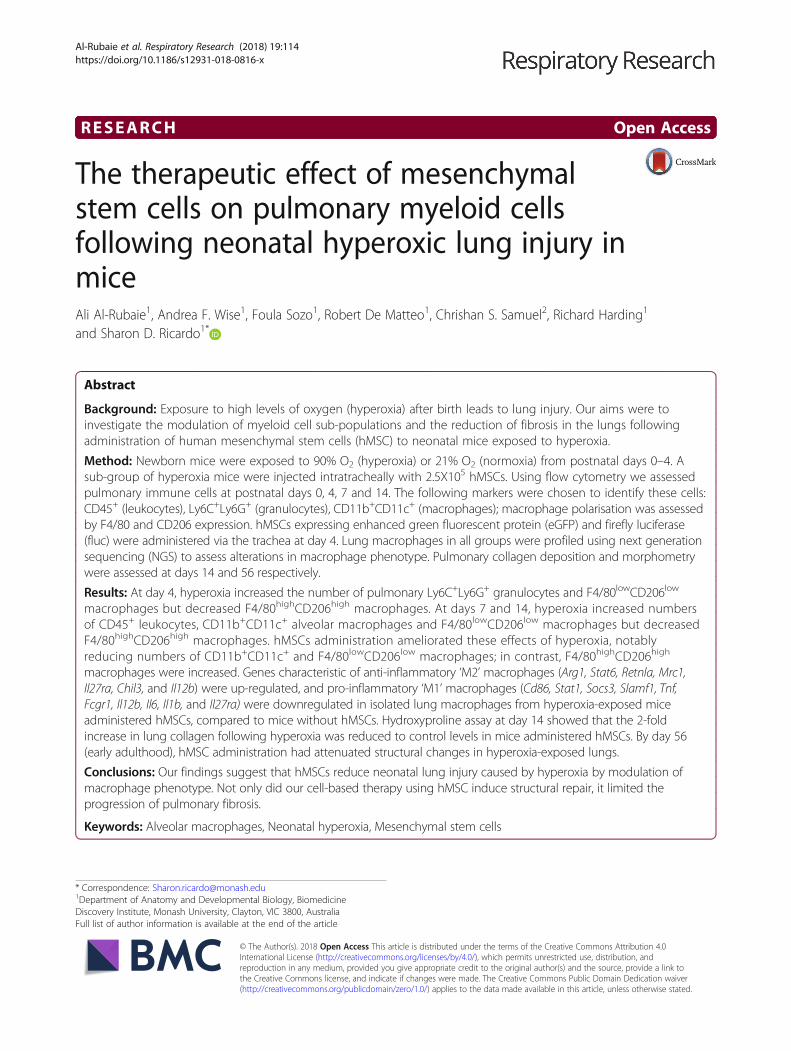
RESEARCH Open Access
The therapeutic effect of mesenchymalstem cells on pulmonary myeloid cellsfollowing neonatal hyperoxic lung injury inmiceAli Al-Rubaie1, Andrea F. Wise1, Foula Sozo1, Robert De Matteo1, Chrishan S. Samuel2, Richard Harding1
and Sharon D. Ricardo1*
Abstract
Background: Exposure to high levels of oxygen (hyperoxia) after birth leads to lung injury. Our aims were toinvestigate the modulation of myeloid cell sub-populations and the reduction of fibrosis in the lungs followingadministration of human mesenchymal stem cells (hMSC) to neonatal mice exposed to hyperoxia.
Method: Newborn mice were exposed to 90% O2 (hyperoxia) or 21% O2 (normoxia) from postnatal days 0–4. Asub-group of hyperoxia mice were injected intratracheally with 2.5X10⁵ hMSCs. Using flow cytometry we assessedpulmonary immune cells at postnatal days 0, 4, 7 and 14. The following markers were chosen to identify these cells:CD45+ (leukocytes), Ly6C+Ly6G+ (granulocytes), CD11b+CD11c+ (macrophages); macrophage polarisation was assessedby F4/80 and CD206 expression. hMSCs expressing enhanced green fluorescent protein (eGFP) and firefly luciferase(fluc) were administered via the trachea at day 4. Lung macrophages in all groups were profiled using next generationsequencing (NGS) to assess alterations in macrophage phenotype. Pulmonary collagen deposition and morphometrywere assessed at days 14 and 56 respectively.
Results: At day 4, hyperoxia increased the number of pulmonary Ly6C+Ly6G+ granulocytes and F4/80lowCD206low
macrophages but decreased F4/80highCD206high macrophages. At days 7 and 14, hyperoxia increased numbersof CD45+ leukocytes, CD11b+CD11c+ alveolar macrophages and F4/80lowCD206low macrophages but decreasedF4/80highCD206high macrophages. hMSCs administration ameliorated these effects of hyperoxia, notablyreducing numbers of CD11b+CD11c+ and F4/80lowCD206low macrophages; in contrast, F4/80highCD206high
macrophages were increased. Genes characteristic of anti-inflammatory ‘M2’ macrophages (Arg1, Stat6, Retnla, Mrc1,Il27ra, Chil3, and Il12b) were up-regulated, and pro-inflammatory ‘M1’ macrophages (Cd86, Stat1, Socs3, Slamf1, Tnf,Fcgr1, Il12b, Il6, Il1b, and Il27ra) were downregulated in isolated lung macrophages from hyperoxia-exposed miceadministered hMSCs, compared to mice without hMSCs. Hydroxyproline assay at day 14 showed that the 2-foldincrease in lung collagen following hyperoxia was reduced to control levels in mice administered hMSCs. By day 56(early adulthood), hMSC administration had attenuated structural changes in hyperoxia-exposed lungs.
Conclusions: Our findings suggest that hMSCs reduce neonatal lung injury caused by hyperoxia by modulation ofmacrophage phenotype. Not only did our cell-based therapy using hMSC induce structural repair, it limited theprogression of pulmonary fibrosis.
Keywords: Alveolar macrophages, Neonatal hyperoxia, Mesenchymal stem cells
* Correspondence: [email protected] of Anatomy and Developmental Biology, BiomedicineDiscovery Institute, Monash University, Clayton, VIC 3800, AustraliaFull list of author information is available at the end of the article
© The Author(s). 2018 Open Access This article is distributed under the terms of the Creative Commons Attribution 4.0International License (http://creativecommons.org/licenses/by/4.0/), which permits unrestricted use, distribution, andreproduction in any medium, provided you give appropriate credit to the original author(s) and the source, provide a link tothe Creative Commons license, and indicate if changes were made. The Creative Commons Public Domain Dedication waiver(http://creativecommons.org/publicdomain/zero/1.0/) applies to the data made available in this article, unless otherwise stated.
Al-Rubaie et al. Respiratory Research (2018) 19:114 https://doi.org/10.1186/s12931-018-0816-x
BackgroundPreterm infants experience a range of debilitating healthissues primarily resulting from lung immaturity, and fewtreatment options are available [1–3]. Owing to lungunderdevelopment, preterm infants often require mech-anical ventilation with hyperoxic gas in order to survive[4, 5]. However, high levels of oxygen or prolonged useof ventilators can damage the lungs and interrupt normalalveolar and bronchiolar development, which may lead tochronic lung diseases known as bronchopulmonary dys-plasia (BPD) [6]. It is common, that preterm infants thatare born at less than 32 weeks of gestation have increasedprogression of other short and long-term respiratory ill-nesses, such as asthma and chronic obstructive pulmonarydisease (COPD) [7].Experimental studies have attempted to reduce the
negative effects of hyperoxia on the developing lungs;however, clinical translation has been disappointing todate. Consequently, there is still no effective treatmentfor BPD in preterm infants [8–10]. BPD is associatedwith the inflammation of lungs, which typically involvesthe recruitment of monocytes that differentiate into al-veolar macrophages [11]. Macrophages are a heteroge-neous cell type that can be broadly categorised into twogroups: “classically activated” M1 macrophages that havepro-inflammatory functions and M2 macrophages are“alternatively activated” cells that play a reparative orregulatory role [12–14].Human mesenchymal stem cells (hMSCs) have the abil-
ity to alter macrophage phenotype from an inflammatoryto anti-inflammatory phenotype that may be therapeutic-ally beneficial to treat injured lungs [15]. There have beencompleted or ongoing clinical trials involving cell therap-ies that delay the progression or reverse a variety of im-mune and non-immune diseases in the lung and otherorgans [16]. Preclinical and clinical data support the useof cell therapy to treat COPD, acute adult lung injury andsevere chronic asthma [17]. Recently, the protective effectsof stem cells in lung diseases, for example BPD, have beendemonstrated [18]. Studies have shown that stem cellsprotect the newborn injured lung from BPD and aid in en-dogenous repair of the injured lung tissue, either by differ-entiation into lung parenchymal cells [19], or into type IIalveolar epithelial cells [20], via the secretion of factors[21]. However, some limitations to the studies have alsobeen reported such as risks associated with possible im-mune reactions against hMSCs [22]. Understanding thetherapeutic benefit of hMSCs in neonatal hyperoxic lunginjury in mice remaining largely unclear, specifically, theability to alter the lung immune cells population/s.This study aimed to determine the effectiveness of hu-
man bone marrow-derived hMSC therapy on neonatalhyperoxia, through the modulation of pulmonary im-mune cells and lung injury–induced collagen deposition
in mice, based on the alteration of macrophage phenotype.We showed that hMSCs tracked to the injured lung fol-lowing intratracheal injection where they modulatedmacrophage phenotype leading to reduced collagen de-position, thus improving lung structure.
MethodsExperimental animalsThis study was performed using pups at embryonic day14 timed-pregnant C57BL/6 J mice obtained from theMonash Animal Research Platform (MARP), housed atMonash University, Clayton. The study was conductedunder animal ethics number MARP2014/092 issued byMonash Animal Ethics Committee. Pups were born atterm, and the day designated as postnatal day 0. Theexposure of lungs to 90% O2 was used to induce injuryand the effectiveness of hMSC treatment on hyperoxia-induced lung injury was assessed. Newborn mice wereexposed to either normoxia 21% O2 (control group) or90% O2 (hyperoxia) from postnatal day 0 to day 4. Theoxygen concentration was maintained and monitoredthroughout the period of hyperoxia dams were rotatedwith non-hyperoxia dams (SWAP dams) every 24 h. Themice received standard chow ad libitum under 12/12 h day/night cycle. At day 4, sub-groups within the hyperoxiagroups were injected directly into the trachea with hMSCspurchased from the Tulane Centre for Stem Cell Researchand Regenerative Medicine (Tulane University, NewOrleans, LA) [23] at passage four. 2.5X10⁵ hMSCs were sus-pended in 10 μl PBS and delivered via intratracheal injectionusing a 1 ml insulin syringe and a 29G hypodermic needle.Mouse lungs were collected at days 0, 4, 7 and 14.
Bioluminescence imagingFor cell tracing of hMSCs in vivo, sub-groups of hyper-oxia-exposed mice (n = 8) were administered 2.5X10⁵
hMSCs expressing enhanced green fluorescent protein(eGFP) and firefly luciferase (fluc) in 10 μl PBS at postnatalday 4 [23]. To image the hMSCs in vivo following delivery,anesthetized mice were injected intraperitoneally with200 μl of D-luciferin (15 mg/ml in PBS; VivoGlo Luciferin,Promega, San Luis Obispo, CA, USA) and imaged on days0, 1 and 3 following cell delivery using a IVIS 200 system(Xenogen, Alameda, CA, USA). The fluc luminescent sig-nal was captured and analysed as photons/sec/cm2 (LivingImage 3.2, Xenogen).
Lung immune cells preparationPreparation of lung tissue Freshly excised lungs werekept in cold FACS -fluorescence-activated cell sorting-buffer (Phosphate-buffered saline accompanied with 0.2%Bovine serum albumin, 0.02% Sodium azide and 5 mMEDTA). With surgical scissors, the main airways were re-moved and the lungs were cut into small portions before
Al-Rubaie et al. Respiratory Research (2018) 19:114 Page 2 of 11
enzymatic digestion, for 45 min, in 1 mL of dissociationmedia consisting of HBSS- Hanks’ Balanced Salt Solu-tion- (Sigma-Aldrich, St. Louis, USA) containing 3 mg/mLcollagenase/dispase (Roche Applied Science, Penzberg,Germany) and 0.2 mg/mL DNase type 1 (Roche AppliedScience). To eliminate red blood cells, the lung single cellsuspensions were incubated in 1 mL of red blood cell lysisbuffer (8.3 g/L NH4Cl, 10 mM Tris-HCl, and pH 7.5) for1 min. Samples were filtered through a 100 μm nylon cellstrainer (BD Bioscience, San Jose, USA) before markerslabelling.
Flow-cytometry Cells counts were performed using aZ2 Coulter Counter (Beckman Coulter, USA). 3X10⁶
cells from single cell suspensions were incubated at 4 °Cfor 20 min with the following fluorochrome-conjugatedanti-mouse markers: anti-CD45 APC-Cy7 (clone 30-F11;Biolegend, San Diego, USA), anti-CD11b PE-Cy7 (cloneM1/70; BD Biosciences), anti-CD11c Pacific Blue (cloneN418; Biolegend), anti-Ly6C FITC (clone HK1.4; Biolegend),anti-Ly6G Alexa Fluor 647 (clone 1A8; Biolegend), anti-F4/80 APC (clone BM8; eBioscience), and anti-CD206(mannose receptor; MR) Alexa Fluor 488 (clone C068C2;Biolegend). Fc receptor block (anti-CD16/32 antibody)was added to all markers cocktails. Intracellular CD206 la-belling was performed using a CytoFix/CytoPerm kit (BDBiosciences, USA). After surface receptor labelling, cellswere permeabilized and incubated with the marker for30 min at 4 °C in the dark before being washed twice in1× Perm/Wash buffer (BD Biosciences) and resuspend inFACS buffer. A BD FACS Canto II flow cytometer (BDBiosciences) was used to acquire data. Data was analysedusing FlowLogic FCS analysis software (Inivai Technolo-gies, Melbourne, Australia).
Alveolar macrophages total-RNA sequencing At post-natal day14, 1X10⁵ CD45+CD11b+CD11c+ alveolar mac-rophages were sorted from mouse lungs (total n = 9; 3from each treatment group) via an influx sorter (Flow-core, Monash University, Victoria, Australia). Total RNAwas extracted from the sorted macrophages using RNeasyKit following the manufacturer’s protocol (Qiagen, Hilden,Germany). The integrity of the samples was measured usingthe Agilent Bioanalyzer 2100 with B.02.08.SI648 (SR3) soft-ware and a microfluidics device, in conjunction the as-sociated hardware and chemistry (Agilent Technologies,Waldbronn, Germany). Samples were used to constructIllumina sequencing libraries using the following TakaraSMART-seq Ultra Low Input version 4 according to themanufacturer’s instructions (Clontech Takara SMART-SeqV4 Ultra Low Input RNA Kit for Sequencing UserManual v.01251). Libraries were quantitated using aQubit DNA HS kit, which incorporates a double-stranded DNA-specific fluorescent dye (Invitrogen, Carlsbad
CA., USA). Libraries were sized and checked for adaptercontamination using the Agilent Bioanalyzer 2100 microflui-dics device, in conjunction with Agilent DNA HS kits andchemistry (Agilent Technologies, Waldbronn, Germany).These libraries were sequenced using the followingchemistry and conditions in NextSeq500 - High-OutputSBS version 2 (Illumina 15,046,563 v02), with libraryconcentration 1.8 pM and reading length 1X75b accord-ing to the manufacturer’s instructions. The differentialgene expression was performed with the voom [24] limmapackage (v3.34.1) [25].
Lung fibrosis profile and structural changesHydroxyproline assay On day 14, lung tissue frommouse pups (n = 8) were assessed for total collagen con-tent, determined by hydroxyproline content as previouslydescribed [23] . Hydroxyproline values were then con-verted to collagen content by multiplying by a factor of6.94 as hydroxyproline represents; 14.4% of the aminoacid composition of collagen in most mammalian tissues[26]. This was determined using a standard curve of puri-fied trans-4-hydroxy-L-proline (Sigma-Aldrich) and fur-ther expressed as a percentage of the dry tissue weight toyield collagen concentration.
Morphometric lung analysis At day 56, lung tissue sec-tions from mouse (n = 8 / group) were randomly selectedfrom the left lung were embedded in paraffin for histo-logical staining. Paraffin-embedded sections of lung tissuewere stained with picrosirius red. Sections 5 μm thickwere examined by light microscopy (X200 magnification)and five random sections were captured using a digitalcamera (SPOT Insight 4Meg Fire Wire Color Mosaic 14.2,Diagnostic Instruments, USA). To assess lung injury,mean linear intercept (MLI) was measured using softwareImageJ, version 1.47 (Wayne Rasband, NIH, Bethesda,MD, USA). Large airways and vessels were avoided. Theprogram placed horizontal lines, 30 μm apart, across eachsection of lung. Then the number of times the lines inter-cepted the alveoli (intercepts) was calculated. All countingmethods have been described previously [27].
Statistical analysisData were analysed using GraphPad Prism software ver-sion 7.0c (GraphPad Software Inc., San Diego, USA) andIBM SPSS statistics (The Apache Software Foundation).A t-test (unpaired, two-tailed) was used to analyse differ-ences between the normoxia and hyperoxia groups atday 4. A one-way analysis of variance with a Tukey’s mul-tiple comparisons test was used to analyse data containedin the normoxia, hyperoxia and hyperoxia + hMSC groupsat day 7 and day 14. Data are given as means ± SEM andP < 0.05 was considered statistically significant.
Al-Rubaie et al. Respiratory Research (2018) 19:114 Page 3 of 11
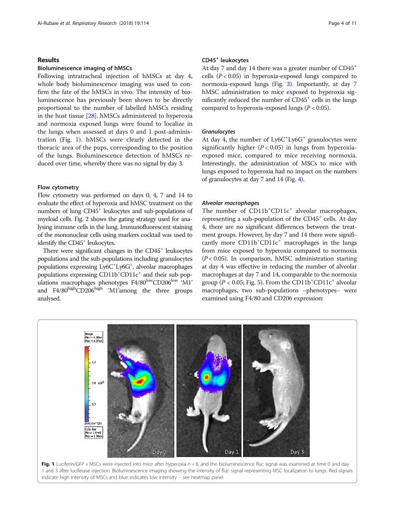
ResultsBioluminescence imaging of hMSCsFollowing intratracheal injection of hMSCs at day 4,whole body bioluminescence imaging was used to con-firm the fate of the hMSCs in vivo. The intensity of bio-luminescence has previously been shown to be directlyproportional to the number of labelled hMSCs residingin the host tissue [28]. hMSCs administered to hyperoxiaand normoxia exposed lungs were found to localize inthe lungs when assessed at days 0 and 1 post-adminis-tration (Fig. 1). hMSCs were clearly detected in thethoracic area of the pups, corresponding to the positionof the lungs. Bioluminescence detection of hMSCs re-duced over time, whereby there was no signal by day 3.
Flow cytometryFlow cytometry was performed on days 0, 4, 7 and 14 toevaluate the effect of hyperoxia and hMSC treatment on thenumbers of lung CD45+ leukocytes and sub-populations ofmyeloid cells. Fig. 2 shows the gating strategy used for ana-lysing immune cells in the lung. Immunofluorescent stainingof the mononuclear cells using markers cocktail was used toidentify the CD45+ leukocytes.There were significant changes in the CD45+ leukocytes
populations and the sub-populations including granulocytespopulations expressing Ly6C+Ly6G+, alveolar macrophagespopulations expressing CD11b+CD11c+ and their sub-pop-ulations macrophages phenotypes F4/80lowCD206low ‘M1’and F4/80highCD206high ‘M1’among the three groupsanalysed.
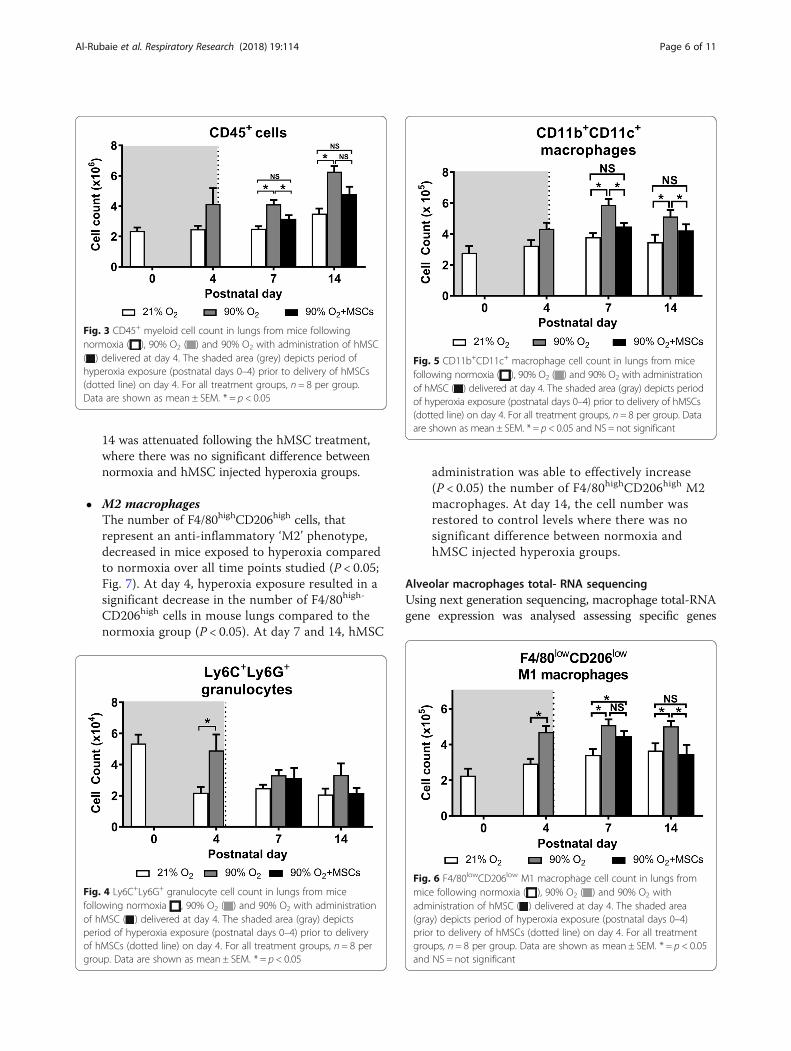
CD45+ leukocytesAt day 7 and day 14 there was a greater number of CD45+
cells (P < 0.05) in hyperoxia-exposed lungs compared tonormoxia-exposed lungs (Fig. 3). Importantly, at day 7hMSC administration to mice exposed to hyperoxia sig-nificantly reduced the number of CD45+ cells in the lungscompared to hyperoxia-exposed lungs (P < 0.05).
GranulocytesAt day 4, the number of Ly6C+Ly6G+ granulocytes weresignificantly higher (P < 0.05) in lungs from hyperoxia-exposed mice, compared to mice receiving normoxia.Interestingly, the administration of MSCs to mice withlungs exposed to hyperoxia had no impact on the numbersof granulocytes at day 7 and 14 (Fig. 4).
Alveolar macrophagesThe number of CD11b+CD11c+ alveolar macrophages,representing a sub-population of the CD45+ cells. At day4, there are no significant differences between the treat-ment groups. However, by day 7 and 14 there were signifi-cantly more CD11b+CD11c+ macrophages in the lungsfrom mice exposed to hyperoxia compared to normoxia(P < 0.05). In comparison, hMSC administration startingat day 4 was effective in reducing the number of alveolarmacrophages at day 7 and 14, comparable to the normoxiagroup (P < 0.05; Fig. 5). From the CD11b+CD11c+ alveolarmacrophages, two sub-populations –phenotypes– wereexamined using F4/80 and CD206 expression:
Fig. 1 Luciferin/GFP + MSCs were injected into mice after hyperoxia n = 8, and the bioluminescence fluc signal was examined at time 0 and day1 and 3 after luciferase injection. Bioluminescence imaging showing the intensity of fluc signal representing MSC localization to lungs. Red signalsindicate high intensity of MSCs and blue indicates low intensity – see heatmap panel
Al-Rubaie et al. Respiratory Research (2018) 19:114 Page 4 of 11
� M1 macrophagesThe number of F4/80lowCD206low macrophages sub-population, that represent an inflammatory ‘M1’phenotype, significantly changed among the
treatment groups. Hyperoxia caused a significant risein the number of F4/80lowCD206low macrophages atdays 4, 7 and 14, compared to the normoxia group(P < 0.05; Fig. 6). This increase in cell number at day
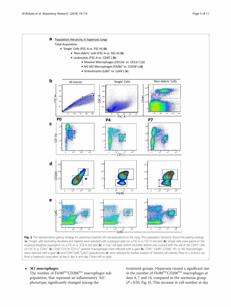
a
b
c
d
e
Fig. 2 The representative gating strategy for assessing myeloid cell sub-populations in the lung. The population hierarchy shows the gating strategy(a). ‘Single’ cells (excluding doublets and triplets) were selected with a polygon gate on a FSC-A vs. FSC-H dot plot (b). Single cells were gated on theresulting daughter population on a FSC-A vs. SCA-A dot plot (b). A ‘Live’ cell gate (which excludes debris) was created with the aid of the CD45+ cellson FSC-A vs. CD45+ (b). CD45+CD11b+CD11c+ alveolar macrophages were selected with a gate (b), CD45+ F4/80+ CD206+ M1 vs. M2 macrophageswere selected with a gate (d) and CD45+Ly6C+Ly6G+ granulocytes (e) were selected for further analysis of myeloid cell subsets. Plots in c, d and e arefrom a hyperoxic lung taken at day 0, day 4, and day 7 from left to right
Al-Rubaie et al. Respiratory Research (2018) 19:114 Page 5 of 11
14 was attenuated following the hMSC treatment,where there was no significant difference betweennormoxia and hMSC injected hyperoxia groups.
� M2 macrophagesThe number of F4/80highCD206high cells, thatrepresent an anti-inflammatory ‘M2’ phenotype,decreased in mice exposed to hyperoxia comparedto normoxia over all time points studied (P < 0.05;Fig. 7). At day 4, hyperoxia exposure resulted in asignificant decrease in the number of F4/80high-
CD206high cells in mouse lungs compared to thenormoxia group (P < 0.05). At day 7 and 14, hMSC
administration was able to effectively increase(P < 0.05) the number of F4/80highCD206high M2macrophages. At day 14, the cell number wasrestored to control levels where there was nosignificant difference between normoxia andhMSC injected hyperoxia groups.
Alveolar macrophages total- RNA sequencingUsing next generation sequencing, macrophage total-RNAgene expression was analysed assessing specific genes
Fig. 3 CD45+ myeloid cell count in lungs from mice followingnormoxia ( ), 90% O2 ( ) and 90% O2 with administration of hMSC( ) delivered at day 4. The shaded area (grey) depicts period ofhyperoxia exposure (postnatal days 0–4) prior to delivery of hMSCs(dotted line) on day 4. For all treatment groups, n = 8 per group.Data are shown as mean ± SEM. * = p < 0.05
Fig. 4 Ly6C+Ly6G+ granulocyte cell count in lungs from micefollowing normoxia , 90% O2 ( ) and 90% O2 with administrationof hMSC ( ) delivered at day 4. The shaded area (gray) depictsperiod of hyperoxia exposure (postnatal days 0–4) prior to deliveryof hMSCs (dotted line) on day 4. For all treatment groups, n = 8 pergroup. Data are shown as mean ± SEM. * = p < 0.05
Fig. 5 CD11b+CD11c+ macrophage cell count in lungs from micefollowing normoxia ( ), 90% O2 ( ) and 90% O2 with administrationof hMSC ( ) delivered at day 4. The shaded area (gray) depicts periodof hyperoxia exposure (postnatal days 0–4) prior to delivery of hMSCs(dotted line) on day 4. For all treatment groups, n = 8 per group. Dataare shown as mean ± SEM. * = p < 0.05 and NS = not significant
Fig. 6 F4/80lowCD206low M1 macrophage cell count in lungs frommice following normoxia ( ), 90% O2 ( ) and 90% O2 withadministration of hMSC ( ) delivered at day 4. The shaded area(gray) depicts period of hyperoxia exposure (postnatal days 0–4)prior to delivery of hMSCs (dotted line) on day 4. For all treatmentgroups, n = 8 per group. Data are shown as mean ± SEM. * = p < 0.05and NS = not significant
Al-Rubaie et al. Respiratory Research (2018) 19:114 Page 6 of 11
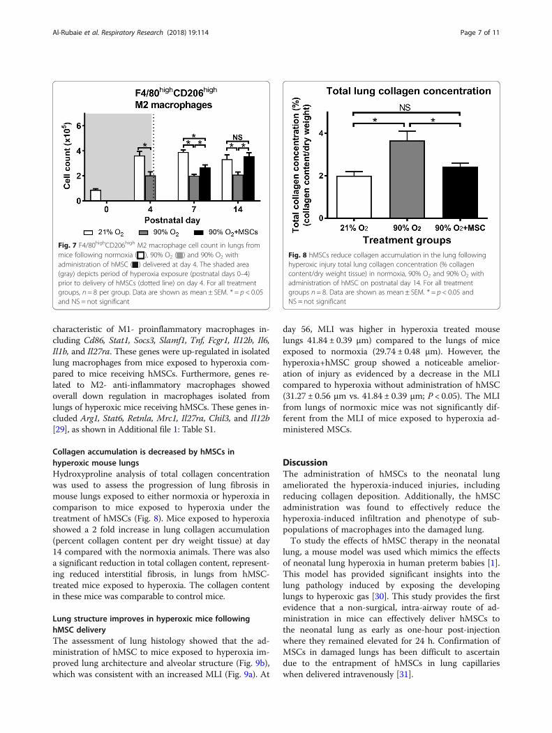
characteristic of M1- proinflammatory macrophages in-cluding Cd86, Stat1, Socs3, Slamf1, Tnf, Fcgr1, Il12b, Il6,Il1b, and Il27ra. These genes were up-regulated in isolatedlung macrophages from mice exposed to hyperoxia com-pared to mice receiving hMSCs. Furthermore, genes re-lated to M2- anti-inflammatory macrophages showedoverall down regulation in macrophages isolated fromlungs of hyperoxic mice receiving hMSCs. These genes in-cluded Arg1, Stat6, Retnla, Mrc1, Il27ra, Chil3, and Il12b[29], as shown in Additional file 1: Table S1.
Collagen accumulation is decreased by hMSCs inhyperoxic mouse lungsHydroxyproline analysis of total collagen concentrationwas used to assess the progression of lung fibrosis inmouse lungs exposed to either normoxia or hyperoxia incomparison to mice exposed to hyperoxia under thetreatment of hMSCs (Fig. 8). Mice exposed to hyperoxiashowed a 2 fold increase in lung collagen accumulation(percent collagen content per dry weight tissue) at day14 compared with the normoxia animals. There was alsoa significant reduction in total collagen content, represent-ing reduced interstitial fibrosis, in lungs from hMSC-treated mice exposed to hyperoxia. The collagen contentin these mice was comparable to control mice.
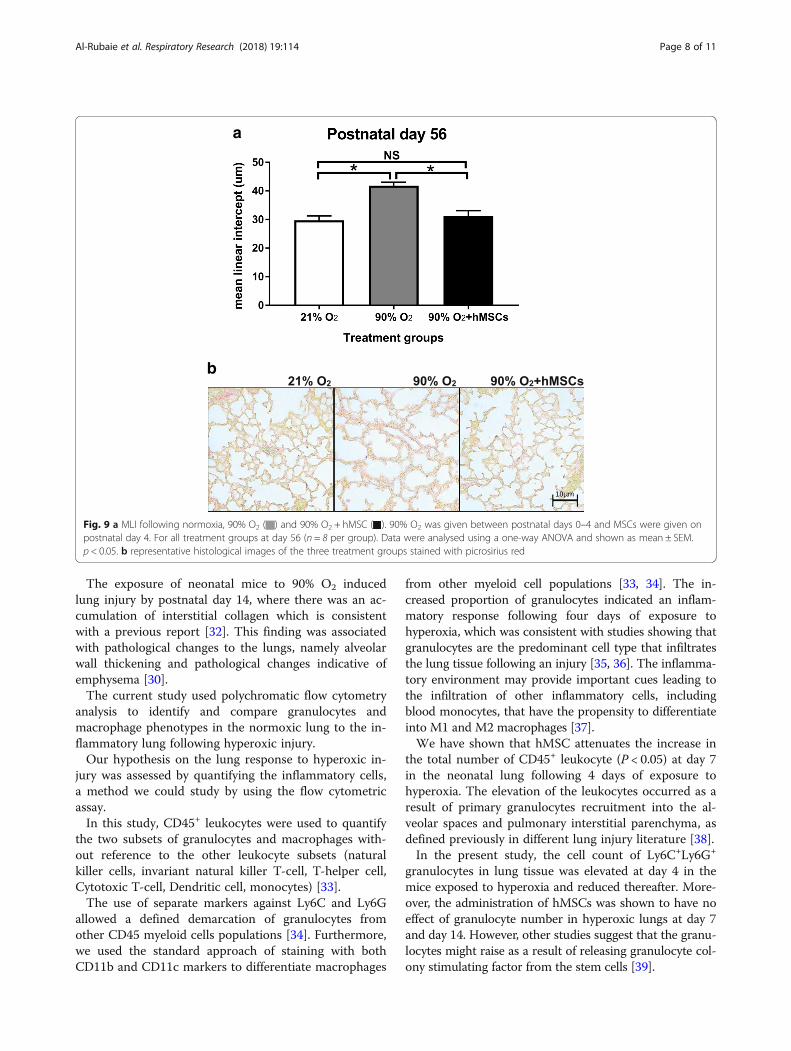
Lung structure improves in hyperoxic mice followinghMSC deliveryThe assessment of lung histology showed that the ad-ministration of hMSC to mice exposed to hyperoxia im-proved lung architecture and alveolar structure (Fig. 9b),which was consistent with an increased MLI (Fig. 9a). At
day 56, MLI was higher in hyperoxia treated mouselungs 41.84 ± 0.39 μm) compared to the lungs of miceexposed to normoxia (29.74 ± 0.48 μm). However, thehyperoxia+hMSC group showed a noticeable amelior-ation of injury as evidenced by a decrease in the MLIcompared to hyperoxia without administration of hMSC(31.27 ± 0.56 μm vs. 41.84 ± 0.39 μm; P < 0.05). The MLIfrom lungs of normoxic mice was not significantly dif-ferent from the MLI of mice exposed to hyperoxia ad-ministered MSCs.
DiscussionThe administration of hMSCs to the neonatal lungameliorated the hyperoxia-induced injuries, includingreducing collagen deposition. Additionally, the hMSCadministration was found to effectively reduce thehyperoxia-induced infiltration and phenotype of sub-populations of macrophages into the damaged lung.To study the effects of hMSC therapy in the neonatal
lung, a mouse model was used which mimics the effectsof neonatal lung hyperoxia in human preterm babies [1].This model has provided significant insights into thelung pathology induced by exposing the developinglungs to hyperoxic gas [30]. This study provides the firstevidence that a non-surgical, intra-airway route of ad-ministration in mice can effectively deliver hMSCs tothe neonatal lung as early as one-hour post-injectionwhere they remained elevated for 24 h. Confirmation ofMSCs in damaged lungs has been difficult to ascertaindue to the entrapment of hMSCs in lung capillarieswhen delivered intravenously [31].
Fig. 7 F4/80highCD206high M2 macrophage cell count in lungs frommice following normoxia ( ), 90% O2 ( ) and 90% O2 withadministration of hMSC ( ) delivered at day 4. The shaded area(gray) depicts period of hyperoxia exposure (postnatal days 0–4)prior to delivery of hMSCs (dotted line) on day 4. For all treatmentgroups, n = 8 per group. Data are shown as mean ± SEM. * = p < 0.05and NS = not significant
Fig. 8 hMSCs reduce collagen accumulation in the lung followinghyperoxic injury total lung collagen concentration (% collagencontent/dry weight tissue) in normoxia, 90% O2 and 90% O2 withadministration of hMSC on postnatal day 14. For all treatmentgroups n = 8. Data are shown as mean ± SEM. * = p < 0.05 andNS = not significant
Al-Rubaie et al. Respiratory Research (2018) 19:114 Page 7 of 11
The exposure of neonatal mice to 90% O2 inducedlung injury by postnatal day 14, where there was an ac-cumulation of interstitial collagen which is consistentwith a previous report [32]. This finding was associatedwith pathological changes to the lungs, namely alveolarwall thickening and pathological changes indicative ofemphysema [30].The current study used polychromatic flow cytometry
analysis to identify and compare granulocytes andmacrophage phenotypes in the normoxic lung to the in-flammatory lung following hyperoxic injury.Our hypothesis on the lung response to hyperoxic in-
jury was assessed by quantifying the inflammatory cells,a method we could study by using the flow cytometricassay.In this study, CD45+ leukocytes were used to quantify
the two subsets of granulocytes and macrophages with-out reference to the other leukocyte subsets (naturalkiller cells, invariant natural killer T-cell, T-helper cell,Cytotoxic T-cell, Dendritic cell, monocytes) [33].The use of separate markers against Ly6C and Ly6G
allowed a defined demarcation of granulocytes fromother CD45 myeloid cells populations [34]. Furthermore,we used the standard approach of staining with bothCD11b and CD11c markers to differentiate macrophages
from other myeloid cell populations [33, 34]. The in-creased proportion of granulocytes indicated an inflam-matory response following four days of exposure tohyperoxia, which was consistent with studies showing thatgranulocytes are the predominant cell type that infiltratesthe lung tissue following an injury [35, 36]. The inflamma-tory environment may provide important cues leading tothe infiltration of other inflammatory cells, includingblood monocytes, that have the propensity to differentiateinto M1 and M2 macrophages [37].We have shown that hMSC attenuates the increase in
the total number of CD45+ leukocyte (P < 0.05) at day 7in the neonatal lung following 4 days of exposure tohyperoxia. The elevation of the leukocytes occurred as aresult of primary granulocytes recruitment into the al-veolar spaces and pulmonary interstitial parenchyma, asdefined previously in different lung injury literature [38].In the present study, the cell count of Ly6C+Ly6G+
granulocytes in lung tissue was elevated at day 4 in themice exposed to hyperoxia and reduced thereafter. More-over, the administration of hMSCs was shown to have noeffect of granulocyte number in hyperoxic lungs at day 7and day 14. However, other studies suggest that the granu-locytes might raise as a result of releasing granulocyte col-ony stimulating factor from the stem cells [39].
a
b
Fig. 9 a MLI following normoxia, 90% O2 ( ) and 90% O2 + hMSC ( ). 90% O2 was given between postnatal days 0–4 and MSCs were given onpostnatal day 4. For all treatment groups at day 56 (n = 8 per group). Data were analysed using a one-way ANOVA and shown as mean ± SEM.p < 0.05. b representative histological images of the three treatment groups stained with picrosirius red
Al-Rubaie et al. Respiratory Research (2018) 19:114 Page 8 of 11
This study showed that the CD11b CD11c expressedmacrophages were relatively high from day zero, althoughthe level of CD11c expression was low in comparison withday 4 and 7 as shown in a representative FACS plots inFig. 2. It is hard to confirm whether these macrophagesare monocytes derived macrophages or foetal monocytes-macrophages, and immature-macrophages, with signifi-cant overlap in expression of marker sets. These intersti-tial macrophages were derived from the yolk sac [12]which initially derived from mesoderm cells that developduring gastrulation from the primitive streak [40].Throughout the study, we demonstrated that CD11b+
CD11c+ macrophages increased in response to hyper-oxia by day 14, including an increased population ofF4/80lowCD206low inflammatory ‘M1’ macrophages anddecreased F4/80highCD206high anti-inflammatory ‘M2’macrophages. In other studies, M1 macrophage infiltra-tion has been confirmed as well, together with myeloiddifferentiation and the alteration of relative function at thesite of inflammation [41, 42]. These changes in phenotyperesult in the activation of monocytes at different matur-ation stages leading to mature macrophages of distinctivefunctional states [43]. Because macrophages are necessaryfor the phagocytosis of apoptotic neutrophils, the expos-ure to neonatal hyperoxia may lead to a reduced numberof macrophages proceeding to necrosis, leading to the ex-pansion of damaged alveoli [11]. This conclusion contra-dicts studies suggesting that macrophages might secretecertain chemokines, which could influence neutrophil in-filtration and recruitment [11].Our data confirms the beneficial effect of hMSC treat-
ment on the phenotype and function of macrophagesafter neonatal hyperoxia. The administration of MSCs tomice with hyperoxia-induced acute lung injury was shownalteration of the macrophages proportion and phenotypes.These findings indicated the presence of an inflammatoryenvironment. These results have also been demonstratedin an experimental model of asthma after bone marrow-derived hMSC administration [44].Following the intra-airway administration of hMSCs,
an increase in M2 macrophages was observed in the in-jured lungs of mice. It is possible that the reduction inlung injury in hMSC-treated mice was due to downregu-lation of proinflammatory factors and the upregulationof anti-inflammatory chemokines. In contrast, the ad-vantageous effects of hMSC treatment were displayed byreducing eosinophil infiltration in mouse models with al-lergic lung injury [45]. These findings and other support-ing studies suggested that hMSCs are able to alter thebalance of macrophage phenotype and function thatoccur during injury to promote repair [46]. We showedthat the protective effect of hMSCs in hyperoxia-inducedlung injury could alter macrophage phenotype. Recentstudies have shown the in vivo interaction between
hMSCs and macrophages to promote M2 polarisation.The in vitro co-culture of hMSCs and macrophages re-sulted in an alternatively activated macrophage phenotypecharacterised as mannose receptor (MR) high, IL-10high,IL-6high, TNF-αlow and IL-12low which exhibits en-hanced phagocytic activity, increased secretion of IL-10and VEGF and decreased secretion of pro-inflammatorycytokines [44]. Further investigation to identify the po-larisation mechanism will be important for the under-standing how hMSCs alter the host response followingtherapeutic delivery, in the context not only the inflam-matory response to hyperoxia, but also lung disorderssuch as allergic asthma and COPD. Apart from the al-teration of lung myeloid cells, lung fibrosis is the finalcommon pathway of lung injury regardless of aetiology.In examining the effect of hMSCs therapy on lung in-jury induced by 90% O2, we chose an end-point of de-livery of oxygen of four days as published data hasshown that delivery of hMSCs may ameliorate estab-lished lung fibrosis after 14 days of hyperoxia- inducedinjury [47]. We found that the treatment with hMSCsinduced decreased lung collagen accumulation follow-ing hyperoxia. We propose that hMSCs are able to cre-ate a more favourable environment, leading to lesstissue damage and fibrosis. In this environment, hMSCsmay possibly not only alter lung immune cells but mayalso have an increased capacity to facilitate their antifi-brotic and reparative effects, resulting in a greater re-duction in fibrosis [48]. The administration of hMSCsto the neonatal lung ameliorated the hyperoxia-inducedstructural injuries, including reducing collagen depos-ition. Additionally, the intratracheal delivery of hMSCwas found to effectively reduce the hyperoxia-inducedinfiltration of myeloid cells, including sub-populationsof macrophages, into the damaged lung tissue. This in-dicates that hMSCs can modulate the inflammatory en-vironment in the lung to reduce the development offibrotic damage and structural injury.Using hMSCs in a wide range of settings has shown
impressive treatment responses as these cells releaseanti-inflammatory factors providing a positive effect bymodulating the inflammatory environments, hence improv-ing tissue healing [46]. Our study has shown the effect ofthe administration of hMSCs immediately, after exposure tohyperoxia, on myeloid cell sub-populations in the lung.Given our findings, it is possible that future clinical trialsmay incorporate the use of hMSCs therapy to reduce thelung injury including injury that may developed following inpreterm birth hyperoxia and other lung diseases that resultsfrom pathologic fibrosis. This study was designed to test theefficacy of hMSCs in limiting hyperoxia-induced lung injury.In order to establish a ‘proof of principle’ we used 90% oxy-gen, which is a higher concentration of oxygen that is usedclinically. It is recognised that our hyperoxic lung injury
Al-Rubaie et al. Respiratory Research (2018) 19:114 Page 9 of 11
mouse model is likely to induce a more severe form of BPDthan in preterm infants in normal physiological condition.In the future, it will be critical to assess the effectiveness ofhMSC in lung injury models that more closely replicate clin-ical conditions. Additionally, the exposure to 90% O2 islikely to cause hyperoxic injury in organs other than thelungs. As hMSCs administered intratracheally will likelyhome to injured tissues, it will be important study the sys-temic effects of hMSC in future studies.
ConclusionsOur study provides evidence for a novel cellular-basedtherapy that can induce lung repair and limit the progres-sion of fibrosis in neonatal lungs exposed to hyperoxicgas. The results from this study demonstrate that hMSCtherapy may provide improved therapeutic options forpreterm infants necessarily exposed to hyperoxic gas, andmay therefore reduce the risk of COPD and asthma inlater life.
Additional file
Additional file 1: Table S1. Table showing the most differentiallyexpressed genes in isolated macrophages from lungs of mice (n = 9)exposed to normoxia or hyperoxia with/without administration of hMSCs.RNA was isolated, then analysed, using next generation sequencing. Thegenes that are related to M1 and M2 macrophages were either up-regulatedor down-regulated in isolated lung macrophages in response to hyperoxia,that were altered following delivery of hMSCs. Abbreviations: Cd86 (Cluster ofDifferentiation 86), Socs3 (suppressor of and cytokine signalling), Slamf1(signalling lymphocytic activation molecule family member), Tnf (tumournecrosis factor), Fcgr1 (Fc receptor, IgG, low affinity), Il12b (Interleukin 12B),Il1b (Interleukin 1 beta), Il6 (Interleukin 6), Arg1 (arginase enzyme gene),Stat6 (Signal transducer and activator of transcription 6) Retnla (resistin likealpha), Mrc1 (Mannose Receptor C-Type 1), Il27ra (Interleukin 27 ReceptorSubunit Alpha), Il12b (Interleukin 12B). Data showing the Log2-Fold changein gene expression between the treatments groups (false discovery rateFDR P-value < 0.05) for each expression. (PDF 16 kb)
AbbreviationsBPD: Bronchopulmonary dysplasia; COPD: Chronic Obstructive PulmonaryDisease; eGFP: Enhanced Green Fluorescent Protein; FACS: Fluorescence-activated cell sorting; FMO: Fluorescence minus one; HBSS: Hanks’ BalancedSalt Solution.; hMSCs: Human mesenchymal stem cells; MLI: Mean linearintercept; NGS: Next generation sequencing; SEM: Standard error of the mean
AcknowledgmentsThe authors acknowledge technical support from the staff at the MonashHistology Platform, Monash University and Monash Animal Research Platform,Core flow cytometry facility and Monash Bioinformatics Platform.
FundingS R. is supported by Faculty of Medicine Nursing and Health Science, MonashUniversity. C S. is supported by a National Health & Medical ResearchCouncil (NHMRC) of Australia Senior Research Fellowship (GNT1041766). A A. issupported by College of Medicine, Al-Nahrain University, Baghdad, Iraq.
Availability of data and materialsThe datasets used and/or analysed during the current study are availablefrom the corresponding author on reasonable request.
Authors’ contributionsAA, SR and RH designed the research; AA performed experiments and analyseddata; AA and AW cultured human mesenchymal stem cells; AA and CS performedthe hydroxyproline assay; AA wrote the paper with edits from all authors; SR, RH,FS and RDM intellectually contributed to project design and interpretation.All authors read and approved the final manuscript.
Ethics approvalThe study was conducted under animal ethics number MARP2014/092issued by Monash Animal Ethics Committee.
Competing interestsThe authors declare that they have no competing interests.
Publisher’s NoteSpringer Nature remains neutral with regard to jurisdictional claims inpublished maps and institutional affiliations.
Author details1Department of Anatomy and Developmental Biology, BiomedicineDiscovery Institute, Monash University, Clayton, VIC 3800, Australia.2Department of Pharmacology, Biomedicine Discovery Institute, MonashUniversity, Clayton, Australia.
Received: 17 January 2018 Accepted: 21 May 2018
References1. O'Reilly M, Sozo F, Harding R. Impact of preterm birth and bronchopulmonary
dysplasia on the developing lung: long-term consequences for respiratoryhealth. Clin Exp Pharmacol Physiol. 2013;40(11):765–73.
2. Stockwell S. Benefits of kangaroo Care for Premature Babies Continue intoyoung adulthood. Am J Nurs. 2017;117(3):15.
3. Maitre NL, et al. Respiratory consequences of prematurity: evolution of adiagnosis and development of a comprehensive approach. J Perinatol.2015;35(5):313–21.
4. Iyer NP, Mhanna MJ. The role of surfactant and non-invasive mechanicalventilation in early management of respiratory distress syndrome inpremature infants. World J Pediatr. 2014;10(3):204–10.
5. Malkar MB, et al. Respiratory severity score on day of life 30 is predictive ofmortality and the length of mechanical ventilation in premature infantswith protracted ventilation. Pediatr Pulmonol. 2015;50(4):363–9.
6. Groneck P, et al. Association of pulmonary inflammation and increasedmicrovascular permeability during the development of bronchopulmonarydysplasia: a sequential analysis of inflammatory mediators in respiratoryfluids of high-risk preterm neonates. Pediatrics. 1994;93(5):712–8.
7. Voltolini C, et al. Understanding spontaneous preterm birth: fromunderlying mechanisms to predictive and preventive interventions. ReprodSci. 2013;20(11):1274–92.
8. Bancalari E, Jain D, Jobe AH. Prevention of bronchopulmonary dysplasia: areIntratracheal steroids with surfactant a magic bullet? Am J Respir Crit CareMed. 2016;193(1):12–3.
9. Doyle LW, et al. Bronchopulmonary dysplasia in very low birth weightsubjects and lung function in late adolescence. Pediatrics. 2006;118(1):108–13.
10. Xu YP. Bronchopulmonary dysplasia in preterm infants born at less than 32weeks gestation. Glob Pediatr Health. 2016;3:2333794X16668773.
11. Vozzelli MA, et al. Antimacrophage chemokine treatment preventsneutrophil and macrophage influx in hyperoxia-exposed newborn rat lung.Am J Physiol Lung Cell Mol Physiol. 2004;286(3):L488–93.
12. Guilliams M, et al. Alveolar macrophages develop from fetal monocytes thatdifferentiate into long-lived cells in the first week of life via GM-CSF. J ExpMed. 2013;210(10):1977–92.
13. Jones CV, Ricardo SD. Macrophages and CSF-1: implications for developmentand beyond. Organogenesis. 2013;9(4):249–60.
14. Jones CV, et al. M2 macrophage polarisation is associated with alveolarformation during postnatal lung development. Respir Res. 2013;14:41.
15. Cho DI, et al. Mesenchymal stem cells reciprocally regulate the M1/M2balance in mouse bone marrow-derived macrophages. Exp Mol Med.2014;46:e70.
16. Wei X, et al. Mesenchymal stem cells: a new trend for cell therapy. ActaPharmacol Sin. 2013;34(6):747–54.
Al-Rubaie et al. Respiratory Research (2018) 19:114 Page 10 of 11
17. Cheng SL, Lin CH, Yao CL. Mesenchymal stem cell Administration inPatients with chronic obstructive pulmonary disease: state of the science.Stem Cells Int. 2017;2017:8916570.
18. Sdrimas K, Kourembanas S. MSC microvesicles for the treatment of lungdisease: a new paradigm for cell-free therapy. Antioxid Redox Signal.2014;21(13):1905–15.
19. van Haaften T, et al. Airway delivery of mesenchymal stem cells preventsarrested alveolar growth in neonatal lung injury in rats. Am J Respir CritCare Med. 2009;180(11):1131–42.
20. Li Y, et al. Hypoxia promotes the skewed differentiation of umbilical cordmesenchymal stem cells toward type II alveolar epithelial cells by regulatingmicroRNA-145. Gene. 2017;630:68–75.
21. Aslam M, et al. Bone marrow stromal cells attenuate lung injury in a murinemodel of neonatal chronic lung disease. Am J Respir Crit Care Med.2009;180(11):1122–30.
22. Akyurekli C, et al. A systematic review of preclinical studies on thetherapeutic potential of mesenchymal stromal cell-derived microvesicles.Stem Cell Rev. 2015;11(1):150–60.
23. Huuskes BM, et al. Combination therapy of mesenchymal stem cells andserelaxin effectively attenuates renal fibrosis in obstructive nephropathy.FASEB J. 2015;29(2):540–53.
24. Law CW, et al. Voom: precision weights unlock linear model analysis toolsfor RNA-seq read counts. Genome Biol. 2014;15(2):R29.
25. Ritchie ME, et al. Limma powers differential expression analyses for RNA-sequencing and microarray studies. Nucleic Acids Res. 2015;43(7):e47.
26. Gallop PM, Paz MA. Posttranslational protein modifications, with specialattention to collagen and elastin. Physiol Rev. 1975;55(3):418–87.
27. O'Reilly M, et al. Bronchiolar remodeling in adult mice following neonatalexposure to hyperoxia: relation to growth. Anat Rec (Hoboken). 2014;297(4):758–69.
28. Togel F, et al. Bioluminescence imaging to monitor the in vivo distributionof administered mesenchymal stem cells in acute kidney injury. Am JPhysiol Renal Physiol. 2008;295(1):F315–21.
29. Gensel JC, et al. Predictive screening of M1 and M2 macrophages revealsthe immunomodulatory effectiveness of post spinal cord injuryazithromycin treatment. Sci Rep. 2017;7:40144.
30. Sozo F, et al. Altered lung function at mid-adulthood in mice followingneonatal exposure to hyperoxia. Respir Physiol Neurobiol. 2015;218:21–7.
31. Liu YY, et al. Hypoxia-preconditioned mesenchymal stem cells ameliorateischemia/reperfusion-induced lung injury. PLoS One. 2017;12(11):e0187637.
32. Mascaretti RS, et al. Lung morphometry, collagen and elastin content:changes after hyperoxic exposure in preterm rabbits. Clinics (Sao Paulo).2009;64(11):1099–104.
33. Misharin AV, et al. Flow cytometric analysis of macrophages and dendriticcell subsets in the mouse lung. Am J Respir Cell Mol Biol. 2013;49(4):503–10.
34. Rose S, Misharin A, Perlman H. A novel Ly6C/Ly6G-based strategy to analyzethe mouse splenic myeloid compartment. Cytometry A. 2012;81(4):343–50.
35. Ohnishi T, et al. Mature hepatocyte growth factor/scatter factor on thesurface of human granulocytes is released by a mechanism involvingactivated factor Xa. J Immunol. 2006;176(11):6945–53.
36. Sarangi PP, et al. Role of beta1 integrin in tissue homing of neutrophilsduring sepsis. Shock. 2012;38(3):281–7.
37. Cuenca J, et al. Infiltration of inflammatory cells plays an important role inmatrix metalloproteinase expression and activation in the heart duringsepsis. Am J Pathol. 2006;169(5):1567–76.
38. Reutershan J, et al. Sequential recruitment of neutrophils into lung andbronchoalveolar lavage fluid in LPS-induced acute lung injury. Am J PhysLung Cell Mol Phys. 2005;289(5):L807–15.
39. Semedo P, et al. Bone marrow mononuclear cells attenuate fibrosisdevelopment after severe acute kidney injury. Lab Investig. 2010;90(5):685–95.
40. Palis J. Interaction of the macrophage and primitive erythroid lineages inthe mammalian embryo. Front Immunol. 2016;7:669.
41. Giamarellos-Bourboulis EJ, Raftogiannis M. The immune response to severebacterial infections: consequences for therapy. Expert Rev Anti-Infect Ther.2012;10(3):369–80.
42. Pillay J, et al. A subset of neutrophils in human systemic inflammationinhibits T cell responses through mac-1. J Clin Invest. 2012;122(1):327–36.
43. Sunderkotter C, et al. Subpopulations of mouse blood monocytes differ inmaturation stage and inflammatory response. J Immunol. 2004;172(7):4410–7.
44. Mathias LJ, et al. Alveolar macrophages are critical for the inhibition of allergicasthma by mesenchymal stromal cells. J Immunol. 2013;191(12):5914–24.
45. de Castro LL, et al. Human adipose tissue mesenchymal stromal cells and theirextracellular vesicles act differentially on lung mechanics and inflammation inexperimental allergic asthma. Stem Cell Res Ther. 2017;8(1):151.
46. Ionescu L, et al. Stem cell conditioned medium improves acute lung injuryin mice: in vivo evidence for stem cell paracrine action. Am J Physiol LungCell Mol Physiol. 2012;303(11):L967–77.
47. Hansmann G, et al. Mesenchymal stem cell-mediated reversal ofbronchopulmonary dysplasia and associated pulmonary hypertension. PulmCirc. 2012;2(2):170–81.
48. Adhyatmika A, et al. The elusive Antifibrotic macrophage. Front Med(Lausanne). 2015;2:81.
Al-Rubaie et al. Respiratory Research (2018) 19:114 Page 11 of 11