Embed Size (px)
Citation preview

DIPLOMARBEIT
Titel der Diplomarbeit
Characterization of a novel porin from Protochlamydia amoebophila
angestrebter akademischer Grad
Magistra der Naturwissenschaften (Mag. rer.nat.)
Verfasserin / Verfasser: Karin Aistleitner
Matrikel-Nummer: 0305728
Studienrichtung /Studienzweig (lt. Studienblatt):
Genetik - Mikrobiologie
Betreuerin / Betreuer: Prof. Dr. Matthias Horn
Wien, im
März 2009

2
3
Contents
1. INTRODUCTION........................................................................................ 6
1.1 Symbiosis ........................................................................................................................ 6
1.2 The discovery of novel chlamydia-like bacteria as symbionts of amoebae ................... 6
The chlamydial developmental cycle ................................................................................... 8
1.4 The chlamydial inclusion ................................................................................................ 9
1.5 The chlamydial cell envelope ........................................................................................ 11 1.5.1 Outer membrane proteins of Chlamydiaceae ........................................................................ 11 1.5.2 The major outer membrane protein ...................................................................................... 12 1.5.3 PorB .................................................................................................................................... 14 1.5.4 Outer membrane proteins of P. amoebophila ....................................................................... 14
1. 6 Aims of this study ........................................................................................................ 15
2. MATERIAL AND METHODS ..................................................................17
2.1 Technical equipment .................................................................................................... 17
2.2 Expendable items and kits ............................................................................................ 18
2.3 Chemicals and enzymes ................................................................................................ 20
2.4 Organisms ..................................................................................................................... 22
2.5 Plasmids ........................................................................................................................ 22
2.6 Antibodies ..................................................................................................................... 23 2.6.1 Primary antibodies............................................................................................................... 23 2.6.2 Secondary antibodies ........................................................................................................... 24
2.7 Primers .......................................................................................................................... 25
2.8 Media and buffers ......................................................................................................... 26 2.8.1 Media for cultivation of organisms ...................................................................................... 26 2.8.2 General buffers .................................................................................................................... 27 2.8.3 Buffers, solutions and standards for gel electrophoresis ....................................................... 28 2.8.4 Buffers, solutions and standards for Sodium dodecyl sulfate polyacrylamide gel
electrophoresis (SDS – PAGE) ..................................................................................................... 29 2.8.5 Buffers and solutions for Western Blot analysis ................................................................... 31 2.8.6 Buffers for purification of pc1489........................................................................................ 32 2.8.7 Buffers and solutions for antibody preparation and immunofluorescence ............................. 33
2.9 Cell culture .................................................................................................................... 35 2.9.1 Cultivation of Acanthamoeba castellanii Neff...................................................................... 35 2.9.2 Harvesting of amoebae ........................................................................................................ 35 2.9.3 Cultivation of E. coli ........................................................................................................... 35

4
2. 10 General methods ........................................................................................................ 36 2.10.1 Amplification of DNA fragments via Polymerase Chain Reaction (PCR) ........................... 36 2.10.2 Quantitative and qualitative analysis of nucleic acids ......................................................... 38 2.10.3 Sequencing of PCR products .............................................................................................. 38 2.10.4 Analysis of proteins by Sodium dodecyl sulfate polyacrylamide gel electrophoresis (SDS-PAGE)................................................................................................................................ 39 2.10.5 Western blot analysis ......................................................................................................... 42
2.11 Purification of P. amoebophila EBs ............................................................................ 42 2.11.1 Partial purification of P. amoebophila EBs......................................................................... 43 2.11.2 High purification of P. amoebophila EBs ........................................................................... 43
2.12 Screening of amoeba cultures .................................................................................... 44 2.12.1 DAPI staining of amoebae ................................................................................................. 44 2.12.2 Screening of cultures via PCR ............................................................................................ 45 2.12.3 Screening of cultures for bacteria present in low amounts................................................... 46 2.12.4 Screening of purified P. amoebophila EBs by PCR and RFLP analysis .............................. 46
2.13 Purification of the putative porin pc1489 from highly-purified elementary
bodies .................................................................................................................................. 47
2.14 Determination of protein concentrations with the BCATM
Protein Assay Kit ......... 49
2.15 Mass spectrometry analysis ....................................................................................... 50 2.15.1 Sample preparation for mass spectrometry ......................................................................... 50 2.15.2 Identification of proteins by nanoLC-MS/MS..................................................................... 51
2.16 Lipid bilayer measurements ....................................................................................... 51
2.17 Fixation of cells ........................................................................................................... 52 2.17.1 Fixation of cells with 4 % PFA .......................................................................................... 52 2.17.2 Fixation of cells with Methanol .......................................................................................... 53
2.18 Immunofluorescence analysis and antibody preparation ......................................... 53 2.18.1 Preparation of antibodies targeting the putative porin pc1489 ............................................. 53 2.18.2 Preparation of amoeba lysate ............................................................................................. 54 2.18.3 Removal of antibodies targeting amoeba proteins ............................................................... 54 2.18.4 Immunofluorescence analysis ............................................................................................ 54 2.18.6 Immunofluorescence on thin sections ................................................................................. 55
2.19 Infection studies .......................................................................................................... 56 2.19.1 Counting of DAPI-stained cells .......................................................................................... 56 2.19.2 Infection cycle ................................................................................................................... 57 2.19. 3 Heat inactivation of EBs ................................................................................................... 58 2.19.4 Infection - inhibition assay using anti-Pam and anti-pc1489-antibody................................. 58
3. RESULTS ....................................................................................................61
3. 1 Purification of the putative porin pc1489 ................................................................... 61
3.2 Analysis of bands of interest by mass spectrometry ................................................... 62
3.3 Heat modifiability of pc1489 ........................................................................................ 64

5
3.4 Western blot analysis of pc1489 ................................................................................... 65
3.5 Immunofluorescence analysis of pc1489 ...................................................................... 66
3.6 Detection of proteins homologous to pc1489 in other chlamydia-like bacteria .......... 68
3.7 Expression of pc1489 during an infectious cycle ......................................................... 69
3.8 Porin activity of pc1489 ................................................................................................ 73
3.9 Heat inactivation of EBs ............................................................................................... 75
3.9 Infection inhibition assay ............................................................................................. 77
3.10 Western blot analysis of Incs ...................................................................................... 80
3.11 Immunofluorescence analysis with anti-Inc-antibodies ............................................ 81
4. DISCUSSION ..............................................................................................84
4.1 Characterization of pc1489 .......................................................................................... 84 4.1.1 Localization of pc1489 ........................................................................................................ 84 4.1.2 Functional characterization of pc1489.................................................................................. 86
4.2 Influencing the infectivity of P. amoebophila ............................................................... 90 4.2.1 Inhibition of infection by pre-incubation with antibodies ..................................................... 90 4.2.2 Inhibition of infection by heat-treatment .............................................................................. 92
4.3 Inc-proteins of P. amoebophila ..................................................................................... 93
5. SUMMARIES .............................................................................................94
5.1 Summary ....................................................................................................................... 94
5.2 Zusammenfassung ........................................................................................................ 95
6. LIST OF ABBREVIATIONS .....................................................................96
7. REFERENCES............................................................................................99
8. ACKNOWLEDGMENTS ........................................................................ 107
9. CURRICULUM VITAE ........................................................................... 109

1. Introduction
6
1. Introduction
1.1 Symbiosis
Microorganisms arose long before the appearance of the first multicellular organisms.
When latter came into existence, their bodies provided new, persistent, stable and nutrient
rich habitats for the microbes. As a result, large organisms have been inhabited by
microorganisms at all times during their evolution (79). The most important of these
interactions is probably the evolution of the eukaryotic organelles. More than 1.5 billion
years ago, the cyanobacterial and alpha – proteobacterial ancestors of today‟s plastids and
mitochondria established a stable symbiosis with their host cell, thereby giving rise to
modern eukaryotes (72).
The term „symbiosis“ was defined by de Bary in 1879 (24) as „the living together of two
differently named organisms“, independent of effects on the organisms involved and
thereby including mutualism, parasitism and commensalism. A microbial symbiont can
evolve to benefit its host, because by survival of its host it ensures survival of its current
habitat. On the other hand symbiosis might also result in damage to the host (101). Today,
a huge number of symbioses between eukaryotes of different hierarchical levels and
bacteria is known, indicating the tremendous importance of symbiotic interactions for life
on Earth.
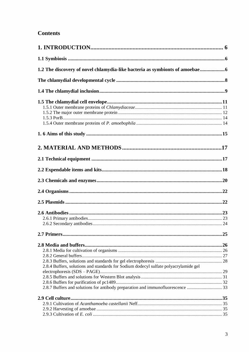
1.2 The discovery of novel chlamydia-like bacteria as symbionts of
amoebae
Free-living amoebae are ubiquitous protozoa that feed on a wide variety of
microorganisms, including bacteria. However, some bacteria have developed strategies to
survive uptake by amoebae and to multiply within them. Amoebae are therefore
considered as “Trojan horses of the microbial world”, acting as vectors and reservoirs for
several bacteria, including numerous human pathogens, such as Legionella pneumophila
and Listeria monocytogenes (52). In a study by Fritsche et al. (34), about 25% of studied
Acanthamoeba isolates were shown to harbour intracellular bacteria. As most of these
bacteria could not be isolated and cultivated in cell-free media, they were considered as
obligate intracellular endosymbionts. In 1997, chlamydia-like bacteria were found as
1. Introduction
7
Fig. 1: Diversity in the phylum Chlamydiae. Phylogenetic tree of known chlamydiae based on
comparative 16S rRNA analysis. From (50)
endosymbionts in amoebae (3, 12) and a later study identified 5% of amoeba
endosymbionts as chlamydia-like bacteria (35). When additionally new chlamydia-like
bacteria were found as contaminants in a laboratory cell culture (62) and in an aborted
bovine foetus (95), this changed the picture of chlamydial diversity dramatically.
Before the discovery of these chlamydia-like bacteria, chlamydiae were believed to be a
small group of closely related pathogens that formed a phylogenetically well separated
phylum in the domain bacteria that contained a single family, the Chlamydiaceae, within
a single order, the Chlamydiales. This family harbours important pathogens like
Chlamydia trachomatis, the most frequently sexually transmitted pathogen (118) and the
world‟s leading cause of preventable blindness (119), or Chlamydophila pneumoniae,
causative agent of human respiratory diseases, which is suggested to play also a role in
artheriosclerosis and coronary heart disease (8, 66).
The discovery of novel chlamydia-like bacteria, also termed „environmental chlamydiae‟
in order to distinguish them from the Chlamydiaceae or „clinical chlamydiae‟, lead to a
significant expansion of the phylum Chlamydiae. Based on 16S rRNA sequence analysis,
it currently contains eight families. Additionally to the Chlamydiaceae, the order
Chlamydiales now consists of the families Parachlamydiaceae, Waddliaceae,
Simkaniaceae, Rhabdochlamydiaceae, Piscichlamydiaceae, Clavochlamydiaceae and

1. Introduction
8
Criblamydiaceae (Fig. 1). As the diversity is thought to be even larger based on detection
of phylogenetically diverse rRNA sequences most similar to known chlamydiae in
various habitats, the number of families might even increase in the next years (50, 53)
The pathogenic potential of environmental chlamydiae is so far not clear. Although there
is some evidence that environmental chlamydiae might be involved in human diseases (5,
21) (42), more research is needed to show a causal relationship between chlamydia-like
bacteria and human disease. However, there is considerable evidence for environmental
chlamydiae infecting animals and thereby causing abortion in cattle (94) and
epitheliocystis in fish (28, 63).
When the genome of Protochlamydia amoebophila, an amoeba endosymbiont, was
sequenced in 2004, it was found to be nearly twice as large as that of clinical chlamydiae.
The genome of P. amoebophila was found to harbour reduced central metabolic and
biosynthetic pathways, comparable to genomes of clinical chlamydiae and other obligate
intracellular bacteria, but to encode a complete tricarboxylic acid cycle in contrast to
chlinical chlamydiae. Comparative genome analysis of members of the clinical
chlamydiae and this member of the environmental chlamydiae showed that their last
common ancestor lived about 700 million years ago and was already adapted to
intracellular survival in early eukaryotes (51). Recent studies indicate that this ancestor
might also have played a fundamental role in the evolution of plastids. The discovery of
genes of chlamydial origin in plant genomes and a high number of plant- and
cyanobacteria-like genes in chlamydial genomes lead to the suggestion that chlamydiae
were involved in establishing a stable interaction between the ancestor of plants and its
cyanobacterial symbiont, thereby giving rise to modern plants (56, 82).
The chlamydial developmental cycle
All known members of the phylum Chlamydiae share a characteristical biphasic lifecycle
that alternates between an infectious extracellular form termed elementary body (EB) and
an intracellular replicative form, the reticulate body (RB) (reviewed in 1). EBs are
metabolically inactive and have a very rigid, osmotically stable cell wall that allows
survival in harsh environments. In contrast, RBs are metabolically active and more fragile
than EBs (43). In addition to these two developmental forms, the existence of crescent
bodies (CB) has been suggested for Parachlamydia acanthamoeba. This form is regarded

1. Introduction
9
as an additional infective developmental stage, whose presence has been associated with
prolonged incubation time (40)
At the beginning of the developmental cycle an infectious EB attaches to a host cell and is
taken up by the host. Upon entry, chlamydiae reside within a host-derived vacuole termed
inclusion. In this inclusion, the EBs differentiate into RBs, which divide by binary fission.
After several rounds of replication, RBs re-differentiate into EBs and leave the host cell
by either lysis of the host or exocytosis (59) in order to infect new host cells (Fig. 2).
Fig. 2: The developmental cycle of chlamydia–like bacteria. Infectious EBs attach to their host cell
and are internalized (A). Inside the host cell, EBs differentiate to RBs and multiply within large or
single cell inclusions (B). After re-differentiation to EBs (C), chlamydiae leave their host by exocytosis
or lysis of the cell (D and E). Modified from (50).
1.4 The chlamydial inclusion
The vacuole in which chlamydiae reside within their host cell is unique among
intracellular bacteria and has been studied intensively for members of the Chlamydiaceae,
but not for other members of the Chlamydiales.
After uptake of the bacteria, the chlamydial inclusion does not enter the endocytic
pathway, but separates itself from this pathway at an early stage of development (98). Rab
GTPases, known regulators of intracellular trafficking, were shown to be recruited to the
chlamydial inclusion and might be involved in the escape from the endocytic pathway
(23, 96). Expansion of the inclusion occurs by hi-jacking host-vesicles containing

1. Introduction
10
sphingomyelin (41) and recently also transport of lipid droplets to the inclusion
membrane has been shown (17). Active participation of chlamydiae is needed during
these events, as the uptake of sphingomyelin and the escape from the endocytic pathway
require chlamydial gene expression (99).
As chlamydiae live intracellularly, they depend on their host for the acquisition of
nutrients. Fusion of the inclusion with vesicles of the phagolysosomal pathway is thought
to be the main source for nutrients. All these interactions of chlamydiae with their host
cell highlight the necessity of communication and transport mechanisms between bacteria
and host cell across the barrier of the inclusion membrane.
Rockey et al. (93) identified the first chlamydial protein in the membrane of the host-
derived inclusion, IncA, in 1995. Besides IncA, several other inclusio nmembrane
proteins (Incs) have been identified in the last years by in silico approaches and for a
subset of these proteins, localization in the inclusion membrane has been demonstrated
(e.g. 68, 69). All Inc-proteins known so far share no discernable homology on the amino-
acid level, but contain a characteristic bilobed hydrophobic domain of 50-80 amino
acids. They are thought to be injected in the chlamydial inclusion membrane by the type-
three-secretion system that is found in all chlamydiae (108).
A function is only known for few of these proteins. IncA plays a role in the fusion of
chlamydial inclusions (26), probably by means of a SNARE-like-motif that was identified
in this protein (26, 27). This case describes the first SNARE mimicry identified in a
bacterium and supports the role of IncA for the fusion of inclusions, as SNARE
signatures are known to play an essential role in targeting and fusion of vesicles in
eukaryotes. Two Inc-proteins were reported to interact with host-cell rab GTPases,
suggesting a role of these proteins in intracellular trafficking (23, 96). IncG is known to
bind to the host-protein 14-3-3-beta, an interaction that is speculated to inhibit apoptosis
of cells infected with C. trachomatis (100, 112).
Recently, putative Inc-proteins have also been identified in the chlamydia-like bacterium
P. amoebophila by in silico analysis (47).
Although the function of most Inc-proteins is not known, these proteins seem to be
central regulators of bacteria-host interactions and play an important role in
communication between chlamydiae and their host cells and thereby in survival of
chlamydiae within their host.
1. Introduction
11
1.5 The chlamydial cell envelope
Chlamydiae have a cell envelope similar to that of other Gram-negative bacteria,
consisting of an inner membrane, a periplasmic space and an outer membrane.
In contrast to other Gram-negative bacteria, chlamydiae seem to lack peptidoglycan,
although the presence or absence of peptidoglycan is controversially discussed (75).
Genome analysis showed the presence of a truncated pathway for the synthesis of
peptidoglycan. Transcription of the genes of this pathway was shown by microarray
analysis (73). Additionally, chlamydiae are susceptible to treatment with antibiotics that
inhibit the peptidoglycan synthesis (81). But until now, detection of N-acetylmuramic
acid, a key constituent of peptidoglycan, has not been possible in chlamydiae (75).
Stability is thought to be conferred to chlamydial cell walls by cysteine-rich proteins of
the outer membrane instead. These proteins are regarded as functional equivalent of
peptidoglycan in chlamydiae (43) and are discussed below in further detail.
1.5.1 Outer membrane proteins of Chlamydiaceae
Proteins in the outer membrane are of great importance as they are involved in attachment
to the host cell, uptake of nutrients, removal of waste and in case of the chlamydiae also
in the differentness of EBs and RBs.
In the Chlamydiaceae, the two cysteine-rich proteins OmcB (60 kDa) and OmcA (12
kDa) are thought to provide rigidity to the cell wall of the EB and to render it osmotically
stable. These two proteins harbour a high number of cysteines that are extensively cross
linked by disulfide bridges in the EB cell envelope. Upon entry of the EB into the host
cell, these disulfide bridges are reduced, resulting in osmotically fragile RBs (44). This
mechanism is of great importance to chlamydiae as it allows them to divide
intracellularly, when cross links are reduced, and to survive extracellularly when proteins
are cross linked (43). Cross linking during conversion from RBs to EBs is thought to
occur either spontaneously when the host cell is lysed (45) or late in the developmental
cycle by an enzymatic reaction before host cell lysis (88), but the exact mechanism is not
known.
The small cysteine-rich protein OmcA is a lipoprotein and thought to be anchored in the
inner leaflet of the outer membrane (43). The large cysteine-rich protein, OmcB, was long
thought to be localized to the periplasm, where it was proposed to form a stabilizing
lattice by inter- and intramolecular disulfide bridges (83, 88). Therefore, it was not

1. Introduction
12
regarded as a true member of the outer membrane, an assumption encouraged by its
solubility in sarcosyl in contrast to other outer membrane proteins of Chlamydiaceae (30).
However, there is strong evidence now for a localization of OmcB to the outer membrane
of C. trachomatis and C. pneumoniae and involvement of this protein in heparin-binding
on the surface of eukaryotic cells (31, 78, 103).
Other well characterized outer membrane proteins are the major outer membrane protein
(MOMP) that forms the chlamydial outer membrane complex (COMC) together with
OmcA and OmcB and is described in more detail below, and the members of the
polymorphic outer membrane protein (POMP) family.
Fig. 3: Model of the cell envelope of Chlamydiaceae. OmcB is shown to be localized in the periplasmic
space and the outer membrane. Modified from (47).
The chlamydial POMPs are a family of autotransporters (48) with heterologous primary
amino acid sequence that can be recognized by two repeated sequence motifs. The
unusually high polymorphism of these proteins is suggested to be due to selective
pressure from the immune system (37) and might contribute to tissue specificity of
different C. trachomatis disease groups (38, 105). Members of this family were shown to
play a role in attachment to host cells (115) and in expansion of the chlamydial inclusion
(61).
1.5.2 The major outer membrane protein
The outer membrane provides a barrier against harmful environmental factors, but also a
barrier to the uptake of nutrients into the cell and the removal of waste from the cell.
Permeability of the membrane is conferred by specific and unspecific transporters.
1. Introduction
13
Unspecific transporters are termed porins. Porins are water-filled channels that allow the
passage of small molecules with an exclusion limit of typically 600 Da and extremes of
5,000 Da, showing a transport rate proportional to the concentration gradient (117).
The major component of the chlamydial outer membrane is the major outer membrane
protein (MOMP), a porin that is thought to make up about 60% of the chlamydial outer
membrane (15). This 40 kDa protein has been intensively studied as it was shown to be
an important chlamydial antigen that elicits a strong immune response (15). In EBs,
MOMP is highly cross linked and mostly present as a trimer, whereas it is found mostly
in its monomeric form in RBs (44). It has been suggest for a long time, that trimers of
MOMP are held together by disulfide bonds (87). More recent studies suggest that
although disulfide bonds are present between peptides of the trimers, they are not required
for its folding, its outer membrane insertion or to stabilize it (32, 110).
The porin function of MOMP in different strains has been demonstrated by different
approaches. Bavoil et al. (6) were the first to show that preparations of the COMC have
porin activity by liposome swelling assays. Opening of the pore appeared to be regulated
by disulfide bonds, because treatment of the isolated COMC with reducing agents
resulted in a strong increase in channel activity, a mechanism also thought to occur after
entry of chlamydiae into the host cell. The function of MOMP as porin was confirmed by
lipid bilayer measurements using purified native and recombinant MOMP (32, 110, 121),
but opening of the pore by reducing agents could not be shown for these preparations.
This suggests that the effect observed by Bavoil was either due to other porins present in
the purified COMC or to disulfide bonds formed between MOMP and other members of
the outer membrane that are not present in preparations of purified MOMP.
MOMP forms a pore consisting of 16 antiparallel beta-sheets that form a barrel-like
structure typical of porins of Gram-negative bacteria (110). MOMP is found in trimeric
conformation in the outer membrane (74, 121). The barrel consists of five well conserved
transmembrane domains that are connected to each other by four variable, surface
exposed loops (4, 126). Removal of these loops has no influence on folding or
incorporation of MOMP into the outer membrane of Escherichia coli, in contrast to
removal of the transmembrane domains (32, 57). The four variable domains that are
exposed to the surface show great variations in sequence (102) and reaction of MOMP
with different antibodies targeting these variable regions is used for serovar typing in C.
trachomatis (113, 126).

1. Introduction
14
Antibodies against the major outer membrane protein were reported to neutralize
infectivity of chlamydiae (16, 92, 106) and to protect mice from abortion after infection
with chlamydiae (25, 91). Although it seems to be a promising candidate, development of
a vaccine based on MOMP has not been successful yet. Vaccines based on recombinant
and therefore non-native MOMP result in only partial protection (91).Vaccines containing
native MOMP have been shown to be protective against chlamydial disease (91, 111), but
high variation in parts of MOMP-sequence leads to difficulties in preparing a general
vaccine and additionally, isolation of high quantities of the native form of MOMP is not
practicable at the moment.
1.5.3 PorB
In 2000, a protein with weak sequence similarity to MOMP was identified in the genome
of C. trachomatis. Localization of this protein to the chlamydial outer membrane complex
and pore-forming activity of this protein, termed PorB, was demonstrated. Diffusion of
sugars and amino acids was not as efficient for PorB as for MOMP. As PorB was only
observed in small amounts in the COMC, the authors of the study suggested a more
specific function of this porin compared to MOMP. Although PorB is present in much
smaller amounts than MOMP, antibodies targeting this protein neutralized infectivity of
C. trachomatis in vitro (65).
1.5.4 Outer membrane proteins of P. amoebophila
In contrast to Chlamydiaceae, little is known about the composition of the outer
membrane of chlamydia-like bacteria. After sequencing the genome of the first member
of the environmental chlamydiae in 2004 (51), only a homologue of the large cysteine-
rich protein OmcB was found. An OmcA homologue was detected due to its position in
the genome and the high amount of cysteines in this gene. No homologue of MOMP,
PorB or members of the POMPs were found in P. amoebophila.
A recent study identified 38 outer membrane proteins of P. amoebophila by combining
1D and 2D gel electrophoresis of outer membrane fractions with mass spectrometry (47).
The identified proteins included the P. amoebophila OmcB, pc0616, and OmcA, pc0617.
In this study, a putative porin family was identified consisting of four proteins. Proteins of
this family share an amino acid sequence identity of 22-28 % and have no homologues in
other organisms except for P. acanthamoeba, a close relative of P. amoebophila. Two of

1. Introduction
15
these proteins, pc1489 and pc1077, were present in high amounts in outer membrane
fractions. Both were predicted to form a beta-barrel by in silico analysis and found to
encode predicted signal peptides. Based on their high abundance and their putative
function as porins, these two proteins were suggested to act as a functional replacement
for MOMP in P. amoebophila, and form the COMC of P. amoebophila by interactions
with pc0616 and pc0617. Pc0978 could be an additional part of the COMC, as it was
found as frequently as pc0616 in outer membrane preparations and has a high number of
cysteines. Therefore, it might be involved in disulfide-cross linking with other proteins to
provide stability to the outer membrane of P. amoebophila.
Fig. 4: Model of the outer membrane of P. amoebophila as proposed by Eva Heinz (47).
1. 6 Aims of this study
The outer membrane of bacteria plays an important role in the attachment to host cells,
uptake of substances from the host cell and in the case of chlamydiae also in the
differentness between EBs and RBs.
The major outer membrane protein is the most abundant component in the cell envelope
of members of the Chlamydiaceae. The outer membrane of P. amoebophila, a member of
the recently discovered environmental chlamydiae, seems to differ substantially from that
of Chlamydiaceae. The most interesting feature is the absence of almost all main
components of the chlamydial outer membrane, including MOMP and POMPs. MOMP
was suggested to be replaced by the two putative porins pc1077 and pc1489. In silico

1. Introduction
16
analysis predicted the presence of a signal peptide and localization to the outer membrane
for these two proteins. Structure prediction indicated formation of a beta-barrel structure,
a typical feature of porins of Gram-negative bacteria (47).
The aim of this study was to characterize the putative porin pc1489 concerning its
localization, its expression during the developmental cycle and its putative function as a
porin in P. amoebophila.
In the second part of this study, the localization of putative Inc-proteins of P.
amoebophila was investigated. Proteins in the inclusion membrane of P. amoebophila are
of exceptionally great interest, as this organism forms single cell inclusions in contrast to
large inclusions containing many bacteria observed in members of the Chlamydiaceae. It
is quite likely that differences in Inc proteins account for this. Additionally, Inc proteins
might play an important role in survival of P. amoebophila in amoebae after
internalization.

2. Material and Methods
17
2. Material and Methods
2.1 Technical equipment
Table 1: Technical equipment used in this study
Equipment Company
Agarose gel electrophoresis apparature Sub–Cell GT Bio-Rad Laboratories GmbH, Munich, Germany
CCD camera AxioCam HRc Carl Zeiss MicroImaging GmbH, Jena, Germany
Centrifuges:
Centrifuge 5840 R
OptimaTM L-100 XP ultracentrifuge
Eppendorf AG, Hamburg, Germany
Beckman Coulter, Inc., Palo Alto, CA, USA
Concentrator 5301 Eppendorf AG, Hamburg, Germany
Electroporator Micro PulserTM Bio-Rad Laboratories GmbH, Munich, Germany
Devices for gelelectrophoresis:
Electrophoresis cell (Sub-Cell GT)
Electrophoresis power supply (PowerPac Basic)
Bio-Rad Laboratories GmbH, Munich, Germany
Bio-Rad Laboratories GmbH, Munich, Germany
icycler Thermal cycler Bio-Rad Laboratories GmbH, Munich, Germany
Microbiological incubator KB 115 Binder GmbH, Tuttlingen, Germany
Laminar flow hood, model 1.8 Holten, Jouan Nordic, Allerod, Denmark
Magnetic stirrer RCT basic IKA® Werke GmbH & Co.KG, Staufen, Germany
Microscopes:
Inverse microscope Axiovert 25
Epifluorescence microscope Axioplan 2 imaging
Confocal laser scanning microscope LSM 510 Meta
Carl Zeiss MicroImaging GmbH, Jena, Germany
Carl Zeiss MicroImaging GmbH, Jena, Germany
Carl Zeiss MicroImaging GmbH, Jena, Germany
Mixing Block MB-102 Biozym Scientific GmbH, Hessisch Oldendorf,
Germany
NanoDrop® ND-1000 UV/Vis spectrophotometer NanoDrop Technologies Inc., Wilmington, DE,
USA
Neubauer counting chamber Paul Marienfeld GmbH & Co KG, Lauda-
Königshofen, Germany
pH meter inoLab pH Level 1 Wissenschaftlich technische Werkstätten (WTW)
GmbH & Co.KG, Weilheim, Germany
Scales:
OHAUS® Analytical Plus balance
Sartorius BL 3100
Ohaus Corporation, Pine Brook, NJ, USA
Sartorius AG, Göttingen, Germany
ScannerEpson Expression 1680 Pro Epson Deutschland GmbH, Meerbusch, Germany
Devices for SDS-PAGE analysis:

2. Material and Methods
18
Mini-PROTEAN Casting Stand
Mini-PROTEAN Tetra Cell
Bio-Rad Laboratories GmbH, Munich, Germany
Bio-Rad Laboratories GmbH, Munich, Germany
Sonicator Bandelin Sonoplus HD2070 Bandelin electronic GmbH & Co.KG, Berlin,
Germany
Sonotrode Bandelin Sonoplus UW 2070 Bandelin electronic GmbH & Co.KG, Berlin,
Germany
Spectral photometer SmartSpecTM 3000 Bio-Rad Laboratories GmbH, Munich, Germany
TE77 semi-dry transfer unit GE Healthcare Bio-Sciences AB, Uppsala, Sweden
Thermostatic circulator MultiTempTM III GE Healthcare Bio-Sciences AB, Uppsala, Sweden
Transilluminator Biostep GmbH, Jahnsdorf, Germany
Ultrasonic Cleaner SC100T VWR International bvba/sprl, Leuven, Belgium
UV sterilizing PCR workstation PeqLab Biotechnologie GmbH, Erlangen, Germany
Water baths:
DC10
GFL® type 1004
Thermo Haake GmbH, Karlsruhe, Germany
Gesellschaft für Labortechnik GmbH, Burgwedel,
Germany
Water purification system MILLI-Q® biocel Millipore GmbH, Vienna, Austria
Watervapour high pressure autoclaves:
Varioclav® 135 S h+P
Varioclav® 25 T H+P
H+P Labortechnik GmbH, Oberschleißheim,
Germany H+P Labortechnik GmbH,
Oberschleißheim, Germany
2.2 Expendable items and kits
Table 2: Expandable items used in this study
Expandable item Company
25 cm2 Tissue culture flasks Asahi Techno Glass Corporation, Iwaki Glass Co.,
Ltd., Funabashi-City, Japan
500 cm2 Tissue culture flasks Nunc, Roskilde, Denmark
Cellulose acetate membrane filters (0.45 µm pore
size, 25 mm diameter)
Sartorius Stedim Biotech GmbH, Göttingen, Germany
Cover glasses 24 x 60 mm Paul Marienfeld GmbH & Co.KG, Lauda-
Königshofen, Germany
Electroporation cuvette (0.2 cm) Bio-Rad Laboratories GmbH, Munich, Germany
Glass beads (0,75-1.0 mm) Macherey-Nagel GmbH&Co.KG, Karlsruhe,
Germany
Greiner tubes (15 ml, 50 ml) Greiner Bio-One GmbH, Frickenhausen, Germany
IsoporeTM polycarbonate membrane filters (0.22
µm pore size, 25 mm diameter, black)
Millipore GmbH, Vienna, Austria

2. Material and Methods
19
Microscope slides (76 x 26 mm) Carl Roth GmbH & Co KG, Karlsruhe, Germany
Microscope slides, 10 well Paul Marienfeld GmbH & Co KG, Lauda-
Königshofen, Germany
Multiwell dishes, polystyrene (24 wells) Nunc, Roskilde, Denmark
Needles Sterican® (ø 0,45 x 25 mm, ø 0,90 x 40
mm), single use, sterile
B.Braun Melsungen AG, Melsungen, Germany
PCR tubes (0.2 ml) Biozym Scientific GmbH, Hessisch Oldendorf,
Germany
Pipette Tipps (various sizes) Carl Roth GMbH Co.KG, Karlsruhe, Germany
Plastic cuvettes Greiner Bio-One GmbH, Frickenhausen, Germany
Plastic pipettes (10 ml, 2 ml), single use, sterile Barloworld Scientific Ltd., Staffordshire, UK
Polyvinylidene fluoride membrane (Hydrobond P) GE Healthcare Bio-Sciences AB, Uppsala, Sweden
Reaction tube (1.5 ml) GenXpress Service & Vertriebs GmbH, Wiener
Neudorf, Austria
Reaction tube (2 ml) Greiner Bio-One GmbH, Frickenhausen, Germany
Round coverslips (12 mm diameter) Carl Roth GmbH & Co., Karlsruhe, Germany
Syringe (1 ml) Inject® - F 1ml, single use, sterile B.Braun Melsungen AG, Melsungen, Germany
Syringe ( 5 ml) Omnifix® single use, sterile B.Braun Melsungen AG, Melsungen, Germany
Syringe filter, single use, sterile, 0.20 µm pore
size
Asahi Techni Glass Corporation, Iwaki Glass Co.,
Ltd., Funabashi-City, Japan
Syringe filter, single use, sterile, 1.20 µm pore
size
Sartorius AG, Goettingen, Germany
Ultracentrifuge tubes Beckham Coulter, Inc. Paolo Alto, CA, USA
Whatman® Chromatography Paper 3MM Chr Whatman Internation Ltd., Maidstone, UK
Table 3:Kits used in this study
Kit Company
DNeasy Blood & Tissue Kit QIAgen, Hilden, Germany
BCA Protein Assay Kit Thermo Scientific Pierce Protein Research Products,
Waltham, Massachusetts, USA
Western Lightning Chemiluminesence Plus Kit Perkin Elmer, Waltham, Massachusetts, USA
HiTrap™ IgY Purification HP Column GE Healthcare Bio-Sciences AB, Uppsala, Sweden
Qiaquick PCR purification Kit QIAgen, Hilden, Germany
Vivapure Q Mini H Spin column Sartorius AG, Göttingen, Germany

2. Material and Methods
20
2.3 Chemicals and enzymes
Table 4: Chemicals used in this study
Chemical Company
4´,6-diamidino-2-phenylindole (DAPI) Laktan Chemikalien und Laborgeräte GmbH, Graz,
Austria
Aceotnitrile Sigma-Aldrich Chemie GmbH, Steinheim,
Germany
Acetic acid Carl Roth GmbH & Co.KG, Karlsruhe, Germany
LE Agarose Biozym Scientific GmbH, Hessisch Oldendorf,
Germany
Ammonium bicarbonate Fluka Chemie AG, Buchs, Switzerland
Ammonium persulfate (APS) GE Healthcare Bio-Sciences AB, Uppsala, Sweden
Ampicillin Sigma-Aldrich Chemie GmbH, Steinheim,
Germany
Boric acid Carl Roth GmbH & Co KG, Karlsruhe, Germany
Bovine serum albumin (BSA) Carl Roth GmbH Co.KG, Karlsruhe, Germany
Brilliant Blue G-250 Carl Roth GmbH & Co KG, Karlsruhe, Germany
Brilliant Blue R-250 Carl Roth GmbH & Co KG, Karlsruhe, Germany
Bromphenol blue Sigma-Aldrich Chemie GmbH, Steinheim,
Germany
Calcium chloride dihydrate Carl Roth GmbH & Co KG, Karlsruhe, Germany
Citifluor AF1 Agar Scientific Ltd., Stansted, UK
Coomassie brillant blue G-250 Carl Roth GmbH Co.KG, Karlsruhe, Germany
Disodiumhydrogenphosphate dihydrate (Na2HPO4 x
2H20)
Carl Roth GmbH Co.KG, Karlsruhe, Germany
Dithiothreitol (DTT) Fluka Chemie AG, Buchs, Switzerland
Ethanol absolute AustrAlco Österreichische Alkoholhandels GmbH,
Spillern, Austria
Ethidium bromide (EtBr) Fluka Chemie AG, Buchs, Switzerland
Ethylenediamine-tetraaceticacid (EDTA) Carl Roth GmbH & Co KG, Karlsruhe, Germany
Ferrous ammonium sulfate hexahydrate Sigma-Aldrich Chemie GmbH, Steinheim,
Germany
Ficoll® 400 Sigma-Aldrich Chemie GmbH, Steinheim,
Germany
Formaldehyde (37% (w/w)) Carl Roth GmbH Co.KG, Karlsruhe, Germany
Gastrografin® Schering Austria GmbH, Vienna, Austria
Glucose Carl Roth GmbH Co.KG, Karlsruhe, Germany
2. Material and Methods
21
Glycerol (87% (w/v)) Carl Roth GmbH Co.KG, Karlsruhe, Germany
Glycine Carl Roth GmbH Co.KG, Karlsruhe, Germany
HEPES Carl Roth GmbH Co.KG, Karlsruhe, Germany
Hydrochloric acid (HCl) (37% (w/w)) Carl Roth GmbH Co.KG, Karlsruhe, Germany
Iodacetamide GE Healthcare Bio-Sciences AB, Uppsala, Sweden
Isopropanol Carl Roth GmbH Co.KG, Karlsruhe, Germany
Magnesium sulfate heptahydrate Merck GmbH, Vienna, Austria
Methanol Carl Roth GmbH Co.KG, Karlsruhe, Germany
Moviol 4-88 Sigma-Aldrich Chemie GmbH, Steinheim,
Germany
N,N,N´,N´-tetramethylethane-1,2-diamine (TEMED) Fluka Chemie AG, Buchs, Switzerland
N-octyl polyoxyethylene (n-octyl POE) BACHEM BIOCHEMICA GmbH, Heidelberg,
Germany
Paraformaldehyde (PFA) Sigma-Aldrich Chemie GmbH, Steinheim,
Germany
Proteose peptone Oxoid Ltd., Hampshire, England
Rotiphorese® NF-Acrylamide/
Bisacrylamide-solution 30 % (29:1)
Carl Roth GmbH Co.KG, Karlsruhe, Germany
Sodium chloride (NaCl) Carl Roth GmbH Co.KG, Karlsruhe, Germany
Sodium dihydrogen phosphate Mallinckrodt Baker B.V., Deventer, Holland
Sodium dodecly sulfate (SDS) Carl Roth GmbH Co.KG, Karlsruhe, Germany
Sodium hydroxide (NaOH) Carl Roth GmbH Co.KG, Karlsruhe, Germany
Sucrose Merck KGaA, Darmstadt, Germany
Trifluoretic acid Fluka Chemie AG, Buchs, Switzerland
Trishydroxymethlaminomethane (Tris-HCl) Carl Roth GmbH Co.KG, Karlsruhe, Germany
Trypticase Soy Broth Oxoid Ltd., Hampshire, England
Trypsin gold powder Promega Corporation, Madison, USA
TweenTM 20 Sigma-Aldrich Chemie GmbH, Steinheim,
Germany
Xylene cyanole FF Sigma-Aldrich Chemie GmbH, Steinheim,
Germany
Yeast extract Oxoid Ltd., Hampshire, England
Table 5: Enzymes and and corresponding enzyme buffers used in this study
Enzyme Company
Benzonase® Nuclease Novagen, Darmstatd, Germany
Buffer O Fermentas Inc., Hanover, MD, USA
Lysozyme human (≥ 100 U/µg) Sigma-Aldrich Chemie GmbH, Steinheim, Germany
2. Material and Methods
22
Taq DNA Polymerase (5U/µl) Fermentas Inc. Hanover, MD, USA
10 x Taq buffer Fermentas Inc. Hanover, MD, USA
VspI (10 U/µl) Fermentas Inc., Hanover, MD, USA
2.4 Organisms
Table 6: Amoeba strains and endosymbionts used in this study
Host Endoymbiont Source Reference
Acanthamoeba
castellanii Neff
none American Type Culture
Collection (ATCC),
Manassas, VA, USA
(86)
Acanthamoeba
castellanii Neff
Protochlamydia
amoebophila UWE25
University of
Washington, Seattle, USA
(19, 36)
Acanthamoeba
sp. UWC1
Parachlamydia
acanthamoeba UV-7
Wastewater treatment
plant, Plattling, Germany
(18, 35)
Acanthamoeba
sp. EI2
Protochlamydia
amoebophila sp. EI2
Soil, Lower Austria,
Austria
(97)
Table 7: Escherichia coli strains used in this study
Strain Source Genotype Reference
BL21 (DE3) Stratagene F-, ompT, hsdS(rB-mB-), gal, dem,
λ(DE3)
(116)
2.5 Plasmids
For expression of proteins in Escherichia coli BL21 (DE3) the vector pet16b was used
(Novagen, Darmstadt, Germany). E. coli cells harbouring the plasmids listed in table 8
were obtained from Eva Heinz (47).
Table 8: Plasmids used in this study
Plasmid Insert Weight of full
length protein
Weight of protein
fragment
Reference
pet16b – pc03991 Fragment of pc0399
(aminoacids 120 – 402)
95.9 kDa 35 kDa (47)
pet16b – pc 0156 Fragment of pc0156
(aminoacids 1 – 180)
35.1 kDa 23.5 kDa (47)
2. Material and Methods
23
pet16b - pc1111 Fragment of pc1111
(aminoacids 1 – 199)
35.6 kDa 25.1 kDa (47)
pet16b – pc0530 Fragment of pc0530
(aminoacids 78 - 252)
28.2 kDa 22.1 kDa (47)
pet16b – pc0577 Fragment of pc0577
(aminoacids 2 - 201)
22.8 kDa 25.3 kDa* (47)
* The higher weight of the protein fragment compared to the full length protein results from a
His-Tag added to the fragment by cloning in the vector pet16b.
2.6 Antibodies
2.6.1 Primary antibodies
Table 9: Primary antibodies used in this study
Antibody Antigen Source of
antibody
Dilution Reference
anti-Pam P. amoebophila elementary bodies rabbit 1 : 2,000 (47)
anti-Pam P. amoebophila elementary bodies chicken 1 : 2,000 (47)
anti-pc0399-1 Fragment of pc0399
(aminoacids 120 – 402)
rabbit 1 : 1,000 (47)
anti-pc0156 Fragment of pc0156
(aminoacids 1 – 180)
rabbit 1 : 500 (47)
anti-pc1111 Fragment of pc1111
(aminoacids 1 – 199)
rabbit 1 : 100 (47)
anti-pc0530 Fragment of pc0530
(aminoacids 78 - 252)
rabbit 1 : 20 (47)
anti-pc0577 Fragment of pc0577
(aminoacids 2 - 201)
rabbit 1 : 20 (47)
preI pc0399-1
(pre-immune serum)
none rabbit 1 : 1,000 (47)
preI pc0156
(pre-immune serum)
none rabbit 1 : 500 (47)
preI pc1111
(pre-immune serum)
none rabbit 1 : 100 (47)
preI pc0530
(pre-immune serum)
none rabbit 1 : 20 (47) )
preI pc0577
(pre-immune serum)
none rabbit 1 : 20 (47)
anti-Hsp60 Hsp60 of Chlamydophila caviae guinea-pig 1 : 2,000 (125)

2. Material and Methods
24
GPIC
anti-pc1489 pc1489 purified from EBs chicken 1 : 2,000
preI pc1489
(pre-immune serum)
none chicken 1 : 2,000
anti-Cld Chloride dismutase purified from
"Candidatus Nitrospira defluvii"
rabbit as indicated (71)
2.6.2 Secondary antibodies
Secondary antibodies were purchased from Dianova (Hamburg, Germany) and GE
Healthcare Bio-Sciences AB (Uppsala, Sweden).
Secondary antibodies used carried one of the following modifications:
- The fluorophor Indocarbocyanine (Cy3)
- The fluorophor Carbocyanine (Cy2)
- The fluorophor Indodicarbocyanine (Cy5)
- The fluorophor Fluorescein (FITC)
- The enzyme Horseraddish-Peroxidase (HRP)
Table 10: Secondary antibodies used in this study
Antibody Antigen Modification Source of
antibody
Dilution Manufacturer
anti-chicken chicken IgY Cy3 donkey 1:1,000 Dianova
anti-chicken chicken IgY Cy2 donkey 1:1,000 Dianova
anti-chicken chicken IgY Cy5 donkey 1:1,000 Dianova
anti-chicken chicken IgY HRP goat 1:10,000 Dianova
anti-guinea pig guinea pig IgG Cy3 goat 1:1,000 Dianova
anti-guinea pig guinea pig IgG Cy2 goat 1:1,000 Dianova
anti-guinea pig guinea pig IgG Cy5 goat 1:1,000 Dianova
anti-guinea pig guinea pig IgG HRP goat 1:10,000 Dianova
anti-rabbit rabbit IgG Cy3 goat 1:1,000 Dianova
anti-rabbit rabbit IgG Cy2 goat 1:1,000 GE Healthcare
Bio-Sciences AB
anti-rabbit rabbit IgG FITC goat 1:1,000 Dianova
anti-rabbit rabbit IgG Cy5 goat 1:1000 Dianova
anti-rabbit rabbit IgG HRP goat 1:10,000 Dianova

2. Material and Methods
25
2.7 Primers
Primers were produced by Thermo Fisher Scientific GmbH (Ulm, Germany).
Table 11: Primers used for the amplification of 16SrRNA gene fragments
Primer
name
Primer sequence*
(5´-3´)
Size of
amplified
fragment
Specificity Annealing
temperature
Reference
PcR
GTC ATC RGC
CYY ACC TTV SRC
RYY TCT
1,260 bp
Parachlamydiaceae,
Waddliaceae
58°C
(53)
PcF TCA GAT TGA
ATG CTG AC
PanF CGTGGATGAGGC
ATGCRAGTCG
1,445 bp
Chlamydiales
65°C
(22)
PanR
GTC ATC RGC
CYY ACC TTV SRC
RYY TCT
1492R GGY TAC CTT GTT
ACG ACT T
1,480 bp
Bacteria, but not
Chlamydiales
52°C
(67)
616 V AGA GTT TGA
TYM TGGC
* According to the NC-IUB the unspecified bases are represented as follows: R = G or A, Y = T or C, M =
A or C, V = G or C or A, S = G or C, N = G or C or A or T;
2. Material and Methods
26
2.8 Media and buffers
Media and buffers described here were sterilized for 20 min at 121°C and 1,013 x 105 Pa
pressure in a watervapour-high pressure autoclave if not stated otherwise. Buffers
containing ethanol or isopropanol were not autoclaved or were autoclaved before the
addition of these components. If not indicated otherwise, media and buffers were stored
at room temperature (RT) until further use.
If indicated in the text, buffers were filter-sterilized by passing them through a 0.2 µm
filter.
2.8.1 Media for cultivation of organisms
2.8.1.1 Medium for axenic cultivation of amoebae
Trypticase Soy Broth with Yeast Extract (TSY)
Trypticase Soy Broth 30 g
Yeast extract 10 g
H2Odd ad 1000 ml
pH 7.3
PYG
Peptone 20 g
Glucose 18 g
Yeast extract 2 g
Sodiumcitrate 1 g
MgSO4 x 7 H2O 980 mg
Na2HPO4 x 7 H2O 355 mg
KH2PO4 340 mg
Fe(NH4)2(SO4)2 x 6 H2O 20 mg
H2Odd ad 1000 ml
pH 6.5
2. Material and Methods
27
2.8.1.2 Medium for cultivation of E. coli
Luria Bertani medium (LB medium)
Tryptone 10 g
Yeast extract 5 g
NaCl 5 g
H2Odd ad1000 ml
pH 7.0 - 7.5
For solid media 15 g/l agar were added to the medium before autoclaving.
Antibiotics
Ampicillin stock solution (Amp) 100 mg/ml
Amp was dissolved in 50 % EtOHabs and stored at -20°C until further use.
Amp was added to medium to reach a final concentration of 100 μg/ml.
2.8.2 General buffers
10 x Page’s Amoebic Saline (PAS)
NaCl 1.20 g
MgSO4 x 7H20 0.04 g
CaCl2 x H20 0.04 g
NaH2PO4 x 2H20 1.78 g
KH2PO4 1.36 g
H2Odd ad 1000 ml
1 x PAS
10 x PAS 100 ml
H2Odd ad 1000 ml

2. Material and Methods
28
PBS stock solution
Solution 1: 35.6 g/l Na2HPO4
Solution 2: 27.6 g/l NaH2PO4
pH of solution 1 was adjusted to 7.2-7.4 by adding solution 2
1 x PBS
PBS stock solution 50 ml
NaCl 7.6 g
H2Odd ad 1000 ml
SPG-buffer
Sucrose 75 g
KH2PO4 0.52 g
Na2HPO4 x 2 H20 2.30 g
Glutamc acid 0.75 g
H2Odd ad l000 ml
2.8.3 Buffers, solutions and standards for gel electrophoresis
Buffers and solutions for gel electrophoresis were not autoclaved.
10 x TBE
Tris-HCl 162.0 g
Boric acid 27.5 g
EDTA 9.3 g
H2Odd ad 1000 ml
pH 8.3 – 8.7
1 x TBE
10 x TBE 100 ml
H2Odd ad 1000 ml
2. Material and Methods
29
Loading buffer
Ficoll 25 % (w/v)
Bromphenol blue 0.5 % (w/v)
Xylencyanol 0.5 % (w/v)
EDTA 50 mM
H2Odd
Ethidium bromide solution
Ethidium bromide stock solution:
10 mg/ml Ethidium bromide (EtBr) in H2Odd
Ethidium bromide staining solution:
Ethidium bromide stock solution diluted 1: 10,000 in H2Odd
DNA ladders
GeneRulerTM
1kb (Fermentas Inc., Hanover, MD, USA)
GeneRuler™ 100 bp (Fermentas Inc., Hanover, MD, USA)
2.8.4 Buffers, solutions and standards for Sodium dodecyl sulfate
polyacrylamide gel electrophoresis (SDS – PAGE)
Buffers and solutions for SDS-PAGE were not autoclaved, except for lower and upper
buffer.
Lower buffer
Tris–HCl 90.8 g
10% (w/v) Sodium dodecyl Sulfat (SDS) 20 ml
H2Odd ad 500 ml
pH 8.8
2. Material and Methods
30
Upper buffer
Tris-HCl 30.3 g
10 (w/w)% SDS 20 ml
H2Odd ad 500 ml
pH 6.8
10 x SDS-PAGE running buffer
Tris-HCl 30.2 g
Glycine 144 g
SDS 10 g
H2Odd ad 1000 ml
4 x SDS - PAGE loading buffer
Tris-HCl pH 6.8 2.42 g
SDS 8 g
Bromphenolblue 0.02 g
Glycerol 40 µl
Dithiothreitol (DTT) 6.2 g
H2Odd ad 100 ml
Coomassie brilliant blue staining solution
Methanol 50 % (v/v)
Acetic acid 10 % (v/v)
Brilliant Blue G-250 2.76 g
H2Odd ad 1000 ml
Destaining solution
Acetic acid 5 % (v/v)
Ethanol 20 % (v/v)
H2Odd ad 1000 ml

2. Material and Methods
31
Colloidal Coomassie solution
(NH4)2SO4 100 g
Orthophosphoric acid 20 g
Methanol 25 % (v/v)
Coomassie Brilliant Blue G-250 0.625 g
H2Odd ad 1000 ml
Fixing solution
Ethanol 40 % (v/v)
Acetic acid 10 % (v/v)
H2Odd ad 2000 ml
Protein Ladders
Unstained Protein Molecular Weight Marker (Fermentas Inc., Hanover, MD, USA)
PageRuler™ Prestained Protein Ladder (Fermentas Inc., Hanover, MD, USA)
2.8.5 Buffers and solutions for Western Blot analysis
Buffers and solutions for Western Blot analysis were not autoclaved, except for 10 x
TBS.
Transfer buffer
Glycine 14.4 g
Tris-HCl 3 g
Methanol 20 % (v/v)
H2Odd ad 1000 ml
10 x TBS
Tris-HCl 12.1 g
NaCl 43.8 g
H2Odd ad 500 ml
pH 7.5; store at 4°C

2. Material and Methods
32
1 x TBS
10 x TBS 100 ml
H2Odd ad 1000 ml
store at 4°C
TBS-T
10 x TBS 50 ml
Tween 20 0.1% (v/v)
H2Odd ad 500 ml
store at 4°C
Developer and Fixer solution
SIGMA Kodak GBX Developer
SIGMA Kodak GBX Fixer
Developer and Fixer were diluted as recommended by the manufacturer
2.8.6 Buffers for purification of pc1489
Buffer A
HEPES pH 7.5 2.9 g
NaCl 0.292 g
n-octyl–Polyoxyethylen (n-octyl-POE) 0.25 g
H2Odd ad 500 ml
Buffer B
HEPES pH 7.5 0.29 g
NaCl 5.84 g
n-octyl–POE 0.025 g
H2Odd ad 50 ml
300 mM NaxPO4
Solution 1: 53.4 g/l Na2HPO4
Solution 2: 41.4 g/l NaH2PO4

2. Material and Methods
33
pH of solution 1 was adjusted to 6.5 by adding solution 2
3 x PEN
EDTA 0.087 g
NaCl 5.84 g
300 mM NaxPO4 ad 1000 ml
POP 05 – buffer
n-octyl–POE 0.5% (w/v)
3 x PEN ad 50 ml
2.8.7 Buffers and solutions for antibody preparation and immunofluorescence
Binding Buffer
NaH2PO4 3.12 g
K2SO4 87.13 g
H2Odd ad 1000 ml
pH 7.5
Elution Buffer
NaH2PO4 3.12 g
H2Odd ad 1000 ml
pH 7.5
Cleaning Buffer
NaH2PO4 3.12 g
Isopropanol 30% (v/v)
H2Odd ad 1000 ml
pH 7.5
2. Material and Methods
34
10 x PBS
Na2HPO4 26.8 g
NaH2PO4 13.8 g
NaCl 81.6 g
H2Odd ad 1000 ml
4 % PFA
1 x PBS 25 ml
Paraformaldehyde (37% w/v) 2.7 ml
FA-Block solution
Bovine Serum Albumine (BSA) 20 g
10 x PBS 100 ml
H2Odd ad 1000 ml
50 ml aliquots of FA-Block solution were stored at -20°C until further use. Upon
thawing, FA-Block solution was stored at 4°C up to 2 weeks.
Mowiol
4 g Mowiol were mixed with 6 g glycerine and 6 ml H2Odd. 12 ml 0.2 M Tris-HCl
solution (pH 8.5) was added and the solution was stirred at 50°C overnight. When the
Mowiol had dissolved completely, the solution was centrifuged at 7,700 x g for 15
minutes (min) to remove air bubbles. The supernatant was collected and stored in aliquots
at -20°C until further use.

2. Material and Methods
35
2.9 Cell culture
2.9.1 Cultivation of Acanthamoeba castellanii Neff
Solutions
TSY medium
PYG medium
Procedure
Acanthamoeba castellanii Neff and Acanthamoeba sp. UWC1 isolates containing either
no endosymbiont or harbouring endosymbionts were grown axenically in small or large
culture flasks in 10 ml or 150 ml TSY or PYG medium. Cultures were grown at a
constant temperature of 20°C in an incubator and were checked for contaminations with
extracellular bacteria or fungi using an inverse microscope at regular intervals.
Medium was exchanged every one or two weeks, depending on the growth of amoebae,
by pouring out the old medium in a glass bottle and adding new, fresh medium to the
flasks.
2.9.2 Harvesting of amoebae
Amoeba cultures were harvested by shaking the culture flasks vigorously to detach
amoebae from the bottom of the flask. The supernatant of the culture containing detached
amoebae was collected in 50 ml Greiner tubes. Cell pellets were obtained by
centrifugation at 7,323 x g for 5 min. TSY or PYG medium was added to the culture
flasks to enable growth of remaining amoebae.
2.9.3 Cultivation of E. coli
Solutions
LB medium
Ampicillin stock solution

2. Material and Methods
36
Procedure
E. coli cells were grown on LB plates or in liquid LB medium at 37°C. For culturing cells
in liquid media, 5 ml of LB medium were inoculated with a single colony from a plate or
cells from a glycerol stock under sterile conditions and incubated on an orbital shaker at
200 rpm. Recombinant E. coli cells harbouring a plasmid were grown in the presence of
100 µg/ml ampicillin to avoid the growth of cells without plasmid.
LB plates with E. coli colonies were stored at 4° for several weeks.
2. 10 General methods
2.10.1 Amplification of DNA fragments via Polymerase Chain Reaction (PCR)
Solutions
MgCl2 (25 mM) (Fermentas Inc. Hanover, MD, USA)
Nucleotide Mix (2.5 mM each dNTP)
10 x Ex Taq polymerase buffer
Taq DNA Polymerase (5U/µl)
Forward primer (50 pmol/µl)
Reverse primer (50 pmol/µl)
Aquabidest. (Mayrhofer Pharmazeutika GmbH & Co KG, Leonding, Austria)
Procedure
To avoid contaminations by external DNA, Aquabidest., MgCl2, 10 x Ex Taq polymerase
buffer and tubes used for PCR were exposed to ultra-violet (UV) light in a UV sterilizing
PCR hood for 20 min before preparing the standard reaction mixture. All pipetting steps,
except for the addition of the positive control, were carried out under the PCR hood.
For amplification of DNA fragments via PCR, the following standard reaction mixture
was prepared:

2. Material and Methods
37
Standard reaction mixture
MgCl2 4 µl
10 x Taq buffer Buffer 5 µl
dNTP – Mix 5 µl
Forward primer (50 pmol/µl) 1 µl
Reverse primer (50 pmol/µl) 1 µl
Template 100 ng
Aquabidest. ad 50 µl
For each PCR, a positive and a negative control were included.
For the amplification of 16S rRNA gene fragments, PCR was performed at the following
conditions:
PCR step Temp. [°C] Time
Number of
cycles
Denaturation 95 90 sec 1
Denaturation 95 30 sec
Annealing * 30 sec 35
Elongation 72 90 sec
Final
elongation 72 420 sec 1
* for annealing temperatures, see Table 11
Obtained PCR products were analyzed quantitatively by photometric analysis and
qualitatively by agarose gel electrophoresis.

2. Material and Methods
38
2.10.2 Quantitative and qualitative analysis of nucleic acids
2.10.2.1 Quantitative, photometric analysis of nucleic acids
Concentration of nucleic acids was measured with a NanoDrop® ND-1000
spectralphotometer. 1.5 µl of nucleic acid solution was pipetted onto the end of the fibre
optic cable of the NanoDrop® device. Measurements were performed at λ = 260 nm
according to the instructions of the manufacturer.
2.10.2.2 Qualitative analysis of nucleic acids by agarose gel electrophoresis
Solutions
1 x TBE
Loading buffer
DNA ladder
EtBr staining solution
Procedure
Nucleic acid samples were analysed on agarose gels with concentrations of 1-2.5 %
agarose (1.5 g–3.75 g agarose in 150 ml 1 x TBE). Samples were mixed with 1 volume of
loading buffer and pipetted into the pockets of the gel. Nucleic acids were separated by
applying a voltage of 80 – 100 V for 60 - 90 min, depending on type of analysis.
Separated nucleic acids were stained with EtBr staining solution for 30 min and
visualized with a transilluminator emitting UV- light (λ = 312 nm). Band patterns were
recorded and digitalised using a gel–documentation system (Biostep, Jahnsdorf,
Germany).
2.10.3 Sequencing of PCR products
Certain PCR products were sequenced by Mag. Christian Baranyi using an ABI 3130xl
DNA sequencer and the BigDye Terminator Cycle Sequencing Kit v3.1 (Applied
Biosystems), which is based on Sanger‟s chain-termination method for sequencing.
Brieflly, incorporation of ddNTPs linked to different fluorescent dyes into a newly
synthezised DNA-strand results in termination of elongation. DNA-fragments of different

2. Material and Methods
39
sizes are separated electrophoretically and recognized by a laser and the resulting
chromatograph representing the DNA sequence is recorded.
For sequencing, the same primers as for PCR analysis were used at a concentration of 10
pmol/µl. Sequences were proof-read manually using the FinchTV software (Geospiza)
and subjected to BLAST search using the general nucleotide collection database
(http://blast.ncbi.nlm.nih.gov/Blast.cgi) to find the most similar sequence (2).
2.10.4 Analysis of proteins by Sodium dodecyl sulfate polyacrylamide gel
electrophoresis (SDS-PAGE)
2.10.4.1 Sample preparation for SDS-PAGE analysis
2.10.4.1.1 Sample preparation from cell cultures
Cell pellets were obtained by centrifugation at 17,949 x g for 2 min for E. coli or
harvesting of amoebae at 7,323 x g for 5 min. Cells were resuspended in 1 x PBS and
centrifuged as before. The obtained pellet was resuspended in 50-60 µl 4 x SDS-PAGE
loading buffer, depending on the size of the pellet. Samples were heated to 95°C for 5
min to break the cells and cooled down again.
Nucleic acids present in the samples were removed by digestion with the nuclease
Benzonase. 1 µl of Benzonase was added to the samples and digestion was performed for
1 hour at 4 °C. Samples were stored at -20°C until further use. Before loading on the gel,
samples were heated to 95°C for 5 min.
2.10.4.1.2 Sample preparation for purified proteins
4 x SDS - PAGE loading buffer without DTT was added to the samples and samples were
applied to the gel without heating.
2.10.4.2 SDS-PAGE-analysis
Solutions
Lower buffer
Upper buffer
Rotiphorese®NF-30% (w/v) Acrylamide/Bisacrylamide-solution
2. Material and Methods
40
10% (w/v) Ammonium persulfate (APS)
N,N,N´,N´-tetramethylethane-1,2-diamine (TEMED)
1 x SDS-PAGE running buffer
Protein Ladder
Procedure
Separating and stacking gel were prepared according to the following standard reaction
mixtures:
Separating gel
Lower buffer 2 ml
H2Odd 2.66 ml
Acrylamide/Bisacrylamide solution 3.33 ml
APS 40 µl
TEMED 8 µl
Stacking gel
Upper buffer 0.63 ml
H2Odd 1.63 ml
Acrylamide/Bisacrylamide solution 0.38 ml
APS 17.5 µl
TEMED 10 µl
12.5% polyacrylamide gels were prepared by use of a Mini-PROTEAN casting stand.
Lower buffer, H2Odd and acrylamide/bisacrylamide solution were mixed and APS and
TEMED were added to initiate polymerization of the gel. The separating gel solution was
poured between the two glass plates of one cassette and gels were overlaid with
isopropanol to produce a smooth surface of the separating gel. After polymerization of the
separating gel, the isopropanol was poured off. The stacking gel was prepared by mixing
upper buffer, H2Odd and acrylamide/bisacrylamide solution and adding APS and TEMED.
A layer of stacking gel was added on top of the separating gel and combs were applied.
Polymerized gels were wrapped in wet paper towels and saran wrap and stored at 4°C for
a few days until further use.
2. Material and Methods
41
For separation of proteins, gels were inserted into a Mini PROTEAN gel electrophoresis
chamber and the chamber was filled with 1 x SDS-PAGE running buffer. Samples and
marker were pipetted into the pockets of the gel. Proteins were separated by applying a
voltage of 60 – 110 V for 1.5 hours, depending on type of analysis. The run was stopped
when the marker front reached the bottom of the gel. The gel apparature was dissembled
and gels were stained as described below.
2.10.4.3 Staining and destaining of SDS-PAGE-gels using Coomassie brilliant blue
staining solution
Solutions
Coomassie brilliant blue staining solution
Destaining solution
Procedure
After electrophoresis, gels were stained in Coomassie brilliant blue staining solution on a
rocking platform for 45 min followed by destaining in Destaining solution overnight.
Gels were rinsed with H2Odd and digitalised by scanning.
2.10.4.4 Fixation, staining and destaining of SDS-PAGE-gels using colloidal
Coomassie staining solution
Solutions
Colloidal Coomassie solution
Fixing solution
Procedure
After electrophoresis, gels were rinsed for 5 min with H2Odd, followed by incubation for
20 min in fixing solution. Gels were rinsed 2 times for 10 minutes in H2Odd and stained
with colloidal Coomassie overnight. On the following day, gels were destained by rinsing
them 2 times for 30 min in H2Odd. All these steps were carried out on a rocking platform.
After destaining, gels were digitalised by scanning.

2. Material and Methods
42
2.10.5 Western blot analysis
Solutions
Transfer buffer
1 x TBS
TBS-T
Developer and Fixer solution
Procedure
Proteins were separated by SDS-PAGE as described in 2.10.4. The gel electrophoresis
chamber was dissembled and the stacking gel was removed from the resolving gel. The
resolving gel was equilibrated for 15 min in transfer buffer. A polyvinylidene fluoride
(PVDF) membrane and 6 pieces of Whatman paper were cut to the size of the separating
gel. The PVDF membrane was activated by shaking in methanol for 5 min, followed by
rinsing with H2Odd for 5 min. Transfer onto the membrane was performed by semi-dry
blotting in a TE77 semi-dry transfer unit filled with transfer buffer for 1.5 hours at
settings as recommended by the manufacturer.
Non-specific binding of proteins was prevented by incubating the membrane in 5% non-
fat dry milk in 1 x TBS on a rocking platform overnight. Blocking of the membrane was
followed by incubation in 10 ml TBS-T with the primary antibody against the respective
protein for 1 hour. The membrane was washed 3 times in TBS-T for 15 min to remove
excess primary antibody, followed by incubation in TBS-T with the corresponding HRP-
labelled secondary antibody for 1 hour. After 3 additional washing steps, the signal was
detected by using the Western Lightning Chemiluminescence Plus Kit as recommended
by the manufacturer. The membrane was exposed to a photographic film in the dark. The
film was developed with Developer Solution, fixed with Fixer Solution and washed in
H2O. Films were digitalised by scanning.
2.11 Purification of P. amoebophila EBs
For purification of the putative porin pc1489 and for infection studies, EBs were purified
from Acanthamoeba castellanii cultures infected with P. amoebophila. Amoebae were
broken up in a procedure that also destroyed RBs present in the sample. The cell debris
was removed by filtration and centrifugation, yielding purified EBs.
2. Material and Methods
43
2.11.1 Partial purification of P. amoebophila EBs
Solutions
1 x PAS
SPG-buffer
Procedure
12 – 15 large cultures of A. castellanii cultures infected with P. amoebophila were
harvested (sec. 2.9.2). The pellet was washed in 1 x PAS, centrifuged (7,323 x g, 5 min)
and the resulting pellet was resuspended in 1 x PAS. 4 freeze-and-thaw steps (freezing at
-20°C/ thawing at 42°C in a water bath) were performed to disrupt amoeba cell
membranes, followed by shearing of the samples with an equal volume of glass beads for
3 minutes. To remove glass beads and cell debris, the lysate was centrifuged for 10 min at
300 x g and 4°C. The resulting supernatant was filtered through a 1.2 µm filter and
transferred to an ultracentrifuge tube. The cell suspension was filled up to 10 ml with
precooled SPG-buffer and centrifuged at 40,000 x g for 40 min a 4°C in an
ultracentrifuge. The resulting pellet was resuspended in 10 ml precooled SPG-buffer and
centrifuged as before. The pellet was resuspended in 6 ml SPG–buffer and passed through
a 0.45 µm syringe to singularize EBs. The resulting suspension was aliquoted to screw
caps. To ensure that purified EBs were not contaminated with other bacteria or fungi, 1
ml of TSY was inoculated with 10 µl of each screw cap and incubated for 5 days at 20°C.
Samples were checked for growth of bacteria or fungi with an inverse microscope. The
remaining EB-suspension was stored at -80°C until further use.
2.11.2 High purification of P. amoebophila EBs
High purification of P. amoebophila involved two additional gradient centrifugation steps
compared to partial purification. During gradient centrifugation, EBs formed a pellet at
the bottom of the tube, whereas amoeba cell debris accumulated at the interface between
gastrografin and sucrose.
Solutions
1 x PAS
SPG-buffer
2. Material and Methods
44
30 mM Tris-HCl buffer (pH 7.3)
30% (v/v) Gastrografin in H2Odd
50% (w/v) sucrose in H2Odd
Procedure
12 – 15 large cultures of A. castellanii infected with P. amoebophila were harvested,
washed in 1 x PAS and the resulting pellet was resuspended in 3 ml 30 mM Tris-HCl
buffer. Amoeba cells were disrupted as described above (2.11.1). To remove glass beads
and amoeba cell debris, the lysate was centrifuged for 10 min at 4°C and 300 x g. The
resulting supernatant was filtered through a 1.2 µm filter and transferred to a clean
ultracentrifuge tube. The cell suspension was filled up to 10 ml with 30 mM Tris-HCl
buffer and centrifuged at 40,000 x g for 1 h at 4°C in an ultracentrifuge. The resulting
pellet was resuspended in 1 ml precooled SPG-buffer. For further purification, a sucrose-
gastrografin-gradient was prepared by overlaying 3 ml 50 % sucrose with 3 ml 30 %
gastrografin and overlaying the gastrografin with 3 ml sample. Gradient centrifugation
was performed at 40,000 x g for 2 h at 4°C two times. Between centrifugation steps, the
cell suspension was passed through a 0.45 µm syringe in order to singularize bacteria.
The pellet of the last centrifugation step was resuspended in 6 ml SPG-buffer and passed
again through a 0.45 µm syringe. The resulting suspension was aliquoted to screw caps
and screened for the presence of other bacteria or fungi as described in 2.11.1. Aliquots
were stored at -80°C until further use.
2.12 Screening of amoeba cultures
To ensure that Acanthamoeba castellanii cultures were not contaminated with
extracellular bacteria or other chlamydiae, cultures were screened at regular intervals and
each time before purification of EBs was performed.
2.12.1 DAPI staining of amoebae
Every 2-3 weeks and before purification of EBs was started, cultures were screened by
staining with 4',6-Diamidino-2-phenylindol (DAPI).

2. Material and Methods
45
Solutions
DAPI
1 x PAS
4% PFA in 1 x PBS
Procedure
DAPI was prepared at a dilution of 1:10,000 and stored at 4°C until further use.
1 ml of an amoeba culture was harvested by centrifugation at 5,974 x g for 5 min. The
resulting pellet was resuspended in 1 x PAS and centrifuged as before. The pellet was
resuspended in 200 µl 1 x PAS. 20 µl of this suspension were applied to the well of a
microscope slide. Amoebae were allowed to attach for 30 min. Then, the liquid was
removed carefully and 10 µl of 4 % PFA were added instead to fix cells. After incubation
for 10 min at RT, the fixative was removed and wells were washed with 10 µl H20dd. 10
µl of the DAPI solution were added onto each well and slides were incubated for 7 min in
the dark. The DAPI-solution was removed and wells were washed with 20 µl H20dd.
Slides were dried at RT in the dark, followed by embedding in Citifluor. Fluorescently
labelled cells were analyzed by epifluorescence microscopy.
2.12.2 Screening of cultures via PCR
If investigation by light microscopy or staining with DAPI indicated the presence of
contaminations with extracellular bacteria or other endosymbionts in amoeba cultures,
DNA was isolated from these cultures and 16S rRNA gene-fragments were amplified by
PCR followed by sequencing and BLAST search.
Procedure
10 ml of an amoeba culture were harvested and DNA was isolated from the resulting
pellet using the DNeasy Blood & Tissue Kit according to instructions of the manufacturer
and using the protocol for Gram-negative bacteria. DNA-concentrations were measured
with a NanoDrop® ND-1000 spectralphotometer and 100 ng of DNAwere used as
template for PCR.
To amplify 16S rRNA genes of bacteria, the primer pair 616V and 1492R was used.
Primers PcF and PcR were used to amplify chlamydial 16S rRNA genes as described
above (sec. 2.10.1). Resulting PCR products were checked quantitatively and

2. Material and Methods
46
qualitatively (sec. 2.10.2), purified using the Qiaquick PCR purification Kit according to
the manufacturer‟s instructions and subjected to sequencing (sec. 2.10.3).
2.12.3 Screening of cultures for bacteria present in low amounts
The primers 616V and 1492R do not only target 16S rRNA genes of bacteria, but also
16S rRNA genes of mitochondria. Mitochondria are present in high amounts in amoebae.
Thus, bacteria that are present in low amounts were not detected by the approach
described in 2.12.2. To detect these bacteria, samples were enriched before PCR analysis.
Solutions
1 x PAS (filter-sterilized)
TSY
Procedure
To detect extracellular bacteria present in low amounts, amoeba cultures were harvested
and resuspended in 3 ml 1 x PAS (filter-sterilized). Amoebae were removed by passing
samples through a 5 µm filter. The resulting suspension was used to inoculate 10 ml of
TSY medium. TSY tubes were incubated for 7 days at RT to allow growth of bacteria.
Samples were centrifuged at 5,974 x g for 5 min and resuspended in 50 µl 1 x PAS. 5 µl
of this solution were subjected to DAPI-staining (2.12.1). The remaining solution was
used to isolate DNA for PCR analysis using the DNeasy Blood & Tissue Kit according to
instructions of the manufacturer and using the protocol for Gram-negative bacteria. 100
ng of DNA were used as template for PCR. Bacterial 16S rRNA gene-fragments were
amplified using the primer pair 616V and 1492R as described in 2.10.1.
Resulting PCR products were checked quantitatively and qualitatively (2.10.2). PCR
products were purified using the Qiaquick PCR purification Kit according to the
manufacturer‟s instructions and subjected to sequencing (2.10.3).
2.12.4 Screening of purified P. amoebophila EBs by PCR and RFLP analysis
After partial and high purification, 16S rRNA genes of the resulting EBs were analysed
by Restriction Fragment Length Polymorphism (RFLP) analysis with the restriction
enzyme VspI. This restriction enzyme recognizes the sequence 5'-ATTAAT-3'. This
sequence is present once in the 16S rRNA sequence of P. amoebophila, but twice in
2. Material and Methods
47
Parachlamydia sp., thereby allowing differentiation of different environmental
chlamydiae by RFLP analysis.
Procedure
Purified EBs of P. amoebophila were thawed at 37°C and centrifuged at 17,949 x g for 30
min at 4°C. From the resulting pellet, DNA was purified using the DNeasy Blood &
Tissue Kit according to instructions of the manufacturer using the protocol for Gram-
negative bacteria. The DNA-concentration was measured using a NanoDrop® ND-1000
spectralphotometer and 100 ng of DNA were used as a template for the following PCR.
PCR was performed as described in 2.10.1 using the primer pair PanF and PanR.
Resulting PCR products were digested using the restriction enzyme VspI according to the
following standard reaction mixture:
Standard reaction mixture
PCR-product 5 µl
Buffer O 1 µl
VspI 0.2 µl
H20dd 3.8 µl
The reaction mixture was incubated for 3 hours at 37°C. Digestion was stopped by
addition of 5 µl loading dye. Samples were analysed by agarose gel electrophoresis as
described in 2.10.2.2.
2.13 Purification of the putative porin pc1489 from highly-purified
elementary bodies
The putative porin pc1489 was purified from elementary bodies to get amounts sufficient
for immunization of one chicken and for performing lipid bilayer measurements.
EBs were incubated with detergent and the reducing agent DTT to solubilize membrane
proteins. Obtained proteins were resuspended in a buffer with pH 7.5. Proteins were
loaded on an anionic-exchange column and eluted from this column by applying a NaCl-
concentration-gradient to the column. Pc1489 is predicted to have an isoelectric point of
6.75 in the environmental chlamydia genome database

2. Material and Methods
48
(http://mips.gsf.de/genre/proj/uwe25/). At pH 7.5, pc1489 should be negatively charged
and therefore be able to bind to the anionic exchange column.
Solutions
Buffer A
Buffer A without n-POE
Buffer B
3 x PEN
POP05–buffer with 100 mM DTT
4 x SDS - PAGE loading buffer without DTT
Procedure
Highly purified EBs were thawed, transferred to new 1.5 ml tubes and pelleted by
centrifugation at 10,621 x g for 15 min at 4°C. The pellet was resuspended in 100 µl
POP05-buffer with 100 mM DTT/3 mg EB (wet weight) and incubated for 1 hour at 37°C
on a rocking platform. After this, cell debris was removed by centrifugation (10,621 x g,
10 min, 4°C) and the resulting supernatant containing solubilized membrane proteins was
transferred into a 1.5 ml tube. A sample of 5 µl was withdrawn for SDS-PAGE analysis.
An equal volume of ice-cold acetone was added to the supernatant and proteins were
precipitated for 1 hour at -20°C. After 1 hour, the suspension was centrifuged as before
and the resulting pellet was resuspended in 400 µl Buffer A. After incubation for 10 min
at room temperature, the suspension was centrifuged at 10,621 x g for 10 min at 20°C to
remove undissolved matter. 12 µl of the supernatant were withdrawn for SDS-PAGE
analysis. A Vivapure Q-Mini-spin column was equilibrated by applying 400 µl Buffer A
on top of the membrane and centrifugation at 2,000 x g for 5 min. The flow-through was
discarded. The supernatant was loaded onto the Vivapure Q-Mini-spin column. Proteins
were bound to the membrane of the column by centrifugation at 2,000 x g for 5 min and
the flow-trough was collected. The column was washed 2 times with Buffer A and the
flow through was collected. Elution of proteins was achieved by applying mixtures of
Buffer A and Buffer B with increasing NaCl concentrations to the membrane, starting
with the lowest concentration, and centrifugation at 2,000 x g for 5 min at 20°C. NaCl-
concentrations used for protein elution are listed in Table 12. The flow-through of all
elution-steps was collected. 12 µl of each fraction and 3 µl of the supernatant after
incubation at 37°C were used for SDS-PAGE analysis. 5 µl of 4 x SDS - PAGE loading

2. Material and Methods
49
buffer without DTT was added to each sample and samples were run on a SDS-PAGE-
gel, followed by staining with colloidal Coomassie (sec. 2.10.4). Fractions that seemed to
contain only a single band for pc1489 at the correct size
after SDS-PAGE analysis were
collected and pooled. Protein
concentrations of these fractions were
determined using the BCATM
Protein
Assay Kit (2.14).
To increase protein concentrations,
another precipitation step was
performed by adding 4 volumes of ice-
cold acetone to 1 volume of sample.
Proteins were precipitated overnight at -
20°C. The sample was centrifuged at
10,621 x g for 30 min at 4°C and the
pellet was resuspended in the amount of
Buffer A without n-POE necessary to
obtain a protein concentration of 1
mg/ml. The purity of the precipitated proteins was checked by SDS-PAGE analysis
(2.10.4).
2.14 Determination of protein concentrations with the BCATM
Protein
Assay Kit
Protein concentrations were measured using the BCATM
Protein Assay Kit.
All samples and standards were prepared and measured as suggested by the manufacturer
for the Enhanced Protocol.
All measurements were performed in duplicates and the mean absorbance of these
duplicates was used for the determination of protein concentrations.
Table 12: Volumes of Buffer A and B mixed to
obtain the required concentration of NaCl for
elution of pc1489.
NaCl-
concentration
Buffer A Buffer B
100 mM NaCl 955 µl 45µl
250 mM NaCl 879 µl 121µl
300 mM NaCl 854µl 146µl
350 mM NaCl 829µl 171µl
400 mM NaCl 804µl 196µl
550 mM NaCl 729µl 271µl
700 mM NaCl 653µl 347µl
850 mM NaCl 578µl 422µl
1000 mM NaCl 503µl 497µl

2. Material and Methods
50
2.15 Mass spectrometry analysis
To identify proteins that were present in gel bands after purification of pc1489, bands of
interest were excised from the gel. Proteins were digested with Trypsin and analysed by
linear trap quadruple – mass spectrometry.
2.15.1 Sample preparation for mass spectrometry
Solutions
10% (v/v) and 0.1% (v/v) trifluoroacetic acid (TFA)
50 mM ammonium bicarbonate (ABC-buffer)
Acetonitrile (ACN)
Alkylation buffer (10 mg/ml iodacetamide in 50 mM ABC)
Destaining solution (100 µl 50 mM ABC + 80 µl ACN (44% (v/v))
Reduction buffer (10 mM DTT in 50 mM ABC)
Trypsin gold working solution:
Trypsin gold was added to 50 mM acetic acid to yield a concentration of 100 ng/µl and
aliquots were stored at -80°C. Immediately before use, 10 µl were thawed and diluted by
addition of 70 µl ABC-buffer, resulting in a final concentration of 12.5 ng/µl
Procedure
Band of interest stained with colloidal Coomassie were excised from gels after SDS-
PAGE and were transferred to screw caps and stored at -80°C for further use or
immediately processed for analysis by mass spectrometry. Gel bands were washed three
times in 200 µl H20dd, followed by destaining of spots by adding 100 µl destaining
solution. The solution was replaced every 15 min, until the gel bands were completely
destained. This was followed by incubation for 5 min in acetonitrile. All these steps were
carried out on a rocking platform at 20°C. ACN was removed and bands were vacuum-
dried for 5 minutes at RT.
Samples were reduced in 200 µl reduction buffer for 30 min at 56°C, followed by
incubation in 160 µl ACN for 5 min. ACN was removed and proteins were alkylated with
100 µl iodacetamide for 20 min in the dark. Samples were washed 3 times with 200 µl
ABC-buffer. Gel bands were dehydrated with 200 µl ACN and vacuum-dried.

2. Material and Methods
51
20 µl of trypsin gold working solution was added to the samples and samples were re-
swollen for 10 min at 4°C. Afterwards, trypsin solution was removed, samples were
covered with ABC-buffer and tryptic digest was performed overnight at 37°C. On the
next day, digestion was stopped by addition of 2 µl 10 % TFA. Peptides were extracted
by sonication for 10 min in a sonication water bath. The peptide solution was transferred
to a clean tube and the sonication step was repeated by addition of 10 µl 0.1 % TFA to the
sample. The peptide extracts were pooled and stored at -80°C until they were subjected to
nano-liquid chromatography matrix-assisted laser desorption-ionization (MALDI) - time
of flight (TOF)/TOF (nanoLC-MS/MS) analysis.
2.15.2 Identification of proteins by nanoLC-MS/MS
Mass spectrometry analyses were performed at the Institut für Biochemie und molekulare
Zellbiologie der Universität Wien at the Vienna Biocenter by Dr. Pichler.
MS/MS spectra were matched against a custom generated database with Mascot version
2.2 (Matrix Science). This database contained the protein sequences of P. amoebophila
UWE25 and protein sequences availabe for other environmental chlamydiae.
Additionally, it included protein sequences availabe for Acanthamoeba sp. and the two
relatives Dictyostelium discoideum and Entamoebae histolytica, whose genomes have
been sequenced, as well as a list of frequently found contaminants in MS samples, like
trypsin, keratins and BSA.
2.16 Lipid bilayer measurements
Lipid-bilayer measurments were performed at the laboratory of Prof. Roland Benz
(Germany) as described before (58). Before addition to the aqueous phase, the samples
containing pc1489 were diluted 1:100 in 1 % Genapol.
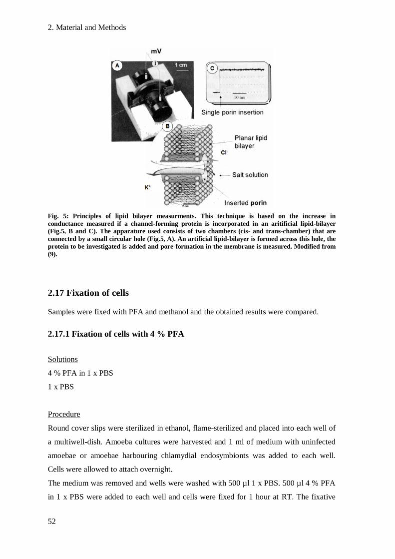
2. Material and Methods
52
Fig. 5: Principles of lipid bilayer measurments. This technique is based on the increase in
conductance measured if a channel-forming protein is incorporated in an aritificial lipid-bilayer
(Fig.5, B and C). The apparature used consists of two chambers (cis- and trans-chamber) that are
connected by a small circular hole (Fig.5, A). An artificial lipid-bilayer is formed across this hole, the
protein to be investigated is added and pore-formation in the membrane is measured. Modified from
(9).
2.17 Fixation of cells
Samples were fixed with PFA and methanol and the obtained results were compared.
2.17.1 Fixation of cells with 4 % PFA
Solutions
4 % PFA in 1 x PBS
1 x PBS
Procedure
Round cover slips were sterilized in ethanol, flame-sterilized and placed into each well of
a multiwell-dish. Amoeba cultures were harvested and 1 ml of medium with uninfected
amoebae or amoebae harbouring chlamydial endosymbionts was added to each well.
Cells were allowed to attach overnight.
The medium was removed and wells were washed with 500 µl 1 x PBS. 500 µl 4 % PFA
in 1 x PBS were added to each well and cells were fixed for 1 hour at RT. The fixative

2. Material and Methods
53
was removed and cells were covered with 1 x PBS. Fixed cells were stored in 1 x PBS up
to 2 weeks at 4°C.
2.17.2 Fixation of cells with Methanol
Solutions
Methanol
1 x PBS
Procedure
Fixation procedure was the same as for fixation with 4% Paraformaldehyde (see 2.17.1),
except that cells were fixed with methanol for only 10 min at RT.
2.18 Immunofluorescence analysis and antibody preparation
2.18.1 Preparation of antibodies targeting the putative porin pc1489
Solutions
Binding Buffer
Elution Buffer
Cleaning Buffer
Procedure
400 µg of pc1489 in Buffer A without n-POE were used for immunization of one
chicken. Antibodies were produced by Eurogentec (Seraing, Belgium). Pre-immune egg
yolks were collected before the first immunization. After the first immunization, 3 boosts
followed. Egg yolks of the immunized chicken were collected, starting 8 days after the
first immunization.
IgY antibodies from different egg yolk collections and also from the pre-immune yolks
were purified using a HiTrap™ IgY Purification HP column according to the
manufacturer‟s instructions.
2. Material and Methods
54
2.18.2 Preparation of amoeba lysate
Solutions
FA-Block solution
Procedure
An amoeba culture was harvested by centrifugation (5 min, 7,323 x g). The pellet was
resuspended in 500 µl – 1 ml FA-Block solution, depending on the size of the pellet.
Amoeba cells were broken by either 4 sonication steps for 30 seconds each or by two
freeze-and-thaw steps (freezing at -20°C/ thawing at 42°C in a water bath). The resulting
lysate was stored at -20°C until further use.
2.18.3 Removal of antibodies targeting amoeba proteins
To remove antibodies that would result in an unspecific background in
immunofluorescence analysis, all sera used were inactivated against amoeba proteins.
Procedure
Antibody sera were mixed 1:1 with amoeba cell lysates, vortexed briefly and incubated
overnight at 4°C. On the following day the solution was mixed and incubated again for 30
min at 4°C. Amoeba cell debris that had bound the host-reactive antibodies was removed
by centrifugation for 2 min at 6,797 x g. The supernatant was transferred to a new tube
and used for further analysis.
2.18.4 Immunofluorescence analysis
Solutions
1 x PBS
10 mM Tris-HCl pH 6.5
0.05% TweenTM
20 in 1 x PBS
FA-Block solution
Mowiol

2. Material and Methods
55
Procedure
For samples fixed with 4% PFA, a permeabilisation step was performed prior to
immunofluorescence analysis in order to permeabilize cells.
1 x PBS was removed and cells were incubated with 300 µl 0.05% TweenTM
20 in 1 x
PBS for 25 min at room temperature. After removal of TweenTM
20, cells were covered
with 300 µl 10 mM Tris-HCl (pH 6.5) containing 5 µg/ml Lysozyme and incubated for 1
hour at room temperature. Permeabilized cells or cells fixed with methanol were
incubated with FA-Block solution for 10 min. After this blocking step, the blocking
solution was replaced with the primary antibody diluted in FA-Block solution and cells
were incubated for 1 hour at RT. Cells were washed 3 times with 1 x PBS, followed by
incubation with secondary antibody diluted in FA-Block solution for 1 hour in the dark.
Cells were washed 3 times with 1 x PBS and cover slips were embedded in Mowiol.
Embedded samples were analyzed by epifluorescence microscopy and confocal laser
scanning microscopy.
2.18.6 Immunofluorescence on thin sections
Solutions
1 x PBS
FA-Block solution
100 mM HEPES buffer, pH 7.2, with 2 % PFA and 0.1 % glutaraldehyde
12 % gelatin
Cryoprotective solution (2.3 M sucrose in 100 mM phosphate buffer)
Procedure
Thin sections (75 nm) of A. castellanii infected with P. amoebophila were provided by
Jacqueline Monatanaro.
Cells were fixed with 2% PFA, 0.1 % glutaraldehyde in 100 mM HEPES buffer (pH 7.2)
for 20 mins at 20 C and then transfered to 2 % PFA in 100 mM HEPES buffer for a
further 2 hours. Fixed cells were pelleted at 3000 x g and washed in 100 mM HEPES
buffer. The cells were encapsulated in 12 % gelatin at 37 C. After 10 min infiltration, the
cells were pelleted in an eppendorf tube and cooled on ice. The gelatin-cell pellet was cut
into 0.1 mm2 cubes and infiltrated in Cryoprotective solution overnight at 4 C. Cell cubes
2. Material and Methods
56
were mounted onto aluminium stubs and frozen in liquid nitrogen. Thin sections were cut
using a Leica Ultracut UC6-FC6 ultramicrotome (Leica Microsystems, Austria) and
mounted onto carbon-coated formvar-pilioform films supported on hexagonal 200 mesh
nickle EM grids (Gilder, Netherlands). Sections were preserved prior to use under Cryo-
EM pick-up solution containing 1% methyl cellulose (Electron Microscopy Sciences,
Germany) and 1.3 M sucrose in 100 mM phosphate buffer at 4 C. 10 µl 1 x PBS were
added to each well to solubilize methyl cellulose on top of the thin-sections. After 10
minutes, 1 x PBS was replaced with 10 µl of the primary antibody diluted in FA-Block
solution and cells were incubated for 1 hour at RT. Cells were washed 3 times with 10 µl
1 x PBS, followed by incubation with 10 µl of the secondary antibody diluted in FA-
block solution for 1 hour in the dark. Cells were washed again and slides were embedded
in Citifluor. Embedded samples were analyzed by epifluorescence microscopy and
confocal laser scanning microscopy.
2.19 Infection studies
2.19.1 Counting of DAPI-stained cells
The number of P. amoebophila cells in suspensions was determined by filtering cells onto
a membrane, followed by DAPI-staining to visualize cells.
Solutions
1 x PBS (filter-sterilized)
DAPI
Procedure
DAPI was prepared at a dilution of 1:10,000 and stored at 4°C until further use.
Partially purified EBs were thawed at 37°C. 1 µl of the EB–suspension was added to 5 ml
of filter-sterilized 1 x PBS and stored on ice. The filter device was flame-sterilized and
assembled. 5 ml 1 x PBS were filtered to rinse the device by applying a vacuum pump
running at 250 mmHg (millimetre of mercury). The bacterial suspension was filtered onto
a 0.22 µm polycarbonate membrane filter and a 0.45 µm cellulose acetate membrane
support underneath the filter. The device was rinsed with 5 ml 1 x PBS to wash down
2. Material and Methods
57
bacteria bound to the glass funnel. 150 µl of DAPI solution were added on top of the filter
and the filter was incubated for 5 min in the dark. The filter was washed with 1 x PBS,
placed on a microscope slide and embedded in Citifluor. Samples were analyzed with an
epifluorescence microscope. The number of cells on 10 x 10 fields on a grid was counted.
The cell number/ml was calculated using the following formula:
M …Microscope factor = 11,264
DF … Dilution factor = 1,000
2.19.2 Infection cycle
Solutions:
TSY
1 x PAS (filter-sterilized)
1 x PBS (filter-sterilized)
4% PFA in 1 x PBS
Methanol
Procedure
A large flask (150 ml) of A. castellanii Neff was harvested (sec. 2.9.2) and amoebae were
counted using a Neubauer counting chamber. The number of amoebae/ml was calculated
using the following formula:
DF … Dilution factor = 100
Depth of chamber = 0.1 mm
After counting, amoebae were pelleted by centrifugation and resuspended in the volume
of TSY needed to obtain the required number of cells/ml. Round cover slips were

2. Material and Methods
58
sterilized in ethanol, flame-sterilizd and placed into each well of a multiwell-dish. 1 ml of
TSY containing a defined number of amoebae was pipetted in the wells of the multiwell-
dish. Samples were incubated for 30 min – 60 min at 20°C to allow attachment of
amoebae. Proper attachment of amoebae was controlled by light microscopy. Partially
purified EBs were thawed at 37°C and placed on ice. Amoebae were infected with EBs at
a multiplicity of infection (MOI) of 5 or a MOI of 10 by adding a defined amount of EB
suspension to each well. Multiwell-dishes were centrifuged at 600 x g for 15 min at 20°C
to enhance infection. The end of centrifugation was regarded as time point 0 hours post
infection (h p.i.). After centrifugation, the medium was replaced with fresh TSY to
remove excess EBs that were not attached to amoebae. Infected cultures were grown at
20°C. At indicated time points, cells were fixed. For fixation, the medium was removed
and cells were washed with 1 x PBS. Cells were fixed with 4% PFA or with methanol
(2.18.1 and 2.18.2). The fixative was replaced by 1 x PBS and cells were stored at 4°C
until immunofluorescence analysis was performed.
2.19. 3 Heat inactivation of EBs
Conditions for heat inactivation were chosen similar to those already published for heat
inactivation of clinical chlamydiae (16).
Procedure
Partially purified EBs were thawed at 37°C and 1 volume of 1 x PBS was added. For heat
inactivation, cells were incubated at 56°C for 30 min.
2.19.4 Infection - inhibition assay using anti-Pam and anti-pc1489-antibody
Infectious EBs were incubated with antibodies targeting the protein of interest and these
EBs were then used for infection of host cells. The ability to attach to host cells and the
number of bacteria/host cell were compared to non-treated EBs at certain time points.
Anti-pc1489-antibody and anti-Pam-antibody, an antibody targeting the immunodominant
proteins of the outer membrane of P. amoebophila, were used.
Solutions
TSY
1 x PAS (filter-sterilized)
2. Material and Methods
59
FA-Block solution (filter-sterilized)
1 x PBS (filter-sterilized)
4% PFA in 1 x PBS
Methanol
Procedure
Anti-pc1489-antibody and anti-Pam-antibody were diluted in FA-Block solution to reach
concentrations of 200 µg/ml and 20 µg/ml. Additionally, a solution containing 200 µg/ml
anti-Cld-antibody, targeting the Chloride dismutase of “Candidatus Nitrospira defluvii”
(generously provided by Frank Maixner) was prepared.
EBs and heat-inactivated EBs were incubated with 20 µg/ml or 200 µg/ml of anti-
pc1489-antibody or anti-Pam–antibody for 30 min at 37°C. As controls, EBs and heat –
inactivated EBs were incubated with FA-Block solution only and additionally, EBs were
incubated with 200 µg/ml anti-Cld-antibody under the same conditions. Amoebae were
infected to obtain a final MOI of 5 as described above (sec. 2.19.2). Infections were done
in duplicates. One of these duplicates was fixed with methanol, the other with 4% PFA
(sec. 2.18.1 and sec. 2.18.2). The following time points were investigated: 0, 6, 12, 24,
48, 72 and 96 hours post infection. After fixation of cells, multiwell-dishes were stored at
4°C until immunofluorescence analysis was performed. Samples were examined by
fluorescence microscopy and the number of infected amoebae at 72 and 96 h p.i. was
counted. The ratio of infected amoebae to all amoebae counted was calculated to allow a
comparison of infectivity of treated and non-treated EBs.
2.15 Heterologous expression of proteins in Escherichia coli
2.15. 1 Overexpression of proteins
Vector-constructs used in this study are listed in table 8 and were provided in the E. coli
expression strain BL21 (DE3) by Eva Heinz.
Solutions
LB medium
Ampicillin
1 M Isopropyl-β-D-thiogalactopyranosid (IPTG) in H2Odd, filter-sterilized
2. Material and Methods
60
4 x SDS - PAGE loading buffer
Procedure
5 ml of LB medium were inoculated with E. coli cells from a glycerol stock and grown
overnight at 37°C. Recombinant E. coli cells harbouring a plasmid were grown in the
presence of ampicillin to avoid the growth of cells without a plasmid. 250 µl of the
overnight-culture were used to inoculate 5 ml of fresh LB medium and cells were grown
for 2 hours at 37°C. Expression of proteins was induced by addition of IPTG to obtain an
end concentration of 1 mM to the culture. After 6 hours, cells were harvested by
centrifugating 300 µl of the culture at 17,949 x g for 2 min. The pellet was resuspended in
60 µl of 4 x SDS - PAGE loading buffer and analysed by SDS-PAGE analysis (sec.
2.10.4).

3. Results
61
3. Results
3. 1 Purification of the putative porin pc1489
The putative porin pc1489 was purified from elementary bodies of P. amoebophila. The
different fractions obtained after applying a NaCl-gradient to the anion-exchange column
were controlled on a SDS-PAGE gel. In the fractions after solubilisation and precipitation
of proteins (Fig.6, lanes 1 and 2), a high number of different proteins was present. In lane
3, the flow-through fraction, one band at ~ 50 kDa can be observed. This band represents
the OmcB homologue pc0616 that was identified in a previous study by mass
spectrometry analysis of gel bands (47). A prominent band corresponding to pc1489 is
present in lanes 1 and 2 at ~30 kDa and also in lanes 5-9, corresponding to the eluates of
the 250 mM, 300 mM, 350 mM and 400 mM NaCl- fractions. The molecular weight of
the observed band is not in accordance with the predicted weight of pc1489 (36 kDa), but
outer membrane proteins have been reported to migrate abnormally in SDS-PAGE gels
(see sec. 3.3). Only very weak additional bands are present in lanes 6, 7 and 8, showing
that pc1489 is highly enriched in these fractions. For some purifications, up to 7 bands
could be observed in the eluate after applying 350mM NaCl, suggesting the presence of
Fig. 6: Typical gel to check the purity of pc1489 after purification. Bands for OmcB (upper arrow)
and pc1489 (lower arrow) are indicated. M:Marker; Lane 1: Proteins after incubation at 37°C; Lane
2: Proteins after precipitation with acetone; Lane 3: Flow-through after applying the sample to the
column; Lane 4: Eluate after applying 100 mM NaCl; Lane 5: Eluate after applying 250 mM NaCl;
Lane 6: Eluate after applying 300 mM NaCl; Lane 7: Eluate after applying 350 mM NaCl; Lane 8:
Eluate after applying 400 mM NaCl; Lane 9: Eluate after applying 550 mM NaCl; Weight of marker
proteins in kDa is indicated on the left.

3. Results
62
various other proteins than pc1489 in these fractions. Fractions where strong additional
bands to that at 30 kDa were observed, were not used for production of antibodies or lipid
bilayer measurements. After the second precipitation step, samples were highly enriched
in pc1489 as can be seen by the presence of a strong band at 30 kDa in Fig.7 (left and
central picture). In the enriched fractions, bands were visible at 55 kDa, 60 kDa and 70
kDa. To see whether these bands represent pc1489 and to identify other proteins present
in these bands, they were subjected to analysis by mass spectrometry.
Fig. 7: SDS-PAGE gels of pc1489 after the second precipitation step (left and central picture) and of a
second, independant purification of pc1489 (right picture). Bands analyzed by mass spectrometry are
indicated by red circles and numbers correspond to table 13. For these scanned images, the contrast
was enhanced. Weight of marker proteins in kDa.
3.2 Analysis of bands of interest by mass spectrometry
In total, 12 bands of two independent purifications of pc1489 were analysed.
Pc1489 was the most abundant protein in most bands in Fig.7. It was present in high
amounts in the band at 30 kDa and also in the bands at 55 kDa, 60 kDa and 70 kDa (table
13). The presence of pc1489 in various bands suggests formation of multimers by pc1489
as found in porins of Gram-negative bacteria or interaction with other proteins of the
outer membrane. The bands at higher molecular weights could represent the dimeric and
trimeric form of pc1489 comparable to those observed for MOMP of Chlamydiaceae
(74). MOMP has been reported to run at a molecular weight of 90 kDa, representing the
porin trimer, when loaded on gels without heating the sample (110). However, for

3. Results
63
Band Protein Function/ Similarity to known
proteins*
Signal peptide
predicted**
1 pc1885 homologue of OmpW yes
pc1077 putative porin yes
pc1974 unkown protein yes
2 pc1489 putative porin yes
pc0383 similar to macrophage infectivity
potentiator (Mip)
yes
pc1499 similar to DnaK (heat shock
protein)
no
3 pc1489 putative porin no
pc1077 putative porin yes
4 pc0383 similar to Mip yes
pc1077 putative porin yes
pc1885 homologue of OmpW yes
5 pc1771 similar to citrate -synthase no
pc1105 conserved hypothetical protein no
pc1489 putative porin yes
6 pc1489 putative porin yes
pc1180 strongly similar to 60 kDa
chaperonin GroEL
no
ACL00009089 *** amoeba protein no
7 pc1489 putative porin yes
pc0675 hypothetical protein yes
pc1070 unknown yes
8 pc1489 putative porin yes
pc1077 putative porin yes
pc1885 homologue of OmpW yes
9 pc1489 putative porin yes
pc1077 putative porin yes
pc0004 hypothetical protein no
10 pc1885 homologue of OmpW yes
pc1489 putative porin yes
pc1077 putative porin yes
11 pc1489 putative porin yes
pc1885 homologue of OmpW yes
pc1077 putative porin yes
12 pc1489 putative porin yes
ACL00000461*** mitochondrial porin from A.
castellanii
no
pc1077 putative porin yes
Table 13: Proteins identified by mass spectrometry in analyzed bands. For each band, only the 3 hits
with the highest protein score are listed. Rows coloured grey were identified in a previous studies in
outer membrane fractions (47).
* according to http://mips.gsf.de/genre/proj/uwe25/
** according to TargetP (29)
*** according to http://tbestdb.bcm.umontreal.ca
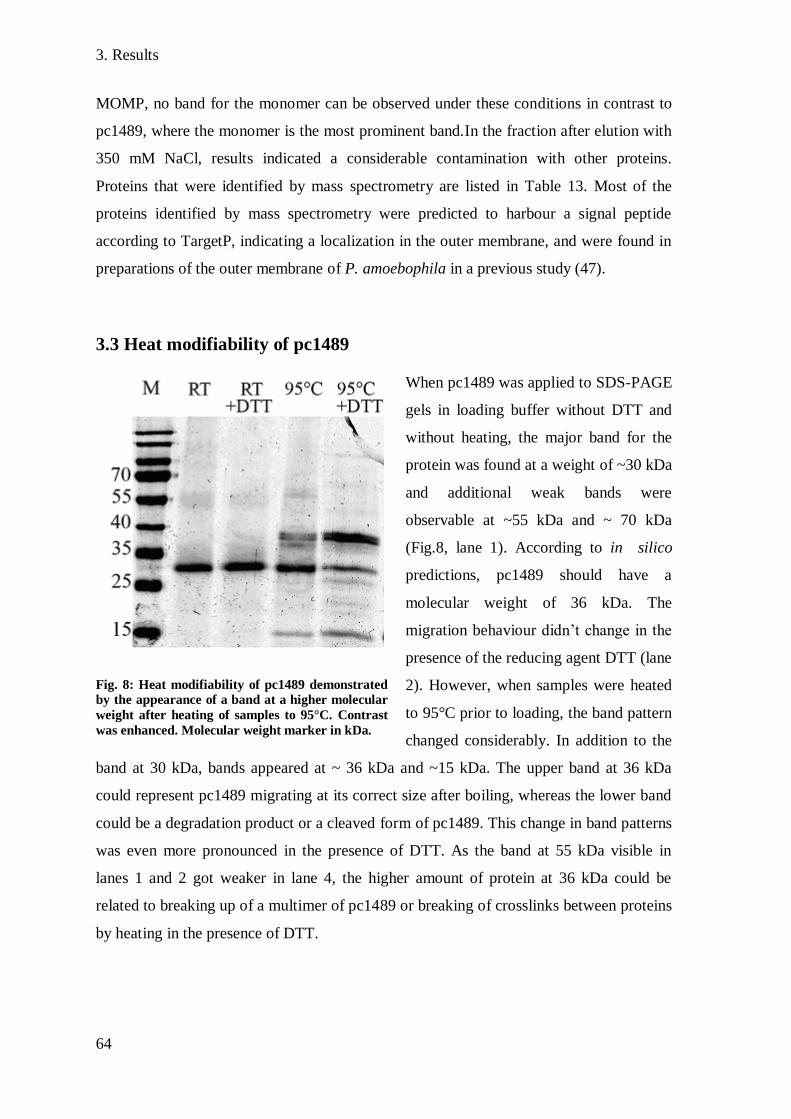
3. Results
64
MOMP, no band for the monomer can be observed under these conditions in contrast to
pc1489, where the monomer is the most prominent band.In the fraction after elution with
350 mM NaCl, results indicated a considerable contamination with other proteins.
Proteins that were identified by mass spectrometry are listed in Table 13. Most of the
proteins identified by mass spectrometry were predicted to harbour a signal peptide
according to TargetP, indicating a localization in the outer membrane, and were found in
preparations of the outer membrane of P. amoebophila in a previous study (47).
3.3 Heat modifiability of pc1489
When pc1489 was applied to SDS-PAGE
gels in loading buffer without DTT and
without heating, the major band for the
protein was found at a weight of ~30 kDa
and additional weak bands were
observable at ~55 kDa and ~ 70 kDa
(Fig.8, lane 1). According to in silico
predictions, pc1489 should have a
molecular weight of 36 kDa. The
migration behaviour didn‟t change in the
presence of the reducing agent DTT (lane
2). However, when samples were heated
to 95°C prior to loading, the band pattern
changed considerably. In addition to the
band at 30 kDa, bands appeared at ~ 36 kDa and ~15 kDa. The upper band at 36 kDa
could represent pc1489 migrating at its correct size after boiling, whereas the lower band
could be a degradation product or a cleaved form of pc1489. This change in band patterns
was even more pronounced in the presence of DTT. As the band at 55 kDa visible in
lanes 1 and 2 got weaker in lane 4, the higher amount of protein at 36 kDa could be
related to breaking up of a multimer of pc1489 or breaking of crosslinks between proteins
by heating in the presence of DTT.
Fig. 8: Heat modifiability of pc1489 demonstrated
by the appearance of a band at a higher molecular
weight after heating of samples to 95°C. Contrast
was enhanced. Molecular weight marker in kDa.

3. Results
65
3.4 Western blot analysis of pc1489
The specificity of purified anti-pc1489-antibody was tested by Western Blot analysis.
Western Blot analysis showed specific detection of purified pc1489 by the antibody,
indicated by the presence of one distinct band at ~ 30 kDa. Additional weak bands were
present at ~ 55 kDa and ~ 70 kDa (Fig.9 A). As pc1489 has been identified in bands of
this weight by mass spectrometry (see sec.3.2), these additional bands could result from
detection of pc1489 by the antibody. Samples with equal amounts of proteins of
uninfected A. castellanii, A. castellanii infected with P. amoebophila and purified EBs of
P. amoebophila were loaded on gels and Western blot analysis was performed. As these
samples had to be heated prior to loading on the gel in order to break up cells, the signal
for pc1489 was expected at ~ 36 kDa.
In samples containing amoebae without symbionts, no band was observed by Western
blot analysis, showing that the anti-pc1489-antibody does not cross-react with amoeba
proteins (Fig.9 B, lane 2).
In samples of A. castellanii infected with P. amoebophila, several bands were detected
(Fig.9 B, lane 1). One prominent band was found at ~ 60 kDa, and a weak signal was
Fig. 9: Western blot analysis with anti-pc1489-antibody. A: Detection of a band at the correct size in
SDS gels with purified pc1489 shows specificity of anti-pc1489-antibody. B: Detection of multiple
bands in A. castellanii infected with P. amoebophila. Lane 1: A. castellanii infected with P.
amoebophila; Lane 2: Uninfected A. castellanii; Lane 3: Purified EBs of P. amoebophila. For
comparison, a SDS gel with samples applied like in B and stained with colloidal Coomassie is shown
in C. Molecular weight marker in kDa.
A B C
3. Results
66
present from 35 kDa to 50 kDa. The smear might represent the monomeric form of
pc1489 after heating to 95°C which results in several bands at these molecular weights
(see sec 3.3). The band at 55 kDa could represent a multimer of pc1489 or the protein
pc1489 cross linked to other proteins in the outer membrane of EBs and RBs as both
developmental forms were present in asynchronized cultures used for this experiment.
The band at 20 kDa could result from degradation products of pc1489. In a study by
Heinz et al. (47), pc1489 was detected in bands of various molecular weights in outer
membrane fractions of P. amoebophila, supporting the detection of several bands by anti-
pc1489-antibody in amoebae infected with P. amoebophila. In EBs, only one prominent
band was observable at ~55 kDa (Fig.9 B, lane 3), possibly representing a tightly cross
linked multimer of pc1489.
3.5 Immunofluorescence analysis of pc1489
The localization of pc1489 in P. amoebophila during co-existence with its host was
studied by immunofluorescence analysis. Anti-pc1489-antibody was used simultaneously
with anti-Pam and anti-Hsp60-antibodies. Anti-Pam-antibodies were raised against whole
elementary bodies of P. amoebophila and therefore target the immunodominant
components of the outer membrane. Anti- Hsp60-antibodies target the chlamydial heat-
shock protein GroEL that is located in the cytoplasm and in the outer membrane of
chlamydiae (120).
Signals for pc1489 showed a halo-like shape and overlapped with signals for the outer
membrane of P. amoebophila by anti-Pam-antibody in methanol and PFA-fixed samples
(Fig.10). Signals observed for the cytoplasmic protein Hsp60 were surrounded by pc1489
(Fig.11 B). The halo-shape of signals was even more evident when immunofluorescence
was performed on thin-sections (Fig.11 A). The shape of the signal for pc1489 indicates a
localization of this protein in either the inner or the outer membrane or in the inclusion
membrane of P. amoebophila. As P. amoebophila thrives in single cell-inclusions in its
host, the inclusion membrane is adjacent to the outer membrane. Differentiation between
signals of the outer membrane and the inclusion membrane is not possible by
immunofluorescence analysis. Hsp60 was detected mostly in the cytoplasm of bacteria,
but also a few halo-like shaped signals could be observed, consistent with recent reports
that Hsp60 is also located to the outer membrane (120).
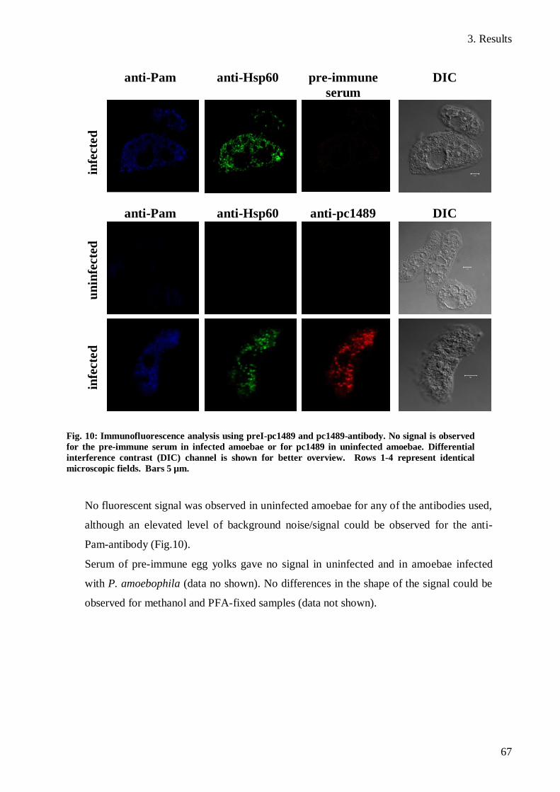
3. Results
67
No fluorescent signal was observed in uninfected amoebae for any of the antibodies used,
although an elevated level of background noise/signal could be observed for the anti-
Pam-antibody (Fig.10).
Serum of pre-immune egg yolks gave no signal in uninfected and in amoebae infected
with P. amoebophila (data no shown). No differences in the shape of the signal could be
observed for methanol and PFA-fixed samples (data not shown).
anti-Pam anti-Hsp60 pre-immune
serum
DIC in
fecte
d
anti-Pam anti-Hsp60 anti-pc1489 DIC
un
infe
cte
d
infe
cte
d
Fig. 10: Immunofluorescence analysis using preI-pc1489 and pc1489-antibody. No signal is observed
for the pre-immune serum in infected amoebae or for pc1489 in uninfected amoebae. Differential
interference contrast (DIC) channel is shown for better overview. Rows 1-4 represent identical
microscopic fields. Bars 5 µm.
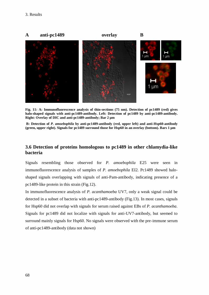
3. Results
68
A anti-pc1489 overlay B
Fig. 11: A: Immunofluorescence analysis of thin-sections (75 nm). Detection of pc1489 (red) gives
halo-shaped signals with anti-pc1489-antibody. Left: Detection of pc1489 by anti-pc1489-antibody.
Right: Overlay of DIC and anti-pc1489-antibody; Bar 2 µm
B: Detection of P. amoebophila by anti-pc1489-antibody (red, upper left) and anti-Hsp60-antibody
(green, upper right). Signals for pc1489 surround those for Hsp60 in an overlay (bottom). Bars 1 µm
3.6 Detection of proteins homologous to pc1489 in other chlamydia-like
bacteria
Signals resembling those observed for P. amoebophila E25 were seen in
immunofluorescence analysis of samples of P. amoebophila EI2. Pc1489 showed halo-
shaped signals overlapping with signals of anti-Pam-antibody, indicating presence of a
pc1489-like protein in this strain (Fig.12).
In immunofluorescence analysis of P. acanthamoeba UV7, only a weak signal could be
detected in a subset of bacteria with anti-pc1489-antibody (Fig.13). In most cases, signals
for Hsp60 did not overlap with signals for serum raised against EBs of P. acanthamoeba.
Signals for pc1489 did not localize with signals for anti-UV7-antibody, but seemed to
surround mainly signals for Hsp60. No signals were observed with the pre-immune serum
of anti-pc1489-antibody (data not shown)
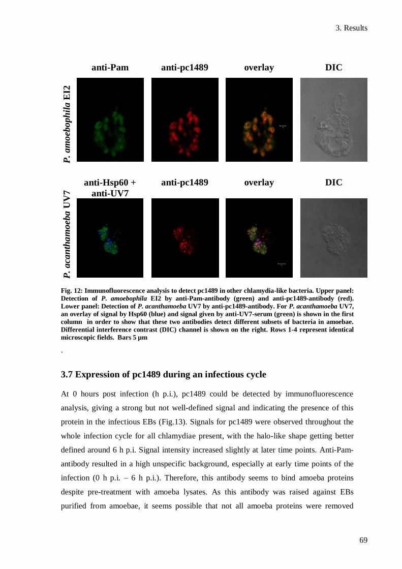
3. Results
69
anti-Pam anti-pc1489 overlay DIC P
. a
mo
ebo
ph
ila
EI2
P.
acan
tha
moeb
a U
V7
anti-Hsp60 +
anti-UV7
anti-pc1489 overlay DIC
Fig. 12: Immunofluorescence analysis to detect pc1489 in other chlamydia-like bacteria. Upper panel:
Detection of P. amoebophila EI2 by anti-Pam-antibody (green) and anti-pc1489-antibody (red).
Lower panel: Detection of P. acanthamoeba UV7 by anti-pc1489-antibody. For P. acanthamoeba UV7,
an overlay of signal by Hsp60 (blue) and signal given by anti-UV7-serum (green) is shown in the first
column in order to show that these two antibodies detect different subsets of bacteria in amoebae.
Differential interference contrast (DIC) channel is shown on the right. Rows 1-4 represent identical
microscopic fields. Bars 5 µm
.
3.7 Expression of pc1489 during an infectious cycle
At 0 hours post infection (h p.i.), pc1489 could be detected by immunofluorescence
analysis, giving a strong but not well-defined signal and indicating the presence of this
protein in the infectious EBs (Fig.13). Signals for pc1489 were observed throughout the
whole infection cycle for all chlamydiae present, with the halo-like shape getting better
defined around 6 h p.i. Signal intensity increased slightly at later time points. Anti-Pam-
antibody resulted in a high unspecific background, especially at early time points of the
infection (0 h p.i. – 6 h p.i.). Therefore, this antibody seems to bind amoeba proteins
despite pre-treatment with amoeba lysates. As this antibody was raised against EBs
purified from amoebae, it seems possible that not all amoeba proteins were removed
3. Results
70
during purification of EBs, thereby giving rise to antibodies targeting proteins of
amoebae. The unspecific background got lower at later time points and halo-shaped
signals of anti-Pam-antibody overlapped with signals for Hsp60 and pc1489. Only very
weak signals could be observed for the heat shock protein Hsp60 at early time points.
Signals for Hsp60 at 0 h p.i. showed a halo-like shape, pointing to the presence of a low
amount of this protein in the outer membrane of EBs. Very weak signals could be seen
for Hsp60 up to 24 h p.i., with a strong increase in signal intensity at 48 h p.i. From 48 h
p.i. to 96 h p.i., nearly all chlamydiae labelled with anti-Pam-antibody and anti-pc1489-
antibody were also detected by anti-Hsp60-antibody and gave a strong signal. Signals for
Hsp60 were surrounded by signals for pc1489 at later time points. Expression of Hsp60
seems to be up-regulated significantly after conversion of EBs in metabolically active
RBs as indicated by the rise of signal for this protein. At later time-points, sometimes
enlarged chlamydiae were seen in amoebae (Fig.14), maybe indicating stress due to
depletion of iron or other nutrients.
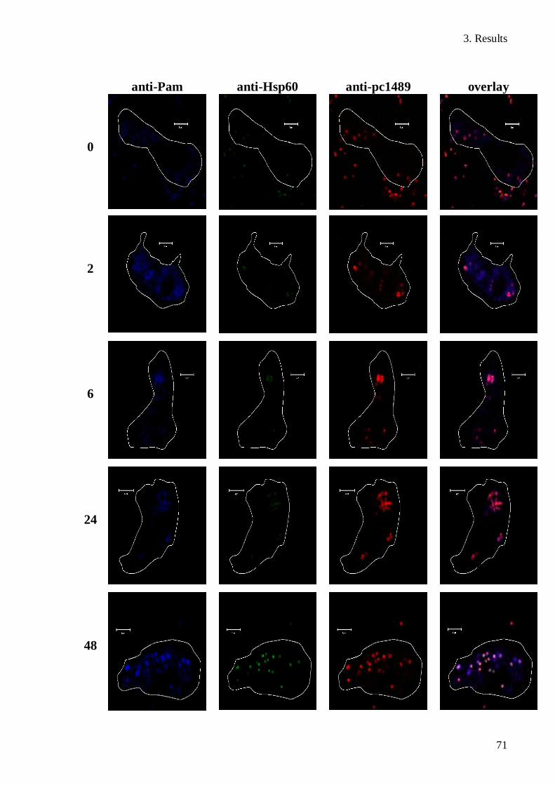
3. Results
71
anti-Pam anti-Hsp60 anti-pc1489 overlay
0
2
6
24
48

3. Results
72
anti-Pam anti-Hsp60 anti-pc1489 overlay
72
96
Fig. 13: Infection cycle of P. amoebophila in A. castellanii. P. amoebophila cells were detected with
anti-Pam-antibody (blue), anti-Hsp60-antibody (green) and anti-pc1489-antibody (red). Amoeba cell
outline are indicated in white. Numbers on the left indicate hours post infection. Rows 1-4 represent
identical microscopic fields. Bars 5 µm
anti-Pam anti-Hsp60 anti-pc1489 overlay
Fig. 14: Enlarged chlamydiae at 96 h p.i. Comparison with Fig.13 shows that chlamydiae are 2-4
times larger than normally. Bars 5 µm

3. Results
73
3.8 Porin activity of pc1489
Lipid-bilayer measurments were
performed at the laboratory of Prof.
Roland Benz (Würzburg, Germany).
Single-channel analysis showed that
pc1489 formed channels in a lipid
bilayer, indicated by a step-wise increase
in the measured conductance after
addition of pc1489 to the lipid bilayer
(Fig.15). The single channel conductance
was 3.25 nS in 1 M KCl. The
dependence of the single-channel
conductance on the KCl-concentration
(Table 14) suggested that ions can pass
the formed channel easily, indicating the
presence of a large water-filled pore in
the membrane.
The single-channel conductance was
shown to be more dependent on the
aqueous mobility of anions than of
cations. Replacement of the mobile Cl-
ion by the less mobile acetate-ion
resulted in a 2.1 –fold decrease in
conductance, whereas replacement of the
mobile K+ -ion by the less mobile Li
+ -
ion resulted in a 1.4 decrease.
These results indicate selectivity of the
pc1489-channel for anions. Anion
selectivity of the porin was confirmed by
additional measurements and calculation
of the Pc/Pa- ratio using the
Fig. 15: Single-channel analysis of purified pc1489
in 1 M KCl. Each step indicates insertion of one
pore in the lipid bilayer. Conductance in nS and
time in seconds
Salt Concentration
(M)
Single-channel
conductance
(nS)
KCl 0.01 0.15
KCl 0.03 0.25
KCl 0.1 0.6
KCl 0.3 1.2
KCl 1 3.25
KCl 3 11
LiCl 1 2.25
KAc 1 1.5
Table 14: Single channel conductance of pc1489
in different salt solutions

3. Results
74
Goldman-Hodgkin-Katz equation (10). This equation shows the ratio of the permeability
coefficient for a cation (Pc) in relation to the permeability coefficient
for an anion (Pa) for a porin. A value for Pc/Pa > 1 indicates selectivity for cations of a
channel, Pc/Pa < 1 indicates selectivity for anions and if Pc/Pa = 1, a channel is not
selective regarding the chosen anion or cation (46). Pc/Pa – ratios were 0.48 for KCl, and
0.78 for LiCl. All calculated Pc/Pa – ratios are smaller than 1, confirming selectivity of
pc1489 for anions.
A typical feature of porins is closure of the pore if high voltage is applied. Channels
formed by pc1489 showed closure for both positive and negative potentials of 30 mV and
-30 mV, shown by a decrease in conductance (Fig.16). Closure behaviour of pores did
only slightly change if pc1489 was added only to the trans- or to the cis-chamber of the
apparature in a way that closure was observed at a voltage of 40 mV and -40 mV. These
results indicate a symmetric response of the pore to the voltage applied and thereby
suggest the formation of a symmetrical pore by pc1489.
Fig. 16: Ratio of the conductance G at a given potential (Vm) divided by the conductance Go at 10 mV
as function of Vm. The aqueous phase contained 1 M KCl. The obtained graph shows voltage
dependent closure of pores formed by pc1489 as seen by a decrease in the measured conductance.
Channels closed if pc1498 was added on both sides (left) or only to the cis-chamber of the apparature.

3. Results
75
3.9 Heat inactivation of EBs
It has been demonstrated in previous studies that incubation of infectious EBs at 60°C for
a period of times as short as 3 min results in a dramatic decrease in attachment to and
uptake by host cells for some members of the Chlamydiaceae (80).
The effect of incubation at 56°C for 30 min on P. amoebophila was tested.
Non-treated and heat treated EBs were compared concerning their uptake by amoebae as
well as their ability to multiply within the amoeba host cells. No difference for uptake by
amoebae and the following 6 h p.i. and 12 h p.i. time points could be noticed (Fig.17, 0
and 6 h p.i.). At 24 h p.i., dividing chlamydiae could be seen in non-treated samples,
although differentiation between heat treated and non-treated bacteria was still difficult.
The number of bacteria in infected host cells started to rise clearly at 48 h p.i. in non-
treated samples, accompanied by an increase in signal for the heat-shock protein Hsp60.
Heat treated P. amoebophila cells appeared unable to divide, as numbers of bacteria per
host-cell dropped instead of increasing. Additionally, no gain in Hsp60 signal was seen
for the heat treated sample. This got even more obvious for the following time points,
where non-treated chlamydiae continued multiplying and filled the host cell, whereas
signals for treated chlamydiae could only be observed for single cells in a few amoebae.
The shape of signals for heat treated cells was well defined up to 72 h p.i., but got diffuse
at 96 h p.i., suggesting slow digestion of heat-inactivated chlamydiae by amoebae.
The obtained results suggest that heat-inactivation of EBs does not inhibit uptake of P.
amoebophila by amoebae, but renders chlamydiae unable to multiply within their host
cells.

1: anti-pc1489 2: anti-Hsp60 3: Overlay Control
0
6
12
24
48
96
Fig. 17: Infection cycle with heat-inactivated EBs. Rows 1-3 represent identical microscopic fields for
infection with heat treated EBs. On the far right, infection with non-treated EBs is shown for
comparison (Hsp60 in green, pc1489 in red) and for this control, amoeba cell outline are indicated in
white. Numbers on the left indicate hours post infection. Bars 5 µm
3.9 Infection inhibition assay
No difference in uptake or the ability to multiply within host cells could be observed
between EBs incubated with antibody and the control (Fig.18). For samples incubated
with 200 µg/ml anti-Pam-antibody, this high concentration of antibody lead to
agglutination of EBs. This resulted in large aggregates of EBs that were taken up by
amoebae in contrast to other samples, where EBs did not stick together. These aggregates
of chlamydiae were observed up to 48 h p.i. in samples treated with 200 µg/ml anti-Pam-
antibody. However, agglutination did not seem to influence multiplication of P.
amoebophila within host cells as an increase of signal for Hsp60 could be observed for
chlamydiae within these aggregates in contrast to heat-inactivated EBs (Fig. 20).
At 72 h p.i., no aggregates were observable any more. The number of amoebae infected
after 72 h p.i. and 96 h p.i. showed no difference between EBs treated with the low
concentration of anti-Pam-antibody and the controls (Fig.19). However, in samples of
EBs treated with the higher anti-Pam-concentration, the percentage of infected amoebae
was even higher than that of the control. However, these results have to be treated with
caution, as no duplicates could be investigated for some time points for samples incubated
with 200 µg/ml anti-Pam-antibody as no amoebae were found on the slides fixed with
PFA. Pre-incubation with anti-pc1489 did not show any effect on infectivity of
chlamydiae even at high concentrations. The number of infected amoebae was
comparable to that observed for untreated EBs and the EBs incubated with anti-Cld-
antibody. No effect of incubation with anti-Cld-antibody, targeting the chloride dismutase
of "Candidatus Nitrospira defluvii" that is not be present in P. amoebophila, was
observable. Heat-inactivated EBs were taken up by amoebae and did not multiply as
expected. No differences in uptake after incubation with antibodies were observable.
Heat-inactivated EBs incubated with high concentrations of anti-Pam-antibody did not
agglutinate and no large aggregates of chlamydiae could be seen. It is possible that
treatment of EBs at 56°C leads to denaturation of heat-sensitive target sites of this
antibody, resulting in lower amounts of anti-Pam-antibody bound to EBs and thus EBs do
not aggregate.
These results suggest that pre-treatment of EBs with anti-Pam-antibody or anti-pc1489-
antibody does not inhibit infection of amoeba host cells. However, as not all samples
from all time points contained amoebae, these results have to be validated by further
experiments.
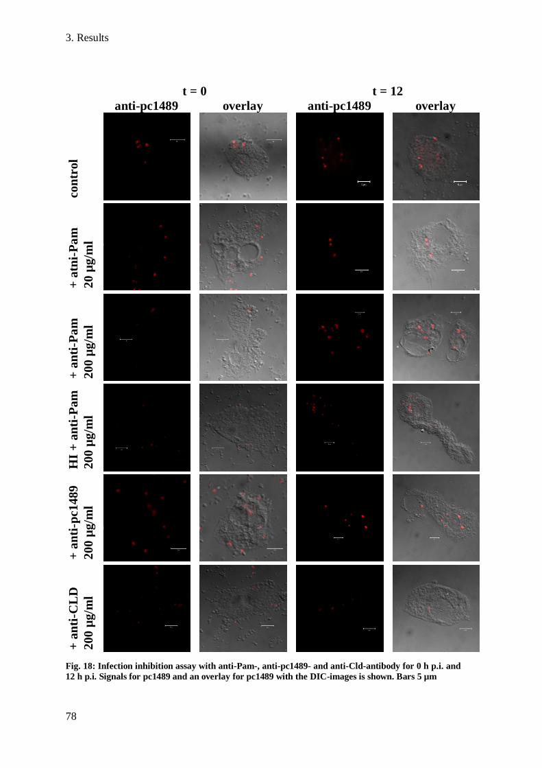
3. Results
78
t = 0 t = 12
anti-pc1489 overlay anti-pc1489 overlay
co
ntr
ol
+ a
tni-
Pa
m
20
µg
/ml
+ a
nti
-Pa
m
200 µ
g/m
l
HI
+ a
nti
-Pam
200 µ
g/m
l
+ a
nti
-pc1489
20
0 µ
g/m
l
+ a
nti
-CL
D
20
0 µ
g/m
l
Fig. 18: Infection inhibition assay with anti-Pam-, anti-pc1489- and anti-Cld-antibody for 0 h p.i. and
12 h p.i. Signals for pc1489 and an overlay for pc1489 with the DIC-images is shown. Bars 5 µm

3. Results
79
t = 48 t = 96
anti-pc1489 overlay anti-pc1489 overlay co
ntr
ol
+ a
tni-
Pa
m
20
µg
/ml
+ a
nti
-Pa
m
200 µ
g/m
l
HI
+ a
nti
-Pam
200 µ
g/m
l
+ a
nti
-pc1489
20
0 µ
g/m
l
+ a
nti
-CL
D
20
0 µ
g/m
l
Fig. 19: Infection inhibition assay with anti-Pam-, anti-pc1489- and anti-Cld-antibody for 48 h p.i.
and 96 h p.i. Signals for pc1489 and and an overlay for pc1489 with DIC-images is shown. Rows 1
and 2 as well as rows 3 and 4 represent identical microscopic fields. In row 3, the outlines of
uninfected amoebae are indicated in white. Bars 5 µm for pictures on the left and 10 µm for pictures
in the right panel.
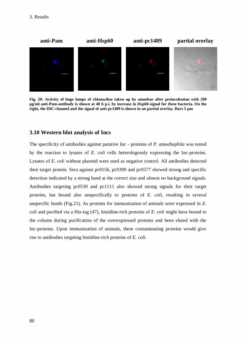
3. Results
80
anti-Pam anti-Hsp60 anti-pc1489 partial overlay
Fig. 20: Activity of huge lumps of chlamydiae taken up by amoebae after preincubation with 200
µg/ml anti-Pam-antibody is shown at 48 h p.i. by increase in Hsp60-signal for these bacteria. On the
right, the DIC-channel and the signal of anti-pc1489 is shown in an partial overlay. Bars 5 µm
3.10 Western blot analysis of Incs
The specificity of antibodies against putative Inc - proteins of P. amoebophila was tested
by the reaction to lysates of E. coli cells heterologously expressing the Inc-proteins.
Lysates of E. coli without plasmid were used as negative control. All antibodies detected
their target protein. Sera against pc0156, pc0399 and pc0577 showed strong and specific
detection indicated by a strong band at the correct size and almost no background signals.
Antibodies targeting pc0530 and pc1111 also showed strong signals for their target
proteins, but bound also unspecifically to proteins of E. coli, resulting in several
unspecific bands (Fig.21). As proteins for immunization of animals were expressed in E.
coli and purified via a His-tag (47), histidine-rich proteins of E. coli might have bound to
the column during purification of the overexpressed proteins and been eluted with the
Inc-proteins. Upon immunization of animals, these contaminating proteins would give
rise to antibodies targeting histidine-rich proteins of E. coli.

3. Results
81
pc0156 pc0399 pc0399 (Coomassie)
pc0530 pc0577 pc1111
Fig. 21: Western blots showing the specificity of anti-Inc-antibodies to parts of putative Inc-proteins
expressed in E. coli. Lysates of E. coli expressing the Inc-proteins are indicated with +, E. coli cells
without expression vector with -. Bands for Inc-proteins are indicated by arrows. For pc0399, a SDS
gel stained with Coomassie is shown for comparison. Molecular weight marker in kDa on the left of
each picture.
3.11 Immunofluorescence analysis with anti-Inc-antibodies
Immunofluorescence analysis showed halo-shaped signals for pc0156, pc0399, pc0530
and pc1111 overlapping with signals of anti-Pam-antibody. Anti-pc0399-antibody and
anti-pc0156-antibody bound to all bacteria detected with anti-Pam-antibody, whereas
pc0530 and pc1111 were detected in only a subset of bacteria (Fig.22). All antibodies
resulted in a high unspecific background even though sera were inactivated against
amoeba proteins several times. Pre-immune sera also yielded high unspecific background,
but no defined signals were observed. Signals for pc0577 were difficult to distinguish
from the high background and were observed in the cytoplasm or the outer membrane.

3. Results
82
anti-Pam anti-Inc DIC p
c0
15
6
pc0
39
91
pc0530
pc0577
pc1
11
1
Fig. 22: Immunofluorescence analysis with antibodies targeting the putative Inc-proteins of P.
amoebophila. Rows 1-3 indicate identical microscopic fields. Bars 5 µm

83
Differences compared to the pre-immune serum were hard to see for this protein (data not
shown).As stated above, differentiation between signals for proteins in the inner
membrane, the outer membrane and the inclusion membrane is not possible by
immunofluorescence analysis in P. amoebophila. To get additional proof for the
localization of the putative Inc-proteins in the inclusion membrane, highly-purified
elementary bodies fixed with methanol were investigated by immunofluorescence
analysis. During high-purification of EBs, the inclusion membrane should be removed as
observed by transmission electron microscopy. Highly-purified EBs gave a strong signal
with anti-Pam-antibody, but no signal could be observed for the putative Inc-proteins
(Fig.23). These results support the localization of these proteins to the inclusion
membrane of P. amoebophila.
Inc protein pc0156 pc0399 pc0530 pc1111
anti-Inc
and anti-
Pam
Fig. 23: Immunofluorescence analysis of highly-purified EBs of P. amoebophila with anti-Pam-
antibody and antibodies targeting four of the putative Inc-proteins. Signals can be seen for anti-Pam-
antibody (blue), but not for Inc-proteins (red), because the inclusion membrane is removed during
purification. Bar 2 µm
4. Discussion
84
4. Discussion
4.1 Characterization of pc1489
4.1.1 Localization of pc1489
MOMP, a porin with a mass of 40 kDa, is the most abundant component in the outer
membrane of Chlamydiaceae (15), but interestingly there is no homologous gene encoded
in the genome of P. amoebophila (19). In a recent study that combined 1D- and 2D- gel
electrophoresis with mass spectrometry, a family of putative porins was identified in
outer membrane preparations of the chlamydia-like bacterium P. amoebophila. The
prediction of a beta-barrel structure and the high abundance in preparations of the outer
membrane suggested that the two proteins pc1489 and pc1077 function as porins and act
as functional replacement of the major outer membrane protein of Chlamydiaceae in P.
amoebophila (47).
In this study, antibodies were raised against pc1489 purified from elementary bodies of P.
amoebophila. Immunfluorescence demonstrated the localization of this protein in the
inner, outer or inclusion membrane of P. amoebophila by forming a halo-shaped signal
surrounding the bacteria. Localization of pc1489 to the outer membrane was supported by
heat modifiability of this protein. Heat modifiability is considered to be a hallmark feature
of outer membrane proteins and has been studied in detail for several outer membrane
proteins like OmpA from E. coli (7) or outer membrane proteins of Rhodopseudomonas
sphaeroides (64). Due to their high beta-sheet content, outer membrane proteins bind
unusually high amounts of SDS. This results in faster migration through the gel compared
to other proteins leading to an underestimation of the weight of the protein. However,
excessive binding of SDS is reduced by boiling and therefore heating of the samples leads
to migration of the protein at the correct size. This behaviour is suggested to result from
the conversion of beta-sheets to alpha-helices by heating (85). Pc1489 was migrating at
30 kDa without heating, but an additional band appeared at 40 kDa after heating, showing
that the migration behaviour of this protein changes upon heating. This heat modifiability
of pc1489 suggests a high beta-sheet content of this protein as also has been proposed by
in silico analysis (47) and points to a localization in the outer membrane of P.
amoebophila.

4. Discussion
85
The final proof of a localization of pc1489 to the outer membrane has recently been given
by immuno-transmission electron microscopy studies by Jacqueline Montanaro. Gold
particles are located at the membrane of P. amoebophila, but not the inclusion membrane
or the
cytoplasm of the bacterium (Fig. 24). To investigate the presence of a homologue of
pc1489 in a member of the same family but different genus than P. amoebophila,
immunofluorescence was performed with P. acanthamoeba. The detection of a signal in
a subset of bacteria is well-supported by the identification of a homologue of pc1489 by
in silico analysis in P. acanthamoeba, suggesting the recognition of a protein similar to
pc1489 in this bacterium by the anti-pc1489-antibody. This protein could be identical
with the one identified by in silico analysis and assigned to the same Pfam domains as
members of the putative porin family of P. amoebophila (pc0870, pc1077, pc1489 and
pc1860) was identified (47). The detection of a protein in P. acanthamoeba by anti-
pc1489-antibody is rather surprising, as antibodies against MOMP are often strain-
specific in clinical chlamydiae and serovar-specific in C. trachomatis, where they are
used for serotyping (102, 104).
The low number of detected cells could be the result of differences in the organisation of
the outer membrane in EBs and RBs of P. acanthamoeba, for example due to extensive
cross linking of proteins in EBs that would mask binding sites for the anti-pc1489-
antibody in proteins of the outer membrane. This hypothesis is supported by the finding
Fig. 24: Pictures taken by transmission electron microscopy using anti-pc1489-antibody (left) and pre-
immune serum (right). Bars 200 nm. Pictures courtesy of Jacqueline Montanaro
4. Discussion
86
that serum raised against whole EBs and therefore targeting proteins of the outer
membrane of EBs only binds to a subset of bacteria, presumably EBs in
immunofluorescence analysis (Fig.12). It is also possible that the antibody does not fit the
protein detected in P. acanthamoeba perfectly and therefore less of the antibody is bound,
leading to fewer cell being detected. To investigate this further, Western blot analysis
with samples of P. acanthamoeba should be performed and differences in band patterns
between purified EBs and bacteria isolated at a time point in the developmental cycle
when mainly RBs are present, should be compared. It will be also interesting to contrast
band patterns of P. acanthamoeba with P. amoebophila.
Pc1489 was shown to be present in P. amoebophila throughout the whole developmental
cycle, demonstrating the presence of this protein in EBs and RBs comparabe to MOMP of
Chlamydiaceae. The observed increase in signal intensity at later time points could be the
result of incorporation of newly synthesized pc1489 into the outer membrane, thereby
increasing the amount of pc1489 present. It could also result from changes in the structure
of pc1489 which lead to better binding of anti-pc1489-antibody to its target protein or be
due to different fixation behaviour of different samples.
4.1.2 Functional characterization of pc1489
In SDS-PAGE gels with purified pc1489 and also in Western blot analysis, weak bands at
55-60 kDa were observed in addition to bands at 30 kDa or 40 kDa. This raises the
question about the organization of pc1489 in the outer membrane of P. amoebophila.
“Classic porins” of other Gram-negative bacteria are usually found as trimers in the outer
membrane, although monomeric forms of porins are known as well (109, 124). These
trimers have been reported to be extremely stable even in the presence of SDS and to
dissolve only by heating above 60°C in SDS (76, 84). This is the case for most known
porins, though exceptions from this rule like the unusually labile PorB-trimers of
Neisseria gonorrhoeae exist, that dissolve in the presence of SDS at room temperature
(89). MOMP of Chlamydiaceae is also present in a trimeric conformation in the outer
membrane and trimers of this proteins only dissociate upon heating to 95°C (74). The
band found at 55 kDa in Western blots with purified pc1489 could represent the trimeric
form of pc1489, because due to the unusual binding of SDS trimers of other porins of
Gram-negative bacteria migrate with a molecular mass of 65 to 70 kDa on SDS-PAGE
gels (13). If pc1489 is present as trimer in the outer membrane of P. amoebophila, it

4. Discussion
87
could be unusually labile, because even for samples in buffer without DTT that were not
heated, the band at 30 kDa, representing the monomer, was dominant. It is possible that
multimers of pc1489 were destroyed during purification, although similar protocols are
used for extraction of other porins and were not reported to destabilize these protein
complexes. However, pc1489 trimers could be comparable to PorB of Neisseria
gonorrhoeae and dissolve in the presence of SDS at room temperature. As in
asynchronized cultures of amoebae infected with P. amoebophila the most prominent
band is observed at around 55 kDa in Western blot analysis, this would support the
presence of multimers of pc1489. However, these results suggest that multimers would
then be quite stable as they were not broken by heating of samples to 95°C, but that the
multimers would be somehow destroyed during purification of pc1489 from elementary
bodies.
If pc1489 is present as a monomer in contrast to MOMP of Chlamydiaceae, bands at
higher molecular weight could result from interactions with other proteins of the outer
membrane, for example the cystein-rich proteins pc0616 and pc0617. This is supported
by the finding that other outer membrane proteins were identified in the same bands as
pc1489 by mass spectrometry (47). In order to further analyze the organization of pc1489
in the outer membrane, cross linking experiments of outer membrane fractions should be
performed.
The putative function of pc1489 as a porin was confirmed by lipid bilayer measurements
that showed a stepwise increase in conductance after addition of pc1489 to lipid bilayers,
each step indicating incorporation of one pore into the membrane. The single-channel
conductance for this porin was 3.25 nS in 1 M KCl and thereby considerably higher than
those observed for the so-called “classic porin” OmpF of E. coli (0.7 nS) (89). It is also
higher than single-channel conductance values observed for MOMPs of the
Chlamydiaceae. MOMP of C. trachomatis and Chlamydophila psittaci have a single-
channel conductance of 1 nS and 1.5
nS in 1 M KCl, respectively (57). This shows that major outer membrane proteins of
members of the Chlamydiaceae differ from each other and porins from environmental
chlamydiae in their porin activity. Values observed for porins of other Gram-negative
bacteria range from 10 pS for Tsx of E. coli (11) up to extremes of 20 nS for the
thermophilic bacterium Thermus thermophilus (70).

4. Discussion
88
Porin Organism Configuration Single channel
conductance in
1 M KCl
Size of pore Reference
MOMP C. trachomatis Trimer 1 nS 2 nm (57, 110)
MOMP C. psittaci Trimer 1.5 nS not known (57)
OmpA E. coli Monomer 50-80 pS or
260-320 pS*
1 nm (109)
OmpF E. coli Trimer 0.7 nS 1.2 nm (89)
OmpG E. coli Monomer 0.81 nS** 1.2 nm (20)
pc1489 P. amoebophila Monomer or
Trimer
3.25 nS not known
PorB N. gonorrhoeae Trimer 1 nS*** 1.6 nm (76)
Table 15: Characteristics of a few porins of gram-negative bacteria
*the two different values presumbably correspond to two different conformational states of this porin
** measured in 1 M NaCl
*** measured in 200 mM NaCl
Dependence of the single-channel conductance on the concentration of KCl was shown,
indicating the presence of a wide water-filled pore formed by pc1489 in lipid bilayers,
although the exact size of pore has not been determined. Formation of a wide pore has
also been demonstrated for the major outer membrane proteins of C. psittaci and C.
trachomatis (57). The pore formed by MOMP is proposed to have a diameter of 2 nm
based on penetration rates of sugars of different sizes through MOMP in liposome
swelling assays (110). With a proposed diameter of 2 nm, the pore formed by MOMP is
considerably larger than channels formed by other Gram-negative bacteria. Diameters
range from 0.3 nm for Tsx over 0.7 nm for OmpF and 1.2 nm for the large porin OmpG
of E. coli (89, 107).
The porin pc1489 was shown to be selective for anions with a Pc/Pa-ratio of 0.48 for KCl,
similar to that measured for native MOMP (0.5) by Wyllie et al. (121). The same authors
reported slight cation selectivity for a truncated recombinant MOMP of C. psittaci and C.
pneumoniae and for a full-length recombinant construct of C. psittaci and C. trachomatis
in later studies (57, 122). They suggested that small proteins or molecules bound to the
channel present in native preparations might have modified selectivity of MOMP in their

4. Discussion
89
previous study (57). MOMPs of C. psittaci and C. trachomatis were shown to differ
significantly in ion selectivity at certain KCl concentrations, again pointing out
differences in this channel for different members of the Chlamydiaceae. Removal of the
largest variable domain of MOMP, VS4, made the pore even more cation selective,
suggesting that selectivity for cations or anions results from variable domains exposed to
the surface (57). As these domains vary largely between different members of the
Chlamydiaceae, this will result in different ion selectivities of MOMPs. Indeed, different
selectivities were reported in other studies where cation selectivity (32) and no selectivity
for anions or cations was reported (60). These variable results may not only result from
different organisms used, but also from the use of native MOMP or different recombinant
constructs in different studies. As it is quite difficult to express the full-length MOMP of
some chlamydiae in E. coli, truncated constructs are often used, probably resulting in
differences in folding of the protein and therefore other pore-forming characteristics. In
this study, native pc1489 was used to determine selectivity for anions. Therefore, biased
results due to recombinant expression can be excluded.
As a family of four putative porins with an amino acid sequence identity of 22-28 % was
identified in P. amoebophila, characterization of the other members of this family will be
interesting. The three major porins of E. coli, OmpC, OmpF and PhoE share ~60 %
homoloy and differ slightly in their ion-selectivity and pore size (77). The expression of
ompC and ompF was shown to be regulated by the omsotic strength of the medium,
whereas phoE is expressed under phosphate limitation (reviewed in 90). In C.
trachomatis, a protein with 20.4% sequence identity to MOMP was identified. This
protein, termed PorB, was only observed in small amounts in the COMC and a more
specific function of this porin compared to MOMP was suggested (65). Similiarly,
different members of the putative porin family of P. amoebophila could be expressed at
different time points in the developmental cycle or differ in ion selectivity. Further
analysis should be performed to show whether the other members of this family do indeed
function as porins and how they differ from each other.
To sum up, pc1489 was shown to be localized to the outer membrane of P. amoebophila,
to be present during the whole developmental cycle of this organism and to function as
porin in this study.
4. Discussion
90
4.2 Influencing the infectivity of P. amoebophila
4.2.1 Inhibition of infection by pre-incubation with antibodies
It has been shown more than 40 years ago that incubation of chlamydiae with sera
prepared against whole chlamydiae resulted in inhibition of infection of host cells by
these organisms (e.g. 55). Caldwell and Perry (16) were the first to show that incubation
of EBs with antibodies targeting MOMP of C. trachomatis resulted in neutralization of
infection of host cells. They reported that incubation of EBs with antibody did not
influence the attachment or entry, but inhibition of infection occurred at a later time point
in the infection cycle. Later studies using antibodies targeting MOMP resulted in either
inhibition of attachment (106, 114) or in neutralization of infection at steps after
internalization of the chlamydiae (92). The results of these studies suggest that different
antibodies against MOMP, targeting different regions of this protein, lead to
neutralization of infection at different time points. Prevention of attachment can be easily
explained by masking of domains important for binding to host cells, comparable to
reports about neutralization of infection by antibodies against the outer membrane
proteins OmcB or PmpD (78, 115). Neutralization at later time points is more difficult to
explain. The study by Caldwell and Perry (16) also demonstrated that inhibition of
infection after uptake of cells depended on the use of intact dimeric IgG molecules,
because Fab fragments targeting MOMP did not neutralize infectivity. Based on this
observation, they suggested that effects on infectivity of C. trachomatis could result from
higher structural rigidity of the outer membrane due to cross linking by IgG, thereby
preventing transformation of EBs to RBs or inhibiting transport of host-derived nutrients
to the bacteria. Alternatively, they proposed that cross linking of MOMP by IgG
molecules could restrict fluidity of the outer membrane and prevent other components of
the outer membrane from interacting with host receptors (16).
In contrast to the pronounced effects of incubation with anti-MOMP-antibody on
infectivity of members of the Chlamydiaceae, no effect was observed on the infectivity of
P. amoebophila after incubation with two different antibodies. Anti-Pam-antibody was
raised against whole EBs of P. amoebophila. EBs that were incubated with this antibody
were still taken up by host cells and were able to multiply within amoebae comparable to
non-treated controls. No effect was observed for pre-incubation with antibodies targeting
pc1489, a protein thought to be the functional replacement of MOMP in P. amoebophila.
These results suggest that neither pc1489 nor proteins targeted by anti-Pam-antibody play
4. Discussion
91
a role in attachment to amoeba host cells. P. amoebophila might attach to amoebae by
means of other proteins that are not targeted by these antibodies, it might attach to
amoebae by unspecific interactions (e.g. electrostatic) or might just be taken up by
amoebae like other bacteria on which these protozoa feed.
In addition, the obtained results show that multiplication is not blocked by treatment with
antibodies in P. amoebophila in contrast to C. trachomatis (16) and therefore either no
cross linking of the outer membrane occurs or cross linking does not influence outer
membrane rigidity or fluidity. It is possible that cross linking is not as effective as in C.
trachomatis, because pc1489 might not be as abundant in the outer membrane as MOMP
that is thought to make up about 60% of the outer membrane of Chlamydiaceae (15). On
the other hand, inhibition of infection has also been observed for pre-incbuation with
antibodies targeting PorB, a porin of C. trachomatis that is present in the outer membrane
in lower amounts than MOMP (65).
Although differences in the abundance of pc1489 and MOMP could account for
differences in inhibition of infection by antibodies targeting these proteins, finding an
explanation for no observed effect by treatment with serum against P. amoebophila is
more difficult.
It was shown that EBs of C. psittaci treated with serum against this bacterium were taken
up by macrophages and destroyed rapidly, because they lost the ability to prevent
phagolysosomal fusion (123). In contrast, P. amoebophila EBs treated with anti-Pam-
antibody were not digested by amoebae, but multiplied within these cells. Therefore, P.
amoebophila must be able to escape from the endocytic pathway even after blocking
proteins of the outer membrane. These observations are similar to results obtained for the
intracellular bacteria Legionella pneumophila and Coxiella burnettii. In both organisms,
treatment with sera against these organisms does not inhibit intracellular multiplication
(49, 54).
In P. amoebophila, proteins in the inclusion membrane could participate in escape of the
bacterium from the endocytic pathway. Inc proteins identified in P. amoebophila differ
substantially from those of clinical chlamydiae (47) and therefore could account for
differences in the ability to survive phagocytosis even after preincubation with antibodies.
However, it can not be excluded that antisera used in this study did not crosslink outer
membrane proteins of P. amoebophila in a way comparable to sera against C. psittaci or
other members of the Chlamydiaceae.

4. Discussion
92
Additionally, it has to be considered that A. castellanii Neff is not the native host of P.
amoebophila. Maybe effects of antibody-treatment on infectivity would be observable in
the native host. If outer membrane proteins of P. amoebophila are adapted to attachment
to only the native host, they would not be required for uptake by A. castellanii. In this
case, A. castellanii that feed on bacteria would take up this bacterium unspecifically.
However, as the native host of P. amoebophila is not known yet, it is not possible to
investigate this issue further at the moment.
4.2.2 Inhibition of infection by heat-treatment
Members of the Chlamydiaceae lose their infectivity upon heat treatment and heat-labile
components of the outer membrane were suggested to be the cause for this, although
slightly contradictory results were reported, maybe generated by different incubation
conditions and different host cell lines used (14, 16, 33, 123) .
Heat treatment resulted in loss of infectivity of P. amoebophila. Heat treated cells were
taken up by amoebae comparable to non-treated control cells, but were not able to
multiply. As no increase of Hsp60-signal, that is thought to be a sign of metabolically
active cells, was observed for the treated cells, heat-inactivated EBs might be unable to
convert to metabolically active RBs. Intriguingly, anti-Pam-signals for P. amoebophila
cells were observed up to 96 h p.i. in amoebae and morphology of cells was preserved up
to 72 hours. This stands in contrast to rapid destruction of heat treated C. psittaci in
macrophages (123) and also to the fact that E. coli normally taken up by amoebae are
digested within 72 hours (Illias Lagkouvardos, personal communication). The observation
that heat inactivated chlamydiae are not immediately destroyed by amoebae is supported
by a report by Greub (39) who found heat-inactivated P. acanthamoeba cells in
macrophages as long as 48 hours post infection. Because this study also showed that the
lysosomal hydrolase Cathepsin D, an enzyme involved in caspase activation, did not
colocalize with heat-inactivated bacteria, Greub suggested a passive modulation of the
intracellular fate of heat-inactivated P. acanthamoeba, maybe by a special composition of
the bacterial outer membrane. As EBs were incubated for a longer period of time and at
higher temperatures in the study by Greub in comparison to this study, this putative
protective effect could be even more pronounced for treatment in this study and explain
the observed results.

4. Discussion
93
4.3 Inc-proteins of P. amoebophila
Modification of the host-derived membrane of the inclusion by incorporation of
chlamydial Inc-proteins was discovered more than 10 years ago (93). The exact role of
most of these proteins for interactions with the host is not known, but they are considered
as important means for the communication between host cell and chlamydiae, to play a
role in transport of nutrients to the chlamydiae and to ensure survival of these bacteria
within the host cell.
Recently, an in silico approach identified 24 putative Inc-proteins in the genome of the
chlamydia-like bacterium P. amoebophila (47). Only three of the predicted Inc-proteins
showed some sequence similarity to proteins of the Chlamydiaceae and one, pc0399,
showed weak sequence similarity to an inclusion membrane protein, IncA, of
Chlamydiaceae (47). For four of these proteins, pc0399, pc0156, pc0530 and pc1111,
localization in the inclusion membrane is supported by halo-shaped signals observed in
immunofluorescence analysis. In addition, no signal was detected for these proteins in
highly-purified EBs where no inclusion membrane should be present. Although additional
proof concerning the localization of these proteins is needed, the obtained results and the
presence of a large bilobed hydrophobicity motif, a key feature of known Inc-proteins,
point to a localization of these proteins to the inclusion membrane. Transmission electron
microscopy studies are in progress to show the exact localization of these proteins.
Inc-proteins of P. amoebophila are of special interest, as this chlamydia-like bacterium
resides within single-cell inclusions and the inclusions inhabited by this organism do not
fuse in contrast to members of the Chlamydiaceae (19). As Inc-proteins and especially the
IncA protein are thought to play an important role in the fusion of inclusions, probably by
means of a SNARE-like motif recently identified in IncA (27) that is not present in
pc0399, differences between Inc-proteins of P. amoebophila and Chlamydiaceae could
help to a better understanding of the function of these proteins.
5. Summaries
94
5. Summaries
5.1 Summary
Proteins in the outer membrane of chlamydiae are of great importance as they are
involved in attachment to host cells, uptake of nutrients, removal of waste and also in the
differentness of the two developmental stages of chlamydiae. Therefore and because of
their potential as vaccine candidates, these proteins have been studied extensively for
members of the Chlamydiaceae. Sequencing of the genome of the first member of the
environmental chlamydiae, the amoeba endosymbiont Protochlamydia amoebophila,
showed that no gene similar to the most abundant outer membrane protein of the
Chlamydiaceae, the major outer membrane protein (MOMP), is encoded (19). In a recent
study, a family of putative porins was identified in the genome of P. amoebophila. Two
of these porins were found to be highly abundant on 2D gels of outer membrane
preparations of this organism and predicted to form beta-barrels by in silico analysis (47).
In this study, the putative porin pc1489 was purified from elementary bodies of P.
amoebophila and antibodies were raised against this protein. The localization of pc1489
to the outer membrane was confirmed by immunofluorescence analysis and immuno-
transmission electron microscopy. It was shown that the protein is present throughout the
whole developmental cycle, thereby indicating an important role of this protein for P.
amoebophila. Its function as a porin was confirmed by lipid bilayer measurements using
purified pc1489. These measurements showed that the pore formed by pc1489 is quite
large and anion selective. These results suggest that pc1489 might be the functional
replacement of MOMP in P. amoebophila, possibly together with other members of the
putative porin family. However, in contrast to MOMP, preincubation of EBs with
antibodies targeting pc1489 as well as antibodies against the immunodominant
components of the outer membrane did not inhibit infection of host cells by P.
amoebophila.
In the second part of this diploma thesis, the localization of putative inclusion proteins in
P. amoebophila was studied. Four of the five proteins studied where shown to be
localized to either the outer membrane or the inclusion membrane of P. amoebophila by
immunofluorescence analysis. Although the function of these proteins is not known yet,
they are suggested to play an important role in communication between symbiont and
host and also in survival of P. amoebophila after internalization by amoebae.
5. Summaries
95
5.2 Zusammenfassung
Außenmembranproteine spielen eine wichtige Rolle für Chlamydien, da sie für die
Bindung an Wirtszellen, die Aufnahme von Nährstoffen in die Zelle und die Entfernung
von Stoffwechselendprodukten benötigt werden und zusätzlich zu Unterschieden
zwischen den beiden Entwicklungsstadien der Chlamydien beitragen. Da sie viel
versprechende Kandidaten für die Entwicklung von Impfstoffen sind, sind die
Außenmembranproteine der Chlamydiaceae gut erforscht. Im Gegensatz dazu ist wenig
über diese Proteine in der kürzlich entdeckten Gruppe der Umweltchlamydien bekannt.
Im ersten sequenzierten Genom eines Vertreters dieser Gruppe, Protochlamydia
amoebophila, wurde kein Homolog für das häufigste Außenmembranprotein der
Chlamydiaceae, das Major outer membrane protein, gefunden (19). Vor kurzem wurde
eine Familie von putativen Porinen in P. amoebophila gefunden. Zwei dieser Proteine,
pc1489 und pc1077, waren in großen Mengen auf 2D-Gelen mit
Außenmembranfraktionen vorhanden (47).
In dieser Diplomarbeit wurde das putative Porin pc1489 aus Elementarkörpern
aufgereinigt und Antikörper gegen dieses Protein hergestellt. Unter Verwendung dieser
Antikörper wurde mittels Immunfluoreszenzanalyse und
Transmissionselektronenmikroskopie gezeigt, dass pc1489 in der Außenmembran von P.
amoebophila lokalisiert ist. Pc1489 konnte während des gesamten Entwicklungszyklus in
der Außenmembran von P. amoebophila detektiert werden, was auf eine wichtige Rolle
dieses Proteins hinweist. Lipid Bilayer Messungen zeigten, dass pc1489 tatsächlich die
Funktion eines Porins besitzt und große, anionen-selektive Proteine in Lipidmembranen
formt. Diese Ergebnisse deuten darauf hin, dass pc1489 in P. amoebophila das Major
outer membrane protein ersetzen könnte, möglicherweise gemeinsam mit anderen
Mitgliedern der putativen Porinfamilie.
Im zweiten Teil dieser Diplomarbeit wurden putative Inklusionsproteine von P.
amoebophila bezüglich ihrer Lokalisation untersucht. Immunfluoreszenzanalyse mit
Antikörpern gegen diese Proteine zeigte, dass 5 der 4 untersuchten Proteine entweder in
der Außenmembran oder der Inklusionsmembran von P. amoebophila lokalisiert sind.
Obwohl die Funktion dieser Proteine noch nicht bekannt ist, wird vermutet, dass sie eine
wichtige Rolle in der Kommunikation mit der Wirtszelle spielen und dadurch auch den
Verdau dieser Bakterien durch Amöben verhindern könnten.
6. List of abbreviations
96
6. List of abbreviations
λ Wavelength
16S rRNA Small ribosomal RNA subunit of prokaryotes
1D One dimensional
2D Two dimensional
A Adenine
A. Acanthamoeba
ABC Ammonium bicarbonate
ACN Acetonitrile
Amp Ampicillin
APS Ammonium persulfate
ATP Adenosine-5'-triphosphate
bp Base pair(s)
BSA Bovine serum albumin
C Cytosine
C. Chlamydia
CLSM Confocal laser scanning microscope
COMC Chlamydia outer membrane complex
Cy2 5,5`-di-sulfo.1,1`-di-(X-carbopentynyl)-3,3,3`,3`-tetra-methylindol-
Cy2.18-derivative N-hydroxysuccimidester
Cy3 5,5`-di-sulfo.1,1`-di-(X-carbopentynyl)-3,3,3`,3`-tetra-methylindol-
Cy3.18-derivative N-hydroxysuccimidester
Cy5 5,5`-di-sulfo.1,1`-di-(X-carbopentynyl)-3,3,3`,3`-tetra-methylindol-
Cy5.18-derivative N-hydroxysuccimidester
Da Dalton
DAPI 4',6-Diamidino-2-phenylindol
DIC Differential interference contrast
DDT Dithiothreitol
DNA Desoxyribonucleic acid
dNTP Desoxynucleotide triphosphate
E. Escherichia
EB(s) Elementary body (-ies)
e.g. example given
6. List of abbreviations
97
et al. Et alterti (lat. and others)
EtOH Ethanol
Fig. Figure
FITC Fluorescein isothiocyanate
g Gram(s); gravity
GTP Guanosine-5'-triphosphate
h Hours
H2Odd Double distilled water
h p.i. hours post infection
HCl hydrochloric acid
His Histidine
HRP Horseraddish-Peroxidase
Hsp Heat shock protein
Ig Immunglobulin
Inc Inclusion
IPTG Isopropyl-β-D-thiogalactopyranosid
k Kilo
KCl Kalium chloride
L Liter(s)
LB Luria Bertani
LC Liquid chromatography
LiCl Lithium chloride
µ Mikro
m Meter(s); milli
M Molar
MALDI Matrix-assisted laser desorption/ionization
mg Milligramm
min Minute(s)
MOI Multiplicity of infection
MOMP Major outer membrane protein
MS Mass spectrometry
MS/MS Tandem mass spectrometry
n Nano
NaCl Sodium chloride

98
NaOH Sodium hydroxide
P. Parachlamydia; Protochlamydia
Pa Permeability coefficient for an anion
Pc Permeability coefficient for a cation
PAGE Polyacrylamide gel electrophoresis
PCR Polymerase chain reaction
PFA Paraformaldehyde
PMP Polymorphic outer membrane protein
RB(s) Reticulate body (-ies)
RFLP Restriction fragment length polymorphism
RNA Ribonucleic acid
rpm Revolutions per minute
rRNA Ribosomal RNA
RT Room temperature
S Siemens
SDS Sodium dodecyl sulfate
sec Second(s); section
SNARE Soluble N-ethylmaleimide-sensitive factor attachment protein
receptors
sp. Species
T Thymine
TEMED N,N,N´,N´-tetramethylethane-1,2-diamine
Tris Trishydroxymethylaminomethane
TSY Trypticase Soy Broth with Yeast Extract
UK United Kingdom
USA United States of America
UV Ultraviolet
V Volt
v/v Volume per volume
W Watt
w/v Weight per volume
w/w Weight per weight

7. References
99
7. References
1. Abdelrahman, Y. M., and R. J. Belland. 2005. The chlamydial developmental
cycle. FEMS Microbiol Rev 29:949-59.
2. Altschul, S. F., W. Gish, W. Miller, E. W. Meyers, and D. J. Lipman. 1990.
Basic Local Alignment Search Tool. Journal of Molecular Biology 215:403-410.
3. Amann, R., N. Springer, W. Schonhuber, W. Ludwig, E. N. Schmid, K. D.
Muller, and R. Michel. 1997. Obligate intracellular bacterial parasites of
acanthamoebae related to Chlamydia spp. Appl Environ Microbiol 63:115-21.
4. Baehr, W., Y. X. Zhang, T. Joseph, H. Su, F. E. Nano, K. D. Everett, and H.
D. Caldwell. 1988. Mapping antigenic domains expressed by Chlamydia
trachomatis major outer membrane protein genes. Proceedings of the National
Academy of Sciences of the United States of America 85:4000-4004.
5. Baud, D., V. Thomas, A. Arafa, L. Regan, and G. Greub. 2007. Waddlia
chondrophila, a potential agent of human fetal death. Emerg Infect Dis 13:1239-
43.
6. Bavoil, P., A. Ohlin, and J. Schachter. 1984. Role of disulfide bonding in outer
membrane structure and permeability in Chlamydia trachomatis. Infect Immun
44:479-85.
7. Beher, M. G., C. A. Schnaitman, and A. P. Pugsley. 1980. Major heat-
modifiable outer membrane protein in gram-negative bacteria: comparison with
the ompA protein of Escherichia coli. J. Bacteriol. 143:906-913.
8. Belland, R., O. S. P., J. Gieffers, and G. I. Byrne. 2004. Chlamydia pneumoniae
and atherosclerosis. Cellular Microbiology 6:117-127.
9. Benz, R. 2004. Bacterial and Eukaryotic Porins. WILEY-CH Verlag GmbH &
Co. KGaA, Weinheim.
10. Benz, R., K. Janko, and P. Läuger. 1979. Ionic selectivity of pores formed by
the matrix protein (porin) of Escherichia coli. Biochim Biophys Acta 551:238-47.
11. Benz, R., A. Schmid, C. Maier, and E. Bremer. 1988. Characterization of the
nucleoside-binding site inside the Tsx channel of Escherichia coli outer
membrane. Reconstitution experiments with lipid bilayer membranes. European
Journal of Biochemistry 176:699-705.
12. Birtles, R. J., T. J. Rowbotham, C. Storey, T. J. Marrie, and D. Raoult. 1997.
Chlamydia-like obligate parasite of free-living amoebae. Lancet 349:925-926.
13. Bolla, J. M., E. Loret, M. Zalewski, and J. M. Pages. 1995. Conformational
analysis of the Campylobacter jejuni porin. J. Bacteriol. 177:4266-4271.
14. Byrne, G. I., and J. W. Moulder. 1978. Parasite-specified phagocytosis of
Chlamydia psittaci and Chlamydia trachomatis by L and HeLa cells. Infect
Immun 19:598-606.
15. Caldwell, H. D., J. Kromhout, and J. Schachter. 1981. Purification and partial
characterization of the major outer membrane protein of Chlamydia trachomatis.
Infect Immun 31:1161-76.
16. Caldwell, H. D., and L. J. Perry. 1982. Neutralization of Chlamydia trachomatis
infectivity with antibodies to the major outer membrane protein. Infect. Immun.
38:745-754.
17. Cocchiaro J.L., Y. K., E.R. Fischer ,T. Hackstadt , R.H. Valdivia 2008.
Cytoplasmic lipid droplets are translocated into the lumen of the Chlamydia
trachomatis parasitophorous vacuole. Proc Natl Acad Sci U S A 105:9379-84.

7. References
100
18. Collingro, A., S. Poppert, E. Heinz, S. Schmitz-Esser, A. Essig, M.
Schweikert, M. Wagner, and M. Horn. 2005. Recovery of an environmental
Chlamydia strain from activated sludge by co-cultivation with Acanthamoeba sp.
Microbiology 151:301-9.
19. Collingro, A., E. R. Toenshoff, M. W. Taylor, T. R. Fritsche, M. Wagner, and
M. Horn. 2005. 'Candidatus Protochlamydia amoebophila', an endosymbiont of
Acanthamoeba spp. Int J Syst Evol Microbiol 55:1863-6.
20. Conlan, S., Y. Zhang, S. Cheley, and H. Bayley. 2000. Biochemical and
Biophysical Characterization of OmpG: A Monomeric Porin. Biochemistry
39:11845-11854.
21. Corsaro, D., and G. Greub. 2006. Pathogenic potential of novel Chlamydiae and
diagnostic approaches to infections due to these obligate intracellular bacteria.
Clin Microbiol Rev 19:283-97.
22. Corsaro, D., D. Venditti, and M. Valassina. 2002. New parachlamydial 16S
rDNA phylotypes detected in human clinical samples. Res Microbiol 153:563-7.
23. Cortes, C., K. A. Rzomp, A. Tvinnereim, M. A. Scidmore, and B. Wizel. 2007.
Chlamydia pneumoniae Inclusion Membrane Protein Cpn0585 Interacts with
Multiple Rab GTPases. Infect. Immun. 75:5586-5596.
24. De Bary, A. 1879. Die Erscheinung der Symbiose. Verlag von Karl J. Trubner,
Strassburg.
25. de Sa, C., A. Souriau, F. Bernard, J. Salinas, and A. Rodolakis. 1995. An
oligomer of the major outer membrane protein of Chlamydia psittaci is recognized
by monoclonal antibodies which protect mice from abortion. Infect. Immun.
63:4912-4916.
26. Delevoye, C., M. Nilges, A. Dautry-Varsat, and A. Subtil. 2004. Conservation
of the biochemical properties of IncA from Chlamydia trachomatis and
Chlamydia caviae: oligomerization of IncA mediates interaction between facing
membranes. J Biol Chem 279:46896-906.
27. Delevoye, C., M. Nilges, P. Dehoux, F. Paumet, S. Perrinet, A. Dautry-Varsat,
and A. Subtil. 2008. SNARE protein mimicry by an intracellular bacterium.
PLOS Pathogens 4:e1000022 doi:10.1371/journal.ppat.1000022.
28. Draghi, A., 2nd, V. L. Popov, M. M. Kahl, J. B. Stanton, C. C. Brown, G. J.
Tsongalis, A. B. West, and S. Frasca, Jr. 2004. Characterization of "Candidatus
piscichlamydia salmonis" (order Chlamydiales), a chlamydia-like bacterium
associated with epitheliocystis in farmed Atlantic salmon (Salmo salar). J Clin
Microbiol 42:5286-97.
29. Emanuelsson, O., H. Nielsen, S. Brunak, and G. von Heijne. 2000. Predicting
subcellular localization of proteins based on their N-terminal amino acid
sequence. J Mol Biol 300:1005-16.
30. Everett, K. D., and T. P. Hatch. 1995. Architecture of the cell envelope of
Chlamydia psittaci 6BC. J. Bacteriol. 177:877-882.
31. Fadel, S., and A. Eley. 2007. Chlamydia trachomatis OmcB protein is a surface-
exposed glycosaminoglycan-dependent adhesin. J Med Microbiol 56:15-22.
32. Findlay, H., H. McClafferty, and R. Ashley. 2005. Surface expression, single-
channel analysis and membrane topology of recombinant Chlamydia trachomatis
Major Outer Membrane Protein. BMC Microbiology 5:5.
33. Friis, R. R. 1972. Interaction of L Cells and Chlamydia psittaci: Entry of the
Parasite and Host Responses to Its Development. J. Bacteriol. 110:706-721.
34. Fritsche, T. R., R. K. Gautom, S. Seyedirashti, D. L. Bergeron, and T. D.
Lindquist. 1993. Occurrence of bacteria endosymbionts in Acanthamoeba spp.

7. References
101
isolated from corneal and environmental specimens and contact lenses. J. Clin.
Microbiol. 31:1122-1126.
35. Fritsche, T. R., M. Horn, M. Wagner, R. P. Herwig, K. H. Schleifer, and R.
K. Gautom. 2000. Phylogenetic diversity among geographically dispersed
Chlamydiales endosymbionts recovered from clinical and environmental isolates
of Acanthamoeba spp. Appl Environ Microbiol 66:2613-9.
36. Fritsche, T. R., D. Sobek, and R. K. Gautom. 1998. Enhancement of in vitro
cytopathogenicity by Acanthamoeba spp. following acquisition of bacterial
endosymbionts. FEMS Microbiol Letters 166:231-236.
37. Gomes, J. P., W. J. Bruno, M. J. Borrego, and D. Dean. 2004. Recombination
in the Genome of Chlamydia trachomatis Involving the Polymorphic Membrane
Protein C Gene Relative to ompA and Evidence for Horizontal Gene Transfer. J.
Bacteriol. 186:4295-4306.
38. Gomes, J. P., A. Nunes, W. J. Bruno, M. J. Borrego, C. Florindo, and D.
Dean. 2006. Polymorphisms in the nine polymorphic membrane proteins of
Chlamydia trachomatis across all serovars: evidence for serovar Da
recombination and correlation with tissue tropism. J Bacteriol 188:275-86.
39. Greub, G., J. L. Mege, J. P. Gorvel, D. Raoult, and S. Meresse. 2005.
Intracellular trafficking of Parachlamydia acanthamoebae. Cell Microbiol 7:581-
9.
40. Greub, G., and D. Raoult. 2002. Crescent bodies of Parachlamydia
acanthamoebae and its life cycle within Acanthamoeba polyphaga: an electron
micrograph study. Appl Environ Microbiol 68:3076-84.
41. Hackstadt, T., D. D. Rockey, R. A. Heinzen, and M. A. Scidmore. 1996.
Chlamydia trachomatis interrupts an exocytic pathway to acquire endogenously
synthesized sphingomyelin in transit from the Golgi apparatus to the plasma
membrane. Embo J 15:964-77.
42. Haider, S., A. Collingro, J. Walochnik, M. Wagner, and M. Horn. 2008.
Chlamydia-like bacteria in respiratory samples of community-acquired pneumonia
patients. FEMS Microbiol Lett 281:198-202.
43. Hatch, T. 1996. Disulfide cross-linked envelope proteins: the functional
equivalent of peptidoglycan in chlamydiae? J. Bacteriol. 178:1-5.
44. Hatch, T. P., I. Allan, and J. H. Pearce. 1984. Structural and polypeptide
differences between envelopes of infective and reproductive life cycle forms of
Chlamydia spp. J. Bacteriol. 157:13-20.
45. Hatch, T. P., M. Miceli, and J. E. Sublett. 1986. Synthesis of disulfide-bonded
outer membrane proteins during the developmental cycle of Chlamydia psittaci
and Chlamydia trachomatis. J. Bacteriol. 165:379-385.
46. Heinz, C. 2003. Strukturelle und funktionelle Analyse des Porins MspA aus
Mycobacterium smegmatis. Friedrich-Alexander-Universität Erlangen-Nürnberg,
Nürnberg.
47. Heinz, E. 2008. The chlamydial inclusion membrane and cell envelope as key
features for host adaption: A genomic, proteomic and evolutionary analysis of the
amoeba symbiont Protochlamydia amoebophila. University of Vienna, Vienna.
48. Henderson, I. R., and A. C. Lam. 2001. Polymorphic proteins of Chlamydia spp.
- autotransporters beyond the Proteobacteria. Trends in Microbiology 9:573-578.
49. Hinrichs, D. J., and T. R. Jerrells. 1976. In vitro evaluation of immunity to
Coxiella burnetii. J Immunol. 117:996-1003.
50. Horn, M. 2008. Chlamydiae as symbionts in eukaryotes. Ann Rev Microbiol
62:in press.

7. References
102
51. Horn, M., A. Collingro, S. Schmitz-Esser, C. L. Beier, U. Purkhold, B.
Fartmann, P. Brandt, G. J. Nyakatura, M. Droege, D. Frishman, T. Rattei, H.
W. Mewes, and M. Wagner. 2004. Illuminating the evolutionary history of
chlamydiae. Science 304:728-30.
52. Horn, M., and M. Wagner. 2004. Bacterial endosymbionts of free-living
amoebae. J Eukaryot Microbiol 51:509-14.
53. Horn, M., and M. Wagner. 2001. Evidence for additional genus-level diversity
of Chlamydiales in the environment. FEMS Microbiol Lett 204:71-4.
54. Horwitz, M. A., Silverstein, S.C. 1981. Interaction of the legionnaires' disease
bacterium (Legionella pneumophila) with human phagocytes. II. Antibody
promotes binding of L. pneumophila to monocytes but does not inhibit
intracellular multiplication. J Exp Med. 153:398-406.
55. Howard, L. V. 1975. Neutralization of Chlamydia trachomatis in cell culture.
Infect. Immun. 11:698-703.
56. Huang, J., and J. Gogarten. 2007. Did an ancient chlamydial endosymbiosis
facilitate the establishment of primary plastids? 8:R99.
57. Hughes, E. S., K. M. Shaw, and R. H. Ashley. 2001. Mutagenesis and
Functional Reconstitution of Chlamydial Major Outer Membrane Proteins: VS4
Domains Are Not Required for Pore Formation but Modify Channel Function.
Infect. Immun. 69:1671-1678.
58. Hünten, P., N. Costa-Riu, D. Palm, F. Lottspeich, and R. Benz. 2005.
Identification and characterization of PorH, a new cell wall channel of
Corynebacterium glutamicum. Biochimica et Biophysica Acta (BBA) -
Biomembranes 1715:25-36.
59. Hybiske, K., and R. S. Stephens. 2007. Mechanisms of host cell exit by the
intracellular bacterium Chlamydia. Proc Natl Acad Sci U S A 104:11430-5.
60. Jones, H. M., A. Kubo, and R. S. Stephens. 2000. Design, expression and
functional characterization of a synthetic gene encoding the Chlamydia
trachomatis major outer membrane protein. Gene 258:173-181.
61. Jorgensen, I., and R. H. Valdivia. 2008. Pmp-Like Proteins Pls1 and Pls2 Are
Secreted into the Lumen of the Chlamydia trachomatis Inclusion. Infect. Immun.
76:3940-3950.
62. Kahane, S., R. Gonen, C. Sayada, J. Elion, and M. G. Friedman. 1993.
Description and partial characterization of a new chlamydia-like microorganism.
FEMS Microbiol Letters 109:329-334.
63. Karlsen, M., A. Nylund, K. Watanabe, J. V. Helvik, S. Nylund, and H. Plarre.
2008. Characterization of 'Candidatus Clavochlamydia salmonicola': an
intracellular bacterium infecting salmonid fish. Environ Microbiol 10:208-18.
64. Kent, N. E., and B. J. Wisnieski. 1983. Heat modifiability and detergent
solubility of outer membrane proteins of Rhodopseudomonas sphaeroides. J.
Bacteriol. 156:956-961.
65. Kubo, A., and R. S. Stephens. 2000. Characterization and functional analysis of
PorB, a Chlamydia porin and neutralizing target. Mol Microbiol 38:772-80.
66. Kuo, C. C., L. A. Jackson, L. A. Campbell, and J. T. Grayston. 1995.
Chlamydia pneumoniae (TWAR). Clin Microbiol Rev 8:451-61.
67. Lane, D. J. 1991. Nucleic acid techniques in bacterial systematics. John Wiley
and Sons, New York, NY.
68. Li, Z., C. Chen, D. Chen, Y. Wu, Y. Zhong, and G. Zhong. 2008.
Characterization of Fifty Putative Inclusion Membrane Proteins Encoded in the
Chlamydia trachomatis Genome. Infect. Immun. 76:2746-2757.

7. References
103
69. Luo, J., G. Liu, Y. Zhong, T. Jia, K. Liu, D. Chen, and G. Zhong. 2007.
Characterization of hypothetical proteins Cpn0146, 0147, 0284 & 0285 that are
predicted to be in the Chlamydia pneumoniae inclusion membrane. BMC
Microbiol 7:38.
70. Maier, E., G. Polleichtner, B. Boeck, R. Schinzel, and R. Benz. 2001.
Identification of the Outer Membrane Porin of Thermus thermophilus HB8: the
Channel-Forming Complex Has an Unusually High Molecular Mass and an
Extremely Large Single-Channel Conductance. J. Bacteriol. 183:800-803.
71. Maixner, F., M. Wagner, S. Lücker, E. Pelletier, S. Schmitz-Esser, K. Hace,
E. Spieck, R. Konrat, and D. Le Paslier, and Holger Daims. 2008.
Environmental genomics reveals a functional chlorite dismutase in the nitrite-
oxidizing bacterium '"Candidatus Nitrospira defluvii'. Environmental
Microbiology 10:3043-3056.
72. Margulis, L. 1981. Symbiosis in cell evolution. Freeman, San Francisco.
73. Mäurer, A. P., A. Mehlitz, H. J. Mollenkopf, and T. F. Meyer. 2007. Gene
expression profiles of Chlamydophila pneumoniae during the developmental cycle
and iron depletion-mediated persistence, p. e83, vol. 3.
74. McCafferty, M. C., A. J. Herring, A. A. Andersen, and G. E. Jones. 1995.
Electrophoretic analysis of the major outer membrane protein of Chlamydia
psittaci reveals multimers which are recognized by protective monoclonal
antibodies. Infect. Immun. 63:2387-2389.
75. McCoy, A. J., and A. T. Maurelli. 2006. Building the invisible wall: updating
the chlamydial peptidoglycan anomaly. Trends Microbiol 14:70-7.
76. Minetti, C. A. S. A., J. Y. Tai, M. S. Blake, J. K. Pullen, S.-M. Liang, and D.
P. Remeta. 1997. Structural and Functional Characterization of a Recombinant
PorB Class 2Protein from Neisseria meningitidis. CONFORMATIONAL
STABILITY AND PORIN ACTIVITY. J. Biol. Chem. 272:10710-10720.
77. Mizuno, T., M. Y. Chou, and M. Inouye. 1983. A comparative study on the
genes for three porins of the Escherichia coli outer membrane. DNA sequence of
the osmoregulated ompC gene. J. Biol. Chem. 258:6932-6940.
78. Moelleken, K., and J. H. Hegemann. 2008. The Chlamydia outer membrane
protein OmcB is required for adhesion and exhibits biovar-specific differences in
glycosaminoglycan binding. Mol Microbiol 67:403-19.
79. Moran, N. A. 2006. Symbiosis. Current Biology 16:R866-R871.
80. Moulder, J. W. 1991. Interaction of chlamydiae and host cells in vitro. Microbiol
Rev 55:143-90.
81. Moulder, J. W. 1993. Why is Chlamydia sensitive to penicillin in the absence of
peptidoglycan? Infect Agents Dis 2:87-99.
82. Moustafa, A., A. Reyes-Prieto, and D. Bhattacharya. 2008. Chlamydiae Has
Contributed at Least 55 Genes to Plantae with Predominantly Plastid Functions.
PLoS ONE 3:e2205.
83. Mygind, P., G. Christiansen, and S. Birkelund. 1998. Topological Analysis of
Chlamydia trachomatis L2 Outer Membrane Protein 2. J. Bacteriol. 180:5784-
5787.
84. Nakae, T., J. Ishii, and M. Tokunaga. 1979. Subunit structure of functional
porin oligomers that form permeability channels in the other membrane of
Escherichia coli. J. Biol. Chem. 254:1457-1461.
85. Nakamura, K., and S. Mizushima. 1976. Effects of Heating in Dodecyl Sulfate
Solution on the Conformation and Electrophoretic Mobility of Isolated Major
Outer Membrane Proteins from Escherichia coli K-12. J Biochem 80:1411-1422.

7. References
104
86. Neff, R. J. 1957. Purification, axenic cultivation, and description of a soil
amoeba, Acanthamoeba sp.. J Protozool 4176-182
87. Newhall V, W. J., and R. B. Jones. 1983. Disulfide-Linked Oligomers of the
Major Outer Membrane Protein of Chlamydiae. J. Bacteriol. 154:998-1001.
88. Newhall, W. J. t. 1987. Biosynthesis and disulfide cross-linking of outer
membrane components during the growth cycle of Chlamydia trachomatis. Infect.
Immun. 55:162-168.
89. Nikaido, H. 2003. Molecular basis of bacterial outer membrane permeability
revisited. Microbiol Mol Biol Rev 67:593-656.
90. Nikaido, H., and M. Vaara. 1985. Molecular basis of bacterial outer membrane
permeability. Microbiol. Mol. Biol. Rev. 49:1-32.
91. Pal, S., I. Theodor, E. M. Peterson, and L. M. de la Maza. 1997. Immunization
with an acellular vaccine consisting of the outer membrane complex of Chlamydia
trachomatis induces protection against a genital challenge. Infect. Immun.
65:3361-3369.
92. Peeling, R., I. W. Maclean, and R. C. Brunham. 1984. In vitro neutralization of
Chlamydia trachomatis with monoclonal antibody to an epitope on the major
outer membrane protein. Infect. Immun. 46:484-488.
93. Rockey, D. D., R. A. Heinzen, and T. Hackstadt. 1995. Cloning and
characterization of a Chlamydia psittaci gene coding for a protein localized in the
inclusion membrane of infected cells. Mol Microbiol 15:617-26.
94. Ruhl, S., N. Casson, C. Kaiser, R. Thoma, A. Pospischil, G. Greub, and N.
Borel. Evidence for Parachlamydia in bovine abortion. Veterinary Microbiology
In Press, Corrected Proof.
95. Rurangirwa, F. R., P. M. Dilbeck, T. B. Crawford, T. C. McGuire, and T. F.
McElwain. 1999. Analysis of the 16S rRNA gene of microorganism WSU 86-
1044 from an aborted bovine foetus reveals that it is a member of the order
Chlamydiales: proposal of Waddliaceae fam. nov., Waddlia chondrophila gen.
nov., sp. nov. Int J Syst. Bacteriol 49:577-581.
96. Rzomp, K. A., A. R. Moorhead, and M. A. Scidmore. 2006. The GTPase Rab4
Interacts with Chlamydia trachomatis Inclusion Membrane Protein CT229. Infect.
Immun. 74:5362-5373.
97. Schmitz-Esser, S., E. R. Toenshoff, S. Haider, E. Heinz, V. M. Hoenninger,
M. Wagner, and M. and Horn. 2008. Diversity of Bacterial Endosymbionts of
Environmental Acanthamoeba Isolates. Applied and Environmental Microbiology
74:5822-5831.
98. Scidmore-Carlson, M., T. Hackstadt. 2000. Chlamydia internalization and
intracellular fate. Subcell Biochem 33:459-478.
99. Scidmore, M. A., E. R. Fischer, and T. Hackstadt. 1996. Sphingolipids and
glycoproteins are differentially trafficked to the Chlamydia trachomatis inclusion.
J Cell Biol 134:363-74.
100. Scidmore, M. A., and T. Hackstadt. 2001. Mammalian 14-3-3beta associates
with the Chlamydia trachomatis inclusion membrane via its interaction with IncG.
Mol Microbiol 39:1638-50.
101. Steinert, M. U. H., J.Hacker. 2000. Symbiosis and Pathogenesis: Evolution of
the Microbe-Host Interaction. Naturwissenschaften 87.
102. Stephens R.S., E. A. W., G.K. Schoolnik 1988. High-resolution mapping of
serovar-specific and common antigenic determinants of the major outer membrane
protein of Chlamydia trachomatis. J Exp Med. 167:817-831.

7. References
105
103. Stephens, R. S., K. Koshiyama, E. Lewis, and A. Kubo. 2001. Heparin-binding
outer membrane protein of chlamydiae. Mol Microbiol 40:691-9.
104. Stephens, R. S., M. R. Tam, C. C. Kuo, and R. C. Nowinski. 1982. Monoclonal
antibodies to Chlamydia trachomatis: antibody specificities and antigen
characterization. J Immunol 128:1083-1089.
105. Stothard, D. R., G. A. Toth, and B. E. Batteiger. 2003. Polymorphic membrane
protein H has evolved in parallel with the three disease-causing groups of
Chlamydia trachomatis. Infect Immun 71:1200-8.
106. Su, H., and H. D. Caldwell. 1991. In vitro neutralization of Chlamydia
trachomatis by monovalent Fab antibody specific to the major outer membrane
protein. Infect. Immun. 59:2843-2845.
107. Subbarao, G. V., and B. van den Berg. 2006. Crystal Structure of the
Monomeric Porin OmpG. Journal of Molecular Biology 360:750-759.
108. Subtil, A., C. Parsot, and A. Dautry-Varsat. 2001. Secretion of predicted Inc
proteins of Chlamydia pneumoniae by a heterologous type III machinery. Mol
Microbiol 39:792-800.
109. Sugawara, E., and H. Nikaido. 1992. Pore-forming activity of OmpA protein of
Escherichia coli. J. Biol. Chem. 267:2507-2511.
110. Sun, G., S. Pal, A. K. Sarcon, S. Kim, E. Sugawara, H. Nikaido, M. J. Cocco,
E. M. Peterson, and L. M. de la Maza. 2007. Structural and Functional Analyses
of the Major Outer Membrane Protein of Chlamydia trachomatis, p. 6222-6235,
vol. 189.
111. Tan, T. W., A. J. Herring, I. E. Anderson, and G. E. Jones. 1990. Protection of
sheep against Chlamydia psittaci infection with a subcellular vaccine containing
the major outer membrane protein. Infect. Immun. 58:3101-3108.
112. Verbeke, P., L. Welter-Stahl, S. Ying, J. Hansen, G. Häcker, T. Darville,
and D. M. Ojcius. 2006. Recruitment of BAD by the Chlamydia trachomatis
Vacuole Correlates with Host-Cell Survival. PLoS Pathogens 2:e45.
113. Wang, S. P., Kuo, C.C., Barnes, R.C., Stephens, R.S., Grayston, J.T. 1985.
Immunotyping of Chlamydia trachomatis with monoclonal antibodies. Journal of
Infectious Diseases 152:791-800.
114. Ward, M. E., Murray, A. 1984. Control mechanisms governing the infectivity of
Chlamydia trachomatis for HeLa cells: mechanisms of endocytosis. J Gen
Microbiol. 130:1765-80.
115. Wehrl, W., V. Brinkmann, P. R. Jungblut, T. F. Meyer, and A. J. Szczepek.
2004. From the inside out--processing of the chlamydial autotransporter PmpD
and its role in bacterial adhesion and activation of human host cells. Mol
Microbiol 51:319-34.
116. Weiner, M. P., C. Anderson, B. Jerpseth, S. Wells, and B. Johnson-Browne.
1994. Strategies 7:41-43.
117. Welte, W., Nestel, U., Wacker, T., Diederichs, K. 1995. Structure and function
of the porin channel. Kidney Int. 48:930-40.
118. WHO. 2001. Global prevalence and incidence of curable STIs. World Health
Organization, Geneva.
119. WHO. 2008. Priority eye diseases.
http://www.who.int/blindness/causes/priority/en/index2.html.
120. Wuppermann, F. N., K. Molleken, M. Julien, C. A. Jantos, and J. H.
Hegemann. 2008. Chlamydia pneumoniae GroEL1 protein is cell surface
associated and required for infection of HEp-2 cells. J Bacteriol 190:3757-67.
7. References
106
121. Wyllie, S., R. H. Ashley, D. Longbottom, and A. J. Herring. 1998. The major
outer membrane protein of Chlamydia psittaci functions as a porin-like ion
channel. Infect Immun 66:5202-7.
122. Wyllie, S., D. Longbottom, A. J. Herring, and R. H. Ashley. 1999. Single
channel analysis of recombinant major outer membrane protein porins from
Chlamydia psittaci and Chlamydia pneumoniae. FEBS Lett 445:192-6.
123. Wyrick, P. B., and E. A. Brownridge. 1978. Growth of Chlamydia psittaci in
macrophages. Infect. Immun. 19:1054-1060.
124. Yoshimura, F., L. S. Zalman, and H. Nikaido. 1983. Purification and properties
of Pseudomonas aeruginosa porin. J. Biol. Chem. 258:2308-2314.
125. Yuan, Y., K. Lyng, Y. X. Zhang, D. D. Rockey, and R. P. Morrison. 1992.
Monoclonal antibodies define genus-specific, species-specific, and cross-reactive
epitopes of the chlamydial 60-kilodalton heat shock protein (Hsp60): specific
immunodetection and purification of chlamydial Hsp60. Infect. Immun. 60:2288-
2296.
126. Yuan, Y., Y. X. Zhang, N. G. Watkins, and H. D. Caldwell. 1989. Nucleotide
and deduced amino acid sequences for the four variable domains of the major
outer membrane proteins of the 15 Chlamydia trachomatis serovars. Infect.
Immun. 57:1040-1049.

8. Acknowledgments
107
8. Acknowledgments
They say that nobody is an island. This is true, especially when writing a diploma thesis.
Therefore, I want to thank the whole DOME team for creating a great working
atmosphere and for all the fun I had during the last year.
I want to specially thank
- Prof. Michael Wagner, for awaking my interest in the field of microbial ecology
- Prof. Matthias Horn, for giving me the opportunity to write my diploma thesis and
for his encouragement during the last year
- Prof. Roland Benz, for performing lipid bilayer measurements with “my” porin
- Dr. Daniel D. Rockey for his input concerning inclusion membrane proteins and
for providing me with antibodies
- Peter Pichler and the team of the Institut für Biochemie und molekulare
Zellbiologie der Universität Wien, especially Sonja Kolar, for mass spectrometry
analysis of my samples and for help with sample preparation
- my supervisor Eva, for teaching me a lot, for introducing me to the fascinating
world of immunofluorescence and for getting me enthusiastic about it and for still
answering tons of questions
- Christian B., for a lot of sequencing, even more ordering (“Wos wüstn scho
wieda?”) and for his fine sense of humour that brightened up many of my lab days
- Schmitzi, for lots of help with various things, especially with all the cloning stuff,
and for his never-ending attempts to teach me at least a few manners
- Christian H., for help with the protein stuff and answering all my protein-porin-
why-is-this-the-way-it-is-questions with endless patience
- Kilian, for help with microscopy and for being really funny – sometimes, at least
- Jacquie, for performing TEM analysis with my samples and for long antibody-
discussions
- my colleagues from the L2-lab, Barbara, Lena, Silvia and Susi, for teaching me a
lot, for helpful discussions and for making it really fun to work there, and Alex,
for joining the chlamydia-club and carrying on the porin-torch

8. Acknowledgments
108
- my diploma-thesis-colleagues Anneli, Chrissy, Diana, Ingo and Sandra, for many
entertaining tea- and coffee-breaks and several beers on several joyful evenings
- my two neighbours in the oval office who bore with me bravely: Illias, for
cheering me up every day, and Roland, for telling me lots of things I wanted to
know and even more things I didn‟t know I wanted to know until he told me about
them
- Albert, for making studying much more fun than it would have been without him
and for being a great friend
- Claudia, the best best-friend ever, for long walks in Dietachdorf “City”, Rome and
New York and for always being there when I need someone to call
- my other friends, for being patient with me during the last year when I didn‟t have
time for them, for listening to all my microbiology-babble and for faking
enthusiasm about it at the right moments
- my brothers, for preparing me for survival in the wild and for still helping me to
do so and
- my parents, for all the encouragement and support I‟ve gotten all my life
- Matteo, for making me laugh at the right moments, for all his support and for all
the other things

9. Curriculum Vitae
109
9. Curriculum Vitae
Karin Aistleitner
Personal Data
Date of birth December 17, 1984
Place of birth Steyr, Austria
Nationality Austrian
Education
1991 - 1995 Volkschule Dietach, Austria
1995 - 2003 Bundesgymnasium Werndlpark Steyr, Austria
July 2003 high school diploma at the Bundesgymnasium Werndlpark,
Steyr, Austria
since October 1, 2003 study of biology at the University of Vienna with focus on
microbiology and genetics
since January 2008 diploma thesis at the Department of Microbial Ecology,
Vienna Ecology Center, University of Vienna