Embed Size (px)
Citation preview

l»
'
THE TEMPORAL RESPONSE OF BONE TO UNLOADING
Ruth K. Globusa/ Daniel D. BiklebJ and Emily Morey-Holtonaj
aN.A.S.A.-Ames Research Center
Biomedical Research Division
Moffett Field, CA 94035
^Department of Medicine
University of California and
Veterans Administration Medical Center
San Francisco, CA 94121
This research was supported by grant NAGW-236 from the National Aeronautics
and Space Administration.
Running Title: The Temporal Response of Bone to Unloading
Address correspondence to Daniel Bikle, M.D., Ph.D., Veterans Administration
Medical Center (111N), 4150 Clement Street, San Francisco, CA 94121.
ftNASA-Tl3-89228) THE 1EKPOBAL. BESPONSE OF N86-30303BONE TO U N L O A D I N G ' ( N A S A ) 34 p CSCL< 06P
Onclasr G3/52 . 43276
https://ntrs.nasa.gov/search.jsp?R=19860020831 2018-05-18T13:41:33+00:00Z

ABSTRACT
A model of weightlessness in which rats are suspended by their tails at a
angle to unload the hindlimbs while maintaining normal weight bearing on
the foreiimbs has been used to simulate certain conditions of space flight.
When we used this model'in growing rats, we found that growth in bone mass
ceased by I week in the hindlimbs and lumbar vertebrae, whereas growth in
bone mass in the foreiimbs and cervical vertebrae remained unaffected. Within
two weeks, however, the accretion of bone mass in the hindlimbs and lumbar
vertebrae returned to normal despite continued skeletal unloading.
Since bone mass in the growing rat is primarily determined by bone
formation (bone resorption is modest), we investigated the effects of selective
skeletal unloading on bone formation during 2 weeks of suspension using
radioisotope incorporation (with ^^Qa and [3H] proline) and histomorphometry
(with tetracycline labeling). The studies using radioisotope incorporation
showed that bone formation was inhibited by the 5th day of skeletal unloading.
By the 10th to 12th day, bone formation had returned toward normal. In
Comparison with cortical bone, cancellous bone (lumbar vertebrae and proximal
tibiae) incorporated more ^^Ca and [ 3rl] proline (indicating greater metabolic
activity) and had a greater absolute response to skeletal unloading. The
results of these studies were confirmed by histomorphometric measurements of
bone formation using triple tetracycline labeling.
We conclude that this model of simulated weightlessness results in an
initial inhibition of bone formation in the unloaded bones. This temporary
cessation of bone formation is followed by a cessation in the accretion of bone
mass, which then resumes at a normal rate by 14 days, despite continued
skeletal unloading. We believe that this cycle of inhibition and resumption of
bone formation has profound implications for understanding bone dynamics
during space flight, immobilization, or bed rest and offers an opportunity to
study the hormonal and mechanical factors that regulate bone formation.

INTRODUCTION
The prolonged absence of skeletal loading, whether caused by
immobilization (1-5), bed rest (6), or exposure to simulated (8,7) or actual (9-11)
weightlessness, consistently results in osteopenia in both the adult and growing
animal. The cellular and molecular basis for these changes remains unclear. In
the adult, the unloaded skeleton may be protected from progressive, uninhibited
bone loss via mechanisms that couple bone resorption to bone formation, but in
the modeling skeleton, where formation predominates, bone resorption appears
to play less of a role.
Long-term exposure to weightlessness provides an excellent model for
investigating the effects of mechanical unloading on skeletal tissue; • Data from
three Skylab astronauts indicate that space flight is associated with an increase
in urinary calcium excretion of 150 mg per day (11) and a continued loss of total
body calcium of 4-8 g per month (12). In two of these astronauts the density of
the calcaneus had decreased after 84 days of flight (11). However, these
investigations did not reveal whether the osteopenia induced in healthy adults
by complete unloading is caused by a reduction in osteoblast activity, a
stimulation of osteoclast activity, or a combination of the two (13).
Studies in adult patients immobilized in a 1 g environment indicate that
osteopenia results from both an inhibition of bone formation and a stimulation
of bone resorption. This was shown by Bronner et al. (14) in patients with
scoliosis who were immobilized by a plaster jacket, as well as by Heaney (1) in
patients who were immobilized by spinal cord injuries. Both studies used
radiocalcium kinetics and calcium balance techniques. Minaire et al. (4), using
histomorphometric analysis of bone biopsies, found an increase in bone
resorption surfaces (although active bone resorption appeared to be reduced
3

according to biochemical assessments) and reduced bone formation in patients
immobilized with spinal cord injuries. In the study by Bronner et al., the
inhibition of bone formation appeared to be quantitatively more important than
the stimulation of bone resorption in the negative calcium balance that occurs
after immobilization. Of interest is that in 2 of the 4 subjects in this study
bone formation resumed despite continued immobilization.
The osteopenia that develops in growing animals as a result of
weightlessness (10,15,16) or immobilization (2) seems to be due primarily to a
reduced rate of bone formation. Klein et al. (2) suggested that a decreased rate
of bone formation was primarily responsible for the smaller net gain in bone
mass of unilaterally denervated limbs of growing rats, although bone resorption
initially increased 10-20% above controls in the immobilized limbs at the same
time that bone formation was reduced. As in patients with spinal cord injuries,
part of the initial response of bone to denervation may be caused by the stress
of the procedure or condition.
We have developed an animal model that uses rats to study the effects of
skeletal unloading without surgical manipulation, and thus minimizes the
effects of stress due to surgery. This model was developed to simulate some of
the changes that occur during space flight. The hindlimbs are selectively
unloaded, but not immobilized, by elevating the hindquarters of the rat while
leaving the forelimbs weighted. Histomorphometric measurements of the
unloaded bones from such animals are comparable to those obtained from rats
following space flight (7,16). We (8) recently reported that the unloaded bones
(tibiae, LI vertebrae) cease to gain bone mass (unlike the same bones in pair-fed
controls) within two weeks of unloading, whereas the normally loaded bones
(humeri, Cl vertebrae, mandibles) are unaffected.

In this report we first investigated whether the inhibition of growth in the
unloaded bones in growing rats would continue beyond 2 weeks. Our results
showed that after the first 2 weeks of suspension, the accretion of bone mass
resumed at a normal rate. We then evaluated more directly the effect of
skeletal unloading on bone formation in growing rats using measurements of
radioisotope incorporation and histomorphometric analysis. These studies
demonstrated that bone formation, which initially is inhibited by skeletal
unloading, recovers despite continued unloading.
MATERIALS AND METHODS
Male Sprague-Dawiey rats (Hilltop Laboratories, Scottdale, PA) were
randomly assigned to groups according to weight, with five or six rats per
group. For the shorter experiments, a 2.5-inch wide strip of orthopedic tape
was wrapped around the base of the tail of experimental rats, and a clip was
attached to the orthopedic tape with filament tape (8). For suspended rats in
the four-week experiment, the method of Sweeney et al. was used (17). Briefly,
the tail was cleaned, dried, and sprayed with benzoyl peroxide (American
Hospital Supply, San Francisco, CA), and a strip of orthopedic traction tape
(Fastrac, Van Nuys, CA) with a clip in the middle was applied to opposite sides
of the tail. The traction tape was secured with elastic bandage and tape. In
both cases experimental rats were attached via the clip to a pulley system
attached to the top of a plexiglass cage. The system permitted the rats free
movement about the cage by pulling with their forelimbs while leaving their
hindlimbs non-weightbearing. Except where otherwise noted, animals were fed
standard lab chow (Wayne Lab Blox F-6, James Grain Co., San 3ose, CA)
containing 1.40% calcium and 0.97% phosphorus.

For the shorter periods of suspension, three consecutive experiments were
performed. Experiment A contained rats suspended for 2 or 5 days plus a
control group. Experiment B contained rats suspended for 7 or 10 days plus a
control group. Experiment C contained rats suspended for 12 or 15 days plus a
control group. In each experiment the mean food intake of the suspended rats
was used to determine the food intake of the nonsuspended rats. All rats were
maintained throughout the experimental period on a 12-hour light/12-hour dark
cycle. Growth was monitored by maintaining a daily record of weight.
After different periods of suspension animals were guillotined, and
selected bones were removed for analysis (see below).
Determination of Bone Mass During Long-Term Suspension . • ;; .- :;
Rats were randomized into groups of five or six for 1, 2, 3 and 4 weeks of
suspension, with a pair-fed control group of five or six rats for each period.
Animals were 6.5 weeks old on the date of suspension. Six animals killed on the
first day of suspension served as base-line controls. All experimental groups
were suspended on the same day. Groups were killed sequentially, week by
week, together with a group of controls maintained in adjacent cages.
Following decapitation, tibiae, humeri, and LI and Cl vertebrae were removed
and cleaned of adhering tissue. Whole bones were extracted 24 h in ethyl
alcohol and then 48 h in diethyl ether in a Soxhlet apparatus. Following
extraction, the bones were placed in pretared crucibles, dried in a vacuum oven
at 105QC for 24 h, and weighed to obtain a fat-free bone weight. The tibiae and
humeri were divided into metaphyseal and diaphyseal fragments, weighed again,
hydrolyzed in H Cl, and evaluated for total calcium as described below.
Radioisotope Uptake
Groups of five or six rats were suspended for 2, 5, 7, 10, 12 or 15 days. At
termination, all animals were 8.5 weeks old. A control group of five or six rats
was maintained for every two suspended groups as described in the first part of
Methods. Twenty-four hours prior to sacrifice, rats were injected
intraperitoneally with lOu Ci [3H] proline per 100 g body weight and ly Ci
^5Ca per 100 g body weight in 0.9% saline.
Following decapitation, tibiae, humeri, and LI and CI vertebrae were
removed and extracted as above. The tibiae and humeri were cut with a dental
saw perpendicular to the longitudinal axis into three segments consisting of the
shaft (diaphysis) and the two ends (metaphyses). Cuts were made at the - :
following sites. ,
a. The proximal tibia was cut at the midpoint between the distal end of
the tibial crest and the epiphyseal-metaphyseal junction.
b. The distal tibia was cut distal to the tibiof ibular junction, where the
anastomosis of the tibia and fibula is complete.
c. The proximal humerus was cut at the midpoint between the distal
end of the deltoid ridge and the epiphyseal-metaphyseal junction.
d. The distal humerus was cut at the point marking the intersection of
the humerus and a line drawn at a 45° angle to the shaft from the
tip of the inner condyle.
Only the proximal end and shaft of the tibia were analyzed. Both ends of the
humerus were combined and analyzed together; the shaft of the humerus was
analyzed separately. Bone segments were dried and weighed to obtain a fat-
free weight.

Concentrated HC1 in the following amounts was then added to the
crucibles: tibial metaphysis, 2.0 ml; tibial diaphysis, 1.5 ml; humeral
metaphysis, 1.5 ml; humeral diaphysis, 1.0 ml; LI vertebrae, 1.0 ml; and Cl
vertebrae, 1.0 ml. The crucibles were loosely capped and heated at 65oC for
30-40 min. The bone digest was transferred to graduated cylinders, and the
crucibles were rinsed with two washes of 0.8 ml of 0.11 M HC1. The bone
hydrolysate was brought to a total volume of 6 ml for the tibial metaphysis and
3 ml for all other bone segments using distilled water. The amounts of 45Ca
and [ 3H] proline in the bon'e digests were determined by liquid scintillation
spectroscopy, and calcium content was determined by atomic absorption
spectrophotometry. : ; . . : : : : ;
Tetracycline Labeling . , : —:'.-^.:•-..:_.-. _:.-_i'd
Animals were fed a synthetic diet containing 1.2% calcium and 0.8%
phosphorus, which was supplemented with 25 ID vitamin 03 (per rat) given
orally in 0.2 ml Wesson oil three times per week beginning 12 days prior to the
first day of the experiment. Rats were injected subcutaneously on day 2 of the
experimental period with 10 mg/kg demeclocycline, on day 8 with 20 mg/kg
oxytetracycline, and on day 13 with 20 mg/kg tetracycline-HCl. The time
between days 2 and 8 was designated as period 1; the time between days 8 and
13, as period 2. Animals were killed on day 14. The tibiae were removed,
adherent soft tissue was excised, and the tibiae were then processed as
previously described (10,16,18). After several days in acetone, the tibiae were
dehydrated in a series of six acetone washes, followed by eight ethyl ether
washes. The tibiae were embedded in polyest casting resin (Chemco, San
Leandro, CA) and dried at 4QoC. The blocks were trimmed to orient the bone
and cut into thin sections (50-70y ) at the tibiofibular junction. Sections were
8

mounted on slides and photographed under UV light. Photographs were enlarged
to 70 X and placed on a digitizing tablet. Each fluorescent label was traced
with a graphic stylus, and the area between labels was calculated by computer.
The amount of bone formed between two labels was calculated using the
formula for the area of a polygon, where
n=max
area= /I (x""Xn+1)(yn + vn+l)/2-n=l
The bone formation rate was calculated by dividing the area of bone formed by
the time interval between labels (in days).
Statistical comparisons were made using Student's t_ test, with P:< 0.05
accepted as significant. Data are reported as mean + S.E. unless otherwise v
indicated.
RESULTS
Bone Mass and Calcium Content
During the 4-week experimental period, tibial, humeral and vertebral bone
mass in control animals increased in a linear fashion (Fig. 1), with a net mean
increase in mass of 93% in the LI vertebrae, 50% in the Cl vertebrae, 70% in
the tibiae and 64% in the humeri. However, no significant net gain in the mass
of the unloaded tibiae or LI vertebrae from suspended rats occurred during the
2nd week of suspension. The subsequent rates of change in mass in the tibiae
and LI vertebrae were comparable to control levels between the 2nd and 4th
weeks of suspension. The humeri and Cl vertebrae of suspended animals, which
were weighted in this model, gained in mass at the same rate as controls
throughout the 4 weeks. No significant differences in the amount of body
weight gained were observed between the suspended and control animals.
9
To determine whether this cessation of bone growth affected both the
cortical (diaphyseal) and cancellous (metaphyseal) regions of the tibia, we
divided the long bones into these two regions and weighed them again. The
results depicted in Figure 2 indicate that both the metaphyseal and diaphyseal
regions of the tibia stopped growing between the 1st and 2nd week of
suspension, whereas the comparable regions of the humerus were unaffected.
Quantitatively, the changes in bone mass induced by suspension were greater in
the metaphyseal region than in the diaphyseal region; after 4 weeks the mass of
the metaphyseal region was reduced 15% in comparison with control values,
whereas the mass of the diaphyseal region was reduced 10%.
Because bone mass includes bone matrix and mineral and because
unloading could affect them differently, we hydrolyzed the bones to determine
the calcium content directly. The calcium content of bones from the control
animals increased linearly with time: 169% in the LI vertebrae, 83% in the Cl
vertebrae, 100% in the tibial metaphysis, 149% in the tibial diaphysis, 104% in
the humeral metaphysis, and 95% in the humeral diaphysis (Fig. 3). These rates
of calcium accumulation are actually greater than the rates of bone mass
accretion. In a pattern similar to that for bone mass accretion, the calcium
content in the tibiae and LI vertebrae of suspended animals failed to increase
significantly between the 1st and 2nd week of suspension.
These results suggested that skeletal unloading resulted in an inhibition of
bone formation by 1 week, with a resumption in bone formation by 2 weeks. To
define this temporal sequence more carefully, we evaluated the effects of 2, 5,
7, 10, 12, and 15 days of suspension on bone formation, which we assessed by both
radioisotope incorporation (45Ca and [3H Jproline) and histomorphometric
analysis. (In preliminary experiments we had determined that both ^5Ca and
10

proline maximally labeled bone by 24 hours.) 45Ca incorporation was used
to assess mineral accretion, and ^H] proline incorporation was used to assess
matrix accretion. All animals were killed at the same age (8.5 weeks) to
eliminate any influence of age on bone formation in the different groups.
Because of the large numbers of animals involved, three successive experiments
were performed in identical fashion, except for the duration of suspension.
Each experiment contained a control group that was pair-fed to the animals
suspended for the longest period in that experiment.
No significant differences in body weight or bone mass (Table 1) were
observed among the three control groups, suggesting that the differences in the
duration of pair-feeding did not influence the results. In suspended rats the
mass (Table 1) and calcium content (Fig. 4) of the tibiae and LI vertebrae were
less than that of controls by 7 days. The humeri and Cl vertebrae were not
affected. The decreases in mass and calcium content in the LI vertebrae and
tibiae of suspended animals were more pronounced in cancellous bone (LI
vertebrae, tibial metaphysis) than in cortical bone (tibial diaphysis). By the
10th day of suspension the mass of the LI vertebrae and the tibial metaphysis
had decreased to 75% and 82% of controls, respectively (P < 0.001). The rate
of decrease appeared to slow or stop between the 10th and 15th day of
unloading. By the 15th day, the mass of the LI vertebrae and the tibial
metaphysis were 81% and 78% of controls, respectively (P< 0.001), and the
calcium content had decreased to 71% and 67% of controls, respectively (P <
O.OOL). This greater loss of calcium in the unloaded LI vertebrae and tibiae, in
comparison with the loss in total mass, resulted in bone that was less
mineralized (87% and 86% of controls, respectively; P < 0.05) than that in
controls.
11

Radioisotope Uptake
All control groups demonstrated comparable levels of 45Ca and
C3Hl proline incorporation (Table 2). Incorporation of these radioisotopes was
approximately threefold greater in the tibial metaphysis than in the shaft
(diaphysis).
^Ca uptake into the diaphysis and metaphysis of the unloaded tibiae and
LI vertebrae in suspended animals reached a nadir between the 5th and 10th day
of suspension; the decrease was quantitatively greater in the tibial metaphysis
than in the diaphysis (Fig. 5). As a percent of control, the maximal decrements
were 29%, 20%, and 24% for the LI vertebrae, the tibial metaphysis, and the
tibial diaphysis, respectively. After 10 days 45Ca incorporation appeared to - ;
increase in both the tibiae and LI vertebrae, this restoration of *-5Ca ' • ' • • ' •
incorporation was especially apparent when the specific activity of calcium in
bone was evaluated (Fig. 6). Using this analysis, 45Ca incorporation into both
the LI vertebrae and the tibial metaphysis exceeded control values after 12 days
of suspension.
[^H ] proline incorporation into unloaded bones showed temporal
variations comparable to those associated with 45Ca incorporation.
t^H ] proline uptake into the LI vertebrae and tibiae reached a nadir between
the 5th and 10th day of suspension, but returned to control levels by the 12th to
15th day (Fig. 7). At its nadir the decrement in [3H] proline incorporation was
22%, 13% and 24% for the LI vertebrae, tibial metaphysis, and tibial diaphysis,
respectively. If [3H] proline incorporation is normalized to bone mass (19) as a
more accurate reflection of the rate of matrix formation, [3H ] proline
incorporation in both the LI vertebrae and tibial metaphysis of suspended
animals exceeded control values by day 15 (Fig. 8).
12

Although initially 45Ca and [3H ]proline incorporation into bone were
parallel, by the 15th day of suspension [ 3H] proline incorporation had recovered
more than had 45Ca incorporation. At this time the ratio of ^5Ca to
[ 3H] proline incorporation was 88%, 91%, and 87% of control values for the LI
vertebrae, tibial metaphysis, and tibial diaphysis, respectively (P < 0.05). These
results are consistent with the observation, based on the ratio of total bone
1calcium to fat-free weight, that skeletal unloading eventuates in T
undermineralized bone. £-• %
Tetracycline Labeling " :
The bone formation rate at the tibiofibular junction in control rats - ~
remained essentially identical for periods 1 (days 2-8) and 2 (days 8-13) (Table . : ;; ;i
3). The bone formation rate in suspended rats decreased to ^9% of control "•"'- ^ - - - • - ,
levels during period 1, but returned toward control levels during period 2. The
difference between these two rates was significant (P < 0.025).
DISCUSSION
Elevating the hindlimbs of a rat causes a cephalad fluid shift and a change
in musculoskeletal loading in the hindlimbs comparable to that induced by
weightlessness (11,20). These rats quickly develop osteopenia in their
hindlimbs. Since elevated levels of corticosteroids are associated with a
reduction in bone formation (21), it would seem possible that the osteopenia
induced in growing animals by suspension (7) and space flight (16) could be the
result of sustained hypersecretion of corticosteroids. It is not known whether
weightlessness per se stresses rats since inflight analysis of serum
corticosteroid levels has not been performed. However, most evidence
indicates that osteopenia induced by tail suspension is not the result of excess
corticosteroids. First of all, the weights of adrenal glands and serum levels of
13

corticosteroids of experimental animals remain comparable to control levels at
1, 2, and 3 weeks of tail suspension (10, and Holton EM, et al., unpublished
observations). Secondly, if elevated levels of circulating corticosteroids were •*•1
primarily responsible for the osteopenia induced by this model, changes in all 1
bones, (i.e., loaded and unloaded) would be expected; however, this was not the 1ti
case. In previous investigations (8) and in the current experiments, osteopenia |j
was localized to unloaded bones, such as the tibia and lumbar vertebrae. 1
Osteopenia was not observed in such bones as the humerus (8), mandible (8,22),
and first cervical vertebra (current report), which remain weightbearing. Thus, t
these data suggest that the osteopenia observed in this model is not caused by a £
sustained hypersecretion of corticosteroids in response to tail suspension. - ; v r:h::^:-..|
Whether other models such as immobilization or surgical denervation result in : , : , . ;
excessive corticosteroid secretion is unknown.
During skeletal modeling, as in the growing rat, bone mass increases when
the rate of bone formation exceeds the rate of bone resorption. In this study
unloading the hindlimbs of growing rats for one week temporarily suppressed
bone growth in the unloaded bones (tibiae, LI vertebrae). After two weeks this
inhibition was reversed, and the rate of growth was comparable to that of
controls. The growth pattern of the humeri and Cl vertebrae in suspended
animals was indistinguishable from that of controls throughout the 4-week
period. Since nonspecific tissue reactions to surgical trauma are avoided in this
model, these data suggest that the induction of osteopenia was a locally-
mediated phenomenon, initiated by mechanical unloading.
The precise mechanism by which weightbearing influences bone formation
and resorption remains poorly defined. Bassett and Becker (23) suggest that
skeletal tissue acts as a transducer, converting the mechanical stimulus

of weightbearing into an electrical signal. The electrical signal, in turn,
initiates the cascade of biochemical events that results in new bone formation.
Findings from studies of growing rats exposed to weightlessness support the
hypothesis that mechanical pressure plays a central role in bone formation.
Arrest lines at the periosteum and endosteum of long bones in rats subjected to
space flight suggest that bone formation may even cease within 12-14 days of
weightlessness (10). It is conceivable that bone formation would be reinitiated
during longer flights, but this has yet to be determined.
Although no arrest lines were evident in the unloaded bones of rats in the
current studies, a 50% reduction in bone formation at the tibiofibular junction
of suspended rats in comparison with controls was observed between: the 2nd
and 8th day of suspension. These data are consistent with previous experiments
using this model (7), and with the finding of a 47% reduction in periosteal bone
formation in rats exposed to weightlessness for 3 weeks (16). In our current
study, the rate of bone formation in the unloaded limbs of rats returned toward
control levels between the 8th and 13th day of suspension, suggesting that the
growing rat responds to continued skeletal unloading with an initial inhibition of
bone formation, followed by its resumption.
Measurements of bone formation using tetracycline as a marker of bone
growth reflect primarily mineral apposition. Therefore, a modification of the
PH] proline test described by Marks (19) was used to evaluate the rate of
matrix formation. This test is based on the assumption that [ ̂ H] proline
injected in vivo is incorporated into matrix that has been synthesized de novo
by osteoblasts. Marks (19) demonstrated that measurements of [ ^H] proline per
mg bone are consistent with the data provided by the quantitative assay for
labeled hydroxyproline, and suggested that the [ ̂ H] proline test provides a
15

specific index of the rate of matrix formation. 45Ca and [ 3H] proline were
both used in the current studies to provide measures of the rates of
mineralization and matrix formation in the same animals. Incorporation of both
[ 3H] proline and 45Ca into the unloaded tibiae was decreased in comparison
with controls at 1 week, suggesting that the initial defect in bone formation in
response to unloading was a decrease in both matrix formation and
mineralization. *5Ca and [ 3H] proline incorporation returned to normal
between the 7th and 15th day of continued unloading. These results are
consistent with the results of experiments that used tetracycline to measure
bone formation.
By separating the metaphyseal and diaphyseal regions of the long bones, :
we were able to show that unloading exerts quantitatively different effects on
regions of bone with different proportions of cancellous and cortical tissue.
The decreases in bone mass and calcium content were greater in the unloaded
metaphyseal tissue than in unloaded diaphyseal tissue. In addition, matrix
formation and mineralization in the metaphysis significantly exceeded control
fevels at 15 days of unloading, whereas the increase in the diaphysis was not
statistically significant. In both regions, the resumption in radiolabel uptake to
control levels or above was temporally associated with the cessation of bone
loss in the unloaded tibiae and LI vertebrae and suggested that adaptation had
been achieved by a reinitiation of bone formation.
Although both matrix formation and mineralization returned toward
control levels between the 7th and 15th day of unloading, there was evidence
that the bones had become hypomineralized. The total bone mass of both the LI
vertebrae and tibiae contained a lower percentage of calcium on the 15th day
of unloading than did bones from controls. In addition, unloaded bone
16

incorporated less 45Ca than [3H] proline at 15 days of unloading. These results
suggest that recovery of matrix formation exceeded that of mineralization.
The possibility that skeletal unloading causes a reduction in the percent of
mineralized bone as well as in total mineral content is supported by the finding
that following space flight the mandibles of rats show an increase in immature,
hypo mineralized bone and a corresponding decrease in mature, fully mineralized
bone (5).
These studies support the hypothesis by Albright et al. (24) that the
absence of mechanical forces causes a reduction in bone formation. The
inhibition of bone formation in growing rats demonstrated in these studies may
be comparable to the decrease in bone formation in immobilized adults that has
been documented by Bronner et al. (14) and Minaire et al. (4). Although results
from growing animals cannot be extrapolated directly to adult humans, it
appears that unloading inhibits bone formation in both the modeling and
remodeling skeletons. Such data suggest that skeletal unloading may have a
primary, if transitory, effect on the number or activity of osteoblasts.
However, at least in the growing rat, bone formation returns to normal despite
continued unloading. In the absence of a pronounced change in bone resorption,
a resumption in the rate of bone formation could reasonably be expected to play
the major role in maintenance of a threshold for bone mass during prolonged
unloading of the modeling skeleton.
In the suspended rat the time required for both the inhibition and recovery
of bone formation during skeletal unloading is short and predictable. The model
has the additional benefit of permitting direct comparisons between loaded and
unloaded bones in the same animal. Thus, we believe that this model will be
useful in studies of the factors that regulate bone formation in vivo.
17

ACKNOWLEDGEMENTS
We thank Robin Holmes, Christopher Maese, and Lisa Nelson for their
excellent technical assistance, Stephen Braitman for secretarial support, and
Mary Jean Moore for editorial support.
18

REFERENCES
1. Heaney, R.P. Radiocalcium metabolism in disuse osteoporosis in man.
Am. 3. Med. 33; 188, 1962.
2. Klein, L., D.LG. Heiple, and B.V. Stromberg. Comparison of growth-
induced resorption and denervation-induced resorption on the release of
[ ^H ]-tetracycline, 45calcium and C3H ]-collagen from whole bones of
growing rats. 3. Orthop. Res. 1: 50, 1983.
3. Klein, L., 3.S. Player, K.G. Heiple, E. Bahniuk, and V.M. Goldberg.
Isotopic evidence for resorption of soft tissues and bone in immobilized
dogs. 3. Bone Joint Surg. 64A; 225, 1982.
4. Minaire, P., P. Meunier, C. Edourard, J. Bernard, P. Courpron, and 3. ' " • • •
Bourret. Quantitative histolgoical data on disuse osteoporosis^ Calcif > "
Tissue Res 17; 57, 1974. • ' : :
5. Simmons, D.3., 3.E. Russell, F. Winter, P. Tran Van, A. Vignery, R. Baron,
G.D. Rosenberg, and W.V. Walker. Effect of spaceflight on the non-
weight-bearing bones of rat skeleton. Am. 3. Physiol. 244; R319, 1983.
6. Donaldson, C.L., S.B. Hulley, J.M. Vogei, R.S. Hattner, 3.H. Bayers, and .
D.E. McMillan. Effect of prolonged bed rest on bone mineral.
Metabolism 19: 1071, 1970.
7. Wronski, T.3., and E.R. Morey. Skeletal abnormalities in rats induced by
simulated weightlessness. Metab. Bone Pis. Relat. Res. 4; 69, 1982.
8. Globus, R.K., D.D. Bikle, and E.R. Morey-Holton. Effects of simulated
weightlessness on bone mineral metabolism. Endocrinology 114: 2264,
1984.
9. Jee, W.S.S., T.3. Wronski, E.R. Morey, and D.B. Kimmel. Effects of
spaceflight on trabecular bone in rats. Am. 3. Physiol. 244: R310,1983.
19

10. Morey, E.R., and D.3. Baylink. Inhibition of bone formation during space
flight. Science 201: 1138, 1978.
1L Smith, M.C., P.C. Rambaut, J.M. Vogel, and M.C. Whittle. Bone mineral ^
measurement — experiment M078. In: Johnston, R.S. and L.F. Dietlein 1"3
(eds.), Biomedical Results from Skylab, NASA SP-377, 1977, p. 183. '*.Jfl
12. Whedon, G.D., L. Lutwak, P.C. Rambaut, M.W. Whittle, M.C. Smith, 3. |
Reid, C. Leach, C.R. Stadler, and D.D. Sanford. Mineral and nitrogen I
metabolic studies, experiment M071. In: Johnston, R.S and L.F. Dietlein
(eds.), Biomedical .Results from Skylab, NASA SP-377, 1977, p. 165.
13. Parfitt, A.M. Bone effects of space flight: analysis by quantum concept
of bone remodeling. Acta Astronautica 8; 1083, 1981. •:•- i :vr=; .:; - _ v . ' : . . . . ; . A/...^ .\5^/or.a-.
14. Bronner, F., L.J. Richelle, P.O. Saville, J.A. Nicholas, and J.R; Cobb; " , :_ _.
Quantitation of calcium metabolism in postmenopausal osteoporosis and in
scoliosis. 3. Clin. Invest. 42:898, 1963.
15. Cann, C.E., and R.R. Adachi. Bone resorption and mineral excretion in
rats during spaceflight. Am. 3. Physiol. 244; R327, 1983.
16. Wronski, T.J., and E.R. Morey. Effect of spaceflight on periosteal bone
formation in rats. Am. 3. Physiol. 244; R305, 1983.
17. Sweeney, J., G.J. Marshall, H. Gruber, R. Tarr. Effects of non-weight
bearing on fracture healing. Transactions, 30th Annual ORS. Feb. 7-9:
299, 1984 (abstract).
18. Morey-Holton, E.R., M.S. Bomalski, E. Enayati-Gordon, M.R. Gonsalves,
and T.J. Wronski. Is suppression of bone formation during simulated
weightlessness related to glucocorticoid levels? Physiologist 25 (6 Suppl):
S145, 1982.
20

19. Marks, S.C. The specificity of the 3H-proline incorporation test as a
measure of bone matrix formation. Biochem. Biophys. Res. Commun. 35:
236, 1969.
20. Thornton, W.E., G.W. Hoffler, and J.A. Rummel. Anthrometric changes
and fluid shifts. In: Johnston, R.S. and L.F. Dietlein (eds.), Biomedical
Results from Skylab, NASA SP-377, 1977, p. 330.
21. Bressott, C., P.J. Meunier, M.C. Chapuy, E. Lejeune, C. Edouard, and A.J.
Darby. Histomorphometric profile, pathophysiology and reversibility of
corticosteroid-induced osteoporosis. Metab. Bone Pis. Relat. Res. 1:
303,1979.
22. Simmons, D.J., B. Grayman, 3.E. Russell, W.V. Waller, D.D. Bikle, and : :
E.R. Morey. Rat model simulating certain aspects of hypogravity: effects
of bone maturation in the non-weight-bearing skeleton. Aviat. Space
Environ. Med. 54: 1080, 1983.
23. Bassett, C.A.L., and R.O. Becker. Generation of electric potentials by
bone in response to mechanical stress. Science 137: 1063, 1962.
24. Albright, F., C.H. Burnett, O. Gope, and W. Parsons. Acute atrophy bone
(osteoporosis) simulating hyperparathyroidism. J^ Clin. Endocrinol. 1: 711,
1941.
21

LEGENDS
FIGURE 1. Temporal Effects of Unloading on Bone Mass during k Weeks of
Suspension.
Male Sprague-Dawley rats were randomized into control and
experimental groups. The latter were suspended at 6.5 weeks of
age. Control and experimental animals were sacrificed in groups of
5-6 at weekly intervals. Tibiae and humeri (A) and Cl and LI
vertebrae (B) were removed, defatted, and weighed. Data are
presented as mean +_ S.E. * denotes significant decrease in bone
mass in comparison with controls.
FIGURE 2. Comparison of the Effects of Unloading on Bone Mass in Cancellous
and Cortical Bone. , - - . . . - • . - . . •
The tibiae and humeri from the rats described in the legend to
Figure 1 were divided into metaphyseal (M) and diaphyseal (D)
fragments and weighed again. Data are presented as mean +_ S.E. *
denotes significant decrease in bone mass in comparison with
controls.
FIGURE 3. Temporal Effects of Unloading on Calcium Content of Bone during
4 Weeks of Suspension.
The bones depicted in Figures 1 and 2 were hydrolyzed and then
analyzed for calcium content. Data are presented as mean _+ S.E.
* denotes significant decrease in bone mass in comparison with
controls.
22
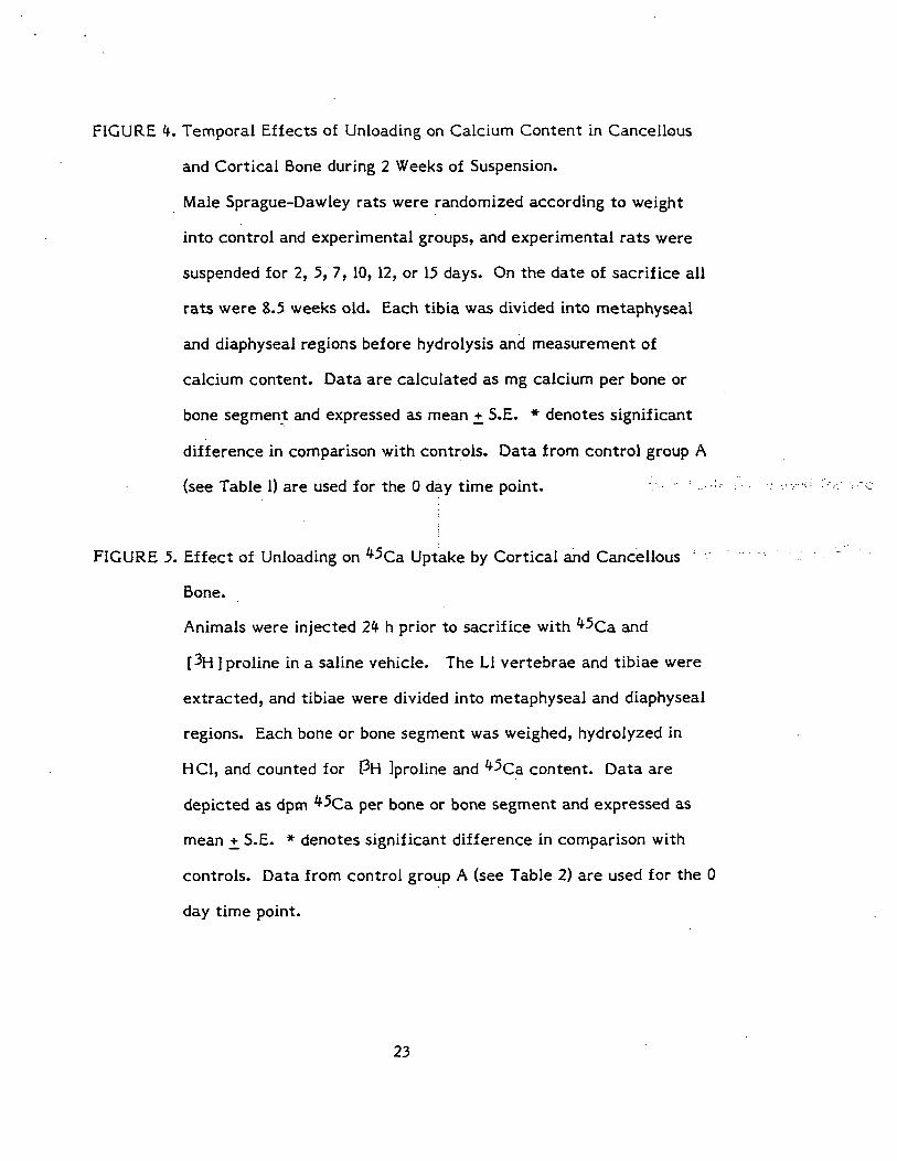
FIGURE 4. Temporal Effects of Unloading on Calcium Content in Cancellous
and Cortical Bone during 2 Weeks of Suspension.
Male Sprague-Dawley rats were randomized according to weight
into control and experimental groups, and experimental rats were
suspended for 2, 5, 7, 10, 12, or 15 days. On the date of sacrifice all
rats were 8.5 weeks old. Each tibia was divided into metaphyseal
and diaphyseai regions before hydrolysis and measurement of
calcium content. Data are calculated as mg calcium per bone or
bone segment and expressed as mean +, S.E. * denotes significant
difference in comparison with controls. Data from control group A
(see Table 1) are used for the 0 day time point. ;
FIGURE 5. Effect of Unloading on ^5Ca Uptake by Cortical and Cancellous
Bone.
Animals were injected 2k h prior to sacrifice with *^Ca and
[3H ] proline in a saline vehicle. The LI vertebrae and tibiae were
extracted, and tibiae were divided into metaphyseal and diaphyseai
regions. Each bone or bone segment was weighed, hydrolyzed in
HC1, and counted for PH Iproline and *^Ca content. Data are
depicted as dptn ^Ca per bone or bone segment and expressed as
mean +_ S.E. * denotes significant difference in comparison with
controls. Data from control group A (see Table 2) are used for the 0
day time point.
23

FIGURE 6. Effect of Unloading on Specific Activity of 45Ca in Bone.
The experimental protocol is described in the legend to Figure 5.
Data are calculated as dpm 45Ca per mg calcium and expressed as
mean +_ S.E. * denotes significant difference in comparison with
controls. Data from control group A (see Table 2) are used for the 0
day time point.
FIGURE 7. Effect of Unloading on [ 3H] Proline Uptake by Cortical and
Cancellous Bone.
The experimental protocol is described in the legend to Figure 5.
Data are calculated as dpm [3H Iproline per bone or bone segment
and expressed as mean +_ S.E. * denotes significant difference in
comparison with controls. Data from control group A (see Table 2)
are used for the 0 day time point.
FIGURE 8. Effect of Unloading on [ 3H] Proline Incorporation per mg Bone
Mass.
The experimental protocol is described in the legend to Figure 5.
Data are calculated as dpm [ 3H] proline per mg fat-free bone and
expressed as mean +_ S.E. * denotes significant difference in
comparison with controls. Data from control group A (see Table 2)
are used for the 0 day time point.

offv
V)>»CO
ORIGINAL PAGE ISOF POOR QUALITY
a i s s 3 ° °. —N 3 S 8
«) vO d- M
aa.ij 6|j\|

ri
aajj JBJ 6W

>. 01 ~ 03 «/*
1/9>•CO
Mi40
36
36
32
|Z8
12
Tibia
M36
32
2S
2*
20
* 16
12
Metaphysis , ControlMetaphysis , SuspendedDiaphysis, ControlDiaphysis, Suspended
32-
Humerus
21 21 21 2S
Days

<uou(/>3
WdQ
ORIGINAL PAGE iSBE POOR QUALITY
* *
t/t
EH

r-
V)a
CO
CO
ovO o o ff>
WdQ Hc
Ui
atvt3
CO
"SCO
B3 fiW/WdO

7.5
7.0
X 6.0
5.5
5.01
Mi£ 10.5
10.0
9.5
9.0
CO S.5
S-0
0 Tibia
5.0
f.5
•—• Metaphysis*-~* Diaphysis
3.5
2.5
2 5 7 1 0 1 2
Days of Suspension
15

Table 1. Effect of Suspensipn on Body Weight and Bone Mass
Groups Body mass Tibial <Ti;bial Humeral Humeral LI vertebra Cl vertebra(g) metaphysis diaphysis metaphysis diaphysis (mg) (mg)
(mg) '-.(mg:) , (mg) (mg)
•Experimental
2 days suspended 266 +_ 7 125.6 +_ 3.3 H4;0+_3.9 109.5 + 2.4 75.5 + 2.9 95.0 + 1.6 69.0 + 2.4
5 days suspended 246 + 8 126.9 +_ 2.9 110;;5,+_4.0 110.5+_4.0 74.9 +_ 5.2 86.0 +_ 2.4 73.0 +_ 0.4
Control Group A 256 +_ 3 124.3 +_ 4.0 116.0 +_ 3.2 111.0+_2.9 69.1+3.5 9].0+_1.6 68.0 + 0.8
Experimental
7 days suspended 254 +_ 5 104.2 +_ 3.3* 108.1+2.2 115.1 +_ 2.6 70.5 +_ 2.8 75.9 +.1.2* 63.3 + 1.6
10 days suspended 252+^7 96.7 + 3.8* 107.6 +_ 3.2 105.7 + 3.9 69.4 + 3.3 69.3 + 2.3* 62.3 + 1.6
Control Group B 262 +_ 3 117.9 +_ 4.4 111.1+2.6 111.1+2.2 74.1+2.8 92.2 + 3.2 65.6 +.1.6
Experimental
12 days suspended 244 + 3* 103.7+^3.0* 110.7 + 3.3* 116.5 +_ 2.3 62.7 + 2.2 75.7 + 1.6* 67.9 + 1.8
15 days suspended 245 + 7 97.1+_1.8* 98.7 + 1.7* 112.8 + 2.0 64.7 + 1.5 73.1+2.7* 66.0 + 2.3i
Control Group C 259 + 3 124.8 + 3.0 112.9 +_ 1.3 112.3 +.1.3 67.6 +_ 1.7 90.4 + 3.6. 69.4 +_ 0.9
Control rats were fed according to the mean food intake of the experimental groups. All rats were 8.5 weeks old onthe date of sacrifice. Body mass was determined on the date of sacrifice. The bones were removed and defatted.Humert a,nd tibiae were split into draphyseal and metaphyseal regions. All bones were then dried in a vacuum oven andweighed to obtain a total bone weight. Data are expressed as mean +_ S.E. * indicates P < 0.05 by Student's t_ testin comparison wrth corresponding control values.

Table 2. Incorporation of Ca and [ H] Proline into Bone of Control Rats
Controlgroup
Tibia!metaphysis
Tibialdiaphysis
LI vertebra
mq Ca per total bone
Group A
Group B
Group C
45Ca X 103
Group A
Group B
Group C45Ca/mg Ca
Group A
Group 8
Group C
[3H] Proli
Group A
Group B
Group C
f3H] Proli
Group A
Group B
Group C
20.4 +2.1
19.6 +1.8
19.2 +1.645(dpm Ca per total
216 + 16
198+12
202 + 15
X 102 (dpm"45Ca per
106 + 6
111 +28
106 + 9
ne X 103 (dpm 3H per
116 + 20
107 +_13
109 +_8
24.8 +_ 2.0
22.9 +1.7
23.7 +1.1
bone)
60.0 +6.6
53.2 +_6.1
57.2 +4.2
mg Ca)
23.9+1.5
23.3 +_3.1
24.2 +2.3
total bone)
33.6 + 5.2
32.3 + 2.6
35.2 +_6.0
16.7 +0.7
17.1 +2.1
16.7 +_2.0
98.4 + 2.9
92.2 +'8.6
96.5+9.6
59.5 +_ 1.6 - ••
f:,?™-. 3 54.4 +_ 7.3 in i- ?P
''••••• -•:••• ''- 68.6 +_6.8 ''•" :. '"'
58.5 +4.2
54.9.+ 4.5
60.0 +_6.3
ne/mg bone (dpm H per mg defatted bone)
917 +86
911 +_ 122
891 +_ 70
289 + 45
290 + 26
318 _+ 53
597 + 68
636 + 21
676 + 72
Control groups were fed according to the mean food intake of the experimentalgroups. Group A was the control for rats suspended 2 or 5 days; Group B, forrats suspended 7 or 10 days; and Group C, for rats suspended 12 or 15 days.All rats were 8.5 weeks old at sacrifice. Twenty-four hours prior to sacrifice,animals were injected intraperitoneally with 10 pCi [^H ] proline per 100 g bodyweight and 1 yCi 4 Ca per 100 g body weight. Bones were removed and defatted,and tibiae were split into diaphyseal and metaphyseal regions. All bones werethen weighed. Following hydrolysis in HC1 , total calcium and ^H and 45Ca contentwere determined. Data are expressed as mean + S.D.

Table 3; Bone Formation Rate at the Tibiofibular Junction during Initial(Days 2-3) and Later (Days 8-13) Periods of Suspension.
Bone Formation Rate(mm^/day)
Days 2-8 Days 8-13
Suspended"(N-4).
Control(N=4)
0
0
.019
.038
±°
±°
.003*
.003
0
0
.030
.041
+ 0
±°
.005
.005
*t
Rats were suspended at 6.5 weeks of age and injected subcutaneously withthree tetracycline labels on days 2, 8, and 13, and then sacrificed onday 14. Bone formation rate was calculated by dividing the area of newlyformed bone by the time interval between the two labels. * indicatesP < 0.05.compared to control values; t indicates P < 0.05 compared, toinitial period. Data are expressed as mean + S.D. :;;•;;.;.,! ;>::; ,....;, •].„[...