Embed Size (px)
Citation preview
The Temporal Lobe
& Limbic System
John R. Hesselink, M.D.
Department of Radiology
University of California
San Diego
The Limbic System
The limbic lobe & its many connections
A set of 3 Arcs containing both gray
& white matter
Phylogenetically, a primitive part of brain
Central role in memory, learning, emotion
neuroendocrine function & autonomic activities
Diseases: Epilepsy, congenital anomalies,
dementias & psychiatric disorders
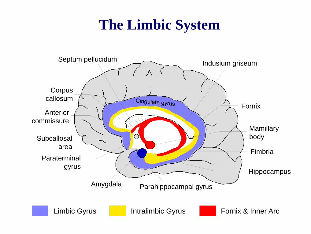
Limbic Gyrus Intralimbic Gyrus Fornix & Inner Arc
Indusium griseum Septum pellucidum
Corpus
callosum
Mamillary
body
Paraterminal
gyrus
Subcallosal
area
Hippocampus
Fimbria
Parahippocampal gyrus Amygdala
Fornix Anterior
commissure
The Limbic System
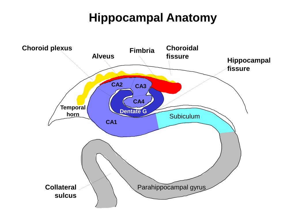
Choroid plexus
Hippocampal
fissure
Temporal
horn
Alveus Fimbria
CA4
CA3 CA2
Subiculum
Parahippocampal gyrus
Choroidal
fissure
CA1
Hippocampal Anatomy
Dentate G
Collateral
sulcus
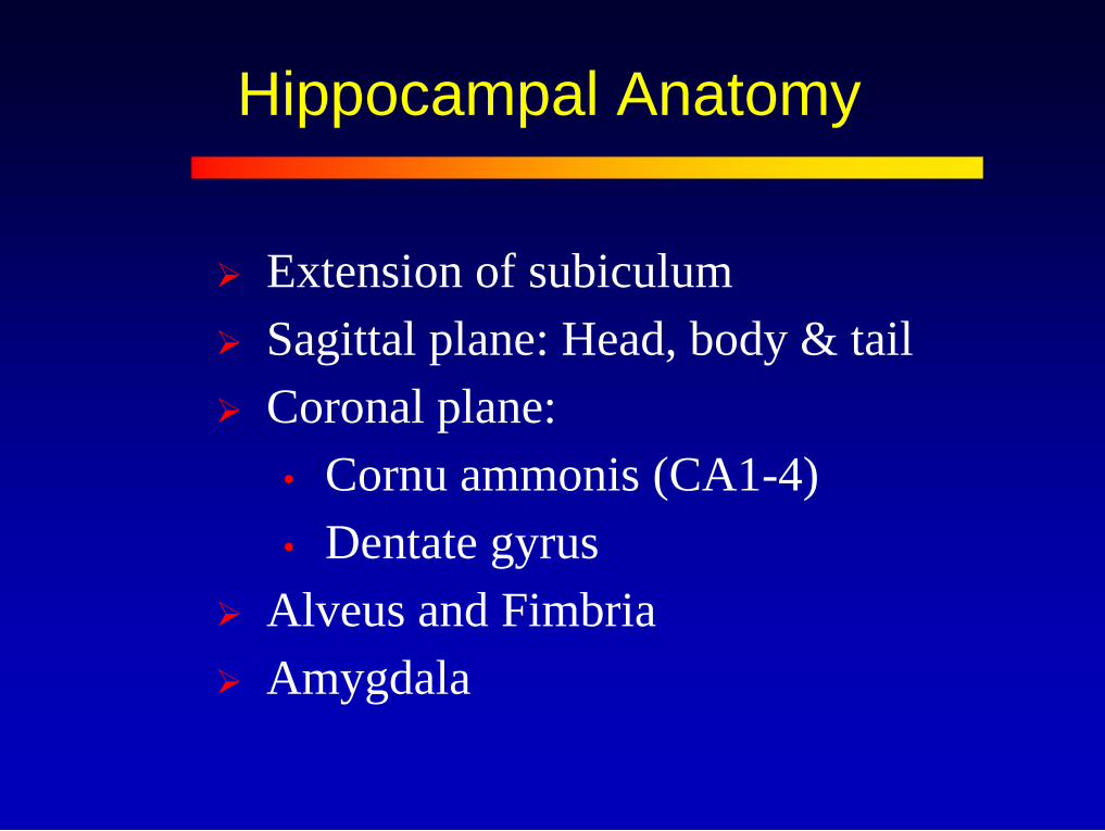
Hippocampal Anatomy
Extension of subiculum
Sagittal plane: Head, body & tail
Coronal plane:
• Cornu ammonis (CA1-4)
• Dentate gyrus
Alveus and Fimbria
Amygdala
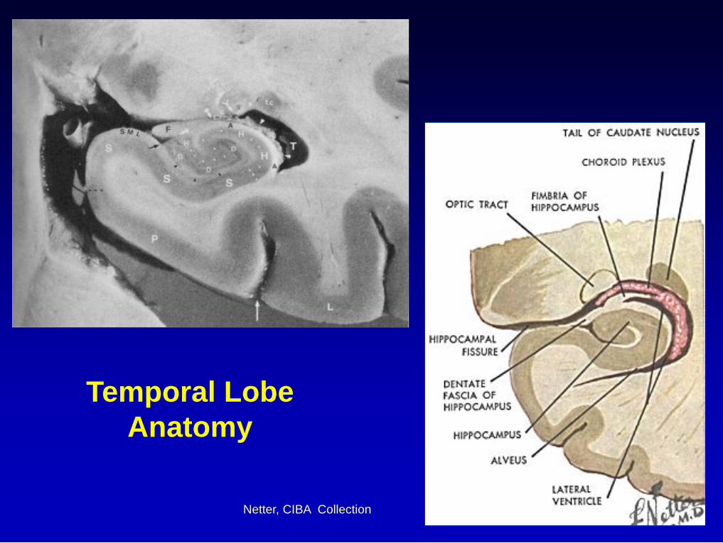
Temporal Lobe
Anatomy
Netter, CIBA Collection
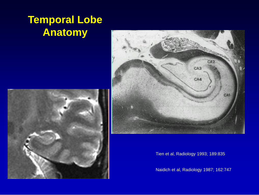
Temporal Lobe
Anatomy
Tien et al, Radiology 1993; 189:835
Naidich et al, Radiology 1987; 162:747
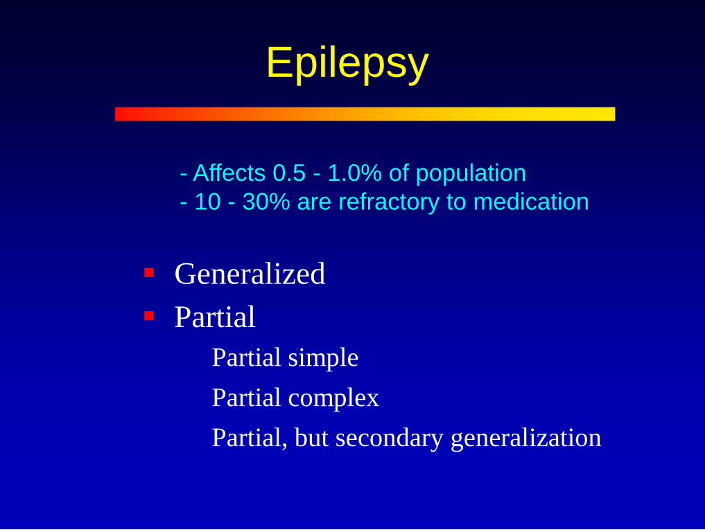
Epilepsy
Generalized
Partial
Partial simple
Partial complex
Partial, but secondary generalization
- Affects 0.5 - 1.0% of population
- 10 - 30% are refractory to medication
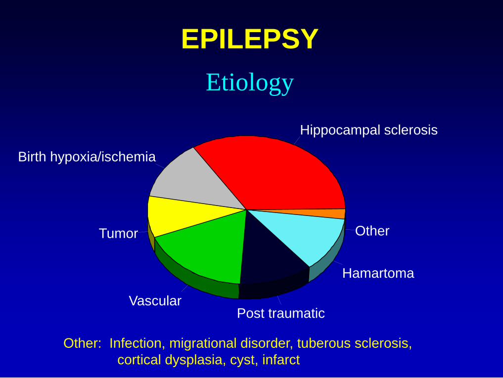
EPILEPSY
Etiology
Hippocampal sclerosis
Birth hypoxia/ischemia
Tumor
Vascular Post traumatic
Hamartoma
Other
Other: Infection, migrational disorder, tuberous sclerosis,
cortical dysplasia, cyst, infarct
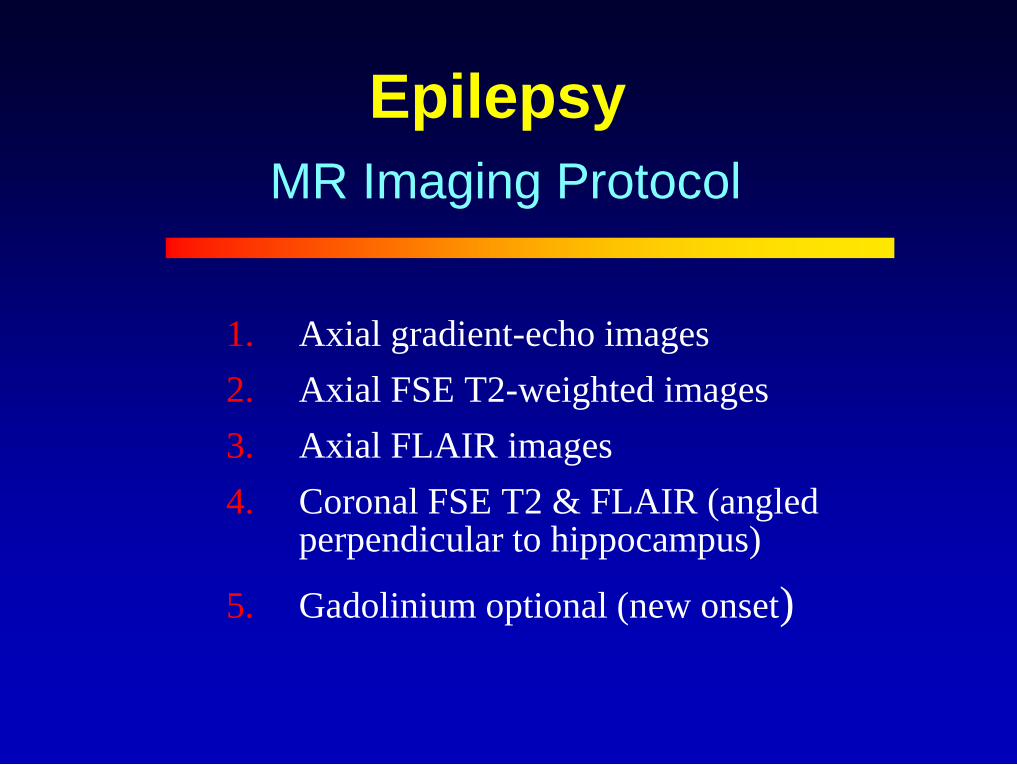
Epilepsy
1. Axial gradient-echo images
2. Axial FSE T2-weighted images
3. Axial FLAIR images
4. Coronal FSE T2 & FLAIR (angled perpendicular to hippocampus)
5. Gadolinium optional (new onset)
MR Imaging Protocol
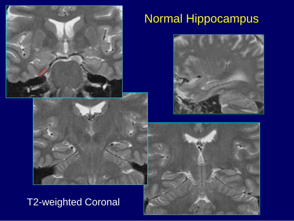
Normal Hippocampus
T2-weighted Coronal
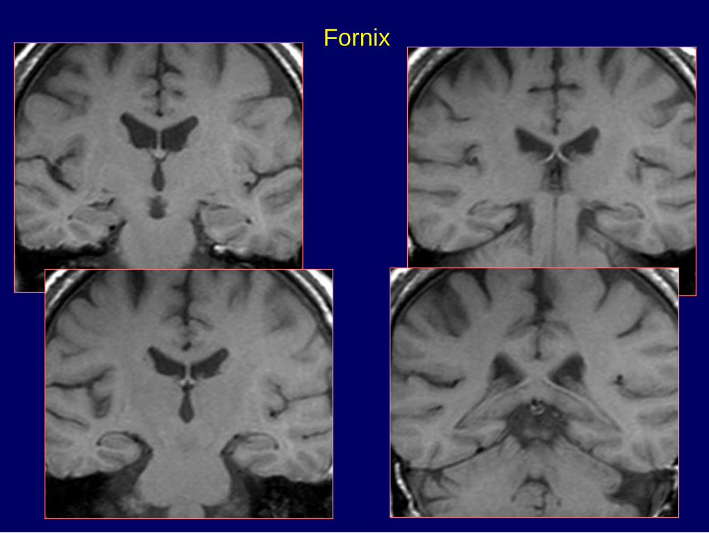
Fornix

Hippocampal Sclerosis
30 - 50% neuronal depletion
Most prominent in CA1 region of
hippocampal body
Loss of inhibitory interneurons
Synaptic reorganization
History of febrile seizures
Temporal Lobe Epilepsy
Hippocampal atrophy
Hyperintensity on T2 images
Temporal horn dilatation
Temporal lobe atrophy
MR Findings
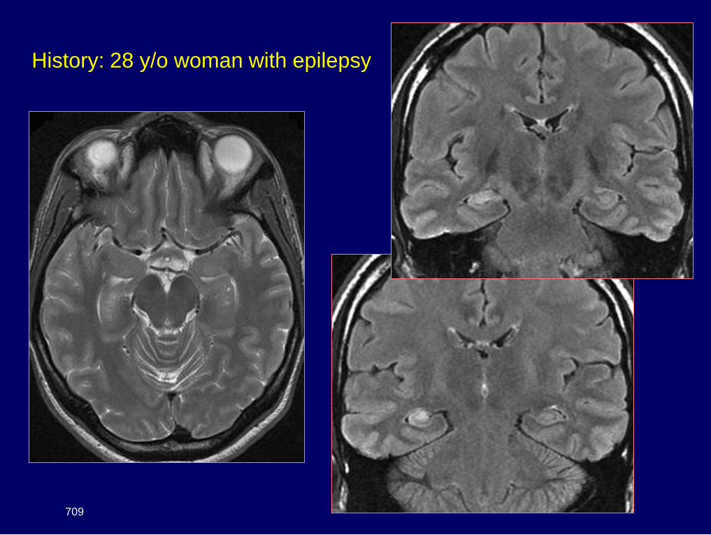
History: 28 y/o woman with epilepsy
709
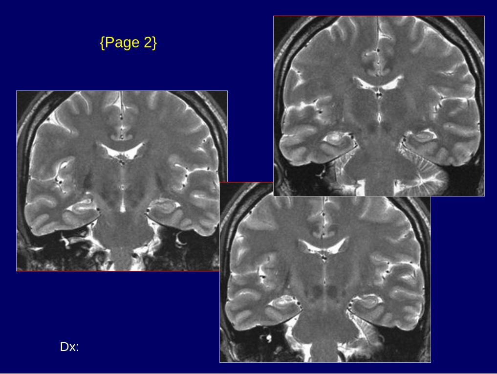
Dx: Hippocampal sclerosis
{Page 2}
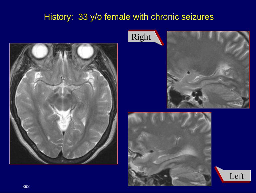
History: 33 y/o female with chronic seizures
392
Right
Left
Dx: Hippocampal sclerosis
{Page 2}
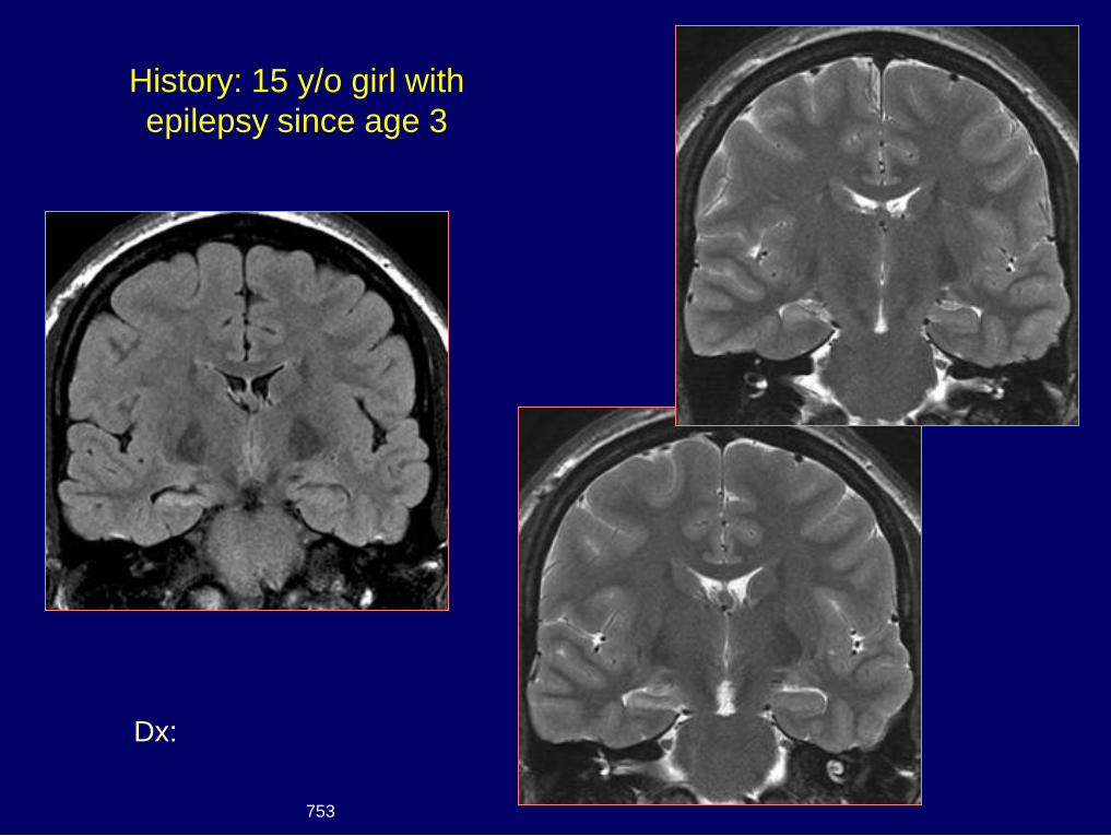
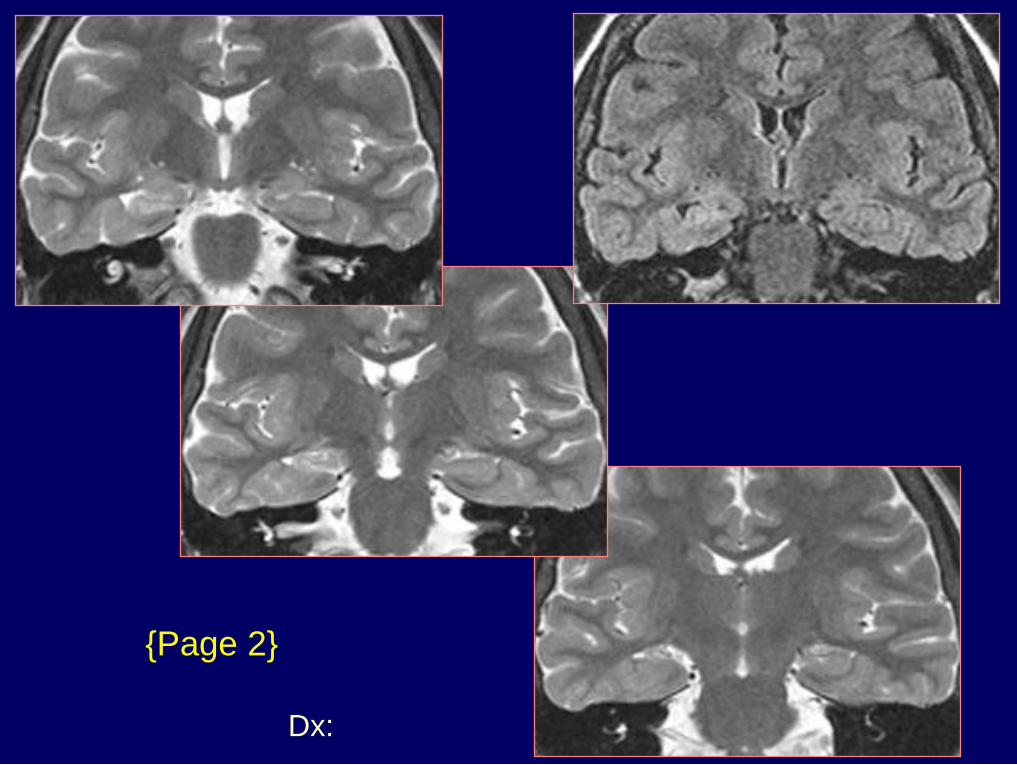
Dx: Hippocampal sclerosis
History: 15 y/o girl with
epilepsy since age 3
753
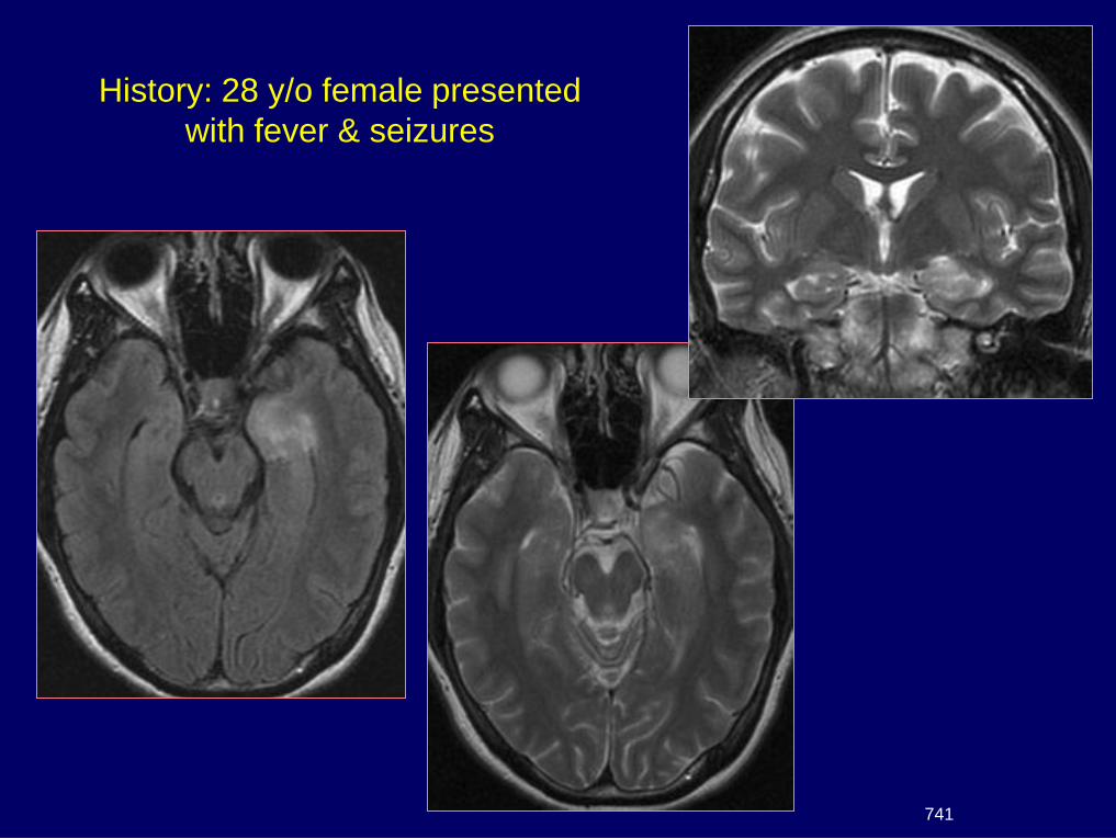
History: 28 y/o female presented
with fever & seizures
741
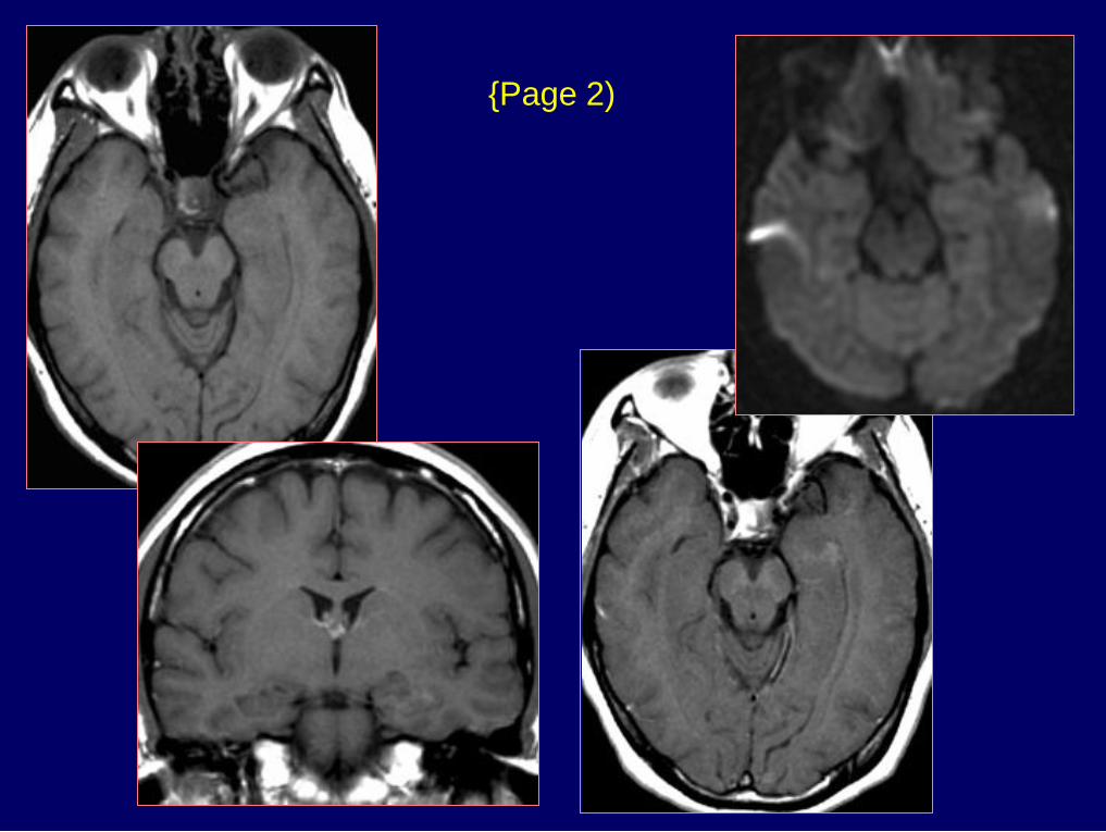
{Page 2)
Dx: HSV encephalitis - MTS
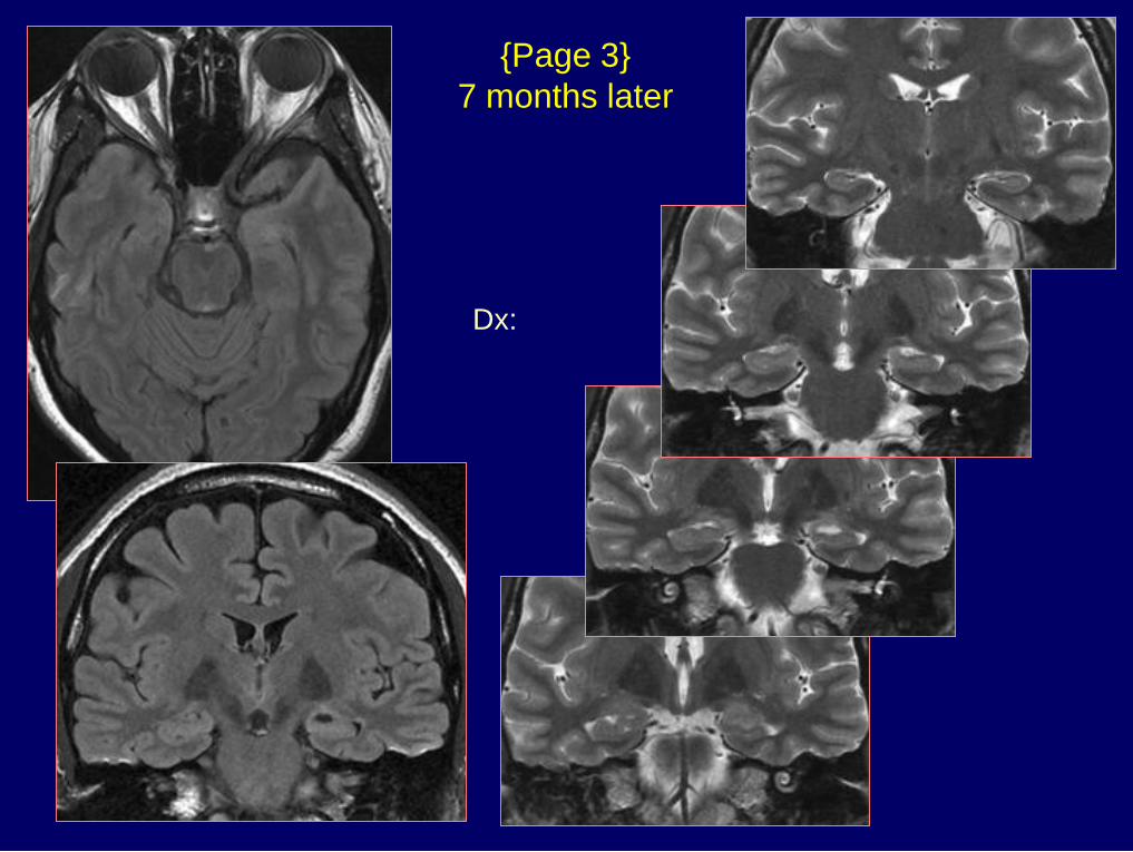
{Page 3}
7 months later
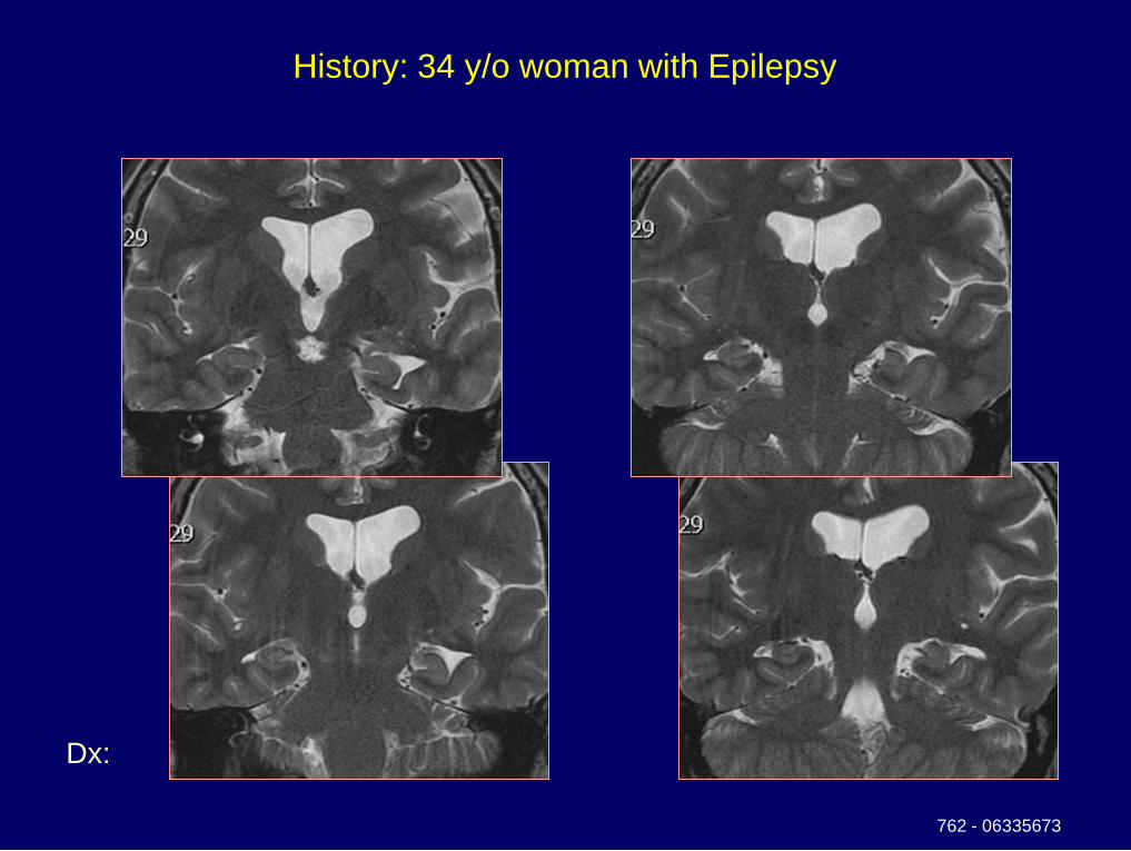
Dx: Malrotation hippocampus
Costello’s syndrome
History: 34 y/o woman with Epilepsy
762 - 06335673
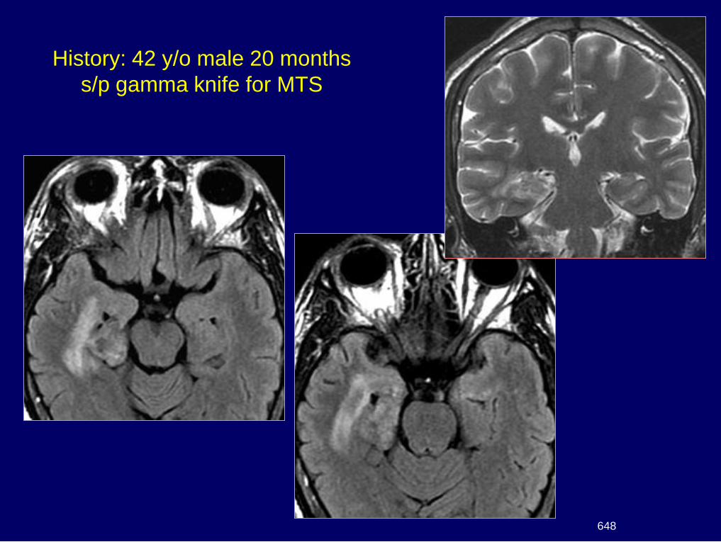
History: 42 y/o male 20 months
s/p gamma knife for MTS
648
Dx: MTS – s/p gamma knife –
Decreased seizures
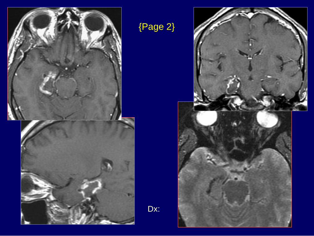
{Page 2}
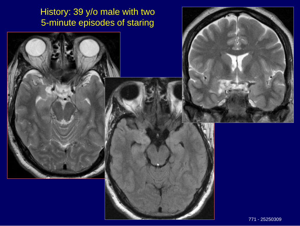
History: 39 y/o male with two
5-minute episodes of staring
771 - 25250309
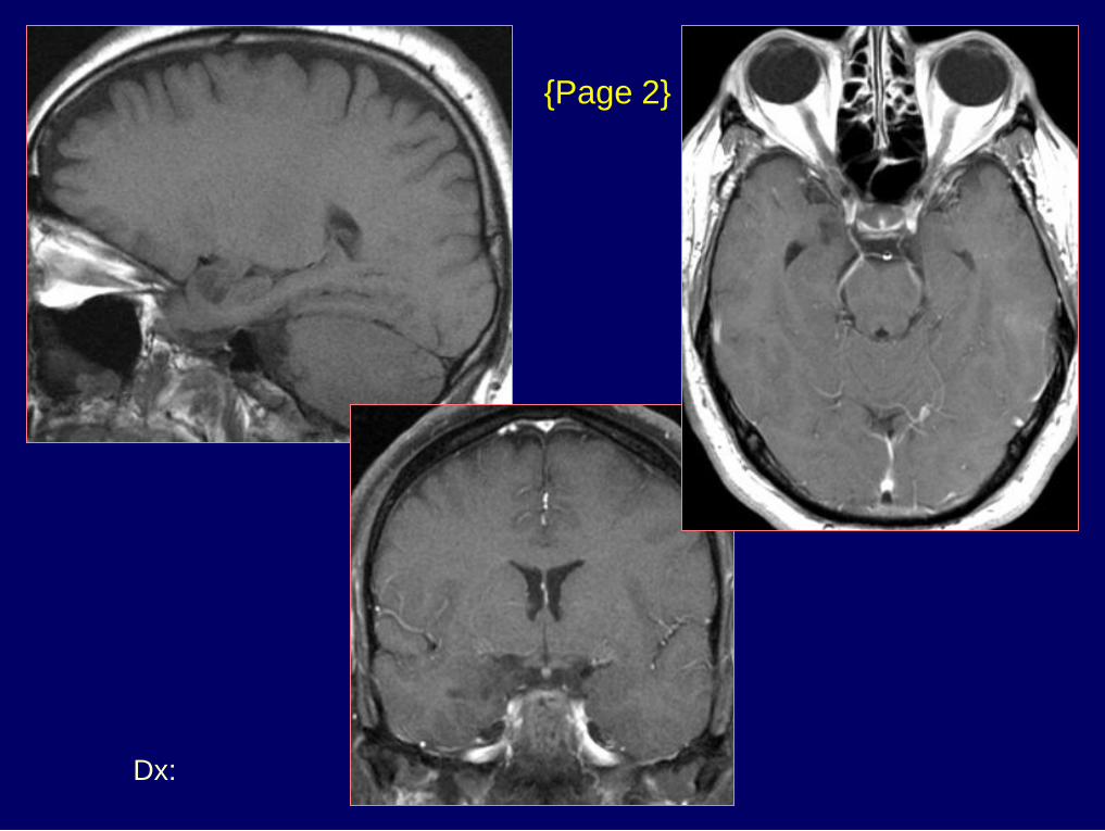
Dx: Cortical dysplasia & DNET
{Page 2}
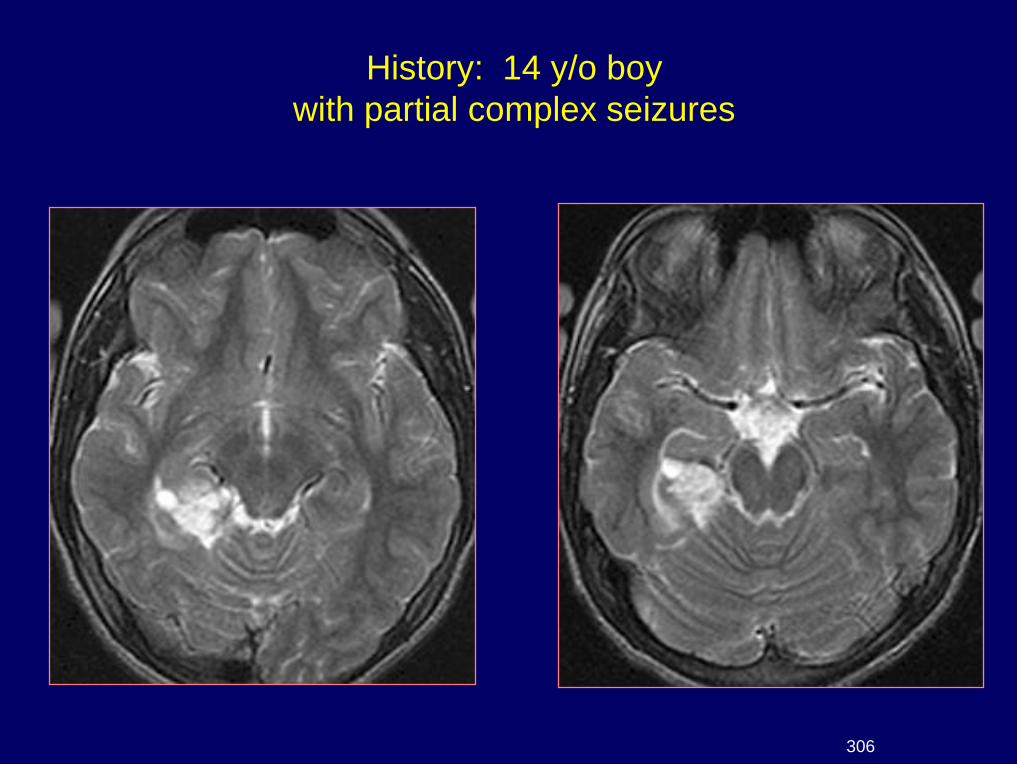
History: 14 y/o boy
with partial complex seizures
306
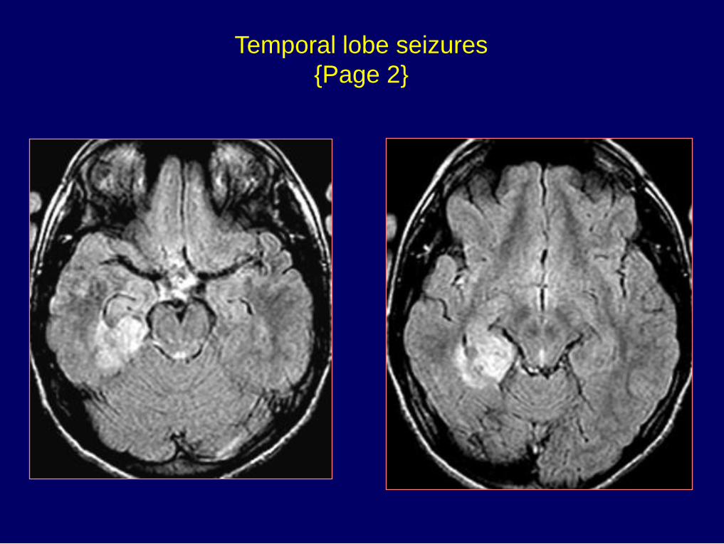
Temporal lobe seizures
{Page 2}
Dx: Ganglioglioma
{Page 3}
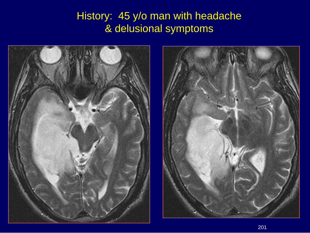
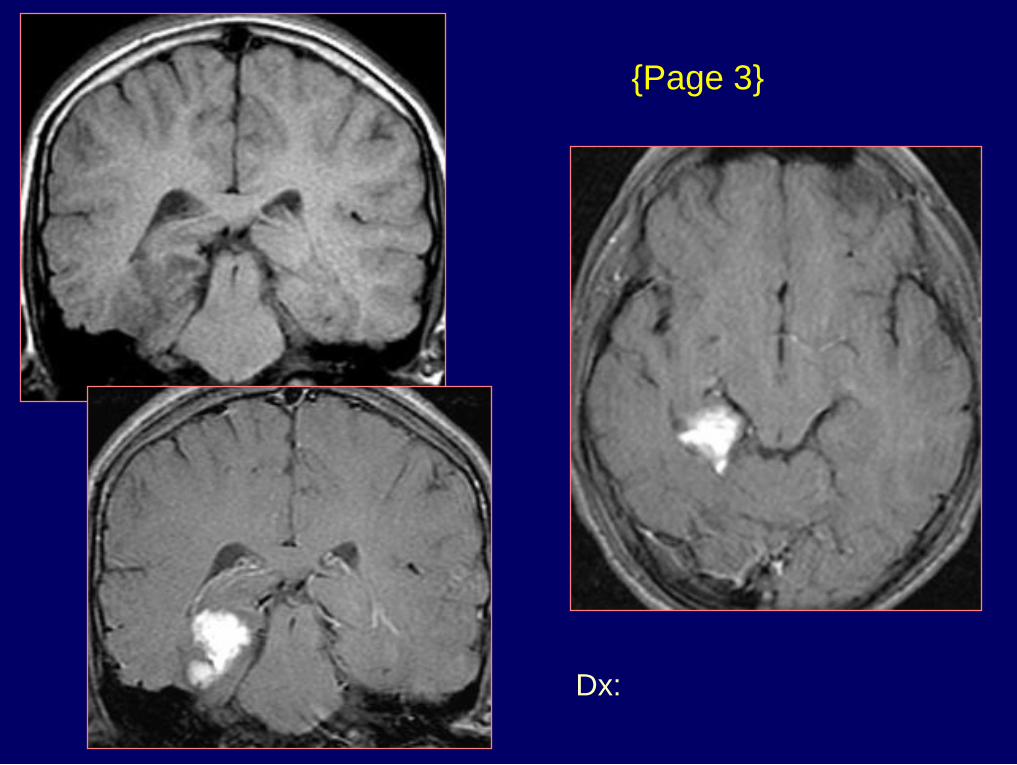
History: 45 y/o man with headache
& delusional symptoms
201
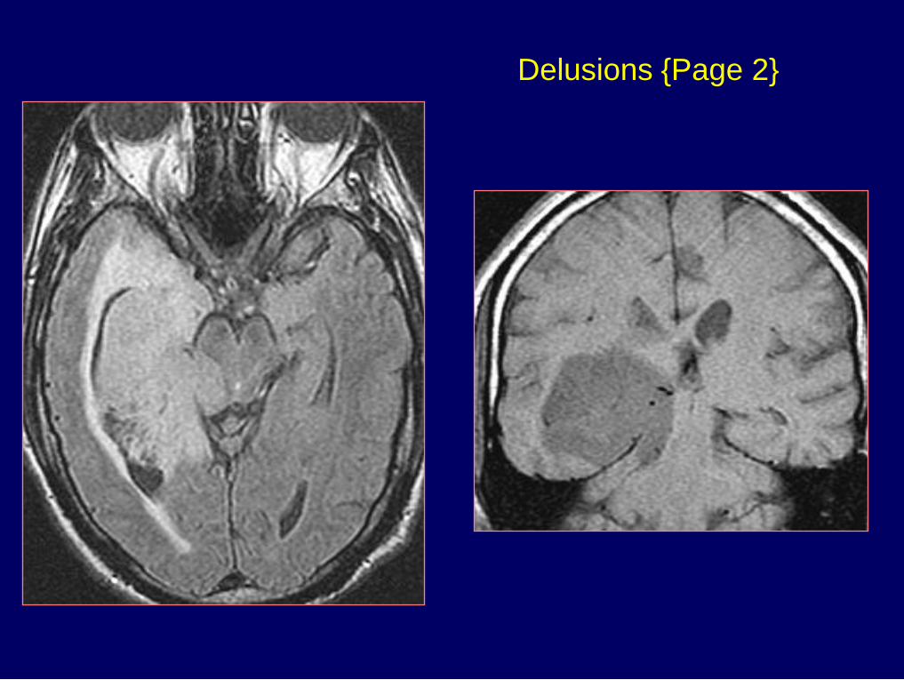
Delusions {Page 2}
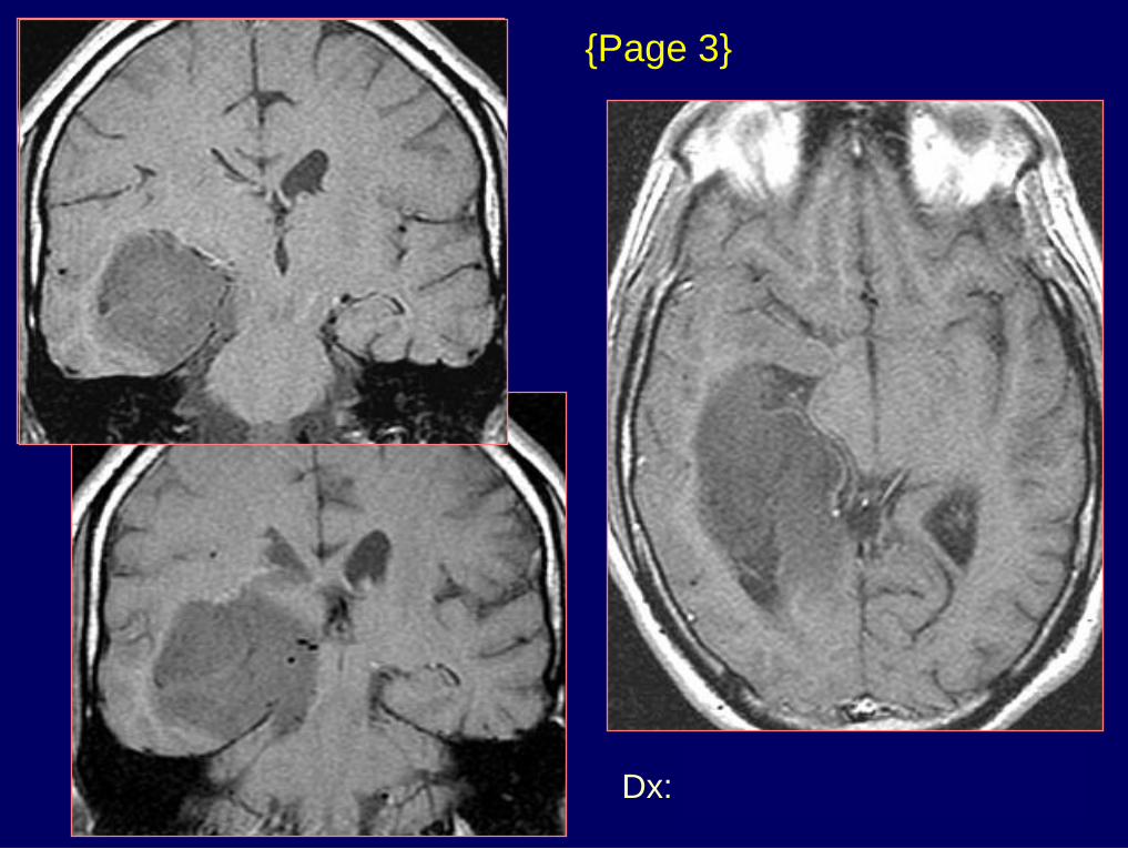
Dx: Astrocytoma (Grade 2-3)
{Page 3}
Dx: Arachnoid cyst in the
choroidal fissure
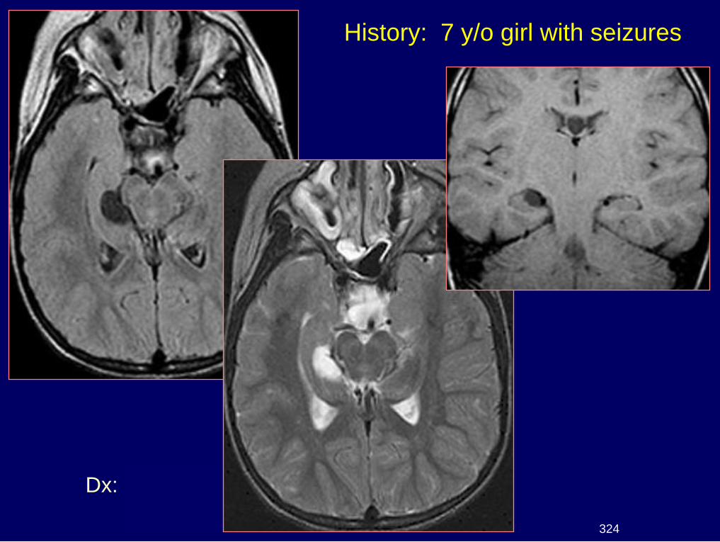
History: 7 y/o girl with seizures
324
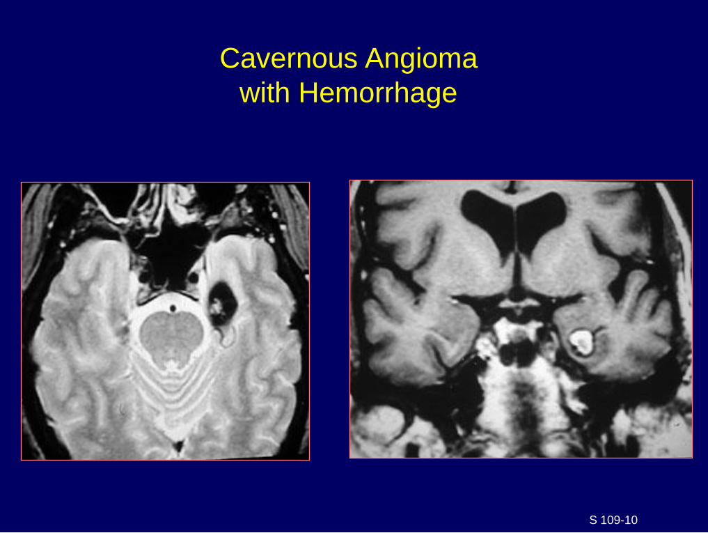
Cavernous Angioma
with Hemorrhage
S 109-10
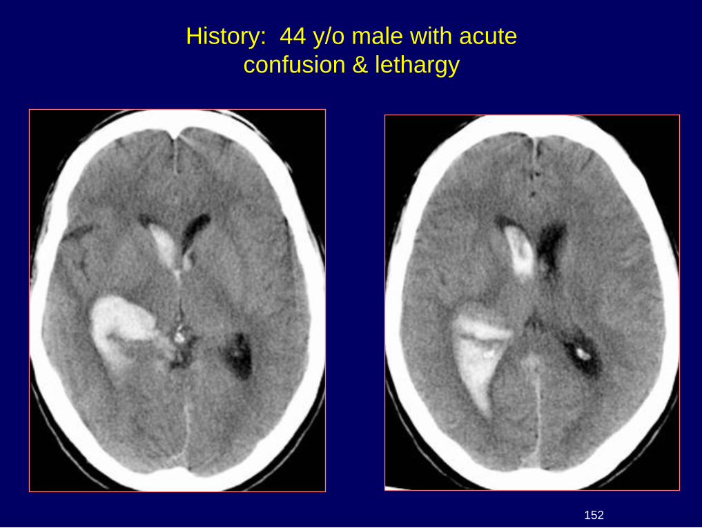
History: 44 y/o male with acute
confusion & lethargy
152
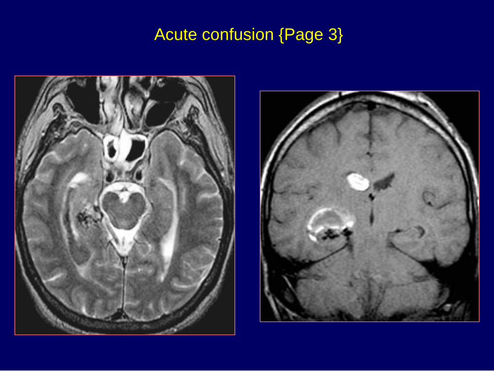
Acute confusion {Page 3}
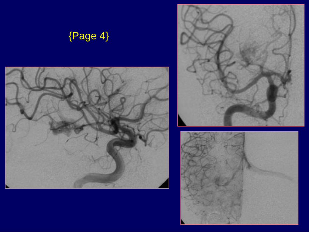
{Page 4}
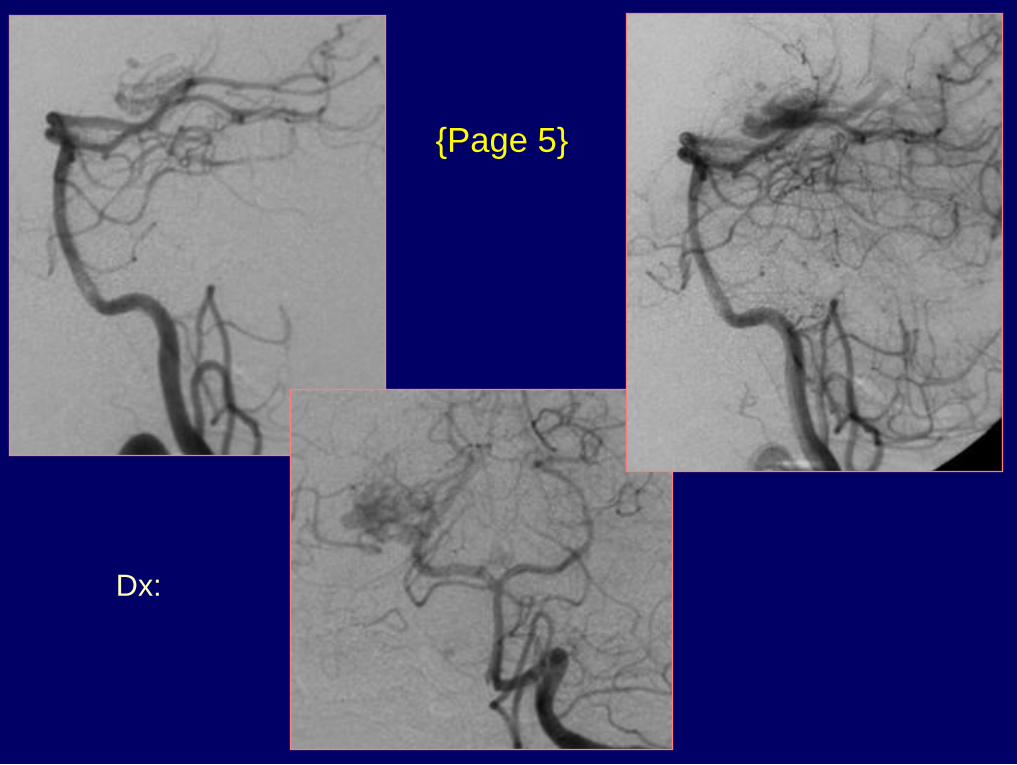
Dx: Choroid plexus / ependymal AVM -
Ventricular + thalamic hemorrhage
{Page 5}
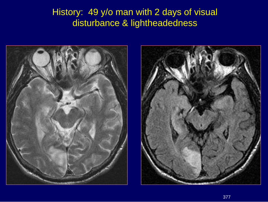
History: 49 y/o man with 2 days of visual
disturbance & lightheadedness
377
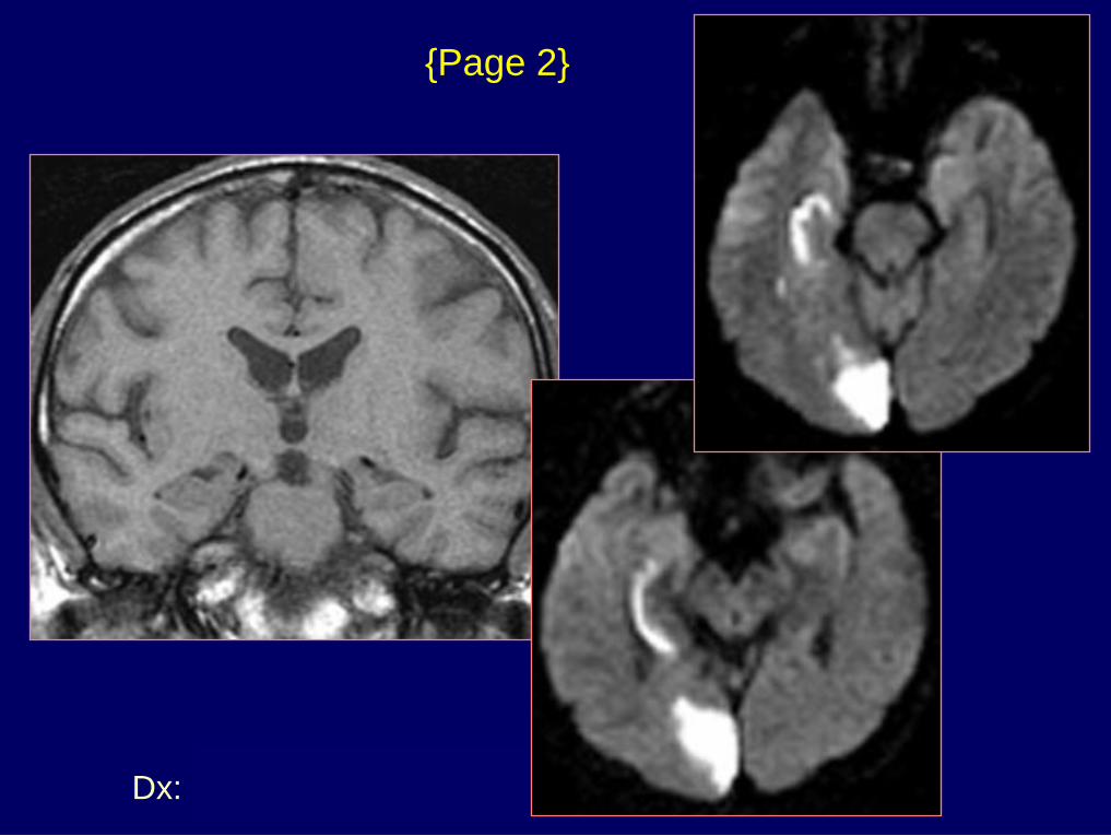
Dx: Hippocampal infarct
{Page 2}
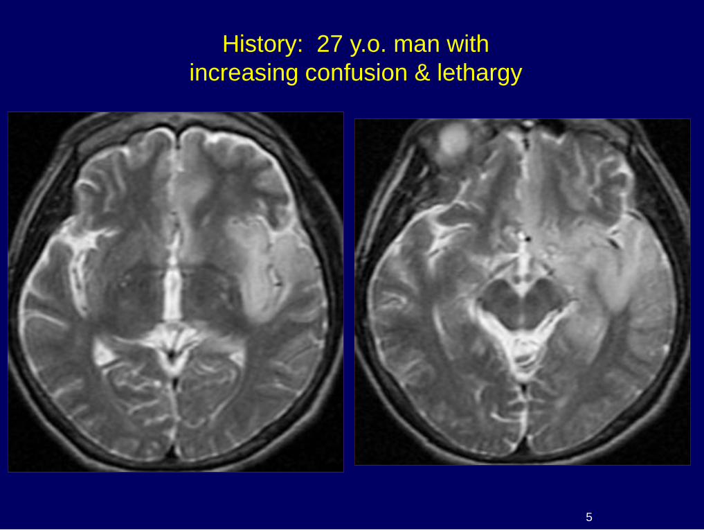
History: 27 y.o. man with
increasing confusion & lethargy
5
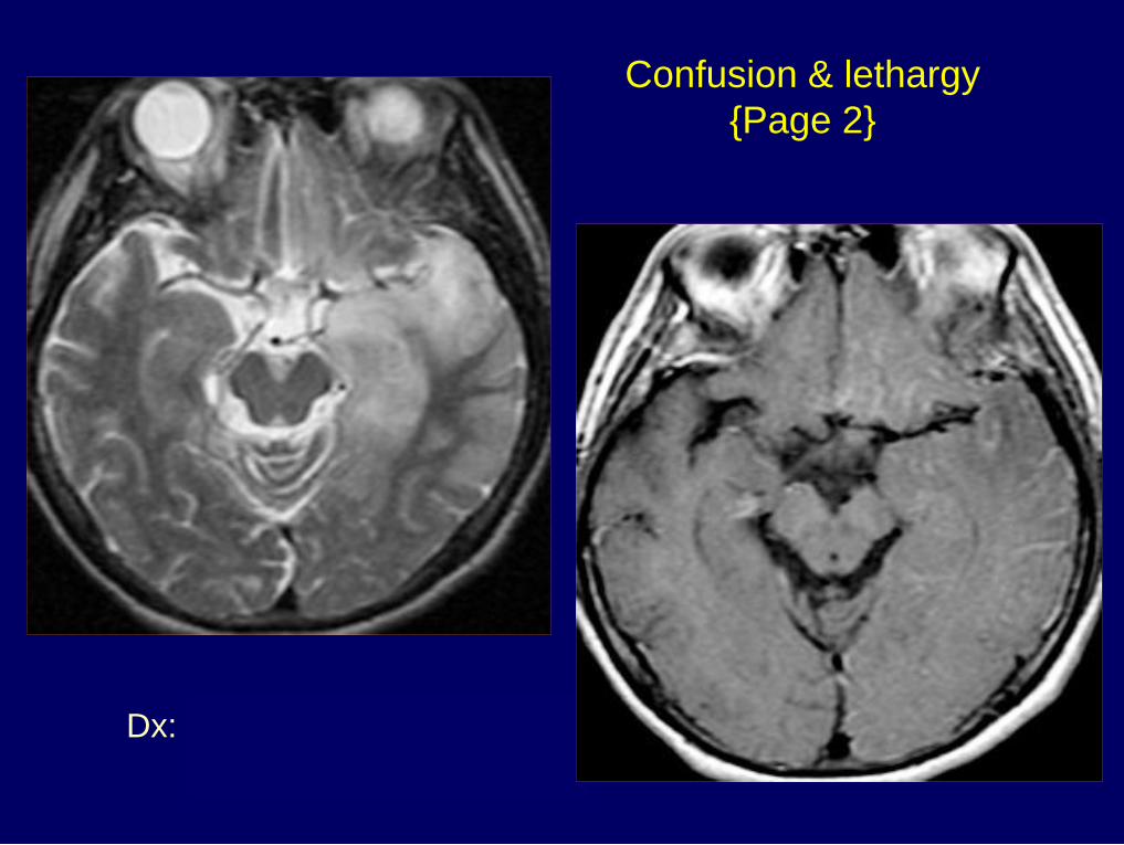
Dx: Herpes simplex
encephalitis
Confusion & lethargy
{Page 2}
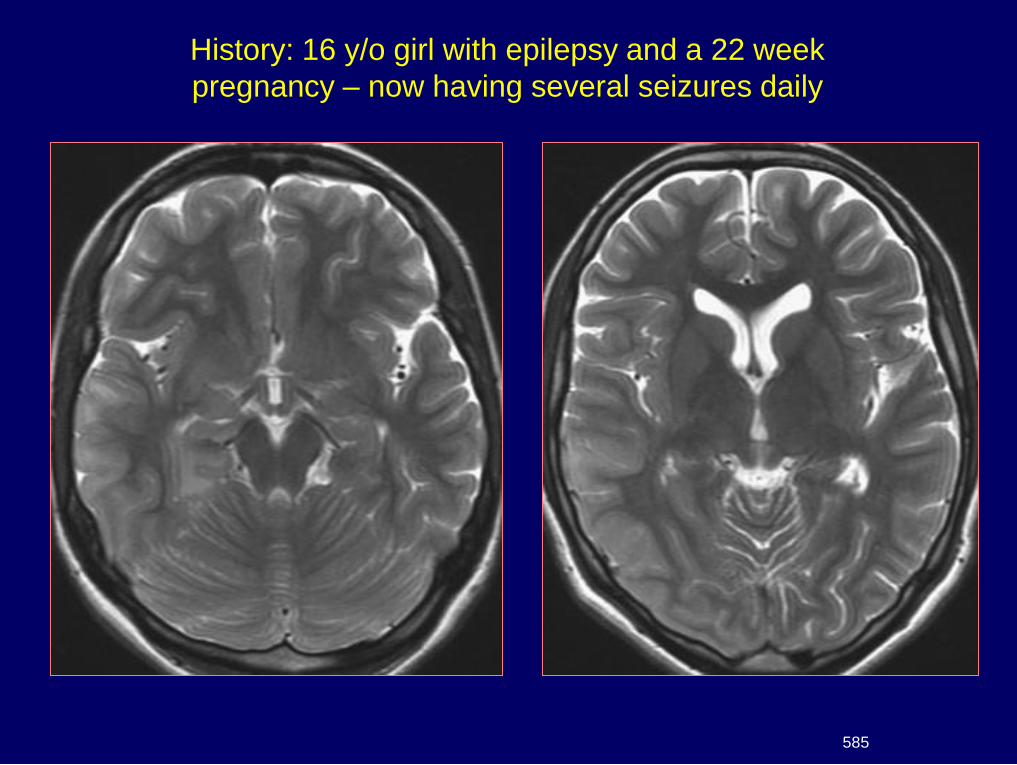
History: 16 y/o girl with epilepsy and a 22 week
pregnancy – now having several seizures daily
585
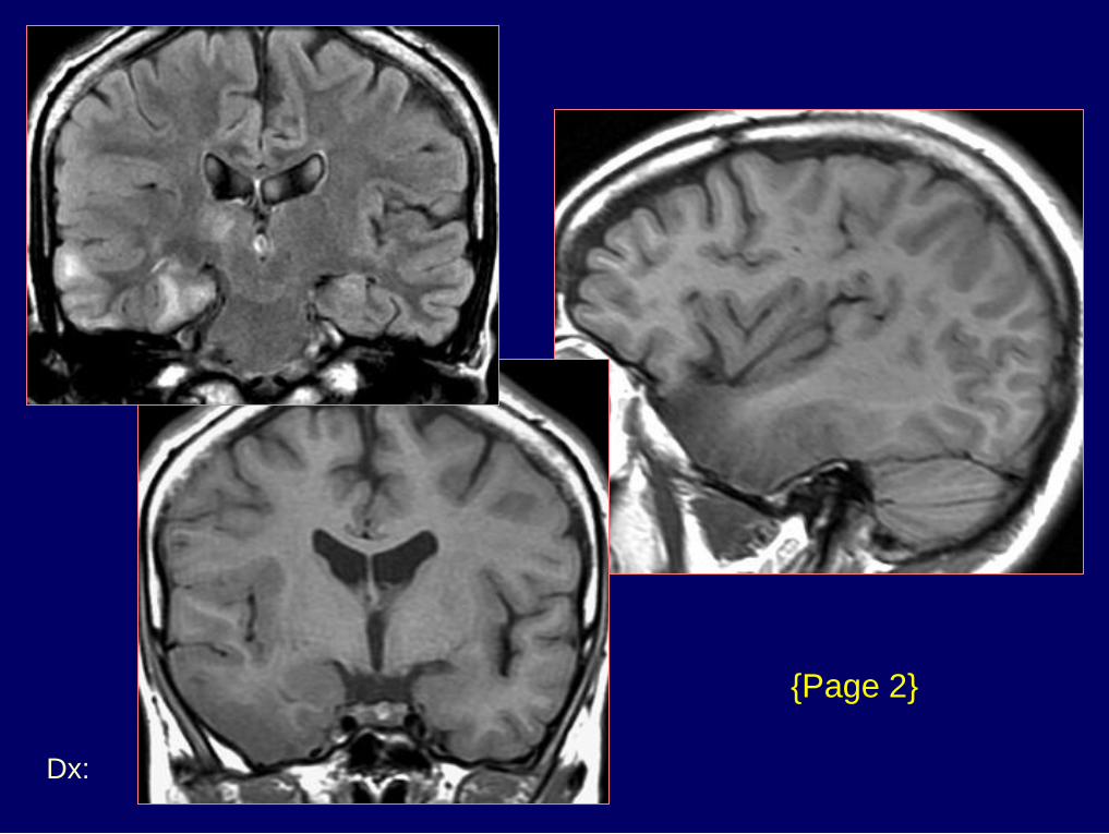
Dx: Seizure-related cortical edema
{Page 2}
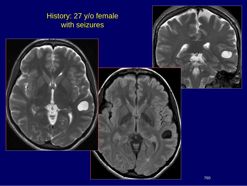
760
History: 27 y/o female
with seizures
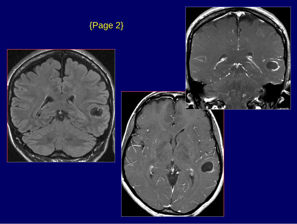
{Page 2}
Dx: Cysticercosis – colloidal phase
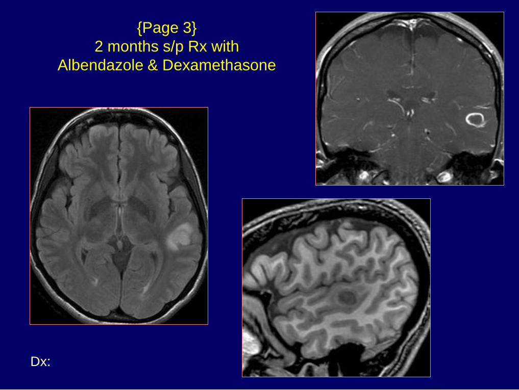
{Page 3}
2 months s/p Rx with
Albendazole & Dexamethasone
Dx: TB meningitis
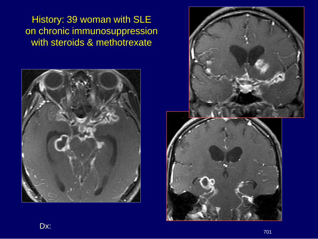
History: 39 woman with SLE
on chronic immunosuppression
with steroids & methotrexate
701

Pachygyria / Lissencephaly
B 10
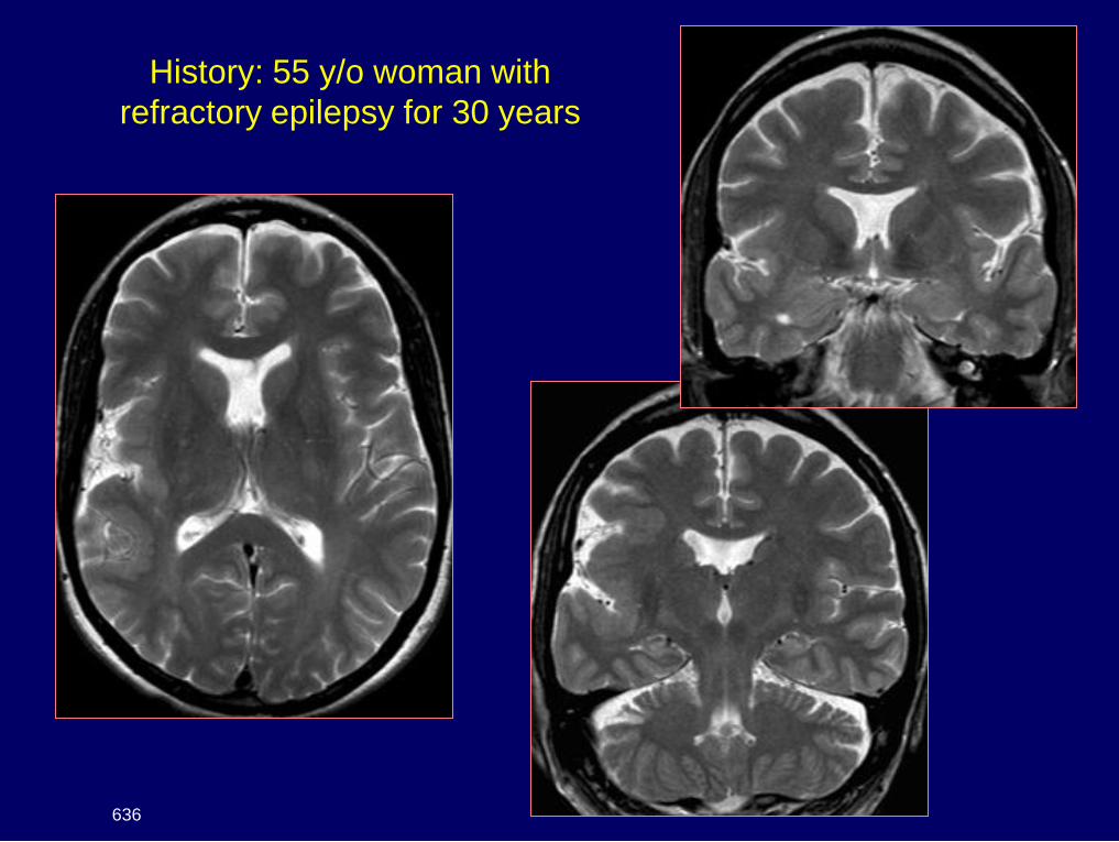
History: 55 y/o woman with
refractory epilepsy for 30 years
636
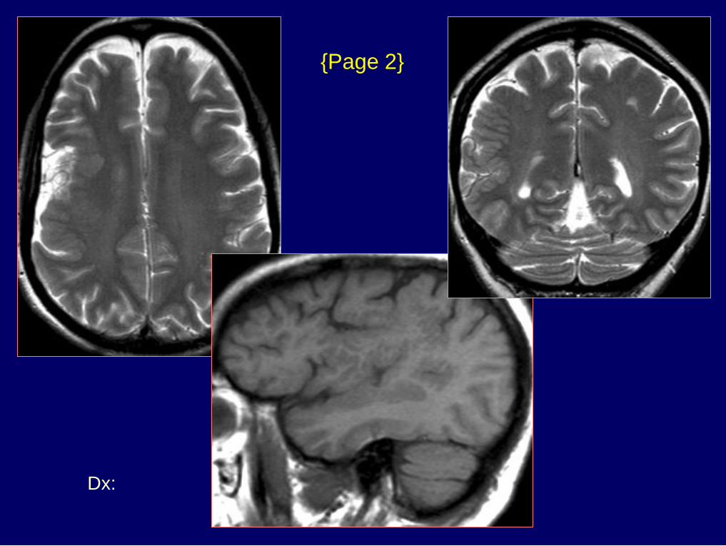
Dx: Polymicrogyria & septo-
optic dysplasia
{Page 2}
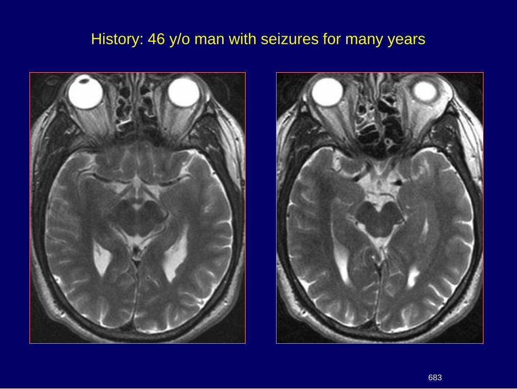
History: 46 y/o man with seizures for many years
683
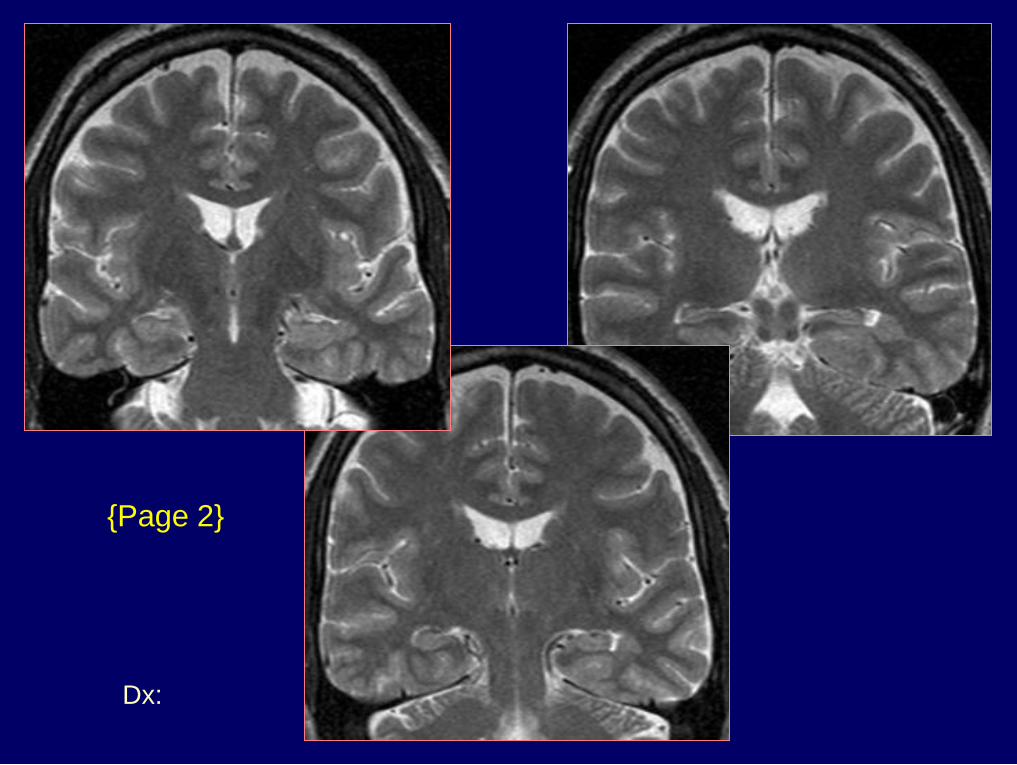
Dx: Heterotopic gray matter
{Page 2}
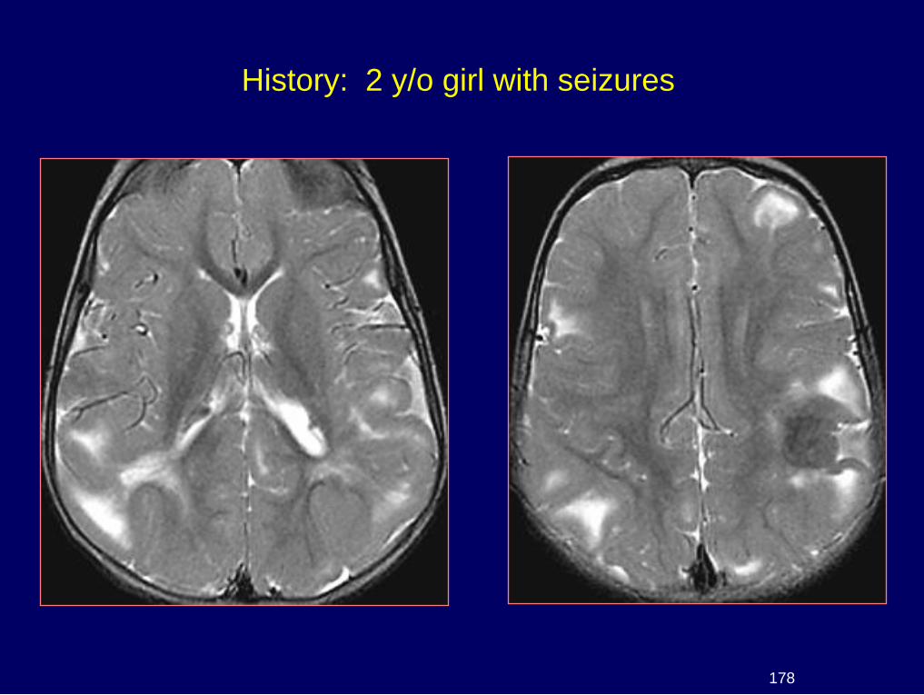
History: 2 y/o girl with seizures
178
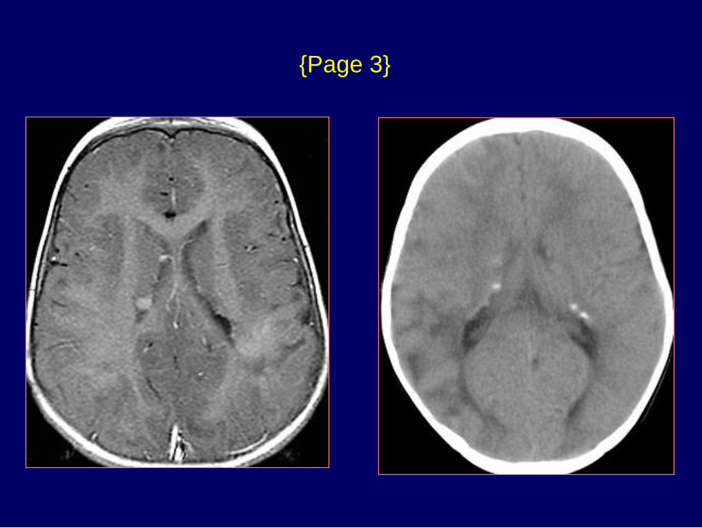
{Page 3}
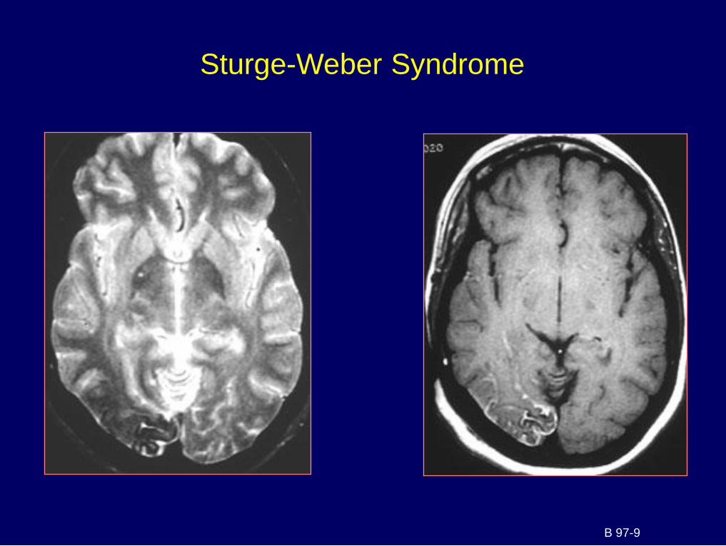
Sturge-Weber Syndrome
B 97-9

The Limbic System
Developmental abnormalities
Limbic encephalitis
Dementia
Neuropsychiatric disorders
Related Diseases
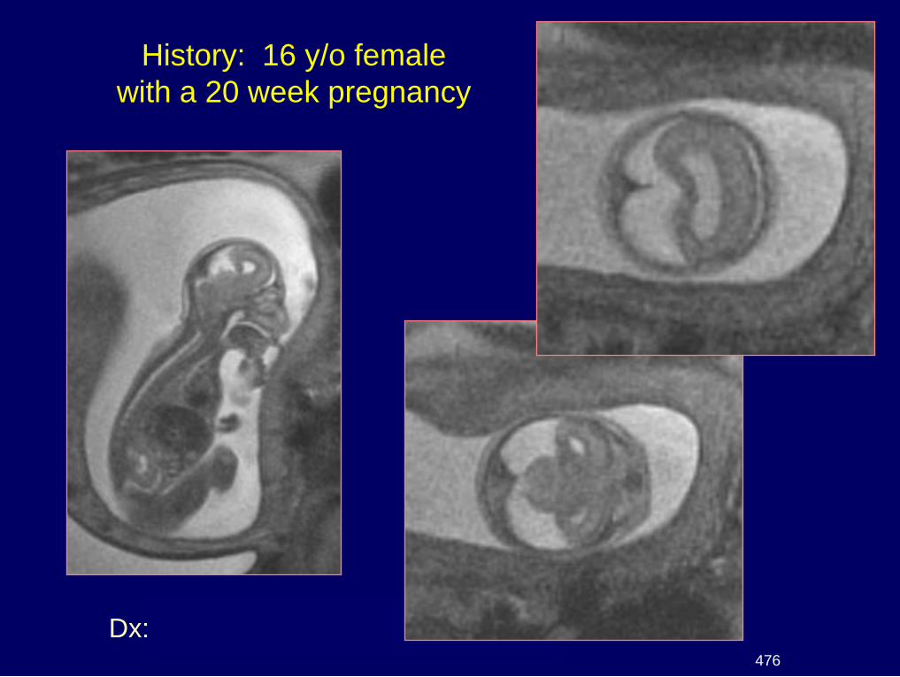
Dx: Alobar holoprosencephaly
History: 16 y/o female
with a 20 week pregnancy
476
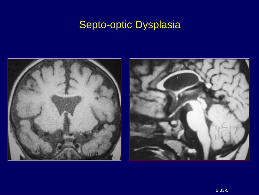
Septo-optic Dysplasia
B 33-5
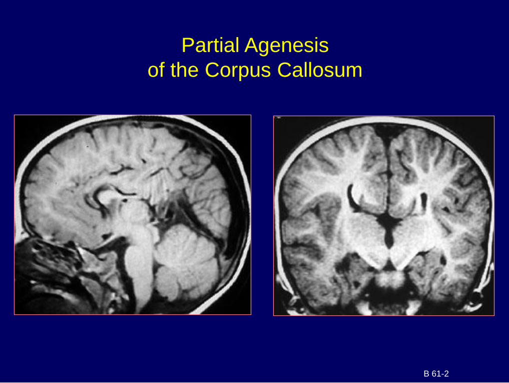
Partial Agenesis
of the Corpus Callosum
B 61-2
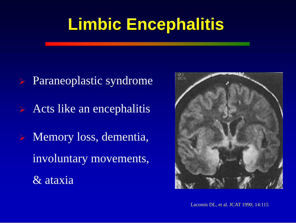
Limbic Encephalitis
Paraneoplastic syndrome
Acts like an encephalitis
Memory loss, dementia,
involuntary movements,
& ataxia
Lacomis DL, et al. JCAT 1990; 14:115
Alzheimer’s Disease
Severe memory deficits
Path: senile plaques & neurofibrillary tangles
Hypometabolism & decreased CBF in parietal &
temporal lobes
Deficiency of cholinergic-producing neurons in the
subcallosal nuclei
Atrophy hippocampus, amygdala & entorhinal cortex
• 10 times faster than normal aging
Imaging: MRI, PET, SPECT
- MRS: Elevated myo-inositol
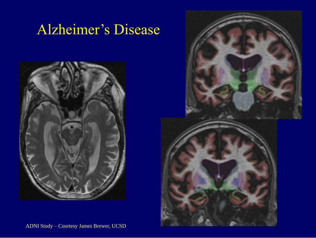
Alzheimer’s Disease
ADNI Study – Courtesy James Brewer, UCSD
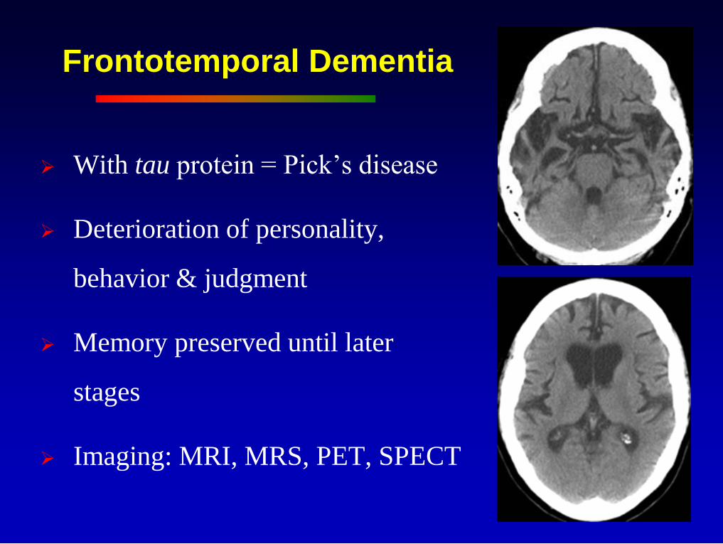
Frontotemporal Dementia
With tau protein = Pick’s disease
Deterioration of personality,
behavior & judgment
Memory preserved until later
stages
Imaging: MRI, MRS, PET, SPECT
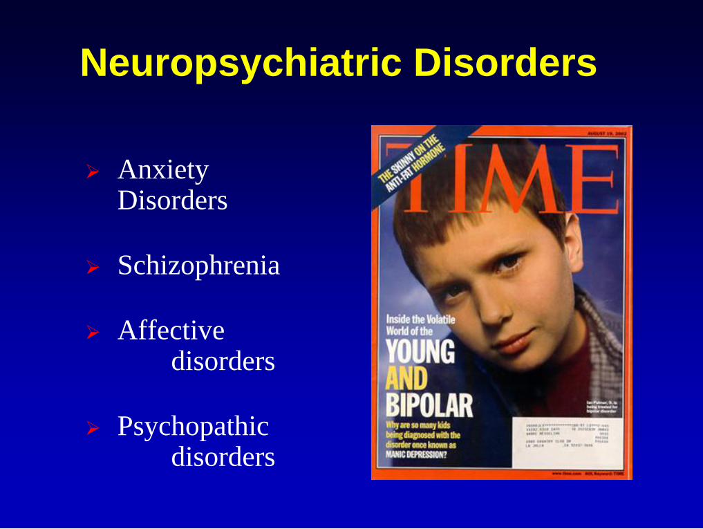
Neuropsychiatric Disorders
Anxiety Disorders
Schizophrenia
Affective disorders
Psychopathic disorders
Anxiety Disorders
Panic, phobias, stress disorders
Amygdala attaches emotional significance to sensory
input
Fear circuitry: Amygdala modulates neural circuitry
in orbital, ventral and prefrontal cortex, the anterior
cingulate and the entorhinal cortex
Failure of the anterior cingulate and hippocampus to
modulate the activity of the amygdala
fMRI: overactivity of the amygdala during facial
recognition tasks
Schizophrenia
Decreased anterior cingulate volume and NAA
fMRI: decreased amygdala response during facial
recognition tasks
Decreased size of hippocampus
Deficit in working memory: decreased activity in dorsolateral prefrontal cortex
Cognitive dysmetria hypothesis: disruption of the cortical-thalamic-cerebellar-cortical loops
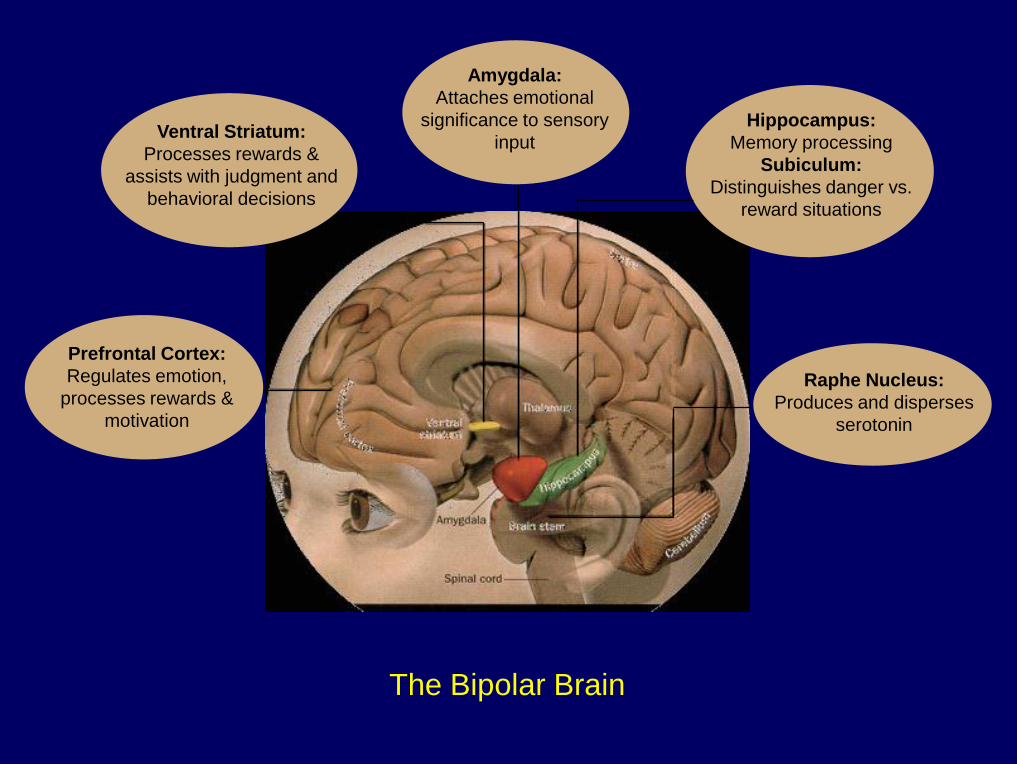
The Bipolar Brain
Prefrontal Cortex:
Regulates emotion,
processes rewards &
motivation
Amygdala:
Attaches emotional
significance to sensory
input Ventral Striatum:
Processes rewards &
assists with judgment and
behavioral decisions
Hippocampus:
Memory processing
Subiculum:
Distinguishes danger vs.
reward situations
Raphe Nucleus:
Produces and disperses
serotonin
Depression
Decreased serotonin levels
Increased ventricles & UBO’s
Decreased frontal lobes, basal ganglia, amygdala & hippocampus
Decreased activity of prefrontal cortex & anterior cingulate
Brain Abnormalities

Severe Depression
Limbic-Cortical Dysregulation
Hypometabolism in dorsal compartment
Prefrontal cortex, anterior cingulate, inferior parietal,
striatum
Symptoms of apathy, attention deficits & psychomotor
slowing
Hypermetabolism in ventral compartment
Hypothalamus, insula, subcallosal region & brain stem
Disturbed sleep, appetite & libido
Mayberg, 1997
Psychopathic Disorders
Impulsive & aggressive behavior
Deficits in informational & emotional
processing
Malfunction of neural circuits in
prefrontal and temporal-limbic regions
(important for emotional learning)


![Radiological Studies on Hippocampal Development ...356455/FULLTEXT01.pdf · Limbic lobe/ system, short overview of the anatomy Limbic lobe, a supernumerary lobe, [49] an arbi-trary](https://img.pdfslide.us/doc/110x75/5e320c89023f1a56c313f8e3/radiological-studies-on-hippocampal-development-356455fulltext01pdf-limbic.jpg)