Embed Size (px)
Citation preview
R E S E A R CH AR T I C L E
The temporal dynamics of structure and content in sentencecomprehension: Evidence from fMRI-constrained MEG
William Matchin1 | Christian Brodbeck2 | Christopher Hammerly3 | Ellen Lau4
1Department of Communication Sciences and
Disorders, University of South Carolina,
Columbia, South Carolina
2Institute for Systems Research, University of
Maryland, College Park, Maryland
3Department of Linguistics, University of
Massachusetts, Amherst, Massachusetts
4Department of Linguistics, University of
Maryland, College Park, Maryland
Correspondence
William Matchin, Department of
Communication Disorders, University of South
Carolina, Discovery 1, Room 202D, Columbia,
South Carolina 29208.
Email: [email protected]
Funding information
University of Maryland
AbstractHumans have a striking capacity to combine words into sentences that express new meanings.
Previous research has identified key brain regions involved in this capacity, but little is known
about the time course of activity in these regions, as hemodynamic methods such as fMRI pro-
vide little insight into temporal dynamics of neural activation. We performed an MEG experi-
ment to elucidate the temporal dynamics of structure and content processing within four brain
regions implicated by fMRI data from the same experiment: the temporo-parietal junction (TPJ),
the posterior temporal lobe (PTL), the anterior temporal lobe (ATL), and the anterior inferior
frontal gyrus (IFG). The TPJ showed increased activity for both structure and content near the
end of the sentence, consistent with a role in incremental interpretation of event semantics.
The PTL, a region not often associated with core aspects of syntax, showed a strong early effect
of structure, consistent with predictive parsing models, and both structural and semantic con-
text effects on function words. These results provide converging evidence that the PTL plays an
important role in lexicalized syntactic processing. The ATL and IFG, regions traditionally associ-
ated with syntax, showed minimal effects of sentence structure. The ATL, PTL and IFG all
showed effects of semantic content: increased activation for real words relative to nonwords.
Our fMRI-guided MEG investigation therefore helps identify syntactic and semantic aspects of
sentence comprehension in the brain in both spatial and temporal dimensions.
KEYWORDS
fMRI, MEG, prediction, semantics, sentence processing, syntax
1 | INTRODUCTION
A central property of human language is our ability to combine words
into sentences to express novel meanings (Chomsky, 1965; Jackend-
off, 2002). In order to identify brain systems involved in combinatory
processing, many neuroimaging studies using functional magnetic res-
onance imaging (fMRI) and positron emission tomography (PET) have
utilized contrasts of structure, comparing highly structured stimuli
such as sentences (e.g., the poet will recite a verse) to less structured
stimuli such as word lists (e.g., rabbit the could extract catch protect).
The key assumption is that brain areas engaged in syntactic and
semantic combinatory processing will show increased activation to
sentences, while noncombinatory effects of sensory, attentional, and
lexical processing are subtracted out (Fedorenko, Nieto-Castanon, &
Kanwisher, 2012; Humphries, Binder, Medler, & Liebenthal, 2006;
Humphries, Love, Swinney, & Hickok, 2005; Matchin, Hammerly, &
Lau, 2017; Mazoyer et al., 1993; Pallier, Devauchelle, & Dehaene,
2011; Rogalsky & Hickok, 2008; Stowe et al., 1998; Vandenberghe,
Nobre, & Price, 2002; see Zaccarella, Schell, & Friederici, 2017, for a
meta-analysis and review). While there is some degree of variability
across studies, four broad portions of the left hemisphere reliably
show effects of sentence structure: the temporo-parietal junction
(TPJ),1 the anterior temporal lobe (ATL), the posterior temporal lobe
(PTL), and the inferior frontal gyrus (IFG).2 While fMRI and PET have
provided precise spatial information about combinatory processing,
these methods have poor temporal resolution, and so the time course
1We note that this is not a precise anatomical term. Some previous authors
have described this region as the angular gyrus, but the relevant functional
region encompasses a more anterior and inferior portion of the angular gyrus,
including activations that extend beyond the angular gyrus into the middle tem-
poral gyrus and supramarginal gyrus. Therefore, we use the term TPJ to be more
inclusive and to fit with our ROI selection procedure.2Here we describe the regions relevant for language quite coarsely; finer ana-
tomical distinctions within these broad regions are clearly relevant. We take this
broad approach because the spatial imprecision of MEG does not allow us to
make finer anatomical claims about our effects.
Received: 23 March 2018 Revised: 4 September 2018 Accepted: 7 September 2018
DOI: 10.1002/hbm.24403
Hum Brain Mapp. 2018;1–16. wileyonlinelibrary.com/journal/hbm © 2018 Wiley Periodicals, Inc. 1
of these structural effects remains largely unknown. The present
study consisted of a within-subjects multimodal imaging approach
using magnetoencephalography (MEG) to reveal the temporal dynam-
ics of sentence processing within language-sensitive regions of inter-
est (ROIs) defined by fMRI. The MEG signal measures magnetic fields
induced by intracranial currents, thought to be due largely to synaptic
activity at cortical pyramidal cells (Buzsáki, Anastassiou, & Koch,
2012). MEG thus reflects similar albeit not identical physiological pro-
cesses as fMRI. Because magnetic fields propagate without temporal
delay, brain activity can be measured with millisecond accuracy, com-
plementing the excellent spatial resolution of fMRI (Hall, Robson,
Morris, & Brookes, 2014). MEG can thus provide information about
the timecourse of effects which previous work only localized spatially.
The added temporal information allows conclusions about the func-
tions of these regions with respect to syntactic and semantic proces-
sing not possible through the use of hemodynamic methods alone.
In natural language, combinatory semantic operations are tightly
tied to syntactic structure. Distinguishing whether structural effects
are due to syntax or semantics is possible using a contrast of content:
comparing natural sentences, with all real words, to jabberwocky sen-
tences (Carroll, 1871), in which the content words are replaced with
nonwords while the function words are the same (e.g., the tevill will
sawl a pand). This greatly reduces conceptual–semantic content while
the syntactic structure is preserved. Interestingly, such contrasts typi-
cally reveal robust increased activation in structure-sensitive regions
(Fedorenko et al., 2012; Humphries et al., 2006; Matchin et al., 2017),
raising questions about the separability of syntax and semantics in the
brain.
However, a separate body of work has associated the IFG and the
PTL with lexical retrieval and lexical storage, respectively (see Lau,
Phillips, & Poeppel, 2008 for a review). All syntactic theories posit
some amount of stored syntactic structure associated with lexical
items (Chomsky, 1981, 1995; Goldberg, 1995; Joshi & Schabes, 1997;
Pollard & Sag, 1994), and stored structure is common in computa-
tional sentence processing models (Demberg, Keller, & Koller, 2013;
Lewis & Vasishth, 2005; Vosse & Kempen, 2000). Thus, lexical and
syntactic processing may be part of a single function of the PTL and
the IFG, namely lexical–syntactic processing (see Snijders et al., 2008
for a similar proposal). On this view, effects of content in the PTL and
the IFG could reflect the more robust activation of stored lexical
entries (with associated syntactic structure) in the natural condition
triggered by real content words, and effects of structure could reflect
the activation of both lexicalized syntax as well as syntactic represen-
tations associated with sentence level structure unconnected to indi-
vidual lexical items (such as clausal nodes with subject noun phrase
and predicate verb phrase nodes; Jackendoff, 2017). By contrast, both
the ATL and the TPJ are strongly associated with conceptual informa-
tion (Binder, Desai, Graves, & Conant, 2009; Lau, Weber, Gramfort,
Hämäläinen, & Kuperberg, 2014; Price, Bonner, Peelle, & Grossman,
2015), suggesting that effects of content in these regions reflects
conceptual–semantic processing rather than lexical or syntactic pro-
cessing. Supporting this hypothesized distinction, contrasts of jabber-
wocky sentences with unstructured jabberwocky lists (e.g., his should
woon pald the must), reliably activates the IFG and the PTL but typi-
cally not the ATL and the TPJ (Fedorenko et al., 2012; Goucha &
Friederici, 2015; Matchin et al., 2017; Pallier et al., 2011), suggesting
that IFG and PTL are involved in syntax regardless of the richness of
conceptual–semantic content.3
A critical test for such hypotheses is the relative timing of activa-
tion across the course of a sentence. In particular, a substantial body
of research has found that syntactic processing can be highly predic-
tive (Levy & Keller, 2013; Omaki et al., 2015; Staub & Clifton, 2006;
Sturt & Lombardo, 2005). If the IFG and the PTL are involved in
lexical–syntactic processing, they might show early effects of struc-
ture. Previous work has suggested that the ATL is involved in local
conceptual–semantic processing (Bemis & Pylkkänen, 2011; Patterson
et al., 2007; Westerlund, Kastner, Al Kaabi, & Pylkkänen, 2015), and
may be tied to the N400 component associated with lexical–
conceptual processing in ERPs (Van Petten & Kutas, 1991; Lau et al.,
2008; Kutas & Hillyard, 1983); this hypothesis would predict that the
ATL would show content effects throughout the sentence. In contrast,
recent work has suggested that the TPJ is more specifically involved
in event semantics (Boylan, Trueswell, & Thompson-Schill, 2015,
2017; Grewe et al., 2007; Thompson et al., 2007), which would pre-
dict that the TPJ would show a late, post-verbal effect of content
associated with the event semantics of natural sentences (which cen-
ter around the verb). Although these distinctions are relatively coarse,
obtaining initial data on the temporal dynamics of sentence processing
within language-sensitive brain regions is a necessary first step toward
the development of more detailed models of neurophysiological
responses associated with combinatorial processing in the brain.
A few recent MEG (Brennan & Pylkkanen, 2017; Halgren et al.,
2002) and electrocortigography (ECoG) studies (Fedorenko et al.,
2016; Nelson et al., 2017) have begun to address how the basic struc-
tural effects observed in fMRI and PET studies emerge over time. For
instance, Nelson et al. (2017) found that the PTL (but not the IFG or
ATL) shows a predictive response profile. By contrast, Fedorenko
et al. (2016) found largely similar temporal dynamics across these
regions. On the other hand, an MEG study by Brennan and Pylkkanen
(2017) found a significant fit of activity in the ATL with a mildly pre-
dictive sentence parser, and did not obtain a significant fit in the IFG.
However, this study did not test for effects in the PTL and did not dis-
tinguish between syntactic and semantic processing in the ATL. The
mixed results of these recent studies are likely due to differences in
sentence materials, task, and neuroanatomical coverage. The goal of
the current study was to more directly evaluate the timecourses of
the fMRI structure and content effects by performing a parallel,
within-subjects MEG and fMRI experiment using the same paradigm
with different materials.
We used a 3 (STRUCTURE) × 2 (CONTENT) parametric design across
both fMRI and MEG sessions. We investigated effects of conceptual–
semantic CONTENT using jabberwocky, replacing open-class words with
phonologically plausible nonwords. We investigated effects of
STRUCTURE by contrasting three lexically matched levels of structure:
six-word sentences, two-word phrases, and randomized word lists.
We used a single sentence structure and a block design in order to
3Some studies have found jabberwocky structure effects in the ATL (Mazoyer
et al., 1993; Humphries et al., 2006), but these effects are notably weaker and
less consistent than in the IFG and the PTL.
2 MATCHIN ET AL.

encourage predictive syntactic processing and to limit the possibility
that subjects would erroneously attempt to impose structure on
unstructured conditions. Group fMRI data from this study have been
previously reported (Matchin et al., 2017). In keeping with the litera-
ture, we found effects of STRUCTURE (sentences > phrases and lists) and
CONTENT (natural sentences > jabberwocky sentences) in the IFG, PTL,
ATL, and TPJ, but we did not find differences between the minimal
two-word phrases and lists in the IFG, PTL, or TPJ.
In the current study, we use the MEG data from the same subjects
to estimate the timecourse of the most robust STRUCTURE and CONTENT
effects observed in fMRI. Prior work has argued that using ROIs derived
from group data results in a significant loss of power due to individual
variability in functional neuroanatomy (Fedorenko, Hsieh, Nieto-Casta-
ñón, Whitfield-Gabrieli, & Kanwisher, 2010; Fedorenko & Kanwisher,
2009; Rogalsky, Almeida, Sprouse, & Hickok, 2015). Therefore, we used
the fMRI data at the subject level to generate individualized IFG, PTL,
ATL, and TPJ ROIs for MEG data analysis. We recognize that analyzing
MEG data within ROIs defined by fMRI data is potentially subject to
error because MEG is less spatially precise than fMRI; therefore, we
supported our ROI analyses with exploratory whole-brain analyses
reported in Supplementary Materials.
We focus our presentation here on a smaller part of the larger
design, choosing pairwise contrasts of STRUCTURE (six-word sentences
vs. two-word phrases) and CONTENT (natural sentences vs. jabberwocky
sentences). We did this for two reasons. First, the reduced design
allows us to present clear and interpretable timecourses that focus on
the maximal fMRI effects. Second, the word list conditions are problem-
atic for MEG because open-class and closed-class words have markedly
different neurophysiological response profiles (Halgren et al., 2002;
Kutas & Hillyard, 1983; Münte et al., 2001; Van Petten & Kutas, 1991).
This means that any contrast between conditions at a particular word
position would be conflated with word class; given our use of dSPM
noise normalization (Dale et al., 2000), selecting only trials where the
open-closed distinction is matched across conditions and positions
would greatly reduce statistical power. We designed our two-word
phrase condition to solve this open/closed problem for MEG by match-
ing the position of open-/closed-class items with the sentence condi-
tion within structurally unconnected phrases (Figure 1). In fMRI this is
not a problem because the signal is integrated across the entire sen-
tence. We therefore included the word list condition in both the fMRI
and MEG sessions for crucial comparison to the existing fMRI literature
and to keep the experiment constant between sessions.
We expected the PTL and the IFG to show effects of STRUCTURE
(greater activity for natural sentences relative to natural phrases) occur-
ring on word 1 and/or word 2, given that these brain regions are associ-
ated with syntactic processing and previous evidence that the PTL
shows a predictive response profile (Nelson et al., 2017). We expected
that the ATL and the TPJ would show effects of STRUCTURE (greater activ-
ity for natural sentences relative to natural phrases) on word 5 and/or
word 6, given that these regions are more associated with conceptual–
semantic processing and previous evidence that the ATL shows a
bottom-up response profile (Nelson et al., 2017; Bemis & Pylkkänen,
2011). We expected that the ATL, PTL, and IFG would show overall
main effects of CONTENT (increased activation for real words relative to
nonwords), given a role for conceptual–semantic processing in the ATL,
and for lexical processing in the PTL and the IFG. We expected that the
TPJ might show effects of CONTENT (greater activity for natural sentences
relative to jabberwocky sentences) at words 4, 5, or 6 (i.e., on or after
the verb), given a possible role for this region in event semantics.
2 | MATERIALS AND METHODS
2.1 | Subjects
15 subjects were included in the analyses presented here (8 women)
(mean age: 24, range: 20–29). One additional subject reported in
Matchin et al. (2017) was excluded here due to a lack of significant acti-
vations in the fMRI experiment to support the individual ROI approach;
one other subject was excluded from both reports due to excessive
movement in the fMRI session. Additionally, 1 of the 15 remaining sub-
jects only had significant fMRI effects in the IFG and TPJ—for this rea-
son, analyses within the IFG and TPJ ROIs included 15 subjects, and
analyses within the ATL and PTL ROIs included 14 subjects. Subjects
were right-handed, native speakers of English, with normal or
corrected-to-normal vision. Consent was acquired from each subject
before the study began, separately for the fMRI and MEG sessions, and
all procedures were approved by the Institutional Review Board of the
University of Maryland. The order of fMRI and MEG sessions for the
total 16 subjects was counterbalanced, with a minimum of 2 days in
between sessions (mean: 12 days, standard deviation: 9 days).
2.2 | Stimuli and design
Our experiment used a 3 (STRUCTURE) × 2 (CONTENT) design, with each
stimulus consisting of a six-word sequence. The three levels of structure
were (i) LIST, (ii) PHRASE, and (iii) SENTENCE, and the two levels of content
were (i) NATURAL and (ii) JABBERWOCKY. All of these conditions were pre-
sented in both the fMRI and MEG experiments. Here we report MEG
analyses of a subset of this fuller design, three conditions: (i) three two-
word phrases with real open-class or content words (NATURAL PHRASE),
(ii) sentences with real open-class or content words (NATURAL SENTENCE),
and (iii) sentences with content words replaced with nonwords (JABBER-
WOCKY SENTENCE). All conditions included real closed-class or function
words. For the reasons described above, the MEG analyses reported in
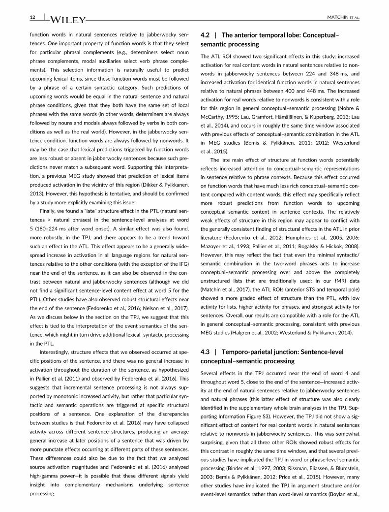
FIGURE 1 Schematic of stimulus design with examples. Tree
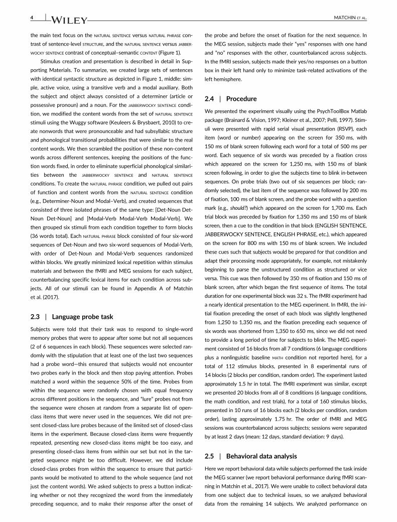
diagrams represent the constituent structure in each condition.Images underneath words indicate conceptual–semanticrepresentations denoted by real words while clouds represent the lackof conceptual–semantic information associated with nonwords. Boldand italicized words indicate open-class/content words [Color figurecan be viewed at wileyonlinelibrary.com]
MATCHIN ET AL. 3
the main text focus on the NATURAL SENTENCE versus NATURAL PHRASE con-
trast of sentence-level STRUCTURE, and the NATURAL SENTENCE versus JABBER-
WOCKY SENTENCE contrast of conceptual–semantic CONTENT (Figure 1).
Stimulus creation and presentation is described in detail in Sup-
porting Materials. To summarize, we created large sets of sentences
with identical syntactic structure as depicted in Figure 1, middle: sim-
ple, active voice, using a transitive verb and a modal auxiliary. Both
the subject and object always consisted of a determiner (article or
possessive pronoun) and a noun. For the JABBERWOCKY SENTENCE condi-
tion, we modified the content words from the set of NATURAL SENTENCE
stimuli using the Wuggy software (Keuleers & Brysbaert, 2010) to cre-
ate nonwords that were pronounceable and had subsyllabic structure
and phonological transitional probabilities that were similar to the real
content words. We then scrambled the position of these non-content
words across different sentences, keeping the positions of the func-
tion words fixed, in order to eliminate superficial phonological similari-
ties between the JABBERWOCKY SENTENCE and NATURAL SENTENCE
conditions. To create the NATURAL PHRASE condition, we pulled out pairs
of function and content words from the NATURAL SENTENCE condition
(e.g., Determiner-Noun and Modal–Verb), and created sequences that
consisted of three isolated phrases of the same type: [Det-Noun Det-
Noun Det-Noun] and [Modal-Verb Modal-Verb Modal-Verb]. We
then grouped six stimuli from each condition together to form blocks
(36 words total). Each NATURAL PHRASE block consisted of four six-word
sequences of Det-Noun and two six-word sequences of Modal-Verb,
with order of Det-Noun and Modal-Verb sequences randomized
within blocks. We greatly minimized lexical repetition within stimulus
materials and between the fMRI and MEG sessions for each subject,
counterbalancing specific lexical items for each condition across sub-
jects. All of our stimuli can be found in Appendix A of Matchin
et al. (2017).
2.3 | Language probe task
Subjects were told that their task was to respond to single-word
memory probes that were to appear after some but not all sequences
(2 of 6 sequences in each block). These sequences were selected ran-
domly with the stipulation that at least one of the last two sequences
had a probe word—this ensured that subjects would not encounter
two probes early in the block and then stop paying attention. Probes
matched a word within the sequence 50% of the time. Probes from
within the sequence were randomly chosen with equal frequency
across different positions in the sequence, and “lure” probes not from
the sequence were chosen at random from a separate list of open-
class items that were never used in the sequences. We did not pre-
sent closed-class lure probes because of the limited set of closed-class
items in the experiment. Because closed-class items were frequently
repeated, presenting new closed-class items might be too easy, and
presenting closed-class items from within our set but not in the tar-
geted sequence might be too difficult. However, we did include
closed-class probes from within the sequence to ensure that partici-
pants would be motivated to attend to the whole sequence (and not
just the content words). We asked subjects to press a button indicat-
ing whether or not they recognized the word from the immediately
preceding sequence, and to make their response after the onset of
the probe and before the onset of fixation for the next sequence. In
the MEG session, subjects made their “yes” responses with one hand
and “no” responses with the other, counterbalanced across subjects.
In the fMRI session, subjects made their yes/no responses on a button
box in their left hand only to minimize task-related activations of the
left hemisphere.
2.4 | Procedure
We presented the experiment visually using the PsychToolBox Matlab
package (Brainard & Vision, 1997; Kleiner et al., 2007; Pelli, 1997). Stim-
uli were presented with rapid serial visual presentation (RSVP), each
item (word or number) appearing on the screen for 350 ms, with
150 ms of blank screen following each word for a total of 500 ms per
word. Each sequence of six words was preceded by a fixation cross
which appeared on the screen for 1,250 ms, with 150 ms of blank
screen following, in order to give the subjects time to blink in-between
sequences. On probe trials (two out of six sequences per block; ran-
domly selected), the last item of the sequence was followed by 200 ms
of fixation, 100 ms of blank screen, and the probe word with a question
mark (e.g., should?) which appeared on the screen for 1,700 ms. Each
trial block was preceded by fixation for 1,350 ms and 150 ms of blank
screen, then a cue to the condition in that block (ENGLISH SENTENCE,
JABBERWOCKY SENTENCE, ENGLISH PHRASE, etc.), which appeared
on the screen for 800 ms with 150 ms of blank screen. We included
these cues such that subjects would be prepared for that condition and
adapt their processing mode appropriately, for example, not mistakenly
beginning to parse the unstructured condition as structured or vice
versa. This cue was then followed by 350 ms of fixation and 150 ms of
blank screen, after which began the first sequence of items. The total
duration for one experimental block was 32 s. The fMRI experiment had
a nearly identical presentation to the MEG experiment. In fMRI, the ini-
tial fixation preceding the onset of each block was slightly lengthened
from 1,250 to 1,350 ms, and the fixation preceding each sequence of
six words was shortened from 1,350 to 650 ms, since we did not need
to provide a long period of time for subjects to blink. The MEG experi-
ment consisted of 16 blocks from all 7 conditions (6 language conditions
plus a nonlinguistic baseline MATH condition not reported here), for a
total of 112 stimulus blocks, presented in 8 experimental runs of
14 blocks (2 blocks per condition, random order). The experiment lasted
approximately 1.5 hr in total. The fMRI experiment was similar, except
we presented 20 blocks from all of 8 conditions (6 language conditions,
the math condition, and rest trials), for a total of 160 stimulus blocks,
presented in 10 runs of 16 blocks each (2 blocks per condition, random
order), lasting approximately 1.75 hr. The order of fMRI and MEG
sessions was counterbalanced across subjects; sessions were separated
by at least 2 days (mean: 12 days, standard deviation: 9 days).
2.5 | Behavioral data analysis
Here we report behavioral data while subjects performed the task inside
the MEG scanner (we report behavioral performance during fMRI scan-
ning in Matchin et al., 2017). We were unable to collect behavioral data
from one subject due to technical issues, so we analyzed behavioral
data from the remaining 14 subjects. We analyzed performance on
4 MATCHIN ET AL.
open-class items only, as open-class probes contained both items from
within the stimulus and lure trials (items not within the stimulus), while
closed-class probes were always within the stimulus (i.e., we never pre-
sented a closed-class item that wasn't in the preceding sequence). Thus
we could only calculate d0 values that correct for response bias for open
class items.
2.6 | fMRI data collection and analysis
The primary focus of the current report is the MEG data. However, we
used individual-subject regions of interest defined by the fMRI data to
constrain the MEG analyses, and therefore we describe fMRI methods
here as well. All fMRI methods, processing, and analysis were identical
to Matchin et al. (2017) except for the use of individual-subject peak
selection. During the fMRI experiment, MR images were obtained in a
Siemens TRIO 3T scanner (Siemens Medical Systems) using a
32-channel head coil. We first collected a high-resolution T1-weighted
anatomical image in the axial plane (voxel dimensions: 0.45 mm ×
0.45 mm × 0.9 mm). We then collected a total of 2,370 T2*-weighted
EPI volumes over 10 runs. Each volume contained 36 oblique slices ori-
ented approximately 20� clockwise relative to the AC-PC axis
(TR = 2 s, TE = 25 ms, flip angle = 90�, in-plane resolution = 3 mm ×
3 mm, slice thickness = 3 mm with 0.3 mm gap).
2.6.1 | Preprocessing
The first four volumes of each run were collected before stimulus pre-
sentation and discarded to control for T1 saturation effects. Slice-timing
correction, motion correction, and spatial smoothing were performed
using AFNI (Cox, 1996; http://afni.nimh.nih.gov/afni). Motion correc-
tion was achieved by using a 6-parameter rigid-body transformation,
with each functional volume in each run first aligned to a single volume
in that run. Functional volumes were aligned to the anatomical image,
and subsequently aligned to Talairach space (Talairach & Tournoux,
1988). Functional images were resampled to 3 mm isotropic voxels and
spatially smoothed using a Gaussian kernel of 6 mm FWHM. The data
were high-pass filtered with a cutoff frequency of 0.0056 Hz at the
first-level analysis stage using AFNI’s 3dDeconvolve function using the
“polort” parameter with a value of 4.
2.6.2 | Defining individual subject regions of interest
Rather than using anatomical ROIs, which do not account for individ-
ual subject anatomical and functional variability (particularly with
respect to language), we generated subject-specific ROIs from the
functional activation peaks in the fMRI data (Fedorenko & Kanwisher,
2009; Fedorenko et al., 2010; Nieto-Castañón & Fedorenko, 2012;
Rogalsky et al., 2015). This ensured that the most functionally relevant
regions in each subject were identified for MEG analysis within
broadly the same anatomical regions across subjects.
Individual subject analyses were performed using AFNI’s 3dDe-
convolve function. The regression analysis was performed to find
parameter estimates that best explained variability in the data. Each
predictor variable representing the time course of stimulus presenta-
tion was entered into a convolution analysis using a canonical hemo-
dynamic response function (AFNI’s BLOCK parameter). The following
seven regressors of interest were included in the model: NATURAL
SENTENCE, JABBERWOCKY SENTENCE, NATURAL PHRASE, JABBERWOCKY PHRASE,
NATURAL LIST, JABBERWOCKY LIST, and MATH. The six motion parameters
were included as regressors of no interest.
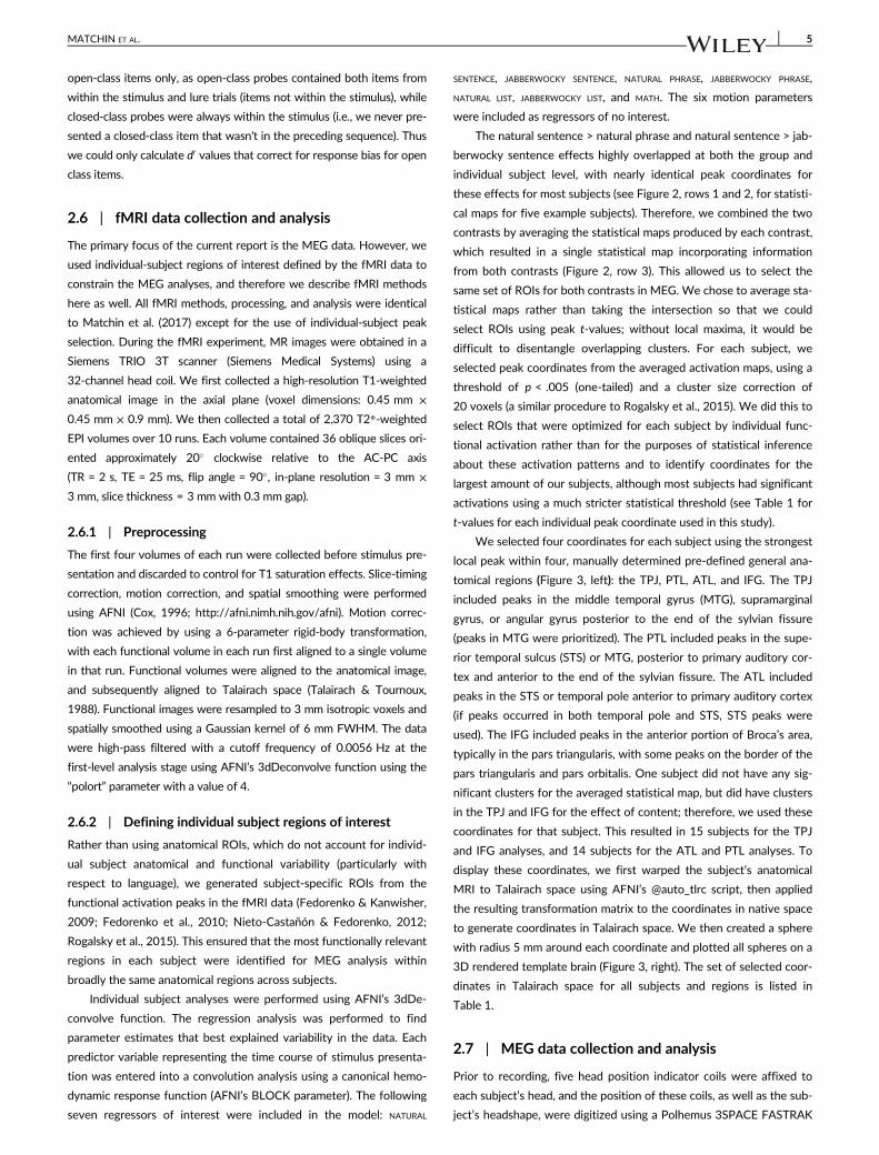
The natural sentence > natural phrase and natural sentence > jab-
berwocky sentence effects highly overlapped at both the group and
individual subject level, with nearly identical peak coordinates for
these effects for most subjects (see Figure 2, rows 1 and 2, for statisti-
cal maps for five example subjects). Therefore, we combined the two
contrasts by averaging the statistical maps produced by each contrast,
which resulted in a single statistical map incorporating information
from both contrasts (Figure 2, row 3). This allowed us to select the
same set of ROIs for both contrasts in MEG. We chose to average sta-
tistical maps rather than taking the intersection so that we could
select ROIs using peak t-values; without local maxima, it would be
difficult to disentangle overlapping clusters. For each subject, we
selected peak coordinates from the averaged activation maps, using a
threshold of p < .005 (one-tailed) and a cluster size correction of
20 voxels (a similar procedure to Rogalsky et al., 2015). We did this to
select ROIs that were optimized for each subject by individual func-
tional activation rather than for the purposes of statistical inference
about these activation patterns and to identify coordinates for the
largest amount of our subjects, although most subjects had significant
activations using a much stricter statistical threshold (see Table 1 for
t-values for each individual peak coordinate used in this study).
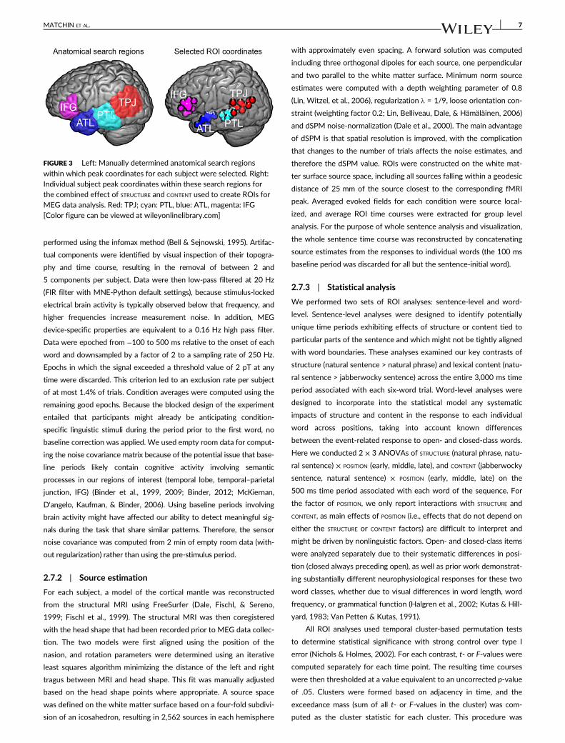
We selected four coordinates for each subject using the strongest
local peak within four, manually determined pre-defined general ana-
tomical regions (Figure 3, left): the TPJ, PTL, ATL, and IFG. The TPJ
included peaks in the middle temporal gyrus (MTG), supramarginal
gyrus, or angular gyrus posterior to the end of the sylvian fissure
(peaks in MTG were prioritized). The PTL included peaks in the supe-
rior temporal sulcus (STS) or MTG, posterior to primary auditory cor-
tex and anterior to the end of the sylvian fissure. The ATL included
peaks in the STS or temporal pole anterior to primary auditory cortex
(if peaks occurred in both temporal pole and STS, STS peaks were
used). The IFG included peaks in the anterior portion of Broca’s area,
typically in the pars triangularis, with some peaks on the border of the
pars triangularis and pars orbitalis. One subject did not have any sig-
nificant clusters for the averaged statistical map, but did have clusters
in the TPJ and IFG for the effect of content; therefore, we used these
coordinates for that subject. This resulted in 15 subjects for the TPJ
and IFG analyses, and 14 subjects for the ATL and PTL analyses. To
display these coordinates, we first warped the subject’s anatomical
MRI to Talairach space using AFNI’s @auto_tlrc script, then applied
the resulting transformation matrix to the coordinates in native space
to generate coordinates in Talairach space. We then created a sphere
with radius 5 mm around each coordinate and plotted all spheres on a
3D rendered template brain (Figure 3, right). The set of selected coor-
dinates in Talairach space for all subjects and regions is listed in
Table 1.
2.7 | MEG data collection and analysis
Prior to recording, five head position indicator coils were affixed to
each subject’s head, and the position of these coils, as well as the sub-
ject’s headshape, were digitized using a Polhemus 3SPACE FASTRAK
MATCHIN ET AL. 5
system. The indicator coils were used to determine the placement of
the subject’s head in the MEG dewar for source analysis (see Source
estimation below). During the experimental sessions, subjects laid
supine in a dark magnetically shielded room (Yokogawa Industries,
Tokyo, Japan). Continuous MEG recording was executed using a
160-channel axial gradiometer whole-head system (Kanazawa Insti-
tute of Technology, Kanazawa, Japan), and data was sampled at
500 Hz (60 Hz online notch filter, DC-200 Hz recording bandwidth).
2.7.1 | Preprocessing
Environmental noise was removed from the data by regressing the sig-
nal at each channel onto three orthogonal reference channels located
far from the participant’s head (Adachi, Shimogawara, Higuchi, Haruta, &
Ochiai, 2001) using the system vendor provided recording software.
Data were then analyzed with MNE-Python v. 0.14 (Gramfort et al.,
2013) and Eelbrain v. 0.26 (Brodbeck, 2017). Bad channels were identi-
fied based on visual inspection of the raw data and excluded (between
0 and 3 per subject), and extraneous artifacts were removed using tem-
poral signal space separation (Taulu & Simola, 2006). Next, Independent
component analysis (ICA) was used to remove ocular and cardiac arti-
facts. ICA decomposition was performed on the entire session’s MEG
data for each subject. First, principal component analysis (PCA) was
used to reduce the complexity of the data, retaining enough PCA com-
ponents to explain 99% of the variance. Then, ICA decomposition was
FIGURE 2 Left hemisphere statistical maps for the effect of structure, natural sentence > natural phrase (top), the effect of content, natural
sentence > jabberwocky sentence (middle), and the combined average of those two effects (bottom) for five individual subjects, displayed on atemplate brain in Talairach space (Talairach & Tournoux, 1988) [Color figure can be viewed at wileyonlinelibrary.com]
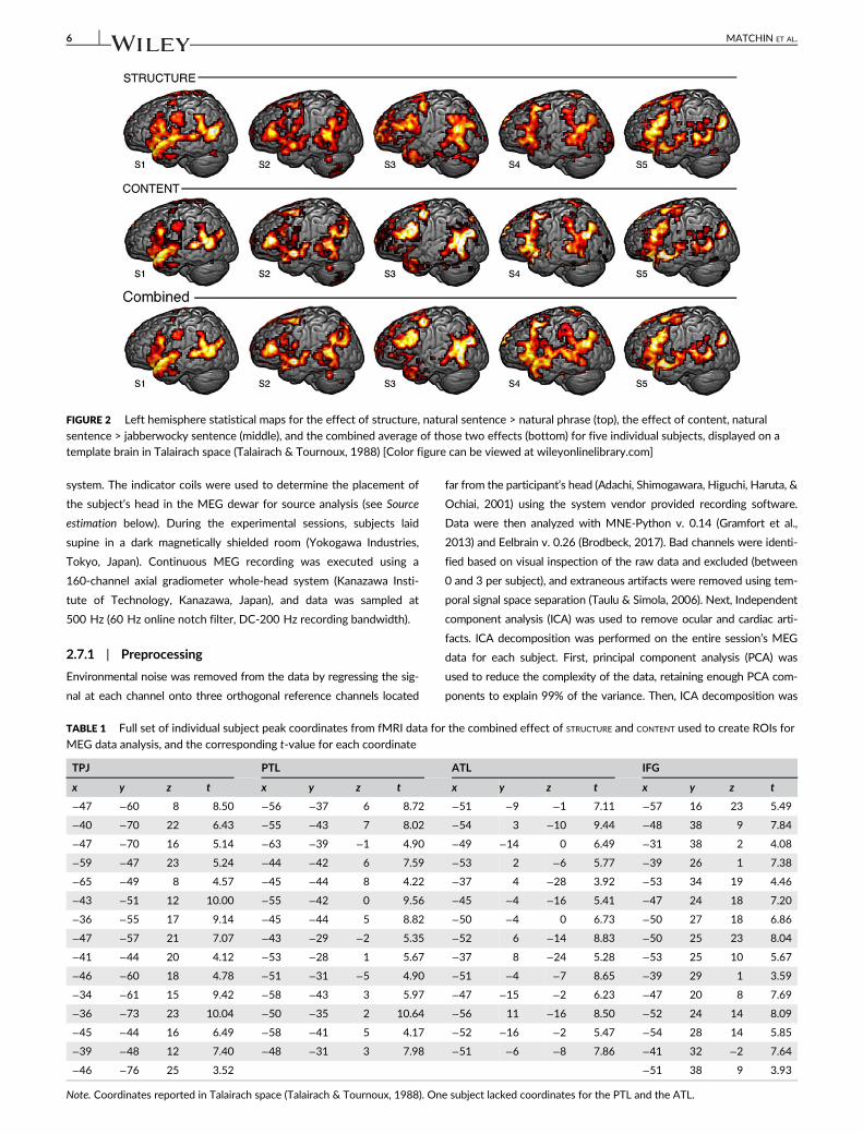
TABLE 1 Full set of individual subject peak coordinates from fMRI data for the combined effect of STRUCTURE and CONTENT used to create ROIs for
MEG data analysis, and the corresponding t-value for each coordinate
TPJ PTL ATL IFG
x y z t x y z t x y z t x y z t
−47 −60 8 8.50 −56 −37 6 8.72 −51 −9 −1 7.11 −57 16 23 5.49
−40 −70 22 6.43 −55 −43 7 8.02 −54 3 −10 9.44 −48 38 9 7.84
−47 −70 16 5.14 −63 −39 −1 4.90 −49 −14 0 6.49 −31 38 2 4.08
−59 −47 23 5.24 −44 −42 6 7.59 −53 2 −6 5.77 −39 26 1 7.38
−65 −49 8 4.57 −45 −44 8 4.22 −37 4 −28 3.92 −53 34 19 4.46
−43 −51 12 10.00 −55 −42 0 9.56 −45 −4 −16 5.41 −47 24 18 7.20
−36 −55 17 9.14 −45 −44 5 8.82 −50 −4 0 6.73 −50 27 18 6.86
−47 −57 21 7.07 −43 −29 −2 5.35 −52 6 −14 8.83 −50 25 23 8.04
−41 −44 20 4.12 −53 −28 1 5.67 −37 8 −24 5.28 −53 25 10 5.67
−46 −60 18 4.78 −51 −31 −5 4.90 −51 −4 −7 8.65 −39 29 1 3.59
−34 −61 15 9.42 −58 −43 3 5.97 −47 −15 −2 6.23 −47 20 8 7.69
−36 −73 23 10.04 −50 −35 2 10.64 −56 11 −16 8.50 −52 24 14 8.09
−45 −44 16 6.49 −58 −41 5 4.17 −52 −16 −2 5.47 −54 28 14 5.85
−39 −48 12 7.40 −48 −31 3 7.98 −51 −6 −8 7.86 −41 32 −2 7.64
−46 −76 25 3.52 −51 38 9 3.93
Note. Coordinates reported in Talairach space (Talairach & Tournoux, 1988). One subject lacked coordinates for the PTL and the ATL.
6 MATCHIN ET AL.
performed using the infomax method (Bell & Sejnowski, 1995). Artifac-
tual components were identified by visual inspection of their topogra-
phy and time course, resulting in the removal of between 2 and
5 components per subject. Data were then low-pass filtered at 20 Hz
(FIR filter with MNE-Python default settings), because stimulus-locked
electrical brain activity is typically observed below that frequency, and
higher frequencies increase measurement noise. In addition, MEG
device-specific properties are equivalent to a 0.16 Hz high pass filter.
Data were epoched from −100 to 500 ms relative to the onset of each
word and downsampled by a factor of 2 to a sampling rate of 250 Hz.
Epochs in which the signal exceeded a threshold value of 2 pT at any
time were discarded. This criterion led to an exclusion rate per subject
of at most 1.4% of trials. Condition averages were computed using the
remaining good epochs. Because the blocked design of the experiment
entailed that participants might already be anticipating condition-
specific linguistic stimuli during the period prior to the first word, no
baseline correction was applied. We used empty room data for comput-
ing the noise covariance matrix because of the potential issue that base-
line periods likely contain cognitive activity involving semantic
processes in our regions of interest (temporal lobe, temporal–parietal
junction, IFG) (Binder et al., 1999, 2009; Binder, 2012; McKiernan,
D'angelo, Kaufman, & Binder, 2006). Using baseline periods involving
brain activity might have affected our ability to detect meaningful sig-
nals during the task that share similar patterns. Therefore, the sensor
noise covariance was computed from 2 min of empty room data (with-
out regularization) rather than using the pre-stimulus period.
2.7.2 | Source estimation
For each subject, a model of the cortical mantle was reconstructed
from the structural MRI using FreeSurfer (Dale, Fischl, & Sereno,
1999; Fischl et al., 1999). The structural MRI was then coregistered
with the head shape that had been recorded prior to MEG data collec-
tion. The two models were first aligned using the position of the
nasion, and rotation parameters were determined using an iterative
least squares algorithm minimizing the distance of the left and right
tragus between MRI and head shape. This fit was manually adjusted
based on the head shape points where appropriate. A source space
was defined on the white matter surface based on a four-fold subdivi-
sion of an icosahedron, resulting in 2,562 sources in each hemisphere
with approximately even spacing. A forward solution was computed
including three orthogonal dipoles for each source, one perpendicular
and two parallel to the white matter surface. Minimum norm source
estimates were computed with a depth weighting parameter of 0.8
(Lin, Witzel, et al., 2006), regularization λ = 1/9, loose orientation con-
straint (weighting factor 0.2; Lin, Belliveau, Dale, & Hämäläinen, 2006)
and dSPM noise-normalization (Dale et al., 2000). The main advantage
of dSPM is that spatial resolution is improved, with the complication
that changes to the number of trials affects the noise estimates, and
therefore the dSPM value. ROIs were constructed on the white mat-
ter surface source space, including all sources falling within a geodesic
distance of 25 mm of the source closest to the corresponding fMRI
peak. Averaged evoked fields for each condition were source local-
ized, and average ROI time courses were extracted for group level
analysis. For the purpose of whole sentence analysis and visualization,
the whole sentence time course was reconstructed by concatenating
source estimates from the responses to individual words (the 100 ms
baseline period was discarded for all but the sentence-initial word).
2.7.3 | Statistical analysis
We performed two sets of ROI analyses: sentence-level and word-
level. Sentence-level analyses were designed to identify potentially
unique time periods exhibiting effects of structure or content tied to
particular parts of the sentence and which might not be tightly aligned
with word boundaries. These analyses examined our key contrasts of
structure (natural sentence > natural phrase) and lexical content (natu-
ral sentence > jabberwocky sentence) across the entire 3,000 ms time
period associated with each six-word trial. Word-level analyses were
designed to incorporate into the statistical model any systematic
impacts of structure and content in the response to each individual
word across positions, taking into account known differences
between the event-related response to open- and closed-class words.
Here we conducted 2 × 3 ANOVAs of STRUCTURE (natural phrase, natu-
ral sentence) × POSITION (early, middle, late), and CONTENT (jabberwocky
sentence, natural sentence) × POSITION (early, middle, late) on the
500 ms time period associated with each word of the sequence. For
the factor of POSITION, we only report interactions with STRUCTURE and
CONTENT, as main effects of POSITION (i.e., effects that do not depend on
either the STRUCTURE or CONTENT factors) are difficult to interpret and
might be driven by nonlinguistic factors. Open- and closed-class items
were analyzed separately due to their systematic differences in posi-
tion (closed always preceding open), as well as prior work demonstrat-
ing substantially different neurophysiological responses for these two
word classes, whether due to visual differences in word length, word
frequency, or grammatical function (Halgren et al., 2002; Kutas & Hill-
yard, 1983; Van Petten & Kutas, 1991).
All ROI analyses used temporal cluster-based permutation tests
to determine statistical significance with strong control over type I
error (Nichols & Holmes, 2002). For each contrast, t- or F-values were
computed separately for each time point. The resulting time courses
were then thresholded at a value equivalent to an uncorrected p-value
of .05. Clusters were formed based on adjacency in time, and the
exceedance mass (sum of all t- or F-values in the cluster) was com-
puted as the cluster statistic for each cluster. This procedure was
FIGURE 3 Left: Manually determined anatomical search regions
within which peak coordinates for each subject were selected. Right:Individual subject peak coordinates within these search regions forthe combined effect of STRUCTURE and CONTENT used to create ROIs forMEG data analysis. Red: TPJ; cyan: PTL, blue: ATL, magenta: IFG[Color figure can be viewed at wileyonlinelibrary.com]
MATCHIN ET AL. 7
repeated under 10,000 random permutations of the data to generate
distributions for the largest exceedance mass value for each effect. In
each permutation, the data were shuffled by switching condition
labels within subject, and the largest exceedance mass value was
retained for each t- or F-map. Finally, p-values were computed for the
clusters found in the original data based on the proportion of permu-
tations that yielded a larger exceedance mass.
Although our focus in this report is on determining the MEG time
course of our structure and content contrasts in regions that elicited
fMRI effects, in Supporting Materials we provide exploratory whole-
brain analyses that provide the opportunity to identify relevant regions
outside of the main language-related regions, as well as exploratory ana-
lyses of the full 3 × 2 design in spite of the open-/closed-class con-
founds that this introduces in event-related MEG.
3 | RESULTS
3.1 | Behavioral data
Subjects performed well on the task overall in the MEG session, with
average d0 values in all conditions (open-class items only) greater than
2.5 (by convention, a d0 value of 1 is considered good performance): NAT-
URAL SENTENCE: 4.319, NATURAL PHRASE: 4.929, NATURAL LIST: 3.444, JABBER-
WOCKY SENTENCE: 3.314, JABBERWOCKY PHRASE: 3.453, JABBERWOCKY LIST:
2.690. A 3 × 2 ANOVA revealed a main effect of structure:
F(2,26) = 8.250, p = .002, a main effect of content: F(1,13) = 9.230,
p = .010, and no interaction: F(2,26) = 1.102, p = .347. Post-hoc tests
of structure revealed that subjects performed significantly better on
sentences relative to lists: t = 2.894, p = .022, better on phrases relative
to lists: t = 3.960, p = .001, and no difference between sentences and
phrases: t = −1.495, p = .440 (Bonferroni corrected). These results indi-
cate facilitatory effects of both structure and content, in line with previ-
ous studies (Brener, 1940; Marks & Miller, 1964; Miller & Isard, 1963;
Miller et al., 1951) as well behavioral performance in the fMRI session
of this experiment (Matchin et al., 2017), suggesting that subjects
indeed parsed the structured material and processed the content of the
words.
3.2 | MEG: Structure
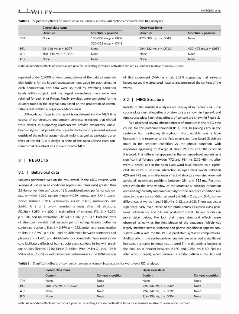
Results of the statistical analyses are displayed in Tables 2–4. Time
course plots illustrating effects of structure are shown in Figure 4, and
time course plots illustrating effects of content are shown in Figure 5.
We observed several distinct effects of structure in the MEG time
course for the posterior temporal (PTL) ROI, beginning early in the
sentence but continuing throughout. Most notable was a large
increase in the response to the first open-class item (word 2; subject
noun) in the sentence condition vs. the phrase condition, with
responses appearing to diverge at about 250 ms after the onset of
the word. This difference appeared in the sentence-level analysis as a
significant difference between 772 and 984 ms (272–484 ms after
word 2 onset), and in the open-class word-level analysis as a signifi-
cant structure × position interaction in open-class words between
420 and 472 ms; a smaller main effect of structure was also observed
across all open-class positions between 284 and 332 ms. Post-hoc
tests within the time window of the structure × position interaction
revealed significantly increased activity for the sentence condition rel-
ative to the phrase condition at word 2 (t[14] = 2.35, p = .034), but no
differences at words 4 and 6 (t[14] ≤ 0.13, p ≥ .902). There was also a
significant early main effect of structure across all closed-class posi-
tions between 92 and 148 ms post-word-onset. As we discuss in
more detail below, the fact that these structural effects were
observed as early as the first phrase of the sequence (which was
largely matched across sentence and phrase conditions) appears con-
sistent with a role for the PTL in predictive syntactic computations.
Additionally, in the sentence-level analysis we observed a significant
increased response to sentences at word 5 (the determiner beginning
the final noun phrase) between 2,180 and 2,284 ms (180–284 ms
after word 5 onset), which mirrored a similar pattern in the TPJ and
TABLE 2 Significant effects of STRUCTURE or STRUCTURE × POSITION interactions for word-level ROI analyses
Closed-class items Open-class items
Structure Structure × position Structure Structure × position
TPJ None 180–300 ms, p = .0002 472–500 ms, p = .0343 None
320–352 ms, p = .0341
PTL 92–148 ms, p = .0297 None 284–332 ms, p = .0013 420–472 ms, p = .0083
ATL 400–448 ms, p = .0321 None None None
IFG None None None None
Note. All reported effects of STRUCTURE are positive, reflecting increased activation for NATURAL SENTENCE relative to NATURAL PHRASE.
TABLE 3 Significant effects of CONTENT or CONTENT × POSITION interactions for word-level ROI analyses
Closed-class items Open-class items
Content Content × position Content Content × position
TPJ None None None None
PTL 208–272 ms, p = .0062 None 228–332 ms, p = .0009 None
ATL None None 224–348 ms, p = .0032 None
IFG None None 216–396 ms, p = .0004 None
Note. All reported effects of CONTENT are positive, reflecting increased activation for NATURAL SENTENCE relative to JABBERWOCKY SENTENCE.
8 MATCHIN ET AL.
the ATL that may reflect interpretive processes triggered by the verb
phrase.
In the TPJ, this increased response to sentences vs. phrases at the
onset of the last function word (word 5) was the largest structure effect,
resulting in two nearly adjacent effects in the sentence analysis
(between 1964–2096 ms and 2,156–2,356 ms, or from 36 ms prior to
word 5 onset to 356 ms post-onset). We also observed a significant
structure × position interaction in the closed class word analysis that
appeared to be due to the strong word 5 effect (t[14] = 3.97, p = .001),
a similar but smaller effect at word 3 (t[14] = 2.54, p = .024), and a pat-
tern in the reverse direction at word 1 (t[14] = 2.31, p = .036).
In the ATL we observed only a main effect of structure for
closed-class words between 400 and 448 ms. In the sentence time
courses, we note the presence of a nonsignificant but numerically
sustained effect of structure between 2,000 and 2,500 ms (word 5)
that parallels the effects observed in PTL and TPJ in the same time-
window. Additionally, there appeared to be some short and sub-
threshold effects of structure at the end of word 2.
In the IFG, there were surprisingly no effects of structure across
all of our analyses. Examining the time courses for this ROI, there
were only some weak and intermittent periods of time where natural
sentences showed numerically increased activation relative to natural
TABLE 4 Significant effects in the sentence-level ROI analyses
Structure Content
TPJ 1964–2096 ms, (word 4–5) p = .0028 2,200–2,280 ms (word 5), p = .0324
2,156–2,356 ms (word 5), p = .0005
PTL 772–984 ms (word 2), p < .0001 1,200–1,272 ms (word 3), p = .0306
2,180–2,284 ms (word 5), p = .0104 1,724–1808 ms (word 4), p = .0071
2,724–2,844 ms (word 6), p = .0039
ATL None None
IFG None 796–896 ms (word 2), p = .0187
1,204–1,292 ms (word 3), p = .0421
1,728–1800 ms (word 4), p = .0411
Note. All reported effects are positive, reflecting increased activation for NATURAL SENTENCE relative to NATURAL PHRASE (effects of STRUCTURE) or increased activa-tion for NATURAL SENTENCE relative to JABBERWOCKY SENTENCE (effects of CONTENT).
FIGURE 4 Analyses of STRUCTURE within each ROI (dSPM). Red: NATURAL SENTENCE, orange: NATURAL PHRASE. Gray lines indicate significant main
effects of STRUCTURE in the word-level analyses, blue lines indicate significant interactions of STRUCTURE and POSITION in the word-level analyses, andgreen lines represent significant effects of STRUCTURE in the sentence-level analyses. X axis is time in milliseconds relative to onset of the first wordin each six-word trial. Shading indicates the within-subject standard error (Loftus & Masson, 1994). The blue text “STRUCTURE × POSITION interaction”and the corresponding blue underline reflects significant time periods for the STRUCTURE x POSITION interaction in the word-level analysis. In the PTL,only the time period at word 2 survived a post-hoc pairwise comparison, while in the TPJ, the time periods at words 1, 3, and 5 all survived apost-hoc pairwise comparisons [Color figure can be viewed at wileyonlinelibrary.com]
MATCHIN ET AL. 9
phrases. This was unexpected, given that we observed robust struc-
ture effects in the IFG in our fMRI data (Matchin et al., 2017).
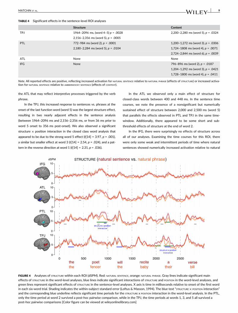
3.3 | MEG: Content
While the timing of structure effects varied considerably across the
four ROIs, the timing of lexical content effects appeared more uni-
form, and included the IFG. The IFG, ATL, and PTL all showed signifi-
cantly greater activity for natural versus jabberwocky materials in the
open-class words, roughly 215–350 ms post-word-onset. The PTL
also showed a main effect of content for closed-class items, but inter-
estingly this occurred in a later time window (208–272 ms) than the
main effect of structure for closed-class items reported above
(92–148 ms) and appeared especially large at the auxiliary verb
position (word 3), suggesting distinct effects of structure and content
on PTL responses to closed-class items. The IFG also showed a similar
content effect at closed-class word 3 in the sentence-level analysis
(204–292 ms post-word-onset). The only significant effect of content
in the TPJ was in the sentence-level analysis at word 5 (200–280 ms
post-word-onset), with approximately the same timing as the struc-
ture effect in this region.
4 | DISCUSSION
We used parallel fMRI and MEG to determine the time course of
structural and conceptual–semantic processing in regions shown to be
involved in combinatory processing in previous fMRI and PET studies
(Fedorenko et al., 2012; Humphries et al., 2005, 2006; Matchin et al.,
2017; Mazoyer et al., 1993; Pallier et al., 2011; Rogalsky & Hickok,
2008; Rogalsky et al., 2015; Stowe et al., 1998; Vandenberghe et al.,
2002). Previous research in neurophysiology has examined general
effects of sentence position, such as words early versus late in a
sentence (Halgren et al., 2002; Fedorenko et al., 2016) or structural
variables like parsing steps or constituent structure complexity
(Brennan & Pylkkänen, 2012; Nelson et al., 2017). Our results comple-
ment these studies by identifying specific points in a sentence when
enhanced activity is seen for sentence structure and conceptual–
semantic content. Given that our experiment was designed to maxi-
mize any effects of structural prediction beyond what is likely to occur
in natural sentence processing, we do not claim that these temporal
dynamics precisely characterize natural sentence processing under all
conditions. Rather, the observed temporal dynamics help to character-
ize the underlying functions of these brain regions. Here we discuss
the effects found in each ROI and their implications with respect to
their functional contributions to sentence processing.
4.1 | The posterior temporal lobe: Lexical–syntacticprocessing
In previous work, we have hypothesized that the PTL underlies
lexical–syntactic processing (Matchin, 2017; Matchin et al., 2017), and
we have suggested two explanations for effects of structure in this
region in fMRI and PET studies: (i) predictive activation of sentence-
level syntactic representations, and (ii) increased attention or
FIGURE 5 Analyses of CONTENT within each ROI (dSPM). Red: NATURAL SENTENCE, blue: JABBERWOCKY SENTENCE. Gray lines indicate significant main
effects of CONTENT in the word-level analyses, and green lines represent significant effects of content in the sentence-level analyses. X axis is timein milliseconds relative to onset of the first word in each six-word trial. Shading indicates the within-subject standard error (Loftus & Masson,1994) [Color figure can be viewed at wileyonlinelibrary.com]
10 MATCHIN ET AL.
maintenance of syntactic representations associated with lexical items
when they are presented in a sentence context. The timing of the
structure effects we observed here in MEG in our PTL ROI (based on
fMRI activations in or near posterior superior temporal sulcus in indi-
vidual subjects) appear to support both of these possibilities.
The early structure effect we observed in MEG at the end of
word 2 (in the sentence-level analysis, 272–484 ms after onset of the
subject noun) provides more direct evidence for the hypothesis that
posterior temporal structure effects in part reflect syntactic predic-
tions. This effect was reinforced by the cluster identified for this con-
trast in the supplementary whole brain analyses in a similar posterior
temporal region, centered on the STS/MTG (Supporting Information
Figure S3). Given the high similarity between the sentence and phrase
conditions at this early stage in the sentence and our blocked experi-
mental design that encouraged structural predictions, a straightfor-
ward interpretation of this effect is that in the sentence condition
subjects processed the subject noun phrase and then projected
sentence-level hierarchical structure before the appearance of the fol-
lowing words that support that structure. In the phrase condition,
when subjects have clear information that there is no hierarchical sen-
tence structure, subjects likely did not generate such structural predic-
tions. This account naturally fits with predictive left-corner parsing
models (Demberg et al., 2013; Lewis & Vasishth, 2005) and psycholin-
guistic data supporting structural predictions during sentence proces-
sing (Levy & Keller, 2013; Staub & Clifton, 2006; Sturt & Lombardo,
2005). These results also converge with those of Nelson et al. (2017),
who found that activity in the PTL was best fit with a predictive top-
down parsing model. Combined, these data suggest that sentence-
level predictive structural processing can be localized at least in part
to posterior regions of the temporal lobe, and more specifically that
the head noun of the subject noun phrase may be a critical position in
the sentence for projecting upcoming structure. Importantly, we do
not claim that structural predictions always unfold with identical tem-
poral dynamics as observed in this study, as our choice of sentence
structure and block design likely substantially enhanced structural pre-
dictions. Rather, we suggest that the PTL encodes structural represen-
tations that can be used in a predictive fashion, particularly when
context encourages the use of such predictions.
One potential caveat to note is that the distribution of words
across positions of the sentence was not perfectly controlled across
the sentence and phrase conditions. In the sentence condition, modal
auxiliaries and verbs always occurred in the middle of each stimulus
and determiners and nouns occurred at the beginning and ends of
sentences, while in the phrase condition, these words were equally
distributed throughout the stimulus. In principle, this might have
affected our results, as a determiner + noun occurred 100% of the
time early in the sentence stimuli, while a determiner + noun occurred
only 67% of the time early in the phrase stimuli. However, we note
that the middle and ends of stimuli are similarly imbalanced—this
would predict an effect in the opposite direction at word 4, and a sim-
ilar effect at word 6. However, this is not the pattern we see—there is
in fact a small effect in the same direction at word 4, and no consis-
tent effect at word 6 (there is a small effect in the same direction
228–332 ms after word onset, but the effect later reverses,
~400–500 ms after word onset). Thus, our results cannot be
straightforwardly attributed to differences in the distribution of words
between the two conditions, but are transparently accounted for by
higher-level structural differences.
The PTL showed two additional effects of structure that were dis-
tributed throughout the duration of the sentence: increased activation
for sentences relative to phrases at closed-class words (words 1, 3,
and 5) between 92 and 148 ms after word onset and at open-class
words (word 2, 4, and 6) between 284 and 332 ms after word onset.
Given that these effects occurred at all positions of the sentence,
these effects could be explained by increased attention or mainte-
nance of individual words with associated syntactic structure during
sentence processing (Snijders et al., 2008). Under this hypothesis, dur-
ing both the phrase and sentence conditions, words activate corre-
sponding lexical–syntactic representations. In both conditions, this
creates local, connected phrases. However, in the sentence condition,
these local phrases are also integrated to create a higher-level sen-
tence structure. This process would involve increased attention to
individual syntactic representations, resulting in increased activation
for these words in sentence context. The difference in the latencies of
the structure effect in closed- and open-class words may simply
reflect the fact that shorter and more frequent closed-class words can
be identified more quickly.
We hypothesized that effects of content—that is, increased
activation for natural sentences relative to jabberwocky sentences—
in the PTL in fMRI and PET studies also reflect lexical–syntactic pro-
cessing. Under this hypothesis, real words result in the selection of
lexical–syntactic representations stored in the PTL while nonwords
do not, resulting in greater overall lexical–syntactic processing in
the PTL for natural sentences that have both real content and func-
tion words relative to jabberwocky sentences that only have real
function words. Consistent with this hypothesis, we observed
greater activation for real content words in natural sentences rela-
tive to nonwords in jabberwocky sentences in the PTL. This content
effect highly overlapped in time with the structure effect for con-
tent words in all sentence positions (roughly 250–330 ms post-
word-onset), suggesting that both of these effects may in fact be
due to a common underlying source such as lexical–syntactic
processing.
We also found greater activation for function words in natural
sentences relative to jabberwocky sentences. Interestingly, while the
effects of structure and content for content words highly overlapped
in time, the effects on function words did not: for structure, the effect
occurred between 92 and 148 ms after word onset; for content, the
effect occurred between 208 and 272 ms. Visual inspection of the
time courses shows a hint of a content effect for function words in
the earlier time window at word 3, but no early effect at words 1 and
5, indicating that this lack of timing overlap was not an artifact of the
temporal clustering procedure.
Here we offer a tentative hypothesis of the distinct timing of
structure and content effects on function words. As discussed above,
the effect of structure at function words between 92 and 148 ms
may reflect increased attention to or maintenance of lexical–syntactic
representations associated with those words in sentences relative to
unconnected phrases. By contrast, the later effect of content may
reflect stronger predictions of upcoming lexical items triggered by
MATCHIN ET AL. 11
function words in natural sentences relative to jabberwocky sen-
tences. One important property of function words is that they select
for particular phrasal complements (e.g., determiners select noun
phrase complements, modal auxiliaries select verb phrase comple-
ments). This selection information is naturally useful to predict
upcoming lexical items, since these function words must be followed
by a phrase of a certain syntactic category. Such predictions of
upcoming words would be equal in the natural sentence and natural
phrase conditions, given that they both have the same set of local
phrases with the same words (in other words, determiners are always
followed by nouns and modals always followed by verbs in both con-
ditions as well as the real world). However, in the jabberwocky sen-
tence condition, function words are always followed by nonwords. It
may be the case that lexical predictions triggered by function words
are less robust or absent in jabberwocky sentences because such pre-
dictions never match a subsequent word. Supporting this interpreta-
tion, a previous MEG study showed that prediction of lexical items
produced activation in the vicinity of this region (Dikker & Pylkkanen,
2013). However, this hypothesis is tentative, and should be confirmed
by a study more explicitly examining this issue.
Finally, we found a “late” structure effect in the PTL (natural sen-
tences > natural phrases) in the sentence-level analyses at word
5 (180–224 ms after word onset). A similar effect was also found,
more robustly, in the TPJ, and there appears to be a trend toward
such an effect in the ATL. This effect appears to be a generally wide-
spread increase in activation in all language regions for natural sen-
tences relative to the other conditions (with the exception of the IFG)
near the end of the sentence, as it can also be observed in the con-
trast between natural and jabberwocky sentences (although we did
not find a significant sentence-level content effect at word 5 for the
PTL). Other studies have also observed robust structural effects near
the end of the sentence (Fedorenko et al., 2016; Nelson et al., 2017).
As we discuss below in the section on the TPJ, we suggest that this
effect is tied to the interpretation of the event semantics of the sen-
tence, which might in turn drive additional lexical–syntactic processing
in the PTL.
Interestingly, structure effects that we observed occurred at spe-
cific positions of the sentence, and there was no general increase in
activation throughout the duration of the sentence, as hypothesized
in Pallier et al. (2011) and observed by Fedorenko et al. (2016). This
suggests that incremental sentence processing is not always sup-
ported by monotonic increased activity, but rather that particular syn-
tactic and semantic operations are triggered at specific structural
positions of a sentence. One explanation of the discrepancies
between studies is that Fedorenko et al. (2016) may have collapsed
activity across different sentence structures, producing an average
general increase at later positions of a sentence that was driven by
more punctate effects occurring at different parts of these sentences.
These differences could also be due to the fact that we analyzed
source activation magnitudes and Fedorenko et al. (2016) analyzed
high-gamma power—it is possible that these different signals yield
insight into complementary mechanisms underlying sentence
processing.
4.2 | The anterior temporal lobe: Conceptual–semantic processing
The ATL ROI showed two significant effects in this study: increased
activation for real content words in natural sentences relative to non-
words in jabberwocky sentences between 224 and 348 ms, and
increased activation for identical function words in natural sentences
relative to natural phrases between 400 and 448 ms. The increased
activation for real words relative to nonwords is consistent with a role
for this region in general conceptual–semantic processing (Nobre &
McCarthy, 1995; Lau, Gramfort, Hämäläinen, & Kuperberg, 2013; Lau
et al., 2014), and occurs in roughly the same time window associated
with previous effects of conceptual–semantic combination in the ATL
in MEG studies (Bemis & Pylkkänen, 2011; 2012; Westerlund
et al., 2015).
The late main effect of structure at function words potentially
reflects increased attention to conceptual–semantic representations
in sentence relative to phrase contexts. Because this effect occurred
on function words that have much less rich conceptual–semantic con-
tent compared with content words, this effect may specifically reflect
more robust predictions from function words to upcoming
conceptual–semantic content in sentence contexts. The relatively
weak effects of structure in this region may appear to conflict with
the generally consistent finding of structural effects in the ATL in prior
literature (Fedorenko et al., 2012; Humphries et al., 2005, 2006;
Mazoyer et al., 1993; Pallier et al., 2011; Rogalsky & Hickok, 2008).
However, this may reflect the fact that even the minimal syntactic/
semantic combination in the two-word phrases acts to increase
conceptual–semantic processing over and above the completely
unstructured lists that are traditionally used: in our fMRI data
(Matchin et al., 2017), the ATL ROIs (anterior STS and temporal pole)
showed a more graded effect of structure than the PTL, with low
activity for lists, higher activity for phrases, and strongest activity for
sentences. Overall, our results are compatible with a role for the ATL
in general conceptual–semantic processing, consistent with previous
MEG studies (Halgren et al., 2002; Westerlund & Pylkkanen, 2014).
4.3 | Temporo-parietal junction: Sentence-levelconceptual–semantic processing
Several effects in the TPJ occurred near the end of word 4 and
throughout word 5, close to the end of the sentence—increased activ-
ity at the end of natural sentences relative to jabberwocky sentences
and natural phrases (this latter effect of structure was also clearly
identified in the supplementary whole brain analyses in the TPJ, Sup-
porting Information Figure S3). However, the TPJ did not show a sig-
nificant effect of content for real content words in natural sentences
relative to nonwords in jabberwocky sentences. This was somewhat
surprising, given that all three other ROIs showed robust effects for
this contrast in roughly the same time window, and that several previ-
ous studies have implicated the TPJ in word or phrase-level semantic
processing (Binder et al., 1997, 2003; Rissman, Eliassen, & Blumstein,
2003; Bemis & Pylkkänen, 2012; Price et al., 2015). However, many
other studies have implicated the TPJ in argument structure and/or
event-level semantics rather than word-level semantics (Boylan et al.,
12 MATCHIN ET AL.
2015; Grewe et al., 2007; Kalénine et al., 2009; Pallier et al., 2011;
Thompson et al., 2007; Thothathiri, Kimberg, & Schwartz, 2012). If
TPJ is primarily involved in processing coherent sentence meaning, its
activity might not be tied to the specific conceptual content associ-
ated with individual lexical items. Our data are consistent with this
view, as the increased activation for natural sentences near the end of
the sentence suggests a response associated with the interpretation
of the event semantics of the sentence, which is more complete at
the end of the sentence than near the beginning.
Interestingly, this effect occurred before the final noun of the sen-
tence. All of the verbs in our study were transitive, which means that
the event’s meaning is incomplete without two arguments (event par-
ticipants). For instance, the meaning of an event associated with a
verb like destroy is incomplete unless there is an agent of destruction
and a recipient of destruction. One possibility is that our results reflect
incremental interpretation of the sentence meaning, using only the
subject and verb. For instance, in the sentence a telepath could destroy
our foe, subjects could integrate telepath and destroy together before
waiting for the object of the verb.
The follow-up pairwise tests to the structure × position interac-
tion showed that there was in fact increased activity for the phrase
condition relative to the sentence condition at word 1, a function
word (180–300 ms after word onset). It is unclear why phrases
would activate the TPJ more than sentences at any time point,
let alone at the beginning of the trial, although we note a similar but
not significant effect in the PTL and ATL ROIs as well. The ECoG
studies by Nelson et al. (2017, supporting information materials) and
Fedorenko et al. (2016, supporting information materials) both
report some indication of increased activation for word lists relative
to sentences at early positions, suggesting that decreased activation
in language regions for sentences relative to less structured condi-
tions early in the trial is a general phenomenon and not an anomaly.
This effect may be due to overall attentional differences between
the two conditions, whereby subjects direct increased attention to
sensory processing in the sentence condition early on, resulting in
increased activation in systems involved in linguistic processing for
the phrase condition. Future studies could attempt to narrow down
more definitively the source of this early decreased activation for lin-
guistic structure.
4.4 | The inferior frontal gyrus: Limited effects
Our parallel fMRI study identified strong effects of structure and con-
tent in the left anterior IFG, replicating prior fMRI findings (Fedorenko
et al., 2012; Humphries et al., 2006). In MEG we obtained robust
effects of content in IFG for real content words relative to nonwords
in a similar time window as the PTL and ATL, but we did not observe
any effects of structure in this region, similar to several previous MEG
studies (Bemis & Pylkkänen, 2011; 2012; Brennan & Pylkkänen, 2012;
2017; Westerlund & Pylkkanen, 2014). The lack of these structure
effects limits our ability to make inferences about the function of IFG
from this study. One factor that could have contributed is reduced sig-
nal from frontal regions, as in standard MEG systems the subject’s
head rests against the back of the helmet, and most of the gap
between the head size and the helmet size is thus in the frontal areas
(Marinkovic, Cox, Reid, & Halgren, 2004). Second, the cortical mor-
phology of the IFG might be such that it is difficult to identify the rele-
vant functional sources using MEG (Ahlfors et al., 2010), suggesting
that future similar studies might profit from using electrophysiological
methods, which have complementary orientation sensitivity. How-
ever, the fact that we still obtained robust effects of our content
manipulation in IFG casts some doubt on this explanation, especially
given that IFG effects of content and structure were relatively compa-
rable in magnitude in our fMRI study. Other possible explanations
relate to the different temporal properties of fMRI and MEG. If IFG
effects of structure occur at variable timepoints from trial to trial, they
would sum across trials in temporally blurred fMRI analyses, but
would not sum across trials in temporally sensitive MEG analyses. Or,
IFG effects of structure might primarily reflect “wrap-up” activity after
the sentence is over; we were unable to evaluate this possibility in the
current MEG dataset because of muscle movement associated with
the probe response and increased ocular artifact post-sentence.
Finally, although both fMRI and MEG signals are correlated with the
local field potential, the relationship between the two measures is a
complex one (Hall et al., 2014), and it is possible that a difference in
sensitivity to different neural processes lead to different response pat-
terns in the two modalities. Although we cannot determine the source
of the fMRI/MEG discrepancy from the current data, these explana-
tions would put very strong constraints on the functional interpreta-
tion of IFG effects of structure, and therefore the discrepancy itself
suggests important and straightforward avenues for future
investigation.
5 | CONCLUSIONS
A growing body of neurophysiological research has focused on how
brain activity in language-sensitive regions of the brain contributes to
the processing of sentence-level structure and conceptual–semantic
content (Brennan & Pylkkänen, 2017; Fedorenko et al., 2016; Halgren
et al., 2002; Nelson et al., 2017). In contrast to previous studies, our
study identified clear effects of linguistic structure and content at spe-
cific sentence positions, rather than averaging across structures or col-
lapsing across syntactic positions and word categories. A major
finding of this study is that PTL activity exhibited a robust structure
effect that likely reflects predictive syntactic processing: increased
activation at the end of the subject noun phrase in sentence context
relative to sequences of three unconnected phrases. This effect con-
verges with predictive parsing models of human sentence comprehen-
sion (Demberg et al., 2008; Hale, 2001; Lewis & Vasishth, 2005) and
provides novel support for the PTL as a critical region for syntax.
These relatively coarse observations about the time course of combi-
natory operations across the sentence provide a potential basis for
future integration with more precise neurophysiological models of sin-
gle word processing derived from ERP responses such as the N400
(Kutas & Hillyard, 1983). Future research should also extend the
approach to a broad range of sentence structures in varying contexts
in order to determine the extent to which these effects generalize to
other syntactic and semantic representations and processing contexts.
MATCHIN ET AL. 13
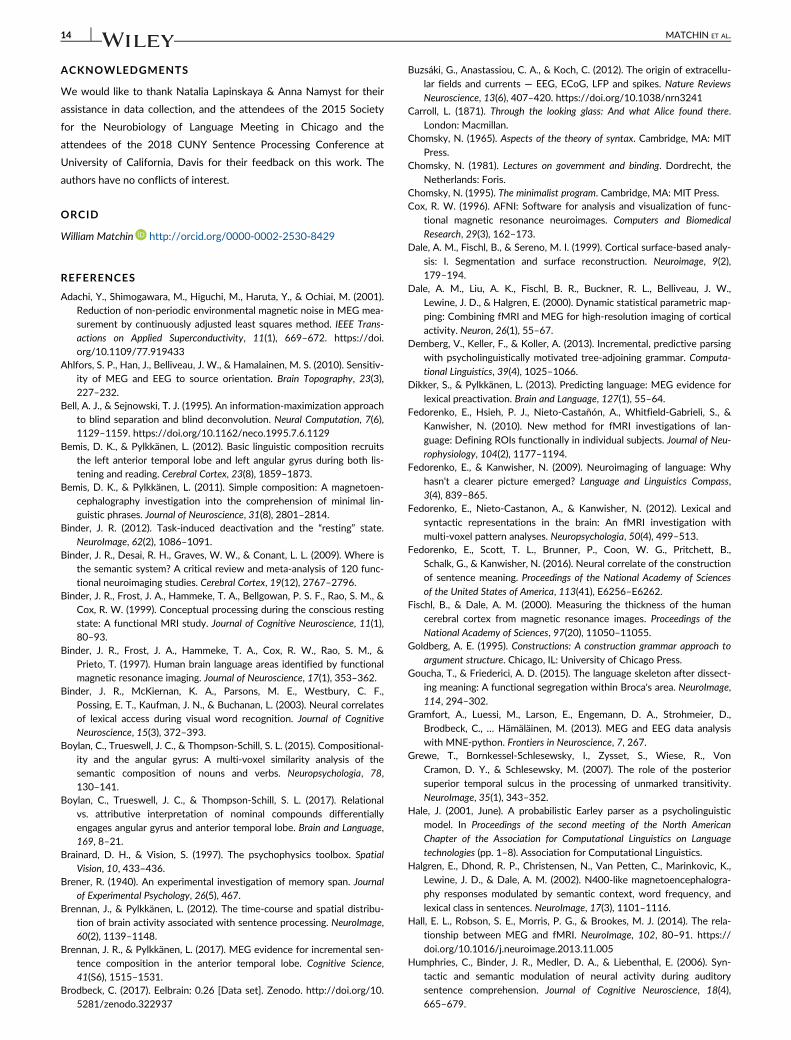
ACKNOWLEDGMENTS
We would like to thank Natalia Lapinskaya & Anna Namyst for their
assistance in data collection, and the attendees of the 2015 Society
for the Neurobiology of Language Meeting in Chicago and the
attendees of the 2018 CUNY Sentence Processing Conference at
University of California, Davis for their feedback on this work. The
authors have no conflicts of interest.
ORCID
William Matchin http://orcid.org/0000-0002-2530-8429
REFERENCES
Adachi, Y., Shimogawara, M., Higuchi, M., Haruta, Y., & Ochiai, M. (2001).Reduction of non-periodic environmental magnetic noise in MEG mea-surement by continuously adjusted least squares method. IEEE Trans-
actions on Applied Superconductivity, 11(1), 669–672. https://doi.org/10.1109/77.919433
Ahlfors, S. P., Han, J., Belliveau, J. W., & Hamalainen, M. S. (2010). Sensitiv-ity of MEG and EEG to source orientation. Brain Topography, 23(3),
227–232.Bell, A. J., & Sejnowski, T. J. (1995). An information-maximization approach
to blind separation and blind deconvolution. Neural Computation, 7(6),1129–1159. https://doi.org/10.1162/neco.1995.7.6.1129
Bemis, D. K., & Pylkkänen, L. (2012). Basic linguistic composition recruitsthe left anterior temporal lobe and left angular gyrus during both lis-tening and reading. Cerebral Cortex, 23(8), 1859–1873.
Bemis, D. K., & Pylkkänen, L. (2011). Simple composition: A magnetoen-
cephalography investigation into the comprehension of minimal lin-guistic phrases. Journal of Neuroscience, 31(8), 2801–2814.
Binder, J. R. (2012). Task-induced deactivation and the “resting” state.NeuroImage, 62(2), 1086–1091.
Binder, J. R., Desai, R. H., Graves, W. W., & Conant, L. L. (2009). Where isthe semantic system? A critical review and meta-analysis of 120 func-tional neuroimaging studies. Cerebral Cortex, 19(12), 2767–2796.
Binder, J. R., Frost, J. A., Hammeke, T. A., Bellgowan, P. S. F., Rao, S. M., &Cox, R. W. (1999). Conceptual processing during the conscious restingstate: A functional MRI study. Journal of Cognitive Neuroscience, 11(1),80–93.
Binder, J. R., Frost, J. A., Hammeke, T. A., Cox, R. W., Rao, S. M., &Prieto, T. (1997). Human brain language areas identified by functionalmagnetic resonance imaging. Journal of Neuroscience, 17(1), 353–362.
Binder, J. R., McKiernan, K. A., Parsons, M. E., Westbury, C. F.,
Possing, E. T., Kaufman, J. N., & Buchanan, L. (2003). Neural correlatesof lexical access during visual word recognition. Journal of CognitiveNeuroscience, 15(3), 372–393.
Boylan, C., Trueswell, J. C., & Thompson-Schill, S. L. (2015). Compositional-
ity and the angular gyrus: A multi-voxel similarity analysis of thesemantic composition of nouns and verbs. Neuropsychologia, 78,130–141.
Boylan, C., Trueswell, J. C., & Thompson-Schill, S. L. (2017). Relational
vs. attributive interpretation of nominal compounds differentiallyengages angular gyrus and anterior temporal lobe. Brain and Language,169, 8–21.
Brainard, D. H., & Vision, S. (1997). The psychophysics toolbox. Spatial
Vision, 10, 433–436.Brener, R. (1940). An experimental investigation of memory span. Journal
of Experimental Psychology, 26(5), 467.Brennan, J., & Pylkkänen, L. (2012). The time-course and spatial distribu-
tion of brain activity associated with sentence processing. NeuroImage,60(2), 1139–1148.
Brennan, J. R., & Pylkkänen, L. (2017). MEG evidence for incremental sen-
tence composition in the anterior temporal lobe. Cognitive Science,41(S6), 1515–1531.
Brodbeck, C. (2017). Eelbrain: 0.26 [Data set]. Zenodo. http://doi.org/10.5281/zenodo.322937
Buzsáki, G., Anastassiou, C. A., & Koch, C. (2012). The origin of extracellu-
lar fields and currents — EEG, ECoG, LFP and spikes. Nature Reviews
Neuroscience, 13(6), 407–420. https://doi.org/10.1038/nrn3241Carroll, L. (1871). Through the looking glass: And what Alice found there.
London: Macmillan.Chomsky, N. (1965). Aspects of the theory of syntax. Cambridge, MA: MIT
Press.Chomsky, N. (1981). Lectures on government and binding. Dordrecht, the
Netherlands: Foris.Chomsky, N. (1995). The minimalist program. Cambridge, MA: MIT Press.Cox, R. W. (1996). AFNI: Software for analysis and visualization of func-
tional magnetic resonance neuroimages. Computers and Biomedical
Research, 29(3), 162–173.Dale, A. M., Fischl, B., & Sereno, M. I. (1999). Cortical surface-based analy-
sis: I. Segmentation and surface reconstruction. Neuroimage, 9(2),
179–194.Dale, A. M., Liu, A. K., Fischl, B. R., Buckner, R. L., Belliveau, J. W.,
Lewine, J. D., & Halgren, E. (2000). Dynamic statistical parametric map-
ping: Combining fMRI and MEG for high-resolution imaging of cortical
activity. Neuron, 26(1), 55–67.Demberg, V., Keller, F., & Koller, A. (2013). Incremental, predictive parsing
with psycholinguistically motivated tree-adjoining grammar. Computa-
tional Linguistics, 39(4), 1025–1066.Dikker, S., & Pylkkänen, L. (2013). Predicting language: MEG evidence for
lexical preactivation. Brain and Language, 127(1), 55–64.Fedorenko, E., Hsieh, P. J., Nieto-Castañón, A., Whitfield-Gabrieli, S., &
Kanwisher, N. (2010). New method for fMRI investigations of lan-
guage: Defining ROIs functionally in individual subjects. Journal of Neu-
rophysiology, 104(2), 1177–1194.Fedorenko, E., & Kanwisher, N. (2009). Neuroimaging of language: Why
hasn't a clearer picture emerged? Language and Linguistics Compass,
3(4), 839–865.Fedorenko, E., Nieto-Castanon, A., & Kanwisher, N. (2012). Lexical and
syntactic representations in the brain: An fMRI investigation with
multi-voxel pattern analyses. Neuropsychologia, 50(4), 499–513.Fedorenko, E., Scott, T. L., Brunner, P., Coon, W. G., Pritchett, B.,
Schalk, G., & Kanwisher, N. (2016). Neural correlate of the construction
of sentence meaning. Proceedings of the National Academy of Sciences
of the United States of America, 113(41), E6256–E6262.Fischl, B., & Dale, A. M. (2000). Measuring the thickness of the human
cerebral cortex from magnetic resonance images. Proceedings of the
National Academy of Sciences, 97(20), 11050–11055.Goldberg, A. E. (1995). Constructions: A construction grammar approach to
argument structure. Chicago, IL: University of Chicago Press.Goucha, T., & Friederici, A. D. (2015). The language skeleton after dissect-
ing meaning: A functional segregation within Broca's area. NeuroImage,
114, 294–302.Gramfort, A., Luessi, M., Larson, E., Engemann, D. A., Strohmeier, D.,
Brodbeck, C., … Hämäläinen, M. (2013). MEG and EEG data analysis
with MNE-python. Frontiers in Neuroscience, 7, 267.Grewe, T., Bornkessel-Schlesewsky, I., Zysset, S., Wiese, R., Von
Cramon, D. Y., & Schlesewsky, M. (2007). The role of the posterior
superior temporal sulcus in the processing of unmarked transitivity.
NeuroImage, 35(1), 343–352.Hale, J. (2001, June). A probabilistic Earley parser as a psycholinguistic
model. In Proceedings of the second meeting of the North American
Chapter of the Association for Computational Linguistics on Language
technologies (pp. 1–8). Association for Computational Linguistics.Halgren, E., Dhond, R. P., Christensen, N., Van Petten, C., Marinkovic, K.,
Lewine, J. D., & Dale, A. M. (2002). N400-like magnetoencephalogra-
phy responses modulated by semantic context, word frequency, and
lexical class in sentences. NeuroImage, 17(3), 1101–1116.Hall, E. L., Robson, S. E., Morris, P. G., & Brookes, M. J. (2014). The rela-
tionship between MEG and fMRI. NeuroImage, 102, 80–91. https://doi.org/10.1016/j.neuroimage.2013.11.005
Humphries, C., Binder, J. R., Medler, D. A., & Liebenthal, E. (2006). Syn-
tactic and semantic modulation of neural activity during auditory
sentence comprehension. Journal of Cognitive Neuroscience, 18(4),
665–679.
14 MATCHIN ET AL.
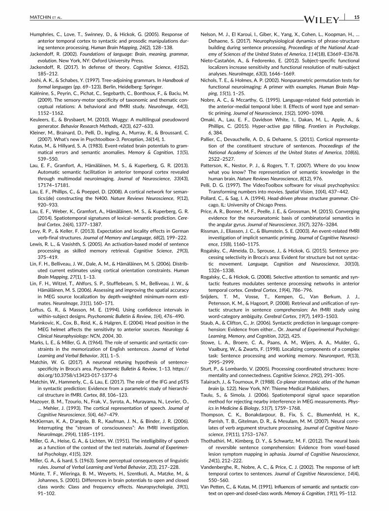
Humphries, C., Love, T., Swinney, D., & Hickok, G. (2005). Response ofanterior temporal cortex to syntactic and prosodic manipulations dur-ing sentence processing. Human Brain Mapping, 26(2), 128–138.
Jackendoff, R. (2002). Foundations of language: Brain, meaning, grammar,evolution. New York, NY: Oxford University Press.
Jackendoff, R. (2017). In defense of theory. Cognitive Science, 41(S2),185–212.
Joshi, A. K., & Schabes, Y. (1997). Tree-adjoining grammars. In Handbook offormal languages (pp. 69–123). Berlin, Heidelberg: Springer.
Kalénine, S., Peyrin, C., Pichat, C., Segebarth, C., Bonthoux, F., & Baciu, M.(2009). The sensory-motor specificity of taxonomic and thematic con-ceptual relations: A behavioral and fMRI study. NeuroImage, 44(3),1152–1162.
Keuleers, E., & Brysbaert, M. (2010). Wuggy: A multilingual pseudowordgenerator. Behavior Research Methods, 42(3), 627–633.
Kleiner, M., Brainard, D., Pelli, D., Ingling, A., Murray, R., & Broussard, C.(2007). What's new in Psychtoolbox-3. Perception, 36(14), 1.
Kutas, M., & Hillyard, S. A. (1983). Event-related brain potentials to gram-matical errors and semantic anomalies. Memory & Cognition, 11(5),539–550.
Lau, E. F., Gramfort, A., Hämäläinen, M. S., & Kuperberg, G. R. (2013).Automatic semantic facilitation in anterior temporal cortex revealedthrough multimodal neuroimaging. Journal of Neuroscience, 33(43),17174–17181.
Lau, E. F., Phillips, C., & Poeppel, D. (2008). A cortical network for seman-tics:(de) constructing the N400. Nature Reviews Neuroscience, 9(12),920–933.
Lau, E. F., Weber, K., Gramfort, A., Hämäläinen, M. S., & Kuperberg, G. R.(2014). Spatiotemporal signatures of lexical–semantic prediction. Cere-bral Cortex, 26(4), 1377–1387.
Levy, R. P., & Keller, F. (2013). Expectation and locality effects in Germanverb-final structures. Journal of Memory and Language, 68(2), 199–222.
Lewis, R. L., & Vasishth, S. (2005). An activation-based model of sentenceprocessing as skilled memory retrieval. Cognitive Science, 29(3),375–419.
Lin, F. H., Belliveau, J. W., Dale, A. M., & Hämäläinen, M. S. (2006). Distrib-uted current estimates using cortical orientation constraints. HumanBrain Mapping, 27(1), 1–13.
Lin, F. H., Witzel, T., Ahlfors, S. P., Stufflebeam, S. M., Belliveau, J. W., &Hämäläinen, M. S. (2006). Assessing and improving the spatial accuracyin MEG source localization by depth-weighted minimum-norm esti-mates. NeuroImage, 31(1), 160–171.
Loftus, G. R., & Masson, M. E. (1994). Using confidence intervals inwithin-subject designs. Psychonomic Bulletin & Review, 1(4), 476–490.
Marinkovic, K., Cox, B., Reid, K., & Halgren, E. (2004). Head position in theMEG helmet affects the sensitivity to anterior sources. Neurology &Clinical Neurophysiology: NCN, 2004, 30.
Marks, L. E., & Miller, G. A. (1964). The role of semantic and syntactic con-straints in the memorization of English sentences. Journal of VerbalLearning and Verbal Behavior, 3(1), 1–5.
Matchin, W. G. (2017). A neuronal retuning hypothesis of sentence-specificity in Broca's area. Psychonomic Bulletin & Review, 1–13. https://doi.org/10.3758/s13423-017-1377-6
Matchin, W., Hammerly, C., & Lau, E. (2017). The role of the IFG and pSTSin syntactic prediction: Evidence from a parametric study of hierarchi-cal structure in fMRI. Cortex, 88, 106–123.
Mazoyer, B. M., Tzourio, N., Frak, V., Syrota, A., Murayama, N., Levrier, O.,… Mehler, J. (1993). The cortical representation of speech. Journal ofCognitive Neuroscience, 5(4), 467–479.
McKiernan, K. A., D'angelo, B. R., Kaufman, J. N., & Binder, J. R. (2006).Interrupting the “stream of consciousness”: An fMRI investigation.NeuroImage, 29(4), 1185–1191.
Miller, G. A., Heise, G. A., & Lichten, W. (1951). The intelligibility of speechas a function of the context of the test materials. Journal of Experimen-tal Psychology, 41(5), 329.
Miller, G. A., & Isard, S. (1963). Some perceptual consequences of linguisticrules. Journal of Verbal Learning and Verbal Behavior, 2(3), 217–228.
Münte, T. F., Wieringa, B. M., Weyerts, H., Szentkuti, A., Matzke, M., &Johannes, S. (2001). Differences in brain potentials to open and closedclass words: Class and frequency effects. Neuropsychologia, 39(1),91–102.
Nelson, M. J., El Karoui, I., Giber, K., Yang, X., Cohen, L., Koopman, H., …Dehaene, S. (2017). Neurophysiological dynamics of phrase-structurebuilding during sentence processing. Proceedings of the National Acad-emy of Sciences of the United States of America, 114(18), E3669–E3678.
Nieto-Castañón, A., & Fedorenko, E. (2012). Subject-specific functionallocalizers increase sensitivity and functional resolution of multi-subjectanalyses. NeuroImage, 63(3), 1646–1669.
Nichols, T. E., & Holmes, A. P. (2002). Nonparametric permutation tests forfunctional neuroimaging: A primer with examples. Human Brain Map-ping, 15(1), 1–25.
Nobre, A. C., & Mccarthy, G. (1995). Language-related field potentials inthe anterior-medial temporal lobe: II. Effects of word type and seman-tic priming. Journal of Neuroscience, 15(2), 1090–1098.
Omaki, A., Lau, E. F., Davidson White, I., Dakan, M. L., Apple, A., &Phillips, C. (2015). Hyper-active gap filling. Frontiers in Psychology,6, 384.
Pallier, C., Devauchelle, A. D., & Dehaene, S. (2011). Cortical representa-tion of the constituent structure of sentences. Proceedings of theNational Academy of Sciences of the United States of America, 108(6),2522–2527.
Patterson, K., Nestor, P. J., & Rogers, T. T. (2007). Where do you knowwhat you know? The representation of semantic knowledge in thehuman brain. Nature Reviews Neuroscience, 8(12), 976.
Pelli, D. G. (1997). The VideoToolbox software for visual psychophysics:Transforming numbers into movies. Spatial Vision, 10(4), 437–442.
Pollard, C., & Sag, I. A. (1994). Head-driven phrase structure grammar. Chi-cago, IL: University of Chicago Press.
Price, A. R., Bonner, M. F., Peelle, J. E., & Grossman, M. (2015). Convergingevidence for the neuroanatomic basis of combinatorial semantics inthe angular gyrus. Journal of Neuroscience, 35(7), 3276–3284.
Rissman, J., Eliassen, J. C., & Blumstein, S. E. (2003). An event-related fMRIinvestigation of implicit semantic priming. Journal of Cognitive Neurosci-ence, 15(8), 1160–1175.
Rogalsky, C., Almeida, D., Sprouse, J., & Hickok, G. (2015). Sentence pro-cessing selectivity in Broca's area: Evident for structure but not syntac-tic movement. Language, Cognition and Neuroscience, 30(10),1326–1338.
Rogalsky, C., & Hickok, G. (2008). Selective attention to semantic and syn-tactic features modulates sentence processing networks in anteriortemporal cortex. Cerebral Cortex, 19(4), 786–796.
Snijders, T. M., Vosse, T., Kempen, G., Van Berkum, J. J.,Petersson, K. M., & Hagoort, P. (2008). Retrieval and unification of syn-tactic structure in sentence comprehension: An fMRI study usingword-category ambiguity. Cerebral Cortex, 19(7), 1493–1503.
Staub, A., & Clifton, C., Jr. (2006). Syntactic prediction in language compre-hension: Evidence from either... Or. Journal of Experimental Psychology:Learning, Memory, and Cognition, 32(2), 425.
Stowe, L. A., Broere, C. A., Paans, A. M., Wijers, A. A., Mulder, G.,Vaalburg, W., & Zwarts, F. (1998). Localizing components of a complextask: Sentence processing and working memory. Neuroreport, 9(13),2995–2999.
Sturt, P., & Lombardo, V. (2005). Processing coordinated structures: Incre-mentality and connectedness. Cognitive Science, 29(2), 291–305.
Talairach, J., & Tournoux, P. (1988). Co-planar stereotaxic atlas of the humanbrain (p. 122). New York, NY: Thieme Medical Publishers.
Taulu, S., & Simola, J. (2006). Spatiotemporal signal space separationmethod for rejecting nearby interference in MEG measurements. Phys-ics in Medicine & Biology, 51(7), 1759–1768.
Thompson, C. K., Bonakdarpour, B., Fix, S. C., Blumenfeld, H. K.,Parrish, T. B., Gitelman, D. R., & Mesulam, M. M. (2007). Neural corre-lates of verb argument structure processing. Journal of Cognitive Neuro-science, 19(11), 1753–1767.
Thothathiri, M., Kimberg, D. Y., & Schwartz, M. F. (2012). The neural basisof reversible sentence comprehension: Evidence from voxel-basedlesion symptom mapping in aphasia. Journal of Cognitive Neuroscience,24(1), 212–222.
Vandenberghe, R., Nobre, A. C., & Price, C. J. (2002). The response of lefttemporal cortex to sentences. Journal of Cognitive Neuroscience, 14(4),550–560.
Van Petten, C., & Kutas, M. (1991). Influences of semantic and syntactic con-text on open-and closed-class words.Memory & Cognition, 19(1), 95–112.
MATCHIN ET AL. 15

Vosse, T., & Kempen, G. (2000). Syntactic structure assembly in humanparsing: A computational model based on competitive inhibition and alexicalist grammar. Cognition, 75(2), 105–143.
Westerlund, M., Kastner, I., Al Kaabi, M., & Pylkkänen, L. (2015). The LATLas locus of composition: MEG evidence from English and Arabic. Brainand Language, 141, 124–134.
Westerlund, M., & Pylkkänen, L. (2014). The role of the left anterior tem-poral lobe in semantic composition vs. semantic memory. Neuropsycho-logia, 57, 59–70.
Zaccarella, E., Schell, M., & Friederici, A. D. (2017). Reviewingthe functional basis of the syntactic merge mechanism forlanguage: A coordinate-based activation likelihood estimationmeta-analysis. Neuroscience & Biobehavioral Reviews, 80,646–656.
SUPPORTING INFORMATION
Additional supporting information may be found online in the Sup-
porting Information section at the end of the article.
How to cite this article: Matchin W, Brodbeck C,
Hammerly C, Lau E. The temporal dynamics of structure and
content in sentence comprehension: Evidence from
fMRI-constrained MEG. Hum Brain Mapp. 2018;1–16. https://
doi.org/10.1002/hbm.24403
16 MATCHIN ET AL.