Embed Size (px)
Citation preview

The tcpO2 handbook

Data subject to change without notice.Radiometer, the Radiometer logo, ABL, AQT, TCM, RADIANCE, AQURE, PICO,CLINITUBES and QUALICHECK are trademarks of Radiometer Medical ApS.
By Franz von Wirth, Radiometer GmbH, Annette Thomsen and Jesper Bryder-Jacobsen, Radiometer Medical ApS.
Copyright © 2012 Radiometer Medical ApS, Denmark.Contents may be freely reproduced if the source is acknowlegded.
Printed in Denmark by Radiometer Medical ApS, DK-2700 Brønshøj, 2012.
ISBN 87-88138-87-9989-969. 201201F.
US Federal law restricts this device to sale,distribution or use by or on the order of a physician.

Introduction
tcpO2 monitoring How to place an sensor ............................................................... 8
Wound care Diagnosis of ischemia .................................................................. 12 Diabetic foot syndrome ............................................................... 13 Healing probability ...................................................................... 14 Prediction of wound healing ........................................................ 16 Spinal cord stimulation ................................................................ 18 Hyperbaric treatment ................................................................... 20 Evaluation of vasodilaters ............................................................ 21
Predicting amputation Suggesting amputation level ....................................................... 24 Amputation level – healing prognosis .......................................... 26
Use of TCM400 The menu structure of the monitor .............................................. 29 Calibration .................................................................................. 30 Cleaning ..................................................................................... 32 Maintenance of the sensor .......................................................... 33
Methods in diagnosis Comparison of methods – wound care ........................................ 38 Comparison of methods – amputation level ................................ 39
Cost savings with tcpO2
Calculate cost savings .................................................................. 41


Introduction
This small handbook is a quick guide to transcutaneous oxygen
(tcpO2) in a clinical perspective.
The intention of this handbook is to give healthcare workers
an overview and short-form information on how to use tcpO2
in their daily work.
For more clinical details, please see the references. For a
more detailed presentation of the technical issues and a
troubleshooting guide of the TCM400, please look in the
operator’s manual.
The validity of the measurement results from this instrument
must be carefully examined by a clinician and related to the
patient’s clinical condition before any clinical decisions are
made on the basis of these results.
The tcpO2 handbook is based on scientific literature and
the operator’s manual and gives suggestions on how the
procedures may be carried out according to clinical studies.
It is the intention that the users of the tcpO2 handbook always
make the relevant modifications according to local policies
and procedures as well as review and approve the procedures
suggested herein prior to their implementation.

6
O2
O2
O2O2
O2
O2
O2
O2
O2
O2
O2
O2 O2
O2
O2
tcpO2 monitoring
Transcutaneous oxygen
Transcutaneous oxygen (tcpO2) is a non-invasive monitoring
of the oxygen tension in the skin. The monitoring is done by
placing a Clark-type sensor on the skin so that it heats up the
skin and provides tcpO2 values.
tcpO2 is a direct indication of the microvascular function. As
opposed to pressure and volume assessments, tcpO2 maps the
actual oxygen supply available for the skin tissue cells. tcpO2
also responds to macrocirculatory events, e.g. change in blood
pressure and provocational maneuvers.
The sensor on the skin
arterial venous
FIG. 1: Transcutaneous pO2 monitoring (TC)The heat from the sensor dilates the capillary and increases local blood flow and the diffusion of O2 through the skin to the sensor. tcpO2 is measured electrochemically inside the sensor.
Venous end of a capillary loop
Arterial end of a capillary loop
7
Practical application
Site selection – an ideal site would be located over a
homogeneous capillary bed without large veins, skin defects,
or hair. Placing the sensor directly over a bone may also give
erroneous results, especially if a change in body position causes
skin to be pulled against a protruding bone.Severe edema can
also give unreliable results.
The sensor
The sensor must be in contact with the tissue through the
contact liquid. If there is air between the tissue and the sensor
the tcpO2 values will be questionable.
Initiation of monitoring
It takes about 15 – 20 minutes after the probe has been placed
on the skin for the tcpO2 to stabilize.
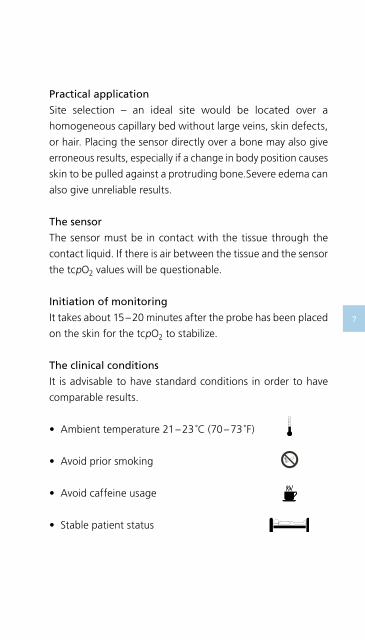
The clinical conditions
It is advisable to have standard conditions in order to have
comparable results.
• Ambienttemperature21–23̊C(70–73̊ F)
• Avoidpriorsmoking
• Avoidcaffeineusage
• Stablepatientstatus
8
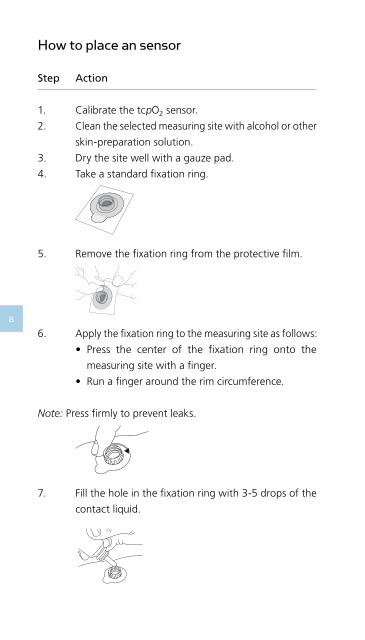
How to place an sensor
Step Action
1. Calibrate the tcpO2 sensor.
2. Clean the selected measuring site with alcohol or other
skin-preparation solution.
3. Dry the site well with a gauze pad.
4. Take a standard fixation ring.
5. Remove the fixation ring from the protective film.
6. Apply the fixation ring to the measuring site as follows:
• Press the center of the fixation ring onto the
measuring site with a finger.
• Runafingeraroundtherimcircumference.
Note: Press firmly to prevent leaks.
7. Fill the hole in the fixation ring with 3-5 drops of the
contact liquid.

9
8. Affix the sensor into the fixation ring as follows:
•Alignthearrowonthesensorwithoneofthemarks
on the fixation ring.
•Turnthesensor90°clockwisetofasten it inthe
fixation ring.
9. Repeat steps 1 to 8 if more sensors are to be applied.
Wait for a stable reading after the sensor has been affixed
to the patient.
Note: The physiological stabilization time of a patient is 15-20
minutes for the tcpO2 reading. During this time the sensor will
slowly heat the skin, making the arteries dilate. Longer time
may indicate an incorrect attachment of the sensor or a poorly
selected measuring site.
Typical tcpO2 mapping sites of the leg [1]
1. Sheffield P. J. Clinical Application of Transcutaneous pO2 in Hyperbaric Oxygen Treatment. Blood Gas News 1998; 7,2:10-13

10
Wound care
tcpO2tcpO2 can be used in a number of clinical situations. In this
section the clinical use of tcpO2 is addressed in:
• Diagnosisofischemia
• Diabeticfootsyndrome
• Healingprobability
• Predictionofwoundhealing(RPI)
• Spinalcordstimulation
• Hyperbarictreatment
• Evaluationofvasodilaters
• Predictingamputation
• Suggestingamputationlevel
• Amputationlevelhealingprognosis
11
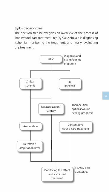
Monitoring the effect and success of
treatment
Determine amputation level
tcpO2
Critical ischemia
No ischemia
Amputation
Revasculization/ surgery
Conservative wound-care treatment
Diagnosis and quantification of disease
Therapeutical options/wound healing prognosis
Control and evaluation
tcpO2 decision tree
The decision tree below gives an overview of the process of
limb wound-care treatment. tcpO2 is a useful aid in diagnosing
ischemia, monitoring the treatment, and finally, evaluating
the treatment.

12
Diagnosis of ischemia
Application
Mapping the oxygenation around the wound and extremities
can recognize ischemia and severity of ischemia.
Diagnosing critical limb ischemia
tcpO2 - criteria for critical limb ischemia [2]. Forefoot tcpO2 in supine
and dependency position to indicate critical limb ischemia [2].
tcpO2 workflow
Use caution on patients with media sclerosis when measuring
ankle pressure
Ankle systolicarterial pressure
< 60 mmHg?
Dependent tcpO2 < 40 – 45 mmHg?
Critical ischemia may be assumed
Yes
Yes
Yes
No
No
No
Critical ischemia should not be
assumed
Supine tcpO2
< 10 – 15 mmHg?
2. Scheffler A et al. Influence of clinical findings, positional maneuvers, and systolic ankle arterial pressure on tcpO2 in PAOD. Eur J F Clin Invest 1992; 22: 420-26.

13
Diabetic foot syndrome
Application
Determination of tcpO2 appears to be a useful tool in screening
type 2 diabetic patients for foot at risk.
Screening
The tcpO2 value is monitored at the dorsum of the foot in
supine and sitting position.
tcpO2 difference between supine and
sitting (mmHg) [3]
Diabetic group with foot at risk 23.5 ± 2.0
Diabetic control group 15.0 ± 1.4
Control group 13.3 ± 1.1
Type 2 diabetic patient with foot at risk was defined as a foot
with neuropathy but without ulceration or previous ulceration.
Diabetic control group was type 2 diabetic patients without
foot lesions or neuropathy and control group was normal
subjects.
Notes
3. Zimny S, Dessel F, Ehren M. Early detection of microcirculatory impairment in diabetic patients with foot at risk. Diabetes Care 2001; 24,10: 1810-14.

14
Healing probability
Application
By performing provocational maneuvers such as elevating the
leg, the macro- and microcirculatory capacity can be evaluated.
For tcpO2 values between 20 and 40 mmHg in supine position
it is advisable to measure tcpO2 on an elevated leg to diagnose
the healing probability [4].
How to do the provocational maneuver
tcpO2 on elevated leg for three minutes.
Predicting healing possibility
According to T.Rooke [4] a supine measurement below 20
mmHg indicates problems in wound healing and values above
40 mmHg indicate that the wound will heal. In the area
between 20 and 40 mmHg, leg elevation can predict outcome.
When the decrease is less than 10 mmHg, 80 % of the wounds
will heal.
When the decrease is greater than 10 mmHg, 80 % of the
wounds will fail to heal.
Decrease of < 10 mmHg 80 % healing
Decrease of > 10 mmHg 80 % fails to heal
30°to45°
4. Rooke T. tcpO2 in non-invasive vascular medicine. Blood Gas News 1998; 7,2: 21-23.

15
Notes
16
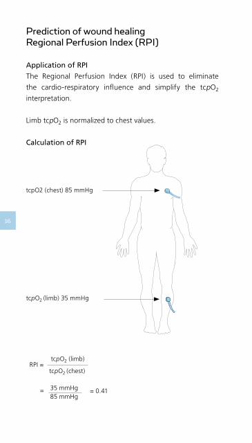
Prediction of wound healingRegional Perfusion Index (RPI)
Application of RPI
The Regional Perfusion Index (RPI) is used to eliminate
the cardio-respiratory influence and simplify the tcpO2
interpretation.
Limb tcpO2 is normalized to chest values.
Calculation of RPI
tcpO2 (chest) 85 mmHg
tcpO2 (limb) 35 mmHg
tcpO2 (limb)
tcpO2 (chest)RPI =
35 mmHg85 mmHg
= = 0.41
17
5. Hauser CJ. Tissue salvage by mapping of skin surface transcutaneous oxygen tension index. Arch Surg 1987; 122: 1128-30.
Prediction of wound healing
RPI = < 0.4 predicts a poor outcome [5]
RPI = > 0.6 predicts an excellent outcome [5]
0.4 < RPI < 0.6 some heal and some do not [5]
Notes

18
Spinal cord stimulation
Application
In a study by W. Amann et al [6] the effect of spinal cord
stimulation (SCS) on patients with unreconstructable critical
leg iscemia (CLI) is documented.
The study indicates that in a preselected group of patients limb
salvage, pain and wound care can be improved.
tcpO2 in spinal cord stimulation treatment
• SelectionofpatientsforSCSonthebasisofmicrocirculation
and microcirculatory response to test stimulation.
• Evaluateeffectoftreatment
Notes

19
Flow chart for patient selection based on tcpO2 measured in
supine position. [6]
6. W. Amann et al. Spinal cord stimulation in the treatment of non-reconstructable stable critical leg ischaemia: Results of the European peripheral vascular disease outcome study (SCS-EPOS). Eur J Vasc Endovasc Surg: 2003; 26: 280-86.
Permanent implant and SCS-Match patient
Atherosclerotic chronic stable
CLI
Wet gangrene/ulcers > 3 cm
Not eligible for study
Non-reconstructable
tcpO2 < 10 mmHg 10 ≥ tcpO2 ≤ 30 mmHg
tcpO2 > 30 mmHg
tcpO2 > 10 mmHg
Trial screening Trial screening
Increase in tcpO2 to ≥ 20 mmHg
Adequate paraesthesia
coverage and adequate pain relief?
SCS-No-Match or No-SCS patient
Yes
Yes
Yes Yes
NoNo
No
Yes
Yes
No No
No
20
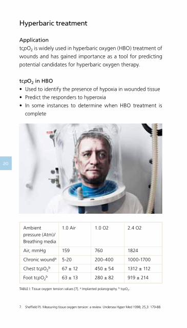
Hyperbaric treatment
Application
tcpO2 is widely used in hyperbaric oxygen (HBO) treatment of
wounds and has gained importance as a tool for predicting
potential candidates for hyperbaric oxygen therapy.
tcpO2 in HBO
• Usedtoidentifythepresenceofhypoxiainwoundedtissue
• Predicttheresponderstohyperoxia
• In some instances to determine when HBO treatment is
complete
TABLE I: Tissue oxygen tension values [7]. a Implanted polarography. b tcpO2.
7. Sheffield PJ. Measuring tissue oxygen tension: a review. Undersea Hyper Med 1998; 25,3: 179-88.
Ambient pressure (Atm)/ Breathing media
1.0 Air 1.0 O2 2.4 O2
Air, mmHg 159 760 1824
Chronic wounda 5-20 200-400 1000-1700
Chest tcpO2b 67 ± 12 450 ± 54 1312 ± 112
Foot tcpO2b 63 ± 13 280 ± 82 919 ± 214

21
tcpO2 [mmHg]
60
50
40
30
200 30 60 120 180 240
Scheffler [8]
[Min]
30 µg PGE1
40 µg PGE1
20 µg PGE1
8. Scheffler et al. Therapeutic efficacy of intravenously applied prostaglandin E1. VASA 1989; -Suppl.28:19-25.
Evaluation of vasodilaters
Application
Until now the only way to document the effect of vasodilator
therapy was improvement of the clinical symptoms. tcpO2
measurements in the beginning of the treatment as well as
during the couse of treatment make it possible to monitor the
effect of the treatment.
Therapeutic effects in pure conservative therapy (e.g. PGE1)
is to be expected on the microcirculatory level and can be
documented through a significant increase in tcpO2.
In the conservative therapy of arterial vascular diseases with
expensive Prostaglandin products, an early prognosis of the
success rate of the treatment can be estimated. If necessary
a change in the therapy can be implemented, which can save
a substantial amount of money.
Example
22
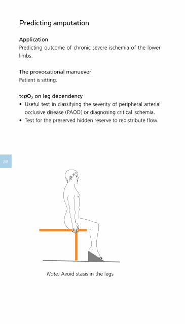
Predicting amputation
Application
Predicting outcome of chronic severe ischemia of the lower
limbs.
The provocational manuever
Patient is sitting.
tcpO2 on leg dependency
• Usefultestinclassifyingtheseverityofperipheralarterial
occlusive disease (PAOD) or diagnosing critical ischemia.
• Testforthepreservedhiddenreservetoredistributeflow.
Note: Avoid stasis in the legs

23
Predictive value
Patients with a tcpO2 below 10 mmHg, measured at the
dorsum of the foot in supine position, have poor prognosis.
To improve predictions, measurements are done on sitting
patients [9].
Forefoot > 40 mmHg 5 % PAOD patients required
amputation [9].
Forefoot < 10 mmHg 85 % PAOD patients were amputated,
in spite of 2.3 ±1.51 arterial
reconstructive operations [9].
Notes
9. Becker et al. Predictive value of tcpO2 in chronic severe ischemia of lower limbs. Int J Microcirc: Clin Exp 1988; 7: 270.

24
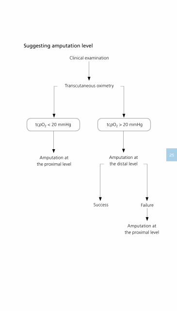
Suggesting amputation level
Application
The healing success after amputation is highly dependent on
oxygenation of the tissue.
By mapping tcpO2 levels on the leg an optimal amputation
level can be suggested, reducing amputation of well-perfused
tissue and reamputations.
In a study by A. Misuri et al [10] a reference level of 20 mmHg
was evaluated as suitable for evaluation of the amputation
level. The decision tree on the following page is from the
article. For further information from the article, please see
page 37 of this handbook.
Notes
10. Misuri A et al. The Journal of Cardiovascular Surgery 2000.
25
Suggesting amputation level
Clinical examination
tcpO2 < 20 mmHg tcpO2 > 20 mmHg
Amputation at the proximal level
Amputation atthe distal level
Success Failure
Amputation at the proximal level
Transcutaneous oximetry

26
O2
Amputation level – healing prognosis
Application
Suggesting the right level of amputation.
The provocational maneuvre
tcpO2 with oxygen inhalation:
100 % oxygen admission for 10 minutes.
Suggesting amputation level
For successful amputation stump healing
Initial tcpO2 values [11] > 10 mmHg
Increased tcpO2 after O2 inhalation [11] > 10 mmHg
*Values from dorsum of the foot and 10 cm distal to the knee
For unsuccessful amputation stump healing
Initial tcpO2 values [11] < 10 mmHg
Increased tcpO2 after O2 inhalation [11] ≤ 10 mmHg
*Values from dorsum of the foot and 10 cm distal to the knee
11. Harward TR, Volny J, Golbranson F et al. Oxygen inhalation-induced tcpO2 changes as a predictor of amputation level. J Vasc Surg 1985; 2: 220-28.

27
Notes
28
Use of TCM400
This section of the handbook gives an overview of how to
work with the TCM400. For further information, please see
the TCM400 Operator´s Manual.
Please refer to the operator’s manual of the TCM400 for further
details prior to using TCM400.
After the sensor has been placed it takes 15 – 20 minutes for
the measuring site to be thoroughly heated and stable results
to be displayed on the monitor. A measuring temperature of
43–45°C(109–113°F)isrecommended.
During monitoring, the results can be viewed as curves, trend
table, and the current results.
A measuring sequence with TCM400 is easy:
1. Start the monitor
2. Calibrate
3. Apply the sensor to the measuring site
4. Wait 15 – 20 minutes
5. Measure and perform provocational maneuvers
6. Print report
29
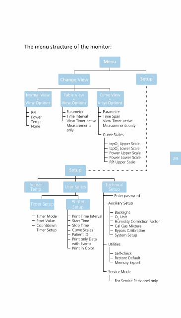
The menu structure of the monitor:
Menu
Setup
Setup
RPIPowerTemp.None
Enter password
Timer ModeStart ValueCountdown Timer Setup
Print Time IntervalStart TimeStop TimeCurve ScalesPatient IDPrint only Data with EventsPrint in Color
Auxiliary Setup
Backlight O2 Unit Humidity Correction Factor Cal Gas Mixture Bypass Calibration System Setup
Utilities
Self-check Restore Default Memory Export
Service Mode
For Service Personnel only
ParameterTime IntervalView Timer-active Measurements only
ParameterTime SpanView Timer-active Measurements only
Curve Scales tcpO2 Upper Scale tcpO2 Lower Scale Power Upper Scale Power Lower Scale RPI Upper Scale
Normal View+
View Options
Change View
Table View+
View Options
Curve View+
View Options
SensorTemp. User Setup
Printer Setup
Timer Setup
Technical Setup

30
Calibration
Radiometer recommends performing a calibration:
• priortoeachmonitoringperiod
• whenchangingmeasuringsites
• everyfourhours
• everytimeansensorhasbeenremembraned
Follow the steps below to calibrate the sensor(s) with
atmospheric air:
Step Action
1. Connect the tcpO2 sensor to the sensor socket on the
TCM400 system.
2. Insert the membraned sensor into the calibration
chamber on the TCM400 system.
3. Swing the sensor retainer into position over the sensor.
4. Adjust the humidity correction factor, if required.
Access: Menu → Setup → Technical Setup → Auxiliary
Setup.
5a. To calibrate all connected sensors simultaneously, press
Calibrate.
Result: If six sensors are connected, the following
screen appears:

31
5b. To calibrate one sensor at a time, press the number of
the relevant sensor.
Result: The Calibrate Single Sensor screen appears:
Press OK to start calibration, or press Cancel to return to the
previous screen without initiating the calibration.
Note: Pressing Calibrate will reset the numbering of events.
Note: While calibrating a single sensor, it is possible to monitor
with the other sensors.

32
Cleaning
Wipe the following parts gently with a soft cloth moistened
with skin antiseptic, e.g. 70 % alcohol:
• thesensorhead
• thecable
Note: Constant use of hand lotion containing isopropanol/
propylalcohol and alcohol prior to handling the sensor may
damage the cable. To avoid transferring lotion to the cable,
dry your hands prior to handling the sensor.
Cleaning the exterior
When cleaning the monitor:
• Shutdownthemonitor.
• Useaclothwhichhasbeenlightlydampenedwithsoapy
water or a mild detergent.
• Donotuseabrasivecleansersorpads:thefinishmaybecome
damaged.
• Do not use ethanol-based substances or aggressive
detergents. Extensive use may cause the plastic to become
brittle and cracks may occur.
Cleaning the touch screen
A dry or lightly dampened soft, lint-free cloth may be used
to clean the monitor’s touch screen. Simply wipe the screen
gently to remove fingerprints and/or dirt. To avoid streaking,
an approved screen cleaner is recommended.
Disinfection
Immerse the sensor and the cable in a 2 – 3 % aqueous solution
of active dialdehydes.
WARNING/CAUTION: Do not immerse the sensor plug in the
disinfection solution, as this will cause the sensor to fail.

33
Maintenance of the sensor
To obtain reliable values, remembrane the sensor every week.
Follow the steps below to prepare the sensor for membraning:
Step Action
1. Remove the old O-rings:
Slide the O-ring remover under the O-ring, just above
the arrow on the sensor house.
2. Turn the O-ring remover clockwise to release the
O-ring.
3. Peel off the old membrane.
WARNING/CAUTION: Do not heat sterilize as the sensor cannot
toleratetemperaturesexceeding70°C(158°F),asthiswill
cause the sensor to fail.
34
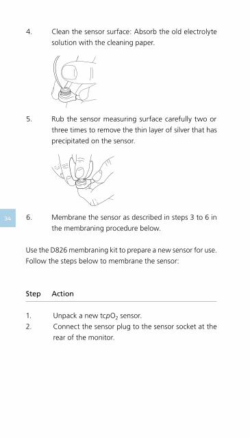
4. Clean the sensor surface: Absorb the old electrolyte
solution with the cleaning paper.
5. Rub the sensor measuring surface carefully two or
three times to remove the thin layer of silver that has
precipitated on the sensor.
6. Membrane the sensor as described in steps 3 to 6 in
the membraning procedure below.
Use the D826 membraning kit to prepare a new sensor for use.
Follow the steps below to membrane the sensor:
Step Action
1. Unpack a new tcpO2 sensor.
2. Connect the sensor plug to the sensor socket at the
rear of the monitor.

35
3. Apply two drops of the tcpO2 electrolyte solution to
the surface of the tcpO2 sensor.
Note: Check that the electrolyte solution
covers the entire surface without air bubbles.
4. Membrane the tcpO2 sensor:
•Placethemembraneunitonahardandstable
surface.
•Insertthesensorheadintothetopofthe
white tcpO2 membrane unit.
5. • Pressthesensorfirmlyintotheunituntila
click is heard.
• Removethesensorfromtheunitandwipe
off the surplus electrolyte solution with the
cleaning paper.
6. Check that the system shows Calibration Required,
and calibrate the sensor as described in chapter 5:
Calibration.

36
Methods in diagnosis
tcpO2 provides non-invasive monitoring of the oxygen tension
of the skin:
• Itgivesdirectindicationofmicrovascularfunction.
• Asopposedtopressureandvolumeassessments,tcpO2
maps the actual oxygen supply available for the skin tissue
cells.
• tcpO2 responds to macrocirculatory events, e.g. change in
blood pressure, provocational maneuvers and removal of
plaque.
Notes

37
Ankle blood pressure – Blood cuffs are placed at the ankle
and inflated to block blood flow. Measurements are made
with a Doppler sensing unit placed distal to the cuffs when
pressure is released. The method measures arterial circulation
and is used to assess disease severity. The method may give
falsely elevated values due to incompressibility of the arteries
caused by calcification – often seen in diabetics – and can be
painful to the patient.
Toe blood pressure – A small cuff is applied to the toe
and inflated to above toe systolic pressure and then slowly
deflated. A Doppler device registers when pressure is released.
The method measures arterial circulation and is used in the
diagnosis of occlusive diseases. It cannot be used if there are
ulcers on the toes or if toe pulse is severely reduced or absent
in critical ischemia.
Duplex scanning – Vessels are visualized by a two-dimensional
ultrasound imaging. Blood flow is displayed on the screen as
a picture. The method can be used to evaluate arterial and
venous circulation and locate blockages. It does not quantify
local oxygen perfusion.
Angiography – An invasive examination with the injection
of a contrast agent through a needle or catheter into one
or more arteries to make them visible by x-ray. The method
provides a road map of blockages in the arteries. It does not
quantify local oxygen perfusion and the contrast agent may
cause discomfort.
Laser Doppler – The method is based on the principle of
Doppler shift of laser light back-scattered by the movement of
red blood cells. The perfusion is measured with laser probes.
38
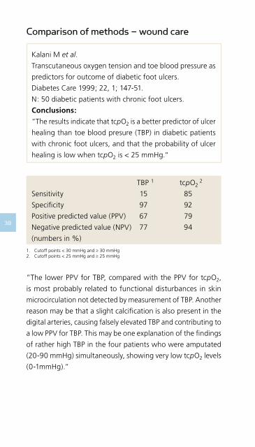
TBP 1 tcpO2 2
Sensitivity 15 85
Specificity 97 92
Positive predicted value (PPV) 67 79
Negative predicted value (NPV) 77 94
(numbers in %)
”The lower PPV for TBP, compared with the PPV for tcpO2,
is most probably related to functional disturbances in skin
microcirculation not detected by measurement of TBP. Another
reason may be that a slight calcification is also present in the
digital arteries, causing falsely elevated TBP and contributing to
a low PPV for TBP. This may be one explanation of the findings
of rather high TBP in the four patients who were amputated
(20-90 mmHg) simultaneously, showing very low tcpO2 levels
(0-1mmHg).”
Comparison of methods – wound care
1. Cutoff points < 30 mmHg and ≥ 30 mmHg2. Cutoff points < 25 mmHg and ≥ 25 mmHg
Kalani M et al.
Transcutaneous oxygen tension and toe blood pressure as
predictors for outcome of diabetic foot ulcers.
Diabetes Care 1999; 22, 1; 147-51.
N: 50 diabetic patients with chronic foot ulcers.
Conclusions:
“The results indicate that tcpO2 is a better predictor of ulcer
healing than toe blood presure (TBP) in diabetic patients
with chronic foot ulcers, and that the probability of ulcer
healing is low when tcpO2 is < 25 mmHg.“

39
Comparison of methods – amputation level
Misuri A et al.
Predictive value of transcutaneous oximetry for selection of the
amputation level.
The Journal of Cardiovascular Surgery 2000; 41: 83-87. N: 30.
Conclusions:
“Following our observations and according to some reported
studies, we believe transcutaneous oximetry to be the best
method for selection of amputation level. This is a simple,
noninvasive and accurate method, which has showed itself
superior to other techniques (i.e. Doppler and radioisotope).”
“An accurate method is the radioisotope technique using
Xenon, proposed by Lassen and Holstein. Recently, using this
technique Dwars et al have reported a positive predictive value
of 89 % and a negative predictive value of 99 %. Nevertheless,
application of this technique is not widely spread, due to the
fact that the necessary equipment is complex to use, expensive,
requires specialized technicians, and use of a radioisotope.
Transcutaneous oximetry is a noninvasive method, which moreover
is quantitative, easy to apply and with high diagnostic accuracy
despite some false-positive or false-negative values, but in rare
cases. It can be employed in all cases, including those with blood
flow in distal (pedal) arteries not found by Doppler technique. It
enables the evaluation of all presumed levels of amputation.
Finally, based on these data of transcutaneous oximetry and
comparison of it with other methods, we can conclude that
transcutaneous oximetry is close to being optimal. Unlike
Doppler and radioisotope techniques, this investigation is
noninvasive (as Doppler technique), easily carried out (while
Doppler technique is not appropiate for calcified arterial wall
and blood flow velocity less than 6 cm/sec). It is simple, does
not generate radiation and is cost-effective.”
40
Cost savings with tcpO2
In this section examples of costs of wound care and amputation
are presented.
Wound care
In a Dutch study [12] by Bouter et al the in-hospital cost of
primary healing was USD 10,000, and Apelqvist et al found
in a Swedish study [13] that the total direct costs until healing
was USD 8,500.
Early identification of ineffective treatments and obtaining
information to better decide how to treat a patient or when
and where to send a patient for further analysis optimize the
clinical process. The improved foundation for making the optimal
decision may lead to cost savings and increased patient life
quality.
Amputation
Several studies of the cost of amputation have been made.
In a Dutch study [12] the in-hospital cost of an amputation
was USD 15,000.
A Swedish study [13] investigated the total direct costs until
healing and found the total average cost for patients who
healed after a primary amputation was USD 48,000 compared
with USD 74,000 for patients with reamputation.By decreasing
the levels of reamputations costs can be reduced. In a
Norwegian study [14] by Witso et al 19 % of patients having
had an amputation were reamputated.
12. Bouter et al. The diabetic foot in Dutch hospitals: Epidemiological features and clinical outcome. The European Journal of Medicine; 1994; 2, 215-18. 13. Apelqvist et al. Diabetic foot ulcers in a multidisciplinary setting. An economic analysis of primary healing and healing with amputation. Journal of International Medicine 1994; 235: 463-71.14. Witso et al. Lower limb amputations: registration of all lower limb amputations performed at the University Hospital of Trondheim, Norway, 1994-1997. Prosthetics and orthotics international 2001; 25,3: 181-85.
41
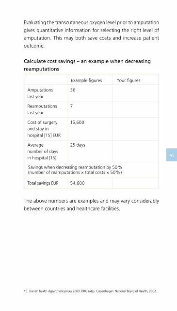
Savings when decreasing reamputation by 50 % (number of reamputations × total costs × 50 %)
Evaluating the transcutaneous oxygen level prior to amputation
gives quantitative information for selecting the right level of
amputation. This may both save costs and increase patient
outcome.
Calculate cost savings – an example when decreasing
reamputations
15. Danish health department prices 2003. DRG rates. Copenhagen: National Board of Health, 2002.
The above numbers are examples and may vary considerably
between countries and healthcare facilities.
Example figures Your figures
Amputations 36 last year
Reamputations 7last year
Cost of surgery 15,600and stay in
hospital [15] EUR
Average 25 daysnumber of days in hospital [15]
Total savings EUR 54,600

42
Notes

43
Notes