Embed Size (px)
Citation preview

184 SYSTEMATIC POSITION OF Pseudomicrothorax
8. FaurC-Fremiet, E. (1950). Morphologie comparke et sys- tCmatique des ciliCs. Bull. Soc. Zool. France, 75, 109-122.
9. Kahl, A. (1926). Neue und wenig bekannte Formen der holotricben und heterotrichen Ciliaten. Arch. Protistenk., 55,
10. - (1931). Holotricha (ausser den im 1. Teil behan- delten Prostomata), part 2 1 of Dahl, F., Die Tierzuelt Deutsch- lands, G. Fischer, Jena, 181-398.
11. Klein, B. M . (1928). Die Silberliniensysteme der Cilia- ten. Weitere Resultate. Arch. Protistenk., 62, 177-260.
12. Maupas, E (1883). Contribution 2 1’Ctude morpho- logique et anatomique des infusoires ciliPs. Arch. sool. e x p t l . e t g h z . (sbr. 2 ) , 1, 427-664.
1.3. Mermod, G. (1914). Recherches sur la faune infusor- ienne des tourbitires et des eaux voisines de Sainte-Croix ( Jura vaudois). Rev. sztisse sool., 22, 31-114.
14. Penard, E. (1922). &udes sur les litfusoires d’Eau doure. Georg & Cie, Gentve.
15. Savoie, A. (1957). Le ciliC Trichopelma agilis n. sp. J . Protozool,, 4, 276-280.
197-438.
16. Sondheim, M. (1929). Protozoen aus der Ausbeute der Voeltzkowschen Reisen in Madagaskar und Ostafrika. A b- handl. senckenberg. naturforsch. Ges., 41, 285-313.
17. Srimek-Husek, R. (1952). Uber einige Bemerkenswerte Ciliaten aus bohmischen Moosen. Cesk. Biol., Praha, 1, 367- 376. [ In Russian with German summary.]
18. Thompson, J . C. , Jr. (1955). Morphology of a new species of Tetrahymena. (Abstr.) J . Protosool., 2 (Suppl.), 12.
19. __. (1956). Observations on the buccal ciliature o i Tetrahymena. (Abstr.) J . Protosool., 3 (Suppl.), 4.
20. __. (1957). Pseudonzicrothoraa gellirti n. sp., a hymenostome ciliate from American moss. (Abstr.) Assoc. Southeastern Biol. Bull., 4, 15.
21. Tuffrau, M. (1954). Discotricha papillifera, n. g., n. sp. CiliC psammobie de la famille des Trichopelmidae. J . Proto- zool., 1, 183-186.
2 2 . Wang, C. C. & Nie, D. (1935). Report on the rare and new species of fresh-water infusoria, Part 11. Sinensia, 6, 399- 524.
J. PROTOZOOL., 5(3), 184-193 (1958)
The Systematic Position of Pseudomicrothorax dubius, Ciliate with a Unique Combination of Anatomical Features”
JOHN 0. CORLISS Department o f Zoology, University o f Illinois, Urbana, Illinois
SYNOPSIS. The rather rarely found holotrichous ciliate Pseudoinicrothorax dztbius, generally considered in the most authoritative literature to be a member of the order Trichostomatida, more likely belongs in the Hymenostomatida. I ts curious combination of trans-ordinal characteristics, however, makes difficult exact systematic allocation of it and of the congeneric species P . agil is . The provocative morphological features include: a gymnostome-like cytopharyngeal “basket” ; a tetrahymenal buccal apparatus, with membranelles comparable with those of primitive hymeno- stomes; an advanced semi-autonomous type of stomatogenesis ; and sensory bristles, flattened rigid form, and restricted ciliature reminiscent of cermtain spirotrichs. Brief discussion is presented con- cerning the influence of modern ideas of ciliate classification upon what is considered the proper taxonomic position of the genus
SEUDOMICROTHOR.4X DUBIUS, a rather P rare edaphic ciliate recently rediscovered in Amer- ica, appears to have been well named: some of its characteristics give a false impression of its taxonomic affinities, a t both high and low levels, and there is considerable doubt as to whether the organism has been allocated to its proper systematic position in the past. The unusual nature of its anatomy and of the morphogenetics of its stomatogenesis has been de- scribed by Dr. Jesse C. Thompson, Jr., and myself in the preceding paper in this number of the JOURNAL OF PROTOZOOLOGY (38). Its unique combination of morphological features, which, individually, generally have been held to be distinctive of several separate taxa a t the ordinal level, and its mode of stomato- genesis have made problematical the exact systematic
* Much of the work involved in the present investigation has been carried out under a grant from the Xational Science
Foundation, #G-3887.
affinities of the species. Thus it has seemed desirable to devote a paper to consideration of this problem. The related, and perhaps even more important, matter of the possible significance of the taxonomic affinities of P. dubius upon over-all problems of ciliate evolu- tion is considered beyond the scope and space of the present work and thus has been reserved for separate publication elsewhere ( 11).
The terminology for ciliate “mouth parts” adopted here essentially follows that proposed briefly 3-4 years ago( 7,8). The nomenclature and classification scheme used in reference to the higher taxa within the sub- phylum Ciliophora also follow that discussed earlier elsewhere (9,lO).
I am indebted to Professor E. Faurk-Fremiet, Col- l6ge de France, Paris, for his keen interest in my treat- ment of this subject and for his critical reading of the manuscript in its final form.
SYSTEMATIC POSITION OF Pseudomicrothorax 185
NATERIALS AND METHODS show several recognizably different traits which, a t
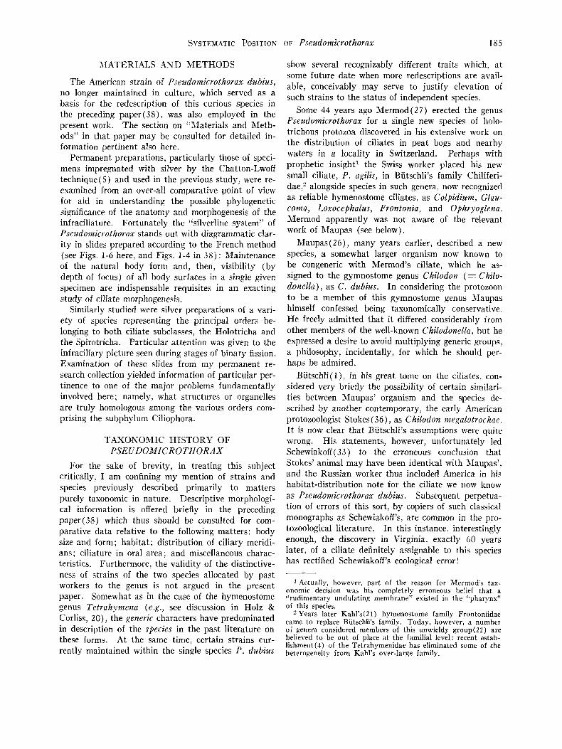
The American strain of Pseudomicrothorax dubius, no longer maintained in culture, which served as a basis for the redescription of this curious species in the preceding paper(38), was also employed in the present work. The section on “Materials and Meth- ods” in that paper may be consulted for detailed in- formation pertinent also here.
Permanent preparations, particularly those of speci- mens impregnated with silver by the Chatton-Lwoff technique(5) and used in the previous study, were re- examined from an over-all comparative point of view for aid in understanding the possible phylogenetic significance of the anatomy and morphogenesis of the infraciliature. Fortunately the “silverline system” of Pscudomicrothoran stands out with diagrammatic clar- ity in slides prepared according to the French method (see Figs. 1-6 here, and Figs. 1-4 in 38): Maintenance of the natural body form and, then, visibility (by depth of focus) of all body surfaces in a single given specimen are indispensable requisites in an exacting study of ciliate morphogenesis.
Similarly studied were silver preparations of a vari- ety of species representing the principal orders be- longing to both ciliate subclasses, the Holotricha and the Spirotricha. Particular attention was given to the infraciliary picture seen during stages of binary fission. Examination of these slides from my permanent re- search collection yielded information of particular per- tinence to one of the major problems fundamentally involved here; namely, what structures or organelles are truly homologous among the various orders com- prising the subphylum Ciliophora.
TASONO?(.IIC HISTORY OF PSEUDOMICROTHORAX
For the sake of brevity, in treating this subject critically, I am confining my mention of strains and species previously described primarily to matters purely taxonomic in nature. Descriptive morphologi- cal information is offered briefly in the preceding paper(38) which thus should be consulted for com- parative data relative to the following matters: body size and form; habitat; distribution of ciliary meridi- ans; ciliature in oral area; and miscellaneous charac- teristics. Furthermore, the validity of the distinctive- ness of strains of the two species allocated by past workers to the genus is not argued in the present paper. Somewhat as in the case of the hymenostome genus Tetrahynzena (e.g., see discussion in Holz & Corliss, 20), the generic characters have predominated in description of the species in the past literature on these forms. At the same time, certain strains cur- rently maintained within the single species P. dubius
some future date when more redescriptions are avail- able, conceivably may serve to justify elevation of such strains to the status of independent species.
Some 44 years ago Mermod(27) erected the genus Pseudomicrothorax for a single new species of holo- trichous protozoa discovered in his extensive work on the distribution of ciliates in peat bogs and nearby waters in a locality in Switzerland. Perhaps with prophetic insight1 the Swiss worker placed his new small ciliate, P. agilis, in Butschli’s family Chiliferi- dae,:! alongside species in such genera, now recognized as reliable hymenostome ciliates, as Colpidizrtn, Glau- coma, Loxocephalus, Frontonia, and Ophryoglena. Mermod apparently was not aware of the relevant work of Maupas (see below).
Maupas(26), many years earlier, described a new species, a somewhat larger organism now known to be congeneric with Mermod’s ciliate, which he as- signed to the gymnostome genus Chilodon (= Chilo- donella), as C . dubius. In considering the protozoon to be a member of this gymnostome genus >\.laupas himself confessed being taxonomically conservative. H e freely admitted that it differed considerably from other members of the well-known Chilodonella, but he expressed a desire to avoid multiplying generic groups, a philosophy, incidentally, for which he should per- haps be admired.
Butschli( l ) , in his great tome on the ciliates, con- sidered very briefly the possibility of certain similari- ties between Maupas’ organism and the species de- scribed by another contemporary, the early American protozoologist Stokes( 36), as Chilodon megalotrochae. I t is now clear that Butschli’s assumptions were quite wrong. His statements, however, unfortunately led Schewiakoff (33) to the erroneous conclusion that Stokes’ animal may have been identical with Maupas’, and the Russian worker thus included America in his habitat-distribution note for the ciliate we now know as Pseudomicrothorax dubius. Subsequent perpetua- tion of errors of this sort, by copiers of such classical monographs as Schewiakoff’s, are common in the pro- tozoological literature. In this instance, interestingly enough, the discovery in Virginia, exactly 60 years later, of a ciliate definitely assignable to this species has rectified Schewiakoff’s ecological error!
1 actually, however, part of the reason for Mermod’s tax- onomic decision was his completely erroneous belief that a “rudimenltary undulating membrane” existed in the “pharynx” of this species.
2 Years later Kahl’s(2 1) hymenostome familv Frontoniidae came to replace Butschli’s family. Today, however, a number of genera considered members of this unwieldy group(22) are believed to be out of place a t the familial level; recent estab- lishment(4) of the Tetrahymenidae has eliminated some of the heterogeneity from Kahl’s over-large family.

186 SYSTEMATIC POSITION OF Pseudoinicrothorax
In 1905. Conn(2) published a manual on the pro- tozoa of the fresh water of Connecticut which has stood as a usable contribution in a practical sort of way but which is exasperatingly incomplete and mis- leading in many specific instances. Conn himself realized many of its limitations and admitted his lack of kno\vledqe of the European literature, for example, but because of widespread acceptance of the work as a precise. authoritative treatise considerable taxonomic confusion has been caused which will take years to straighten out. In the case of the present problem, Conn portrayed an organism, in his Plate S I V , Figs. 167, 167a. strikingly reminiscent of P. dubius. I t is labeled “Sew genus ( ?)”, and is not even mentioned in the test except for the cryptic statement in his key ( 2 , p. 4 2 ) which one can only assume refers to the same ciliate: “Body crenate in cross section; without a movable tail . . . *Yew gt:nus.” (This is not the only “new genus” vaguely mentioned in his work.)
In 1915, writing with Edmondson the section on flagellates and ciliates in Ward & Whipple’s handy volume. Conn(3, p. 276) reproduced his figures of 1905 mentioned above, labeling the organism “Chilo- donopsis crcnula Conn 1905.” His description in the key reads in toto: “Body with ridges on dorsal and ventral surfaces, crenate in cross section, pharynx with rods.” Although Conn committed a number of taxonomic errors3 in his treatment of the matter, his ciliate must be considered in the present paper because it is very likely congeneric, if not identical, with P. dubius, in spite of the lack of descriptive information given. Conn did include magnification sizes in both papers which allow one to determine that the organ- ism’s length was about 80 p. This isolated fact plus i t s striking features recognizable in his passably accu- rate drawing of the whole body, ventral view, strongly
supports the likelihood that the ciliate is a member of the genus Pseudomicrothorax. It is regrettable that more information concerning Chilodonopsis crenula Conn in Conn & Edmondson, 1918, will never be available.
Penard( 29) gave excellent redescriptions of filer- mod’s P. QgdiS and of Maupas’ similar ciliate, which he was the first to recognize clearly as a congeneric form, having rediscovered both of them in the Geneva area of Switzerland. But this skilled investigator. in keeping with his usual rather disappointing practice of avoiding discussion of such matters, made no direct assignment of the species to higher ciliate taxa.
Four years after Penard’s study, Kahl(2 1 ) described what he considered to be a third species, P. gracilis, the smallest in the genus. Later(22), however. he considered it to be identical with the type, P. agilis. Kahl(2 1 ) placed the genus in his small family Lepto- pharyngidae [subsequently (2 2 ) renamed Trichopel- midae] which he considered to be one of some ten families comprising his “suborder Trichostomata” ( 23 ) .
Sondheim(34) found P. dubius in great abundance, and of an unusually large size, in the mud of rice fields in Madagascar. Although her work was not published until 1929, the MS for her paper had been completed in 1914; thus she could have had no knowl- edge of the enlightening works of Penard and Kahl published in 1922 and 1926, respectively. A very careful worker, she recognized the close similarity be- tween her ciliate and that described by Maupas, and also realized that Maupas’ allocation of it to the genus Chilodo?? (el la) was poor. Sondheim proposed a new generic name, Craspedothorax, and placed her own ciliate and Maupas’ in the genus as strains (in effect) of C. dubius.
Although it is not absolutely certain that the Mada-
Figs. 1 - t i . Aill figures a re unretoached pliotoniicrograplls takcn iiiicler oil iinmersion of spccimeiis of Pse~tt lo~tr iootko,’a.~ 0 c g i l i . s . (~’oiiv;irncau strain, and P. dubizrs, Virgiuia strain, impregnated with silver accordiiig to the Cliat.ton-Lwoff tech- ~iiquv. Aitproxiniate niagiiification for Figs. 1-5 is lSOOX ; f o r Fig. 6 , 2OOOX. The figures are offered, in conjuiiction with T;1111r I, :IS a direct I I I~ : I I IS of conipariiig the two species; for full explanation of cytological details see especial17 t.cxt ;ii i11 figures of the preceding papcr(38). The writer wishes to takr this opportunity to thank Miss Margaret Dysart
Fig. 1, ventral surfncc~, high focus, showing hasal granules of the UM-honioloyue iii the buccal area. Note CVP (and its tubule) 311d
C Y P ;IS ljoiiits of reference. Fig. 2, ventral surface, deeper focus, sliowing with clarity the infraciliary bases of tlie 3 rnr~libr~iiiellrs of tlie AZM deep in the buccal cavity; CYP now iii sharper focus. Fig. 3 , coiivex dorsal surface, with cell- tral nwitliiiiis (rows of “ sensory bristles ’ ,) in sliarpest focus.
E’igs. 4-5. P. t l~ ib iz t s , single specinieii, a t 2 focal levels, sanie magnification as in Figs. 1-3. Total nuniber of iiieridi:uis III:I)- )I(’ S W I I to hc 13. Fig. 4, ventral surface, infraciliature of all buccal structures in fairly good focus. Fig. 5, COIIWS
dors:il surface, with central ciliary rows in sharpest focus. Fig. ti. P. t l ~ r b i t t s , enlarged picture of the buccal area of a differelit individual with eniphnsis 011 a single structure, the
e ~ t o s t c ~ m r ~ - c ~ t u ~ ~ l ~ : i r ~ i i g e : i l coirlplex. Crown of groxin~al ends of tlie minute individaal “ trichitrs, ’ 19 in rtuiiiber, are in as sharp :I focus :IS possible. For orientation note infraciliary bases of tlie tetraliynieual buccal apparatus: AZM 011 (view- er’s 1 right ant1 UM-honiologue, in higher focus, on left. The strougly gyniiiostonic-like feature of a cy topharpgea l “ bas- ket ’ ’ has nwrr before 1,eeii ilcscribed for a hpienostoiiie ciliate niid is the princi])al cause of the taxolioniir controrers?. co~~cer i i ing this genus.
and especially Mr. Luis de la Torre for invaluable tec1inic:il assistance ill obtaining these pictures. Figs. 1-3 . 1’. n g i l i s , single specimen, a t 3 focal levels. Note tot;il iiunil)er of ciliary meridians is IS.
i: To which may be added t h e fact that “Chilodonopsis” was a very poor choice of a name in view of the already well- established Chilodontopsis Blochmann, 1895.

SYSTEMATIC POSITION OF Psmdoniicrothorax 187
gascar strain“ is identical with p . dubius, the ciliates only 19-21 in P. dubius. In the case of the type species a similar problem, also requiring new work for its resolution,
4Size alone (her ciliate. 1.50 p in length; Maupas’: 80-110 exists: P. agilis generally has been reported to range from 40- p ) might not be of great significance, striking though it is 62 p in length; but Kahl’s(21) taxonomically short-lived “P. here, but other differences may be involved. &.g., Sondheim g r a d i s ” was only 30 long, showed a unique median break reports .32 trichites in the cytopharynx; we(38) have found in certain of the ciliary rows, and possibly had no trichocysts.

188 SYSTEMATIC POSITION OF Pseudomicrothorax
are obviously congeneric, and thus Sondheim’s generic name must fall as a synonym of Pseudomicrothorax, as already suggested briefly by Kahl(23, p. 832).
Wang & Nie(41) found the type species, P. agilis, in China. Their redescription adds nothing particu- larly new to our knowledge of that ciliate. They fol- lowed Kahl’s lead in placing it among the trichostome ciliates. About five years ago Sramek-Husek(35) noted the presence of P. agilis in a moss collection from Bohemia : his description also agreed with Kahl’s (23) summary and no discussion was offered concern- ing the ordinal position of the species.
Recently Thompson (37)’ in a brief abstract, de- scribed as “Pseudomicrothorax gelle‘rti” a ciliate which I (see also 38) consider to be very likely a strain of P. dubius. He assigned this edaphic organism found in I’irginia to the “suborder Hymenostomata,” on the
Fig. T. Seiiii-tliag.r:i~~i~ii:~tic tlrawing of P,sc.rrtlottiiclolI/orn.c c c ! g i l i s , tF1je species of tlie genus, coinpoaite ventral view, ~ii;ide csselitially froiii silver-iiiiprcgiiateil speeinieiis of the (‘oiic:inir:{ii straili, with represe~~tative somatic ciliat.ure :itltlctl 1:i t~~r:i lI~ f o r sake of roiiiplete~iess. Characteristics of tlw ycnns :ire so sfrikingly rvvenled ill the iiif raciliarg pic- ture of these ciliates that labels are hardly ~iecessary (see test for drscriptire details). It, should prove helpful to ronip:ire this so~iiewliat scllelnatized drawing with the sev- c w l 1111otoiiiic.rogr:~phr; of both species in this and the prc- cetliiig p:iper(38 ). The darker stippled area indicates the sl~:illo~v IJucral cavity ; the stippling of the bases of the tripartite AZBl is not to IIC interpreted as portraying incli- vifliial h s t i l gr:iniilrs. [Xo tlirect references are made to this figiiw iii the tes t because i t \vas not submitted until the time :it which proofs were read.]
basis of his analysis of the adoral ciliature which he recognized as a primitive left-hand adoral zone of membranelles. The following year Thompson & Cor- liss (38) described the morphology and morphogenesis of this strain fairly extensively; they allocated it to the order Hymenostomatida, but offered no detailed discussion substantiating this decision.
The taxonomic history of the two species belonging to the genus Pseudomicrothorax Mermod, 19 14 (syns. Craspedothorax Sondheim, 1929, and probably Chilo- donopsis Conn in Conn Kr Edmondson, 1918), in the family Trichopelmidae Kahl, 193 1 (syn. Leptopha- ryngidae Kahl, 19261, may be summarized as follows:
1. Pseudomicrothorax agilis Mermod, 1914. Type by monotypy. Synonym: P. gracilis Kahl, 1926.
Discovered and described as a chiliferid holotrich by Mermod(27). Redescribed by Penard(29) and by Kah1(21,22), assigned by the latter worker to the family Trichopelmidae among the trichostome holo- trichs. Reported from China and Bohemia by Wang Kr Nie (4 1 ) and SrPmek-Husek (3 5 ) respectively.
2. Pseudomicrothorax dubius (Maupas, 1883) Pen- ard, 1922. Second species, added to the genus by Penard( 29).
Synonyms: Chilodon dubius Maupas, 1883 : pos- sibly Chilodonopsis crenula Conn in Conn 81 Edmondson, 19 18 ; probably Csaspedothorax dubius (Maupas, 1883) Sondheim, 1929: Psezrdomicrothoyax gellCrti Thompson, 1957.
Discovered and described by Maupas(26) as a gym- nostome species. Possibly the organism figured, but not described, as a “new genus” by Conn ( 2 ) , named by him 13 years later(3) without reference to Maupas. Redescribed by Penard (29) and by Sondheim (34), and assigned to the trichostomes by Kahl(22), along with the type species. Redescribed very briefly by Thompson(37), who called it a hymenostome, and in detail by Thompson Kr Corliss(38), who allocated it to the order Hymenostomatida without discussion.
PROPOSED SYSTEMATIC POSITION
Until very recently, consideration of Pseudomicro- thorax as a primitive member of the order Tricho- stomatida, following Kahl’s( 2 2 ) allocation. has not been questioned. Now that its reassignment to the Hymenostomatida has been suggested( 38) ~ the prob- lem, side-stepped previously, arises as to its exact place in this allegedly more highly evolved order of ciliates. Although the more involved evolutionary aspects of this problem are reserved for discussion in a separate paper ( 1 l ) , brief consideration of certain phylogenetic matters must be included here in order to defend the conclusions drawn with regard to the
SYSTEMATIC POSITION OF Pseudomicrothorax 189
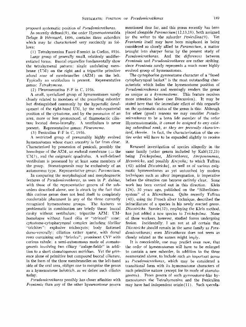
proposed systematic position of Pseudomicrothorax. As recently defined ( 9 ) , the order Hymenostomatida
Delage & Hi.rouard, 1896, contains three suborders which may be characterized very succinctly as fol- lows:
( 1) Tetrahymenina FaurC-Fremiet in Corliss, 1956. Large group of generally small, relatively undiff er-
entiated forms. Buccal organelles fundamentally show the tetrahymenal pattern: single undulating mem- brane (LX) on the right and a tripartite primitive adoral zone of membranelles (AZM) on the left. Typically no vestibulum is present. Representative genus: Tetrahymena.
(2) Pleuronematina F-F in C, 1956. A small, specialized group of hymenostomes surely
closely related to members of the preceding suborder but distinguished commonly by the hypertelic devel- opment of the right-hand UM, by the sub-equatorial position of the cytostome, and by the possession of an area, more or less pronounced, of thigmotactic cilia- ture located dorso-laterally. A vestibulum is never present. Representative genus: Pleuronerna.
(3) Peniculina F-F in C, 1956. A restricted group of presumably highly evolved
hymenostomes whose exact ancestry is far from clear. Characterized by possession of peniculi, possibly the homologue of the AZAI, an endoral membrane (= the UM?) , and the enigmatic quadrulus. A well-defined vestibulum is possessed by a t least some members of the group. Stomatogenesis may be exclusively of an autonomous type. Representative genus: Paramecium.
I n comparing the morphological and morphogenetic features of Pseudomicrothorax, as seen in P. dubius, with those of the representative genera of the sub- orders described above, one is struck by the fact that this curious genus does not lend itself to completely comfortable placement in any of the three currently recognized hymenostome groups. The features so problematic in combination are briefly these: buccal cavity without vestibulum; tripartite AZM : U N - homologue without fused cilia or “striated” zone: cytostome-cytopharyngeal complex including fibrillar “trichites”; explosive trichocysts; body flattened dorso-ventrally : ciliation rather sparse, with dorsal rows containing only “bristles” ; prominent CVP with curious tubule ; a semi-autonomous mode of stomato- genesis involving two ciliary “anlage-field~’~ in addi- tion to a short stomatogenous meridian. Yet the pres- ence alone of primitive but compound buccal ciliature, in the form of the three membranelles on the left-hand side of the oral area, obliges one to recognize the genus as a hymenostome holotrich, as we define such ciliates today.
Pseudomicrothorax possibly has closer affinities with Fiontonia than any of the other hymenostome senera
mentioned thus far, and this genus recently has been placed alongside Paramecium ( 12,13,18), both assigned by the writer to the suborder Peniculina(9). Yet Frontonia itself may have been misplaced in being considered so closely allied to Paramecium, a matter brought into sharper focus by the present study of Pseudomicrothorax. And the diflerences between Frontonia and Pseudomicrothorax are rather striking. since Frontonia surely represents a much more highly evolved group of hymenostomes.
The cyrtophorine gymnostome character of a “fused cytopharyngeal basket” is the most outstanding char- acteristic which belies the hymenostome position of Pseudomicrothorax and seemingly renders the genus so unique as a hynzenostome. This feature receives more attention below (see Discussion). It may be stated here that the immediate effect of this organelle on the systematic status of the genus is this: Although for other (good) reasons we may consider Pseudo- microthorax to be a bona fide member of the order Hymenostomatida, it cannot be assigned to any exist- ing subordinal rank, as they are presently character- ized, therein. I n fact, the characterization of the en- tire order will have to be expanded slightly to receive it.
Renewed investigation of species allegedly in the same family [other genera included by Kah1(22,23) being Trichopelma, Microthorax, Drepanomonas, Hexotricha, and possibly Kreyella; to which Tuff rau (40) added Discotricha] , as well as of various enig- matic hymenostomes as yet untouched by modern techniques such as silver impregnation, is imperative before the situation can become entirely clear. Some work has been carried out in this direction. Klein (24) , 30 years ago, published on the “Silberlinien- system” of a Microthorax. Quite recently Tuffrau (40), using the French silver technique, described the infraciliature of a species in his newly erected genus, Discotricha. Savoie( 32), employing the Klein method, has just added a new species to Trichopelma. None of these workers, however, studied forms undergoing fission. Incidentally, I am not a t all certain that Discotricha should remain in the same family as Pseu- domicrothorax; even Microthorax does not seem as closely related as the names might imply.
It is conceivable, one may predict even now, that the order of hymenostomes will have to be enlarged to contain a new suborder, in addition to the three enumerated above, to include such an important genus as Pseudomicrothorax, which may be considered a transitional form with its hymenostome characters of such primitive nature (except for its mode of stomato- genesis). From genera of such gymnostome-like hy- menostomes the Tetrahymenina and the Peniculina may have had independent origin ( 1 1 ) . Such specula-

190 SYSTEMATIC POSITION OF Pseudomicruthoran
tion, not unfounded, may serve a t least as “food for thought” and perhaps will aid in stimulating much- needed research on such problematic ciliates.
An alternative, although, i t seems to me, much less acceptable, solution to the problem of Pseudomicro- thorax’s ordinal location should not be ignored. The genus could be considered a representative member of the Trichostomatida (the order in which Kahl already has firmly placed i t ) , insisting that it enjoys an inter- mediate position between certain gymnostomes (a group sorely in need of revision itself, incidentally) and the hymenostomes. This proposition also receives more attention below. The principal drawback to such a taxonomic decision regarding the fate of Pseudomirro- thorax is that it changes radically the modern, well- established concept of a trichostome. Among other alterations forced upon us by retention of the genus in the Trichostomatida would be obligate elimination of such well-known families as the Colpodidae and Plagiopylidae, since such forms are characterized by possession solely of vestibular ciliature( 9,15,22,39). It seems to me that the revision of the Hymenostoma- tida demanded by addition of Pseudomicrothorax to its ranks is far less drastic than the change in our basic concept of the Trichostomatida which would be required by continued inclusion of the genus there, now that we are aware of the anatomical complexities of species in this genus, characteristics which undeni- ably are of significance in systematics.
DISCUSSION
The exact systematic position of the anatomically enigmatic Pseudomicrothorax dubius depends upon the completeness of our knowledge in two areas: the first is related to the degree of evolution attained or repre- sented by the structures characteristic of the genus Psczrdomicrothorax; the second is concerned with the degree of apparent relationship between this genus and supposedly “representative” genera of various orders of holotrich ciliates. Unfortunately, but as inevitably the case in all such taxonomic problems, our informa- tion is meager in both instances. The rather extensive phylogenetic implications of the entire problem are too lengthy for inclusion in the present discussion (see l l ) , although obviously they are intimately re- lated to the restricted problem at hand, viz., consid- eration of the most appropriate ordinal position for the ciliate. The apparent relationship of Pseudomi- crothorax to other ciliophoran orders is summarized briefly below. It should be kept in mind that the characterizations of these orders themselves have un- dergone and are continuing to undergo substantial revision ( 9 , l l . 13. and unpublished 1lSS of FaurC- Fremiet and the writer) ; it is in light of and in accord
with such modern analyses and interpretations that the classification of Pseudomicrothorax is being treated here.
Relationship to g?jmnostorms. The cytostome-cyto- pharyngeal complex possessed by Pseudomicrothorax (see Fig. 6 ) is an undeniably strong gymnostome-like feature; other characters. such as the dorso-ventrally flattened body, nature of the pellicle, location of the cytostome, and disposition of the ventral ciliature. could have been realized through parallel evolution or through a common remote ancestry. The three ciliary “organelles” involved in stomatogenesis in the “higher” cyrtophorine gymnostomes( 14), however, are not com- pound ciliary organelles such as the membranelles found in Pseudomicrothorax and they are far different from the ciliary anlage-fields. Thus although certain of its morphological features indicate a general phylo- genetic relationship with the cyrtophorine gymno- stomes which should not be discounted, the presence. in particular, of a true adoral zone of membranelles in Pseudomicrothora.r, unknown for any bona fide gym- nostome described to date, forces one to look to a more highly evolved group of ciliates for its immediate relatives.
Relationship t o trichostowzes. Pseudamrirrothorax cannot remain a member of the order of trichostomes even if one adheres to Kahl’s (23 ) own well-established characterization of that order. It is too highly evolved, in spite of its gymnostome-like cytopharyn-i. as is especially realizable by recognition today of its possession of a true buccal cavity equipped with com- pound ciliary organelles. Kahl and others are not to be criticized for their failure to detect or appreciate the significance of such a relatively inconspicuous AZM-type of oral ciliature, but one can no longer ignore either its existence or its taxonomic importance. The modern view(9) that posession of a vestibulum equipped solely with non-fused vestibular (really just more or less modified somatic) ciliature is as compleh as true trichostomes can become in their oral areas requires exclusion of forms such as Pseudoniirrothora1.1.. If this problematic genus is removed from the tricho- stomes, however, it is apparent that a number of other “trichostome” genera, belonging to several families. will have to join in the exodus, sooner or later. The fact that the order Trichostomatida is generally ad- mitted to comprise a polyphyletic assemblaqe of forms is no basis for increasing even further the heterogene- ity of the group.
The presence of truly compound buccal ciliature, best represented in Pseudomicrothorax by its primitive but definite AZM located in a recognizable buccal cavity, particularly dictates inclusion of the genus in the Hymenostomat-
Relationship to hymenostomes.
SYSTEMATIC POSITION OF Pseudomicrothorax 191
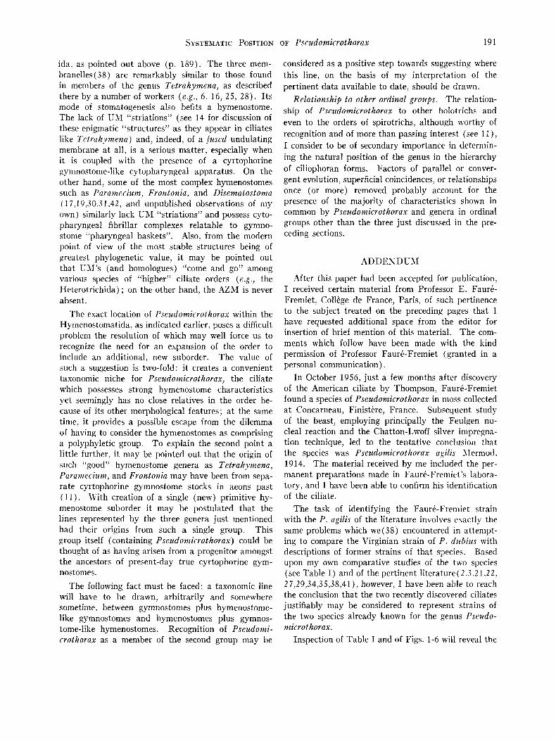
ida, as pointed out above (p. 189) . The three mem- branelles( 38) are remarkably similar to those found in members of the genus Tetrahymena, as described there by a number of workers (e.g., 6, 16, 2 5 , 28). I ts mode of stomatogenesis also befits a hymenostome. The lack of UM “striations” (see 14 for discussion of these enigmatic “structures” as they appear in ciliates like Tetrahyinena) and, indeed, of a fused undulating membrane a t all, is a serious matter, especially when it is coupled with the presence of a cyrtophorine gymnostome-like cytopharyngeal apparatus. On the other hand, some of the most complex hymenostomes such as Paramecium, Frontonia, and Disematostoma ( 17,19,30.3 1,42, and unpublished observations of my own) similarly lack UM “striations” and possess cyto- pharyngeal fibrillar complexes relatable to gymno- stome “pharyngeal baskets”. Also, from the modern point of view of the most stable structures being of greatest phylogenetic value, i t may be pointed out that UJI’s (and homologues) “come and go” among various species of “higher” ciliate orders (e.g., the Heterotrichida) ; on the other hand, the AZM is never absent.
The exact location of Pseudomicrothorax within the Hymenostomatida, as indicated earlier, poses a difficult problem the resolution of which may well force us to recognize the need for an expansion of the order to include an additional, new suborder. The value of such a suggestion is two-fold: i t creates a convenient taxonomic niche for Pseudomicrothorax, the ciliate which possesses strong hymenostome characteristics yet seemingly has no close relatives in the order be- cause of its other morphological features: a t the same time, it provides a possible escape from the dilemma of having to consider the hymenostomes as comprising a polyphyletic group. To explain the second point a little further, it may be pointed out that the origin of such “good” hymenostome genera as Tetrahymena, Paramecium, and Frontonia may have been from sepa- rate cyrtophorine gymnostome stocks in aeons past ( 11). With creation of a single (new) primitive hy- menostome suborder it may be postulated that the lines represented by the three genera just mentioned had their origins from such a single group. This group itself (containing Pseudomicrothorax) could be thought of as having arisen from a progenitor amongst the ancestors of present-day true cyrtophorine gym- nostomes.
The following fact must be faced: a taxonomic line will have to be drawn, arbitrarily and somewhere sometime, between gymnostomes plus hymenostome- like gymnostomes and hymenostomes plus gymnos- tome-like hymenostomes. Recognition of Pseudomi- crothorax as a member of the second group may be
considered as a positive step towards suggesting where this line, on the basis of my interpretation of the pertinent data available to date, should be drawn.
Relationship to other ordinal groups. The relation- ship of Pseudomicrothorax to other holotrichs and even to the orders of spirotrichs, although worthy of recognition and of more than passing interest (see I I ) , I consider to be of secondary importance in determin- ing the natural position of the genus in the hierarchy of ciliophoran forms. Factors of parallel or conver- gent evolution, superficial coincidences, or relationships once (or more) removed probably account for the presence of the majority of characteristics shown in common by Pseudomicrothorax and genera in ordinal groups other than the three just discussed in the pre- ceding sections.
ADDENDUM
After this paper had been accepted for publication, I received certain material from Professor E. Faurb- Fremiet, Collkge de France, Paris, of such pertinence to the subject treated on the preceding pages that I have requested additional space from the editor for insertion of brief mention of this material. The com- ments which follow have been made with the kind permission of Professor FaurC-Fremiet (granted in a personal communication).
I n October 1956, just a few months after discovery of the American ciliate by Thompson, F a d - F r e m i e t found a species of Pseudomicrothorax in moss collected a t Concarneau, Finistkre, France. Subsequent study of the beast, employing principally the Feulgen nu- cleal reaction and the Chatton-Lwoff silver inipregna- tion technique, led to the tentative conclusion that the species was Pseudomicrothorax agilis Jlermod. 1914. The material received by me included the per- manent preparations made in Faurk-Fremiet’s labora- tory, and I have been able to confirm his identification of the ciliate.
The task of identifying the FaurC-Freniiet strain with the P. agilis of the literature involves exactly the same problems which we(38) encountered in attempt- ing to compare the Virginian strain of P. dubius with descriptions of former strains of that species. Based upon my own comparative studies of the two species (see Table I ) and of the pertinent literature( 2.3.21.22, 27,29,34,35,38,41), however, I have been able to reach the conclusion that the two recently discovered ciliates justifiably may be considered to represent strains of the two species already known for the genus Pseudo- microthorax.
Inspection of Table I and of Figs. 1-6 will reveal the

192 SYSTEMATIC POSITION OF Pseudoinicrothorax
TABTAE I. Comparison of Psr2iilonlIc,.otkol.n.c agiZis, Concarneau strain, and P. cliibi2t.9, Virginia strain. A nuniht.r of the strricturcs consitleretl liere niay be seen in the accoinpanying illustrations (Figs. 1-G). Note horn often the data f o r hoth species :ire either idelltical or relatively the sanie. Generally a niininiun~ of 30 fixed and stained specimens, selected rail-
c1onily, were used for obtaiiling measurements reported here as averages,
P. ~7rrblrr.s P. n p l i s
NuiiilJer of ciliary iiieridians ~ u n i l ~ c r of iiifraciliary granules:
I11 rN-llolllologue 111 nic~riclinii No. 1 I11 nivri(1iaii 8 or 9
Diniueter of C V P Lellgtll of C T P IYiuniber of “ tricliites” in wall of
cy t op11:i r y s C’!tcil’li:ir)-iigc,nl crowii (dicini., long
Bt~cltllrrs of AZM :isis J
(‘!‘st*
See Figs. 1-3
51 p X 31 p
Considerable (avg. nias. thickness, 14 p )
13
5-8 1 6 4 5 2.5 p
i s r c . 20? (Est.)
c. 2.5 @
Tripartitc ; infmeilinry picture distinct. M, largest: 3 p long, 3 IOWS of G gr:inules each
Prest.11 t
Single, sulmplierical, diani. c . 0 p
Generally short rod-sliaped, 9 p X 4 p (measured only iu encysted forms)
Rouiidetl, resistnnt type observed, diam. 2 1 p. Heavy cyst wall, 3 p thick.
Erhphic. In moss, including dried sam ples
Similar. See Figs. 4-5 Sigiiificantly larger, i 8 p X 48 Relatively slightly greater (avg. nias.
Ollr illore, 13 thickness, 20 p )
8-12 24 49
3.5 p
1’ P 19-21 (sce Fig. G )
c . 5.0 p
Siniilnr, but larger. M,: 6 p long, 3 rows of c. 15 granules e:icli
Similarly preseiit
Siiiiihr in all respects Siniilar, but iiiore elongate, 16 p X 4 p
S o t 1~110w11 to csist (tropliont)
E(Iq~1iir. Associate& with algae (plus moss?) in (or near) fresh water
striking morphological similarity of the two species.5 The principal differences are in body size, total num- ber of ciliary meridians (although the difference is slight here), and in presence or absence of a cystic stage. Although the number of forms fixed while undergoing fission is insufficient for a full study of stomatogenesis in the FaurC-Fremiet material, stages which are available indicate clearly that the mode of reproduction of structures in the oral area is identical in the two members of the genus. The preceding paper (38) may be consulted for details of the process in the larger species, P. dubius.
Professor FaurC-Fremiet also has sent me a copy of a manuscript of his, being readied for publication, entitled “La differenciation des structures buccales au
ZThe accompanying Table and Figs. 1-3 are concerned sclely with the Concarneau strain of P . agilis. The slides sent t o me, however, also included silver preparations of a second strain collected by Faun-Fremiet at a later date from Gif, near Paris. The Gif strain averages slightly larger in size ( 5 5 p X 33 p), reemphasizing the very close similarity between P . a g i h and P . dtcbbs, but it also never shows more than 12 ciliary meridians.
cours de I’Cvolution des Ciliata.” In this paper FaurC- Fremiet provisionally assigns his strain of Pseudo- microthorax to the species P. agilis. What is of much greater significance is his excellent discussion of the phylogenetic implications of the curious morphological structures found in the ciliate. Unfortunately treat- ment of this aspect of his MS is beyond the scope of the present paper; the reader is referred, however, to an addendum in a related work of mine already sub- mitted for publication elsewhere( 11 ). FaurC-Fremiet has suggested that Pseudomicrothorax might be con- sidered as a “prkcurseur gymnostomien” of the hy- menostome suborder Tetrahymenina. Phylogenetically speaking, this hypothesis differs little from my own: we agree that Tetrahymena-like hymenostomes very possibly arose from Pseudomicrothorax-like progeni- tors (see pp. 190 and 191, above). From a taxonomic or classificational point of view, however, our views are not identical, since FaurC-Fremiet, although simi- larly removing Pseudomicrothorax from the tricho- stomes, considers it as an advanced cyrtophorine gym-

SYSTEMATIC POSITION OF Pseudo?nb.othorax 193
nostome, whereas I have considered it as a primitive hymenostome just one step on the evolutionary scale beyond the (other) cyrtophorine-like ciliates retained as gymnostomes. These matters are discussed in more detail from over-all comparative points of view, and thus in more proper perspective, in both Faurk- Fremiet’s RIS and my own separate paper in press (11). In any case, conclusions drawn a t this time must be considered quite tentative and fairly specula- tive until more pertinent information is made available through additional studies on a wider variety of re- lated forms.
REFERENCES
1. Butschli, 0. (1887-1889). Infusoria und System der Radiolaria, in Bronn, H. G., Klassen t i . Ordnung d . Thier- Reichs. Leipzig, 1 (I11 Abt.), 1098-2035.
2. Conn, H . W. (1905). A preliminary report on the proto- zoa of the fresh waters of Connecticut. Conn. Geol. Nut . Hist. Survey, Bull. No. 2, 5-69.
3. Conn, H. W. & Edmondson, C. H. (1918). Flagellate and ciliate protozoa (Mastigophora et Infusoria), in Ward, H. B. & Whipple, G. C., Fresh-water Biology, John Wiley & Sons, Inc., New York, 238-300.
4. Corliss, J. 0. (1952). Characterization of the family Tetrahymenidae nov. fam. (Abstr.) Proc. Soc. Protozool., 3, 4.
3 . ___ (1953). Silver impregnation of ciliated protozoa by the Chatton-Lwoff technique. Stain Technol., 28, 97-100.
6. - (1953). Comparative studies on holotrichous cili- ates in the Colpidiiim-Glaziconza-Le~~cophrys-Te~rah~~iirna group. 11. Morphology, life cycles and systematic status of strains in pure culture. Parasitology, 43, 49-87.
7. -__ (1954). The buccal apparatus and systematic status of Glaucoma frontata (“Dallasia frontata Stokes”). J . Morphol., 94, 199-219.
s. __ (1955). Proposed uniformity in naming “mouth parts” in ciliates. (Abstr.) J . Protozool., 2 (Suppl.), 12.
9. ___ (1956). On the evolution and systematics of cili- ated protozoa. Syst. Zool., 5, 68-91, 121-140.
10. ~ (1957). Nomenclatural history of the higher taxa in the subphylum Ciliophora. Arch. Protistenk., 102, 113-146.
11. - ( 1958). The phylogenetic significance of the genus Pseudomicrothorax in the evolution of holotrichous cili- ates. Acta Biol. Acad. Sci. Hung., 7 (in press).
I? . FaurP-Fremiet, E. (1949). Morphologie compare des s holotriches Trichostomata et Hymenostomata. Compt.
rend. 23th congr. intern. zool., 215-216, Paris, France. 13. - (1950). Morphologie comparCe et systematique
des cilits. Bitll. SOC. 2001. France, 75, 109-122. 14. - (1950). MCcanismes de la morphogknese chez
quelques cilits gymnostomes hypostomiens. Arch. anat. micr. morphol. exptl., 39, 1-14.
15. ~ (1950). Morphologie comparCe des cilits holo- triches trichostomes. Anais mad. brasil. cienc., 22, 257-261.
16. Furgason, W. H. (1940). The significant cytostomal pat- tern of the “GEa2icoma-Colpidii~mt group,” and a proposed new genus and species, Tetrahyniena geleii. Arch. Protistenk., 94, 224-266.
17. Gelei, J . v. (1934). Der feinere Bau des Cytopharynx von Paramecium und seine systematische Bedeutung. Arch. Protistenk., 82, 331-362.
18. -- (1952). Nehiny sz6 a csill6sok Trichostomata alrendjbnek rendszertanihoz. Einiges uber die Systematik der Unterordnung Trichostomata der Ciliaten. Ann. biol. Univ.
Hung., 1, 351-360. [ In Hungarian with German summary.] 19. ___ (1954). Uber die Lebensgemeinschaft einiger
temporarer Tiimpel auf einer Bergwiese im Borzsonygebirge (Oberungarn) . 111. Ciliaten. Acfa Biol. Hung., 5, 259-343. 20. Holz, G . G., Jr . & Corliss, J. 0. (1956). Tetrahymena
setifera n. sp., a member of )the genus Tetrahymenu with a caudal cilium. J . Protozool., 3, 112-118.
21. Kahl, A. (1926). Neue und wenig bekannte Formen der holotrichen und heterotrichen Ciliaten. Arch. Protistenk., 55, 197-438.
22. ___ (1931). Holotricha (ausser den im 1. Teil be- handelten Prostomata), part 2 1 of Dahl, F., Die Tierwelt Uiutschlands, G . Fischer, Jena, 181-398.
23. ___ (1930-1935). I . Wimperthiere oder Ciliata (In- fusoria), in Dahl, F., Die Tirrwelt Dezitschlands, G. Fischer, Jena, 1-886.
24. Klein, B. M. (1928). Die Silberliniensystem der Ciliaten. Weitere Resultate. Arch. Protistenk., 62, 177-260.
25. Kozloff, E. N. (1946). The morphology and systematic position of a holotrichous ciliate parasitizing Deroceras agreste ( L ) . J. Morphol., 79, 445-465.
26. Maupas, E . (1883). Contribution k I’ktude morpholo- gique et anatomique des infusoires cilies. Arch. zool. exptl. et g&. (ser. Z), 1, 427-664.
27. Mermod, G. (1914). Recherches sur la faune infusori- enne des tourbieres et des eaux voisines de Sainte-Croix (Jura vaudois). R e v . suisse zool., 22, 31-114.
28. Mugard, H. (1949). Contribution k l’ktude des infu- soires hymhostomes histiophages. Ann. sci. nut. zool. e t biol. aninzale (sPr. l l ) , 10, 171-268.
29. Penard, E. (1922). Etudes sur les Infusoires d’Eau douce. Georg & Cie, Genkve.
30. Roque, M. (1956). La stomatogCnkse pendant I’auto- gamie, la conjugaison et la division chez Paramecium azirelia. Coittpt. rend., 243, 1564-1565.
31. __- (1957). Ciliature eomatique et ciliature buccale chez Disematostonza tetraedrica (ciliC holotriche). Compt. rend., 244, 2657-2660.
32. Savoie, A. (1957). Le ciliC Tvichopelma agilis n. sp. J . Protocool., 4, 276-280.
33. Schewiakoff, W. (1896). The organization and syste- matics of the Infusoria Aspirotricha (Holotricha auctorum) . M&n. acad. imptr . xi. St. Petersbourg (str. 8), 4, 1-395. [In Russian. 1
34. Sondheim, M. (1929). Prcrtozoen aus der Ausbeute der Voeltzkowschen Reisen in Madagaskar und Ostafrika. Ab- handl. senckenberg. naturforsch. Ges., 41, 285-313.
35. Srimek-Husek, R. (1952). o b e r einige Bemerkenswerte Ciliaten aus bohmischen Moosen. Cesk. Biol., Praha, 1, 367- 376. [ In Russian with German summary.]
36. Stokes, A. C. (1884). Notices of some new parasitic infusoria. Ant. Naturalist, 18, 1081-1086.
37. Thompson, J. C., Jr. (1957). Pseudomicrothorax gel- 16 t i n. sp., a hymenostome ciliate from American moss. (Abstr.) Assoc. Southeastern Biol. Bull., 4, 15.
38. Thompson, J. C., Jr. & Corliss, J. 0. (1958). A re- description of the holotrichous ciliate Pseudomicrothorax dubiiis with particular attention to its morphogenesis. J . Protozool., 5 ( in press).
39. Tuffrau, M. (1952). L a morphogCnese de division chez les Colpodidae. Bull. biol. France et Belg., 86, 309-320.
40. ~ (1954). Discotricha papillifera, n.g., n. sp. Cili6 psammobic de la famille des Trichopelmidae. J . Protozool., 1, 18.3 - 186,
41. Wang, C. C. & Nie, D. (1935). Report on the rare and new species of fresh-water infusoria, Par t 11. Sinmsia, 6,
42. Yusa, A. (1957). The morphology and morphogenesis of the buccal organelles in Paramecium with particular reference to their systematic significance. J . Protozool., 4, 128-142. ’ 1
399-524.


![Metabolites from the Euryhaline Ciliate Pseudokeronopsis ......Metabolites from the Euryhaline Ciliate Pseudokeronopsis erythrina Andrea Anesi,*[a] Federico Buonanno,[b] Graziano di](https://img.pdfslide.us/doc/110x75/5eb6046dce73b216293aaa74/metabolites-from-the-euryhaline-ciliate-pseudokeronopsis-metabolites-from.jpg)