Embed Size (px)
Citation preview
ORIGINAL RESEARCHpublished: 23 October 2020
doi: 10.3389/fcimb.2020.572849
Frontiers in Cellular and Infection Microbiology | www.frontiersin.org 1 October 2020 | Volume 10 | Article 572849
Edited by:
Mathew Upton,
University of Plymouth,
United Kingdom
Reviewed by:
Keith Miller,
Sheffield Hallam University,
United Kingdom
Roderich Roemhild,
Uppsala University, Sweden
*Correspondence:
Ke-Jian Wang
Specialty section:
This article was submitted to
Clinical Microbiology,
a section of the journal
Frontiers in Cellular and Infection
Microbiology
Received: 15 June 2020
Accepted: 02 September 2020
Published: 23 October 2020
Citation:
Liu J, Chen F, Wang X, Peng H,
Zhang H and Wang K-J (2020) The
Synergistic Effect of Mud Crab
Antimicrobial Peptides Sphistin and
Sph12–38 With Antibiotics Azithromycin
and Rifampicin Enhances Bactericidal
Activity Against Pseudomonas
Aeruginosa.
Front. Cell. Infect. Microbiol.
10:572849.
doi: 10.3389/fcimb.2020.572849
The Synergistic Effect of Mud CrabAntimicrobial Peptides Sphistin andSph12−38 With AntibioticsAzithromycin and RifampicinEnhances Bactericidal ActivityAgainst Pseudomonas Aeruginosa
Jie Liu 1, Fangyi Chen 1,2, Xiaofei Wang 1, Hui Peng 1,2, Hua Zhang 1 and Ke-Jian Wang 1,2*
1 State Key Laboratory of Marine Environmental Science, College of Ocean & Earth Sciences, Xiamen University, Xiamen,
China, 2 State-Province Joint Engineering Laboratory of Marine Bioproducts and Technology, College of Ocean & Earth
Sciences, Xiamen University, Xiamen, China
Overuse or abuse of antibiotics has undoubtedly accelerated the increasing prevalence
of global antibiotic resistance crisis, and thus, people have been trying to explore
approaches to decrease dosage of antibiotics or find new antibacterial agents for many
years. Antimicrobial peptides (AMPs) are the ideal candidates that could kill pathogens
and multidrug-resistant bacteria either alone or in combination with conventional
antibiotics. In the study, the antimicrobial efficacy of mud crab Scylla paramamosain
AMPs Sphistin and Sph12−38 in combination with eight selected antibiotics was
evaluated using a clinical pathogen, Pseudomonas aeruginosa. It was interesting to note
that the in vitro combination of rifampicin and azithromycin with Sphistin and Sph12−38
showed significant synergistic activity against P. aeruginosa. Moreover, an in vivo study
was carried out using a mouse model challenged with P. aeruginosa, and the result
showed that the combination of Sph12−38 with either rifampicin or azithromycin could
significantly promote the healing of wounds and had the healing time shortened to 4–5
days compared with 7–8 days in control. The underlying mechanism might be due to
the binding of Sphistin and Sph12−38 with P. aeruginosa lipopolysaccharides (LPS) and
subsequent promotion of the intracellular uptake of rifampicin and azithromycin. Taken
together, the significant synergistic antibacterial effect on P. aeruginosa in vitro and in
vivo conferred by the combination of low dose of Sphistin and Sph12−38 with low dose
of rifampicin and azithromycin would be beneficial for the control of antibiotic resistance
and effective treatment of P. aeruginosa-infected diseases in the future.
Keywords: antimicrobial peptides, Sphistin, Sph12−38, Pseudomonas aeruginosa, rifampicin, azithromycin,
synergistic efficacy
Liu et al. AMPs and Antibiotics Synergistic Against P. aeruginosa
INTRODUCTION
Pseudomonas aeruginosa is an opportunistic Gram-negativebacterial pathogen and can cause infections and mass mortalityin patients that have cystic fibrosis, AIDS, severe burns, organtransplants, and cancer (Lyczak et al., 2000; Blonder et al.,2004). The current treatment regimen of P. aeruginosa includes awide range of antibiotics including β-lactams, aminoglycosides,fluoroquinolones, or even the inter-combination of thoseantibiotics (Hancock and Speert, 2000); however, the clinicalpathogen P. aeruginosa is less susceptible to almost all of theroutinely used antibiotics and fairly easy to develop resistance.For example, from 2003 to 2011, the rates of carbapenem-resistant P. aeruginosa (CRPA) isolated from patients withhospital-acquired infections in a tertiary care hospital innortheast China were 14.3, 17.1, 21.1, 24.6, 37.0, 48.8, 56.4,51.2, and 54.1% over time (Xu et al., 2013). In another hospital,First Affiliated Hospital of Nanjing Medical University, in2008, the resistant rates of P. aeruginosa to cephalosporins(Ceftazidime, Cefotaxime, and Cefepime) were 5.9, 82.4, and17.6%, respectively, while by the end of 2011, only 4 years passed,those numbers increased to 37.8, 85.7, and 27.8%, respectively(Zhang et al., 2015). Owing to its high intrinsic resistance toantibiotics and wide repertoire of virulence factors, the therapyfor the P. aeruginosa-infected diseases becomes an intractableissue (Hancock and Speert, 2000). As reported, the resistanceof P. aeruginosa is mainly due to the low permeability of itsouter membrane (Hancock, 1998). Besides, the transmembraneefflux pumps are also considered for the intrinsic resistanceof P. aeruginosa by which the incoming antibiotics can betaken out of the bacteria efficiently (Li et al., 1994). Therefore,exploration of new antipseudomonal agents is desperately neededto take control of the ubiquitous and acute drug resistance ofP. aeruginosa.
To date, multifarious highlighted new strategies againstthe multidrug-resistant (MDR) bacteria have been proposedand some potential biological products or pharmaceuticals areexpected to be applied in clinic, including antimicrobial peptides(AMPs), anti-virulence compounds, phage therapy, and newmolecules (Pacios et al., 2020). For example, both Enterobacter
cloacae (Mu208) and Klebsiella pneumoniae (Mu1343) displaymultiple heteroresistance to the antibiotics; the simultaneouscombinations of antibiotics targeting multiple heteroresistanceare effective to kill these two kinds of bacteria, whereas thosetargeting homogeneous resistance are ineffective (Band et al.,2019). The sequential therapy is considered as a sustainablestrategy to counter the antibiotic crisis because this therapeuticmethod can constrain the emergence of drug resistance andenhance the bactericidal activity (Roemhild and Schulenburg,2019). Collateral sensitivity means that the mutations inbacteria cause multidrug resistance but simultaneously enhancesensitivity to many other unrelated drugs, and this newmechanism might be developed to alternative antimicrobialstrategies against the multidrug bacteria (Pal et al., 2015). AMPsare widespread distributed in various organisms whose manytissues and cell types could produce different functional AMPs(Vizioli and Salzet, 2002; Zasloff, 2002; Brogden et al., 2003).
Most AMPs attach to and permeate the target membrane bilayersto induce pore formation and cause the leakage of cytoplasm(Shai, 2002; Brogden, 2005). Besides that, some peptides can alterthe septum formation of cytoplasmic membrane and inhibit thesynthesis of cell wall, nucleic acid, and protein or enzymaticactivity to kill the bacteria (Brogden, 2005). In addition, AMPscan damage the bacterial cell wall, resulting in the radical changeof the bacterial morphology, while simple mutations of thebacteria could not reserve the situations (Shai, 2002; Zasloff,2002; Chongsiriwatana et al., 2008). Although the AMPs possessbetter antibacterial activity and a broad antibacterial spectrum,antibiotics have not been successfully substituted by AMPs yet.One reason was that the bacteria also developed resistance toAMPs (Habets et al., 2012; Dobson et al., 2013; Makarova et al.,2018; El Shazely et al., 2020); for example, the point mutationsinduced conformational changes in BraS or BraR, resulting inthe constitutive expression of VraDE, conferring Staphylococcusaureus to evolve high resistance to nisin A (Arii et al., 2019).The second reason is that there is also cross-resistance of evolvedstrains to other AMPs, not much but it still exists; for instance,the melittin-resistant S. aureus displays cross-resistance againstpexiganan (El Shazely et al., 2020). However, despite resistanceevolution to AMPs conferred by a few bacteria or cross-resistance of evolved strains to other AMPs, according to thepharmacodynamic studies of AMPs, compared with antibiotics,the evolution of resistance to AMPs is much lower (Yu et al.,2018). Therefore, AMPs are considered to be the potential idealsubstituents for antibiotics to be used to some extent in the future.Some studies also have shown that the combination of AMPswithconventional antibiotics has synergistic effect against the targetedpathogenic microorganisms (Li et al., 2017; Zheng et al., 2017;Koppen et al., 2019).
Rifampicin is one kind of derivative of rifamycin. It displaysa broad spectrum of antibacterial spectrum against Gram-positive bacteria, particularly Mycobacteria and, to a lesserextent, Gram-negative bacteria such as Escherichia coli, Neisseriameningitides, etc (Walter and Staehelin, 1971; Heinz Floss andYu, 2005). The antibacterial mechanism of rifampicin rootsin its high affinity binding to and inhibition of the bacterialDNA-dependent RNA polymerase (Campbell et al., 2001).Azithromycin is a kind of macrolide antibiotic, which has a15-member macrocyclic lactone ring. It is derived from theerythromycin 14-member ring that is inserted into an aminogroup (Alvarez-Elcoro and Enzler, 1999). Azithromycin alsohas a broad spectrum of antibacterial spectrum against Gram-positive bacteria including S. aureus, parts of Streptococci,Streptococcus pneumoniae etc.; Gram-negative bacteria includingHaemophilus influenzae, Haemophilus ducreyi, Neisseriagonorrhoeae, Bordetella pertussis, etc.; and other pathogenssuch as Chlamydia trachomatis, Ureaplasma urealyticum,Mycoplasma pneumoniae, etc (Retsema et al., 1987; Peterset al., 1992; Alvarez-Elcoro and Enzler, 1999). Azithromycininhibits the bacterial growth by interfering with their proteinsynthesis. It could also inhibit RNA-dependent protein synthesisby reversibly binding to the 50 S subunits of the bacterialribosome (Mazzei et al., 1993; Alvarez-Elcoro and Enzler,1999).
Frontiers in Cellular and Infection Microbiology | www.frontiersin.org 2 October 2020 | Volume 10 | Article 572849
Liu et al. AMPs and Antibiotics Synergistic Against P. aeruginosa
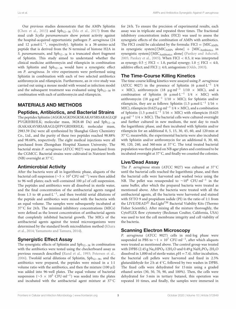
Our previous studies demonstrate that the AMPs Sphistin(Chen et al., 2015) and Sph12−38 (Ma et al., 2017) from themud crab Scylla paramamosain show potent activity againstthe hospital-acquired opportunistic pathogen P. aeruginosa (24and 12 µmol·L−1, respectively). Sphistin is a 38-amino-acidpeptide that is derived from the N-terminal of histone H2A inS. paramamosain, and Sph12−38 is a truncated short fragmentof Sphistin. This study aimed to understand whether theclinical medicine azithromycin and rifampicin in combinationwith Sphistin and Sph12−38 would have a synergistic effecton P. aeruginosa. In vitro experiments were performed usingSphistin in combination with each of two selected antibioticsazithromycin and rifampicin. Furthermore, an in vivo study wascarried out using a mouse model with wound as infection modeland the subsequent treatment was evaluated using Sph12−38 incombination with each of azithromycin and rifampicin.
MATERIALS AND METHODS
Peptides, Antibiotics, and Bacterial StrainsThe peptides Sphistin (AGGKAGKDSGKSKAKAVSRSARAGLQFPVGRIHRHLK; molecular mass, 3828.48 Da) and Sph12−38
(KAKAKAVSRSARAGLQFPVGRIHRHLK; molecular mass,2983.59 Da) were all synthesized by Shanghai Glory ChemistryCo., Ltd., and the purity of these two peptides reached 98.88%and 98.68%, respectively. The eight medical injections were allpurchased from Zhongshan Hospital Xiamen University. Thebacterial strain P. aeruginosa (ATCC 9027) was purchased fromthe CGMCC. Bacterial strains were cultivated in Nutrient broth(NB) overnight at 37◦C.
Antimicrobial ActivityAfter the bacteria were all in logarithmic phase, aliquots of thebacterial cell suspension (∼5× 10⁵ CFU·ml−1) were then addedto 96-well plates; each well-contained 100 µl of cell suspension.The peptides and antibiotics were all dissolved in sterile water,and the final concentration of the antibacterial agents rangedfrom 1.5 to 48 µmol·L−1, and then twofold serial dilutions ofthe peptide and antibiotics were mixed with the bacteria withan equal volume. The samples were subsequently incubated at37◦C for 24 h. The minimal inhibitory concentrations (MICs)were defined as the lowest concentration of antibacterial agentsthat completely inhibited bacterial growth. The MICs of theantibacterial agents against the tested microorganisms weredetermined by the standard broth microdilution method (Kharaet al., 2014; Yamamoto and Tamura, 2014).
Synergistic Effect AssayThe synergistic effects of Sphistin and Sph12−38 in combinationwith the antibiotics were tested using the checkerboard assay asprevious research described (Rand et al., 1993; Petersen et al.,2006). Twofold serial dilutions of Sphistin, Sph12−38, and theantibiotics were prepared, the peptides were mixed in a 1:1volume ratio with the antibiotics, and then the mixture (100 µl)was added into 96-well plates. The equal volume of bacterialsuspension (∼5 × 10⁵ CFU·ml−1) was seeded into the platesand incubated with the antibacterial agent mixture at 37◦C
for 24 h. To ensure the precision of experimental results, eachassay was in triplicate and repeated three times. The fractionalinhibitory concentration index (FICI) was used to assess thesynergistic effects of the combination of AMPs with antibiotics.The FICI could be calculated by the formula: FICI = [MICAMPs
in synergistic system]/[MICAMPs alone] + [MICAntibiotics insynergistic system]/[MICAntibiotics alone] (Pankey and Ashcraft,2005; Pankey et al., 2005). When FICI < 0.5, it was interpretedas synergy; 0.5 ≤ FICI < 1.0, partial synergy; 1.0 ≤ FICI < 4.0,additive effect; and FICI ≥ 4.0, antagonism (Odds, 2003).
The Time-Course Killing KineticsThe time-course killing kinetics were assayed using P. aeruginosa(ATCC 9027) in the presence of Sphistin (6 µmol·L−1 1/4× MIC), azithromycin (18 µg·ml−1 1/10 × MIC), and acombination of Sphistin (6 µmol·L−1 1/4 × MIC) withazithromycin (18 µg·ml−1 1/10 × MIC); for Sphistin and/orrifampicin, they are as follows: Sphistin (1.5 µmol·L−1 1/16 ×
MIC), rifampicin (0.625µg·ml−1 1/4×MIC), and a combinationof Sphistin (1.5 µmol·L−1 1/16 × MIC) with rifampicin (0.625µg·ml−1 1/4×MIC). The bacterial cells were cultured overnightand further cultured in new medium, the next day to reachthe logarithmic phase, and then incubated with Sphistin and/orrifampicin for an additional 0, 5, 15, 30, 45, 60, and 120min at37◦C; meanwhile, the experimental bacteria were also incubatedwith Sphistin and/or azithromycin for an additional 0, 30, 60,90, 120, 240, and 360min at 37◦C. The total treated bacterialpopulation was then plated onNB agar plates and continued to beincubated overnight at 37◦C, and finally we counted the colonies.
Live/Dead AssayThe P. aeruginosa strain (ATCC 9027) was cultured at 37◦Cuntil the bacterial cells reached the logarithmic phase, and thenthe bacterial cells were harvested and washed twice using theNB. The pellet was resuspended to ∼106 CFU·ml−1 in thesame buffer, after which the prepared bacteria were treated asmentioned above. After the bacteria were treated with all theantibacterial agents, all the bacteria were harvested and stainedwith SYTO 9 and propidium iodide (PI) in the ratio of 1:1 fromthe LIVE/DEAD R© BacLightTM Bacterial Viability Kits (ThermoFisher Scientific). After mixing all the mixture thoroughly, theCytoFLEX flow cytometry (Beckman Coulter, California, USA)was used to test the cell membrane integrity and cell viability ofthe bacteria.
Scanning Electron MicroscopyP. aeruginosa (ATCC 9027) cells in mid-log phase weresuspended in PBS to ∼1 × 10⁷ CFU·ml−1, after which aliquotswere treated as mentioned above. The control group was treatedwith DPBS (2.45 g Na2HPO4·12H2O and 0.49 g NaH2PO4·2H2Odissolved in 1,000ml of sterile water, pH= 7.4). After incubation,the bacterial cell pellets were harvested and fixed in 2.5%glutaraldehyde for 2 h at 4◦C, followed by two washes in DPBS.The fixed cells were dehydrated for 15min using a gradedethanol series (30, 50, 70, 90, and 100%). Then, the cells weredehydrated for 5min in tertiary butanol, this operation wasrepeated 10 times, and finally, the samples were immersed in
Frontiers in Cellular and Infection Microbiology | www.frontiersin.org 3 October 2020 | Volume 10 | Article 572849
Liu et al. AMPs and Antibiotics Synergistic Against P. aeruginosa
tertiary butanol overnight at 4◦C. When the prepared specimensdried, conductive coating was applied to the specimens andthey were examined using a field emission scanning electronmicroscopy (SUPRA 55; ZEISS, Germany).
Bacterial Cell Membrane PermeabilizationAssayThe permeability of bacterial cell membranes was determinedby measuring the leakage of intracellular ATP levels out of thebacterial cells as described by previous research (Koshlukovaet al., 1999). Briefly, P. aeruginosa (ATCC 9027) was culturedovernight at 37◦C, the cells were harvested and washed twice,and the bacterial cells were resuspended in DPBS. The preparedbacterial cells were treated as mentioned above. After incubation,samples were centrifuged to get the supernatant and then 10µl of the supernatant was added into 90 µl of the standardreaction solution that comes from the Molecular Probes’ ATPDetermination Kit (Thermo Fisher Scientific). Prior to testing theluminescence of the samples, use the luminometer to measurethe background luminescence and then subtract the backgroundluminescence and read the luminescence of the samples. Usingthe gradient dilution ATP standard solution to generate astandard curve for a series of ATP concentrations, and accordingto the standard curve, we could calculate the amounts of theleakage of the intracellular ATP.
Transmission Electron Microscopy (TEM)The treated bacteria were fixed in 2.5% glutaraldehyde overnightat 4◦C, the samples were washed in PBS, the bacteria wereharvested and resuspended in PBS (1.5 × 10⁹ CFU·ml−1), andthe samples were added into the agar models. The mixturewas centrifuged and the supernatant was removed, and theprepared samples were put into 2% molten agar solution, afterwhich the mixed soulution was served on ice until agar solutionsolidification. The agar block was cut into the size of a ricegrain and washed twice, and the agar granules were resuspendedand fixed in 2.5% glutaraldehyde overnight at 4◦C. Finally, thefixed agar granules were suspended in PBS; after embeddingand sectioning, the samples were examined by TEM (Tecnai G2Spirit, FEI, USA).
Closure of Wounds Infected WithP. aeruginosaSix to eight week old BALB/c male mice weighting 25–28 g(n = 42) were used in the study. The wound was produced byusing the medical pressure-sensitive adhesive tape to removea 2 cm × 2 cm area of the epidermis on the backs of mice.P. aeruginosa (ATCC 9027) (1 × 10⁸ CFU per 20 µl in PBS)was then immediately smeared onto the artificial wound. Twohours after bacterial infection at the wound site, rifampicin (1.25µg·ml−1, 1/2 × MIC), azithromycin (90 µg·ml−1, 1/2 × MIC),Sph12−38 (24 µmol·L−1, 2 × MIC) alone and in combinationwith rifampicin (1.25 µg·ml−1, 1/2 × MIC), and azithromycin(90 µg·ml−1, 1/2 × MIC), respectively, in 20 µl of PBS wereadministered in the wound site by hypodermic injection. Themice without infection of P. aeruginosa (ATCC 9027) were used
TABLE 1 | MIC data for Sphistin alone and in combination with antibiotics against
P. aeruginosa.
Compounds MIC
Antibiotics
(µg·ml−1)
FIC
Antibiotics
MIC
Sphistin
(µmol·L−1)
FIC
Sphistin
FIC
index
Vancomycin 4.5 >2.25 24 >12 >1
Penicillin >18 >9 24 >12 >1
Ceftizoxime >20 >10 24 >12 >1
Cefotiam >30 >15 24 >12 >1
Clindamycin >25 >12.5 24 >12 >1
Tinidazole >12 >6 24 >12 >1
Azithromycin 180 18 24 6 0.35
Rifampicin 2.5 0.625 24 1.5 0.3125
as uninfected controls. The wounds were photographed at adefinite time to record the wound healing.
Statistical AnalysisAll experiments were performed three independent times, witheach sample performed in triplicate. All data were expressed asmeans ± standard deviations. Differences among groups wereevaluated by using one-way analysis of variance. P < 0.05 wereconsidered statistically significant.
RESULTS
The Synergistic and Additive AntibacterialEffects of Sphistin and Sph12−38 inCombination With Eight Commonly UsedAntibioticsAs reported previously (Chen et al., 2015), the synthetic Sphistinhas no cytotoxicity toward mouse osteoblastic cell MC3T3-E1 and crab hemocytes even at high tested concentrations(100 mg·ml−1). Similarly, Sph12−38 also exhibits no cytotoxicityon HeLa cell and crab hemocytes (Ma et al., 2017). Both ofthe AMPs had strong antibacterial activity and also showedpotent activity against P. aeruginosa, whose MIC values were24 and 12 µmol·L−1, respectively. The antimicrobial activitiesof Sphistin and Sph12−38 in combination with the commonlyused clinical antibiotics rifampicin, vancomycin, penicillin,ceftizoxime, cefotiam, clindamycin, tinidazole, and azithromycinagainst P. aeruginosa were individually determined using thebroth microdilution method in accordance with the Clinicaland Laboratory Standards Institute (CLSI) recommendation(C. L. S. Institute, 2012), and the results are summarized inTables 1, 2. Among the eight selected antibiotics, only Sphistinand Sph12−38 in combination with azithromycin and rifampicinexhibited significant synergistic activity against P. aeruginosa.The mixture of 4-fold reduction of Sphistin (reduced from24 to 6 µmol·L−1) with 10-fold reduction of azithromycin(reduced from 180 to 18 µg·ml−1) and the mixture of 16-fold reduction of Sphistin (reduced from 24 to 1.5 µmol·L−1)with 4-fold reduction of rifampicin (reduced from 2.5 to 0.625µg·ml−1) could inhibit the growth of P. aeruginosa. Similar
Frontiers in Cellular and Infection Microbiology | www.frontiersin.org 4 October 2020 | Volume 10 | Article 572849
Liu et al. AMPs and Antibiotics Synergistic Against P. aeruginosa
TABLE 2 | MIC data for Sph12−38 alone and in combination with antibiotics
against P. aeruginosa.
Compounds MIC
Antibiotics
(µg·ml−1)
FIC
Antibiotics
MIC
Sph12−38
(µmol·L−1)
FIC
Sph12−38
FIC index
Vancomycin 4.5 >2.25 12 >6 >1
Penicillin >18 >9 12 >6 >1
Ceftizoxime >20 >10 12 >6 >1
Cefotiam >30 >15 12 >6 >1
Clindamycin >25 >12.5 12 >6 >1
Tinidazole >12 >6 12 >6 >1
Azithromycin 180 18 12 1.5 0.225
Rifampicin 2.5 0.625 12 1.5 0.375
situations also occurred when Sph12−38 in combination withazithromycin and rifampicin and the mixture of 8-fold reductionof Sph12−38 (reduced from 24 to 1.5 µmol·L−1) with 10-foldreduction of azithromycin (reduced from 180 to 18 µg·ml−1) orwith 4-fold reduction of rifampicin (reduced from 2.5 to 0.625µg·ml−1) could also significantly minimize the growth of P.aeruginosa. The FICIs of these four combinations were all <0.5(Tables 1, 2), which could be considered as a synergistic effect.However, for other antibiotics, including vancomycin, penicillin,ceftizoxime, cefotiam, clindamycin, and tinidazole, when theywere combined with either Sphistin or Sph12−38 to treat P.aeruginosa, the FICIs were all more than 1, which showed nosynergistic effect.
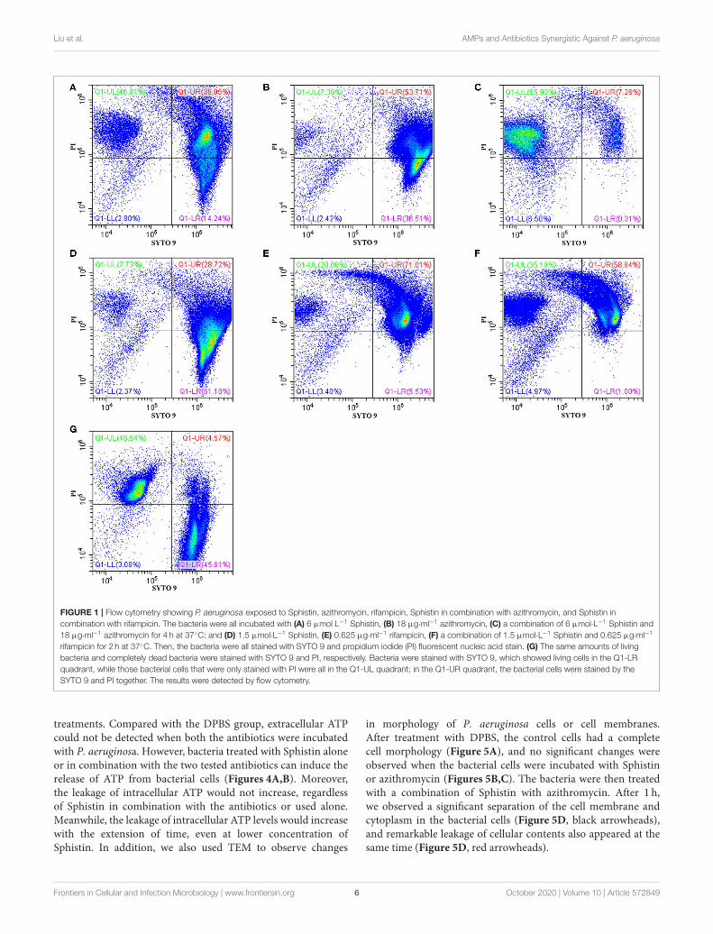
Effects of Sphistin With Azithromycin andRifampicin on Viability of P. aeruginosaThe effects of Sphistin in combination with azithromycin andrifampicin on viability and membrane integrity of P. aeruginosawere tested by using the LIVE/DEAD R© BacLightTM BacterialViability Kits and flow cytometry. This kit has two-colorfluorescence: the SYTO 9 green-fluorescent nucleic acid stain,which could stain all living cells green, and the red-fluorescentnucleic acid stain, PI, which could specifically penetrate thebacterial cells such that the cell membrane is damaged andcells are stained red. When the flow cytometry was used todetect the mixture of the same number of living bacteria andcompletely dead bacteria, the living bacteria were stained withSYTO 9, and they were all almost distributed in the Q1-LRquadrant, while the completely dead bacteria were stained withPI, and they were all almost distributed in the Q1-UL quadrant(Figure 1G). The results showed that ∼46.01% and ∼7.36% ofthe bacteria treated with Sphistin and azithromycin, respectively,were stained by PI (Figures 1A,B), and the combination ofSphistin and azithromycin could totally kill 85.93% of thebacterial cells (Figure 1C). As for Sphistin and/or rifampicintreatment, only ∼7.73% and ∼20.06% of the bacterial cellswere completely killed by Sphistin and rifampicin, respectively(Figures 1D,E), while when Sphistin is in combination withrifampicin, ∼35.19% of the bacterial cells were completelykilled (Figure 1F). These findings indicated that exposure to
both Sphistin in combination with azithromycin or rifampicinresulted in the uptake of PI by more bacterial cells thanSphistin and these two antibiotics alone, suggesting a significantincrease in cell permeability and hence the synergistic activityof Sphistin in combination with these two antibiotics, especiallywith the azithromycin.
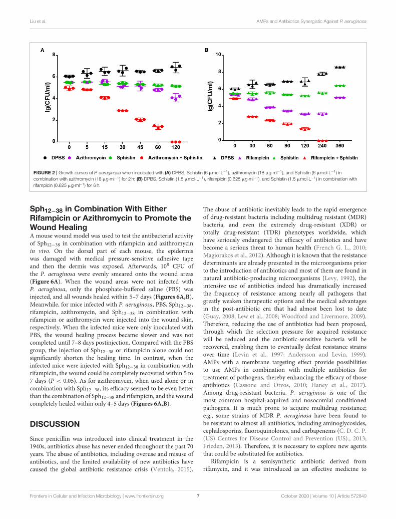
The Time-Course Killing KineticsAccording to the results, Sphistin in combination withazithromycin and rifampicin had synergistic effects againstP. aeruginosa. We also conducted a time-course killingexperiment to examine the effects of Sphistin and/or these twoantibiotics against P. aeruginosa. Sphistin in combination withazithromycin reduced the number of bacteria by more than twoorders of magnitude after 45min, and after 2 h, all the bacteriawere killed. By contrast, the Sphistin or azithromycin used alonedid not inhibit the bacterial viability efficiently (Figure 2A).Similarly, Sphistin in combination with rifampicin also inhibitedthe growth of P. aeruginosa; after 1 h, the number of bacteriawas also reduced more than two orders of magnitude, and thecombination of Sphistin and rifampicin could kill all the bacteriaafter 4 h. However, if Sphistin or rifampicin was incubated withthe bacteria alone, each of them could not inhibit bacterialviability, and the concentration of bacteria increased after 4 h(Figure 2B).
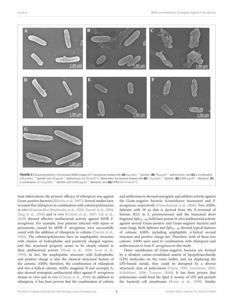
Visualization of the Interaction of Sphistinand/or Rifampicin and Azithromycin WithP. aeruginosaScanning electron microscopy (SEM) was used to visualizethe bacterial cell membrane damaged by Sphistin and/orrifampicin and azithromycin. Compared with the controlgroup (Figure 3G), P. aeruginosa treated with Sphistin orazithromycin alone showed slight cell shrinkage (Figures 3A,B),but the cell membrane was intact. When the bacteria wereincubated with a combination of Sphistin and azithromycin,the entire cell membrane was completely damaged alongwith the leakage of cytoplasmic contents (Figure 3C). WhenSphistin or rifampicin was used alone, each reagent onlyinduced slight changes in cellular morphology (Figures 3D,E);however, when the bacteria were treated with Sphistin incombination with rifampicin, obvious depressions were observedon the bacterial cell membrane, but no leakage of cytoplasmiccontent was present and the cellular morphology remainedintact (Figure 3F).
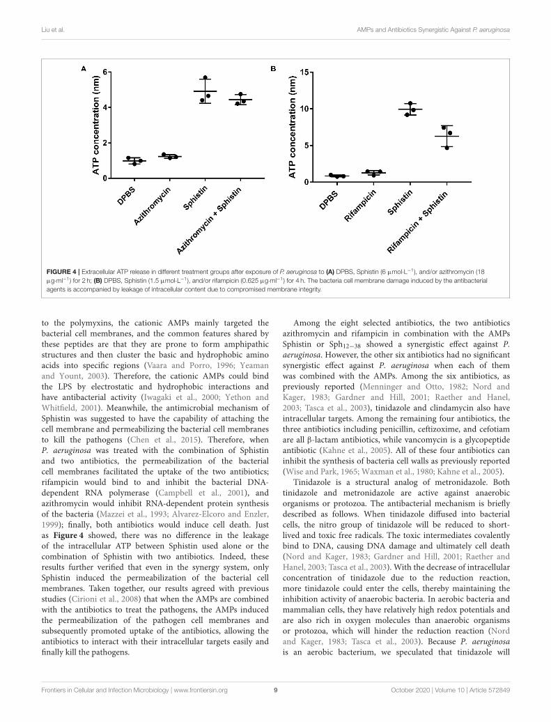
Antimicrobial Mechanism of Sphistin inCombination With Rifampicin andAzithromycinAs reported, when the cell membrane was compromised,the barrier function of the cell membrane will be impaired,resulting in the leakage of critical cellular contents (Kharaet al., 2015). To further investigate the mechanism of thecombination of Sphistin with rifampicin and azithromycin, weevaluated the changes in membrane permeability by measuringthe extracellular ATP levels after the two combination group
Frontiers in Cellular and Infection Microbiology | www.frontiersin.org 5 October 2020 | Volume 10 | Article 572849
Liu et al. AMPs and Antibiotics Synergistic Against P. aeruginosa
FIGURE 1 | Flow cytometry showing P. aeruginosa exposed to Sphistin, azithromycin, rifampicin, Sphistin in combination with azithromycin, and Sphistin in
combination with rifampicin. The bacteria were all incubated with (A) 6 µmol L−1 Sphistin, (B) 18 µg·ml−1 azithromycin, (C) a combination of 6 µmol·L−1 Sphistin and
18 µg·ml−1 azithromycin for 4 h at 37◦C; and (D) 1.5 µmol·L−1 Sphistin, (E) 0.625 µg·ml−1 rifampicin, (F) a combination of 1.5 µmol·L−1 Sphistin and 0.625 µg·ml−1
rifampicin for 2 h at 37◦C. Then, the bacteria were all stained with SYTO 9 and propidium iodide (PI) fluorescent nucleic acid stain. (G) The same amounts of living
bacteria and completely dead bacteria were stained with SYTO 9 and PI, respectively. Bacteria were stained with SYTO 9, which showed living cells in the Q1-LR
quadrant, while those bacterial cells that were only stained with PI were all in the Q1-UL quadrant; in the Q1-UR quadrant, the bacterial cells were stained by the
SYTO 9 and PI together. The results were detected by flow cytometry.
treatments. Compared with the DPBS group, extracellular ATPcould not be detected when both the antibiotics were incubatedwith P. aeruginosa. However, bacteria treated with Sphistin aloneor in combination with the two tested antibiotics can induce therelease of ATP from bacterial cells (Figures 4A,B). Moreover,the leakage of intracellular ATP would not increase, regardlessof Sphistin in combination with the antibiotics or used alone.Meanwhile, the leakage of intracellular ATP levels would increasewith the extension of time, even at lower concentration ofSphistin. In addition, we also used TEM to observe changes
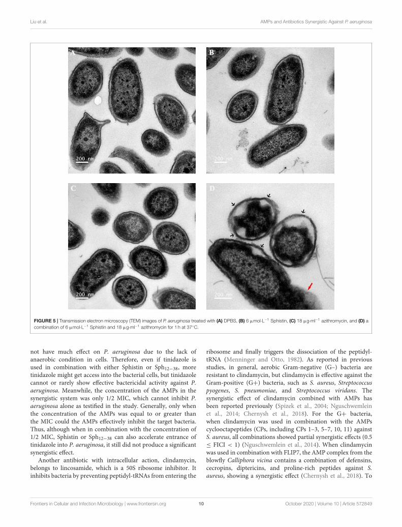
in morphology of P. aeruginosa cells or cell membranes.After treatment with DPBS, the control cells had a completecell morphology (Figure 5A), and no significant changes wereobserved when the bacterial cells were incubated with Sphistinor azithromycin (Figures 5B,C). The bacteria were then treatedwith a combination of Sphistin with azithromycin. After 1 h,we observed a significant separation of the cell membrane andcytoplasm in the bacterial cells (Figure 5D, black arrowheads),and remarkable leakage of cellular contents also appeared at thesame time (Figure 5D, red arrowheads).
Frontiers in Cellular and Infection Microbiology | www.frontiersin.org 6 October 2020 | Volume 10 | Article 572849
Liu et al. AMPs and Antibiotics Synergistic Against P. aeruginosa
FIGURE 2 | Growth curves of P. aeruginosa when incubated with (A) DPBS, Sphistin (6 µmol·L−1), azithromycin (18 µg·ml−1 ), and Sphistin (6 µmol·L−1) in
combination with azithromycin (18 µg·ml−1 ) for 2 h; (B) DPBS, Sphistin (1.5 µmol·L−1), rifampicin (0.625 µg·ml−1), and Sphistin (1.5 µmol·L−1) in combination with
rifampicin (0.625 µg·ml−1 ) for 6 h.
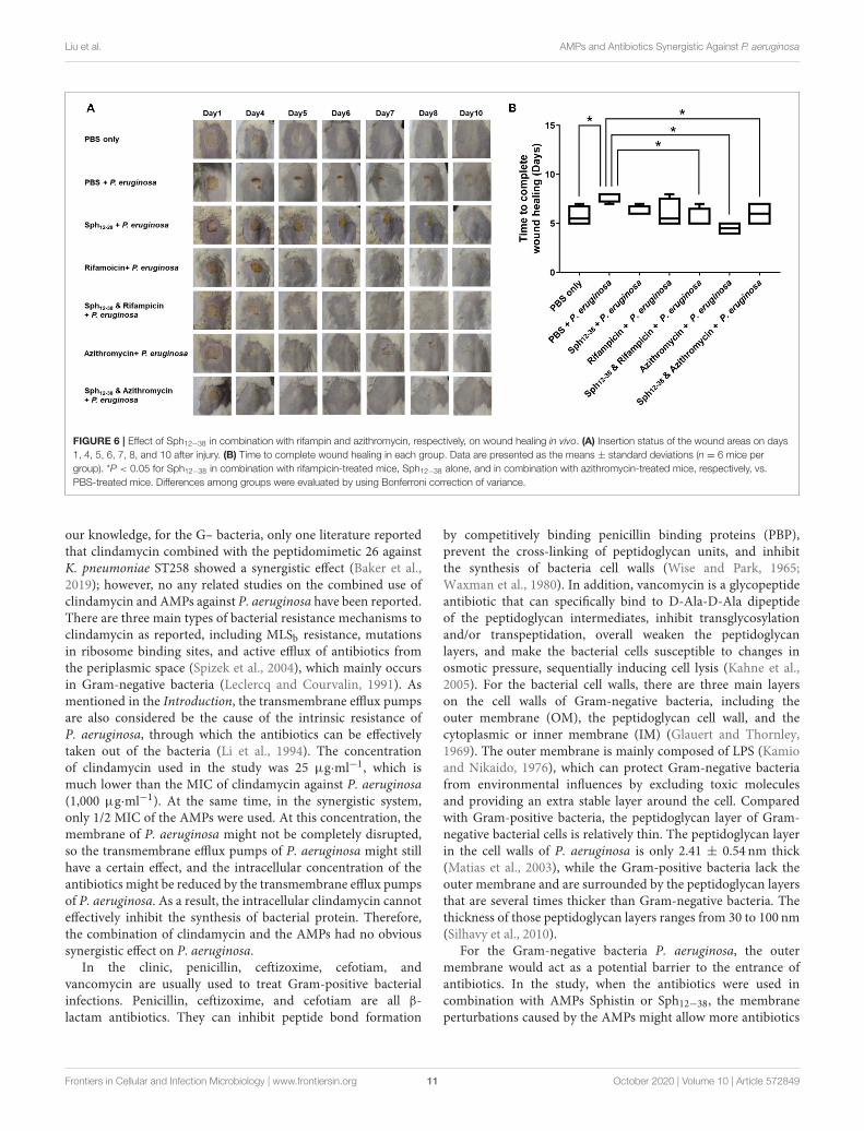
Sph12−38 in Combination With EitherRifampicin or Azithromycin to Promote theWound HealingA mouse wound model was used to test the antibacterial activityof Sph12−38 in combination with rifampicin and azithromycinin vivo. On the dorsal part of each mouse, the epidermiswas damaged with medical pressure-sensitive adhesive tapeand then the dermis was exposed. Afterwards, 108 CFU ofthe P. aeruginosa were evenly smeared onto the wound areas(Figure 6A). When the wound areas were not infected withP. aeruginosa, only the phosphate-buffered saline (PBS) wasinjected, and all wounds healed within 5–7 days (Figures 6A,B).Meanwhile, for mice infected with P. aeruginosa, PBS, Sph12−38,rifampicin, azithromycin, and Sph12−38 in combination withrifampicin or azithromycin were injected into the wound skin,respectively. When the infected mice were only inoculated withPBS, the wound healing process became slower and was notcompleted until 7–8 days postinjection. Compared with the PBSgroup, the injection of Sph12−38 or rifampicin alone could notsignificantly shorten the healing time. In contrast, when theinfected mice were injected with Sph12−38 in combination withrifampicin, the wound could be completely recovered within 5 to7 days (P < 0.05). As for azithromycin, when used alone or incombination with Sph12−38, its efficacy seemed to be even betterthan the combination of Sph12−38 and rifampicin, and the woundcompletely healed within only 4–5 days (Figures 6A,B).
DISCUSSION
Since penicillin was introduced into clinical treatment in the1940s, antibiotics abuse has never ended throughout the past 70years. The abuse of antibiotics, including overuse and misuse ofantibiotics, and the limited availability of new antibiotics havecaused the global antibiotic resistance crisis (Ventola, 2015).
The abuse of antibiotic inevitably leads to the rapid emergenceof drug-resistant bacteria including multidrug resistant (MDR)bacteria, and even the extremely drug-resistant (XDR) ortotally drug-resistant (TDR) phenotypes worldwide, whichhave seriously endangered the efficacy of antibiotics and havebecome a serious threat to human health (French G. L., 2010;Magiorakos et al., 2012). Although it is known that the resistancedeterminants are already presented in the microorganisms priorto the introduction of antibiotics and most of them are found innatural antibiotic-producing microorganisms (Levy, 1992), theintensive use of antibiotics indeed has dramatically increasedthe frequency of resistance among nearly all pathogens thatgreatly weaken therapeutic options and the medical advantagesin the post-antibiotic era that had almost been lost to date(Guay, 2008; Lew et al., 2008; Woodford and Livermore, 2009).Therefore, reducing the use of antibiotics had been proposed,through which the selection pressure for acquired resistancewill be reduced and the antibiotic-sensitive bacteria will berecovered, enabling them to eventually defeat resistance strainsover time (Levin et al., 1997; Andersson and Levin, 1999).AMPs with a membrane targeting effect provide possibilitiesto use AMPs in combination with multiple antibiotics fortreatment of pathogens, thereby enhancing the efficacy of thoseantibiotics (Cassone and Otvos, 2010; Haney et al., 2017).Among drug-resistant bacteria, P. aeruginosa is one of themost common hospital-acquired and nosocomial conditionedpathogens. It is much prone to acquire multidrug resistance;e.g., some strains of MDR P. aeruginosa have been found tobe resistant to almost all antibiotics, including aminoglycosides,cephalosporins, fluoroquinolones, and carbapenems (C. D. C. P.(US) Centres for Disease Control and Prevention (US)., 2013;Frieden, 2013). Therefore, it is necessary to explore new agentsthat could be substituted for antibiotics.
Rifampicin is a semisynthetic antibiotic derived fromrifamycin, and it was introduced as an effective medicine to
Frontiers in Cellular and Infection Microbiology | www.frontiersin.org 7 October 2020 | Volume 10 | Article 572849
Liu et al. AMPs and Antibiotics Synergistic Against P. aeruginosa
FIGURE 3 | Scanning electron microscope (SEM) images of P. aeruginosa treated with (A) 6 µmol·L−1 Sphistin, (B) 18 µg·ml−1 azithromycin, and (C) a combination
of 6 µmol·L−1 Sphistin and 18 µg·ml−1 azithromycin for 2 h at 37◦C. Meanwhile, the bacteria treated with (D) 1.5 µmol·L−1 Sphistin, (E) 0.625 µg·ml−1 rifampicin, (F)
a combination of 1.5 µmol·L−1 Sphistin and 0.625 µg·ml−1 rifampicin, and (G) DPBS for 4 h at 37◦C.
treat tuberculosis; the primary efficacy of rifampicin was againstGram-positive bacteria (Bliziotis et al., 2007). Several studies haverevealed that rifampicin in combination with colistin/polymyxinsin vitro (Giamarellos-Bourboulis et al., 2003; Tascini et al., 2004;Yang et al., 2016) and in vivo (Cirioni et al., 2007; Cai et al.,2018) showed effective antibacterial activity against MDR P.aeruginosa. For example, four patients infected with sepsis orpneumonia caused by MDR P. aeruginosa were successfullycured with the addition of rifampicin to colistin (Tascini et al.,2004). The colistin/polymyxins have an amphipathic structurewith clusters of hydrophobic and positively charged regions,and this structural property seems to be closely related totheir antibacterial activity (Wade et al., 1990; Scott et al.,1999). In fact, the amphipathic structure with hydrophobicand positive charge is also the classical structural feature ofthe cationic AMPs; therefore, the combination of rifampicinand two α-helical cationic AMPs, magainin II and cecropin A,also showed synergistic antibacterial effect against P. aeruginosastrains in vitro and in vivo (Cirioni et al., 2008). In addition torifampicin, it has been proven that the combination of colistin
and azithromycin showed synergistic and additive activity againstthe Gram-negative bacteria Acinetobacter baumannii and P.aeruginosa, respectively (Timurkaynak et al., 2006). Two AMPs,Sphistin with 38 aa that is derived from the N-terminal ofhistone H2A in S. paramamosain and the truncated shortfragment Sph12−38, both have potent in vitro antibacterial activityagainst several Gram-positive and Gram-negative bacteria andsome fungi. Both Sphistin and Sph12−38 showed typical featuresof cationic AMPs, including amphiphilic α-helical secondstructure and positive charge net. Therefore, both of these twocationic AMPs were used in combination with rifampicin andazithromycin to treat P. aeruginosa in this study.
Outer membranes of Gram-negative bacteria are formedby a divalent cation-crosslinked matrix of lipopolysaccharide(LPS) molecules on the outer leaflet, and via displacing theLPS-bound metals, they could be disrupted by a diversestructural class of polycations (Vaara, 1993; Livermore, 2002;Schuldiner, 2006; Tenover, 2006). It has been proven thatpolymyxins could bind the lipid A moiety of LPS and perturbthe bacterial cell membranes (Evans et al., 1999). Similar
Frontiers in Cellular and Infection Microbiology | www.frontiersin.org 8 October 2020 | Volume 10 | Article 572849
Liu et al. AMPs and Antibiotics Synergistic Against P. aeruginosa
FIGURE 4 | Extracellular ATP release in different treatment groups after exposure of P. aeruginosa to (A) DPBS, Sphistin (6 µmol·L−1), and/or azithromycin (18
µg·ml−1 ) for 2 h; (B) DPBS, Sphistin (1.5 µmol·L−1), and/or rifampicin (0.625 µg·ml−1 ) for 4 h. The bacteria cell membrane damage induced by the antibacterial
agents is accompanied by leakage of intracellular content due to compromised membrane integrity.
to the polymyxins, the cationic AMPs mainly targeted thebacterial cell membranes, and the common features shared bythese peptides are that they are prone to form amphipathicstructures and then cluster the basic and hydrophobic aminoacids into specific regions (Vaara and Porro, 1996; Yeamanand Yount, 2003). Therefore, the cationic AMPs could bindthe LPS by electrostatic and hydrophobic interactions andhave antibacterial activity (Iwagaki et al., 2000; Yethon andWhitfield, 2001). Meanwhile, the antimicrobial mechanism ofSphistin was suggested to have the capability of attaching thecell membrane and permeabilizing the bacterial cell membranesto kill the pathogens (Chen et al., 2015). Therefore, whenP. aeruginosa was treated with the combination of Sphistinand two antibiotics, the permeabilization of the bacterialcell membranes facilitated the uptake of the two antibiotics;rifampicin would bind to and inhibit the bacterial DNA-dependent RNA polymerase (Campbell et al., 2001), andazithromycin would inhibit RNA-dependent protein synthesisof the bacteria (Mazzei et al., 1993; Alvarez-Elcoro and Enzler,1999); finally, both antibiotics would induce cell death. Justas Figure 4 showed, there was no difference in the leakageof the intracellular ATP between Sphistin used alone or thecombination of Sphistin with two antibiotics. Indeed, theseresults further verified that even in the synergy system, onlySphistin induced the permeabilization of the bacterial cellmembranes. Taken together, our results agreed with previousstudies (Cirioni et al., 2008) that when the AMPs are combinedwith the antibiotics to treat the pathogens, the AMPs inducedthe permeabilization of the pathogen cell membranes andsubsequently promoted uptake of the antibiotics, allowing theantibiotics to interact with their intracellular targets easily andfinally kill the pathogens.
Among the eight selected antibiotics, the two antibioticsazithromycin and rifampicin in combination with the AMPsSphistin or Sph12−38 showed a synergistic effect against P.aeruginosa. However, the other six antibiotics had no significantsynergistic effect against P. aeruginosa when each of themwas combined with the AMPs. Among the six antibiotics, aspreviously reported (Menninger and Otto, 1982; Nord andKager, 1983; Gardner and Hill, 2001; Raether and Hanel,2003; Tasca et al., 2003), tinidazole and clindamycin also haveintracellular targets. Among the remaining four antibiotics, thethree antibiotics including penicillin, ceftizoxime, and cefotiamare all β-lactam antibiotics, while vancomycin is a glycopeptideantibiotic (Kahne et al., 2005). All of these four antibiotics caninhibit the synthesis of bacteria cell walls as previously reported(Wise and Park, 1965; Waxman et al., 1980; Kahne et al., 2005).
Tinidazole is a structural analog of metronidazole. Bothtinidazole and metronidazole are active against anaerobicorganisms or protozoa. The antibacterial mechanism is brieflydescribed as follows. When tinidazole diffused into bacterialcells, the nitro group of tinidazole will be reduced to short-lived and toxic free radicals. The toxic intermediates covalentlybind to DNA, causing DNA damage and ultimately cell death(Nord and Kager, 1983; Gardner and Hill, 2001; Raether andHanel, 2003; Tasca et al., 2003). With the decrease of intracellularconcentration of tinidazole due to the reduction reaction,more tinidazole could enter the cells, thereby maintaining theinhibition activity of anaerobic bacteria. In aerobic bacteria andmammalian cells, they have relatively high redox potentials andare also rich in oxygen molecules than anaerobic organismsor protozoa, which will hinder the reduction reaction (Nordand Kager, 1983; Tasca et al., 2003). Because P. aeruginosais an aerobic bacterium, we speculated that tinidazole will
Frontiers in Cellular and Infection Microbiology | www.frontiersin.org 9 October 2020 | Volume 10 | Article 572849
Liu et al. AMPs and Antibiotics Synergistic Against P. aeruginosa
FIGURE 5 | Transmission electron microscopy (TEM) images of P. aeruginosa treated with (A) DPBS, (B) 6 µmol·L−1 Sphistin, (C) 18 µg·ml−1 azithromycin, and (D) a
combination of 6 µmol·L−1 Sphistin and 18 µg·ml−1 azithromycin for 1 h at 37◦C.
not have much effect on P. aeruginosa due to the lack ofanaerobic condition in cells. Therefore, even if tinidazole isused in combination with either Sphistin or Sph12−38, moretinidazole might get access into the bacterial cells, but tinidazolecannot or rarely show effective bactericidal activity against P.aeruginosa. Meanwhile, the concentration of the AMPs in thesynergistic system was only 1/2 MIC, which cannot inhibit P.aeruginosa alone as testified in the study. Generally, only whenthe concentration of the AMPs was equal to or greater thanthe MIC could the AMPs effectively inhibit the target bacteria.Thus, although when in combination with the concentration of1/2 MIC, Sphistin or Sph12−38 can also accelerate entrance oftinidazole into P. aeruginosa, it still did not produce a significantsynergistic effect.
Another antibiotic with intracellular action, clindamycin,belongs to lincosamide, which is a 50S ribosome inhibitor. Itinhibits bacteria by preventing peptidyl-tRNAs from entering the
ribosome and finally triggers the dissociation of the peptidyl-tRNA (Menninger and Otto, 1982). As reported in previousstudies, in general, aerobic Gram-negative (G–) bacteria areresistant to clindamycin, but clindamycin is effective against theGram-positive (G+) bacteria, such as S. aureus, Streptococcuspyogenes, S. pneumoniae, and Streptococcus viridans. Thesynergistic effect of clindamycin combined with AMPs hasbeen reported previously (Spizek et al., 2004; Nguschwemleinet al., 2014; Chernysh et al., 2018). For the G+ bacteria,when clindamycin was used in combination with the AMPscyclooctapeptides (CPs, including CPs 1–3, 5–7, 10, 11) againstS. aureus, all combinations showed partial synergistic effects (0.5≤ FICI < 1) (Nguschwemlein et al., 2014). When clindamycinwas used in combination with FLIP7, the AMP complex from theblowfly Calliphora vicina contains a combination of defensins,cecropins, diptericins, and proline-rich peptides against S.aureus, showing a synergistic effect (Chernysh et al., 2018). To
Frontiers in Cellular and Infection Microbiology | www.frontiersin.org 10 October 2020 | Volume 10 | Article 572849
Liu et al. AMPs and Antibiotics Synergistic Against P. aeruginosa
FIGURE 6 | Effect of Sph12−38 in combination with rifampin and azithromycin, respectively, on wound healing in vivo. (A) Insertion status of the wound areas on days
1, 4, 5, 6, 7, 8, and 10 after injury. (B) Time to complete wound healing in each group. Data are presented as the means ± standard deviations (n = 6 mice per
group). *P < 0.05 for Sph12−38 in combination with rifampicin-treated mice, Sph12−38 alone, and in combination with azithromycin-treated mice, respectively, vs.
PBS-treated mice. Differences among groups were evaluated by using Bonferroni correction of variance.
our knowledge, for the G– bacteria, only one literature reportedthat clindamycin combined with the peptidomimetic 26 againstK. pneumoniae ST258 showed a synergistic effect (Baker et al.,2019); however, no any related studies on the combined use ofclindamycin and AMPs against P. aeruginosa have been reported.There are three main types of bacterial resistance mechanisms toclindamycin as reported, including MLSb resistance, mutationsin ribosome binding sites, and active efflux of antibiotics fromthe periplasmic space (Spizek et al., 2004), which mainly occursin Gram-negative bacteria (Leclercq and Courvalin, 1991). Asmentioned in the Introduction, the transmembrane efflux pumpsare also considered be the cause of the intrinsic resistance ofP. aeruginosa, through which the antibiotics can be effectivelytaken out of the bacteria (Li et al., 1994). The concentrationof clindamycin used in the study was 25 µg·ml−1, which ismuch lower than the MIC of clindamycin against P. aeruginosa(1,000 µg·ml−1). At the same time, in the synergistic system,only 1/2 MIC of the AMPs were used. At this concentration, themembrane of P. aeruginosa might not be completely disrupted,so the transmembrane efflux pumps of P. aeruginosa might stillhave a certain effect, and the intracellular concentration of theantibiotics might be reduced by the transmembrane efflux pumpsof P. aeruginosa. As a result, the intracellular clindamycin cannoteffectively inhibit the synthesis of bacterial protein. Therefore,the combination of clindamycin and the AMPs had no obvioussynergistic effect on P. aeruginosa.
In the clinic, penicillin, ceftizoxime, cefotiam, andvancomycin are usually used to treat Gram-positive bacterialinfections. Penicillin, ceftizoxime, and cefotiam are all β-lactam antibiotics. They can inhibit peptide bond formation
by competitively binding penicillin binding proteins (PBP),prevent the cross-linking of peptidoglycan units, and inhibitthe synthesis of bacteria cell walls (Wise and Park, 1965;Waxman et al., 1980). In addition, vancomycin is a glycopeptideantibiotic that can specifically bind to D-Ala-D-Ala dipeptideof the peptidoglycan intermediates, inhibit transglycosylationand/or transpeptidation, overall weaken the peptidoglycanlayers, and make the bacterial cells susceptible to changes inosmotic pressure, sequentially inducing cell lysis (Kahne et al.,2005). For the bacterial cell walls, there are three main layerson the cell walls of Gram-negative bacteria, including theouter membrane (OM), the peptidoglycan cell wall, and thecytoplasmic or inner membrane (IM) (Glauert and Thornley,1969). The outer membrane is mainly composed of LPS (Kamioand Nikaido, 1976), which can protect Gram-negative bacteriafrom environmental influences by excluding toxic moleculesand providing an extra stable layer around the cell. Comparedwith Gram-positive bacteria, the peptidoglycan layer of Gram-negative bacterial cells is relatively thin. The peptidoglycan layerin the cell walls of P. aeruginosa is only 2.41 ± 0.54 nm thick(Matias et al., 2003), while the Gram-positive bacteria lack theouter membrane and are surrounded by the peptidoglycan layersthat are several times thicker than Gram-negative bacteria. Thethickness of those peptidoglycan layers ranges from 30 to 100 nm(Silhavy et al., 2010).
For the Gram-negative bacteria P. aeruginosa, the outermembrane would act as a potential barrier to the entrance ofantibiotics. In the study, when the antibiotics were used incombination with AMPs Sphistin or Sph12−38, the membraneperturbations caused by the AMPs might allow more antibiotics
Frontiers in Cellular and Infection Microbiology | www.frontiersin.org 11 October 2020 | Volume 10 | Article 572849

Liu et al. AMPs and Antibiotics Synergistic Against P. aeruginosa
to enter the bacterial cells. Nevertheless, since the main targetfor the β-lactam antibiotics (like three antibiotics in thestudy) and vancomycin is peptidoglycan synthesis, even if themembrane perturbations accelerated the entry of these fourantibiotics into bacteria and possibly affect the peptidoglycansynthesis of P. aeruginosa, their action could not affectthe integrity of the outer membrane. In addition, a lowconcentration of the AMPs (<1/2 MIC) could not completelydestroy the structure of the outer membrane, and the cellmorphology of P. aeruginosa can maintain relative integrity,indicating that the bacteria remained alive. Therefore, whenthese two antibiotics were used in combination with theAMPs against P. aeruginosa, no significant synergistic effectwas observed.
The in vitro antibacterial tests indicated that the combinationof Sphistin with rifampicin and azithromycin killed thepathogens efficiently. To demonstrate the synergy effectsfurther, we tested the antibacterial efficiency in vivo. Similarto the experimental results in vitro, the remarkable effectappeared using Sph12−38 in combination with rifampicin thatpromoted the wound healing significantly (Figures 6A,B),whereas no significant effect was found using Sph12−38 orrifampicin alone. The underlying mechanism was presumedas follows. The AMPs could induce the permeabilization ofbacterial cells, facilitating rifampicin to access the cells andbind their binding sites; alternatively, the peptides stimulatethe immune systems of the host and then rifampicin couldplay an antibacterial role independently of the AMPs (Vaaraand Porro, 1996; Yeaman and Yount, 2003; Balakrishna et al.,2006). Nevertheless, unlike rifampicin, azithromycin aloneor in combination with Sph12−38 significantly facilitated thewound healing. Otherwise, Sphistin/Sph12−38 could bindto LPS and permeabilize the bacterial membrane; whencombined with rifampicin and azithromycin, Sphistin/Sph12−38
promoted the intracellular uptake of the antibiotics andsubsequently enhanced the bactericidal activity of bothagents against P. aeruginosa. Although P. aeruginosa wasnon-susceptible to rifampicin or azithromycin, when incombination with Sphistin/Sph12−38, they all showed higherantibacterial efficiency; the combination of Sphistin/Sph12−38
with rifampicin and azithromycin might be potentially usedfor the prevention and treatment of infections caused byP. aeruginosa; however, more work needs to be done inthe future.
DATA AVAILABILITY STATEMENT
The raw data supporting the conclusions of this article will bemade available by the authors, without undue reservation.
ETHICS STATEMENT
The animal study was reviewed and approved byXiamen University Laboratory Animal Management andEthics Committee.
AUTHOR CONTRIBUTIONS
K-JW design the research work, supervised, and revised themanuscript. JL performed the experiment, co-designed, andwrote the paper. XW and HZ participated the antibacterialexperiment and the transmission electron microscopy (TEM)experiment, and verified the overall replication/reproducibility ofresults/experiments and other research outputs. HP provided theantimicrobial peptides and co-designed the in vivo experiment.K-JW and FC supervised and revised the research work.All authors contributed to the article and approved thesubmitted version.
FUNDING
This work was sponsored by the National Natural ScienceFoundation of China (grant # 41676158 and 41806162); theFundamental Research Funds for the Central Universities (grant# 20720190109 and 20720180100); and the Fujian MarineEconomic Development Subsidy Fund Project from the FujianOcean and Fisheries Department (grant # FJHJF-L-2019-1). TheNational Natural Science Foundation of China (NSFC) is incharge of the administration of NSFC in accordance with thelaw and operates relatively independently. It is responsible for theorganization and implementation of subsidy plans, project setupand review, project approval and supervision. The FundamentalResearch Funds for the Central Universities funded by Ministryof Education of the People’s Republic of China. Fujian MarineEconomic Development Subsidy Fund Project from the FujianOcean and Fisheries Department. The funds received for openaccess publication fees from Xiamen University. This study alsoreceived funding from Xiamen Science and Technology PlanningProject (grant # 3502Z20203012).
REFERENCES
Alvarez-Elcoro, S., and Enzler, M. J. (1999). The macrolides: erythromycin,clarithromycin, and azithromycin. Mayo Clin. Proc. 74, 613–34.doi: 10.4065/74.6.613
Andersson, D. I., and Levin, B. R. (1999). The biological costof antibiotic resistance, Curr. Opin. Microbiol. 2, 489–493.doi: 10.1016/S1369-5274(99)00005-3
Arii, K., Kawada-Matsuo, M., Oogai, Y., Noguchi, K., and Komatsuzawa,H. (2019). Single mutations in BraRS confer high resistance againstnisin A in Staphylococcus aureus. Microbiologyopen 8:e791. doi: 10.1002/mbo3.791
Baker, K. R., Jana, B., Hansen, A. M., Nielsen, H. M., Franzyk, H., and Guardabassi,L. (2019). Repurposing azithromycin and rifampicin against gram-negativepathogens by combination with peptidomimetics. Front. Cell. Infect. Microbiol.
9:236. doi: 10.3389/fcimb.2019.00236Balakrishna, R., Wood, S. J., Nguyen, T. B., Miller, K. A., Kumar, E. S.,
Datta, A., et al. (2006). Structural correlates of antibacterial and membrane-permeabilizing activities in acylpolyamines. Antimicrob. Agents Chemother. 50,852–861. doi: 10.1128/AAC.50.3.852-861.2006
Band, V. I., Hufnagel, D. A., Jaggavarapu, S., Sherman, E. X., Wozniak, J. E.,Satola, S.W., et al. (2019). Antibiotic combinations that exploit heteroresistanceto multiple drugs effectively control infection. Nat. Microbiol. 4, 1627–1635.doi: 10.1038/s41564-019-0480-z
Frontiers in Cellular and Infection Microbiology | www.frontiersin.org 12 October 2020 | Volume 10 | Article 572849
Liu et al. AMPs and Antibiotics Synergistic Against P. aeruginosa
Bliziotis, I., Ntziora, F., Lawrence, K., and Falagas, M. (2007). Rifampin asadjuvant treatment of Gram-positive bacterial infections: a systematic reviewof comparative clinical trials. Eur. J. Clin. Microbiol. Infect. Dis. 26:849.doi: 10.1007/s10096-007-0378-1
Blonder, J., Ghose, M. B., Xiao, W., Camp, D. G., Wingred, M., Davis, R. W.,et al. (2004). Global analysis of the membrane subproteome of pseudomonasaeruginosa using liquid chromatography-tandem mass spectrometry. J.
Proteome Res. 3, 434–444. doi: 10.1021/pr034074wBrogden, K. A. (2005). Antimicrobial peptides: pore formers or metabolic
inhibitors in bacteria?Nat. Rev. Microbiol. 3, 238–50. doi: 10.1038/nrmicro1098Brogden, K. A., Ackermann, M., McCray, P. B., and Tack, B. F. (2003).
Antimicrobial peptides in animals and their role in host defences. Int. J.Antimicrob. Agents 22, 465–478. doi: 10.1016/S0924-8579(03)00180-8
Cai, Y., Yang, D., Wang, J., and Wang, R. (2018). Activity of colistin alone orin combination with rifampicin or meropenem in a carbapenem-resistantbioluminescent Pseudomonas aeruginosa intraperitoneal murine infectionmodel. J. Antimicrob. Chemother. 73, 456–461. doi: 10.1093/jac/dkx399
Campbell, E. A., Korzheva, N., Mustaev, A., Murakami, K., Nair, S., Goldfarb, A.,et al. (2001). Structural mechanism for rifampicin inhibition of bacterial rnapolymerase. Cell 104, 901–912. doi: 10.1016/S0092-8674(01)00286-0
Cassone, M., and Otvos, L. (2010). Synergy among antibacterial peptides andbetween peptides and small-molecule antibiotics. Expert. Rev. Anti Infect. Ther.8, 703–716. doi: 10.1586/eri.10.38
C. D. C. P. (US) Centres for Disease Control and Prevention (US). (2013).Antibiotic resistance threats in the United States, 2013. Centres for DiseaseControl and Prevention, US Department of Health and Human Services.
Chen, B., Fan, D. Q., Zhu, K. X., Shan, Z. G., Chen, F. Y., Hou,L., et al. (2015). Mechanism study on a new antimicrobial peptideSphistin derived from the N-terminus of crab histone H2A identified inhaemolymphs of Scylla paramamosain. Fish. Shellfish. Immunol. 47, 833–46.doi: 10.1016/j.fsi.2015.10.010
Chernysh, S. I., Gordya, N., Tulin, D., and Yakovlev, A. B. (2018). Biofilminfections between scylla and charybdis: interplay of host antimicrobialpeptides and antibiotics. Infect. Drug Resist. 11, 501–514. doi: 10.2147/IDR.S157847
Chongsiriwatana, N. P., Patch, J. A., Czyzewski, A. M., Dohm, M. T., Ivankin, A.,Gidalevitz, D., et al. (2008). Peptoids that mimic the structure, function, andmechanism of helical antimicrobial peptides. Proc. Natl. Acad. Sci. U.S.A. 105,2794–2799. doi: 10.1073/pnas.0708254105
Cirioni, O., Ghiselli, R., Orlando, F., Silvestri, C., Mocchegiani, F., Rocchi,M., et al. (2007). Efficacy of colistin/rifampin combination in experimentalrat models of sepsis due to a multiresistant Pseudomonas aeruginosastrain. Crit. Care Med. 35, 1717–1723. doi: 10.1097/01.CCM.0000266685.25436.03
Cirioni, O., Silvestri, C., Ghiselli, R., Orlando, F., Riva, A., Mocchegiani, F., et al.(2008). Protective effects of the combination of α-helical antimicrobial peptidesand rifampicin in three rat models of Pseudomonas aeruginosa infection. J.Antimicrob. Chemother. 62, 1332–1338. doi: 10.1093/jac/dkn393
C. L. S. Institute. (2012).Methods for Dilution Antimicrobial Susceptibility Tests for
Bacteria That Grow Aerobically; Approved Standards—Ninth Edition (Wayne:CLSI document M07-A9).
Dobson, A. J., Purves, J., Kamysz, W., and Rolff, J. (2013). Comparing selection ons. aureus between antimicrobial peptides and common antibiotics. PLOS ONE,8:e0076521. doi: 10.1371/journal.pone.0076521
El Shazely, B., Yu, G. Z., Johnston, P. R., and Rolff, J. (2020). Resistanceevolution against antimicrobial peptides in staphylococcus aureusalters pharmacodynamics beyond the MIC. Front. Microbiol. 11:103.doi: 10.3389/fmicb.2020.00103
Evans, M. E., Feola, D. J., and Rapp, R. P. (1999). Polymyxin B sulfate andcolistin: old antibiotics for emerging multiresistant gram-negative bacteria.Ann. Pharmacother. 33, 960–967. doi: 10.1345/aph.18426
French G. L. (2010). The continuing crisis in antibiotic resistance. Int. J.
Antimicrob. Agents 36, S3–S7. doi: 10.1016/S0924-8579(10)70003-0Frieden, T. (2013). Antibiotic resistance Threats in the United States, 2013, Vol.
23. Centers for Disease Control and Prevention, US Department of Health andHuman Services, 11–28.
Gardner, T. B., and Hill, D. R. (2001). Treatment of giardiasis. Clin. Microbiol. Rev.
14, 114–128. doi: 10.1128/CMR.14.1.114-128.2001
Giamarellos-Bourboulis, E., Sambatakou, H., Galani, I., and Giamarellou,H. (2003). In vitro interaction of colistin and rifampin on multidrug-resistant Pseudomonas aeruginosa. J. Chemother. 15, 235–238.doi: 10.1179/joc.2003.15.3.235
Glauert, A. M., and Thornley, M. J. (1969). The topography of thebacterial cell wall. Ann. Rev. Microbiol. 23, 159–198. doi: 10.1146/annurev.mi.23.100169.001111
Guay, D. R. J. D. (2008). Contemporary management of uncomplicatedurinary tract infections. Drugs. 68, 1169–1205. doi: 10.2165/00003495-200868090-00002
Habets, M. G. J. L., Rozen, D. E., and Brockhurst, M. A. (2012). Variationin Streptococcus pneumoniae susceptibility to human antimicrobial peptidesmay mediate intraspecific competition. Proc. Biol. Sci. 279, 3803–3811.doi: 10.1098/rspb.2012.1118
Hancock, R. E. (1998). Resistance mechanisms in Pseudomonas aeruginosa andother nonfermentative gram-negative bacteria. Clin. Infect. Dis. 27, S93–S99doi: 10.1086/514909
Hancock, R. E., and Speert, D. P. (2000). Antibiotic resistance in Pseudomonasaeruginosa: mechanisms and impact on treatment. Drug Resist. Updat. 3,247–255. doi: 10.1054/drup.2000.0152
Haney, E. F., Mansour, S. C., and Hancock, R. E. (eds.). (2017). “Antimicrobialpeptides: an introduction,” in Antimicrobial Peptides (New York, NY: Springer)doi: 10.1007/978-1-4939-6737-7_1
Heinz Floss, G., and Yu, W.-T. (2005). Rifamycin-mode of action, resistance, andbiosynthesis. Chem. Rev. 105, 621–32. doi: 10.1021/cr030112j
Iwagaki, A., Porro,M., and Pollack,M. (2000). Influence of synthetic antiendotoxinpeptides on lipopolysaccharide (LPS) recognition and LPS-inducedproinflammatory cytokine responses by cells expressing membrane-boundCD14. Infect. Immun. 68, 1655–1663. doi: 10.1128/IAI.68.3.1655-1663.2000
Kahne, D., Leimkuhler, C., Lu, W., and Walsh, C. T. (2005). Glycopeptideand lipoglycopeptide antibiotics. Chem. Rev. 105, 425–448. doi: 10.1021/cr030103a
Kamio, Y., and Nikaido, H. (1976). Outer membrane of salmonella typhimurium:accessibility of phospholipid head groups to phospholipase c and cyanogenbromide activated dextran in the external medium. Biochemistry 15, 2561–2570.doi: 10.1021/bi00657a012
Khara, J. S., Lim, F. K., Wang, Y., Ke, X.-Y., Voo, Z. X., Yang, Y. Y., et al. (2015).Designing α-helical peptides with enhanced synergism and selectivity againstmycobacterium smegmatis: discerning the role of hydrophobicity and helicity.Acta Biomater. 28, 99–108 doi: 10.1016/j.actbio.2015.09.015
Khara, J. S., Wang, Y., Ke, X. Y., Liu, S. Q., Newton, S. M., Langford, P. R.,et al. (2014). Anti-mycobacterial activities of synthetic cationic alpha-helicalpeptides and their synergism with rifampicin. Biomaterials, 35, 2032–2038.doi: 10.1016/j.biomaterials.2013.11.035
Koppen, B. C., Mulder, P. P., G., de Boer, L., Riool, M., Drijfhout, J. W., et al.(2019). Synergistic microbicidal effect of cationic antimicrobial peptides andteicoplanin against planktonic and biofilm-encased Staphylococcus aureus. Int.J. Antimicrob. Agents 53, 143–151. doi: 10.1016/j.ijantimicag.2018.10.002
Koshlukova, S. E., Lloyd, T. L., Araujo,M.W., B., and Edgerton,M. (1999). Salivaryhistatin 5 induces non-lytic release of ATP fromCandida albicans leading to celldeath. J. Biol. Chem. 274, 18872–18879. doi: 10.1074/jbc.274.27.18872
Leclercq, R., and Courvalin, P. (1991). Bacterial resistance to macrolide,lincosamide, and streptogramin antibiotics by target modification. Antimicrob.
Agents Chemother. 35, 1267–1272 doi: 10.1128/AAC.35.7.1267Levin, B., Lipsitch, M., Perrot, V., Schrag, S., Antia, R., Simonsen, L., et al. (1997).
The population genetics of antibiotic resistance. Clin. Infect. Dis. 24, S9-S16.doi: 10.1093/clinids/24.Supplement_1.S9
Levy, S. B. (1992). The Antibiotic Paradox. (New York, NY: Springer)Lew, W., Pai, M., Oxlade, O., Martin, D., and Menzies, D. J. A.
(2008). Initial drug resistance and tuberculosis treatment outcomes:systematic review and meta-analysis, Ann. Intern. Med. 149, 123–134.doi: 10.7326/0003-4819-149-2-200807150-00008
Li, D., Yang, Y., Tian, Z., Lv, J., Sun, F., Wang, Q., et al. (2017).Synergistic antibiotic effect of looped antimicrobial peptide CLP-19with bactericidal and bacteriostatic agents. Oncotarget 8, 55958–55966.doi: 10.18632/oncotarget.18124
Li, X. Z., Livermore, D. M., and Nikaido, H. (1994). Role of effluxpump(s) in intrinsic resistance of Pseudomonas aeruginosa: resistance to
Frontiers in Cellular and Infection Microbiology | www.frontiersin.org 13 October 2020 | Volume 10 | Article 572849
Liu et al. AMPs and Antibiotics Synergistic Against P. aeruginosa
tetracycline, chloramphenicol, and norfloxacin. Antimicrob Agents Chemother,38, 1732–1741 doi: 10.1128/AAC.38.8.1732
Livermore, D. M. (2002). Multiple mechanisms of antimicrobial resistance inPseudomonas aeruginosa: our worst nightmare? Clin. Infect. Dis. 34, 634–640.doi: 10.1086/338782
Lyczak, J. B., Cannon, C. L., and Pier, G. B. (2000). Establishment of Pseudomonasaeruginosa infection: lessons from a versatile opportunist. Microbes Infect 2,1051–60. doi: 10.1016/S1286-4579(00)01259-4
Ma, X. W., Hou, L., Chen, B., Fan, D. Q., Chen, Y. C., Yang, Y., et al. (2017).A truncated Sph12-38 with potent antimicrobial activity showing resistanceagainst bacterial challenge in Oryzias melastigma. Fish. Shellfish Immunol. 67,561–570. doi: 10.1016/j.fsi.2017.06.013
Magiorakos, A. P., Srinivasan, A., Carey, R. B., Carmeli, Y., Falagas, M. E.,Giske, C. G., et al. (2012). Multidrug-resistant, extensively drug-resistantand pandrug-resistant bacteria: an international expert proposal for interimstandard definitions for acquired resistance. Clinical Microbiol. Infect. 18,268–281. doi: 10.1111/j.1469-0691.2011.03570.x
Makarova, O., Johnston, P. R., Rodriguezrojas, A., Shazely, B. E., Morales, J. M.,and Rolff, J. (2018). Genomics of experimental adaptation of Staphylococcusaureus to a natural combination of insect antimicrobial peptides. Sci. Rep.8:15359 (2018) doi: 10.1038/s41598-018-33593-7
Matias, V. R., F., Alamoudi, A., Dubochet, J., and Beveridge, T. J. (2003).Cryo-Transmission Electron Microscopy of Frozen-Hydrated Sections ofEscherichia coli and Pseudomonas aeruginosa. J. Bacteriol. 185, 6112–6118.doi: 10.1128/JB.185.20.6112-6118.2003
Mazzei, T., Mini, E., Novelli, A., and Periti, P. (1993). Chemistry andmode of action of macrolides. J. Antimicrob. Chemother. 31(Suppl C), 1–9doi: 10.1093/jac/31.suppl_C.1
Menninger, J. R., and Otto, D. P. (1982). Erythromycin, carbomycin, andspiramycin inhibit protein synthesis by stimulating the dissociation ofpeptidyl-tRNA from ribosomes. Antimicrob. Agents Chemother. 21, 811–818.doi: 10.1128/AAC.21.5.811
Nguschwemlein, M., Dumond, J., Rudd, L., and Rigaud, J. (2014). In vitro synergybetween some cationic amphipathic cyclooctapeptides and antibiotics. Aust. J.Chem. 68, 218–223. doi: 10.1071/CH14427
Nord, C. E., and Kager, L. (1983). Tinidazole - microbiology, pharmacology andefficacy in anaerobic infections. Infection 11, 54–60. doi: 10.1007/BF01651361
Odds, F. C. (2003). Synergy, antagonism, and what the chequerboard puts betweenthem. J. Antimicrob. Chemother. 52, 1–1. doi: 10.1093/jac/dkg301
Pacios, O., Blasco, L., Bleriot, I., Fernandezgarcia, L., Bardanca, M. G., Ambroa, A.,et al. (2020). Strategies to combat multidrug-resistant and persistent infectiousdiseases. J. Antib. 9:65 doi: 10.3390/antibiotics9020065
Pal, C., Papp, B., and Lazar, V. (2015). Collateral sensitivity of antibiotic-resistantmicrobes. Trends Microbiol. 23, 401–407. doi: 10.1016/j.tim.2015.02.009
Pankey, G., Ashcraft, D., and Patel, N. (2005). In vitro synergy of daptomycinplus rifampin against Enterococcus faecium resistant to both linezolidand vancomycin. Antimicrob. Agents Chemother. 49, 5166–5168doi: 10.1128/AAC.49.12.5166-5168.2005
Pankey, G. A., and Ashcraft, D. S. (2005). In vitro synergy ofciprofloxacin and gatifloxacin against ciprofloxacin-resistantPseudomonas aeruginosa. Antimicrob. Agents Chemother. 49, 2959–2964.doi: 10.1128/AAC.49.7.2959-2964.2005
Peters, D. H., Friedel, H. A., and McTavish, D. (1992). azithromycin - a review ofits antimicrobial activity, pharmacokinetic properties and clinical efficacyDrugs44, 750–799. doi: 10.2165/00003495-199244050-00007
Petersen, P. J., Labthavikul, P., Jones, C. H., and Bradford, P. A. (2006).In vitro antibacterial activities of tigecycline in combination withother antimicrobial agents determined by chequerboard and time-killkinetic analysis. J. Antimicrob. Chemother. 57, 573–576. doi: 10.1093/jac/dki477
Raether, W., and Hanel, H. (2003). Nitroheterocyclic drugs with broad spectrumactivity. Parasitol. Res. 90(Supp 1), S19–39. doi: 10.1007/s00436-002-0754-9
Rand, K. H., Houck, H. J., Brown, P., and Bennett, D. (1993). Reproducibility of themicrodilution checkerboard method for antibiotic synergy. Antimicrob. Agents
Chemother. 37, 613–615. doi: 10.1128/AAC.37.3.613Retsema, J., Girard, A., Schelkly, W., Manousos, M., Anderson, M., Bright,
G., et al. (1987). Spectrum and mode of action of azithromycin (CP-62,993), a new 15-membered-ring macrolide with improved potency against
gram-negative organisms. Antimicrob. Agents Chemother. 31, 1939–47.doi: 10.1128/AAC.31.12.1939
Roemhild, R., and Schulenburg, H. (2019). Evolutionary ecology meets theantibiotic crisis: can we control pathogen adaptation through sequentialtherapy? Evol. Med. Public Health 2019, 37–45. doi: 10.1093/emph/eoz008
Schuldiner, S. (2006). The ins and outs of drug transport. Nature 443, 157–157doi: 10.1038/443156b
Scott, M. G., Yan, H., and Hancock, R. E. (1999). Biological properties ofstructurally related α-helical cationic antimicrobial peptides. Infect. Immun. 67,2005–2009. doi: 10.1128/IAI.67.4.2005-2009.1999
Shai, Y. (2002). Mode of action of membrane active antimicrobial peptides.Biopolymers 66, 236–48. doi: 10.1002/bip.10260
Silhavy, T. J., Kahne, D., and Walker, S. (2010). The Bacterial Cell Envelope. ColdSpring Harbor Perspec. Biol. 2:414. doi: 10.1101/cshperspect.a000414
Spizek, J., Novotna, J., and Rezanka, T. (2004). Lincosamides: chemical structure,biosynthesis, mechanism of action, resistance, and applications. Adv. Appl.Microbiol. 56, 121–154 doi: 10.1016/S0065-2164(04)56004-5
Tasca, T., Borges, F. P., Bonan, C. D., De Carli, G. A., Battastini, A. M., O., et al.(2003). Effects of metronidazole and tinidazole on NTPDase1 and ecto-5’-nucleotidase from intact cells of Trichomonas vaginalis. Fems Microbiol. Lett.
226, 379–384. doi: 10.1016/S0378-1097(03)00637-2Tascini, C., Gemignani, G., Ferranti, S., Tagliaferri, E., Leonildi, A., Lucarini,
A., et al. (2004). Microbiological activity and clinical efficacy of a colistinand rifampin combination in multidrug-resistant Pseudomonas aeruginosainfections. J. Chemother. 16, 282–287. doi: 10.1179/joc.2004.16.3.282
Tenover, F. C. (2006). Mechanisms of antimicrobial resistance in bacteria. Am. J.
Med. 119, S3-S10 doi: 10.1016/j.amjmed.2006.03.011Timurkaynak, F., Can, F., Azap, Ö. K., Demirbilek, M., Arslan, H., and Karaman,
S. Ö. (2006). In vitro activities of non-traditional antimicrobials alone or incombination against multidrug-resistant strains of Pseudomonas aeruginosaand Acinetobacter baumannii isolated from intensive care units. Int. J.
Antimicrob. Agents 27, 224–228. doi: 10.1016/j.ijantimicag.2005.10.012Vaara, M. (1993). Outer membrane permeability barrier to azithromycin,
clarithromycin, and roxithromycin in gram-negative enteric bacteria.Antimicrob. Agents Chemother. 37, 354–356. doi: 10.1128/AAC.37.2.354
Vaara, M., and Porro, M. (1996). Group of peptides that act synergistically withhydrophobic antibiotics against gram-negative enteric bacteria. Antimicrob.
Agents Chemother. 40, 1801–1805. doi: 10.1128/AAC.40.8.1801Ventola, C. L. (2015). The antibiotic resistance crisis: part 1: causes and threats.
Pharm. Therap. 40, 277−83.Vizioli, J., and Salzet, M. (2002). Antimicrobial peptides from
animals: focus on invertebrates. Trends Pharmacol. Sci. 23, 494–6.doi: 10.1016/S0165-6147(02)02105-3
Wade, D., Boman, A., Wåhlin, B., Drain, C., Andreu, D., Boman, H. G., et al.(1990). All-D amino acid-containing channel-forming antibiotic peptides. Proc.Natl. Acad. Sci.U.S.A. 87, 4761–4765. doi: 10.1073/pnas.87.12.4761
Walter, W., and Staehelin, M. (1971). Actions of the Rifamycins. Bacteriol. Rev.35:290. doi: 10.1128/MMBR.35.3.290-309.1971
Waxman, D. J., Yocum, R. R., and Strominger, J. L. (1980). Penicillins andcephalosporins are active site-directed acylating agents - evidence in supportof the substrate-analog hypothesis. Philos. Trans. Roy. Soc. B Biol. Sci. 289,257–271. doi: 10.1098/rstb.1980.0044
Wise, E. M., and Park, J. T. (1965). Penicillin: its basic site of action as an inhibitorof a peptide cross-linking reaction in cell wall mucopeptide synthesis. Proc.Natl. Acad. Sci.U.S.A. 54, 75–81. doi: 10.1073/pnas.54.1.75
Woodford, N., and Livermore, D. M. J. J. o. I. (2009). Infections caused byGram-positive bacteria: a review of the global challenge. J. Infect. 59, S4-S16.doi: 10.1016/S0163-4453(09)60003-7
Xu, J. C., Duan, X. M., Wu, H., and Zhou, Q. (2013). Surveillance and Correlationof Antimicrobial Usage and Resistance of Pseudomonas aeruginosa: AHospital Population-Based Study. PLoS ONE 8:7 doi: 10.1371/journal.pone.0078604
Yamamoto, N., and Tamura, A. (2014). Designing cell-aggregatingpeptides without cytotoxicity. Biomacromolecules 15, 512–523 (2014)doi: 10.1021/bm4014414
Yang, D., Ni, W., Wang, R., and Wang, J. (2016). In vitro activity of antibioticcombination against carbapenems resistant Pseudomonas aeruginosa. Chin. J.Clin. Pharmacol. 32, 2269–72. doi: 10.1590/0037-8682-0012-2013
Frontiers in Cellular and Infection Microbiology | www.frontiersin.org 14 October 2020 | Volume 10 | Article 572849
Liu et al. AMPs and Antibiotics Synergistic Against P. aeruginosa
Yeaman, M. R., and Yount, N. Y. (2003). Mechanisms of antimicrobial peptideaction and resistance. Pharmacol. Rev. 55, 27–55. doi: 10.1124/pr.55.1.2
Yethon, J., and Whitfield, C. (2001). Lipopolysaccharide as a target forthe development of novel therapeutics in gram-negative bacteria.Curr. Drug Targets Infect. Disord. 1, 91–106. doi: 10.2174/1568005014606143
Yu, G., Baeder, D. Y., Regoes, R. R., and Rolff, J. (2018). Predicting drug resistanceevolution: insights from antimicrobial peptides and antibiotics. Proc. Roy. Soc.B Biol. Sci. 285:20172687 doi: 10.1098/rspb.2017.2687
Zasloff, M. (2002). Antimicrobial peptides of multicellular organisms. Nature 415,389–95. doi: 10.1038/415389a
Zhang, X. L., Gu, B. N., Mei, Y. N., Wen, Y., and Xia, W. Y. (2015). Increasingresistance rate to carbapenem among blood culture isolates of Klebsiellapneumoniae, Acinetobacter baumannii and Pseudomonas aeruginosa in auniversity-affiliated hospital in China, 2004–2011. J. Antib. 68, 115–120.doi: 10.1038/ja.2014.119
Zheng, Z., Tharmalingam, N., Liu, Q., Jayamani, E., Kim, W., Fuchs, B. B., et al.(2017). Synergistic efficacy of aedes aegypti antimicrobial peptide cecropinA2 and tetracycline against pseudomonas aeruginosa. Antimicrob. Agents
Chemother. 61, e00686-17. doi: 10.1128/AAC.00686-17
Conflict of Interest: The authors declare that the research was conducted in theabsence of any commercial or financial relationships that could be construed as apotential conflict of interest.
Copyright © 2020 Liu, Chen, Wang, Peng, Zhang and Wang. This is an open-access
article distributed under the terms of the Creative Commons Attribution License (CC
BY). The use, distribution or reproduction in other forums is permitted, provided
the original author(s) and the copyright owner(s) are credited and that the original
publication in this journal is cited, in accordance with accepted academic practice.
No use, distribution or reproduction is permitted which does not comply with these
terms.
Frontiers in Cellular and Infection Microbiology | www.frontiersin.org 15 October 2020 | Volume 10 | Article 572849