Embed Size (px)
Citation preview
on July 3, 2018http://rstb.royalsocietypublishing.org/Downloaded from
rstb.royalsocietypublishing.org
ReviewCite this article: Takeuchi T, Duszkiewicz AJ,
Morris RGM. 2014 The synaptic plasticity and
memory hypothesis: encoding, storage
and persistence. Phil. Trans. R. Soc. B 369:
20130288.
http://dx.doi.org/10.1098/rstb.2013.0288
One contribution of 35 to a Discussion Meeting
Issue ‘Synaptic plasticity in health and disease’.
Subject Areas:neuroscience, behaviour, cognition, physiology
Keywords:synaptic plasticity, memory, long-term
potentiation, engram, initial consolidation,
dopamine
Author for correspondence:Richard G. M. Morris
e-mail: [email protected]
& 2013 The Author(s) Published by the Royal Society. All rights reserved.
The synaptic plasticity and memoryhypothesis: encoding, storageand persistence
Tomonori Takeuchi, Adrian J. Duszkiewicz and Richard G. M. Morris
Centre for Cognitive and Neural Systems, University of Edinburgh, 1 George Square, Edinburgh EH8 9JZ, UK
The synaptic plasticity and memory hypothesis asserts that activity-dependent
synaptic plasticity is induced at appropriate synapses during memory for-
mation and is both necessary and sufficient for the encoding and trace
storage of the type of memory mediated by the brain area in which it is
observed. Criteria for establishing the necessity and sufficiency of such plas-
ticity in mediating trace storage have been identified and are here reviewed
in relation to new work using some of the diverse techniques of contemporary
neuroscience. Evidence derived using optical imaging, molecular-genetic and
optogenetic techniques in conjunction with appropriate behavioural analyses
continues to offer support for the idea that changing the strength of connec-
tions between neurons is one of the major mechanisms by which engrams
are stored in the brain.
1. IntroductionThe idea that changes in the efficacy of synapses within diverse neural circuits
could mediate the storage of information acquired during learning has a long
history. Theoretical hypotheses about the growth of neuronal connections in
the brain and the circumstances in which such growth might take place date
back to Ramon y Cajal [1] and, in the mid-twentieth century, to Hebb [2] and
Konorski [3]. The first experimental evidence emerged from studies of habitu-
ation and sensitization in the marine mollusc Aplysia [4,5]. The discovery of
long-term potentiation (LTP) [6] acted as a further stimulus to the concept,
not least because LTP was first discovered in an area of the brain—the hippo-
campal formation—that had been implicated in memory from clinical
observations of amnesia [7]. Indeed, the last sentence of Bliss & Lomo’s
paper raises the question. Noting the possibility that the time-scale of LTP
was long enough to be potentially useful for information storage, they go on
to conclude in characteristically quizzical fashion:
whether or not the intact animal makes use in real life of a property which has beenrevealed by synchronous, repetitive volleys to a population of fibres the normal rateand pattern of activity along which are unknown, is another matter. [6, p. 355].
In our quest for understanding ‘mechanisms’ in neuroscience, a focus in
research on activity-dependent synaptic plasticity such as LTP and long-term
depression (LTD) has been on identifying the causal steps that occur at individual
synapses mediating lasting changes in synaptic efficacy in terms of changes
in presynaptic transmitter release, alterations in postsynaptic glutamatergic
receptors, the action of neuromodulatory transmitters, the signal transduction
pathways activated, gene activation and synthesis of new proteins. A contempor-
ary focus is on the endo- and exocytosis of specific sub-types of glutamate
receptors, and alterations in the scaffolding molecules that make up the pre-
and postsynaptic elements of neuronal connectivity [8]. Recent reviews of the
persistence of LTP/LTD collectively point to the importance of translational con-
trol of dendritic mRNAs, and that spine dynamics may be remarkably fast to be in
register with the relatively immediate functional changes [9–11]. Accompanying
papers in this issue discuss similar themes.
rstb.royalsocietypublishing.orgPhil.T
2
on July 3, 2018http://rstb.royalsocietypublishing.org/Downloaded from
However, in our view, no less important is Bliss & Lomo’s
now 40-year-old question—do animals actually make use of
this capacity for change and, if so, how? This systems-level
question was initially approached in terms of seeking correl-
ations between learning and synaptic potentiation [12], but
the diverse techniques of contemporary neuroscience offer
new tools for securing a definitive and causal answer
[13,14], and addressing whether such understanding could
be a route towards more effective cognition [15]. Here, we
review progress in identifying whether and how synaptic
plasticity may mediate distinct aspects of learning and
memory with focus on the hippocampus, beginning with
the generic hypothesis.
rans.R.Soc.B369:201302882. The synaptic plasticity and memoryhypothesis
The synaptic plasticity and memory (SPM) hypothesis is
not identified with any one individual scientist, it being an
idea that has come forward in various guises over the years
(see [16] for review). At its heart is the notion that the
memory of prior experience is mediated by the reactivation
of ‘traces’ or ‘engrams’ whose basis involves alterations,
possibly bidirectional alterations, in synaptic efficacy. The
intellectual debt to Hebb, in particular, is very clear but the con-
cept of ‘memory of prior experience’ has to be unpacked with
respect to the distinct ways in which it can be interpreted.
The most everyday sense of memory is that of an event happen-
ing to someone, it somehow being recorded in their brain, and
then this person later bringing to mind some representation of
that same event. One can think of this loosely as ‘real’ memory
in the everyday ‘folk-psychology’ sense of the term. However,
prior experience may also induce changes in the nervous
system, possibly mediated by synaptic plasticity, that in
no sense allow recall of the prior experience that occurred.
A runner training for a marathon will gradually build up
muscle mass and all manner of changes in his or her body
that reflect the experience of training. However, changes in
muscle mass in no sense ‘represent’ the actual events that con-
stitute that training, even though they are triggered by it. These
two examples of putative ‘memory’ can be thought of as quali-
tatively distinct ways in which experience drives plasticity,
or alternatively, as two ends of a continuum. Huebener &
Bonhoeffer [17] take a ‘continuum’ point of view, suggesting
that study of the neural mechanisms of ocular dominance or
orientation-sensitive plasticity could be a valid route to discov-
ering the neural mechanisms of memory. The fine-tuning of
cortical connections in the visual system during the sensitive
period of early development is now known to involve bidirec-
tional changes in synaptic efficacy mediated by LTP-like and
LTD-like mechanisms, and various tantalizing phenomena
potentially relevant to memory have recently been discovered
such as ‘savings’ with respect to future plasticity after incom-
plete experiences during infancy. Specifically, Hofer et al. [18]
have demonstrated successful adult cortical plasticity in mice
that had a period of eye closure during the sensitive period
that was then terminated early on. This phenomenon is remin-
iscent of the ‘savings’ that can be seen in learning a second
language if a person has been exposed, at least for a period,
to that language early in life. Huebener and Bonhoeffer’s
view is that drawing a sharp categorical boundary between
‘real’ memory and the diverse array of experience-dependent
changes that occur in the nervous system throughout life is
both unnecessary and misleading.
However, while accepting the force of this argument, a
problem is that while the underlying neural mechanisms of
plasticity at glutamatergic synapses may be very similar for
changes right across this continuum, the function(s) that
activity-dependent synaptic plasticity serves will, in our
view, depend critically on the neural circuit in which that
plasticity is embedded in a non-monotonic manner. Let us
contrast two cases. Pre-synaptic facilitation at the sensory to
motor neuron synapse of the abdominal ganglion of Aplysiais thought to realize an increase in synaptic throughput
that mediates a behavioural facilitation of responsiveness.
A stimulus to the siphon then gives rise to a larger and
longer withdrawal of the gill and siphon reflex. Conversely,
habituation is associated with a decrease in transmitter release
measured using quantal analysis [5]. In these cases, there is an
elegant isomorphism between the physiological change and
the behavioural change that has been likened to a ‘cellular
alphabet of learning’ [19]. A similar principle may also apply
to fear conditioning mediated by the amygdala of mammals
in which an initially neutral stimulus can, through synaptic
potentiation mediated by changes in post-synaptic receptor
expression [20], give rise to graded evocation of fear through
associative conditioning with a painful or biologically danger-
ous stimulus (e.g. a predator). The amygdala’s efferent
connections trigger the various expressions of that fear via
downstream connections to the autonomic nervous system
[14]. Isomorphism may be a common characteristic of learned
reflex systems.
What is less well appreciated is, however, that other neural
circuits of memory do not operate on an isomorphism prin-
ciple. Rather, synaptic potentiation and depression sculpt the
possibility of associations between stimuli such that learning,
rather than giving rise to larger or smaller responses, enables
one stimulus representation to evoke the memory represen-
tation of another. This is qualitatively different. For example,
a key characteristic of episodic-like memory is what has been
called the ‘automatic recording of experience’ [21] whereby
the memory of an event is automatically associated with the
context in which it happens. Biologically significant events
such as reward and punishment are not necessary for
making such associations; nor need they enter into them in
the conventional sense of Pavlovian conditioning between a
conditional stimulus (CS) and a biologically significant uncon-
ditioned stimulus (US). Instead, the co-occurrence of events
and contexts enables, in the distributed associative machinery
of the hippocampal formation operating on Hebb-like prin-
ciples, an association that ties these two entities together.
That is, if you remember an event you will automatically
remember the context where it happened. Realizing this kind
of association requires distributed temporo-spatial represen-
tations of events and contexts that can be discriminated
(pattern separation) and sometimes generalized (pattern com-
pletion), with multiple events and contexts overlaid in a
distributed manner within a common neural circuit [22]. Heb-
bian synaptic plasticity, possibly coupled to associative
synaptic depression, within a neural circuitry that enables dis-
tributed associative memory through the pattern of excitatory
and inhibitory interconnections will realize this function—
albeit subject to the myriad of inhibitory feed-forward and
feedback connections that regulate excitability in different com-
partments of the dendritic tree [23]. The Huebener–Bonhoeffer
rstb.royalsocietypublishing.orgPhil.Trans.R.Soc.B
369:20130288
3
on July 3, 2018http://rstb.royalsocietypublishing.org/Downloaded from
continuum principle, with its focus on mechanism at the
level of individual synapses, does not yet provide an effective
framework for thinking about these systems-level issues. In
fact, at a systems-level, we are a long way from understanding
the detailed manner in which information is represented
in temporo-spatial codes, how the interplay of excitatory and
inhibitory activation enables encoding and later retrieval,
and even the distinct and sometimes sparse representational
codes of different sub-regions of the hippocampal formation
[24]. However, computational neuroscience techniques are
proving invaluable in tackling these issues and a systems
approach is essential if the representational issues are to be
addressed mechanistically.
Two last points to make about the generic SPM hypothesis
concern plasticity itself and new techniques. Research over
the 40 years since LTP was discovered has revealed a family
of different forms of activity-dependent synaptic plasticity.
In addition to NMDA receptor-dependent LTP and LTD,
there are forms of synaptic potentiation and depression that
are NMDA receptor-independent. Homeostatic plasticity has
been discovered and it may play both a normalizing and
protective role in neural circuits that might otherwise be risk
at seizure through potentiation of too high a proportion of
neural afferents [25]. Spike-timing-dependent plasticity [26,27]
has been implicated in the learning and expression of, for
example, remembered sequences [28]. Second, contemporary
neuroscience is characterized by an array of novel technologies
including new molecular-genetic technologies, optical imaging
in living animals and optogenetic manipulations of individual
neurons [29–34] as well as new behavioural paradigms
[35–37]. Their use, illustrated below, is offering the opportunity
to secure more definitive answers to the validity of the SPM
hypothesis.
To conclude, this hypothesis can be stated, in its most
general form as follows:
activity-dependent synaptic plasticity is induced at appropriatesynapses during memory formation, and is both necessary andsufficient for the encoding and trace storage of the type ofmemory mediated by the brain area in which that plasticity isobserved. [16, p. 650]
This definition is intended to be both inclusive and suffi-
ciently precise for the hypothesis to be falsifiable. We turn
now to four critical tests of the hypothesis that have been
examined in numerous studies over the past 25 years.
3. Criteria for assessing the synaptic plasticityand memory hypothesis
Martin et al. [16] identified four distinct criteria of assess-
ment and corresponding tests of the SPM hypothesis. The
first they called detectability whereby if learning involves
activity-dependent synaptic plasticity, it should be possible
to detect changes in synaptic efficacy following learning. This
is one aspect of a ‘sufficiency’ criterion—that synaptic change
occurs during learning—though it falls short of establishing
that such a change is actually sufficient. Second, they argued
that if some treatment (pharmacological, physiological and
molecular-genetic) were to be given prior to learning, the rate
of learning should be blocked, enhanced or otherwise altered
in a predictable manner if the treatment in question were to
alter the induction or expression of synaptic plasticity. They
called this the anterograde alteration criterion, a component of
‘necessity’. Third, if learning were to occur and then, after learn-
ing, certain retrograde manipulations were made that might
affect the expression of earlier changes in synaptic weights,
the ability of the neural circuit to reconstruct the appropriate
representational pattern should be affected. The experimental
subject might then behave as if it had retrieved different infor-
mation from that which had been learned. This is the retrogradealteration criterion—a second component of necessity. Last, if
memory resides in specific distributed patterns of altered
synaptic weights, the artificial creation of such a pattern
should result in the creation of a ‘false memory’ for an event
that did not happen or some aspect of knowledge or skill
that had not been taught or trained. This mimicry criterion,
the second component of sufficiency and essentially the
engineering criterion, is arguably the most demanding.
(a) DetectabilityThere is now strong evidence that learning can be associated
with the induction of changes in synaptic weights in apparently
relevant neural circuits. This is the essence of the ‘detectability’
criterion—with critical issues arising over what constitute rele-
vant neural circuits for any particular instance of learning (see
[38] for a detailed discussion).
The earliest attempts to detect changes in synaptic weight
in association with specific experiences revealed changes in
synaptic strength and the magnitude of population spikes
in the hippocampal formation in association with exposure of
animals, normally in isolation, to a complex social living environ-
ment [39]. It later transpired that these may, at least in part, be
associated with alterations in brain temperature rather than
exploration-associated changes in synaptic weights [40]. A later
study, with suitable calibration for temperature, did reveal tran-
sitory changes in excitatory postsynaptic potentials (EPSPs)
associated with novelty exposure, but these rapidly decayed to
baseline [41].
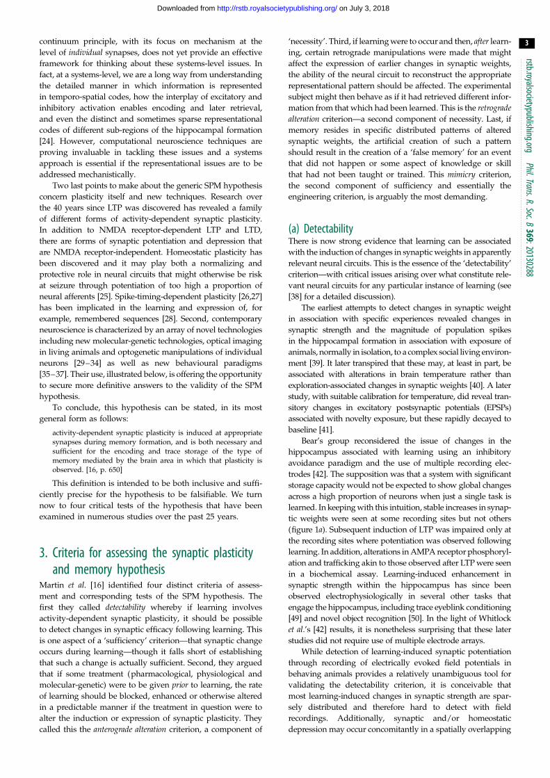
Bear’s group reconsidered the issue of changes in the
hippocampus associated with learning using an inhibitory
avoidance paradigm and the use of multiple recording elec-
trodes [42]. The supposition was that a system with significant
storage capacity would not be expected to show global changes
across a high proportion of neurons when just a single task is
learned. In keeping with this intuition, stable increases in synap-
tic weights were seen at some recording sites but not others
(figure 1a). Subsequent induction of LTP was impaired only at
the recording sites where potentiation was observed following
learning. In addition, alterations in AMPA receptor phosphoryl-
ation and trafficking akin to those observed after LTP were seen
in a biochemical assay. Learning-induced enhancement in
synaptic strength within the hippocampus has since been
observed electrophysiologically in several other tasks that
engage the hippocampus, including trace eyeblink conditioning
[49] and novel object recognition [50]. In the light of Whitlock
et al.’s [42] results, it is nonetheless surprising that these later
studies did not require use of multiple electrode arrays.
While detection of learning-induced synaptic potentiation
through recording of electrically evoked field potentials in
behaving animals provides a relatively unambiguous tool for
validating the detectability criterion, it is conceivable that
most learning-induced changes in synaptic strength are spar-
sely distributed and therefore hard to detect with field
recordings. Additionally, synaptic and/or homeostatic
depression may occur concomitantly in a spatially overlapping

10
5
0
NMDA receptor blockade
abolition of late-LTP by ZIP
PS a
mpl
itude
(m
V)
abolition of long-term memory by ZIP
saline
electrodes
opposite
opposite
Mea
n pa
ltfor
m c
ross
ings
6
5
4
3
2
1
0
T29–1
+/+–/–
CA1–KO
–20 –15 –10 –5 0 5 10 15 20right
right
thin
thin0
20
40
60
80
100
GFP
-Glu
A1+
spi
nes
(%)
stubby mushroom
stubby mushroomapical *
* *
UP 24 hCT 24 hFC 24 hFC 72 h
Dil
GFP
merged–0.5 0
1
1 2 1.0 mV5 m s
IA training
50
75
100
125
150
FP s
lope
(%
bas
elin
e)
2 1 + 2
1 2 3 4
trained
trained
left
left
ZIP
0 1
HFS time (h)
time (h) relative to conditioning
10 12 14 16 18 20 22 24 26 28 30
injectioncannula
pretraining training(trial 8)
24 h retention
ZIP
field-potentials following learning
GluN2A knock-out CA1-specific GluN1 deletion
learning-induced AMPA receptor trafficking to specific spine types
saline D-AP5
(b)(a)
(c) (d )
(g)
(e)
( f )
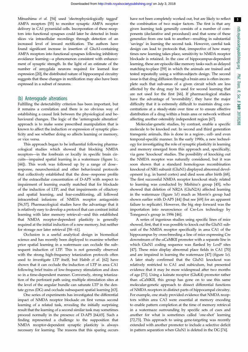
Figure 1. Illustrative findings relevant to the established criteria for assessing the SPM hypothesis. (a,b) Detectability. Field-potentials are increased on some but not allelectrodes of a multi-electrode array in area CA1 following inhibitory avoidance learning (Adapted with permission from Whitlock et al. [42] & AAAS) (a). AMPA receptortrafficking detected optically using a GFP label in association with learning, with GluA1 targeted specifically at mature, mushroom-shaped spines (Adapted with permissionfrom Matsuo et al. [43] & AAAS) (b). (c – e) Anterograde alterations. Pharmacological blockade of NMDA receptors in rats with chronic infusion of D-AP5 impairs spatiallearning (Adapted with permission from Morris et al. [44]) (c). Genetic knock-out of GluN2A in mice also impairs spatial learning in the watermaze (Adapted with permissionfrom Sakimura et al. [45]&Macmillan Publishers Ltd) (d ). CA1 pyramidal cell-specific knockout of GluN1 in mice also impairs selective searching in the watermaze (Adaptedwith permission from Tsien et al. [46,47] & Elsevier) (e). ( f,g) Retrograde alterations. Successful abolition by ZIP of long-lasting LTP 22 h after its initial induction (f ).Corresponding abolition of long-term place-memory on a rotating platform by ZIP (Adapted with permission from Pastalkova et al. [48] & AAAS) (g).
rstb.royalsocietypublishing.orgPhil.Trans.R.Soc.B
369:20130288
4
on July 3, 2018http://rstb.royalsocietypublishing.org/Downloaded from
set of synapses, further reducing the possibility of observing a
net enhancement of the evoked field response. Therefore,
the use of various molecular and structural hallmarks nor-
mally associated with LTP as markers for learning-induced
potentiation is a more robust way of determining whether
synaptic potentiation has taken place.
AMPA receptor insertion into the postsynaptic membrane
is a hallmark of NMDA receptor-dependent synaptic poten-
tiation [51,52]. Several studies used modified AMPA receptors
to monitor AMPA receptor trafficking in the hippocampus
after a learning session. In a clever set of experiments,
Matsuo et al. [43] fused GluR1 (GluA1) subunit of AMPA recep-
tors to green fluorescent protein (GFP). Synthesis of those
receptors was regulated using a tetracycline-controlled tran-
scriptional activation system and was also dependent on
neural activity (via an immediate early gene (IEG) c-Fos promo-
ter). The results showed a significant increase in GFP-positive
spines on CA1 pyramidal neurons after contextual fear con-
ditioning as well as after exposure to the context or footshock
alone (figure 1b). Critically, when spines were sorted according
to their type, contextual learning resulted in a relative increase
in GFP-positive mushroom spines, when compared to the two
control conditions. This provides strong evidence that learning
results in enhanced AMPA receptor trafficking at mature, stable
synapses in the hippocampus (for review of the role of
mushroom-type spines in memory, see [53]). Similarly,
rstb.royalsocietypublishing.orgPhil.Trans
5
on July 3, 2018http://rstb.royalsocietypublishing.org/Downloaded from
Mitsushima et al. [54] used ‘electrophysiologically tagged’
AMPA receptors [55] to monitor synaptic AMPA receptor
delivery in CA1 pyramidal cells. Incorporation of these recep-
tors into functional synapses could later be detected in brain
slices via intracellular recordings through detection of an
increased level of inward rectification. The authors have
found significant increase in insertion of GluA1-containing
AMPA receptors into functional synapses following inhibitory
avoidance learning—a phenomenon consistent with enhance-
ment of synaptic strength. In the light of an estimate of the
number of amygdala neurons required for learned fear
expression [20], the distributed nature of hippocampal circuitry
suggests that these changes in rectification may also have been
expressed in a subset of neurons.
.R.Soc.B369:20130288
(b) Anterograde alterationsFulfilling the detectability criterion has been important, but
it remains a correlation and there is no obvious way of
establishing a causal link between the physiological and be-
havioural changes. The logic of the ‘anterograde alteration’
approach is to make some prescribed manipulation that is
known to affect the induction or expression of synaptic plas-
ticity and see whether doing so affects learning or memory,
or vice versa.
This approach began to be influential following pharma-
cological studies which showed that blocking NMDA
receptors—in the forebrain or locally in specific brain cir-
cuits—impaired spatial learning in a watermaze (figure 1c,
[44]). This work was followed up by a range of dose–
response, neurochemical and other behavioural protocols
that collectively established that the dose–response profile
of the extracellular concentration of D-AP5 with respect to
impairment of learning exactly matched that for blockade
of the induction of LTP, and that impairments of olfactory
and spatial learning, and fear-conditioning, all followed
intracerebral infusions of NMDA receptor antagonists
[56,57]. Pharmacological studies have the advantage that it
is relatively easy to develop a protocol that can contrast initial
learning with later memory retrieval—and this established
that NMDA receptor-dependent plasticity is generally
required at the initial induction of a new memory, but neither
for storage nor later retrieval [58–61].
Occlusion is a useful analytical design in biomedical
science and has recently been deployed to examine whether
prior spatial learning in a watermaze can occlude the sub-
sequent induction of LTP. This is not generally observed
with the strong high-frequency tetanization protocols often
used to investigate LTP itself, but Habib et al. [62] have
shown that it can occlude the induction of LTP in area CA1
following brief trains of low-frequency stimulation and does
so in a time-dependent manner. Conversely, strong tetaniza-
tion of the perforant path using multiple stimulation sites at
the level of the angular bundle can saturate LTP in the den-
tate gyrus (DG) and occlude subsequent spatial learning [63].
One series of experiments has investigated the differential
impact of NMDA receptor blockade on first versus second
learning of a related task, revealing the initially surprising
result that the learning of a second similar task may sometimes
proceed normally in the presence of D-AP5 [64,65]. Such a
finding represented a challenge to the supposition that
NMDA receptor-dependent synaptic plasticity is always
necessary for learning. The reasons that this sparing occurs
have not been completely worked out, but are likely to reflect
the combination of two major factors. The first is that any
given learning task generally consists of a number of com-
ponents (declarative and procedural) and that some of these
generalize from one task to another—resulting in substantial
‘savings’ in learning the second task. However, careful task
design can lead to protocols that, irrespective of how many
times new learning takes place, sensitivity to NMDA receptor
blockade is retained. In the case of hippocampus-dependent
learning, these are episodic-like memory tasks such as delayed
matching-to-place [59] in which the animals are trained and
tested repeatedly using a within-subjects design. The second
issue is that drug diffusion through a brain area is often incom-
plete such that sub-areas of a given circuit which are less
affected by the drug may be used for second learning that
are not used for the first [66]. If pharmacological studies
have the advantage of ‘reversibility’, they have the major
difficulty that it is extremely difficult to maintain drug con-
centrations at a steady-state over time or to ensure effective
distribution of a drug within a brain area or network without
affecting another ostensibly independent region [67].
Molecular-genetic studies, by contrast, enable a specific
molecule to be knocked out. In second and third generation
transgenic animals, this is done in a region-, cell- and even
temporal-specific manner. In the early 1990s, a new technol-
ogy for investigating the role of synaptic plasticity in learning
and memory emerged from this approach and, specifically,
via gene ‘knockout’ studies. The possibility of knocking out
the NMDA receptor was naturally considered, but it was
soon shown that a standard homologous recombination
knockout of NR1 subunit (GluN1) displayed abnormal devel-
opment (e.g. in barrel cortex) and died soon after birth [68].
The first successful NMDA receptor knockout study related
to learning was conducted by Mishina’s group [45], who
showed that deletion of NR2A (GluN2A) affected learning
in the watermaze (figure 1d ) much as Morris’s group had
shown earlier with D-AP5 [44] (but see [69] for an apparent
failure to replicate). However, the big step forward was the
importation into neuroscience of Cre-Lox technology by
Tonegawa’s group in 1996 [46].
A series of ingenious studies using specific lines of mice
showed, first, that it was possible to knock out the GluN1 sub-
unit of the NMDA receptor specifically in area CA1 of the
hippocampus by cross-breeding a line of mice expressing Cre
downstream of the aCaMKII promoter with a separate line in
which GluN1 coding sequence was flanked by LoxP sites
[47]. These mice display abnormal place fields in CA1 [70]
and are impaired in learning the watermaze [47] (figure 1e).
A later study confirmed that the GluN1 knockout was
relatively restricted to CA1 and subiculum, but presented
evidence that it may be more widespread after two months
of age [71]. Using a kainate receptor (GluK4) promoter rather
than aCaMKII, this group has gone on to use this same
molecular-genetic approach to dissect differential functions
of NMDA receptors in distinct parts of hippocampal circuitry.
For example, one study provided evidence that NMDA recep-
tors within area CA3 were essential at memory encoding
to enable pattern completion at the time of memory retrieval
in a watermaze surrounding by specific sets of cues and
another for what is sometimes called ‘one-shot’ learning
[72,73]. This approach to using gene-targeting was recently
extended with another promoter to include a selective deficit
in pattern separation when GluN1 is deleted in the DG [74].
rstb.royalsocietypublishing.orgPhil.Trans.R.Soc.B
369:20130288
6
on July 3, 2018http://rstb.royalsocietypublishing.org/Downloaded from
Another group led by Seeburg and Sakmann, with behav-
ioural studies led by Rawlins, have also used ‘knockout’
technology to investigate the role of glutamate receptors in learn-
ing and memory. They observed that whole brain deletion of
GluA1 can cause deficits in LTP at CA3–CA1 synapses but,
importantly, a behavioural dissociation between impaired spatial
working-memory alongside intact reference-memory [75,76].
However, the LTP deficit in these mice may have been overesti-
mated in the original study [77]. Nevertheless, that the deficit
in spatial working memory can be rescued by transgenic
expression of GluA1 on the knockout background is important
[78]. This group has recently shown that GluA1 knockout
impairs short-term spatial habituation but, surprisingly,
enhances long-term spatial habituation [79]. These findings
raise the possibility that the spatial working memory deficit in
GluA1 knockout mice might be indirect, and reflect only the
impairment in non-associative short-term habituation [80].
More recently, they have turned their attention to NMDA
receptors using a cell-type and region-specific strategy, and
observed that selective deletion of GluN1 in DG also causes
the behavioural dissociation between spatial reference and
spatial working memories [81]. A very recent paper using a
new line of mice in which GluN1 is deleted in both CA1
and the DG again shows the relative sparing of spatial refer-
ence memory in the watermaze, but suggests that deficits can
be observed if a beacon task is used which maximizes the
opportunity for navigational interference, particularly when
a path has to be inhibited [82]. The lesson from all these
studies is that the specific type of memory being investigated
has to be considered carefully with respect to the brain area
targeted—a key argument of §2.
In addition to the glutamate receptor knockout studies
described above, a wide range of genetically modified mice
having mutations in different components of the postsynaptic
machinery and downstream signalling molecules have been
created. The now extensive list of mutated genes includes
those coding for PSD scaffolding proteins [83], kinases and
phosphatases [84–86], motor proteins [87,88], regulators for
epigenetic mechanisms [89], translational regulators [9,90]
and immediate early genes [91]. Interpreting the underlying
complexity is difficult as some of these mutations have effects
on the nervous system that go beyond changes specific to
synaptic plasticity. In addition, simple monotonic changes
in the expression or magnitude of synaptic potentiation con-
comitant to parallel changes in learning and memory are not
always seen. While this may to some undermine the simplest
versions of the SPM hypothesis, the vast majority of studies
in which changes in synaptic plasticity are observed also
show changes in memory in the mutant animals.
(c) Retrograde alterationsIf synaptic weights are indeed the core substrate of hippo-
campal memory traces, alteration of the spatial distribution
of synaptic efficacy across neurons and their dendrites within
the hippocampus should interfere with memories of past
events. Such ‘retrograde alteration’ can be achieved either by
erasure of any learning-induced synaptic changes or by artifi-
cial induction of additional synaptic potentiation soon after
memory encoding. The latter would effectively scramble the
pattern of synaptic weights and thus render it behaviourally
meaningless. As an example of this approach, Brun et al. [92]
tetanized the perforant pathway input to the DG of rats trained
on a water maze reference memory task. As expected, induction
of hippocampal LTP in vivo after 5 days of training result-
ed in a profound deficit in memory retrieval—an effect
blocked by intraperitoneal administration of 3-(2-carboxy-
piperazin-4-yl)propyl-1-phosphonic acid. By contrast, rats
that received control stimulation of the same pathway
showed normal memory of the hidden platform, indicating
that NMDA receptor-dependent potentiation of hippocampal
synapses interfered with the activated memory trace. If recently
induced hippocampal LTP is followed by trains of low-
frequency stimulation, enhanced field potentials often return
to the pre-LTP baseline, effectively erasing the effects of the tet-
anus [93]. Though this phenomenon has, to date, only been used
to depotentiate artificially induced LTP, it could in theory be
used to reverse learning-induced changes at hippocampal
synapses, as discussed in our previous review [38].
Sacktor’s group have pursued the idea that a particular form
of protein kinase C (PKC) may be involved in memory. They first
showed that infusion of a peptide blocker of a constitutively
active but atypical form of PKC (PKM-z) called ZIP could
block LTP maintenance in vivo and abolish well-established
place avoidance memory (figure 1f,g) [48]. These landmark find-
ings relevant to retrograde alteration of synaptic efficacy were
followed by papers implicating the role of PKM-z in mainten-
ance of synaptic plasticity and memory in a variety of brain
areas [94]. It therefore came as a surprise that PKM-z knockout
mice are reported to have no deficits in plasticity and memory,
while maintaining sensitivity to ZIP treatment [95,96]. Although
these findings call the specificity of ZIP into question and, with
it, the role of PKM-z as the quintessential ‘memory molecule’,
they do not undermine the fact that a drug that causes a post-
learning reversal of synaptic enhancement is associated with a
complete erasure of a recently encoded memory (see Sacktor
and co-workers’ paper [97] in this issue). Recent work by
Migues and Hardt is consistent with this association in demon-
strating that PKM-z also maintains long-term memory for the
location of recently explored objects in the rat hippocampus
[98] and does so by regulating the trafficking of GluA2-contain-
ing AMPA receptors with memory strength positively correlated
with post-synaptic GluA2 levels [99].
Some evidence points to another kinase, CaMKII, as a key
player in LTP maintenance [86] in addition to its widely estab-
lished role in LTP induction. Redondo & Morris [100] have
implicated CaMKII as being on the pathway to the setting of
synaptic tags—one essential step for lasting memory. Other
work has shown that blocking the interaction between CaMKII
and NMDA receptors with a CN21 peptide reverses hippocam-
pal LTP in vitro [101], though it is yet to be demonstrated whether
the same peptide interferes with maintenance of hippocampal
memories. In line with the role of the CaMKII–NMDA receptor
complex in maintenance of hippocampal plasticity and memory,
‘knockin’ mice with the NMDA receptor GluN2B subunit that is
incapable of forming this complex show a deficit in consolidation
of spatial reference memory [102]. The same animals also show
a reduction of LTP magnitude on the Schaffer collateral/commis-
sural-CA1 pathway, but we are not aware of any published
data regarding a possible impairment of LTP maintenance in
these animals.
(d) MimicryThe creation of an artificial engram using a putative mechan-
ism of memory formation would be a particularly stringent

rstb.royalsocietypublishing.orgPhil.Trans.R.Soc.B
369:20130288
7
on July 3, 2018http://rstb.royalsocietypublishing.org/Downloaded from
test of the SPM hypothesis. The test in question would be
to artificially introduce changes in synaptic weights in a dis-
tributed pattern and show that this results in a predictable
display of ‘memory’ for something that in practice had either
not happened or had happened earlier and then had been
demonstrably forgotten. There is a pleasing irony here, for
studies of false memory by neuropsychologists are generally
seen as studies of how memory fails; for the neurobiologist,
the possibility of artificially creating a false memory represents
an intriguing experimental opportunity.
We may be getting very close to a true demonstration of
mimicry in some brain structures with unambiguously
defined CS and US inputs, most notably the amygdala in
which learning follows Pavlovian principles (for review, see
[14]). It is postulated that associative fear learning occurs
through Hebbian LTP at synapses onto cells in lateral amyg-
dala (LA). An association between CS and US occurs when
cortical/thalamic projections carrying information about the
CS fire coincidently with US-associated depolarization of
postsynaptic LA cells. LeDoux’s group demonstrated this
using optogenetic stimulation of LA neurons at the time of
tone delivery, which resulted in the formation of an artificial
fear memory without the need for a foot shock [103].
The distributed-associative network of the hippocampus,
on the other hand, makes designing a potential mimicry exper-
iment much more difficult—and also brings out some of the
key issues discussed in §2. One way of approaching the
issue is to use learning to create a pattern, allow the memory
to fade and be forgotten but hopefully not the molecular mar-
kers of its earlier existence. An analogy might be to the act of
bringing back to life a ghost town in the wild west of America.
But how might this resurrection of memory be achieved?
In recent years, optogenetics was developed as a way to
control the activity of spatially and genetically defined popu-
lations of neurons with millisecond precision [31]. Coupling
optogenetics to advanced temporal gene expression control sys-
tems enables one to tag a population of neurons active during a
specific event and subsequently reactivate them at will. Tonega-
wa’s group have used tetracycline-controlled transcriptional
activation system to selectively express channelrhodopsin
(ChR2) in those neurons of DG that were active during the
encoding phase of a contextual fear conditioning task [104].
Crucially, tetracycline transactivator (tTA) was placed under
the control of c-Fos promoter and ChR2 was controlled by the
tetracycline-responsive element (TRE). As long as the mice
were kept on doxycycline (Dox), expression of ChR2 was
blocked by preventing tTA from binding to the TRE site.
When taken off Dox on the day of critical memory encoding,
ChR2 expression could be induced in DG neurons in an
activity-dependent manner. Having expressed ChR2 in DG
neurons that were active during engram formation, these inves-
tigators were able to reactivate the engram with light in a
different context. Reactivation of the memory trace resulted in
a robust increase in freezing behaviour—an observation con-
sistent with fear memory recall. In a related approach,
Mayford’s group have described their use of the DREADD
system to generate a synthetic memory trace [105]. Powerful
and striking as these two landmark studies are, we believe
that to fulfil the mimicry criterion of the SPM hypothesis, it is
critical to move from the level of neuronal assemblies down
to the level of synapses.
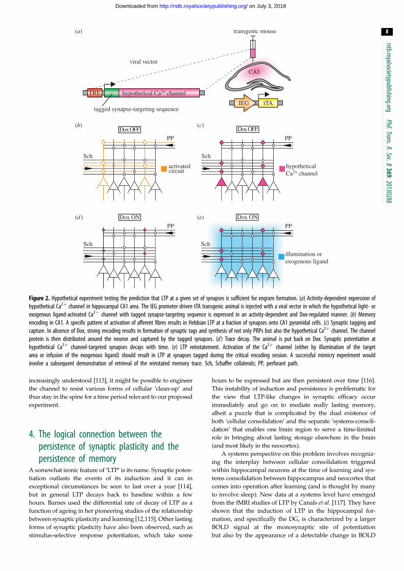
As suggested previously by Neves et al. [106], an experiment
testing whether hippocampal NMDA receptor-dependent
plasticity is sufficient for episodic memory could involve a
hypothetical light or exogenous ligand-activated Ca2þ channel.
As Ca2þ influx into the spine may be sufficient to induce early-
LTP [107], activation of this Ca2þ channel once it has made its
way to the spine should result in synapse-specific potentiation.
Though a remotely controlled ion channel selective for Ca2þ has
not yet been described, it is worth noting that some new ChR2
variants show enhanced Ca2þ conductivity [108] and it would
be interesting to see whether activation of these channels
within a dendritic spine could lead to LTP without the need
for presynaptic activation.
In a ‘dream experiment’, the hypothetical Ca2þ channel
would be expressed in all hippocampal neurons postsynaptic
to the set of synapses under investigation, its transcription
would be tTA-regulated and IEG-dependent, and the channel
protein itself would contain a targeting sequence that directs
it to recently potentiated (tagged) spines in the same way
these synapses recognize plasticity-related products (PRPs)
as outlined by the synaptic tagging and capture hypothesis
[100,109,110] (figure 2a). Though we are beginning to unravel
how PRPs like Homer-1a/Vesl-1S and Arc/Arg 3.1 are trans-
ported around the neuron [111,112], the precise mechanisms
of PRP capture by tagged synapses remain elusive and will
have to be uncovered before an experiment like this can be
carried out.
The toolkit described above could in principle be used
to study any IEG-expressing population of neurons in the
hippocampus and beyond. The experiment outlined below
focuses on principal neurons in the hippocampal area CA1
(figure 2b). The hypothetical channel would be expressed
selectively at tagged synapses at the time of encoding
(figure 2c), and these steps followed by forgetting and loss
of the underlying memory trace (figure 2d ). The question
then is whether the trace could be brought back (figure 2e).
Specifically, experimental animals would be trained in the
presence of Dox on a one-shot spatial memory task in
which the location of the goal—either a hidden platform
in the watermaze [59], or the availability of food in a sandwell in
the event arena [37]—changes every day. On a specific day, ani-
mals would be taken off Dox and subjected to strong memory
encoding (figure 2b), which should induce IEG expression (and
thus the Ca2þ channel) in hippocampal neurons (figure 2c).
Animals would then be put back on Dox and the ‘tagged’
memory trace would be allowed to decay or would be over-
written by extensive training (figure 2d). The animals would
then need to be tested to be confident that forgetting has
occurred. The experimental question is then addressed by
attempting to reintroduce this specific memory. This would
be attempted optogenetically or pharmacogenetically with
the effect that the only subset of synapses that would be poten-
tiated would be those expressing the hypothetical Ca2þ
channel and therefore those that were tagged during the critical
memory encoding phase (figure 2e). If LTP at a specific set of
hippocampal synapses is sufficient to construct a meaningful
memory trace, re-inducing LTP via activation of this hypo-
thetical channel should mimic weak memory of the correct
location of the goal on the ‘tagged’ day.
One potential obstacle of such experimental set-up is protein
turnover. Endocytosis and degradation of the hypothetical Ca2þ
channel would lead to its progressive elimination from spines.
Since by that time the synaptic tag would be long gone, selective
redelivery of the channel protein to the spines of interest would
be impossible. As mechanisms of protein degradation are being
transgenic mouse
tagged synapse-targeting sequence
Dox OFF Dox OFF
Dox ON
Sch
Sch
Sch
PP PP
PP
activatedcircuit
viral vector
TRE hypothetical Ca2+ channel
hypothetical Ca2+ channel
Dox ON
Sch
PP
illumination orexogenous ligand
CA1
IEG tTA
(b)
(a)
(c)
(d ) (e)
Figure 2. Hypothetical experiment testing the prediction that LTP at a given set of synapses is sufficient for engram formation. (a) Activity-dependent expression ofhypothetical Ca2þ channel in hippocampal CA1 area. The IEG promoter-driven tTA transgenic animal is injected with a viral vector in which the hypothetical light- orexogenous ligand-activated Ca2þ channel with tagged synapse-targeting sequence is expressed in an activity-dependent and Dox-regulated manner. (b) Memoryencoding in CA1. A specific pattern of activation of afferent fibres results in Hebbian LTP at a fraction of synapses onto CA1 pyramidal cells. (c) Synaptic tagging andcapture. In absence of Dox, strong encoding results in formation of synaptic tags and synthesis of not only PRPs but also the hypothetical Ca2þ channel. The channelprotein is then distributed around the neuron and captured by the tagged synapses. (d ) Trace decay. The animal is put back on Dox. Synaptic potentiation athypothetical Ca2þ channel-targeted synapses decays with time. (e) LTP reinstatement. Activation of the Ca2þ channel (either by illumination of the targetarea or infusion of the exogenous ligand) should result in LTP at synapses tagged during the critical encoding session. A successful mimicry experiment wouldinvolve a subsequent demonstration of retrieval of the reinstated memory trace. Sch, Schaffer collaterals; PP, perforant path.
rstb.royalsocietypublishing.orgPhil.Trans.R.Soc.B
369:20130288
8
on July 3, 2018http://rstb.royalsocietypublishing.org/Downloaded from
increasingly understood [113], it might be possible to engineer
the channel to resist various forms of cellular ‘clean-up’ and
thus stay in the spine for a time period relevant to our proposed
experiment.
4. The logical connection between thepersistence of synaptic plasticity and thepersistence of memory
A somewhat ironic feature of ‘LTP’ is its name. Synaptic poten-
tiation outlasts the events of its induction and it can in
exceptional circumstances be seen to last over a year [114],
but in general LTP decays back to baseline within a few
hours. Barnes used the differential rate of decay of LTP as a
function of ageing in her pioneering studies of the relationship
between synaptic plasticity and learning [12,115]. Other lasting
forms of synaptic plasticity have also been observed, such as
stimulus-selective response potentiation, which take some
hours to be expressed but are then persistent over time [116].
This instability of induction and persistence is problematic for
the view that LTP-like changes in synaptic efficacy occur
immediately and go on to mediate really lasting memory,
albeit a puzzle that is complicated by the dual existence of
both ‘cellular consolidation’ and the separate ‘systems-consoli-
dation’ that enables one brain region to serve a time-limited
role in bringing about lasting storage elsewhere in the brain
(and most likely in the neocortex).
A systems perspective on this problem involves recogniz-
ing the interplay between cellular consolidation triggered
within hippocampal neurons at the time of learning and sys-
tems consolidation between hippocampus and neocortex that
comes into operation after learning (and is thought by many
to involve sleep). New data at a systems level have emerged
from the fMRI studies of LTP by Canals et al. [117]. They have
shown that the induction of LTP in the hippocampal for-
mation, and specifically the DG, is characterized by a larger
BOLD signal at the monosynaptic site of potentiation
but also by the appearance of a detectable change in BOLD
rstb.royalsocietypublishing.orgPhil.Trans.R.Soc.B
369:20130288
9
on July 3, 2018http://rstb.royalsocietypublishing.org/Downloaded from
in other circuits, including the prefrontal lobe. This indicates
that potentiation at one level may somehow be projected
to or otherwise affect other levels of the neural circuit
mediating memory processing. Preliminary data secured by
Canals’ group indicate that similar remote changes in neural
activation occur following induction of LTP in CA1. The
reason(s) for this systems aspect of LTP induction remain to
be investigated, but could include an alteration in the exci-
tation–inhibition balance following some types of LTP that
is detectable through observation of a change in the coupling
of excitatory post-synaptic potentials to population spike gen-
eration in field-potential recordings. The work of Canals and
co-workers (see [118] in this issue) discusses LTP-induced
alterations in feed-forward disinhibition.
A separate cellular consolidation possibility is that early-
LTP decays precisely in order to re-set hippocampal circuits
back to a level whereby they can most effectively process
new information at a later time—not least the same day.
This gets round the need for lasting potentiation within the
hippocampus, though it is of course still necessary in the neo-
cortex, but also raises the issue of the timeframe for which
information must be retained by cellular consolidation for
the systems-consolidation aspect to work effectively.
Central to addressing this issue is the work of the Magde-
burg group of Matthies and Frey that established the separate
existence of early- and late-forms of LTP, the latter being
defined as protein synthesis-dependent. Their work provided
the first experimental evidence suggesting that neuromodu-
lators, especially dopamine (DA), play a significant role in
gating of plasticity and memory persistence. In vivo and
in vitro electrophysiological studies have revealed a specific
role for DA in control of temporal persistence of LTP
[119–122]. Pharmacological manipulations of DA receptors
also indicate that DA is required for the persistence of memories
in the hippocampus [123–125]. DA receptor activation can lead
to enhanced somatic and dendritic protein synthesis essential
for the establishment of lasting plasticity and memory
[126,127]. This function is mediated by protein kinase A, extra-
cellular-signal regulated kinase (ERK) 1/2, CaMKIV and cAMP
response element-binding protein CREB [128–131].
The midbrain dopaminergic neurons of the ventral teg-
mental area (VTA) project to the hippocampal formation
[132,133] and are thought to release DA under circumstances
of novelty or surprise [134,135]. In addition, a recent paper
suggests that noradrenergic (NA) terminals of the locus coer-
uleus (LC) might have a role in DA transmission in the
hippocampus [136]. The necessity of DA-dependent mechan-
isms within hippocampal neurons in cellular consolidation
implies that cellular consolidation depends not just on the
intrahippocampal, cellular processes, but also on the action
of systems-level components. Therefore, we propose that ‘cel-
lular consolidation’ is renamed ‘initial consolidation’.
There are many lines of evidence suggesting that the per-
sistence of memory is determined largely by neural activity
that occurs at the time of memory encoding. However, the
synaptic tagging and capture hypothesis of protein syn-
thesis-dependent LTP developed by Frey & Morris [109,110]
offers the intriguing but distinct perspective that the persist-
ence of memory is also dependent on independent neural
activity afferent to the same pool of neurons mediating LTP
that occurs before or after memory traces are encoded.
According to this hypothesis, the local setting of ‘synaptic
tags’ at activated glutamatergic synapses during memory
encoding can be dissociated from DA-dependent synthesis
and distribution of PRPs that is induced by surrounding
events. PRPs are then captured by synaptic tags in order to
stabilize synaptic change—a process that is critical for initial
consolidation.
Until recently, a challenge for the synaptic tagging and
capture hypothesis has been to assess its relevance to real
memory. Considering that exploration of a novel environ-
ment probably activates VTA DA neurons to release DA in
the hippocampus and thus cause upregulation of IEGs such
as Arc and Homer-1a [137,138], the synaptic tagging and cap-
ture hypothesis predicts that unrelated novelty exploration
before or after memory encoding should enhance the persist-
ence of a recently encoded memory [139]. This prediction was
first confirmed using a hippocampus-dependent inhibitory
avoidance task in rats [140]. Moncada and Viola showed that
weak memory could be consolidated into long-term memory
by unrelated exploration of a novel environment. This
novelty-induced memory persistence was blocked by intra-
hippocampal injection of DA D1/D5 receptor antagonist and
b-adrenergic receptors antagonist [140,141] or by inhibition of
induction or expression of CA1 LTD [142]. Complementary
results have been obtained using different learning tasks includ-
ing taste memory, spatial object recognition, contextual fear
conditioning and spatial memory [37,143,144].
Our laboratory has developed a realistic everyday appeti-
tive paradigm for rats in order to establish whether unrelated
novel experiences can facilitate the persistence of reward-
motivated spatial memory [37]. We have expanded our analy-
sis to different systems domains and, for this purpose, have
recently altered the protocol for the spatial memory task in
the event arena and made it suitable for mice (figure 3a). The
object of switching the task to mice is to enable genetically
modified animals to be tested. The tyrosine hydroxylase-Cre
knockin mice with C57BL/6 genetic background [145] were
trained on this one-shot spatial memory task over four
weeks. The mice have to learn a different location of food
each day and, with five sandwells to choose from, performance
increases quite rapidly from the chance level of two errors on
the daily choice trial to a consistent pattern of less than one
error per day (figure 3b). A series of post-learning tests, of
which we here show only one, established that: (i) persistence
of one-shot memory depends on reward magnitude, last-
ing only 1 h with two pellets but 24 h with eight pellets;
(ii) 5-min exploration of an open field with a variable novel
floor substrate 30 min after weak two-pellet encoding success-
fully transformed 1-h memory into 24-h memory (figure 3c)
and (iii) pharmacological interruption of DA D1/D5 receptors,
but not b-adrenergic receptors in hippocampus during novelty
exploration prevented novelty-induced memory persistence
over 24 h. Those characteristics are consistent with our earlier
rat data [37]. The next step is to substitute novelty exploration
with photo-activation of VTA DA neurons or LC NA neurons
using optogenetics (figure 3d ). With this procedure, our predic-
tion is that activating these neurons in some appropriate
temporal pattern should convert a recently encoded short-
term memory into a long-term memory.
5. ConclusionMartin et al. [16] laid out a framework for testing rigorously
the widely held notion that synaptic potentiation and
sand-wellreward
event arena
trial 1 trial 2 choice
~10 min ~10 min
sample trial
correct
incorrect
% d
ig ti
me
novelty box
0 h
0 h
24 h
24 h
0.5 h
0.5 h
probe
LC
TH-Cre mouse with AAV-Flex-ChR2
VTA
HPC
laser
DA
laser
DA
0
20
novelty
sample trial
noveltybox
light stim.
probe
24 h memory(predicted)
w/o novelty
40
60
80
0 2 4 6 8 10 12 14 16 18 20 22 24
0.5
1.0
1.5
2.0
2.5
3.0
chance level
sessions
erro
rs
sample location(b)(a)
(c) (d )
Figure 3. One-shot spatial memory task on the event arena for mice. (a) Event arena for one-shot spatial memory task. The event arena during a daily choice phase.Five sand wells are open but only one contains the reward pellets. All open sand wells contain several pellets that are inaccessible to the mouse in order to controlfor olfactory artefacts. (b) Daily spatial memory performance (errors). Every day mice have two trials to encode the new sand well location, followed by a choicephase. They quickly reach a stable performance level of less than one error (with two errors being the chance level). (c) Novelty-induced enhancement of memorypersistence. Critical sessions involve one sample trial followed by an unrewarded probe test 24 h later. 5 min exploration of a novel environment 30 min afterencoding results in enhanced persistence of one-shot spatial memory, as demonstrated by increased dig time in the correct location. (d ) Prediction ofmemory enhancement by optogenetic stimulation of catecholaminergic nuclei. We predict that photoactivation of DA cells of the VTA or DA-releasing NA cellsof LC in TH-Cre mice injected with Cre-dependent ChR2 virus (AAV-Flex-ChR2) after weak memory encoding will result in enhancement of memory persistencethat mimics the novelty effect. Error bars, +s.e.m; dotted lines, chance level.
rstb.royalsocietypublishing.orgPhil.Trans.R.Soc.B
369:20130288
10
on July 3, 2018http://rstb.royalsocietypublishing.org/Downloaded from
depression are key players in mediating the creation of
memory traces or engrams. That framework has stood the
test of time, with exciting new approaches using contempor-
ary techniques exploring the idea further with respect to
detectability, anterograde and retrograde alteration. Perhaps
most exciting are the first steps being taken towards testing
and possibly satisfying the mimicry criterion using opto-
genetic and other cell-type-specific molecular tools. Critical
experiments remain to be done, but the neuroscience
community can justifiably feel tantalizingly close to having
tested one of the great ideas of modern neuroscience. Forty
years on, LTP continues to excite us all as it slowly gives
up its mechanistic secrets and reveals its important functional
role in learning and memory.
Acknowledgements. We are grateful to Patrick Spooner for technicalassistance with the event arena and Jacqueline Friel for assistancewith behavioural training of mice.
rstb
11
on July 3, 2018http://rstb.royalsocietypublishing.org/Downloaded from
Funding statement. This work was supported by a European ResearchCouncil Advanced Investigator Grant to R.G.M.M. and GuillenFernandez (NEUROSCHEMA, no. 268800). T.T. was supported by the
Mitsubishi Tanabe Pharma Corporation and the UK Medical ResearchCouncil, to whom we are also grateful for past funding to R.G.M.M.and for a studentship and in vivo skills award currently held by A.J.D.
.royalsociety
Referencespublishing.orgPhil.Trans.R.Soc.B
369:20130288
1. Jones EG. 1994 Santiago Ramon y Cajal and theCroonian Lecture. Trends Neurosci. 17, 190 – 192.(doi:10.1016/0166-2236(94)90100-7)
2. Hebb DO. 1949 The organization of behaviour.New York, NY: Wiley.
3. Konorski J. 1948 Conditioned reflexes and neuronorganisation. Cambridge, UK: Cambridge UniversityPress.
4. Kandel ER. 1978 A cell-biological approach tolearning. Bethesda, MD: Society for Neuroscience.
5. Kandel ER. 2001 The molecular biology of memorystorage: a dialogue between genes and synapses.Science 294, 1030 – 1038. (doi:10.1126/science.1067020)
6. Bliss TV, Lomo T. 1973 Long-lasting potentiation ofsynaptic transmission in the dentate area of theanaesthetized rabbit following stimulation of theperforant path. J. Physiol. 232, 331 – 356.
7. Scoville WB, Milner B. 1957 Loss of recent memoryafter bilateral hippocampal lesions. J. Neurol.Neurosurg. Psychiatry 20, 11 – 21. (doi:10.1136/jnnp.20.1.11)
8. Ho VM, Lee JA, Martin KC. 2011 The cell biologyof synaptic plasticity. Science 334, 623 – 628.(doi:10.1126/science.1209236)
9. Costa-Mattioli M, Sossin WS, Klann E, Sonenberg N.2009 Translational control of long-lasting synapticplasticity and memory. Neuron 61, 10 – 26.(doi:10.1016/j.neuron.2008.10.055)
10. Kasai H, Fukuda M, Watanabe S, Hayashi-Takagi A,Noguchi J. 2010 Structural dynamics of dendriticspines in memory and cognition. Trends Neurosci.33, 121 – 129. (doi:10.1016/j.tins.2010.01.001)
11. Murakoshi H, Yasuda R. 2012 Postsynaptic signalingduring plasticity of dendritic spines. Trends Neurosci.35, 135 – 143. (doi:10.1016/j.tins.2011.12.002)
12. Barnes CA, McNaughton BL. 1985 An agecomparison of the rates of acquisition andforgetting of spatial information in relation to long-term enhancement of hippocampal synapses.Behav. Neurosci. 99, 1040 – 1048. (doi:10.1037/0735-7044.99.6.1040)
13. Alberini CM. 2009 Transcription factors inlong-term memory and synaptic plasticity.Physiol. Rev. 89, 121 – 145. (doi:10.1152/physrev.00017.2008)
14. Johansen JP, Cain CK, Ostroff LE, LeDoux JE.2011 Molecular mechanisms of fear learning andmemory. Cell 147, 509 – 524. (doi:10.1016/j.cell.2011.10.009)
15. Lee YS, Silva AJ. 2009 The molecular and cellularbiology of enhanced cognition. Nat. Rev. Neurosci.10, 126 – 140. (doi:10.1038/nrn2572)
16. Martin SJ, Grimwood PD, Morris RG. 2000 Synapticplasticity and memory: an evaluation of the
hypothesis. Annu. Rev. Neurosci. 23, 649 – 711.(doi:10.1146/annurev.neuro.23.1.649)
17. Hubener M, Bonhoeffer T. 2010 Searching forengrams. Neuron 67, 363 – 371. (doi:10.1016/j.neuron.2010.06.033)
18. Hofer SB, Mrsic-Flogel TD, Bonhoeffer T, Hubener M.2009 Experience leaves a lasting structural tracein cortical circuits. Nature 4577, 313 – 317.(doi:10.1038/nature07487)
19. Hawkins RD, Kandel ER. 1984 Is there a cell-biological alphabet for simple forms of learning?Psychol. Rev. 91, 375 – 391. (doi:10.1037/0033-295X.91.3.375)
20. Rumpel S, LeDoux J, Zador A, Malinow R. 2005Postsynaptic receptor trafficking underlying a formof associative learning. Science 308, 83 – 88.(doi:10.1126/science.1103944)
21. Morris RG, Frey U. 1997 Hippocampal synapticplasticity: role in spatial learning or the automaticrecording of attended experience? Phil. Trans. R. Soc.Lond. B 352, 1489 – 1503. (doi:10.1098/rstb.1997.0136)
22. McNaughton BL, Morris RGM. 1987 Hippocampalsynaptic enhancement and information storagewithin a distributed memory system. TrendsNeurosci. 10, 408 – 415. (doi:10.1016/0166-2236(87)90011-7)
23. Klausberger T, Somogyi P. 2008 Neuronal diversityand temporal dynamics: the unity of hippocampalcircuit operations. Science 321, 53 – 57. (doi:10.1126/science.1149381)
24. Dayan P, Abbott L. 2005 Theoretical neuroscience.Cambridge, MA: MIT Press.
25. Turrigiano GG, Nelson SB. 2000 Hebb andhomeostasis in neuronal plasticity. Curr. Opin.Neurobiol. 10, 358 – 364. (doi:10.1016/S0959-4388(00)00091-X)
26. Levy WB, Steward O. 1983 Temporal contiguityrequirements for long-term associativepotentiation/depression in the hippocampus.Neuroscience 8, 791 – 797. (doi:10.1016/0306-4522(83)90010-6)
27. Markram H, Lubke J, Frotscher M, Sakmann B. 1997Regulation of synaptic efficacy by coincidence ofpostsynaptic APs and EPSPs. Science 275, 213 – 215.(doi:10.1126/science.275.5297.213)
28. Xu S, Jiang W, Poo MM, Dan Y. 2012 Activity recallin a visual cortical ensemble. Nat. Neurosci. 15,449 – 455. (doi:10.1038/nn.3036)
29. O’Connor DH, Huber D, Svoboda K. 2009 Reverseengineering the mouse brain. Nature 461,923 – 929. (doi:10.1038/nature08539)
30. Rogan SC, Roth BL. 2011 Remote control ofneuronal signaling. Pharmacol. Rev. 63, 291 – 315.(doi:10.1124/pr.110.003020)
31. Yizhar O, Fenno LE, Davidson TJ, Mogri M,Deisseroth K. 2011 Optogenetics in neural systems.Neuron 71, 9 – 34. (doi:10.1016/j.neuron.2011.06.004)
32. Packer AM, Roska B, Hausser M. 2013 Targetingneurons and photons for optogenetics. Nat.Neurosci. 16, 805 – 815. (doi:10.1038/nn.3427)
33. Knopfel T. 2012 Genetically encoded opticalindicators for the analysis of neuronal circuits. Nat.Rev. Neurosci. 13, 687 – 700. (doi:10.1038/nrm3461)
34. Wilt BA, Burns LD, Ho ETW, Ghosh KK, Mukamel EA,Schnitzer MJ. 2009 Advances in light microscopy forneuroscience. Annu. Rev. Neurosci. 32, 435 – 506.(doi:10.1146/annurev.neuro.051508.135540)
35. Tse D, Langston RF, Kakeyama M, Bethus I,Spooner PA, Wood ER, Witter MP, Morris RGM. 2007Schemas and memory consolidation. Science 316,76 – 82. (doi:10.1126/science.1135935)
36. Harvey CD, Collman F, Dombeck DA, Tank DW. 2009Intracellular dynamics of hippocampal place cellsduring virtual navigation. Nature 461, 941 – 946.(doi:10.1038/nature08499)
37. Wang SH, Redondo RL, Morris RG. 2010 Relevanceof synaptic tagging and capture to the persistenceof long-term potentiation and everyday spatialmemory. Proc. Natl Acad. Sci. USA 107, 19 537 –19 542. (doi:10.1073/pnas.0913844107)
38. Martin SJ, Morris RG. 2002 New life in an old idea:the synaptic plasticity and memory hypothesisrevisited. Hippocampus 12, 609 – 636. (doi:10.1002/hipo.10107)
39. Green EJ, McNaughton BL, Barnes CA. 1990Exploration-dependent modulation of evokedresponses in fascia dentata: dissociation of motor,EEG, and sensory factors and evidence for a synapticefficacy change. J. Neurosci. 10, 1455 – 1471.
40. Moser E, Mathiesen I, Andersen P. 1993 Associationbetween brain temperature and dentate fieldpotentials in exploring and swimming rats. Science259, 1324 – 1326. (doi:10.1126/science.8446900)
41. Moser EI, Moser MB, Andersen P. 1994 Potentiationof dentate synapses initiated by exploratory learningin rats: dissociation from brain temperature, motoractivity, and arousal. Learn. Mem. 1, 55 – 73.
42. Whitlock JR, Heynen AJ, Shuler MG, Bear MF. 2006Learning induces long-term potentiation in thehippocampus. Science 313, 1093 – 1097.(doi:10.1126/science.1128134)
43. Matsuo N, Reijmers L, Mayford M. 2008 Spine-type-specific recruitment of newly synthesized AMPAreceptors with learning. Science 319, 1104 – 1107.(doi:10.1126/science.1149967)
44. Morris RGM, Anderson E, Lynch GS, Baudry M. 1986Selective impairment of learning and blockade oflong-term potentiation by an N-methyl-D-aspartate
rstb.royalsocietypublishing.orgPhil.Trans.R.Soc.B
369:20130288
12
on July 3, 2018http://rstb.royalsocietypublishing.org/Downloaded from
receptor antagonist, AP5. Nature 319, 774 – 776.(doi:10.1038/319774a0)
45. Sakimura K et al. 1995 Reduced hippocampal LTPand spatial learning in mice lacking NMDA receptorepsilon 1 subunit. Nature 373, 151 – 155.(doi:10.1038/373151a0)
46. Tsien JZ, Chen DF, Gerber D, Tom C, Mercer EH,Anderson DJ, Mayford M, Kandel ER, Tonegawa S.1996 Subregion- and cell type-restricted geneknockout in mouse brain [see comments]. Cell 87,1317 – 1326. (doi:10.1016/S0092-8674(00)81826-7)
47. Tsien JZ, Huerta PT, Tonegawa S. 1996 The essentialrole of hippocampal CA1 NMDA receptor-dependentsynaptic plasticity in spatial memory. Cell 87,1327 – 1338. (doi:10.1016/S0092-8674(00)81827-9)
48. Pastalkova E, Serrano P, Pinkhasova D, Wallace E,Fenton AA, Sacktor TC. 2006 Storage of spatialinformation by the maintenance mechanism of LTP.Science 313, 1141 – 1144. (doi:10.1126/science.1128657)
49. Gruart A, Munoz MD, Delgado-Garcia JM. 2006Involvement of the CA3-CA1 synapse in theacquisition of associative learning in behaving mice.J. Neurosci. 26, 1077 – 1087. (doi:10.1523/JNEUROSCI.2834-05.2006)
50. Clarke JR, Cammarota M, Gruart A, Izquierdo I,Delgado-Garcia JM. 2010 Plastic modificationsinduced by object recognition memory processing.Proc. Natl Acad. Sci. USA 107, 2652 – 2657.(doi:10.1073/pnas.0915059107)
51. Bredt DS, Nicoll RA. 2003 AMPA receptor traffickingat excitatory synapses. Neuron 40, 361 – 379.(doi:10.1016/S0896-6273(03)00640-8)
52. Kessels HW, Malinow R. 2009 Synaptic AMPAreceptor plasticity and behavior. Neuron 61,340 – 350. (doi:10.1016/j.neuron.2009.01.015)
53. Bourne J, Harris KM. 2007 Do thin spines learn tobe mushroom spines that remember? Curr. Opin.Neurobiol. 17, 381 – 386. (doi:10.1016/j.conb.2007.04.009)
54. Mitsushima D, Ishihara K, Sano A, Kessels HW,Takahashi T. 2011 Contextual learning requiressynaptic AMPA receptor delivery in thehippocampus. Proc. Natl Acad. Sci. USA 108,12 503 – 12 508. (doi:10.1073/pnas.1104558108)
55. Hayashi Y, Shi SH, Esteban JA, Piccini A, Poncer JC,Malinow R. 2000 Driving AMPA receptors intosynapses by LTP and CaMKII: requirement forGluR1 and PDZ domain interaction. Science 287,2262 – 2267. (doi:10.1126/science.287.5461.2262)
56. Davis S, Butcher SP, Morris RGM. 1992 The NMDAreceptor antagonist D-2-amino-5-phosphonopentanoate (D-AP5) impairs spatiallearning and LTP in vivo at intracerebralconcentrations comparable to those that block LTPin vitro. J. Neurosci. 12, 21 – 34.
57. Danysz W, Zajaczkowski W, Parsons CG. 1995Modulation of learning processes by ionotropicglutamate receptor ligands. Behav. Pharmacol. 6,455 – 474. (doi:10.1097/00008877-199508000-00007)
58. Staubli U, Thibault O, DiLorenzo M, Lynch G. 1989Antagonism of NMDA receptors impairs acquisition
but not retention of olfactory memory. Behav.Neurosci. 103, 54 – 60. (doi:10.1037/0735-7044.103.1.54)
59. Steele RJ, Morris RGM. 1999 Delay-dependentimpairment of a matching-to-place task withchronic and intrahippocampal infusion of theNMDA-antagonist D-AP5. Hippocampus 9,118 – 136. (doi:10.1002/(SICI)1098-1063(1999)9:2,118::AID-HIPO4.3.0.CO;2-8)
60. Day M, Langston R, Morris RG. 2003 Glutamate-receptor-mediated encoding and retrieval of paired-associate learning. Nature 424, 205 – 209.(doi:10.1038/nature01769)
61. Bast T, da Silva BM, Morris RG. 2005 Distinctcontributions of hippocampal NMDA and AMPAreceptors to encoding and retrieval of one-trialplace memory. J. Neurosci. 25, 5845 – 5856.(doi:10.1523/JNEUROSCI.0698-05.2005)
62. Habib D, Tsui CK, Rosen LG, Dringenberg HC.In press. Occlusion of low-frequency-induced,heterosynaptic long-term potentiation in the rathippocampus in vivo following spatial training.Cereb Cortex. (doi:10.1093/cercor/bht174)
63. Moser EI, Krobert KA, Moser MB, Morris RG. 1998Impaired spatial learning after saturation of long-term potentiation. Science 281, 2038 – 2042.(doi:10.1126/science.281.5385.2038)
64. Saucier D, Cain DP. 1995 Spatial learning withoutNMDA receptor-dependent long-term potentiation.Nature 378, 186 – 189. (doi:10.1038/378186a0)
65. Bannerman DM, Good MA, Butcher SP, Ramsay M,Morris RG. 1995 Distinct components of spatiallearning revealed by prior training and NMDAreceptor blockade. Nature 378, 182 – 186.(doi:10.1038/378182a0)
66. Wang SH, Finnie PS, Hardt O, Nader K. 2012 Dorsalhippocampus is necessary for novel learning butsufficient for subsequent similar learning.Hippocampus 22, 2157 – 2170. (doi:10.1002/hipo.22036)
67. Inglis J, Martin SJ, Morris RGM. 2013 Upstairs-downstairs revisited: spatial pretraining-inducedrescue of normal spatial learning during selectiveblockade of hippocampal N-methyl-D-aspartatereceptors. Eur. J. Neurosci. 37, 718 – 727.(doi:10.1111/ejn.12087)
68. Li Y, Erzurumlu RS, Chen C, Jhaveri S, Tonegawa S.1994 Whisker-related neuronal patterns fail todevelop in the trigeminal brainstem nuclei ofNMDAR1 knockout mice. Cell 76, 427 – 437.(doi:10.1016/0092-8674(94)90108-2)
69. Bannerman DM et al. 2008 NMDA receptor subunitNR2A is required for rapidly acquired spatialworking memory but not incremental spatialreference memory. J. Neurosci. 28, 3623 – 3630.(doi:10.1523/JNEUROSCI.3639-07.2008)
70. McHugh TJ, Blum KI, Tsien JZ, Tonegawa S,Wilson MA. 1996 Impaired hippocampalrepresentation of space in CA1-specific NMDAR1knockout mice. Cell 87, 1339 – 1349. (doi:10.1016/S0092-8674(00)81828-0)
71. Fukaya M, Kato A, Lovett C, Tonegawa S, WatanabeM. 2003 Retention of NMDA receptor NR2 subunits
in the lumen of endoplasmic reticulum in targetedNR1 knockout mice. Proc. Natl Acad. Sci. USA 100,4855 – 4860. (doi:10.1073/pnas.0830996100)
72. Nakazawa K et al. 2002 Requirement forhippocampal CA3 NMDA receptors in associativememory recall. Science 297, 211 – 218. (doi:10.1126/science.1071795)
73. Nakazawa K, Sun LD, Quirk MC, Rondi-Reig L,Wilson MA, Tonegawa S. 2003 Hippocampal CA3NMDA receptors are crucial for memory acquisitionof one-time experience. Neuron 38, 305 – 315.(doi:10.1016/S0896-6273(03)00165-X)
74. McHugh TJ et al. 2007 Dentate gyrus NMDAreceptors mediate rapid pattern separation in thehippocampal network. Science 317, 94 – 99.(doi:10.1126/science.1140263)
75. Reisel D, Bannerman DM, Schmitt WB, Deacon RM,Flint J, Borchardt T, Seeburg PH, Rawlins JN.2002 Spatial memory dissociations in micelacking GluR1. Nat. Neurosci. 5, 868 – 873.(doi:10.1038/nn910)
76. Schmitt WB, Deacon RM, Seeburg PH, Rawlins JN,Bannerman DM. 2003 A within-subjects, within-taskdemonstration of intact spatial reference memoryand impaired spatial working memory in glutamatereceptor-A-deficient mice. J. Neurosci. 23,3953 – 3959.
77. Zamanillo D et al. 1999 Importance of AMPAreceptors for hippocampal synaptic plasticity butnot for spatial learning. Science 284, 1805 – 1811.(doi:10.1126/science.284.5421.1805)
78. Schmitt WB, Sprengel R, Mack V, Draft RW,Seeburg PH, Deacon RM,, Rawlins JN, BannermanDM. 2005 Restoration of spatial working memory bygenetic rescue of GluR-A-deficient mice. Nat.Neurosci. 8, 270 – 272. (doi:10.1038/nn1412)
79. Sanderson DJ, Good MA, Skelton K, Sprengel R,Seeburg PH, Rawlins JN, Bannerman DM. 2009Enhanced long-term and impaired short-termspatial memory in GluA1 AMPA receptor subunitknockout mice: evidence for a dual-process memorymodel. Learn. Mem. 16, 379 – 386. (doi:10.1101/lm.1339109)
80. Sanderson DJ, Bannerman DM. 2012 The role ofhabituation in hippocampus-dependent spatialworking memory tasks: ecvidence from GluA1 AMPAreceptor subunit knockout mice. Hippocampus 22,981 – 994. (doi:10.1002/hipo.20896)
81. Niewoehner B, Single FN, Hvalby O, Jensen V, Meyerzum Alten Borgloh S, Seeburg PH, Rawlins JN,Sprengel R, Bannerman DM. 2007 Impaired spatialworking memory but spared spatial referencememory following functional loss of NMDAreceptors in the dentate gyrus. Eur. J. Neurosci. 25,837 – 846. (doi:10.1111/j.1460-9568.2007.05312.x)
82. Bannerman DM et al. 2012 Dissecting spatialknowledge from spatial choice by hippocampalNMDA receptor deletion. Nat. Neurosci. 15,1153 – 1159. (doi:10.1038/nn.3166)
83. Xu W. 2011 PSD-95-like membrane associatedguanylate kinases (PSD-MAGUKs) and synapticplasticity. Curr. Opin. Neurobiol. 21, 306 – 312.(doi:10.1016/j.conb.2011.03.001)
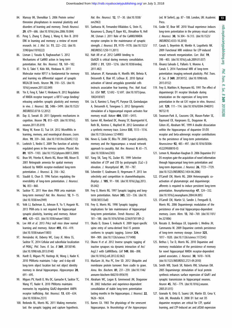
rstb.royalsocietypublishing.orgPhil.Trans.R.Soc.B
369:20130288
13
on July 3, 2018http://rstb.royalsocietypublishing.org/Downloaded from
84. Mansuy IM, Shenolikar S. 2006 Protein serine/threonine phosphatases in neuronal plasticity anddisorders of learning and memory. Trends Neurosci.29, 679 – 686. (doi:10.1016/j.tins.2006.10.004)
85. Peng S, Zhang Y, Zhang J, Wang H, Ren B. 2010ERK in learning and memory: a review of recentresearch. Int. J. Mol. Sci. 11, 222 – 232. (doi:10.3390/ijms11010222)
86. Lisman J, Yasuda R, Raghavachari S. 2012Mechanisms of CaMKII action in long-termpotentiation. Nat. Rev. Neurosci. 13, 169 – 182.
87. Yin X, Takei Y, Kido MA, Hirokawa N. 2011Molecular motor KIF17 is fundamental for memoryand learning via differential support of synapticNR2A/2B levels. Neuron 70, 310 – 325. (doi:10.1016/j.neuron.2011.02.049)
88. Yin X, Feng X, Takei Y, Hirokawa N. 2012 Regulationof NMDA receptor transport: a KIF17-cargo binding/releasing underlies synaptic plasticity and memoryin vivo. J. Neurosci. 32, 5486 – 5499. (doi:10.1523/JNEUROSCI.0718-12.2012)
89. Day JJ, Sweatt JD. 2011 Epigenetic mechanisms incognition. Neuron 70, 813 – 829. (doi:10.1016/j.neuron.2011.05.019)
90. Wang W, Kwon EJ, Tsai LH. 2012 MicroRNAs inlearning, memory, and neurological diseases. Learn.Mem. 19, 359 – 368. (doi:10.1101/lm.026492.112)
91. Loebrich S, Nedivi E. 2009 The function of activity-regulated genes in the nervous system. Physiol. Rev.89, 1079 – 1103. (doi:10.1152/physrev.00013.2009)
92. Brun VH, Ytterbo K, Morris RG, Moser MB, Moser EI.2001 Retrograde amnesia for spatial memoryinduced by NMDA receptor-mediated long-termpotentiation. J. Neurosci. 2, 356 – 362.
93. Staubli U, Chun D. 1996 Factors regulating thereversibility of long-term potentiation. J. Neurosci.16, 853 – 860.
94. Sacktor TC. 2011 How does PKM zeta maintainlong-term memory? Nat. Rev. Neurosci. 12, 9 – 15.(doi:10.1038/nrn2949)
95. Volk LJ, Bachman JL, Johnson R, Yu Y, Huganir RL.2013 PKM-zeta is not required for hippocampalsynaptic plasticity, learning and memory. Nature493, 420 – 423. (doi:10.1038/nature11802)
96. Lee AM et al. 2013 Prkcz null mice show normallearning and memory. Nature 493, 416 – 419.(doi:10.1038/nature11803)
97. Hernandez AI, Oxberry WC, Crary JF, Mirra SS,Sacktor TC. 2014 Cellular and subcellular localizationof PKMz. Phil. Trans. R. Soc. B 369, 20130140.(doi:10.1098/rstb.2013.0140)
98. Hardt O, Migues PV, Hastings M, Wong J, Nader K.2010 PKMzeta maintains 1-day- and 6-day-oldlong-term object location but not object identitymemory in dorsal hippocampus. Hippocampus 20,691 – 695.
99. Migues PV, Hardt O, Wu DC, Gamache K, Sacktor TC,Wang YT, Nader K. 2010 PKMzeta maintainsmemories by regulating GluR2-dependent AMPAreceptor trafficking. Nat. Neurosci. 13, 630 – 634.(doi:10.1038/nn.2531)
100. Redondo RL, Morris RG. 2011 Making memorieslast: the synaptic tagging and capture hypothesis.
Nat. Rev. Neurosci. 12, 17 – 30. (doi:10.1038/nrn2963)
101. Sanhueza M, Fernandez-Villalobos G, Stein IS,Kasumova G, Zhang P, Bayer KU,, Otmakhov N, HellJW, LIsman J. 2011 Role of the CaMKII/NMDAreceptor complex in the maintenance of synapticstrength. J. Neurosci. 31, 9170 – 9178. (doi:10.1523/JNEUROSCI.1250-11.2011)
102. Halt AR et al. 2012 CaMKII binding toGluN2B is critical during memory consolidation.EMBO J. 31, 1203 – 1216. (doi:10.1038/emboj.2011.482)
103. Johansen JP, Hamanaka H, Monfils MH, Behnia R,Deisseroth K, Blair HT, LeDoux JE. 2010 Opticalactivation of lateral amygdala pyramidal cellsinstructs associative fear learning. Proc. Natl Acad.Sci. USA 107, 12 692 – 12 697. (doi:10.1073/pnas.1002418107)
104. Liu X, Ramirez S, Pang PT, Puryear CB, GovindarajanA, Deisseroth K, Tonegawa S. 2012 Optogeneticstimulation of a hippocampal engram activates fearmemory recall. Nature 484, U381 – U415.
105. Garner AR, Rowland DC, Hwang SY, Baumgaertel K,Roth BL, Kentros C, Mayford M. 2012 Generation ofa synthetic memory trace. Science 335, 1513 – 1516.(doi:10.1126/science.1214985)
106. Neves G, Cooke SF, Bliss TV. 2008 Synaptic plasticity,memory and the hippocampus: a neural networkapproach to causality. Nat. Rev. Neurosci. 9, 65 – 75.(doi:10.1038/nrn2303)
107. Yang SN, Tang YG, Zucker RS. 1999 Selectiveinduction of LTP and LTD by postsynaptic [Ca2þ]ielevation. J. Neurophysiol. 81, 781 – 787.
108. Schneider F, Gradmann D, Hegemann P. 2013 Ionselectivity and competition in channelrhodopsins.Biophys. J. 105, 91 – 100. (doi:10.1016/j.bpj.2013.05.042)
109. Frey U, Morris RG. 1997 Synaptic tagging and long-term potentiation. Nature 385, 533 – 536. (doi:10.1038/385533a0)
110. Frey U, Morris RG. 1998 Synaptic tagging:implications for late maintenance of hippocampallong-term potentiation. Trends Neurosci. 21,181 – 188. (doi:10.1016/S0166-2236(97)01189-2)
111. Okada D, Ozawa F, Inokuchi K. 2009 Input-specificspine entry of soma-derived Vesl-1S proteinconforms to synaptic tagging. Science 324,904 – 909. (doi:10.1126/science.1171498)
112. Okuno H et al. 2012 Inverse synaptic tagging ofinactive synapses via dynamic interaction of Arc/Arg3.1 with CaMKIIbeta. Cell 149, 886 – 898.(doi:10.1016/j.cell.2012.02.062)
113. MacGurn JA, Hsu PC, Emr SD. 2012 Ubiquitin andmembrane protein turnover: from cradle to grave.Annu. Rev. Biochem. 81, 231 – 259. (doi:10.1146/annurev-biochem-060210-093619)
114. Abraham WC, Logan B, Greenwood JM, DragunowM. 2002 Induction and experience-dependentconsolidation of stable long-term potentiationlasting months in the hippocampus. J. Neurosci. 22,9626 – 9634.
115. Barnes CA. 1983 The physiology of the senescenthippocampus. In Neurobiology of the hippocampus
(ed. W Siefert), pp. 87 – 108. London, UK: AcademicPress.
116. Cooke SF, Bear MF. 2010 Visual experience induceslong-term potentiation in the primary visual cortex.J. Neurosci. 30, 16 304 – 16 313. (doi:10.1523/JNEUROSCI.4333-10.2010)
117. Canals S, Beyerlein M, Merkle H, Logothetis NK.2009 Functional MRI evidence for LTP-inducedneural network reorganization. Curr. Biol. 19,398 – 403. (doi:10.1016/j.cub.2009.01.037)
118. Alvarez-Salvado E, Pallares V, Moreno A,Canals S. 2014 Functional MRI of long-termpotentiation: imaging network plasticity. Phil. Trans.R. Soc. B 369, 20130152. (doi:10.1098/rstb.2013.0152)
119. Frey U, Matthies H, Reymann KG. 1991 The effect ofdopaminergic D1 receptor blockade duringtetanization on the expression of long-termpotentiation in the rat CA1 region in vitro. Neurosci.Lett. 129, 111 – 114. (doi:10.1016/0304-3940(91)90732-9)
120. Swanson-Park JL, Coussens CM, Mason-Parker SE,Raymond CR, Hargreaves EL, Dragunow M,Cohen AS, Abraham WC. 1999 A double dissociationwithin the hippocampus of dopamine D1/D5receptor and beta-adrenergic receptor contributionsto the persistence of long-term potentiation.Neuroscience 92, 485 – 497. (doi:10.1016/S0306-4522(99)00010-X)
121. Lemon N, Manahan-Vaughan D. 2006 Dopamine D1/D5 receptors gate the acquisition of novel informationthrough hippocampal long-term potentiation andlong-term depression. J. Neurosci. 26, 7723 – 7729.(doi:10.1523/JNEUROSCI.1454-06.2006)
122. O’Carroll CM, Morris RG. 2004 Heterosynaptic co-activation of glutamatergic and dopaminergicafferents is required to induce persistent long-termpotentiation. Neuropharmacology 47, 324 – 332.(doi:10.1016/j.neuropharm.2004.04.005)
123. O’Carroll CM, Martin SJ, Sandin J, Frenguelli B,Morris RG. 2006 Dopaminergic modulation of thepersistence of one-trial hippocampus-dependentmemory. Learn. Mem. 13, 760 – 769. (doi:10.1101/lm.321006)
124. Rossato JI, Bevilaqua LR, Izquierdo I, Medina JH,Cammarota M. 2009 Dopamine controls persistenceof long-term memory storage. Science 325,1017 – 1020. (doi:10.1126/science.1172545)
125. Bethus I, Tse D, Morris RG. 2010 Dopamine andmemory: modulation of the persistence of memoryfor novel hippocampal NMDA receptor-dependentpaired associates. J. Neurosci. 30, 1610 – 1618.(doi:10.1523/JNEUROSCI.2721-09.2010)
126. Smith WB, Starck SR, Roberts RW, Schuman EM.2005 Dopaminergic stimulation of local proteinsynthesis enhances surface expression of GluR1 andsynaptic transmission in hippocampal neurons.Neuron 45, 765 – 779. (doi:10.1016/j.neuron.2005.01.015)
127. Granado N, Ortiz O, Suarez LM, Martin ED, Cena V,Solis JM, Moratalla R. 2008 D1 but not D5dopamine receptors are critical for LTP, spatiallearning, and LTP-Induced arc and zif268 expression

rstb.royalsocietypublishing.orgPhil.Trans.R.Soc.B
369:20130288
14
on July 3, 2018http://rstb.royalsocietypublishing.org/Downloaded from
in the hippocampus. Cereb. Cortex 18, 1 – 12.(doi:10.1093/cercor/bhm026)
128. Abel T, Nguyen PV, Barad M, Deuel TA, Kandel ER,Bourtchouladze R. 1997 Genetic demonstrationof a role for PKA in the late phase of LTPand in hippocampus-based long-term memory.Cell 88, 615 – 626. (doi:10.1016/S0092-8674(00)81904-2)
129. Barco A, Alarcon JM, Kandel ER. 2002 Expression ofconstitutively active CREB protein facilitates the latephase of long-term potentiation by enhancingsynaptic capture. Cell 108, 689 – 703. (doi:10.1016/S0092-8674(02)00657-8)
130. Kelleher 3rd RJ, Govindarajan A, Jung HY, Kang H,Tonegawa S. 2004 Translational control by MAPKsignaling in long-term synaptic plasticity andmemory. Cell 116, 467 – 479. (doi:10.1016/S0092-8674(04)00115-1)
131. Redondo RL, Okuno H, Spooner PA, Frenguelli BG,Bito H, Morris RG. 2010 Synaptic tagging and capture:differential role of distinct calcium/calmodulinkinases in protein synthesis-dependent long-termpotentiation. J. Neurosci. 30, 4981 – 4989.(doi:10.1523/JNEUROSCI.3140-09.2010)
132. Gasbarri A, Packard MG, Campana E, Pacitti C. 1994Anterograde and retrograde tracing of projectionsfrom the ventral tegmental area to the hippocampalformation in the rat. Brain Res. Bull. 33, 445 – 452.(doi:10.1016/0361-9230(94)90288-7)
133. Gasbarri A, Sulli A, Packard MG. 1997 Thedopaminergic mesencephalic projections to the
hippocampal formation in the rat. Prog.Neuropsychopharmacol. Biol. Psychiatry 21, 1 – 22.(doi:10.1016/S0278-5846(96)00157-1)
134. Lisman JE, Grace AA. 2005 The hippocampal-VTAloop: controlling the entry of information into long-term memory. Neuron 46, 703 – 713. (doi:10.1016/j.neuron.2005.05.002)
135. Lisman J, Grace AA, Duzel E. 2011 A neoHebbianframework for episodic memory; role of dopamine-dependent late LTP. Trends Neurosci. 34, 536 – 547.(doi:10.1016/j.tins.2011.07.006)
136. Smith CC, Greene RW. 2012 CNS dopaminetransmission mediated by noradrenergicinnervation. J. Neurosci. 32, 6072 – 6080. (doi:10.1523/JNEUROSCI.6486-11.2012)
137. Guzowski JF, McNaughton BL, Barnes CA, Worley PF.1999 Environment-specific expression of theimmediate-early gene Arc in hippocampal neuronalensembles. Nat. Neurosci. 2, 1120 – 1124. (doi:10.1038/16046)
138. Vazdarjanova A, Guzowski JF. 2004 Differences inhippocampal neuronal population responses tomodifications of an environmental context: evidencefor distinct, yet complementary, functions of CA3and CA1 ensembles. J. Neurosci. 24, 6489 – 6496.(doi:10.1523/JNEUROSCI.0350-04.2004)
139. Morris RG. 2006 Elements of a neurobiologicaltheory of hippocampal function: the role of synapticplasticity, synaptic tagging and schemas.Eur. J. Neurosci. 23, 2829 – 2846. (doi:10.1111/j.1460-9568.2006.04888.x)
140. Moncada D, Viola H. 2007 Induction of long-termmemory by exposure to novelty requires proteinsynthesis: evidence for a behavioral tagging.J. Neurosci. 27, 7476 – 7481. (doi:10.1523/JNEUROSCI.1083-07.2007)
141. Moncada D, Ballarini F, Martinez MC, Frey JU, ViolaH. 2011 Identification of transmitter systems andlearning tag molecules involved in behavioraltagging during memory formation. Proc. Natl Acad.Sci. USA 108, 12 931 – 12 936. (doi:10.1073/pnas.1104495108)
142. Dong Z, Gong B, Li H, Bai Y, Wu X, Huang Y, He W,Li T, Wang YT. 2012 Mechanisms of hippocampallong-term depression are required for memoryenhancement by novelty exploration. J. Neurosci.32, 11 980 – 11 990. (doi:10.1523/JNEUROSCI.0984-12.2012)
143. Merhav M, Rosenblum K. 2008 Facilitation of tastememory acquisition by experiencing previous noveltaste is protein-synthesis dependent. Learn. Mem.15, 501 – 507. (doi:10.1101/lm.986008)
144. Ballarini F, Moncada D, Martinez MC, Alen N, ViolaH. 2009 Behavioral tagging is a general mechanismof long-term memory formation. Proc. Natl Acad.Sci. USA 106, 14 599 – 14 604. (doi:10.1073/pnas.0907078106)
145. Lindeberg J, Usoskin D, Bengtsson H, Gustafsson A,Kylberg A, Soderstrom S, Ebendal T. 2004Transgenic expression of Cre recombinase from thetyrosine hydroxylase locus. Genesis 40, 67 – 73.(doi:10.1002/gene.20065)

![LowFrequency Rhythmic Electrocutaneous Hand ... - Sleep.Ruattracted by Tononi’s hypothesis [19], suggesting that the effect of sleep on memory consolidation is related to plastic](https://img.pdfslide.us/doc/110x75/5f7a98d4436c1c41950b483e/lowfrequency-rhythmic-electrocutaneous-hand-sleepru-attracted-by-tononias.jpg)