Embed Size (px)
Citation preview
Science Arena Publications
Specialty Journal of Chemistry ISSN: 2521-3164
Available online at www.sciarena.com 2019, Vol, 4 (1): 1-8
The Study of Morphology and Grain Size of Hydroxyapatite/Zeolite Nanocomposite Using SEM
and TEM Analyses
Fereshteh Khorasani*, Maryam Khavarpour, Mohamad Shokrzade Lamuki
Chemical Engineering – Process Engineering, Faculty of Engineering, Islamic Azad University – South Tehran Branch, Iran.
* Corresponding Author
Abstract: Bone is a real nanocomposite composed of organic (collagen) and inorganic (nano-hydroxyapatite) phases. The construction of new composites as bone and tissue substitute is one of the attractive topics in bone and tissue engineering. Nano-hydroxyapatite and its composites containing 2.5, 5 and 7.5 percent of zeolite have been synthesized in this research. SEM and TEM analyses were utilized to study morphology and evaluate the particle size of the composites. The results showed that the particle size in pure hydroxyapatite and its composite was 40-200 nm. The surface characterization study indicated that the accumulation of particles on the surface increases and the particle size would be more uniform as the concentration of the zeolite increases. Finally, according to the results, the use of 7.5% zeolite is recommended for the synthesis of hydroxyapatite-zeolite nano-composite as the bone substitute. Keywords: Hydroxyapatite, Zeolite, Nanocomposite, Tissue Engineering, Bone Substitute
INTRODUCTION
The nanotechnology is defined by the National Nanotechnology Organization as “understanding and control of materials in the dimensions of about 1-100 nm in such a way that the unique processes lead to the novel applications”. Medical nanotechnology means the utilization of nanotechnology in medicine and healthcare that has the ability to act in the prevention, diagnosis, reliable and early control and curing of the diseases. One of the goals of the medical nanotechnology is to understand the biological processes for diagnosis and curing the diseases (Freitas, 2002). The reconstruction and repair of bone defects, which are caused by impacts, infections or removing a bone tumor, are considered as the main and important problems in orthopedic and oral and maxillofacial surgeries (Mikos et al., 1993). The bone repair by tissue engineering is one of the spotlights of specialists in various areas including Orthopedic, Cranium and Oral and Maxillofacial area. The scaffolds that are used in tissue engineering are categorized into four categories of inorganic, synthetic and natural polymers and the combination of these groups (Li and Wozney, 2001). Hydroxyapatite is a calcium phosphate that has the bio-active and bio-compatible properties that are very similar to the tooth and natural bone in terms of natural structure and crystalline structure. It has been proved that its nanostructures compound has much more functional features compared to its micro-scaled
Spec. J. Chem, Vol, 4 (1): 1-8
2
structure. It has been proposed to use the biomaterials that beside the proper biocompatible and bioactive properties, compensate the lack of strength and adhesion of hydroxyapatite. The hydroxyapatite not only is bioactive but also is the ossification tool (Hammerle et al., 1997; Denissen et al., 1980; Hollinger and Battistone, 1986), non-toxic and non-immunogenetic (Krause et al., 2001). The hydroxyapatite has various applications in medicine and dentistry such as the replacement of bone tissue and coating the body implants. The researches have shown that the nanostructured hydroxyapatite shows higher mechanical and more proper biocompatible properties compared to the micrometer samples in the body environment. These properties are in the optimal mode when the hydroxyapatite nanoparticles have uniform shape and size and minimum agglomeration (ShafieiZaadeh, 2010). The zeolites are hydrated aluminosilicates that have the cations of alkali metals and alkaline earth metal have unlimited structure. The zeolites have the network structure with the internal and external surface of 100 m2/g and the cationic exchange capacity up to several mili-equivalents per grams (Kazemian, 2004). The applications of the natural and synthetic zeolites are dependent on their physical and chemical properties that these properties are also dependent on the crystal structure and chemical composition of the zeolite (Rasouli, 2010). There are various techniques for characterizing the structure, properties and physicochemical specifications of zeolites. Among these most important methods are the methods that utilize the X-ray including X-Ray Diffraction (XRD), X-Ray Fluorescence (XRF) and Scanning Electron Microscope (SEM), Infrared Radiation (IR), Ion Exchange Chromatography (IEC) and etc. The electron microscopes are basically two types: the first type is called the Scanning Electron Microscope (SEM) and the second type is called the Transmission Electron Microscope (TEM) that in it the electron passes through the sample so the internal structure of the sample could be imaged. In the first type of the electron microscopes, i. e. SEM, the incident electron to the surface is reflected and collected by detectors and finally is converted to the light photons so the visible image could be formed. In other words, this type of microscope gives only the images of the surface structure (Rasouli, 2010). A research was carried out by Salahi et al., in 2012, titled “The effect of temperature on the hardness and microstructure properties of hydroxyapatite/zirconia-alumina nanocomposites”. The obtained hardnesses in the two-step sintering samples were three times and their fracture toughnesses were twice of the samples sintered by common methods. Also, in the two-step sintering samples, according to the XRD results, there was minimum Tri-Calcium Phosphate (TCP) formed (Salahi, 2012). A research was carried out by Taherian et al., in 2012, titled as “Synthesis of bioactive mesoporous silica/hydroxyapatite nanocomposite by sol-gel method”. The TEM analysis confirmed the confinement of hydroxyapatite crystals by a layer of mesoporous materials. The results showed that the silica mesoporous biocompatible behavior is improved due to the presence of hydroxyapatite in nanocomposite and having larger cavities (Taherian, 2012). Neha Gupta et al., in a study in 2010 investigated the effect of surficial adsorption of cobalt (II) on the hydroxyapatite/zeolite (HApZ) composite from the aqueous solution. The hydroxyapatite/zeolite composite was synthesized by the precipitation method and was used as the adsorbent for removing the cobalt ions from the aqueous solutions. The materials were characterized by the SEM and Fourier-Transform Infrared Spectroscopy (FTIR). The results showed that the HApZ composite has better surficial adsorption compared to the Hydroxyapatite (HAp). A small amount of it (mg/L) has the adsorption capability of about 65% of cobalt (II) in the pH equal to 6, the temperature of about 30oC in about 20 minutes. The surficial adsorption amount has been decreased by the decrease in the main metallic ion concentration (Neha et al., 2011). The nanocomposites have drawn much attention due to the excellent bending strength compared to the composites with large grain sizes. The hydroxyapatite-based nanocomposites have found the high potential for replacing the bones due to the improvement of mechanical and biological properties. The aim of this research is to synthesize the hydroxyapatite/zeolite nanocomposite. The structural characterization of the products was performed by the SEM and TEM analyses.
Spec. J. Chem, Vol, 4 (1): 1-8
3
Materials and Methods
All the chemicals used in this research are high grades and are used without purification. Also, deionized water has been used for washing and preparing some of the solutions. All the reactions were taken place at room temperature. In this project, the raw zeolite was used that was provided by the Afrand tooska company and was the commercial grade clinoptilot that was obtained from the Semnan district. The di-ammonium phosphate (NH4)2HPO4 with the molecular mass of 132.05 g/mol, tetrahydrate calcium nitrate Ca(NO3)2.4H2O with the molecular mass of 236.15 g/mol and also 25% purity ammonia (NH4OH) with the molecular mass of 17.031 g/mol was used. The performed tests in this research were categorized into two main groups of products synthesis and structural features characterization. In order to synthesize the hydroxyapatite, at first, the solutions containing the calcium and phosphorus were prepared separately by the tetrahydrate calcium nitrate with the chemical formula of Ca (NO3)2.4H2O and di-ammonium phosphate with the chemical formula of (NH4)2HPO4, respectively. The synthesis of the powder was performed in the pH near to 11 and at room temperature. The used reactor contained Pyrex three-spin balloons that were used according to the different volumes. These balloons were positioned on the mixer with the connection to the refrigerant equipped with the cold water input and output in the paraffin bath and in case of requirement of the homogeneous solution preparation, the magnetic stirrers were used. The required amount of tetrahydrate calcium nitrate and di-ammonium phosphate were weighted with the precision of 0.0001 g by the one-piece digital scale made by A&D Japanese Company. Then, the solutions containing Ca and P were prepared by the demineralized water. In all the synthesis steps, the pH of the solutions was adjusted to the desired value by ammonia. For simultaneous measurement of the pH and temperature, the Swiss-made Metrohm 691pH meter was used. The calcium-containing solution transferred to the reactor and the phosphorus-containing solution was gradually added to it using a burette in 60 minutes and drop by drop. All the time that the reaction took place, the solution in the reactor was stirred so the product would form uniformly. At the end of the two solutions combination, the obtained solution was put at room temperature for one night. After the passage of the mentioned time, the obtained solution was filtered, dried and was put in the oven with the temperature of 100oC for 24 hours and was dried at the intended temperature and related process so finally a white powder was obtained (Zhang et al., 2009). In order to synthesize the hydroxyapatite/zeolite nanocomposite, various compounds of zeolite solution were obtained by dissolving 2.5, 5 and 7.5 percent zeolite powder. Then the tetrahydrate calcium nitrate was added to it in the stirring state and after four hours of vigorous stirring, the di-ammonium phosphate was gradually and in 60 minutes added to it using a dropper and the stirring continued for two more hours so a good and uniform suspension could be obtained. Throughout the process, the pH of the solution was kept constant around 11 using the ammonia solution. Finally, the obtained concentrated suspension was put at room temperature for one night. Then the obtained solution was filtered, dried and was put in the oven with the temperature of 100oC for 24 hours and finally, a white powder was obtained. The synthesized solutions were named as HA/Z 2.5, HA/Z 5 and HA/Z 7.5. In this research, two methods for analysis and characterization of the obtained materials and composites were used that were: 1- Scanning Electron Microscope (SEM) for surface and morphology characterization. 2- Transmission Electron Microscope (TEM) to determine the electron diffraction pattern and the size of the produced nanograins. In order to study the surface morphology, topography and size distribution, the SEM was used. The SEM used in this study was Germany-made VEGA II, TESCAN Company at the Razi Metallurgical Research Center. In this kind of microscope, an electron beam is radiated on the sample surface. The electrons are emitted from a hot filament and by acceleration in an electric field and passing a magnetic field lens are focused and scan the sample surface. The electrons are diffracted after incidence to the surface and by detection and analyzing
Spec. J. Chem, Vol, 4 (1): 1-8
4
them with a computer only an image of the surface will be obtained. The maximum voltage utilized by this kind of microscopes is about 30 kV. This voltage is used for accelerating the electrons so they could have more energy and acceleration. The surface of the samples that are characterized by the SEM must have electric conductivity. By coating the surface with a fine layer of gold or carbon, the surface of the non-conductive samples would gain electric conductivity and the surficial electron are repulsed and as a result, the resolution will be improved. The TEM was utilized for determination of the nano-grains size and more precise characterization of the grain size morphology. In a transmission electron microscope, the accelerated electron beam passes through a very thin sample and an image with very high resolution will be formed on the display tool. The display tool could be a fluorescence display tool or a special camera. In this research, the Zeiss EM900 electron microscope of the Kafa Nano Technology-Laboratory Complex was used.
Results
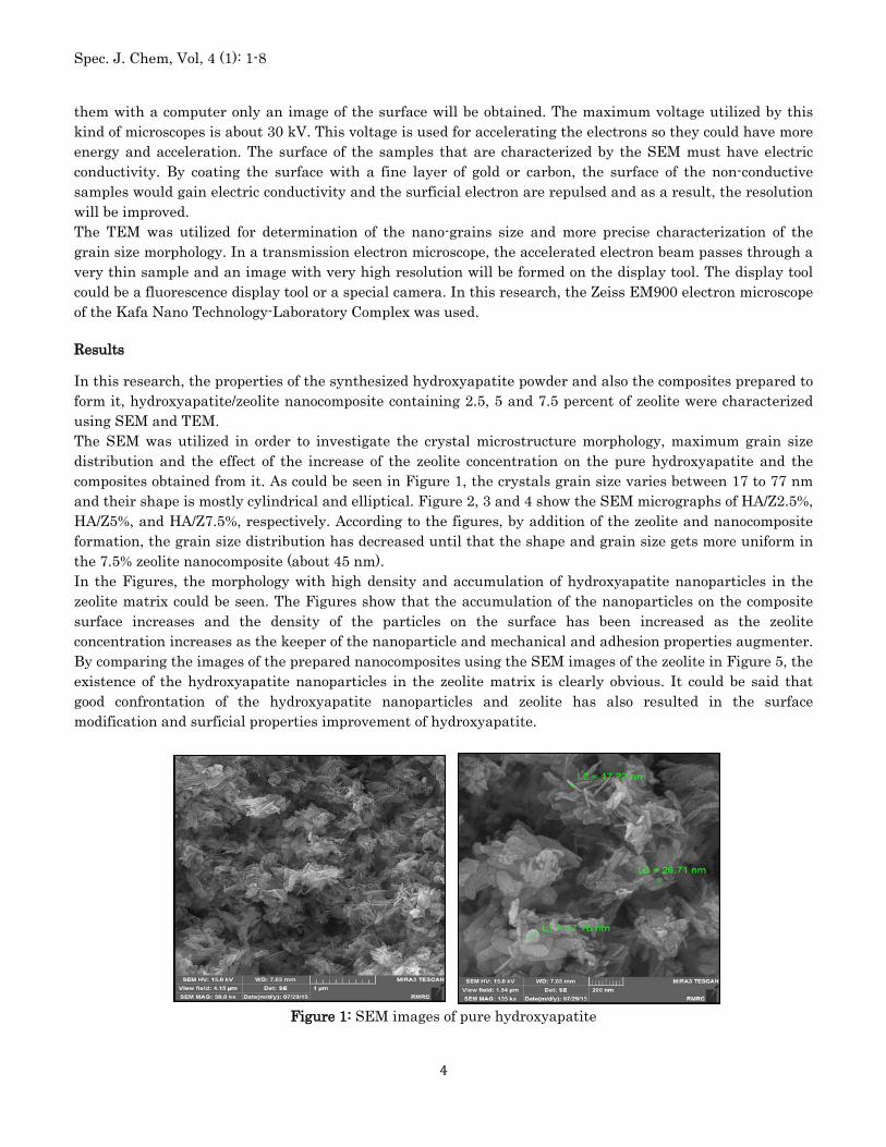
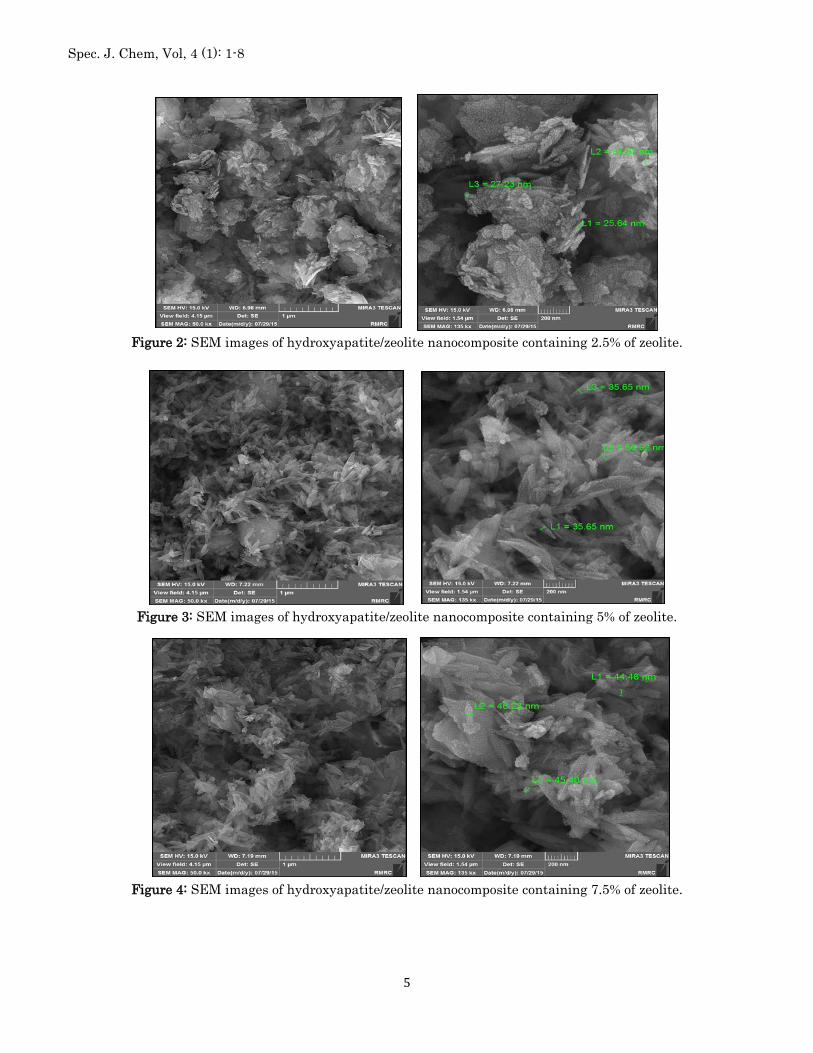
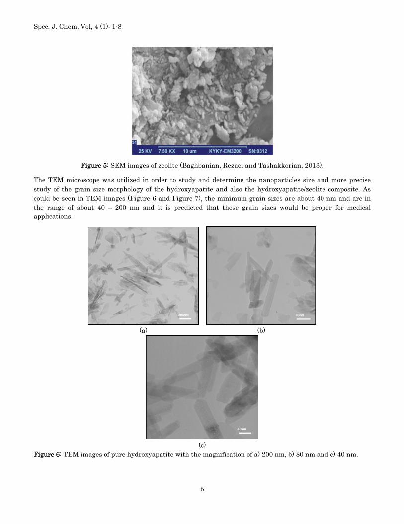
In this research, the properties of the synthesized hydroxyapatite powder and also the composites prepared to form it, hydroxyapatite/zeolite nanocomposite containing 2.5, 5 and 7.5 percent of zeolite were characterized using SEM and TEM. The SEM was utilized in order to investigate the crystal microstructure morphology, maximum grain size distribution and the effect of the increase of the zeolite concentration on the pure hydroxyapatite and the composites obtained from it. As could be seen in Figure 1, the crystals grain size varies between 17 to 77 nm and their shape is mostly cylindrical and elliptical. Figure 2, 3 and 4 show the SEM micrographs of HA/Z2.5%, HA/Z5%, and HA/Z7.5%, respectively. According to the figures, by addition of the zeolite and nanocomposite formation, the grain size distribution has decreased until that the shape and grain size gets more uniform in the 7.5% zeolite nanocomposite (about 45 nm). In the Figures, the morphology with high density and accumulation of hydroxyapatite nanoparticles in the zeolite matrix could be seen. The Figures show that the accumulation of the nanoparticles on the composite surface increases and the density of the particles on the surface has been increased as the zeolite concentration increases as the keeper of the nanoparticle and mechanical and adhesion properties augmenter. By comparing the images of the prepared nanocomposites using the SEM images of the zeolite in Figure 5, the existence of the hydroxyapatite nanoparticles in the zeolite matrix is clearly obvious. It could be said that good confrontation of the hydroxyapatite nanoparticles and zeolite has also resulted in the surface modification and surficial properties improvement of hydroxyapatite.
Figure 1: SEM images of pure hydroxyapatite
Spec. J. Chem, Vol, 4 (1): 1-8
5
Figure 2: SEM images of hydroxyapatite/zeolite nanocomposite containing 2.5% of zeolite.
Figure 3: SEM images of hydroxyapatite/zeolite nanocomposite containing 5% of zeolite.
Figure 4: SEM images of hydroxyapatite/zeolite nanocomposite containing 7.5% of zeolite.
Spec. J. Chem, Vol, 4 (1): 1-8
6
Figure 5: SEM images of zeolite (Baghbanian, Rezaei and Tashakkorian, 2013).
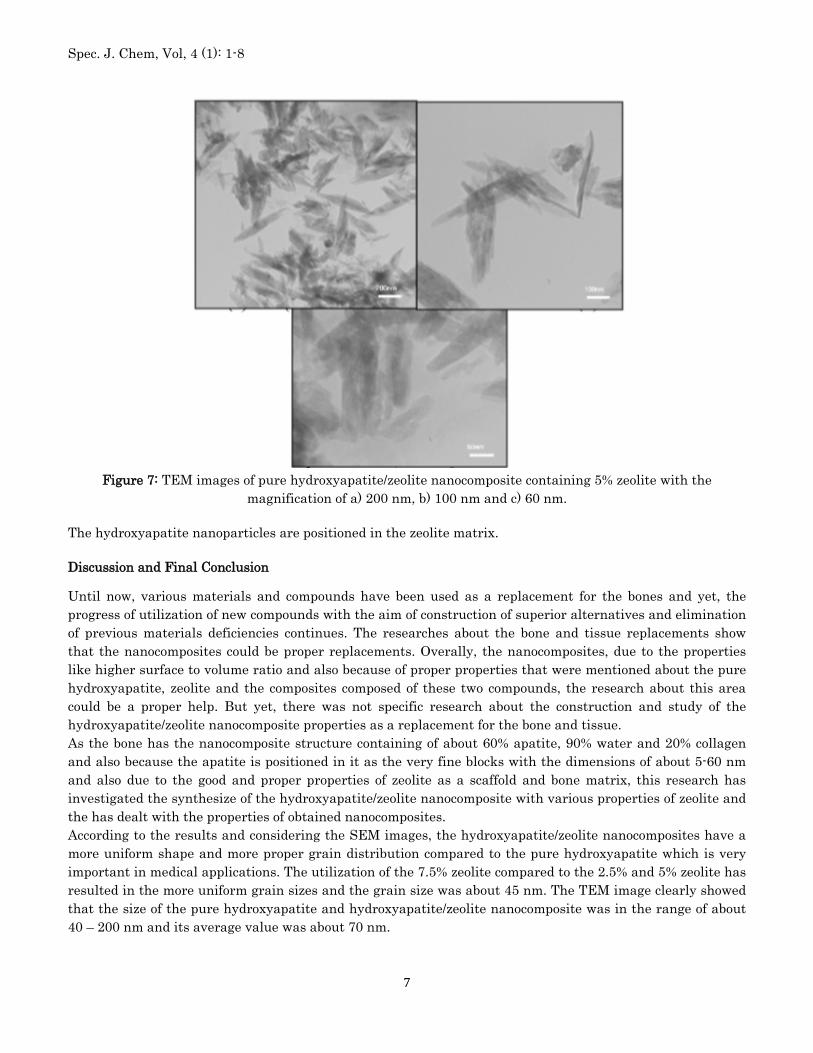
The TEM microscope was utilized in order to study and determine the nanoparticles size and more precise study of the grain size morphology of the hydroxyapatite and also the hydroxyapatite/zeolite composite. As could be seen in TEM images (Figure 6 and Figure 7), the minimum grain sizes are about 40 nm and are in the range of about 40 – 200 nm and it is predicted that these grain sizes would be proper for medical applications.
(a) (b)
(c)
Figure 6: TEM images of pure hydroxyapatite with the magnification of a) 200 nm, b) 80 nm and c) 40 nm.
Spec. J. Chem, Vol, 4 (1): 1-8
7
Figure 7: TEM images of pure hydroxyapatite/zeolite nanocomposite containing 5% zeolite with the
magnification of a) 200 nm, b) 100 nm and c) 60 nm.
The hydroxyapatite nanoparticles are positioned in the zeolite matrix.
Discussion and Final Conclusion
Until now, various materials and compounds have been used as a replacement for the bones and yet, the progress of utilization of new compounds with the aim of construction of superior alternatives and elimination of previous materials deficiencies continues. The researches about the bone and tissue replacements show that the nanocomposites could be proper replacements. Overally, the nanocomposites, due to the properties like higher surface to volume ratio and also because of proper properties that were mentioned about the pure hydroxyapatite, zeolite and the composites composed of these two compounds, the research about this area could be a proper help. But yet, there was not specific research about the construction and study of the hydroxyapatite/zeolite nanocomposite properties as a replacement for the bone and tissue. As the bone has the nanocomposite structure containing of about 60% apatite, 90% water and 20% collagen and also because the apatite is positioned in it as the very fine blocks with the dimensions of about 5-60 nm and also due to the good and proper properties of zeolite as a scaffold and bone matrix, this research has investigated the synthesize of the hydroxyapatite/zeolite nanocomposite with various properties of zeolite and the has dealt with the properties of obtained nanocomposites. According to the results and considering the SEM images, the hydroxyapatite/zeolite nanocomposites have a more uniform shape and more proper grain distribution compared to the pure hydroxyapatite which is very important in medical applications. The utilization of the 7.5% zeolite compared to the 2.5% and 5% zeolite has resulted in the more uniform grain sizes and the grain size was about 45 nm. The TEM image clearly showed that the size of the pure hydroxyapatite and hydroxyapatite/zeolite nanocomposite was in the range of about 40 – 200 nm and its average value was about 70 nm.
Spec. J. Chem, Vol, 4 (1): 1-8
8
References
1. Baghbanian, S. M., Rezaei, N., & Tashakkorian, H. (2013). Nanozeolite clinoptilolite as a highly efficient heterogeneous catalyst for the synthesis of various 2-amino-4 H-chromene derivatives in aqueous media. Green Chemistry, 15(12), 3446-3458.
2. Denissen HW, deGroot K, Kakkes P, van den Hooff A, Klopper PJ. (1980). Tissue response to dense apatite implants in rats. Biomed Mater.Res. 14, 713–721.
3. Freitas RA Jr. (2002). The future of nanofabrication and molecular scale devices in nanomedicine. Stud Health Technol Inform. 80, 45–59.
4. Hammerle CH, Olah AJ, Schid J, Fluckiger L, Gogolewski S,Winkler JR, et al. (1997). The biological effect of natural bone mineral on bone neoformation on the rabbit skull. Clin Oral Implants Res. 8, 198–207.
5. Hollinger JO, Battistone GC. (1986). Biodegradable bone repair materials.Clin Orthop Rel Res. 207, 290–305.
6. Kazemian H., (2004). An Introduction to zeolites: The Magic Minerals, Tehran-Iran, Beheshti Publication, 53-68.
7. Krause D, Theise N, Collector M et al. (2001). Multi–organ, multi–lineage engraftment by a single bone marrow derived stem cell. Cell. 105, 369–377.
8. Li, R. H., & Wozney, J. M. (2001). Delivering on the promise of bone morphogenetic proteins. Trends in biotechnology, 19(7), 255-265.
9. Mikos A.G., Sarakinos G., Leite S.M., Vacanti J.P, Langer R, (1993). Laminated three-dimensional biodegradable foams for use in tissue engineering. Biomaterial. 14, 323.
10. Neha Gupta, Atul K. Kushwaha and M.C. Chattopadhyaya. (2011). Adsorption of cobalt (II) from aqueous solution onto hydroxyapatite/zeolite composite. 2(4), 309-312
11. Rasouli, L., (2010). “Evaluation of the efficiency of the modified natural zeolite with the cationic surfactant for chromate removal from aqueous solutions”, M.Sc. thesis, Sanitary Engineering, Tarbiat Modares University.
12. Salahi, A., (2012). “The effect of temperature on the hardness and microstructure properties of hydroxyapatite/zirconia-alumina nanocomposites”, M.Sc. thesis, Faculty of Metallurgy and Material science Engineering, Iran University of Science and Technology.
13. ShafieiZaadeh Sh., (2010). “The investigation on the nanotechnology application in the teeth whitening”, M.Sc. thesis, Chemical engineering faculty of Amirkabir University of Technology, Supervisor: Parivash HoseinPour.
14. Taherian, Z., (2012), “Synthesis of bioactive mesoporous silica/hydroxyapatite nanocomposite by sol-gel method”, Applied Chemical Researches, 9, 47 – 55.
15. Y. Zhang, C, Xia, Appl, Catal. A, Gen, 366(2009) 141-147