Embed Size (px)
Citation preview

The studies presented in this thesis were conducted at the Department of Ophthalmology and the
Department of Immunology, Erasmus MC, University Medical Center Rotterdam, the Netherlands.
The work described in this thesis was financially supported by Stichting Lijf en Leven, Prof. Dr.
Henkes Stichting, Rotterdamse Stichting Blindenbelangen, Algemene Nederlandse Vereniging ter
Voorkoming van Blindheid, Landelijke Stichting voor Blinden en Slechtzienden, Stichting Neder-
lands Oogheelkundig Onderzoek, Stichting Blindenhulp.
The printing of this thesis was financially supported by Prof. Dr. Henkes Stichting, Rotterdamse
Stichting Blindenbelangen, Stichting Blindenhulp, Landelijke Stichting voor Blinden en Slechtzien-
den, Stichting voor Ooglijders.
Cover & lay-out: evelienjagtman.com
Printed by: Gildeprint
ISBN: 978-94-6233-879-1
© 2018 J.C.E.M. ten Berge
All rights reserved. No part of this thesis may be reproduced, stored in a retrieval system or
transmitted in any form or by any means, without permission of the author or, when appropriate,
of the publishers of the publication.
AUTOIMMUNITY IN UVEITIS AND OTHER CHORIORETINAL DISEASES
Autoimmuniteit in uveïtis en andere chorioretinale ziektebeelden
Proefschrift
ter verkrijging van de graad van doctor aan de
Erasmus Universiteit Rotterdam
op gezag van de
rector magnificus
Prof.dr. H.A.P. Pols
en volgens besluit van het College voor Promoties.
De openbare verdediging zal plaatsvinden op
donderdag 8 maart 2018 om 15.30 uur
Josianne Carina Elvire Maria ten Berge
geboren te Delft

PROMOTIECOMMISSIE
Promotoren: Prof.dr. J.R. Vingerling
Prof.dr. A. Rothova
Overige leden: Prof.dr. P.M. van Hagen
Prof.dr. J.H. de Boer
Prof.dr. C.J.F. Boon
Copromotor: Dr. M.W.J. Schreurs
CONTENTS
Chapter 1 General introduction 7
Chapter 2 Autoimmunity in uveitis 15
Accepted for publication in Acta Ophthalmologica
Chapter 3 Antinuclear antibody profiling in uveitis 29
Adapted version accepted for publication in Acta Ophthalmologica
Chapter 4 Prevalence and clinical impact of antiretinal antibodies in uveitis 39
Acta Ophthalmol. 2016 May;94(3):282-8
Chapter 5 Serum autoantibody profiling of patients with paraneoplastic and non-paraneoplastic autoimmune retinopathy
57
PLoS One. 2016 Dec;11(12):e0167909
Chapter 6 Ernstige visusdaling door auto-immuun retinopathie (Severe visual loss caused by autoimmune retinopathy)
79
Ned Tijdschr Geneeskd. 2015;159:A8039
Chapter 7 Antiretinal antibodies in central serous chorioretinopathy: prevalence and clinical implications
89
Acta Ophthalmol. 2017 Apr 26, Epub ahead of print
Chapter 8 Antiretinal and antinuclear antibodies in uveitis with latent and active tuberculosis
105
Accepted for publication in Acta Ophthalmologica
Chapter 9 Antiretinal antibodies in Mexican children with severe pars planitis 111
Submitted for publication
Chapter 10 Autoantibody profiling in intraocular fluid of patients with uveitis 117
Submitted for publication
Chapter 11 Intraocular cytokine profile and autoimmune reactions in retinitis pig-mentosa, age-related macular degeneration, glaucoma and cataract
133
Submitted for publication
Chapter 12 Summary and conclusions 151
Appendices Samenvatting (Summary in Dutch) 161
Dankwoord (Acknowledgements) 167
About the author 171
PhD portfolio 173
List of publications 175


GENERAL INTRODUCTION
1

General introduction
9
CHAPTER 1GENERAL INTRODUCTION
Uveitis
Uveitis is an inflammation of the vascular layer of the eye (uveal tract), which includes the iris,
ciliary body and choroid. However, in practice the term uveitis is usually used as a collective
term for any form of intraocular inflammation. Uveitis is a major cause of visual impairment or
even blindness. The annual prevalence of uveitis in the western world is increasing and varies
between 85-115 cases per 100.000 persons. The incidence is around 25-52 cases per 100.000
person-years with a peak at the age of 25-44 year, affecting predominantly the adult working
population.1,2
Uveitis is usually classified according the anatomical location of the inflammation into anterior,
intermediate, posterior or panuveitis group.3 The most common location encountered by primary
care ophthalmologists is anterior uveitis, whereas the involvement of posterior eye segment is
typically referred to tertiary care institutions.1 In approximately one third of uveitis cases the cause
remains elusive (idiopathic uveitis), but the remainder may be either associated with systemic
infectious (e.g. syphilis, toxoplasmosis), or an underlying systemic autoimmune or auto-inflam-
matory diseases.4-6 In these systemic non-infectious diseases, the eye is usually one of the several
organs involved and uveitis might be the first clinical sign of a more widespread systemic disease.
Scleritis represents also an inflammatory disease of the eye, involving predominantly the sclera,
but corneal, episcleral and retinal tissue may also be involved. Scleritis can be very severe, pain-
ful and result in blindness. As in uveitis, scleritis is sometimes associated with an underlying
non-infectious systemic disease such as rheumatoid arthritis or granulomatosis with polyangiitis.
Scleritis is formally not a subtype of uveitis, but these ocular inflammations may have similar
causes and associations, as well as comparable diagnostic and therapeutic approaches.
Autoimmunity in uveitis
The pathogenesis of uveitis is not fully clarified, but a crucial role of autoimmune reactions has
been suggested. Autoimmunity is characterized by an aberrant activity of the immune system
directed against the body’s own cells and tissues. It occurs when the immune system stops
tolerating ‘self’ antigens and autoreactive cells attack the body’s own antigens. An exogenous or
endogenous trigger (for example tissue damage) causes activation of the immune system, result-
ing in production of pathogenic antibodies and/or T-cells directed against ocular antigens. Several
theories have been proposed about why this expansion occurs, including molecular mimicry in
which autoantigens are mistaken for peptides from micro-organisms.7
10
CHAPTER 1 Insights about autoimmunity in eye-specific disease are limited, especially in ocular diseases
without systemic manifestations (e.g. birdshot chorioretinopathy). Indirect evidence for involve-
ment of autoimmunity in uveitis has been provided by induction of autoimmune uveitis after
immunization of animals with retinal autoantigens in combination with Freund adjuvant.8-10 These
animal models, representing experimental autoimmune uveitis (EAU), have provided insight into
the pathogenesis of human uveitis. In EAU predominantly mice are injected with different anti-
gens (such as S-arrestin and interphotoreceptor retinal binding protein) causing inflammation
of intraocular tissue similar to human uveitis. More recently, models with genetic manipulated
mouse and spontaneously emerging uveitis, have been developed.2,5 Similar to the heterogeneity
of human uveitis, clinical manifestations of uveitis in animal models may differ and is probably
related dose and type of immunization as well as genetic sensibility.
Antiretinal antibodies
The activity of our humoral immune system can be studied in the laboratory by measuring auto-
antibodies directed against retinal tissue. These so-called antiretinal antibodies (ARAs) can be
detected by various laboratory techniques including immunohistochemistry, Western blot and
enzyme-linked immunosorbent assay (ELISA). However, a standardized assay to measure ARAs
is lacking and results may vary depending on the laboratory tool.11
The exact role of ARAs in uveitis and other chorioretinal diseases such as retinitis pigmentosa
(RP), age-related macular degeneration (AMD) and glaucoma, was scarcely investigated. It has
been suggested that ARAs might be involved in the inciting process of the ocular disease. Another
hypothesis addresses a secondary phenomenon of ARAs induced by retinal damage. It has been
proposed that ARAs in ocular disease might cause a mild inflammation in the retina and subse-
quently aggravate and/or prolong the ocular disease.12,13
Autoimmune retinopathy
ARAs have been described also in the context of autoimmune retinopathy (AIR). AIR encompasses
a spectrum of rare autoimmune diseases that primarily affect retinal cells, and includes cancer-as-
sociated retinopathy (CAR), melanoma-associated retinopathy (MAR) and non-paraneoplastic
autoimmune retinopathy. The affected patients produce ARAs directed against their own retina,
which are thought to play a pathogenic role and being able to attack and destroy retinal cells,
leading rapidly to visual loss or even blindness. It is hypothesized that the underlying mechanism
of AIR is an immune response to tumor antigens sharing homology with retinal antigens (molec-
ular mimicry).14,15 AIR has been associated with the presence of various serum ARAs including
antibodies directed against recoverin, α-enolase, transducin-α, carbonic anhydrase, arrestin and
various other retinal antigens. While the patients with anti-recoverin antibodies frequently suffer
from associated cancer and severe loss of rod and cone function, the anti-enolase retinopathy
is characteristically associated with cone dysfunction and is also prevalent in patients without
General introduction
11
CHAPTER 1cancer. The autoreactivity to specific antigens and the clinical manifestations of AIR indicate that
different antigens might be associated with distinct clinical signs. However, since most AIR patients
exhibit multiple antiretinal antibodies, it is not yet known which antibodies are pathologic and
clinically relevant, and which represent innocent bystanders.
Current dilemmas
Although humoral autoimmune reactions directed against retinal tissue are thought to play an
important role in either initiation or modification of diverse chorioretinal disorders including
uveitis, they were not as yet systematically measured and their possible clinical impact in retinal
diseases was not examined. It is not known which specific retinal antigens provoke the forma-
tion of antibodies (and the repertoire of retinal autoimmune reactions). Furthermore, possible
associations between ARAs and clinical characteristics, such as phase of the disease and being
on immunosuppressive treatment, were not systematically examined. In addition, determination
of ARAs was so far performed predominantly in serum of patients with chorioretinal diseases,
which does not give precise information on what is exactly happening within the eye itself. The
eye represents an immune-privileged organ, in which the immune reactions might be downplayed
and/or different than observed in the peripheral blood. Information on the autoimmune reactions
measured in intraocular fluid is scarce and the prevalence of ARAs in chorioretinal diseases has
not been determined.
Understanding of autoimmune processes in ocular diseases might help to further elucidate
their pathogeneses and may have consequences for the design of new diagnostic and treatment
modalities. More detailed insight in the immuno-pathogenesis may be extremely valuable for
patients, because the inhibition (or prevention) of inflammation (if present) in specific phases
of the ocular disease might beneficially influence the course of disease and hopefully its visual
outcome.
AIM AND SCOPE OF THIS THESIS
This thesis aims to assess the presence of humoral autoimmunity in uveitis and other chorio-
retinal diseases, including AIR, and to gain insight its role. To achieve this, we start by providing
an overview of a large series of patients with uveitis and/or scleritis and examine the prevalence
of systemic autoimmune and autoinflammatory diseases in this population. We review specific
ocular diagnoses and clinical manifestations of patients affected by systemic autoimmune dis-
eases. Further, we critically evaluate the term “autoimmune uveitis” (chapter 2). In chapter 3 we
measure the prevalence of common systemic autoantibodies (antinuclear antibodies; ANA) in
serum of uveitis patients. Subsequently, we determined the presence of retina specific antibod-
ies (ARAs) in serum of patients with uveitis, AIR and central serous chorioretinopathy (CSC), and

12
CHAPTER 1 discuss their possible pathogenic role (chapter 4-9). In the last chapters of this thesis we explore
the determination of humoral autoimmune reactions in intraocular fluid samples of patients with
uveitis (chapter 10). In addition, in chapter 11 we investigate intraocular fluid samples further
and determine the presence of ARAs and inflammatory cytokines in intraocular fluid samples of
patients with diverse ocular diseases, including RP, AMD, glaucoma and cataract.
General introduction
13
CHAPTER 1REFERENCES
1. Gritz DC, Wong IG. Incidence and prevalence of uveitis in Northern California; the Northern California
Epidemiology of Uveitis Study. Ophthalmology. 2004;111(3):491-500; discussion 500.
2. Acharya NR, Tham VM, Esterberg E, et al. Incidence and prevalence of uveitis: results from the Pacific
Ocular Inflammation Study. JAMA Ophthalmol. 2013;131(11):1405-1412.
3. Trusko B, Thorne J, Jabs D, et al. The Standardization of Uveitis Nomenclature (SUN) Project. Development
of a clinical evidence base utilizing informatics tools and techniques. Methods Inf Med. 2013;52(3):259-
265, S251-256.
4. Pras E, Neumann R, Zandman-Goddard G, et al. Intraocular inflammation in autoimmune diseases. Semin
Arthritis Rheum. 2004;34(3):602-609.
5. Lee RW, Nicholson LB, Sen HN, et al. Autoimmune and autoinflammatory mechanisms in uveitis. Semin
Immunopathol. 2014;36(5):581-594.
6. Willermain F, Rosenbaum JT, Bodaghi B, et al. Interplay between innate and adaptive immunity in the
development of non-infectious uveitis. Prog Retin Eye Res. 2012;31(2):182-194.
7. Forrester JV. Autoimmunity and autoimmune disease of the eye. Dev Ophthalmol. 1999;30:167-186.
8. Adamus G, Chan CC. Experimental autoimmune uveitides: multiple antigens, diverse diseases. Int Rev
Immunol. 2002;21(2-3):209-229.
9. Forrester JV, Klaska IP, Yu T, Kuffova L. Uveitis in mouse and man. Int Rev Immunol. 2013;32(1):76-96.
10. Forrester JV. Uveitis: pathogenesis. Lancet. 1991;338(8781):1498-1501.
11. Forooghian F, Macdonald IM, Heckenlively JR, et al. The need for standardization of antiretinal antibody
detection and measurement. Am J Ophthalmol. 2008;146(4):489-495.
12. Chant SM, Heckenlively J, Meyers-Elliott RH. Autoimmunity in hereditary retinal degeneration. I. Basic
studies. Br J Ophthalmol. 1985;69(1):19-24.
13. Heckenlively JR, Aptsiauri N, Nusinowitz S, Peng C, Hargrave PA. Investigations of antiretinal antibodies in
pigmentary retinopathy and other retinal degenerations. Trans Am Ophthalmol Soc. 1996;94:179-200;
discussion 200-176.
14. Grange L, Dalal M, Nussenblatt RB, Sen HN. Autoimmune retinopathy. Am J Ophthalmol. 2014;157(2):266-
272 e261.
15. Rahimy E, Sarraf D. Paraneoplastic and non-paraneoplastic retinopathy and optic neuropathy: evaluation
and management. Surv Ophthalmol. 2013;58(5):430-458.


AUTOIMMUNITY IN UVEITIS
Josianne C.E.M. ten Berge, Marco W.J. Schreurs, Paul L.A. van Daele, Aniki Rothova
Accepted for publication in Acta Ophthalmologica
2
16
CHAPTER 2
ABSTRACT
Purpose: Recent insights into the pathogenesis of immune-mediated diseases proposed a new
classification, which includes autoimmune and auto-inflammatory diseases. The prevalence of
specific autoimmune and auto-inflammatory diseases in uveitis and/or scleritis is not yet known.
In this study we examine the presence of systemic immune-mediated diseases in patients with
uveitis and/or scleritis and put a special emphasis on autoimmune disorders by reporting on
their clinical manifestations and visual prognosis.
Methods: In this retrospective study we reviewed data of 1327 patients presenting with uveitis
and/or scleritis between January 2010 and July 2016 at the Erasmus Medical Center Rotterdam,
the Netherlands. All patients with non-infectious uveitis and/or scleritis were classified according
to novel criteria for immune-mediated diseases. Various clinical data, including visual acuity, of
patients with uveitis of autoimmune origin were registered during five-year follow-up.
Results: The origin of uveitis was in 5% (62/1327) autoimmune, in 15% (197/1327) auto-inflam-
matory and in 14% (180/1327) mixed autoimmune/auto-inflammatory. Patients with classical
autoimmune connective tissue disease (N=17) suffered mostly from rheumatoid arthritis and
granulomatosis with polyangiitis and exhibited predominantly scleritis (53%). After five years of
follow-up none of the eyes of these patients developed legal blindness (visual acuity of <0.1).
The visual acuity in patients with uveitis associated with autoimmune neuro-ophthalmological
diseases (multiple sclerosis and neuromyelitis optica; N=27) remained stable over time.
Conclusion: Uveitis and scleritis of autoimmune origin were observed in 5% of the total series.
The term autoimmune uveitis should not be used as a synonym for intraocular inflammation of
non-infectious origin.

Autoimmunity in uveitis
17
CHAPTER 2
INTRODUCTION
Uveitis is a potentially blinding ocular disease of multiple causes. It may be associated with various
systemic infectious and non-infectious diseases. Various non-infectious uveitis cases are caused
by an underlying systemic autoimmune or auto-inflammatory disease.1-3
The label “autoimmune uveitis” is commonly (and in our view unjustly) used for all types of uveitis
associated with a systemic disease. Autoimmune diseases are characterized by an aberrant activ-
ity of the immune system directed against the body’s own cells and tissues. Recent advances in the
understanding of the pathogenesis of immune-mediated diseases proposed a new classification,
which includes autoimmune and auto-inflammatory diseases.4-6 The prevalence of these specific
diseases in patients with uveitis and scleritis has not yet been assessed.
In this study, we examine the presence of autoimmune diseases, according to current classifi-
cation of autoimmune- and auto-inflammatory diseases in a large series of patients with uveitis
and/or scleritis. Further, we report on the prevalence, clinical features and visual prognosis of
patients with autoimmune uveitis.
METHODS
At the department of Ophthalmology at the Erasmus Medical Center Rotterdam (a tertiary referral
center), we conducted a retrospective study in patients with uveitis and/or scleritis to examine the
prevalence of associated diseases. All patients presenting with uveitis and/or scleritis between
January 2010 and July 2016 were identified. A total of 1327 patient files were reviewed. This
study was performed in accordance with the Declaration of Helsinki and in agreement with our
institutional regulations and after approval of our institutional review board.
Clinical data of patients were collected, and included patient demographics (age, gender and
race), specific diagnoses and anatomical location of uveitis. Furthermore, all patients were divided
according to specific cause or association with systemic diseases into following groups: infectious
origin, associated with a non-infectious systemic disease, clinically established ocular syndrome,
masquerade syndrome and idiopathic types.7 To be classified as infectious uveitis, either micro-
biological proof for presence of specific pathogens in ocular fluids or evidence of active systemic
infection was required. Patients with a positive IGRA test in the presence of otherwise unex-
plained uveitis were classified as of unknown origin and further specified as IGRA positive uveitis
of unknown cause.
18
CHAPTER 2
Patients with uveitis associated with a non-infectious systemic disease were further classified into
four groups based on recent classification of their degree of autoimmunity: autoimmune, mixed
autoimmune / auto-inflammatory, auto-inflammatory or not classified.4-6,8-10 Additional clinical data
of patients with uveitis and autoimmune disease were collected, and included the manifestation
at the time of first presentation (ocular versus non-ocular). Data regarding visual acuity (of both
eyes) and use of systemic immunosuppressive medication were collected at onset of uveitis, and
during follow-up at 1, 3 and 5 years. In patients with sympathetic ophthalmia also both eyes were
included for visual acuity outcomes. Furthermore, we registered ocular complications including
the presence of cystoid macular edema (CME) and optic neuropathy.
All patients underwent a standardized diagnostic investigation protocol according to the local-
ization of the inflammation.7 This protocol included erythrocyte sedimentation rate (ESR), blood
counts, serum angiotensin-converting enzyme levels, serology for syphilis and Lyme disease,
interferon gamma release assay (IGRA) test (QuantiFERON–TB Gold In-Tube test) and radiologic
chest imaging. In patients with scleritis, anterior uveitis or panuveitis presence of Human Leu-
kocyte Antigen (HLA) B27 was determined. Depending on the clinical manifestations, additional
examinations were performed (tailored approach). Patients with juvenile idiopathic arthritis were
screened for the presence of antinuclear antibodies (ANA). In patients with scleritis anti-neutrophil
cytoplasmic antibodies (ANCA), anti-citrullinated protein antibodies (ACPA) and rheumatoid factor
were determined. All diagnoses were made according to current diagnostic and internationally
accepted criteria.
RESULTS
The causes and associations in the entire uveitis series are depicted in Table 1. Specific causes
of uveitis and/or associations with systemic diseases were found in the majority of patients (62%,
820/1327), of which 186/1327 (14%) were of infectious origin and 438/1327 (33%) were associ-
ated with non-infectious systemic diseases.
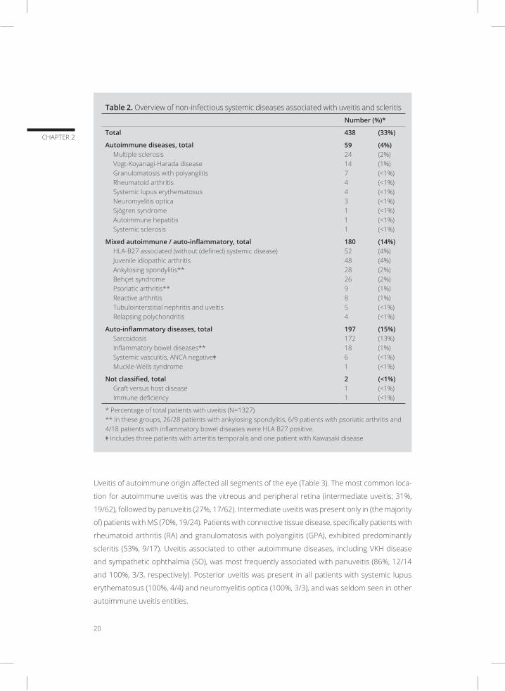
The classification of systemic non-infectious diseases according to current classification of auto-
immune- and auto-inflammatory diseases is indicated in Table 2. An association with a systemic
disease of established autoimmune origin was identified in 4% (59/1327), which was lower than
the percentage of patients with auto-inflammatory (15%, 197/1327) and mixed autoimmune-/
auto-inflammatory diseases (14%, 180/1327). The association with classic autoimmune connec-
tive tissue diseases was even lower, (1%, 17/1327). The most commonly associated autoimmune
disease was MS (24 patients) followed by VKH (14 patients). In our series, sarcoidosis (38% pre-
sumed and 62% biopsy proven) represented the most common non-infectious systemic disease
associated with uveitis (13%, 172/1327).
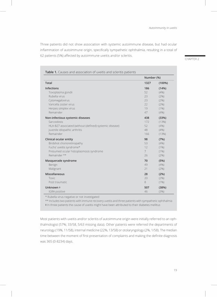
Autoimmunity in uveitis
19
CHAPTER 2
Three patients did not show association with systemic autoimmune disease, but had ocular
inflammation of autoimmune origin, specifically sympathetic ophthalmia, resulting in a total of
62 patients (5%) affected by autoimmune uveitis and/or scleritis.
Table 1. Causes and association of uveitis and scleritis patients
Number (%)
Total 1327 (100%)
InfectionsToxoplasma gondiiRubella virusCytomegalovirusVaricella zoster virus Herpes simplex virusRemainder
186 (14%)52 (4%)23 (2%)23 (2%)22 (2%)19 (1%)47 (4%)
Non-infectious systemic diseasesSarcoidosisHLA-B27 associated (without (defined) systemic disease)Juvenile idiopathic arthritisRemainder
438 (33%)172 (13%)52 (4%)48 (4%)166 (13%)
Clinical ocular entityBirdshot chorioretinopathyFuchs’ uveitis syndrome*Presumed ocular histoplasmosis syndromeRemainder **
98 (7%)53 (4%)12 (1%)7 (1%)26 (2%)
Masquerade syndromeBenignMalignant
70 (5%)49 (4%)21 (2%)
MiscellaneousToxicPost traumatic
28 (2%)20 (2%)8 (1%)
Unknown ǂIGRA positive
507 (38%)46 (3%)
* Rubella virus negative or not investigated ** Includes two patients with immune recovery uveitis and three patients with sympathetic ophthalmiaǂ In three patients the cause of uveitis might have been attributed to their diabetes mellitus
Most patients with uveitis and/or scleritis of autoimmune origin were initially referred to an oph-
thalmologist (57%; 33/58, 5/63 missing data). Other patients were referred the departments of
neurology (19%, 11/58), internal medicine (22%, 13/58) or otolaryngology (2%, 1/58). The median
time between the moment of first presentation of complaints and making the definite diagnosis
was 365 (0-8234) days.
20
CHAPTER 2
Table 2. Overview of non-infectious systemic diseases associated with uveitis and scleritis
Number (%)*
Total 438 (33%)
Autoimmune diseases, totalMultiple sclerosisVogt-Koyanagi-Harada diseaseGranulomatosis with polyangiitisRheumatoid arthritisSystemic lupus erythematosusNeuromyelitis opticaSjögren syndromeAutoimmune hepatitis Systemic sclerosis
59 (4%)24 (2%)14 (1%)7 (<1%)4 (<1%)4 (<1%)3 (<1%)1 (<1%)1 (<1%)1 (<1%)
Mixed autoimmune / auto-inflammatory, totalHLA-B27 associated (without (defined) systemic disease)Juvenile idiopathic arthritisAnkylosing spondylitis**Behçet syndromePsoriatic arthritis**Reactive arthritisTubulointerstitial nephritis and uveitis Relapsing polychondritis
180 (14%)52 (4%)48 (4%)28 (2%)26 (2%)9 (1%)8 (1%)5 (<1%)4 (<1%)
Auto-inflammatory diseases, totalSarcoidosisInflammatory bowel diseases**Systemic vasculitis, ANCA negativeǂMuckle-Wells syndrome
197 (15%)172 (13%)18 (1%)6 (<1%)1 (<1%)
Not classified, totalGraft versus host diseaseImmune deficiency
2 (<1%)1 (<1%)1 (<1%)
* Percentage of total patients with uveitis (N=1327)** In these groups, 26/28 patients with ankylosing spondylitis, 6/9 patients with psoriatic arthritis and 4/18 patients with inflammatory bowel diseases were HLA B27 positive.ǂ Includes three patients with arteritis temporalis and one patient with Kawasaki disease
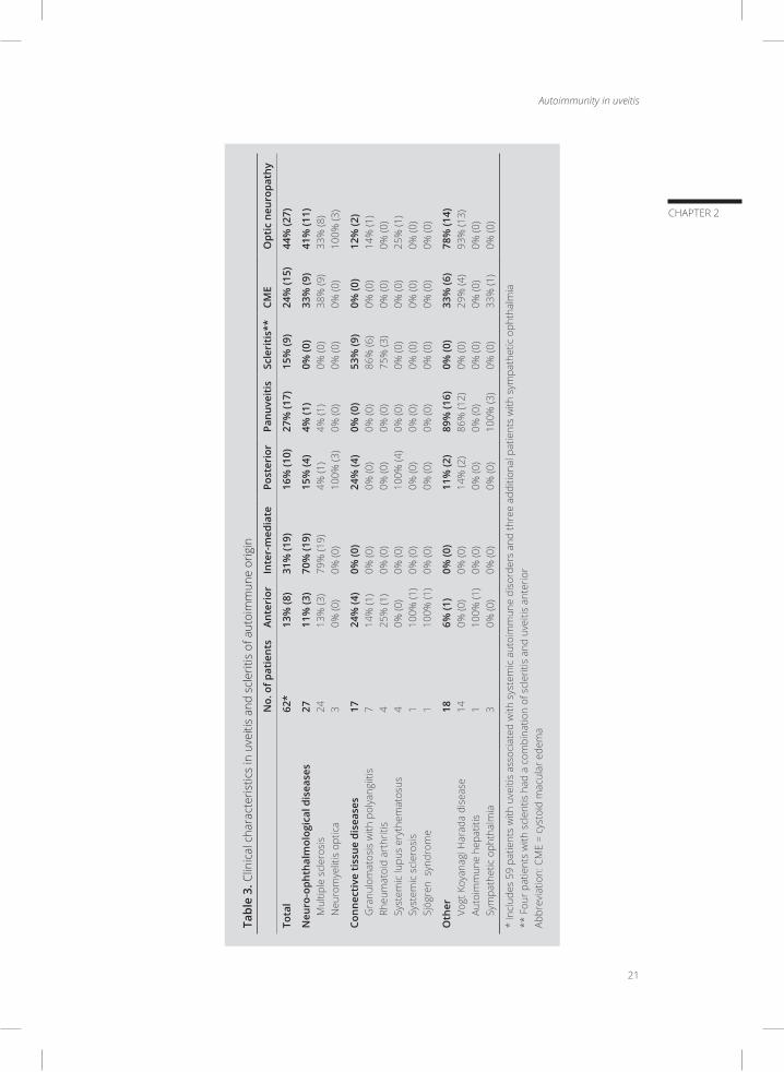
Uveitis of autoimmune origin affected all segments of the eye (Table 3). The most common loca-
tion for autoimmune uveitis was the vitreous and peripheral retina (intermediate uveitis; 31%,
19/62), followed by panuveitis (27%, 17/62). Intermediate uveitis was present only in (the majority
of) patients with MS (70%, 19/24). Patients with connective tissue disease, specifically patients with
rheumatoid arthritis (RA) and granulomatosis with polyangiitis (GPA), exhibited predominantly
scleritis (53%, 9/17). Uveitis associated to other autoimmune diseases, including VKH disease
and sympathetic ophthalmia (SO), was most frequently associated with panuveitis (86%, 12/14
and 100%, 3/3, respectively). Posterior uveitis was present in all patients with systemic lupus
erythematosus (100%, 4/4) and neuromyelitis optica (100%, 3/3), and was seldom seen in other
autoimmune uveitis entities.
Autoimmunity in uveitis
21
CHAPTER 2
Tabl
e 3.
Clin
ical
cha
ract
eris
tics
in u
veiti
s an
d sc
lerit
is o
f aut
oim
mun
e or
igin
No.
of p
atie
nts
Ante
rior
Inte
r-m
edia
tePo
ster
ior
Panu
veit
isSc
leri
tis*
*CM
EO
ptic
neu
ropa
thy
Tota
l62
*13
% (8
)31
% (1
9)16
% (1
0)27
% (1
7)15
% (9
)24
% (1
5)44
% (2
7)
Neu
ro-o
phth
alm
olog
ical
dis
ease
sM
ultip
le s
cler
osis
Neu
rom
yelit
is o
ptic
a
27 24 3
11%
(3)
13%
(3)
0% (0
)
70%
(19)
79%
(19)
0% (0
)
15%
(4)
4% (1
)10
0% (3
)
4% (1
)4%
(1)
0% (0
)
0% (0
)0%
(0)
0% (0
)
33%
(9)
38%
(9)
0% (0
)
41%
(11)
33%
(8)
100%
(3)
Conn
ecti
ve ti
ssue
dis
ease
sG
ranu
lom
atos
is w
ith p
olya
ngiit
isRh
eum
atoi
d ar
thrit
isSy
stem
ic lu
pus
eryt
hem
atos
usSy
stem
ic s
cler
osis
Sjög
ren
syn
drom
e
17 7 4 4 1 1
24%
(4)
14%
(1)
25%
(1)
0% (0
)10
0% (1
)10
0% (1
)
0% (0
)0%
(0)
0% (0
)0%
(0)
0% (0
)0%
(0)
24%
(4)
0% (0
)0%
(0)
100%
(4)
0% (0
)0%
(0)
0% (0
)0%
(0)
0% (0
)0%
(0)
0% (0
)0%
(0)
53%
(9)
86%
(6)
75%
(3)
0% (0
)0%
(0)
0% (0
)
0% (0
)0%
(0)
0% (0
)0%
(0)
0% (0
)0%
(0)
12%
(2)
14%
(1)
0% (0
)25
% (1
)0%
(0)
0% (0
)
Oth
er Vogt
Koy
anag
i Har
ada
dise
ase
Auto
imm
une
hepa
titis
Sy
mpa
thet
ic o
phth
alm
ia
18 14 1 3
6% (1
)0%
(0)
100%
(1)
0% (0
)
0% (0
)0%
(0)
0% (0
)0%
(0)
11%
(2)
14%
(2)
0% (0
)0%
(0)
89%
(16)
86%
(12)
0% (0
)10
0% (3
)
0% (0
)0%
(0)
0% (0
)0%
(0)
33%
(6)
29%
(4)
0% (0
)33
% (1
)
78%
(14)
93%
(13)
0% (0
)0%
(0)
* In
clud
es 5
9 pa
tient
s w
ith u
veiti
s as
soci
ated
with
sys
tem
ic a
utoi
mm
une
diso
rder
s an
d th
ree
addi
tiona
l pat
ient
s w
ith s
ympa
thet
ic o
phth
alm
ia
** F
our p
atie
nts
with
scl
eriti
s ha
d a
com
bina
tion
of s
cler
itis
and
uvei
tis a
nter
ior
Abbr
evia
tion:
CM
E =
cyst
oid
mac
ular
ede
ma

22
CHAPTER 2
The prevalence of complications in patients with uveitis of autoimmune origin is presented in
Table 3. CME was present in 24% (15/62) and was most frequently observed in patients with
MS (38%, 9/24), VKH disease (29%, 4/14) and SO (33%, 1/3). None of the patients with uveitis/
scleritis associated with connective tissue disease exhibited CME. The optic disk was involved
in 44% (27/62) of patients with uveitis of autoimmune origin. Almost all patients with VKH had
involvement of the optic disk (93%, 13/14). In patients with neuro-ophthalmological diseases, the
optic disk was involved in 41% (11/27), predominantly in patients with neuromyelitis optica (100%,
3/3). Involvement of the optic disk in other entities of autoimmune uveitis was only occasionally
observed.
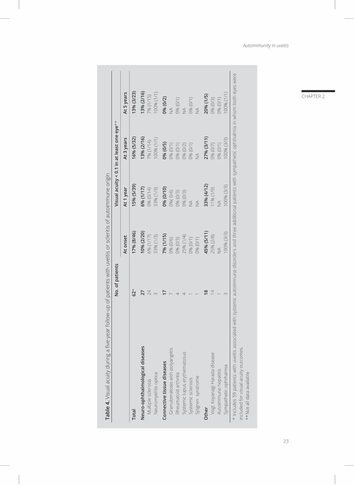
Overall, visual prognosis was favorable as none of the patients developed bilateral visual acuity
of less than 0.1 after five years of follow-up. At onset, visual acuity less than 0.1 in at least one
eye was present in 8/46 (17%) patients. The overall prevalence of a visual acuity of <0.1 in at
least one eye remained stable over the first five years of follow-up (Table 4). Visual acuity <0.1
in at least one eye in patients with uveitis associated with neurological diseases varied between
6% and 13% and did not change over time. Patients with VKH disease improved; 25% of these
patients started with a visual acuity of <0.1 in at least one eye, but after five years this percentage
was reduced to none.
The use of systemic immunosuppressive medications and/or systemic corticosteroids for sys-
temic and/or ocular inflammation during the first five years of follow-up in patients with uveitis of
autoimmune origin reached 76% (Table 4). The majority of patients with uveitis associated with
connective tissue disease were successfully treated with systemic immunosuppressive medica-
tion and/or systemic corticosteroids during the follow-up period of five years (75% - 83%). In the
group of patients with neuro-ophthalmological diseases immunosuppressive systemic medication
was least frequently used (37% during year one and 56% during year 3-5).
Autoimmunity in uveitis
23
CHAPTER 2
Tabl
e 4.
Vis
ual a
cuity
dur
ing
a fiv
e-ye
ar fo
llow
-up
of p
atie
nts
with
uve
itis
or s
cler
itis
of a
utoi
mm
une
orig
in
N
o. o
f pat
ient
sVi
sual
acu
ity
< 0.
1 in
at l
east
one
eye
**
At o
nset
At 1
yea
r At
3 y
ears
At 5
yea
rs
Tota
l62
*17
% (8
/46)
15%
(5/3
9)16
% (5
/32)
13%
(3/2
3)
Neu
ro-o
phth
alm
olog
ical
dis
ease
sM
ultip
le s
cler
osis
Neu
rom
yelit
is o
ptic
a
27 24 3
10%
(2/2
0)6%
(1/1
7)33
% (1
/3)
6% (1
/17)
0% (0
/14)
33%
(1/3
)
13%
(2/1
6)7%
(1/1
4)10
0% (1
/1)
13%
(2/1
6)7%
(1/1
5)10
0% (1
/1)
Conn
ecti
ve ti
ssue
dis
ease
sG
ranu
lom
atos
is w
ith p
olya
ngiit
isRh
eum
atoi
d ar
thrit
isSy
stem
ic lu
pus
eryt
hem
atos
usSy
stem
ic s
cler
osis
Sjög
ren
syn
drom
e
17 7 4 4 1 1
7% (1
/15)
0% (0
/6)
0% (0
/3)
25%
(1/4
)0%
(0/1
)0%
(0/1
)
0% (0
/10)
0%( 0
/4)
0% (0
/3)
0% (0
/3)
NA
NA
0% (0
/5)
0% (0
/1)
0% (0
/1)
0% (0
/2)
0% (0
/1)
NA
0% (0
/2)
NA
0% (0
/1)
NA
0% (0
/1)
NA
Oth
er Vogt
Koy
anag
i Har
ada
dise
ase
Auto
imm
une
hepa
titis
Sy
mpa
thet
ic o
phth
alm
ia
18 14 1 3
45%
(5/1
1)25
% (2
/8)
NA
100%
(3/3
)
33%
(4/1
2)11
% (1
/9)
NA
100%
(3/3
)
27%
(3/1
1)0%
(0/7
)0%
(0/1
)10
0% (3
/3)
20%
(1/5
)0%
(0/3
)0%
(0/1
)10
0% (1
/1)
* In
clud
es 5
9 pa
tient
s w
ith u
veiti
s as
soci
ated
with
sys
tem
ic a
utoi
mm
une
diso
rder
s an
d th
ree
addi
tiona
l pat
ient
s w
ith s
ympa
thet
ic o
phth
alm
ia in
who
m b
oth
eyes
wer
e in
clud
ed fo
r vis
ual a
cuity
out
com
es.
** N
ot a
ll da
ta a
vaila
ble
24
CHAPTER 2
DISCUSSION
Our results show that uveitis and/or scleritis of autoimmune origin was identified in 5% (62/1327)
of all patients. The most common autoimmune disease in patients with uveitis was MS (39%,
24/62), followed by VKH disease (23%, 14/62) and GPA (11%, 7/62). Scleritis was only observed in
patients with uveitis associated with connective tissue diseases (RA and GPA), and intermediate
uveitis was present only in patients with MS. Optic neuropathy was the most frequent complica-
tion (44%, 27/62). Use of systemic immunosuppressive treatment was frequent (up to 76%), and
visual outcomes were favorable as none of the patients developed permanent bilateral visual
acuity of less than 0.1 and only 13% of patients with uveitis of autoimmune origin developed
unilateral visual acuity < 0.1 after five years of follow-up.
Autoimmune diseases are classically defined by the Witebsky’s postulates: 1. presence of an
autoantibody or cell-mediated autoimmune reaction, 2. identification of a corresponding auto-
antigen, and 3. an analogous autoimmune response inducible in an experimental animal model
with development of a similar disease.11 These postulates have been revisited in 1993 by Rose
and Bona, which resulted in three types of evidence to establish an autoimmune origin: direct
proof, indirect proof and circumstantial evidence.12 Reproducing the disease by transfer of auto-
antibody or auto-reactive T-cells, or documenting the involvement of immunological reactions
after immunization with the autoantigen provides (in)direct proof of autoimmunity. Associations
with other autoimmune diseases, favorable response to immunosuppression or other distinctive
clinical clues such as presence of autoantibodies represent circumstantial evidence.
Autoimmune diseases predominantly involve the adaptive immune system, and are characterized
by the production of autoantibodies and / or auto-reactive T-cells that recognize specific cells or
tissues.4 Various autoantibodies are specific for individual autoimmune diseases, although their
exact role in the pathogenesis of the disease is often unknown. Since diverse autoantibodies (e.g.
antinuclear antibodies) appear also in healthy subjects, the mere presence of autoantibodies
does not always indicate the presence of an autoimmune disease.13 Self-directed inflamma-
tion by auto-inflammatory diseases is caused by an over-activity of the adaptive and/or innate
immune system, without specific identification of auto-reactive B- and T-cell responses (e.g. Crohn
disease).4 Recent classification of inflammatory diseases into autoimmune, mixed and auto-in-
flammatory diseases takes these differences into account.4-6,8-10
The eye is an immune privileged organ, which indicates that immune responses to foreign- and
self-antigens are suppressed or inhibited.14-16 This phenomenon prevents ocular damage and
preserves vision. Features that contribute to the mechanism of ocular immune privilege include
the blood-retina barrier, decreased lymphatic drainage, and soluble factors with immunosuppres-
sive properties in aqueous humor known as the anterior chamber associated immune deviation.

Autoimmunity in uveitis
25
CHAPTER 2
Autoimmune reactions against retinal antigens have repeatedly been suggested to play a crucial
role in diverse clinical uveitis entities. Direct evidence for an autoimmune pathogenesis has been
described in cancer-associated retinopathies by reproducing the disease after transfer of auto-
antibodies.17,18 In uveitis however definite proof of autoimmune reactions and inciting antigens
is very limited. Secondary contribution of autoimmune reactions has been proposed to play a
role diverse uveitis entities, including intraocular infections.19 Indirect evidence for autoimmunity
in uveitis has been provided by induction of autoimmune uveitis after immunization of animals
with retinal antigens and Freund adjuvant.20,21 These animal models, so called experimental auto-
immune uveitis (EAU), have provided insight into the immuno-pathogenesis of human uveitis. In
EAU predominantly mice are injected with different antigens (such as S-arrestin and interphoto-
receptor retinoid-binding protein) causing inflammation of intraocular tissue similar to human
uveitis. Other animal models induced autoimmune uveitis by transfer of retina specific T-cells.
For autoimmunity in human uveitis only circumstantial evidence was reported, for example by an
increasing number of T-helper 17 cells during active uveitis and scleritis, and a decreasing number
during treatment.22 So far, human autoimmune uveitis was only proven in uveitis when it is part
of a systemic autoimmune disease and is highly suspected in sympathetic ophthalmia. It is not
unlikely that other ocular entities (e.g. birdshot chorioretinopathy) might also be of autoimmune
origin, but direct evidence for an autoimmune pathogenesis is lacking.
Despite the high number of patients included in our series from a tertiary center, our study has
certain limitations. First, a bias to a more severe uveitis population is evident and is valid for
most studies from tertiary centers. Furthermore, it should be noted that our hospital represents
one of the national referral centers for sarcoidosis, which is probably in part responsible for a
somewhat higher prevalence of ocular sarcoidosis in our series (13%, 172/1327).23 In addition, at
5-year follow-up a significant number of patients with autoimmune uveitis was lost to follow-up
(65%, 40/62). The most probable explanation is that in the majority of these cases uveitis stabi-
lized or diminished and the patients were referred back to the ophthalmologists in peripheral
centers. Last, it cannot be ruled out that uveitis appearing with a systemic disease represent an
epiphenomena and is not associated with the systemic disease, although this is highly unlikely.
In conclusion, autoimmune uveitis is a rare diagnosis, which comprises 5% of our large uveitis/
scleritis population. It is feasible that secondary autoimmune reactions might play a role in some
uveitis entities (e.g. infections), as a consequence of damage and subsequent exposure of (so
far hidden or altered) retinal/choroidal antigens. Clinicians caring for uveitis patients should be
aware of the variety of diagnoses and the high prevalence of uveitis associated to sarcoidosis.
In our view, the term autoimmune uveitis should be reserved for intraocular inflammations of
confirmed autoimmune origin and should not be used as a synonym for non-infectious uveitis.
26
CHAPTER 2
REFERENCES
1. Pras E, Neumann R, Zandman-Goddard G, et al. Intraocular inflammation in autoimmune diseases. Semin
Arthritis Rheum. 2004;34(3):602-609.
2. Lee RW, Nicholson LB, Sen HN, et al. Autoimmune and autoinflammatory mechanisms in uveitis. Semin
Immunopathol. 2014;36(5):581-594.
3. Willermain F, Rosenbaum JT, Bodaghi B, et al. Interplay between innate and adaptive immunity in the
development of non-infectious uveitis. Prog Retin Eye Res. 2012;31(2):182-194.
4. McGonagle D, McDermott MF. A proposed classification of the immunological diseases. PLoS Med.
2006;3(8):e297.
5. Kastner DL, Aksentijevich I, Goldbach-Mansky R. Autoinflammatory disease reloaded: a clinical perspective.
Cell. 2010;140(6):784-790.
6. Pathak S, McDermott MF, Savic S. Autoinflammatory diseases: update on classification diagnosis and
management. J Clin Pathol. 2017;70(1):1-8.
7. Trusko B, Thorne J, Jabs D, et al. The Standardization of Uveitis Nomenclature (SUN) Project. Development
of a clinical evidence base utilizing informatics tools and techniques. Methods Inf Med. 2013;52(3):259-
265, S251-256.
8. Abramovits W, Oquendo M. Introduction to autoinflammatory syndromes and diseases. Dermatol Clin.
2013;31(3):363-385.
9. van Kempen TS, Wenink MH, Leijten EF, Radstake TR, Boes M. Perception of self: distinguishing autoim-
munity from autoinflammation. Nat Rev Rheumatol. 2015;11(8):483-492.
10. Davila-Seijo P, Hernandez-Martin A, Torrelo A. Autoinflammatory syndromes for the dermatologist. Clin
Dermatol. 2014;32(4):488-501.
11. Witebsky E. Experimental evidence for the role of auto-immunization in chronic thyroiditis. Proc R Soc
Med. 1957;50(11):955-958.
12. Rose NR, Bona C. Defining criteria for autoimmune diseases (Witebsky’s postulates revisited). Immunol
Today. 1993;14(9):426-430.
13. Marin GG, Cardiel MH, Cornejo H, Viveros ME. Prevalence of antinuclear antibodies in 3 groups of healthy
individuals: blood donors, hospital personnel, and relatives of patients with autoimmune diseases. J Clin
Rheumatol. 2009;15(7):325-329.
14. Kaplan HJ, Streilein JW. Immune response to immunization via the anterior chamber of the eye. II. An
analysis of F1 lymphocyte-induced immune deviation. J Immunol. 1978;120(3):689-693.
15. Kaplan HJ, Streilein JW. Immune response to immunization via the anterior chamber of the eye. I. F.
lymphocyte-induced immune deviation. J Immunol. 1977;118(3):809-814.
16. Streilein JW. Ocular immune privilege: the eye takes a dim but practical view of immunity and inflammation.
J Leukoc Biol. 2003;74(2):179-185.
17. Ohguro H, Ogawa K, Maeda T, Maeda A, Maruyama I. Cancer-associated retinopathy induced by both
anti-recoverin and anti-hsc70 antibodies in vivo. Invest Ophthalmol Vis Sci. 1999;40(13):3160-3167.
18. Adamus G, Machnicki M, Elerding H, Sugden B, Blocker YS, Fox DA. Antibodies to recoverin induce apop-
tosis of photoreceptor and bipolar cells in vivo. J Autoimmun. 1998;11(5):523-533.
19. Whittle RM, Wallace GR, Whiston RA, Dumonde DC, Stanford MR. Human antiretinal antibodies in toxo-
plasma retinochoroiditis. Br J Ophthalmol. 1998;82(9):1017-1021.
20. de Kozak Y, Sakai J, Thillaye B, Faure JP. S antigen-induced experimental autoimmune uveo-retinitis in rats.
Curr Eye Res. 1981;1(6):327-337.
21. Broekhuyse RM, Winkens HJ, Kuhlmann ED. Induction of experimental autoimmune uveoretinitis

Autoimmunity in uveitis
27
CHAPTER 2
and pinealitis by IRBP. Comparison to uveoretinitis induced by S-antigen and opsin. Curr Eye Res.
1986;5(3):231-240.
22. Amadi-Obi A, Yu CR, Liu X, et al. TH17 cells contribute to uveitis and scleritis and are expanded by IL-2
and inhibited by IL-27/STAT1. Nat Med. 2007;13(6):711-718.
23. Tsirouki T, Dastiridou A, Symeonidis C, et al. A Focus on the Epidemiology of Uveitis. Ocul Immunol
Inflamm. 2016:1-15.

ANTINUCLEAR ANTIBODY PROFILING IN UVEITIS
Josianne C.E.M. ten Berge, F. Groen-Hakan, Aniki Rothova, Marco W.J. Schreurs
Adapted version accepted for publication in Acta Ophthalmologica
3
30
CHAPTER 3
ABSTRACT
Purpose: Antinuclear antibody (ANA) profiling plays an important role in diagnosis of various
autoimmune and autoinflammatory diseases. ANA is associated with the development of uveitis
in children and its poor prognosis. In contrast, the diagnostic value of ANA in work-up of adults
with uveitis is debatable. The aim of this study is to assess the diagnostic value of ANA profiling
in adult patients with uveitis.
Methods: In this prospective study, we assessed the presence of ANA in serum of 105 consecu-
tive adult patients with uveitis. In samples positive for ANA, ANA titer, ANA subtypes and staining
patterns on IIF were also determined. Clinical data from uveitis patients were collected and
statistical analyses were performed to relate laboratory results to clinical data of the patients.
Results: A positive ANA result was observed in 29/105 (28%) patients with uveitis, and the median
ANA titer was 160. Positive ANA titers were associated with longer duration of uveitis (p=0.037). No
other associations were found between the presence of ANA, ANA titer or ANA staining pattern
and specific diagnosis and various clinical characteristics of uveitis (all p-values > 0.05).
Conclusion: A positive ANA was found in 28% of patients with uveitis. The ANA profile was not
distinctive for specific causes or clinical manifestations of uveitis. The diagnostic value of ANA
assessment in the adult uveitis population is limited.
Antinuclear antibody profiling in uveitis
31
CHAPTER 3
INTRODUCTION
Uveitis is a clinical syndrome, which can be associated with different causes, including infections
and systemic diseases. The pathogenesis of most uveitis entities in not clarified, although the
immune system has been considered to play a major role. Various uveitis entities are associated
with autoimmune and autoinflammatory diseases.1
Antinuclear antibodies (ANA) are antibodies directed against a variety of nuclear antigens, and
can be detected in patients with autoimmune diseases. The presence of ANA is not specific for
disease, since it has also been observed in the healthy population (predominantly women and
elderly).2 In the past, ANA were determined in all patients with uveitis for diagnostic screening
purposes, but this approach has been abandoned since its diagnostic value in adult patients
with uveitis shown to be limited.3 In contrast, in patients with juvenile idiopathic arthritis (JIA) the
presence of ANA has been demonstrated to be valuable, because its presence increases the risk
of developing uveitis.4,5
In the last decades, the analysis of ANA has been improved and various subtypes and staining
patterns are being determined. Profiling of ANA has been proven to play a significant role for diag-
nostic purposes in various diseases, including systemic lupus erythematosus, Sjögren syndrome
and systemic sclerosis.6,7 The diagnostic relevance of the ANA profile and its possible association
with clinical features in uveitis are not known. The aim of this study is to assess the presence,
subtypes and titers of ANA in adult patients with uveitis of different etiologies and evaluate its
possible value for diagnostic screening in uveitis.
METHODS
We conducted a prospective study at the department of Ophthalmology, Erasmus MC, University
Medical Center Rotterdam and determined ANA profile in 105 consecutive adult patients with
uveitis who underwent a standardized screening protocol for the cause of their uveitis between
January 2016 and July 2017. The study was performed in accordance with the Declaration of
Helsinki and in agreement with the institutional regulations and approval of our institutional
review board.
In addition to ANA screening, all patients underwent a diagnostic screening protocol, which was
related to the location of uveitis (according to the Standardization of Uveitis Nomenclature (SUN)
Working Group) and included chest radiography, erythrocyte sedimentation rate, blood counts,
serum angiotensin converting enzyme levels, serology for syphilis and Lyme disease and inter-
feron gamma release assay test (QuantiFERON–TB Gold In-Tube test). Patients with anterior
32
CHAPTER 3
uveitis or panuveitis were also tested for presence of human leucocyte antigen-B27. A tailored
approach was applied for further examinations.
Data from included patients were collected from medical charts and registered were patients’
demographics (age, gender and race), definitive diagnosis of uveitis as well as ocular character-
istics (laterality, duration and activity of uveitis), use of systemic immunomodulating medications
and ANA characteristics (presence, titer, staining pattern and ANA subtype).
Screening for ANA in serum samples from included patients was performed by indirect immu-
nofluorescence (IIF) according to standard protocol. In short; HEp-2 cells (Inova, San Diego, CA)
were incubated with 1:80 diluted serum samples for 30 minutes, and after being washed, the
slides were incubated for 30 minutes with goat anti-human IgG conjugated with fluorescein iso-
thiocyanate with propidium iodide for counterstaining (Inova, San Diego, CA) to label antibodies.
ANA titers of 1:80 or higher were considered positive and in these samples the ANA pattern
and exact ANA titer were also analyzed. ANA patterns were classified according to international
consensus and include only nuclear and mitotic patterns, whereas cytoplasmic HEp-2 staining
was considered negative.8 In ANA positive samples, further identification for detection of anti-ex-
tractable nuclear antigens (anti-ENA antibodies) and anti-double stranded DNA (anti-dsDNA) was
performed by EliA (Thermo Fisher Scientific/Phadia, Freiburg, Germany), ELISA (Inova, San Diego,
CA) and/or LIA (Euroimmun, Lübeck, Germany). The ENA-panel consisted of anti-SS-A, anti-SS-B,
anti-RNP, anti-Smith (anti-Sm), anti-CenpB, anti-Scl-70, and anti-Jo-1.
Statistical analyses were performed to evaluate the presence and characteristics of ANA in uveitis
patients. Continuous variables were described by median and range, and categorical variables
were summarized by percentages (proportions). We used Fisher’s exact test for categorical data
and Mann Whitney U test, Kruskall-Wallis 1-way ANOVA test and Spearman’s Rank Correlation
for continues variables. All statistical analyses were performed using SPSS software (version 22.0,
Chicago, IL). A p-value of <0.05 was considered statistically significant and all tests were two-sided.
RESULTS
The clinical characteristics of included uveitis patients are shown in Table 1. The majority of
patients were female (69/105, 66%) and the median age was 51 years. Most patients had an
active uveitis (77/105 73%) and did not use systemic immunosuppressive medication (96/105,
91%) during blood sampling.
Positive ANA results were observed in 29/105 (28%) of patients with uveitis. The presence of ANA
was equally distributed between genders and no association was observed between age and

Antinuclear antibody profiling in uveitis
33
CHAPTER 3
presence of ANA. Most ANA positive samples were observed in idiopathic uveitis (18/55, 33%)
and no positive ANA were seen in patients with uveitis classified as a clinical ocular syndromes
(e.g. birdshot chorioretinopathy; Table 1). Prevalence of ANA was higher in patients with anterior
uveitis (12/28, 43%) compared to other locations, but this difference did not reach significance.
A visual acuity of <0.5 was observed in 7/26 (27%) ANA positive patients and in 22/82 (27%) of
patients without ANA. Positive ANA titers were associated with longer duration of uveitis (p=0.037).
All other clinical characteristics of uveitis were not significantly associated to the presence of ANA
(all p-values > 0.05). Activity of uveitis could not be related to ANA presence (p=0.90).
Table 1. Presence of antinuclear antibodies (ANA) in uveitis patients
All uveitis ANA positive ANA negative
Total 105 (100%) 29/105 (28%) 76/105 (72%)
Median age in years (range) 51 (19-88) 49 (21-87) 52 (19-88)
Gender MaleFemale
36/105 (34%)69/105 (66%)
11/36 (31%)18/69 (26%)
25/36 (69%)51/69(74%)
Race CaucasianNon-Caucasian
73/105 (70%)32/105 (30%)
18/73 (25%)11/32 (34%)
55/73 (75%)21/32 (66%)
Cause of uveitisClinical ocular syndromeImmune mediated systemic diseaseInfectionMasqueradeIdiopathic
6/105 (6%)15/105 (14%)16/105 (15%)13/105 (12%)55/105 (52%)
0/6 (0%)3/15 (20%)5/16 (31%)3/13 (23%)
18/55 (33%)
6/6 (100%)12/15 (80%)11/16 (69%)10/13 (77%)37/55 (67%)
Laterality of uveitisUnilateralBilateral
49/105 (47%)56/105 (53%)
13/49 (27%)16/56 (29%)
36/49 (73%)40/56 (71%)
Median duration of uveitis in years* (range) 1 (0-50) 1 (0-50) 0 (0-19)
Location of uveitisAnteriorIntermediatePosteriorPanuveitisSclero-/kerato-uveitis
28/105 (27%)9/105 (9%)
36/105 (34%)20/105 (19%)12/105 (11%)
12/28 (43%)2/9 (22%)
9/36 (25%)4/20 (20%)2/12 (17%)
16/28 (57%)7/9 (78%)
27/36 (75%)16/20 (80%)10/12 (83%)
Activity of uveitisActiveQuiet
77/105 (73%)28/105 (27%)
21/77 (27%)8/28 (29%)
56/77 (73%)20/28 (71%)
Immunocompromised **YesNo
14/105 (13%)91/105 (87%)
5/14 (36%)24/91 (26%)
9/14 (64%)67/91 (74%)
*p = 0.037** Use of immunosuppressive medications, malignant disorder or HIV with CD4 cell count of <300 during blood collection
34
CHAPTER 3
The median ANA titer (within ANA positive uveitis patients) was 160 and ranged from 80 to 640
(Table 2). Clinical features of uveitis were not associated to ANA titer (all p-values > 0.05). The
ANA pattern was classified as nuclear in 23/25 (92%) of patients and as mitotic in the remaining
2/25 (8%) patients. A speckled ANA pattern was the most frequent observed pattern (13/29, 45%),
followed by a homogeneous ANA (9/29, 31%). The speckled pattern was most frequently observed
in uveitis with an unknown cause (9/13, 69%). The distribution of ANA patterns was however not
characteristic for specific causes, locations or clinical manifestations of uveitis. Two patients were
anti-dsDNA positive and one patient was positive for anti- ribonucleoprotein (anti-RNP); none
exhibited any signs of autoimmune systemic disorder on examination by immunologist and all
three were (so far) classified as uveitis of unknown origin.
Table 2. Antinuclear antibody (ANA) characteristics of uveitis population (N=29)
Number (%)
Total ANA positive uveitis 29/105 (28%)
Median ANA titer 160 (80-640)
ANA patternsSpeckledHomogeneousNucleolarSpeckled + nucleolar CentrioleMitotic spindle apparatus
13/29 (45%)9/29 (31%)3/29 (10%)2/29 (7%)1/29 (3%)1/29 (3%)
Anti-double stranded DNA 2/29 (7%)
Anti-extractable nuclear antigens (ENA)(anti- ribonucleoprotein (RNP))
1/29 (3%)
DISCUSSION
Our prospective study shows that ANA is positive in 28% of patients with uveitis. No significant asso-
ciations were found between the presence of ANA, ANA titer, or specific ANA patterns and various
clinical characteristics of uveitis, including specific diagnoses or activity of intraocular inflammation.
The prevalence of 28% is higher than in the age-matched healthy population.9-11 The prevalence
of ANA in healthy population varies between 8-17% and tends to be higher in female individuals
and elderly.9-11 In our series, the relationship between age, gender and positive ANA could not be
confirmed, probably due to the limited number of patients in various age groups. Although the
ANA prevalence in our study was higher compared to the healthy age-matched population, no
clinical relevance for the work-up of uveitis in adult patients could be identified. Therefore, routine
Antinuclear antibody profiling in uveitis
35
CHAPTER 3
ANA determination as a part of the diagnostic testing of uveitis patients cannot be recommended.
Determining ANA should however be performed in cases with signs suggesting specific systemic
diseases such as systemic lupus erythematosus.
Three decades ago, approximately 14% prevalence of ANA in uveitis population was observed
in a setting similar to our series.3,12 This percentage is lower compared to the 28% ANA positivity
found in our cohort and might reflect the changing spectrum of uveitis entities over time. Further
it is possible that referral pattern might also play a role. In line with previous studies we did not
find associations between specific uveitis entities, their characteristics and ANA, with exception
of a borderline association between ANA positivity and longer duration of uveitis. Although our
study is prospective, it includes a limited number of patients and therefore we cannot exclude
that a specific uveitis entity could be associated with ANA.
The prevalence of positive ANA in autoimmune- and autoinflammatory diseases varies widely.
Almost all patients with systemic lupus erythematosus (SLE) are ANA positive and in systemic
sclerosis and rheumatoid arthritis ANA prevalence varies between 30%-70%.13 Therefore, one
could expect a higher ANA prevalence in uveitis associated with systemic non-infectious disor-
ders. However, in our series only 3/15 (20%) of the patients with systemic immune mediated
disorders were ANA positive. Interestingly, ANA was more prevalent in patients with infectious
uveitis (5/16, 31%) than in other uveitis entities. A transiently positive ANA test was previously
noted in systemic infectious diseases.14 The relationship between ANA and ocular infections has
not been specified in earlier studies.
Reactivity to specific ENA discriminates between various types of systemic autoimmune diseases
and plays herein also a prognostic role. For example, in SLE, antibodies directed against the Sm
antigen are specific for the disease and presence of anti-Topo-I antibodies is associated with more
severe course of systemic sclerosis.15 We identified 3/29 (10%) with positive anti-ENA in our ANA
positive patients but found no associations with uveitis characteristics, including its severity. This
low proportion of anti-ENA positivity is not surprising, since still many anti-ENA specificities are
not known. Usually anti-ENA antibodies occur more frequently in patients with high ANA titers.16
The low to moderate ANA titers in our study appears in agreement with the small proportion of
anti-ENA presence. The distribution of ANA patterns in our cohort seems similar compared to
previous studies on ANA positive samples.17
Our findings in adult uveitis population differ from uveitis in children. Specifically, in JIA-associated
uveitis the proportion of ANA positivity has been described in up to 86%.3,12 Presence of ANA in
JIA has been documented to impose a significant risk for the development of uveitis.18 The most
common pattern of ANA in JIA-uveitis is (partly) homogeneous (86%) and no specific anti-ENA
have been identified.18

36
CHAPTER 3
In conclusion, positive serum ANA was observed in 28% of adult patients with uveitis. Specific
associations between ANA positivity, ANA titer and ANA subtype, and ocular characteristics of
uveitis were not identified. Based on our results, we do not recommend including ANA for the
screening purposes of patients with uveitis.

Antinuclear antibody profiling in uveitis
37
CHAPTER 3
REFERENCES
1. Ten Berge JC, Schreurs MW, Vermeer J, Meester-Smoor MA, Rothova A. Prevalence and clinical impact of
antiretinal antibodies in uveitis. Acta Ophthalmol. 2016;94(3):282-288.
2. Solomon DH, Kavanaugh AJ, Schur PH, American College of Rheumatology Ad Hoc Committee on Immuno-
logic Testing G. Evidence-based guidelines for the use of immunologic tests: antinuclear antibody testing.
Arthritis Rheum. 2002;47(4):434-444.
3. Murray P. Serum autoantibodies and uveitis. Br J Ophthalmol. 1986;70(4):266-268.
4. Heiligenhaus A, Niewerth M, Ganser G, Heinz C, Minden K, German Uveitis in Childhood Study G. Prev-
alence and complications of uveitis in juvenile idiopathic arthritis in a population-based nation-wide
study in Germany: suggested modification of the current screening guidelines. Rheumatology (Oxford).
2007;46(6):1015-1019.
5. Saurenmann RK, Levin AV, Feldman BM, et al. Prevalence, risk factors, and outcome of uveitis in juvenile
idiopathic arthritis: a long-term followup study. Arthritis Rheum. 2007;56(2):647-657.
6. Hamaguchi Y. Autoantibody profiles in systemic sclerosis: predictive value for clinical evaluation and
prognosis. J Dermatol. 2010;37(1):42-53.
7. Smeenk R, Brinkman K, van den Brink H, et al. Antibodies to DNA in patients with systemic lupus erythe-
matosus. Their role in the diagnosis, the follow-up and the pathogenesis of the disease. Clin Rheumatol.
1990;9(1 Suppl 1):100-110.
8. Chan EK, Damoiseaux J, Carballo OG, et al. Report of the First International Consensus on Standardized
Nomenclature of Antinuclear Antibody HEp-2 Cell Patterns 2014-2015. Front Immunol. 2015;6:412.
9. Tan EM, Feltkamp TE, Smolen JS, et al. Range of antinuclear antibodies in “healthy” individuals. Arthritis
Rheum. 1997;40(9):1601-1611.
10. Nisihara R, Kubis MM, Rodrigues PC, Skare T, Mocelin V, Utiyama S. Antinuclear antibodies and rheuma-
toid factor positivity in healthy elderly adults: a cross-sectional study in 336 individuals. J Am Geriatr Soc.
2013;61(11):2044-2046.
11. Fernandez SA, Lobo AZ, Oliveira ZN, Fukumori LM, AM Pr, Rivitti EA. Prevalence of antinuclear autoanti-
bodies in the serum of normal blood dornors. Rev Hosp Clin Fac Med Sao Paulo. 2003;58(6):315-319.
12. Hundert I, Bakimer R, Amital-Teplizki H, et al. Antinuclear autoantibodies in uveitis. Clin Exp Rheumatol.
1989;7(3):237-241.
13. Wichainun R, Kasitanon N, Wangkaew S, Hongsongkiat S, Sukitawut W, Louthrenoo W. Sensitivity and
specificity of ANA and anti-dsDNA in the diagnosis of systemic lupus erythematosus: a comparison using
control sera obtained from healthy individuals and patients with multiple medical problems. Asian Pac J
Allergy Immunol. 2013;31(4):292-298.
14. Litwin CM, Binder SR. ANA testing in the presence of acute and chronic infections. J Immunoassay Immu-
nochem. 2016;37(5):439-452.
15. Damoiseaux JG, Tervaert JW. From ANA to ENA: how to proceed? Autoimmun Rev. 2006;5(1):10-17.
16. Bossuyt X, Hendrickx A, Frans J. Antinuclear antibody titer and antibodies to extractable nuclear antigens.
Arthritis Rheum. 2005;53(6):987-988.
17. Avery TY, van de Cruys M, Austen J, Stals F, Damoiseaux JG. Anti-nuclear antibodies in daily clinical practice:
prevalence in primary, secondary, and tertiary care. J Immunol Res. 2014;2014:401739.
18. Kotaniemi K, Kautiainen H, Karma A, Aho K. Occurrence of uveitis in recently diagnosed juvenile chronic
arthritis: a prospective study. Ophthalmology. 2001;108(11):2071-2075.

PREVALENCE AND CLINICAL IMPACT OF ANTIRETINAL ANTIBODIES IN UVEITIS
Josianne C.E.M. ten Berge, Marco W.J. Schreurs, Jacolien Vermeer, Magda A. Meester-Smoor, Aniki Rothova
Acta Ophthalmol. 2016 May;94(3):282-8
4
40
CHAPTER 4
ABSTRACT
Purpose: To determine the prevalence of serum antiretinal antibodies (ARAs) among patients
with uveitis and establish their clinical relevance.
Methods: This prospective study assessed the presence of ARAs by indirect immunofluorescence
(IIF) using primate retina in 126 patients with uveitis and 60 healthy controls. Clinical data of uveitis
patients were collected from medical charts and included the classification of uveitis, cause of
uveitis or its association with systemic disease, stage and activity of uveitis and specific retinal
features. Correlations between the presence of specific ARAs and various clinical characteristics
were analyzed.
Results: The presence of ARAs was observed in 49/104 (47%) of patients with uveitis and in 10/59
(17%) of healthy controls (p<0.001). Staining of the nuclear layers and the photoreceptors were
both more often observed in patients with uveitis compared to healthy controls (p=0.002 and
p=0.047, respectively). No specific associations were found between the presence of serum ARAs
and various clinical characteristics.
Conclusion: Serum ARAs were more frequent in patients with uveitis compared to healthy con-
trols, but their clinical role remains elusive. The assessment of intraocular production of specific
ARAs may provide further insight into the role of ocular autoantibodies in diverse uveitis entities.
Antiretinal antibodies in uveitis
41
CHAPTER 4
INTRODUCTION
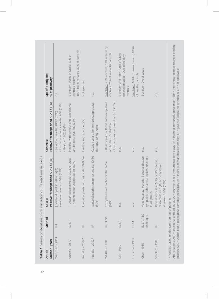
Ocular autoimmunity characterized by the presence of multiple antiretinal antibodies (ARAs)
has been documented in auto-immune retinopathy (AIR), including carcinoma associated reti-
nopathy, melanoma associated retinopathy or non-paraneoplastic autoimmune retinopathy.1-3
In addition, retinal autoimmune reactions are considered to play an important role in the patho-
genesis of diverse retinal and uveo-retinal disorders. Multiple serum ARAs have been observed
in various cohorts of patients with diverse uveitis entities but were also observed in up to 62%
of the healthy population (Table 1).4-13 In addition, the decrease of serum ARAs in patients with
exudative age-related macular degeneration following treatment with bevacuzimab injections has
been described.14 Previous reports hypothesized that retinal damage caused by inflammation
might induce a secondary formation of autoantibodies and cellular auto-immune responses
which might subsequently contribute to continuation, recurrence rate and/or aggravation of the
original inciting process. The precise sequence of events that might result in autoimmune attack
of retinal cells is not yet elucidated.
Uveitis is an inflammatory process of the uvea and is a major cause of blindness, resulting in
10% of all cases of blindness. Uveitis can be caused by infection, systemic inflammatory disease,
trauma or malignancy, however the etiology of uveitis remains unknown for up to 50% of the
cases. Usually uveitis is classified according to its localization in the eye; anterior, intermediate,
posterior or panuveitis.15 The eye is an immune privileged organ, because of its blood-retina
barrier and the absence of lymphatic drainage. Furthermore, the introduction of foreign anti-
gens into the anterior chamber of the eye can induce a tolerance to the foreign antigen, called
the anterior chamber associated immune deviation (ACAID). Absence of these features might
enhance intraocular inflammation and subsequent loss of vision.
The clinical relevance of serum ARAs in uveitis is still unknown. Previous studies on serum ARAs
in uveitis lacked clinical data such as ocular features, activity of uveitis and use of medications
(with the exception of a Polish publication).7 Furthermore, only small cohorts of specific uveitis
entities were analyzed using S-antigen, interphotoreceptor retinoid-binding protein (IRBP) or
crude human or bovine retinal extract. The identification of autoimmune processes in uveitis
will help to elucidate the pathogenesis, and will also aid in the development of new diagnostic
and treatment modalities.
In this study, we investigate the presence of serum ARAs in 126 patients with uveitis and 60
healthy controls and correlate their clinical manifestations to laboratory findings.
42
Tabl
e 1.
Sur
vey
of li
tera
ture
on
retin
al a
utoi
mm
une
reac
tions
in u
veiti
s
Arti
cle
(aut
hor
- yea
r)M
etho
dCa
ses:
Po
siti
ve fo
r un
spec
ified
ARA
/ al
l (%
)Co
ntro
ls:
Posi
tive
for
uns
peci
fied
ARA
/ all
(%)
Spec
ific
anti
gens
:%
of p
osit
ivit
y
Wal
sche
id -
2014
IIHJu
veni
le id
iopa
thic
art
hriti
s (JI
A)
asso
ciat
ed u
veiti
s: 4
2/89
(47%
)- J
IA w
ithou
t uve
itis:
48/
72 (6
7%)
- Idi
opat
hic
ante
rior u
veiti
s: 7
/58
(12%
)- H
ealth
y: 1
2/23
(52%
)
n.a.
Curs
ino
- 201
0EL
ISA
- O
cula
r tox
opla
smos
is:
30/3
0 (1
00%
)-
Non
infe
ctio
us u
veiti
s: 5
0/50
(60
%)
Hea
lthy
(with
/with
out a
nti-t
oxop
lasm
a an
tibod
ies)
: 106
/500
(21%
)S-
antig
en: 1
00%
of c
ases
; 43%
of
cont
rols
pos
itive
IRBP
: 100
% o
f cas
es; 8
7% o
f con
trol
s
Kubi
cka
– 20
04*
IIFId
iopa
thic
pos
terio
r uve
itis:
49/
50 (9
8%)
Hea
lthy:
(not
spe
cifie
d)/5
0N
ot s
peci
fied
Kubi
cka
– 20
02*
IIFAc
tive
idio
path
ic p
oste
rior u
veiti
s: 4
0/50
(8
0%)
Case
s 1
year
afte
r im
mun
osup
pres
sive
th
erap
y: 1
0/50
(20%
)n.
a.
Whi
ttle
– 1
998
IIF, E
LISA
To
xopl
asm
a re
tinoc
horo
iditi
s: 3
4/36
(9
4%)
- Hea
lthy
(with
/with
out a
nti-t
oxop
lasm
a an
tibod
ies)
: 6/1
6 (3
8%)
- Idi
opat
hic
retin
al v
ascu
litis
: 3/1
2 (2
5%)
S-an
tigen
: 75%
of c
ases
; 63%
of h
ealth
y co
ntro
ls; 7
5% o
f vas
culit
is c
ontr
ols
Lelij
- 19
90EL
ISA
n.a.
n.a.
S-an
tigen
and
IRBP
: 100
% o
f cas
es
(onc
hoce
rcia
sis)
; 100
% o
f hea
lthy
cont
rols
Forr
este
r - 1
989
ELIS
A n.
a.n.
a.S-
antig
en: 1
00%
of c
ases
(uve
itis)
; 100
%
of h
ealth
y co
ntro
ls
Chan
- 19
85EL
ISA,
ABC
te
chni
que
Vogt
-Koy
anag
i-Har
ada,
Beh
cet’s
dis
ease
, sy
mpa
thic
oph
thal
mia
: pos
itive
reac
tion
in a
ll gr
oups
n.a.
S-an
tigen
: 0%
of c
ases
Stan
ford
- 19
88IIF
Retin
al v
ascu
litis
(22
Behc
et’s
dise
ase,
3
sarc
oido
sis,
1 JI
A, 2
6 no
sys
tem
ic
dise
ase)
: 35/
52 (6
7%)
n.a.
n.a.
* Po
ssib
ly b
ased
on
the
sam
e co
hort
of p
atie
nts
Abbr
evia
tions
: ARA
= a
ntire
tinal
ant
ibod
ies,
ELI
SA =
enz
yme-
linke
d im
mun
o so
rben
t ass
ay, I
IF =
indi
rect
imm
unofl
uore
scen
ce, I
RBP
= in
terp
hoto
rece
ptor
retin
oid-
bind
ing
prot
ein,
ABC
= A
vidi
n-bi
otin
-per
oxid
ase
com
plex
tech
niqu
e, II
H =
indi
rect
imm
unoh
isto
chem
istr
y, JI
A =
juve
nile
idio
path
ic a
rthr
itis,
n.a
. = n
ot a
pplic
able
Antiretinal antibodies in uveitis
43
CHAPTER 4
METHODS
Patients and sample collection
Blood samples from 126 patients with uveitis were prospectively collected from April 2013 until
November 2014 at the department of Ophthalmology, Erasmus MC, University Medical Center
Rotterdam and stored at minus 80°Celsius at the biobank of our department. All patient signed
informed consent. Our laboratory assessments were performed in May and June 2015. The study
was performed in accordance with the Declaration of Helsinki and in agreement with the institu-
tional regulations and approval of our institutional review board. Patients were classified according
to SUN classification of their anatomical locations.16 All patients with uveitis underwent a standard-
ized diagnostic protocol according to the localization of the inflammation. This protocol included
radiologic chest examination erythrocyte sedimentation rate, blood counts, serum angiotensin-con-
verting enzyme levels, serology for syphilis and Lyme disease as well as interferon gamma release
assay (IGRA) test (QuantiFERON–TB Gold In-Tube test). In those with anterior and panuveitis Human
Leukocyte Antigen (HLA)-B27 testing was also performed. According to the clinical manifestations,
additional examinations were performed (tailored approach). Serum samples of 60 presumably
healthy individuals (blood bank donors) were used as controls (gender and age unknown).
Data collection
Clinical data of uveitis patients were collected from medical data files and included patient demo-
graphic and ocular characteristics such as age, gender, onset and duration of uveitis, cause of
uveitis or association with systemic disorder, anatomical location and activity of uveitis, visual
acuity, use of immunosuppressive medications, laterality, presence of retinal lesions, vasculitis,
cystoid macular edema (CME) and presence of glaucoma.
Detection of ARAs using indirect immunofluorescence (IIF)
Initial screening of sera was performed using primate retinal tissue. Cryosections manufactured
by Euroimmun (Lubeck, Germany) were left unfixed. The quality of the cryosections of primate
retinal tissue was checked with immunohistochemistry and staining with DAPI. Serum was diluted
1:100 with phosphate phosphate buffered saline (PBS), pH 7.8. Retinal tissue was then incu-
bated with the diluted serum from a patient for 30 minutes at room temperature. Sections were
washed in stagnant PBS (pH 7.8) for 15 minutes. After the washing, sections were incubated with
goat-anti-human IgG conjugated with fluorescein isothiocyanate that was provided in the previous
mentioned Euroimmun kit for 30 minutes at room temperature to label the bound antibodies.
The sections were washed again in stagnant PBS (pH 7.8) for 15 minutes. Embedding medium was
placed onto the cryosections and were covered with a cover glass. The positive control consisted
of retinal tissue incubated with 1:100 diluted serum of an antinuclear antibody (ANA) positive
patient, which also allowed visual identification of the different retinal layers. Retinal tissue incu-
bated with PBS or with 1:100 diluted serum of a healthy control were used as negative controls.

44
CHAPTER 4
Detection of ANA using IIF
All sera showing any kind of antiretinal immunoreactivity by IIF were analyzed for the presence of
antinuclear antibodies. ANA detection was performed by IIF using Hep-2 cells (Inova, San Diego,
California). Serum samples were diluted 1:80 with PBS (pH 7.8). Hep-2 cells were incubated with
the diluted serum for 30 minutes at room temperature. After washing in PBS with continuous
stirring, slides were incubated for another 30 minutes with goat anti-human IgG conjugated with
fluorescein isothiocyanate with propidium iodide for counterstaining (Inova, San Diego, California)
to label specifically bound antibodies. Subsequently, slides were washed and embedded.
Evaluation of laboratory results
All slides were evaluated on a fluorescence microscope (20x magnification) by two independent
observers. Specific retinal layers (ganglion cell layer, inner plexiform layer, inner nuclear layer,
outer plexiform layer, outer nuclear layer, and rods and cones layer) were separately evaluated
for presence of fluorescent staining and the location of staining was noted. In addition, specific
repetitive patterns of staining were noted (Figure 1); band A (fluorescence between the inner
nuclear layer and the outer plexiform layer) and band B (fluorescence between the outer nuclear
layer and the photoreceptors). In case both ANA and ARA were positive, a sample was scored as
‘unknown’ because this combination does not allow a proper discrimination between the pres-
ence or absence of ARAs (by possible masking of ANAs). These samples (n= 23) were excluded
from the final analyses.
Statistical analysis
Chi-square tests and Mann Whitney U-tests were employed to evaluate differences in auto-
antibody presence between groups and to determine possible associations between clinical
characteristics and presence of ARAs. P<0.05 was considered as statistically significant.
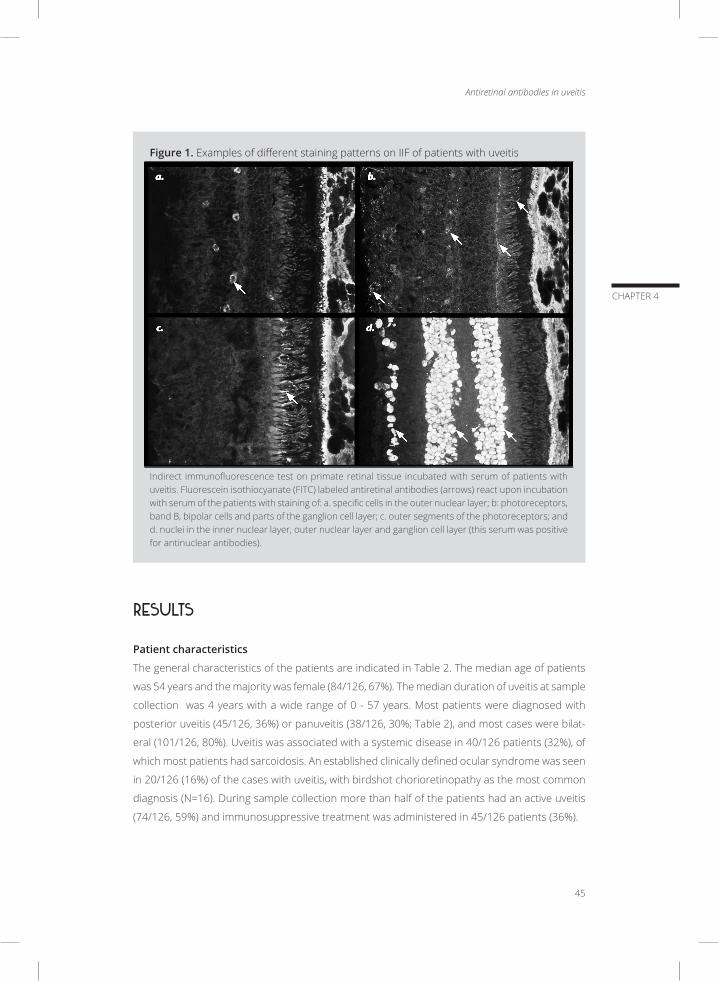
Antiretinal antibodies in uveitis
45
CHAPTER 4
Figure 1. Examples of different staining patterns on IIF of patients with uveitis
Indirect immunofluorescence test on primate retinal tissue incubated with serum of patients with uveitis. Fluorescein isothiocyanate (FITC) labeled antiretinal antibodies (arrows) react upon incubation with serum of the patients with staining of: a. specific cells in the outer nuclear layer; b: photoreceptors, band B, bipolar cells and parts of the ganglion cell layer; c. outer segments of the photoreceptors; and d. nuclei in the inner nuclear layer, outer nuclear layer and ganglion cell layer (this serum was positive for antinuclear antibodies).
RESULTS
Patient characteristics
The general characteristics of the patients are indicated in Table 2. The median age of patients
was 54 years and the majority was female (84/126, 67%). The median duration of uveitis at sample
collection was 4 years with a wide range of 0 - 57 years. Most patients were diagnosed with
posterior uveitis (45/126, 36%) or panuveitis (38/126, 30%; Table 2), and most cases were bilat-
eral (101/126, 80%). Uveitis was associated with a systemic disease in 40/126 patients (32%), of
which most patients had sarcoidosis. An established clinically defined ocular syndrome was seen
in 20/126 (16%) of the cases with uveitis, with birdshot chorioretinopathy as the most common
diagnosis (N=16). During sample collection more than half of the patients had an active uveitis
(74/126, 59%) and immunosuppressive treatment was administered in 45/126 patients (36%).
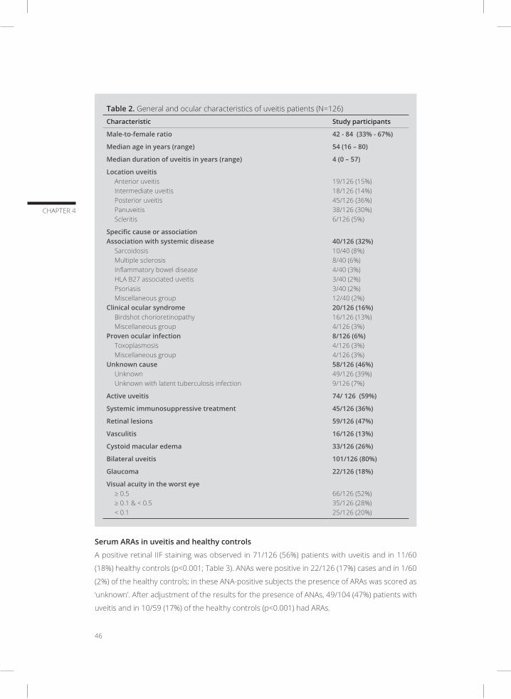
46
CHAPTER 4
Table 2. General and ocular characteristics of uveitis patients (N=126)
Characteristic Study participants
Male-to-female ratio 42 - 84 (33% - 67%)
Median age in years (range) 54 (16 – 80)
Median duration of uveitis in years (range) 4 (0 – 57)
Location uveitis Anterior uveitisIntermediate uveitisPosterior uveitisPanuveitisScleritis
19/126 (15%)18/126 (14%)45/126 (36%)38/126 (30%)6/126 (5%)
Specific cause or associationAssociation with systemic disease
SarcoidosisMultiple sclerosisInflammatory bowel diseaseHLA B27 associated uveitisPsoriasisMiscellaneous group
Clinical ocular syndromeBirdshot chorioretinopathyMiscellaneous group
Proven ocular infectionToxoplasmosisMiscellaneous group
Unknown causeUnknown Unknown with latent tuberculosis infection
40/126 (32%)10/40 (8%)8/40 (6%)4/40 (3%)3/40 (2%)3/40 (2%)12/40 (2%)20/126 (16%)16/126 (13%)4/126 (3%)8/126 (6%)4/126 (3%)4/126 (3%)58/126 (46%)49/126 (39%)9/126 (7%)
Active uveitis 74/ 126 (59%)
Systemic immunosuppressive treatment 45/126 (36%)
Retinal lesions 59/126 (47%)
Vasculitis 16/126 (13%)
Cystoid macular edema 33/126 (26%)
Bilateral uveitis 101/126 (80%)
Glaucoma 22/126 (18%)
Visual acuity in the worst eye≥ 0.5≥ 0.1 & < 0.5< 0.1
66/126 (52%)35/126 (28%)25/126 (20%)
Serum ARAs in uveitis and healthy controls
A positive retinal IIF staining was observed in 71/126 (56%) patients with uveitis and in 11/60
(18%) healthy controls (p<0.001; Table 3). ANAs were positive in 22/126 (17%) cases and in 1/60
(2%) of the healthy controls; in these ANA-positive subjects the presence of ARAs was scored as
‘unknown’. After adjustment of the results for the presence of ANAs, 49/104 (47%) patients with
uveitis and in 10/59 (17%) of the healthy controls (p<0.001) had ARAs.
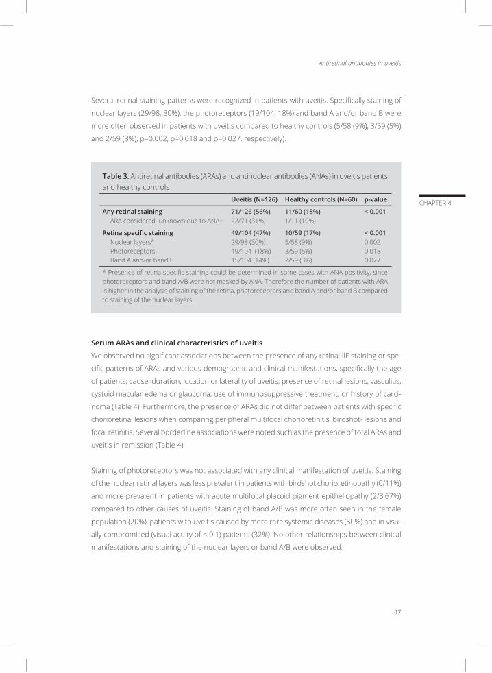
Antiretinal antibodies in uveitis
47
CHAPTER 4
Several retinal staining patterns were recognized in patients with uveitis. Specifically staining of
nuclear layers (29/98, 30%), the photoreceptors (19/104, 18%) and band A and/or band B were
more often observed in patients with uveitis compared to healthy controls (5/58 (9%), 3/59 (5%)
and 2/59 (3%); p=0.002, p=0.018 and p=0.027, respectively).
Table 3. Antiretinal antibodies (ARAs) and antinuclear antibodies (ANAs) in uveitis patients and healthy controls
Uveitis (N=126) Healthy controls (N=60) p-value
Any retinal stainingARA considered unknown due to ANA+
71/126 (56%)22/71 (31%)
11/60 (18%)1/11 (10%)
< 0.001
Retina specific stainingNuclear layers*PhotoreceptorsBand A and/or band B
49/104 (47%)29/98 (30%)19/104 (18%)15/104 (14%)
10/59 (17%)5/58 (9%)3/59 (5%)2/59 (3%)
< 0.0010.0020.0180.027
* Presence of retina specific staining could be determined in some cases with ANA positivity, since photoreceptors and band A/B were not masked by ANA. Therefore the number of patients with ARA is higher in the analysis of staining of the retina, photoreceptors and band A and/or band B compared to staining of the nuclear layers.
Serum ARAs and clinical characteristics of uveitis
We observed no significant associations between the presence of any retinal IIF staining or spe-
cific patterns of ARAs and various demographic and clinical manifestations, specifically the age
of patients; cause, duration, location or laterality of uveitis; presence of retinal lesions, vasculitis,
cystoid macular edema or glaucoma; use of immunosuppressive treatment; or history of carci-
noma (Table 4). Furthermore, the presence of ARAs did not differ between patients with specific
chorioretinal lesions when comparing peripheral multifocal chorioretinitis, birdshot- lesions and
focal retinitis. Several borderline associations were noted such as the presence of total ARAs and
uveitis in remission (Table 4).
Staining of photoreceptors was not associated with any clinical manifestation of uveitis. Staining
of the nuclear retinal layers was less prevalent in patients with birdshot chorioretinopathy (0/11%)
and more prevalent in patients with acute multifocal placoid pigment epitheliopathy (2/3,67%)
compared to other causes of uveitis. Staining of band A/B was more often seen in the female
population (20%), patients with uveitis caused by more rare systemic diseases (50%) and in visu-
ally compromised (visual acuity of < 0.1) patients (32%). No other relationships between clinical
manifestations and staining of the nuclear layers or band A/B were observed.

48
CHAPTER 4
Tabl
e 4.
Pre
senc
e of
ant
iretin
al a
ntib
odie
s (A
RAs)
and
sta
inin
g pa
tter
ns o
f ARA
s on
indi
rect
imm
unofl
uore
scen
ce (a
djus
ted
for p
rese
nce
of a
ntin
ucle
ar
antib
odie
s) in
rela
tion
to c
linic
al c
hara
cter
istic
s in
pat
ient
s w
ith u
veiti
s
Pres
ence
of A
RAs
in
uvei
tis
Stai
ning
pat
tern
s of
ARA
s in
uve
itis
**
Nuc
lear
laye
rs*
Phot
o-re
cept
ors
Band
-A/-
B
Tota
l49
/104
(47%
)29
/98
(30%
)19
/104
(18%
)15
/104
(14%
)
Gen
der
Mal
eFe
mal
e
p>0.
0514
/38
(37%
)35
/66
(53%
)
p>0.
057/
36 (1
9%)
22/6
2 (3
6%)
p>0.
055/
38 (1
3%)
14/6
6 (2
1%)
p=0.
044
2/38
(5%
)13
/66
(20%
)
Age
p>0.
05; n
/ap>
0.05
; n/a
p>0.
05; n
/ap>
0.05
; n/a
Dur
atio
n uv
eiti
sp>
0.05
; n/a
p>0.
05; n
/ap>
0.05
; n/a
p>0.
05; n
/a
Loca
tion
uve
itis
Ante
rior u
veiti
sIn
term
edia
te u
veiti
sPo
ster
ior u
veiti
sPa
nuve
itis
Scle
ritis
All p
>0.0
58/
16 (5
0%)
8/16
(50%
)20
/38
(53%
)11
/30
(37%
)2/
4 (5
0%)
All p
>0.0
54/
16 (2
5%)
5/15
(33%
)11
/36
(31%
)7/
27 (2
6%)
2/4
(50%
)
All p
>0.0
54/
16 (2
5%)
3/16
(18%
)5/
38 (1
3%)
7/30
(23%
)0/
4 (0
%)
All p
>0.0
53/
16 (1
9%)
3/16
(19%
)4/
38 (1
1%)
5/30
(17%
)0/
4 (0
%)
Etio
logy
of u
veit
is ǂ
Asso
ciat
ion
with
sys
tem
ic d
isea
seCl
inic
al o
cula
r syn
drom
ePr
oven
ocu
lar i
nfec
tion
Unk
now
n ca
use
All p
>0.0
514
/32
(44%
)8/
16 (5
0%)
3/7
(43%
)24
/49
(49%
)
All p
>0.0
58/
29 (2
8%)
2/15
(13%
)2/
7 (2
9%)
17/4
7 (3
6%)
All p
>0.0
57/
32 (2
2%)
4/16
(25%
)1/
7 (1
4%)
7/49
(14%
)
All p
>0.0
55/
32 (1
6%)
2/16
(13%
)1/
7 (1
4%)
7/49
(14%
)
Acti
vity
uve
itis
Activ
eRe
mis
sion
p=0.
022
23/6
1 (3
8%)
26/4
3 (6
1%)
p>0.
0514
/59
(24%
)15
/39
(39%
)
p>0.
0510
/61
(16%
)9/
43 (2
1%)
p>0.
059/
61 (1
5%)
6/43
(14%
)
Syst
emic
imm
uno-
supp
ress
ive
trea
tmen
tYe
sN
o
p>0.
0517
/37
(46%
)32
/67
(48%
)
p>0.
057/
32 (2
2%)
22/6
6 (3
3%)
p>0.
058/
37(2
2%)
11/6
7 (1
6%)
p>0.
058/
37 (2
2%)
7/67
(10%
)
Reti
nal l
esio
nsYe
sN
o
p>0.
0525
/49
(51%
)24
/55
(44%
)
p>0.
0516
/47
(34%
)13
/51
(26%
)
p>0.
058/
49 (1
6%)
11/5
5 (2
0%)
p>0.
056/
49 (1
2%)
9/55
(16%
)
Vasc
ulit
is
Yes
N
o
p>0.
058/
15 (5
3%)
41/8
9 (4
6%)
p>0.
054/
13 (3
1%)
25/8
5 (2
9%)
p>0.
055/
15 (3
3%)
14/8
9 (1
6%)
p>0.
054/
15 (2
7%)
11/8
9 (1
2%)
Cyst
oid
mac
ular
ede
ma
Yes
No
p>0.
058/
24 (3
3%)
41/8
0 (5
1%)
p>0.
057/
24 (2
9%)
22/7
4 (3
0%)
p>0.
051/
24 (4
%)
18/8
0 (2
3%)
p>0.
052/
24 (8
%)
13/8
0 (1
6%)
Late
ralit
y uv
eiti
sU
nila
tera
l uve
itis
Bila
tera
l uve
itis
p>0.
057/
22 (3
2%)
42/8
2 (5
1%)
p>0.
055/
22 (2
3%)
24/7
6 (3
2%)
p>0.
052/
22 (9
%)
17/8
2 (2
1%)
p>0.
0513
/82
(16%
)2/
22 (9
%)
Gla
ucom
a
Yes
N
o
p>0.
057/
18 (3
9%)
42/8
6 (4
9%)
p>0.
054/
18 (2
2%)
25/8
0 (3
1%)
p>0.
053/
18 (1
7%)
16/8
6 (1
9%)
p>0.
052/
18 (1
1%)
13/8
6 (1
5%)
His
tory
of c
arci
nom
aYe
sN
o
p>0.
057/
15 (4
7%)
42/8
9 (4
7%)
p>0.
052/
13 (1
5%)
27/8
5 (3
2%)
p>0.
053/
15 (2
0%)
16/8
9 (1
8%)
p>0.
052/
15 (1
3%)
13/8
9 (1
5%)
Visu
al a
cuit
y in
the
wor
st e
ye≥
0.5
≥ 0.
1 &
< 0
.5<
0.1
p>0.
05; 2
7/55
(49%
)p=
0.03
4; 8
/27
(30%
)p>
0.05
; 14/
22 (6
4%)
All p
>0.0
517
/53
(32%
)4/
26 (1
5%)
8/19
(42%
)
All p
>0.0
510
/55
(18%
)4/
27 (1
5%)
5/22
(23%
)
p>0.
05; 6
/55
(11%
) p>
0.0
5; 2
/27
(7%
) p=
0.01
6; 7
/22
(32%
)
* Pr
esen
ce o
f ret
ina
spec
ific
stai
ning
cou
ld b
e de
term
ined
in s
ome
case
s w
ith A
NA
posi
tivity
, sin
ce r
etin
al A
NA
stai
ning
did
not
mas
k ph
otor
ecep
tors
and
/or
band
A/B
. Th
eref
ore
the
num
ber
of p
atie
nts
with
ARA
is h
ighe
r in
the
anal
ysis
of s
tain
ing
of th
e re
tina,
pho
tore
cept
ors
and
band
A a
nd/o
r ba
nd B
com
pare
d to
ret
inal
sta
inin
g of
th
e nu
clea
r lay
ers.
** S
ome
stai
ning
pat
tern
wer
e pr
esen
t in
com
bina
tions
ǂ Su
b-an
alys
is o
f spe
cific
etio
logi
c en
titie
s w
ithin
the
indi
cate
d gr
oups
did
not
sho
w a
ny a
dditi
onal
pos
itive
cor
rela
tions

Antiretinal antibodies in uveitis
49
CHAPTER 4
Tabl
e 4.
Pre
senc
e of
ant
iretin
al a
ntib
odie
s (A
RAs)
and
sta
inin
g pa
tter
ns o
f ARA
s on
indi
rect
imm
unofl
uore
scen
ce (a
djus
ted
for p
rese
nce
of a
ntin
ucle
ar
antib
odie
s) in
rela
tion
to c
linic
al c
hara
cter
istic
s in
pat
ient
s w
ith u
veiti
s
Pres
ence
of A
RAs
in
uvei
tis
Stai
ning
pat
tern
s of
ARA
s in
uve
itis
**
Nuc
lear
laye
rs*
Phot
o-re
cept
ors
Band
-A/-
B
Tota
l49
/104
(47%
)29
/98
(30%
)19
/104
(18%
)15
/104
(14%
)
Gen
der
Mal
eFe
mal
e
p>0.
0514
/38
(37%
)35
/66
(53%
)
p>0.
057/
36 (1
9%)
22/6
2 (3
6%)
p>0.
055/
38 (1
3%)
14/6
6 (2
1%)
p=0.
044
2/38
(5%
)13
/66
(20%
)
Age
p>0.
05; n
/ap>
0.05
; n/a
p>0.
05; n
/ap>
0.05
; n/a
Dur
atio
n uv
eiti
sp>
0.05
; n/a
p>0.
05; n
/ap>
0.05
; n/a
p>0.
05; n
/a
Loca
tion
uve
itis
Ante
rior u
veiti
sIn
term
edia
te u
veiti
sPo
ster
ior u
veiti
sPa
nuve
itis
Scle
ritis
All p
>0.0
58/
16 (5
0%)
8/16
(50%
)20
/38
(53%
)11
/30
(37%
)2/
4 (5
0%)
All p
>0.0
54/
16 (2
5%)
5/15
(33%
)11
/36
(31%
)7/
27 (2
6%)
2/4
(50%
)
All p
>0.0
54/
16 (2
5%)
3/16
(18%
)5/
38 (1
3%)
7/30
(23%
)0/
4 (0
%)
All p
>0.0
53/
16 (1
9%)
3/16
(19%
)4/
38 (1
1%)
5/30
(17%
)0/
4 (0
%)
Etio
logy
of u
veit
is ǂ
Asso
ciat
ion
with
sys
tem
ic d
isea
seCl
inic
al o
cula
r syn
drom
ePr
oven
ocu
lar i
nfec
tion
Unk
now
n ca
use
All p
>0.0
514
/32
(44%
)8/
16 (5
0%)
3/7
(43%
)24
/49
(49%
)
All p
>0.0
58/
29 (2
8%)
2/15
(13%
)2/
7 (2
9%)
17/4
7 (3
6%)
All p
>0.0
57/
32 (2
2%)
4/16
(25%
)1/
7 (1
4%)
7/49
(14%
)
All p
>0.0
55/
32 (1
6%)
2/16
(13%
)1/
7 (1
4%)
7/49
(14%
)
Acti
vity
uve
itis
Activ
eRe
mis
sion
p=0.
022
23/6
1 (3
8%)
26/4
3 (6
1%)
p>0.
0514
/59
(24%
)15
/39
(39%
)
p>0.
0510
/61
(16%
)9/
43 (2
1%)
p>0.
059/
61 (1
5%)
6/43
(14%
)
Syst
emic
imm
uno-
supp
ress
ive
trea
tmen
tYe
sN
o
p>0.
0517
/37
(46%
)32
/67
(48%
)
p>0.
057/
32 (2
2%)
22/6
6 (3
3%)
p>0.
058/
37(2
2%)
11/6
7 (1
6%)
p>0.
058/
37 (2
2%)
7/67
(10%
)
Reti
nal l
esio
nsYe
sN
o
p>0.
0525
/49
(51%
)24
/55
(44%
)
p>0.
0516
/47
(34%
)13
/51
(26%
)
p>0.
058/
49 (1
6%)
11/5
5 (2
0%)
p>0.
056/
49 (1
2%)
9/55
(16%
)
Vasc
ulit
is
Yes
N
o
p>0.
058/
15 (5
3%)
41/8
9 (4
6%)
p>0.
054/
13 (3
1%)
25/8
5 (2
9%)
p>0.
055/
15 (3
3%)
14/8
9 (1
6%)
p>0.
054/
15 (2
7%)
11/8
9 (1
2%)
Cyst
oid
mac
ular
ede
ma
Yes
No
p>0.
058/
24 (3
3%)
41/8
0 (5
1%)
p>0.
057/
24 (2
9%)
22/7
4 (3
0%)
p>0.
051/
24 (4
%)
18/8
0 (2
3%)
p>0.
052/
24 (8
%)
13/8
0 (1
6%)
Late
ralit
y uv
eiti
sU
nila
tera
l uve
itis
Bila
tera
l uve
itis
p>0.
057/
22 (3
2%)
42/8
2 (5
1%)
p>0.
055/
22 (2
3%)
24/7
6 (3
2%)
p>0.
052/
22 (9
%)
17/8
2 (2
1%)
p>0.
0513
/82
(16%
)2/
22 (9
%)
Gla
ucom
a
Yes
N
o
p>0.
057/
18 (3
9%)
42/8
6 (4
9%)
p>0.
054/
18 (2
2%)
25/8
0 (3
1%)
p>0.
053/
18 (1
7%)
16/8
6 (1
9%)
p>0.
052/
18 (1
1%)
13/8
6 (1
5%)
His
tory
of c
arci
nom
aYe
sN
o
p>0.
057/
15 (4
7%)
42/8
9 (4
7%)
p>0.
052/
13 (1
5%)
27/8
5 (3
2%)
p>0.
053/
15 (2
0%)
16/8
9 (1
8%)
p>0.
052/
15 (1
3%)
13/8
9 (1
5%)
Visu
al a
cuit
y in
the
wor
st e
ye≥
0.5
≥ 0.
1 &
< 0
.5<
0.1
p>0.
05; 2
7/55
(49%
)p=
0.03
4; 8
/27
(30%
)p>
0.05
; 14/
22 (6
4%)
All p
>0.0
517
/53
(32%
)4/
26 (1
5%)
8/19
(42%
)
All p
>0.0
510
/55
(18%
)4/
27 (1
5%)
5/22
(23%
)
p>0.
05; 6
/55
(11%
) p>
0.0
5; 2
/27
(7%
) p=
0.01
6; 7
/22
(32%
)
* Pr
esen
ce o
f ret
ina
spec
ific
stai
ning
cou
ld b
e de
term
ined
in s
ome
case
s w
ith A
NA
posi
tivity
, sin
ce r
etin
al A
NA
stai
ning
did
not
mas
k ph
otor
ecep
tors
and
/or
band
A/B
. Th
eref
ore
the
num
ber
of p
atie
nts
with
ARA
is h
ighe
r in
the
anal
ysis
of s
tain
ing
of th
e re
tina,
pho
tore
cept
ors
and
band
A a
nd/o
r ba
nd B
com
pare
d to
ret
inal
sta
inin
g of
th
e nu
clea
r lay
ers.
** S
ome
stai
ning
pat
tern
wer
e pr
esen
t in
com
bina
tions
ǂ Su
b-an
alys
is o
f spe
cific
etio
logi
c en
titie
s w
ithin
the
indi
cate
d gr
oups
did
not
sho
w a
ny a
dditi
onal
pos
itive
cor
rela
tions
50
CHAPTER 4
DISCUSSION
Our results show that serum ocular antibodies, specifically ARAs, are more prevalent in patients
with uveitis as compared to healthy controls (47% versus 17%, p<0.001). However, specific associ-
ations between the presence of serum ARAs (or typical staining patterns on IIF) and clinical ocular
characteristics were not observed so far, except for the association between patients with a visual
acuity of less than 0.1 and the presence of positive staining of band A/B (p=0.013). Furthermore,
some marginal associations were noted, though this probably a coincidence occurring due to
multiple comparisons in a small cohort (in perspective to number of tested variables). Our study
includes a heterogeneous population of uveitis patients in which major groups of etiology are rep-
resented. Due to the limited number of patients in specific subgroups, it cannot be fully excluded
that some associations were missed and that in a larger population of patients with specific
uveitis entities the associations between clinical manifestations and serum ARA’s could be found.
The involvement of autoimmunity in uveitis has repeatedly been implicated and the term autoim-
mune uveitis was sometimes even used as a synonym for all non-infectious types of uveitis. The
proposed autoimmune pathogenesis is often being explained by mimicry between the ocular
and other infectious or non-infectious antigens, but actual autoimmune processes in uveitis have
seldom been proved. The autoimmune reactions against S-antigen, the best-known uveitogenic
antigen, were examined and found to be involved in the pathogenesis of infectious uveitis.5,10,11
In several animal models with monkeys and rats, vaccination with retinal S antigen induced an
ocular disease similar to human uveoretinitis.17-19
The results of our study on the prevalence of serum ARAs in uveitis are in line with previous
reports. Cohorts with different specific entities of uveitis have been analyzed for the presence
of ARAs in serum, including cohorts of patients with juvenile associated uveitis, idiopathic retinal
vasculitis, toxoplasmosis, onchocherciasis, or a variation of noninfectious or idiopathic posterior
uveitis (Table 1). The presence of any ARA in serum was noted in approximately 70% in patients
with retinal vasculitis and up to 100% in patients with toxoplasmosis.5,10,12 Since the aforemen-
tioned studies usually lacked clinical data and information on laboratory techniques the exact
comparison with previous results is not possible. In our series, we have chosen to use (commer-
cially available) primate retinal tissue to investigate the presence of ARAs by IIF. Primate tissue
from an evolutionary perspective closely resembles human tissue and is also used for diagnostic
purposes. Furthermore, in human retinal tissue certain antigens may induce higher background
staining, including blood group-associated antigens. In addition, the use of commercially available
tissues may facilitate future comparisons of results.
The presence of diverse patterns on IIF indicates that different ARAs are produced by individual
patients. However, IIF does not allow the precise identification of ARAs, i.e. their antigen speci-
Antiretinal antibodies in uveitis
51
CHAPTER 4
ficity. To clarify the type and complexity of the ARAs detected in our study, specific IIF patterns
must be correlated to specific retinal antigen reactivity. In the past, patterns of specific ARAs
have been determined, however, IIF showed different patterns in various studies even with the
use of purified antibodies.20-23 The cause of this inconsistency could be explained by the use of
variable tissues and reagents and the recognition of different epitopes. More useful techniques
for confirmation of specific type of ARA and determination of multiple ARAs consist of Western
Blot and/or ELISA. 3,24
The higher prevalence of serum ARAs in uveitis remains unexplained as well as their possible clin-
ical role. Hypothetically, the presence of serum ARA’s (and absence of their clinical impact) might
be due to local tissue damage, leakage of (altered) retinal antigens into the circulation and subse-
quent reaction of the immune system and production of diverse serum ARAs. This implicates that
the production of serum ARAs might develop due to the general response of the immune system
to ocular tissue damage. The systemic production of coincidental ARAs might be responsible for
the lack of associations between the serum ARAs and ocular clinical manifestations in parallel to
the presence of multiple autoantibodies, which are commonly observed in systemic autoimmune
disorders and sometimes in healthy elderly population. In addition, the absence of ARAs in a
subset of uveitis patients indicates that probably not antibodies but cell-mediated autoimmune
reactions might play a primary role in the development of presumed autoimmunity in uveitis.
Further, the levels of antibodies in the serum reflect their total systemic production and are
probably not influenced by the additional production of specific antibodies within the eye. This
phenomenon has also been observed in the diagnosis of infectious uveitis in which local pro-
duction of specific antibodies within the eye rather than serum levels is regarded as a proof of
the ocular infection (determined with a Goldmann-Witmer coefficient.25 The importance of locally
produced antibodies has also been shown in cerebrospinal fluid for the central nervous system,
another immune privileged organ.26 The local production of ARAs might show an entirely differ-
ent pattern and clinical importance than ARAs found in the peripheral circulation and has - to
our knowledge - so far not been examined. Further research assessing the intraocular levels of
specific ARAs might elucidate the clinical relevance of autoimmune processes directed against
the retinal tissue in uveitis.
Serum ARAs were observed in the majority of patients with uveitis and were more frequent
compared to healthy controls. The cause of enhanced serum ARA production in patients with
uveitis is currently not known. Specific associations between the presence of serum ARAs (or
typical staining patterns on IIF) and clinical ocular characteristics were not identified. The role
of presumed autoimmune reactions in uveitis could be further investigated by assessment of
intraocular fluids within the eye instead of peripheral blood samples.

52
CHAPTER 4
ACKNOWLEDGEMENTS
We would like to thank Dr. G. Adamus of Oregon Health and Science University, U.S.A for con-
tributing antibody samples for research and support.
Antiretinal antibodies in uveitis
53
CHAPTER 4
REFERENCES
1. Braithwaite T, Vugler A, Tufail A. Autoimmune retinopathy. Ophthalmologica. 2012;228(3):131-142.
2. Grange L, Dalal M, Nussenblatt RB, Sen HN. Autoimmune retinopathy. Am J Ophthalmol. 2014;157(2):266-
272 e261.
3. Adamus G, Ren G, Weleber RG. Autoantibodies against retinal proteins in paraneoplastic and autoimmune
retinopathy. BMC Ophthalmol. 2004;4:5.
4. Walscheid K, Hennig M, Heinz C, et al. Correlation between disease severity and presence of ocular autoan-
tibodies in juvenile idiopathic arthritis-associated uveitis. Invest Ophthalmol Vis Sci. 2014;55(6):3447-3453.
5. Cursino SR, Costa TB, Yamamoto JH, Meireles LR, Silva MA, Andrade Junior HF. Increased frequency
of anti-retina antibodies in asymptomatic patients with chronic t. gondii infection. Clinics (Sao Paulo).
2010;65(10):1027-1032.
6. Kubicka-Trzaska A. Poziom przeciwcial przeciwsiatkowkowych w surowicy a nasilenie objawow endogen-
nego zapalenia tylnego odcinka blony naczyniowej. Klin Oczna. 2002;104(3-4):231-234.
7. Kubicka-Trzaska A. Typy przeciwcial przeciwsiatkowkowych (PPS) u chorych na samoistne zapalenie
tylnego odcinka blony naczyniowej w tescie immunofluorescencji posredniej. Klin Oczna. 2004;106(1-
2):45-49.
8. Chan CC, Palestine AG, Nussenblatt RB, Roberge FG, Benezra D. Anti-retinal auto-antibodies in
Vogt-Koyanagi-Harada syndrome, Behcet’s disease, and sympathetic ophthalmia. Ophthalmology.
1985;92(8):1025-1028.
9. Forrester JV, Stott DI, Hercus KM. Naturally occurring antibodies to bovine and human retinal S antigen:
a comparison between uveitis patients and healthy volunteers. Br J Ophthalmol. 1989;73(2):155-159.
10. Whittle RM, Wallace GR, Whiston RA, Dumonde DC, Stanford MR. Human antiretinal antibodies in toxo-
plasma retinochoroiditis. Br J Ophthalmol. 1998;82(9):1017-1021.
11. Van der Lelij A, Doekes G, Hwan BS, et al. Humoral autoimmune response against S-antigen and IRBP in
ocular onchocerciasis. Invest Ophthalmol Vis Sci. 1990;31(7):1374-1380.
12. Stanford MR, Graham E, Kasp E, Sanders MD, Dumonde DC. A longitudinal study of clinical and immuno-
logical findings in 52 patients with relapsing retinal vasculitis. Br J Ophthalmol. 1988;72(6):442-447.
13. Shimazaki K, Jirawuthiworavong GV, Heckenlively JR, Gordon LK. Frequency of anti-retinal antibodies in
normal human serum. J Neuro-Ophthalmol. 2008;28(1):5-11.
14. Kubicka-Trzaska A, Wilanska J, Romanowska-Dixon B, Sanak M. Circulating anti-retinal antibodies in
response to anti-angiogenic therapy in exudative age-related macular degeneration. Acta Ophthalmol.
2014;92(8):e610-614.
15. Jabs DA, Nussenblatt RB, Rosenbaum JT, Standardization of Uveitis Nomenclature Working G. Standard-
ization of uveitis nomenclature for reporting clinical data. Results of the First International Workshop. Am
J Ophthalmol. 2005;140(3):509-516.
16. Trusko B, Thorne J, Jabs D, et al. The Standardization of Uveitis Nomenclature (SUN) Project. Development
of a clinical evidence base utilizing informatics tools and techniques. Methods Inf Med. 2013;52(3):259-
265, S251-256.
17. LeHoang P, Sterkers M, Thillaye B, de Kozak Y, Coscas G, Faure JP. Primate model of uveoretinitis and
vasculitis/experimental autoimmune uveoretinitis induced in cynomolgus monkeys by retinal S antigen.
Ophthal Res. 2008;40(3-4):181-188.
18. Roberts AJ, Kasp E, Stanford M, Dumonde DC, Banga JP. Induction of Experimental Autoimmune Uveoret-
initis in Lewis Rats with Purified Recombinant Human Retinal S-Antigen Fusion Protein. European Journal
of Immunology. 1992;22(4):951-956.
54
CHAPTER 4
19. Faure JP, Phuc LH, Takano S, Sterkers M, Thillaye B, Dekozak Y. Experimental Uveoretinitis Induced in
Monkeys by Retinal S-Antigen - Induction, Histopathology. J Fr Ophtalmol. 1981;4(6-7):465-472.
20. Adamus G, Aptsiauri N, Guy J, Heckenlively J, Flannery J, Hargrave PA. The occurrence of serum autoanti-
bodies against enolase in cancer-associated retinopathy. Clin Immunol Immunop. 1996;78(2):120-129.
21. Ren GY, Adamus G. Cellular targets of anti-alpha-enolase autoantibodies of patients with autoimmune
retinopathy. Journal of Autoimmunity. 2004;23(2):161-167.
22. Whitcup SM, Vistica BP, Milam AH, Nussenblatt RB, Gery I. Recoverin-associated retinopathy: A clinically
and immunologically distinctive disease. Am J Ophthalmol. 1998;126(2):230-237.
23. Keltner JL, Thirkill CE. Cancer-associated retinopathy vs recoverin-associated retinopathy. Am J Ophthal-
mol. 1998;126(2):296-302.
24. Adamus G. Autoantibody targets and their cancer relationship in the pathogenicity of paraneoplastic
retinopathy. Autoimmun Rev. 2009;8(5):410-414.
25. Rothova A, de Boer JH, Ten Dam-van Loon NH, et al. Usefulness of aqueous humor analysis for the diag-
nosis of posterior uveitis. Ophthalmology. 2008;115(2):306-311.
26. Dalmau J, Lancaster E, Martinez-Hernandez E, Rosenfeld MR, Balice-Gordon R. Clinical experience and
laboratory investigations in patients with anti-NMDAR encephalitis. Lancet Neurol. 2011;10(1):63-74.


SERUM AUTOANTIBODY PROFILING OF PATIENTS WITH PARANEOPLASTIC AND NON-PARANEOPLASTIC AUTOIMMUNE RETINOPATHY
Josianne C.E.M. ten Berge, Joost van Rosmalen, Jacolien Vermeer, Cecilia Hellström, Cecilia Lindskog, Peter Nilsson, Ulrika Qundos, Aniki Rothova, Marco W.J. Schreurs
PLoS One. 2016 Dec;11(12):e0167909
5
58
CHAPTER 5
ABSTRACT
Purpose: Although multiple serum antiretinal autoantibodies (ARAs) have been reported in
patients with paraneoplastic and non-paraneoplastic autoimmune retinopathy ((n)pAIR), not all
retinal antigens involved in (n)pAIR are specified. This study aims to serologically identify patients
with presumed (n)pAIR through determination of both known and unknown ARAs by autoanti-
body profiling.
Methods: An antigen suspension bead array using 188 different antigens representing 97 ocular
proteins was performed to detect ARAs in serum samples of patients with presumed (n)pAIR
(N=24), uveitis (N=151) and cataract (N=21). Logistic regressions were used to estimate the
associations between ocular antigens and diagnosis. Validation of interphotoreceptor matrix
proteoglycan 2 (IMPG2) and recoverin antigens was performed by immunohistochemistry and
immunoblot, respectively.
Results: Samples of patients with presumed (n)pAIR exhibited a broad spectrum of ARAs. We
identified retinal antigens that have already been described previously (e.g. recoverin), but also
identified novel ARA targets. Most ARAs were not specific for (n)pAIR since their presence was
also observed in patients with cataract or uveitis. High titers of autoantibodies directed against
photoreceptor-specific nuclear receptor and retinol-binding protein 3 were more common in
patients with presumed (n)pAIR compared to uveitis (p=0.015 and p=0.018, respectively). The
presence of all other ARAs did not significantly differ between groups. In patients with presumed
(n)pAIR, anti-recoverin autoantibodies were the most prevalent ARAs. Validation of bead array
results by immunohistochemistry (anti-IMPG2) and immunoblot (anti-recoverin) showed concor-
dant results in (n)pAIR patients.
Conclusions: Patients with (n)pAIR are characterized by the presence of a broad spectrum of
ARAs. The diagnosis of (n)pAIR cannot be based on the mere presence of serum ARAs, as these
are also commonly present in uveitis as well as in age-related cataract patients.
Autoantibody profiling of patients with autoimmune retinopathy
59
CHAPTER 5
INTRODUCTION
Paraneoplastic and non-paraneoplastic autoimmune retinopathy ((n)pAIR) is a rare blinding reti-
nal disorder of unknown pathogenesis. It is presumed that antiretinal autoantibodies (ARAs) are
involved in the pathogenesis of (n)pAIR and damage ocular tissue causing poor visual outcome.
Symptoms associated with (n)pAIR are progressive visual loss (most often bilateral), visual field
loss frequently associated with a ring scotoma or loss of the peripheral field, and decreased
amplitudes on electroretinogram (ERG).1-4
Paraneoplastic autoimmune retinopathy (pAIR) includes two subgroups: cancer associated ret-
inopathy (CAR) and melanoma associated retinopathy (MAR). In pAIR the presence of the same
auto-antigens in both retinal tissue and malignant tissue has previously been described (e.g.
recoverin).5-7 The presence of ARAs however is not conclusive for the diagnosis of (n)pAIR, since
several ARAs were also reported in patients with other ocular disorders and individuals without
ocular disease.8 Nevertheless, ARAs are considered to support the diagnosis of (n)pAIR, which is
often difficult to confirm by clinical symptoms only.9
Multiple serum ARAs have regularly been reported in affected patients (Table 1), although not
all retinal autoantibodies involved in the pathogenesis of (n)pAIR are known and information
regarding their exact pathological roles is lacking.10 Further, a gold standard for the determination
of ARAs is missing.11-13 The optimal approach for the determination and specification of ARAs is
currently unknown. Different techniques, including indirect immunofluorescence, western blot
and enzyme-linked immunosorbent assay (ELISA), have been used for the detection of ARAs;
however, results and conclusions differ and cannot be reliably compared.
Currently, antigen bead arrays are being used to profile autoantibody reactivity in body fluids.38
With this technique, very small volumes of body fluids can be tested for IgG reactivity across hun-
dreds of samples towards hundreds of different antigens. This technique has already successfully
been used for the analysis of autoantibodies in serum and cerebrospinal fluid.39-41
Our study aimed to serologically identify patients with presumed (n)pAIR through determination
of ARAs. For this purpose, we used a bead array-based multiplex assay for autoantibody profiling
using 188 ocular antigens representing 97 different retinal proteins.
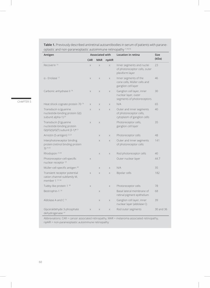
60
CHAPTER 5
Table 1. Previously described antiretinal autoantibodies in serum of patients with parane-oplastic and non-paraneoplastic autoimmune retinopathy 1,14,15
Antigen Associated with Location in retina Size (kDa)CAR MAR npAIR
Recoverin 16 x x x Inner segments and nuclei of photoreceptor cells, outer plexiform layer
23
α - Enolase 17 x x x Inner segments of the cone cells, Müller cells and ganglion cell layer
46
Carbonic anhydrase II 18 x x x Ganglion cell layer, inner nuclear layer, outer segments of photoreceptors
30
Heat shock cognate protein 70 19 x x x N/A 65
Transducin α (guanine nucleotide-binding protein G(t) subunit alpha-1) 20
x x x Outer and inner segments of photoreceptor cells, cytoplasm of ganglion cells
40
Transducin β (guanine nucleotide-binding protein G(I)/G(S)/G(T) subunit β-1)* 21
x x Photoreceptor cells, ganglion cell layer
35
Arrestin (S-antigen) 22,23 x x Photoreceptor cells 48
Interphotoreceptor binding protein (retinol binding protein 3) 24-26
x x Outer and inner segments of photoreceptor cells
141
Rhodopsin 27,28 x x Rod photoreceptor cells 40
Photoreceptor-cell-specific nuclear receptor 29
x Outer nuclear layer 44.7
Müller-cell-specific antigen 30 x x N/A 35
Transient receptor potential cation channel subfamily M, member 1 31-34
x x x Bipolar cells 182
Tubby-like protein 1 35 x x Photoreceptor cells 78
Bestrophin-1 36 x Basal lateral membrane of retinal pigment epithelium
68
Aldolase A and C 15 x x Ganglion cell layer, inner nuclear layer (aldolase C)
39
Glyceraldehyde 3-phosphate dehydrogenase 37
x x x Rod outer segments 30 and 36
Abbreviations: CAR = cancer associated retinopathy, MAR = melanoma associated retinopathy, npAIR = non-paraneoplastic autoimmune retinopathy
Autoantibody profiling of patients with autoimmune retinopathy
61
CHAPTER 5
METHODS
Sample collection and patient selection
Serum samples were either collected during routine diagnostic analysis for the presence of
anti-recoverin autoantibodies in the Laboratory of Medical Immunology of the Erasmus Univer-
sity Medical Center between April 2013 and August 2015 or were obtained from biobank of our
department. The study was approved by the local ethical committee from the Erasmus University
Medical Center (Medical Ethics Committee Erasmus MC) and adhered to the tenets of the Declara-
tion of Helsinki. The ethical committee decided that no informed consent of patients was required
for the use of the remainder of the diagnostic material, as the samples were anonymized and
the patients were not subjected to additional risk or procedures. Samples which were obtained
from the biobank (for which an approval of the ethical committee was obtained) included signed
informed consent from all participants. All whole blood samples were centrifuged after at least
30’ clotting time at 3,000 rpm for 10 minutes, and serum was stored at -80°C.
According to the recently published report on the nomenclature of (n)pAIR, the general term
autoimmune retinopathy (AIR) is recommended to indicate the non-paraneoplastic autoimmune
retinopathy (npAIR) subtype. In our present series we indicate the specific subtype(s) of AIR (pAIR,
npAIR or (n)pAIR) to prevent any misunderstanding regarding nomenclature.9 The diagnosis of
presumed (n)pAIR was made if the patients fulfilled all of the following inclusion criteria: 1. visual
complaints, 2. markedly decreased amplitudes on ERG, 3. visual field loss, and 4. no alternative
explanation for their ocular disorder. In addition, patients with genetically proven retinitis pig-
mentosa or a family history of retinitis pigmentosa were excluded. A total of 17 patients fulfilled
the criteria indicated above and were included in this study. Patients fulfilling the criteria without
a malignancy were indicated as presumed npAIR (N=9), and patients with a malignancy were
indicated as patients with presumed pAIR (N=8).
An additional group of presumed pAIR (CAR or MAR) patients (N=7) in whom ERG or visual field
tests were not performed (choice of the patient or poor general condition), but who fulfilled all
other inclusion criteria was included separately. An additional required criterion for these patients
comprised the development of a malignancy before or within 3 months after presentation with
ocular problems.
We collected various clinical data of the patients with presumed (n)pAIR, including patient demo-
graphics (age and gender) and ocular characteristics such as complaints of photopsia, complaints
of nyctalopia, subjective or objective problems with colour-vision, unilateral or bilateral visual
problems and the presence of a malignancy in the medical history or during follow-up.

62
CHAPTER 5
Controls consisted of two groups: 21 serum samples from cataract patients without retinal
damage and 151 samples from patients with uveitis of different causes. Patients with age related
cataract were included as controls rather than healthy people, as this disorders does not involve
retina nor exhibits retinal damage, and represents a clinical setting in which the tests might be
employed. Samples of control patients were collected at the department of Ophthalmology of the
Erasmus University Medical Center between February 2009 and April 2015. Patient demographics
(age and gender) and known malignancies of these patients were registered.
Antigen suspension bead array
Autoantibody profiling was performed in all serum samples from patients with presumed (n)
pAIR (N=24), uveitis (N=151) and cataract (N=21). Antigens used for the autoantibody profiling
were selected based on potential relevance to ocular diseases according to literature and pre-
vious positive retinal immunohistochemistry staining, resulting in 188 antigens (human protein
fragments) representing 97 unique proteins. The protein fragments were produced within the
Human Protein Atlas and designed to represent unique parts of each target protein.42,43 Protein
fragments were 20-150 amino acids long (median 78 aa) and produced in Escherichia coli, with
an affinity tag consisting of six histidines and an albumin binding domain from streptococcal
protein G (His6ABP) (Supplementary Table). Immobilization onto color-coded magnetic beads
was conducted as described previously.39 In short, diluted antigens were covalently coupled
to activated carboxy groups on color coded polystyrene beads (MagPlex, Luminex Corp.) by
undirected amine coupling. In addition to the selected protein targets, one bead identity was
used for immobilization of anti-human IgG (positive control), one for Epstein-Barr virus nuclear
antigen 1 (second positive control), one for His6ABP (negative control, to monitor binding to the
affinity tag) and one bead identity went through the coupling process without addition of antigen
(second negative control, to monitor binding to bare beads). After incubation, the coupled beads
were washed and stored in a blocking reagent before combining all bead identities to create a
bead array in suspension. Samples were distributed across 96-well microtiter plates, together
with triplicate aliquots of a sample pool and a buffer blank in each plate for determination of the
intra- and inter-reproducibility. Serum samples were diluted 1:250 in assay buffer before being
mixed with the bead array. Incubation was performed at room temperature for 2 hours followed
by detection of the IgG reactivity by a fluorophore conjugated anti-human IgG Fab fragment and
measured in a FlexMap3D instrument (Luminex Corp.).
Recoverin immunoblot
For validation purposes, samples that tested positive for anti-recoverin autoantibodies on the
antigen bead array, and all samples from patients with presumed (n)pAIR, were analysed on a
recoverin specific immunoblot (Euroimmun AG, Lubeck, Germany). Membrane strips coated with
recombinant human recoverin were incubated with a sample buffer for 5 minutes. After aspiration
of the sample buffer, the membrane strips were incubated with diluted serum samples for 30
Autoantibody profiling of patients with autoimmune retinopathy
63
CHAPTER 5
minutes on a shaking platform. Subsequently membrane strips were washed three times, incu-
bated with secondary antibodies (enzyme conjugated anti-human IgG), washed again for three
times and stained with a substrate solution which was capable of promoting an enzymatic colour
reaction. To identify positive reactions, assessment of visible bands was performed relative to
the included control. Results from the antigen suspension bead array and the recoverin specific
immunoblot were compared.
Immunohistochemistry of interphotoreceptor matrix proteoglycan 2 on human retina tissue
Another method for validation was performed with the antigens of interphotoreceptor matrix
proteoglycan 2 (IMPG2). Polyclonal antibodies affinity purified against the IMPG2 antigens no. 214
and 205 were used as antigens for immunization of rabbits to generate polyclonal antibodies for
immunohistochemistry on normal human tissues, in order to determine retina specificity and cell
type expression. The antibodies were applied on tissue microarrays (TMAs) containing samples
from 45 different human tissues, including retina from two individuals. TMAs from human tissues
were generated essentially as previously described.44 The TMAs contained 1 mm diameter forma-
lin-fixed and paraffin-embedded tissue cores from 45 different histologically normal tissue types,
including two samples of human eye: one male 75 years and one female 54 years. All samples
were received from the Department of Pathology, Uppsala University Hospital, Sweden, approved
by the local Research Ethics Committee (Uppsala, Sweden, Ups 02–577). Four-micrometer sec-
tions were cut from the TMA blocks, mounted on adhesive slides and baked at 60 °C for 45 min.
TMA slides were then deparaffinised in Neo-Clear® (Merck Millipore, Darmstadt, Germany), fol-
lowed by hydration in graded alcohols and blocking for endogenous peroxidase in 0.3 % hydrogen
peroxide. For antigen retrieval, slides were immersed and boiled in Citrate buffer®, pH6 (Lab
Vision, Freemont, CA) for 4 min at 125 °C and then allowed to cool to 90 °C. Automated immu-
nohistochemistry was performed essentially as previously described, using an Autostainer 480
instrument® (Lab Vision).44 Affinity purified polyclonal antibodies towards IMPG2 (HPA008779,
antigen number 205, diluted 1:250 and HPA015907, antigen number 214, diluted 1:2500, both
Atlas Antibodies AB) and a dextran polymer visualization system (UltraVision LP HRP polymer®,
Lab Vision) were incubated for 30 min each at room temperature. Slides were developed for 10
min using Diaminobenzidine (Lab Vision) as chromogen. All incubations were followed by rinse
in Wash buffer® (Lab Vision) for 5 min. The slides were counterstained in Mayers hematoxylin
(Histolab) and cover slipped using Pertex® (Histolab) as mounting medium. Digital whole slide
high-resolution images were captured with a 20× objective using an AperioScanScope XT Slide
Scanner (Aperio Technologies, Vista, CA).
Data analysis
Continuous variables were summarized using medians and ranges, and categorical variables
were summarized using percentages. Patient demographics were compared between diagnosis
groups using Mann Whitney U tests for continuous data and Fisher’s exact tests for categorical
64
CHAPTER 5
data. All data from the antigen suspension bead array were represented as ratios (antigen specific
reactivity over patient background (represented by the His6ABP negative control bead)). A ratio of
>2 was considered positive and a ratio of >25 was considered highly positive for the presence of
ARAs. Logistic regressions with correction for age and gender were performed to analyse differ-
ences between the diagnosis groups ((n)pAIR versus uveitis and (n)pAIR versus cataract) for both
ratio’s. In the logistic regression analyses, confidence intervals of the estimated odds ratios were
calculated using a profile likelihood method, and the differences between groups were tested
using a likelihood ratio test. To adjust for the multiple comparisons of the different antigens, a
Bonferroni correction was used for the p-values of the logistic regression analyses, so that only
p-values ≤ 0.0002 were considered statistically significant in these analyses. Intra- and inter-assay
reproducibility was calculated with the coefficient of variation using the technical replicates within
and between plates, based on the pooled serum samples.
The distribution of age, gender and the most prevalent ARAs (using the cut-off values for the ratio
of 2 and 25) were compared between the subtypes of AIR (pAIR, npAIR) using Mann Whitney U
tests for continuous data and Fisher’s exact tests for categorical data. The number of different
ARAs per patient in highly positive titres were counted and compared between groups using a
linear-by-linear association chi-square test. The association between the number of ARAs and age
was assessed using Spearman’s rank correlation coefficient. All statistical tests were two-sided
and used a significance level of 0.05. The analyses were performed using SPSS and R.45
RESULTS
Patient characteristics
Characteristics of the patients with presumed (n)pAIR (N=24) are specified in Table 2. The median
age of patients was 67 years, with a range of 27-86 years. The majority of the patients were
female (17/24, 71%). Most patients had bilateral visual complaints (21/24, 88%), and photopsia,
nyctalopia and colour vision problems were noted frequently (12/19, 63%; 11/13, 85%; 9/11, 82%).
A malignancy was seen in 15/24 (63%) patients (indicative for pAIR: CAR or MAR), of whom 8/24
(33%) patients had a malignancy in the past and 7/24 (29%) patients developed a malignancy
during follow-up. The most frequently diagnosed malignancy was a lung carcinoma (6/15; 40%).
A total of 9/24 (38%) patients did not have a malignancy and were diagnosed with presumed
npAIR. Comparison of patient demographics (age and gender) between groups showed that
patients with uveitis were significantly younger than patients with AIR (p<0.001). Gender did not
differ between groups.
Autoantibody profiling of patients with autoimmune retinopathy
65
CHAPTER 5
Table 2. General characteristics of patients
Patient characteristics (n)pAIR (N=24) Uveitis (N=151) Cataract (N=21)
Gender (male-female) 7 (29%) - 17 (71%) 63 (42%) – 88 (58%)
p=0.271 **
10 (48%) – 11 (52%)
p=0.233 **
Age in years (median; min-max) 67; 27-86 49; 17-86
p<0.001 **
69; 48-83
p=0.339 **
Bilateral visual complaints 21/24 (88%)
Complaints of photopsia 12/19 (63%) *
Complaints of nyctalopia 11/13 (85%) *
Colour-vision problems 9/11 (82%) *
Presence of malignancy (pAIR)
Malignancy in history
Malignancy during follow-up
15/24 (63%)
8/24 (33%)
7/24 (29%)
* Data not available for all patients
** p-value of comparison with (n)pAIR patients
Abbreviations: (n)pAIR = non-paraneoplastic and paraneoplastic autoimmune retinopathy
Antigen suspension bead array: highly positive titres of ARAs (ratio > 25)
Patients with presumed (n)pAIR were characterized by the presence of a wide spectrum of ARAs
(Figure 1 and Supplementary Figure). There was no specific ARA associated with a majority of
patients with presumed (n)pAIR. In patients with presumed (n)pAIR, anti-recoverin autoantibodies
were the most prevalent ARAs (12.5%). The presence of anti-recoverin autoantibodies was not
fully specific for (n)pAIR, since high titres were also present in sporadic patients with cataract
(4.8%; p = 0.351) or uveitis (1.3%; p = 0.061). Further, no association between the presence of
anti-recoverin autoantibodies and a malignancy was found. High titre autoantibodies to photo-
receptor-specific nuclear receptor and retinol-binding protein 3 were more prevalent in patients
with (n)pAIR than in patients with uveitis (p=0.015 and p=0.018, respectively; p-values were not
significant after applying correction for multiple testing). Autoantibodies towards IMPG2 (antigen
number 205) were prevalent with highly positive titres in two patient samples with (n)pAIR (8.3%)
and with lower prevalence in uveitis patients (2%). The results of the most prevalent ARAs present
in high titres (ratio > 25) in patients with presumed (n)pAIR are shown in Table 3. The ARAs (in
highly positive titres) indicated in Table 3 were only found in patients with presumed pAIR with
the exception of two patients with presumed npAIR (one patients with npAIR was positive for high
titres of antibodies against progressive rod-cone degeneration protein and one patient for high
titres of Cbp/p300-interacting transactivator 1). The prevalence of high-titre ARAs (from Table 3),
age and gender did not significantly differ between patients with npAIR and pAIR (all p > 0.05).
66
Table 3. Prevalence of antiretinal autoantibodies in paraneoplastic and non-paraneoplastic auto-immune retinopathy, uveitis and cataract *
Ratio > 25 Ratio >2
Prevalence of ARAs (n)pAIR vs uveitis (n)pAIR vs cataract Prevalence of ARAs (n)pAIR vs uveitis (n)pAIR vs cataract
Antigen number
Antigen (n)pAIR
Uveitis Cataract OR; 2.5% - 97.5%
p-value OR; 2.5% - 97.5%
p-value (n)pAIR Uveitis Cataract OR; 2.5% - 97.5%
p-value OR; 2.5% - 97.5%
p-value
225 Recoverin ** 12.5% (3/24)
1.3% (2/151)
4.8% (1/21)
6.3;0.91 - 54.7
0.061 2.98; 0.32 - 6.49
0.351 20.8% (5/24)
11.9% (18/151)
14.3% (3/21) 2.21;0.64 - 6.87
0.199 1.30; 0.25 - 7.63
0.753
303 Progressive rod-cone degeneration protein
12.5% (3/24)
8.6% (13/151)
14.3% (3/21)
2.01; 0.39 - 8.21
0.370 1.21; 0.19 - 8.23
0.837 50.0% (12/24)
34.4% (52/151)
57.1% (12/21) 1.83;0.73 - 4.58
0.195 0.73; 0.20 - 2.50
0.611
205 Interphoto-receptor matrix proteoglycan 2
8.3% (2/24)
2.0% (3/151)
0% (0/21)
3.82; 0.46 - 6.43
0.195 NA 0.062 16.7% (4/24)
25.2% (38/151)
19.0% (4/21) 0.59; 0.16 - 1.75
0.354 1.28; 0.244 - 6.95
0.766
207 Photoreceptor-specific nuclear receptor **
8.3% (2/24)
0% (0/151)
0% (0/21)
NA 0.015 NA 0.062 8.3% (2/24)
1.3% (2/151) 0% (0/21) 5.15; 0.55 - 49.67
0.141 NA 0.062
378 G protein-coupled receptor kinase 7
8.3% (2/24)
8.6% (13/151)
0% (0/21)
1.10; 0.16 - 4.77
0.905 NA 0.081 41.7% (10/24)
55.6% (84/151)
57.1% (12/21) 0.67; 0.26 - 1.69
0.399 0.47; 0.12 - 1.72
0.253
245 Serotonin N-acetyl-transferase
4.2% (1/24)
0% (0/151)
0% (0/21)
NA 0.126 NA 0.251 12.5% (3/24)
0% (0/151) 4.8% (1/21) NA 0.003 3.72; 0.39 - 84.11
0.262
296 Retinol-binding protein 3 **
4.2% (1/24)
0% (0/151)
0% (0/21)
NA 0.018 NA 0.272 4.2% (1/24)
2.6% (4/151) 4.8% (1/21) 2.65; 0.12 - 22.72
0.456 0.71; 0.03 - 19.31
0.815
335 Cbp/p300-interacting transactivator 1
4.2% (1/24)
0.6% (1/151)
0% (0/21)
7.07; 0.27 - 7.13
0.203 NA 0.377 16.7% (4/24)
6.0% (9/151) 9.5% (2/21) 6.52; 1.43 - 28.34
0.017 1.22; 0.17 - 10.56
0.842
239 Retinitis pigmentosa 1-like 1 protein
0% (0/24)
0.6% (1/151)
0% (0/21)
NA 0.537 NA NA 33.3% (8/24)
15.2% (23/151)
23.8% (5/21) 3.54; 1.23 - 10.02
0.020 1.90; 0.47 - 8.75
0.375
325 Sodium / potassium / calcium exchanger 1
0% (0/24)
1.3% (2/151)
0% (0/21)
NA 0.674 NA NA 25.0% (6/24)
17.9% (27/151)
4.8% (1/21) 1.41; 0.46 - 3.93
0.530 8.67; 1.13 - 188.88
0.037
270 Pigment epithelium-derived factor
0% (0/24)
0% (0/151)
0% (0/21)
NA NA NA NA 16.7% (4/24)
10.6% (16/151)
0% (0/21) 1.64; 0.41 - 5.48
0.456 NA 0.030
* Calculation of OR was not possible in case of an ARA prevalence of 0 in either group** ARAs which have been described also in previous studies as autoantibodies associated with (n)pAIR
Abbreviations: (n)pAIR = non-paraneoplastic and paraneoplastic autoimmune retinopathy, ARAs = antiretinal antibodies, OR = odds ratio, NA = not available

Autoantibody profiling of patients with autoimmune retinopathy
67
Table 3. Prevalence of antiretinal autoantibodies in paraneoplastic and non-paraneoplastic auto-immune retinopathy, uveitis and cataract *
Ratio > 25 Ratio >2
Prevalence of ARAs (n)pAIR vs uveitis (n)pAIR vs cataract Prevalence of ARAs (n)pAIR vs uveitis (n)pAIR vs cataract
Antigen number
Antigen (n)pAIR
Uveitis Cataract OR; 2.5% - 97.5%
p-value OR; 2.5% - 97.5%
p-value (n)pAIR Uveitis Cataract OR; 2.5% - 97.5%
p-value OR; 2.5% - 97.5%
p-value
225 Recoverin ** 12.5% (3/24)
1.3% (2/151)
4.8% (1/21)
6.3;0.91 - 54.7
0.061 2.98; 0.32 - 6.49
0.351 20.8% (5/24)
11.9% (18/151)
14.3% (3/21) 2.21;0.64 - 6.87
0.199 1.30; 0.25 - 7.63
0.753
303 Progressive rod-cone degeneration protein
12.5% (3/24)
8.6% (13/151)
14.3% (3/21)
2.01; 0.39 - 8.21
0.370 1.21; 0.19 - 8.23
0.837 50.0% (12/24)
34.4% (52/151)
57.1% (12/21) 1.83;0.73 - 4.58
0.195 0.73; 0.20 - 2.50
0.611
205 Interphoto-receptor matrix proteoglycan 2
8.3% (2/24)
2.0% (3/151)
0% (0/21)
3.82; 0.46 - 6.43
0.195 NA 0.062 16.7% (4/24)
25.2% (38/151)
19.0% (4/21) 0.59; 0.16 - 1.75
0.354 1.28; 0.244 - 6.95
0.766
207 Photoreceptor-specific nuclear receptor **
8.3% (2/24)
0% (0/151)
0% (0/21)
NA 0.015 NA 0.062 8.3% (2/24)
1.3% (2/151) 0% (0/21) 5.15; 0.55 - 49.67
0.141 NA 0.062
378 G protein-coupled receptor kinase 7
8.3% (2/24)
8.6% (13/151)
0% (0/21)
1.10; 0.16 - 4.77
0.905 NA 0.081 41.7% (10/24)
55.6% (84/151)
57.1% (12/21) 0.67; 0.26 - 1.69
0.399 0.47; 0.12 - 1.72
0.253
245 Serotonin N-acetyl-transferase
4.2% (1/24)
0% (0/151)
0% (0/21)
NA 0.126 NA 0.251 12.5% (3/24)
0% (0/151) 4.8% (1/21) NA 0.003 3.72; 0.39 - 84.11
0.262
296 Retinol-binding protein 3 **
4.2% (1/24)
0% (0/151)
0% (0/21)
NA 0.018 NA 0.272 4.2% (1/24)
2.6% (4/151) 4.8% (1/21) 2.65; 0.12 - 22.72
0.456 0.71; 0.03 - 19.31
0.815
335 Cbp/p300-interacting transactivator 1
4.2% (1/24)
0.6% (1/151)
0% (0/21)
7.07; 0.27 - 7.13
0.203 NA 0.377 16.7% (4/24)
6.0% (9/151) 9.5% (2/21) 6.52; 1.43 - 28.34
0.017 1.22; 0.17 - 10.56
0.842
239 Retinitis pigmentosa 1-like 1 protein
0% (0/24)
0.6% (1/151)
0% (0/21)
NA 0.537 NA NA 33.3% (8/24)
15.2% (23/151)
23.8% (5/21) 3.54; 1.23 - 10.02
0.020 1.90; 0.47 - 8.75
0.375
325 Sodium / potassium / calcium exchanger 1
0% (0/24)
1.3% (2/151)
0% (0/21)
NA 0.674 NA NA 25.0% (6/24)
17.9% (27/151)
4.8% (1/21) 1.41; 0.46 - 3.93
0.530 8.67; 1.13 - 188.88
0.037
270 Pigment epithelium-derived factor
0% (0/24)
0% (0/151)
0% (0/21)
NA NA NA NA 16.7% (4/24)
10.6% (16/151)
0% (0/21) 1.64; 0.41 - 5.48
0.456 NA 0.030
* Calculation of OR was not possible in case of an ARA prevalence of 0 in either group** ARAs which have been described also in previous studies as autoantibodies associated with (n)pAIR
Abbreviations: (n)pAIR = non-paraneoplastic and paraneoplastic autoimmune retinopathy, ARAs = antiretinal antibodies, OR = odds ratio, NA = not available
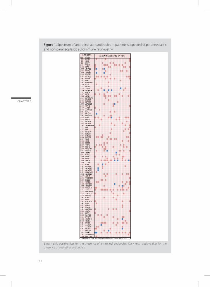
68
CHAPTER 5
Figure 1. Spectrum of antiretinal autoantibodies in patients suspected of paraneoplastic and non-paraneoplastic autoimmune retinopathy
no . abbr.193 M PP5195 CA2196 M ITF201 VSX1203 RP1205 IMPG2206 VAX2207 NR2E3208 CDHR1214 IM PG2215 OPN4217 RHO218 OPN1SW220 EYS221 LRIT1223 TRPM 1225 RCVRN232 CNGA1237 RP1L1239 RP1L1241 RPGRIP1242 PDE6A243 RPE65245 AANAT246 OPN5247 AIPL1248 ARM C9250 EYS257 PTGFR259 M YO7A262 OPTC263 M BP267 IM PG1268 IM PG1270 SERPINF1272 NRL273 NRL279 ALDOC281 ENO1-3282 ENO1-3283 BEST1284 SAG285 CA2286 RGS9287 TRPM 1289 RBP3290 UNC119294 GNGT2296 RBP3297 CRB2299 RAX2300 GNAT1303 PRCD309 TYRP1312 CRX316 BFSP2317 SPATA7318 SPATA7319 C1QTNF5325 SLC24A1327 GRK7329 TM EM 98330 BCO2331 CITED1332 GAPDH335 CITED1338 CNGA3341 TYR343 RPGRIP1345 ABCA4348 PDE6B350 CRB1351 GNA*355 CNGA3356 RS1357 GRK1358 CNGB1359 GAPDH360 CNGA1361 RGR362 IM PG1364 PTGFR365 CNGB3366 CNGA1372 M PP5373 ALDOA374 ALDOC375 ROM 1377 PDE6A378 GRK7379 SERPINF1380 UNC119
ant igens n(p)A IR pat ients (N =24)
* GNAS;GNAZ;GNA11-15;GNAL;GNAQ;GNAI1-I3;GNAO1;GNAT1-T3
Blue: highly positive titer for the presence of antiretinal antibodies. Dark red: positive titer for the presence of antiretinal antibodies.

Autoantibody profiling of patients with autoimmune retinopathy
69
CHAPTER 5
The number of highly positive ARAs present in individual patients is shown in Table 4. A higher
number of different ARAs per patient was most prevalent in patients with presumed (n)pAIR and
least present in patients with cataract. Three or more different ARAs were present in 29% of the
patients with presumed (n)pAIR, compared to 24% of the patients with uveitis and 14% of the
patients with cataract. The number of highly positive ARAs did not show any statistical differences
between presumed (n)pAIR and uveitis (p=0.457) or cataract (p=0.385). Furthermore, there was
no correlation between the number of ARAs and age (p=0.926).
Table 4. Number of highly positive antiretinal autoantibodies per patient
No. of highly positive ARAs (ratio > 25)
(n)pAIR (N=24) Uveitis (N=151) Cataract (N=21)
0 37.5% (9/24) 45.7% (69/151) 52.4% (11/21)
1 33.3% (8/24) 29.8% (45/151) 33.3% (7/21)
2 12.5% (3/24) 13.2% (20/151) 4.8% (1/21)
3 12.5% (3/24) 7.9% (12/151) 4.8% (1/21)
4 4.2% (1/24) 2.6% (4/151) 0% (0/21)
5 0% (0/24) 0.6% (1/151) 4.8% (1/21)
Abbreviations: (n)pAIR = non-paraneoplastic and paraneoplastic autoimmune retinopathy, ARAs = antiretinal antibodies
Antigen suspension bead array: positive titres of ARAs (ratio > 2)
The samples of patients with presumed (n)pAIR as well as both control cohorts exhibited a broad
spectrum of positive ARAs (Figure 1 and Supplementary Figure). None of the ARAs were specific
for presumed (n)pAIR only. Autoantibodies directed against serotonin N-acetyltransferase, cbp/
p300-interacting transactivator 1 and retinitis pigmentosa 1-like 1 protein were more prevalent
in patients with presumed (n)pAIR than in patients with uveitis (p=0.003, p=0.017 and p=0.020;
p-values were not significant after applying correction for multiple testing). When comparing the
serum of patients with presumed (n)pAIR to the serum of patients with cataract, in presumed
(n)pAIR autoantibodies directed against sodium/potassium/calcium exchanger 1 and pigment
epithelium-derived factor were more often present (p=0.037 and p=0.030). The presence of most
ARAs indicated in Table 3 was predominantly found in patients with presumed pAIR (CAR or MAR),
but (often less frequently) also in patients with presumed npAIR. The prevalence of low-titre ARAs
(from Table 3) was not significantly different between patients with npAIR and pAIR (p > 0.05).
The coefficient of variation based on replicates of the serum pools within and across plates (indi-
cating the intra- and inter-reproducibility) ranged between 5 and 23% (median = 13%) for all 188
antigens. ARAs were present in all patients with presumed (n)pAIR and consequently all fulfilled
the recent criteria for the diagnosis of (n)pAIR.9
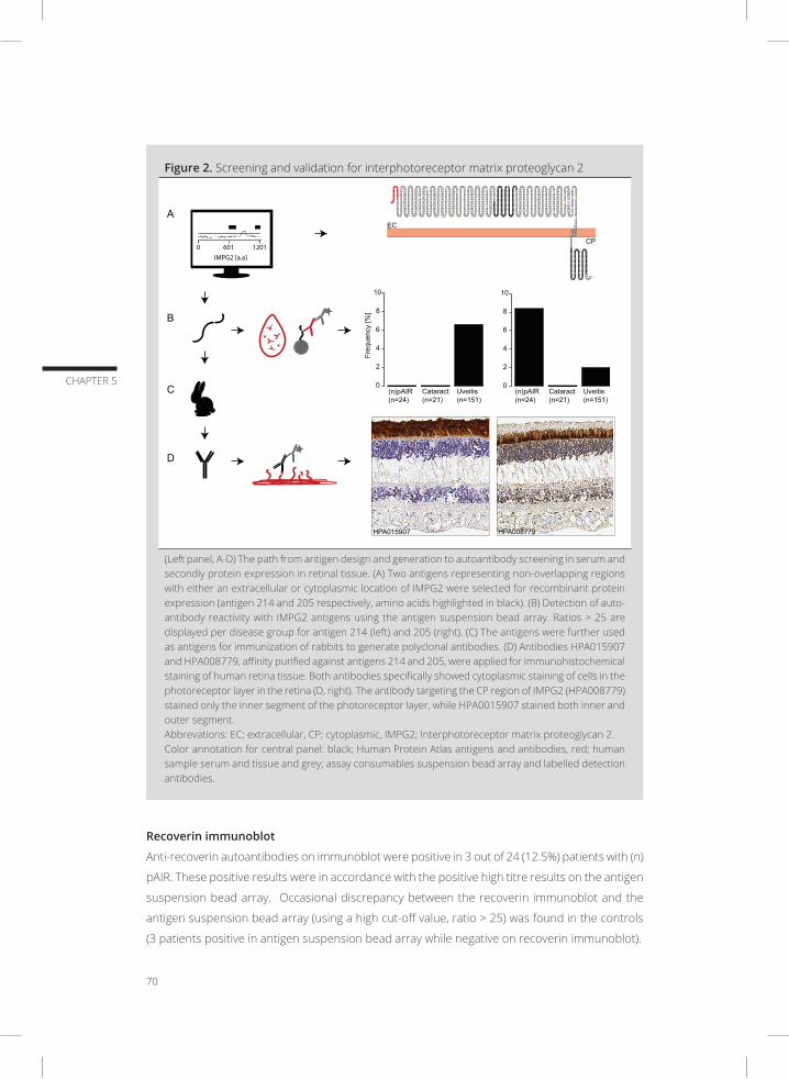
70
CHAPTER 5
Figure 2. Screening and validation for interphotoreceptor matrix proteoglycan 2
D
A
B
C (n)pAIR (n=24)
Uveitis (n=151)
Cataract (n=21)
0
2
4
6
8
10
0
2
4
6
8
10
Freq
uenc
y [%
]
EC
CP
(n)pAIR (n=24)
Uveitis (n=151)
Cataract (n=21)
HPA008779 HPA015907
0 601 1201 IMPG2 [a.a]
(Left panel, A-D) The path from antigen design and generation to autoantibody screening in serum and secondly protein expression in retinal tissue. (A) Two antigens representing non-overlapping regions with either an extracellular or cytoplasmic location of IMPG2 were selected for recombinant protein expression (antigen 214 and 205 respectively, amino acids highlighted in black). (B) Detection of auto-antibody reactivity with IMPG2 antigens using the antigen suspension bead array. Ratios > 25 are displayed per disease group for antigen 214 (left) and 205 (right). (C) The antigens were further used as antigens for immunization of rabbits to generate polyclonal antibodies. (D) Antibodies HPA015907 and HPA008779, affinity purified against antigens 214 and 205, were applied for immunohistochemical staining of human retina tissue. Both antibodies specifically showed cytoplasmic staining of cells in the photoreceptor layer in the retina (D, right). The antibody targeting the CP region of IMPG2 (HPA008779) stained only the inner segment of the photoreceptor layer, while HPA0015907 stained both inner and outer segment. Abbrevations: EC; extracellular, CP; cytoplasmic, IMPG2; Interphotoreceptor matrix proteoglycan 2. Color annotation for central panel: black; Human Protein Atlas antigens and antibodies, red; human sample serum and tissue and grey; assay consumables suspension bead array and labelled detection antibodies.
Recoverin immunoblot
Anti-recoverin autoantibodies on immunoblot were positive in 3 out of 24 (12.5%) patients with (n)
pAIR. These positive results were in accordance with the positive high titre results on the antigen
suspension bead array. Occasional discrepancy between the recoverin immunoblot and the
antigen suspension bead array (using a high cut-off value, ratio > 25) was found in the controls
(3 patients positive in antigen suspension bead array while negative on recoverin immunoblot).
Autoantibody profiling of patients with autoimmune retinopathy
71
CHAPTER 5
IMPG2 expression in human retina tissue
The antigens towards IMPG2 (antigen number 214 and 205) represent two non-overlapping
domains of IMPG2, located either extracellularly or in the cytoplasm (Figure 2A).46 Antibodies
directed against antigens 214 and 205, showed staining exclusively in cells in the photoreceptor
layer of the retina. The antibodies targeting the cytoplasmic region of IMPG2 (against antigen
number 205) stained only the inner segment of the photoreceptor layer, while HPA0015907
(antibodies against antigen number 214) stained both inner and outer segment (Figure 2D).
DISCUSSION
Our study shows that patients with (n)pAIR are characterized by the presence of a broad spectrum
of various ARAs. We identified ARAs that have already been described in previous studies, such as
anti-recoverin autoantibodies, but also identified new retinal targets. Our findings illustrate that
serum ARAs are not only present in patients with (n)pAIR, but also in patients with cataract and
uveitis. Though some ARAs appeared to be specific for (n)pAIR, their prevalence and consequently
their sensitivity as markers for (n)pAIR were low. This autoantibody screening using 188 antigen
provides insight into the autoimmune repertoire of patients with (n)pAIR and a base for further
validation with independent methods for protein analysis and independent sample cohorts.
A gold standard for the determination of ARAs is currently lacking.11 Different techniques are being
used, hampering the comparison of results from various laboratories.47 Moreover, the mere pres-
ence of ARAs does not provide any information on the role of this specific antibody. In addition,
information on clinical relevance of the specific ARAs and their pathological titres are lacking. A
combination of different ARAs was observed in some cases and therefore their individual effects
on retinal tissue could not be distinguished.
In our study, we performed statistical analyses using different cut-off levels. By using a high cut-off
value, a ratio > 25, false positive results were minimized and retinal targets with a high specificity
for (n)pAIR were found. The low cut-off value, a ratio of >2 (indicating ARAs with at least twice the
reactivity of the negative control), was used for a more sensitive approach, limiting the exclusion
of possibly relevant ARA targets with a lower titre. However, with both cut-off values, no ARAs
were found eligible for diagnostic purposes. Some ARAs were specific for (n)pAIR, but had low
prevalence while others were more frequently identified but lacked specificity.
In concordance with previous findings, positive results of serum anti-recoverin autoantibodies
were not only observed in patients with presumed (n)pAIR, but also in patients with uveitis and
cataract. Furthermore, no association between the presence of autoantibodies directed against
recoverin and the presence of a malignancy was found. A discrepancy between the antigen
72
CHAPTER 5
suspension bead array results for anti-recoverin and the anti-recoverin immunoblot was found
in three control patients (one with cataract and two with uveitis). The difference in results could
be explained by the different techniques used for determination of ARAs imposing differences in
analytical performance. Possibly, the number and/or availability of recoverin antigenic epitopes
differed between the antigen suspension bead array and the immunoblot technique. The protein
fragments we used in this study to screen for autoantibody reactivity in serum were designed to
represent unique parts of each target protein. The binding of the autoantibodies towards their
target may be influenced by the protein folding of antigens and may differ in comparison to full-
length protein arrays or peptide arrays. Both linear and conformational epitopes, recognized by
some ARAs, might be missed for some proteins, preventing recognition by certain autoantibodies.
The identification of new ARAs in (n)pAIR is in line with findings from previous studies using
Western blot analysis for the determination of ARAs.11,48 Although many ARAs have already been
identified, several studies have described so far unknown retinal autoantibodies presumably
damaging retinal tissue and causing loss of vision.14 In our study, we were able to identify novel
ARAs possibly associated with (n)pAIR, e.g. serotonin N-acetyltransferase. Serotonin N-acetyl-
transferase plays a role in melatonin synthesis and is expressed only in the pineal gland and
retina.49 Autoantibodies directed against serotonin N-acetyltransferase have to our knowledge
not been described in (n)pAIR so far. Another novel, although unspecific ARA found in this study
is anti-G protein-coupled receptor kinase 7. Interestingly, it has been suggested previously that G
protein-coupled receptor kinases in cancer cell lines are functionally associated with recoverin.50
Moreover, protein IMPG2 was identified as an ARA and reactivity towards the cytoplasmic pro-
tein region (antigen number 205) was associated with (n)pAIR. Autoantibody reactivity towards a
second antigen representing an extracellular region of IMPG2 was in contrast present in serum
from uveitis patients. In short, ARAs targeting IMPG2 were identified using antigen arrays in serum
samples and a retina specific protein expression of IMPG2 identified using immunohistochemistry
in healthy human tissue.
Our present study focused on the autoantibodies prevalent in serum, which reflects systemic
production and is probably not influenced by potential (additional) production or accumulation of
specific autoantibodies within the eye. Analysis of local, intraocular retinal autoantibodies might
show an entirely different pattern and may differ in clinical importance compared to retinal auto-
antibodies found in the peripheral circulation. The importance of locally produced autoantibodies
has already been shown in cerebrospinal fluid for the central nervous system. In addition, it is
unknown which autoantibodies penetrate from the circulation, through the blood retina bar-
rier, into the retina and cause a local inflammation. Further research addressing the intraocular
presence of specific retinal autoantibodies might elucidate the clinically relevant autoimmune
processes directed against the retinal tissue in (n)pAIR.
Autoantibody profiling of patients with autoimmune retinopathy
73
CHAPTER 5
A gold standard for the definitive diagnosis of (n)pAIR is currently lacking. Also in this study, the
diagnosis of presumed (n)pAIR was based on clinical symptoms. To compensate for this inaccu-
racy, we used strict inclusion criteria and selected a uniform cohort of patients with unexplained
visual loss, visual field defects and decreased or absent ERG while other diagnostic possibilities
leading to this configuration of clinical characteristics were (so far as possible) excluded. The
presence of ARAs was found in all our patients with presumed (n)pAIR and therefore all fulfilled
the criteria for the diagnosis of (n)pAIR.9
Although the mere presence of ARAs supports the diagnosis of (n)pAIR, it has been stated that
there are no specific ARAs which would be exclusive for (n)pAIR and none of the ARAs were iden-
tified to be of higher diagnostic value than other ARAs.4,9,14 Our results are in full agreement with
this statement. Proof for the definitive diagnosis of (n)pAIR is still missing and even the presence
of ARAs is not specific for (n)pAIR, which has been illustrated by the finding of ARAs in control
groups and healthy individuals.8,51,52
In conclusion, our study identified a heterogenous reactivity pattern of ARAs in serum of patients
with (n)pAIR, although the presence of ARAs was not discriminatory between (n)pAIR, cataract
and uveitis and exhibited a low sensitivity. Therefore, the diagnosis of (n)pAIR cannot be based on
the mere presence of serum ARAs and such presence thus warrants careful interpretation. The
determination of ARAs in intraocular fluid might provide more insight into the pathogenesis of (n)
pAIR and might indicate more sensitive and specific diagnostic tools. Therefore, future research
on the prevalence of ARAs in ocular fluid represents an important next step.
SUPPLEMENTARY DATA
Supplementary Table. Amino acid sequence and uniprot ID of ocular antigens used for the auto-
antibody profiling
https://doi.org/10.1371/journal.pone.0167909.s001
Supplementary Figure. Spectrum of antiretinal autoantibodies in patients suspected of parane-
oplastic and non-paraneoplastic autoimmune retinopathy, uveitis and cataract
https://doi.org/10.1371/journal.pone.0167909.s002
74
CHAPTER 5
REFERENCES
1. Comlekoglu DU, Thompson IA, Sen HN. Autoimmune retinopathy. Curr Opin Ophthalmol. 2013;24(6):598-
605.
2. Braithwaite T, Vugler A, Tufail A. Autoimmune retinopathy. Ophthalmologica. 2012;228(3):131-142.
3. Heckenlively JR, Ferreyra HA. Autoimmune retinopathy: a review and summary. Semin Immunopathol.
2008;30(2):127-134.
4. Grange L, Dalal M, Nussenblatt RB, Sen HN. Autoimmune retinopathy. Am J Ophthalmol. 2014;157(2):266-
272 e261.
5. Adamus G. Autoantibody targets and their cancer relationship in the pathogenicity of paraneoplastic
retinopathy. Autoimmun Rev. 2009;8(5):410-414.
6. Bazhin AV, Savchenko MS, Shifrina ON, et al. Recoverin as a paraneoplastic antigen in lung cancer: the
occurrence of anti-recoverin autoantibodies in sera and recoverin in tumors. Lung Cancer. 2004;44(2):193-
198.
7. Polans AS, Witkowska D, Haley TL, Amundson D, Baizer L, Adamus G. Recoverin, a Photoreceptor-Specific
Calcium-Binding Protein, Is Expressed by the Tumor of a Patient with Cancer-Associated Retinopathy. P
Natl Acad Sci USA. 1995;92(20):9176-9180.
8. Shimazaki K, Jirawuthiworavong GV, Heckenlively JR, Gordon LK. Frequency of anti-retinal antibodies in
normal human serum. J Neuro-Ophthalmol. 2008;28(1):5-11.
9. Fox AR, Gordon LK, Heckenlively JR, et al. Consensus on the Diagnosis and Management of Nonparane-
oplastic Autoimmune Retinopathy using a Modified Delphi Approach. Am J Ophthalmol. 2016.
10. Adamus G, Ren G, Weleber RG. Autoantibodies against retinal proteins in paraneoplastic and autoimmune
retinopathy. BMC Ophthalmol. 2004;4:5.
11. Forooghian F, Macdonald IM, Heckenlively JR, et al. The need for standardization of antiretinal antibody
detection and measurement. Am J Ophthalmol. 2008;146(4):489-495.
12. Forooghian F. The Uncertainty Regarding Antiretinal Antibodies. JAMA Ophthalmol. 2015;133(7):744-745.
13. Braithwaite T, Holder GE, Lee RW, Plant GT, Tufail A. Diagnostic features of the autoimmune retinopathies.
Autoimmun Rev. 2014;13(4-5):534-538.
14. Grewal DS, Fishman GA, Jampol LM. Autoimmune retinopathy and antiretinal antibodies: a review. Retina.
2014;34(5):827-845.
15. Lu Y, Jia L, He S, et al. Melanoma-Associated Retinopathy A Paraneoplastic Autoimmune Complication.
Arch Ophthalmol-Chic. 2009;127(12):1572-1580.
16. Thirkill CE, Tait RC, Tyler NK, Roth AM, Keltner JL. The cancer-associated retinopathy antigen is a recover-
in-like protein. Invest Ophthalmol Vis Sci. 1992;33(10):2768-2772.
17. Adamus G, Aptsiauri N, Guy J, Heckenlively J, Flannery J, Hargrave PA. The occurrence of serum autoanti-
bodies against enolase in cancer-associated retinopathy. Clin Immunol Immunop. 1996;78(2):120-129.
18. Adamus G, Karren L. Autoimmunity against carbonic anhydrase II affects retinal cell functions in autoim-
mune retinopathy. J Autoimmun. 2009;32(2):133-139.
19. Ohguro H, Ogawa K, Nakagawa T. Recoverin and Hsc 70 are found as autoantigens in patients with can-
cer-associated retinopathy. Invest Ophthalmol Vis Sci. 1999;40(1):82-89.
20. Adamus G, Brown L, Weleber RG. Molecular biomarkers for autoimmune retinopathies: significance of
anti-transducin-alpha autoantibodies. Exp Mol Pathol. 2009;87(3):195-203.
21. Potter MJ, Adamus G, Szabo SM, Lee R, Mohaseb K, Behn D. Autoantibodies to transducin in a patient
with melanoma-associated retinopathy. Am J Ophthalmol. 2002;134(1):128-130.
22. Bazhin AV, Dalke C, Willner N, et al. Cancer-retina antigens as potential paraneoplastic antigens in mela-

Autoantibody profiling of patients with autoimmune retinopathy
75
CHAPTER 5
noma-associated retinopathy. Int J Cancer. 2009;124(1):140-149.
23. Gurevich EV, Gurevich VV. Arrestins: ubiquitous regulators of cellular signaling pathways. Genome Biol.
2006;7(9):236.
24. Bianciotto C, Shields CL, Thirkill CE, Materin MA, Shields JA. Paraneoplastic retinopathy with multiple
detachments of the neurosensory retina and autoantibodies against interphotoreceptor retinoid binding
protein (IRBP) in cutaneous melanoma. Br J Ophthalmol. 2010;94(12):1684-1685, 1696.
25. Korf HW, Korf B, Schachenmayr W, Chader GJ, Wiggert B. Immunocytochemical demonstration of interpho-
toreceptor retinoid-binding protein in cerebellar medulloblastoma. Acta Neuropathol. 1992;83(5):482-487.
26. Heckenlively JR, Jordan BL, Aptsiauri N. Association of antiretinal antibodies and cystoid macular edema
in patients with retinitis pigmentosa. Am J Ophthalmol. 1999;127(5):565-573.
27. Hartmann TB, Bazhin AV, Schadendorf D, Eichmuller SB. SEREX identification of new tumor antigens linked
to melanoma-associated retinopathy. Int J Cancer. 2005;114(1):88-93.
28. Adamus G, Chan CC. Experimental autoimmune uveitides: multiple antigens, diverse diseases. Int Rev
Immunol. 2002;21(2-3):209-229.
29. Kobayashi M, Takezawa S, Hara K, et al. Identification of a photoreceptor cell-specific nuclear receptor.
Proc Natl Acad Sci U S A. 1999;96(9):4814-4819.
30. Flynn MF FG, Adamus G. Antiretinal Muller cell antibodies in patients with melanoma associated and
autoimmune retinopathy. Abstract presented at ARVO. 2000.
31. Kondo M, Sanuki R, Ueno S, et al. Identification of autoantibodies against TRPM1 in patients with para-
neoplastic retinopathy associated with ON bipolar cell dysfunction. PLoS One. 2011;6(5):e19911.
32. Wang Y, Abu-Asab MS, Li W, Aronow ME, Singh AD, Chan CC. Autoantibody against transient receptor
potential M1 cation channels of retinal ON bipolar cells in paraneoplastic vitelliform retinopathy. BMC
Ophthalmol. 2012;12:56.
33. Nakamura M, Sanuki R, Yasuma TR, et al. TRPM1 mutations are associated with the complete form of
congenital stationary night blindness. Mol Vis. 2010;16:425-437.
34. Dhingra A, Fina ME, Neinstein A, et al. Autoantibodies in melanoma-associated retinopathy target TRPM1
cation channels of retinal ON bipolar cells. J Neurosci. 2011;31(11):3962-3967.
35. Kikuchi T, Arai J, Shibuki H, Kawashima H, Yoshimura N. Tubby-like protein 1 as an autoantigen in can-
cer-associated retinopathy. J Neuroimmunol. 2000;103(1):26-33.
36. Eksandh L, Adamus G, Mosgrove L, Andreasson S. Autoantibodies against bestrophin in a patient with
vitelliform paraneoplastic retinopathy and a metastatic choroidal malignant melanoma. Arch Ophthalmol.
2008;126(3):432-435.
37. Adamus G, Brown L, Schiffman J, Iannaccone A. Diversity in autoimmunity against retinal, neuronal, and
axonal antigens in acquired neuro-retinopathy. J Ophthalmic Inflamm Infect. 2011;1(3):111-121.
38. Ayoglu B, Schwenk JM, Nilsson P. Antigen arrays for profiling autoantibody repertoires. Bioanalysis.
2016;8(10):1105-1126.
39. Ayoglu B, Haggmark A, Khademi M, et al. Autoantibody profiling in multiple sclerosis using arrays of human
protein fragments. Mol Cell Proteomics. 2013;12(9):2657-2672.
40. Ayoglu B, Mitsios N, Kockum I, et al. Anoctamin 2 identified as an autoimmune target in multiple sclerosis.
Proc Natl Acad Sci U S A. 2016;113(8):2188-2193.
41. Haggmark A, Hamsten C, Wiklundh E, et al. Proteomic profiling reveals autoimmune targets in sarcoidosis.
Am J Respir Crit Care Med. 2015;191(5):574-583.
42. Persson A, Hober S, Uhlen M. A human protein atlas based on antibody proteomics. Curr Opin Mol Ther.
2006;8(3):185-190.
43. Uhlen M, Bjorling E, Agaton C, et al. A human protein atlas for normal and cancer tissues based on anti-
body proteomics. Mol Cell Proteomics. 2005;4(12):1920-1932.
76
CHAPTER 5
44. Kampf C, Olsson I, Ryberg U, Sjostedt E, Ponten F. Production of tissue microarrays, immunohistochem-
istry staining and digitalization within the human protein atlas. J Vis Exp. 2012(63).
45. R Core Team. R: A language and environment for statistical computing. R Foundation for Statistical Com-
puting, Vienna, Austria 2013.
46. Omasits U, Ahrens CH, Muller S, Wollscheid B. Protter: interactive protein feature visualization and inte-
gration with experimental proteomic data. Bioinformatics. 2014;30(6):884-886.
47. Faez S, Loewenstein J, Sobrin L. Concordance of antiretinal antibody testing results between laboratories
in autoimmune retinopathy. JAMA Ophthalmol. 2013;131(1):113-115.
48. Ferreyra HA, Jayasundera T, Khan NW, He S, Lu Y, Heckenlively JR. Management of autoimmune retinop-
athies with immunosuppression. Arch Ophthalmol. 2009;127(4):390-397.
49. Coon SL, Mazuruk K, Bernard M, Roseboom PH, Klein DC, Rodriguez IR. The human serotonin N-acet-
yltransferase (EC 2.3.1.87) gene (AANAT): structure, chromosomal localization, and tissue expression.
Genomics. 1996;34(1):76-84.
50. Miyagawa Y, Ohguro H, Odagiri H, et al. Aberrantly expressed recoverin is functionally associated with
G-protein-coupled receptor kinases in cancer cell lines. Biochem Biophys Res Commun. 2003;300(3):669-
673.
51. Ten Berge JC, Schreurs MW, Vermeer J, Meester-Smoor MA, Rothova A. Prevalence and clinical impact of
antiretinal antibodies in uveitis. Acta Ophthalmol. 2016;94(3):282-288.
52. Cai Y, Pulido JS. False-positive anti-retinal antibodies as a cause of psychogenic vision loss. Ocul Immunol
Inflamm. 2014;22(4):330-332.


ERNSTIGE VISUSDALING DOOR AUTO-IMMUUN RETINOPATHIE SEVERE VISUAL LOSS CAUSED BY AUTOIMMUNE RETINOPATHY
Josianne C.E.M. ten Berge, Marco W.J. Schreurs, Diana Dufour-van den Goorbergh, Pauline M. de Witte, Mary J. van Schooneveld, Aniki Rothova
Ned Tijdschr Geneeskd. 2015;159:A8039
6
80
CHAPTER 6
SAMENVATTING
Achtergrond: Auto-immuunretinopathie (AIR) is een zeldzaam ziektebeeld dat onder andere
als een paraneoplastisch syndroom kan voorkomen. AIR is geassocieerd met de aanwezigheid
van anti-retinale antistoffen. Er wordt verondersteld dat deze antistoffen schade aan de retina
veroorzaken, met progressieve visusdaling als gevolg.
Casus: Een 74-jarige man kwam bij de oogarts met ernstige, progressieve visusdaling, zonder
noemenswaardige afwijkingen bij het standaard oogheelkundig onderzoek. Het elektroretinogram
was kenmerkend voor functieverlies van de fotoreceptoren. In het serum werden anti-retinale
antistoffen tegen recoverine aangetoond. Na doorverwijzing naar de internist vanwege de ver-
denking op een paraneoplastische AIR, werd een longcarcinoom gediagnostiseerd. De diagnose
‘carcinoom-geassocieerde paraneoplastische AIR’ werd hiermee bevestigd.
Conclusie: De verdenking op een paraneoplastische AIR is hoog bij een onbegrepen visusdaling,
ook bij patiënten zonder bekende maligniteit. Recent zijn in Nederland laboratoriumtechnieken
geïmplementeerd voor de bepaling van de anti-retinale antistof tegen recoverine, waardoor het
stellen van de diagnose ‘AIR’ beter mogelijk is.
Ernstige visusdaling door auto-immuun retinopathie
81
CHAPTER 6
ABSTRACT
Background: Autoimmune retinopathy (AIR) is a rare disorder which may present as a paraneo-
plastic syndrome. AIR is associated with the presence of antiretinal antibodies. These antibodies
are assumed to cause damage to the retina, resulting in progressive vision loss.
Case description: A 74-year-old man visited the ophthalmologist with a serious, progressive loss
of vision, without any noteworthy abnormalities at routine ophthalmological examination. The
electroretinogram was characteristic of loss of photoreceptor function. Anti-retinal antibodies
against recoverin were detected in serum. After referral to an internist on account of a suspected
diagnosis of paraneoplastic AIR, the patient was diagnosed with a lung carcinoma, confirming the
diagnosis of cancer-associated paraneoplastic AIR.
Conclusion: An unexplained loss of vision is highly suggestive of paraneoplastic AIR, even in
patients without a known malignancy. Laboratory techniques for the detection of the antiretinal
antibody against recoverin have recently been implemented in the Netherlands, facilitating the
diagnosis of AIR.
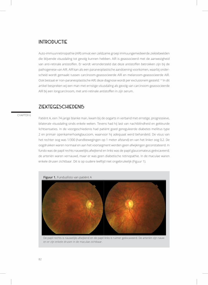
82
CHAPTER 6
INTRODUCTIE
Auto-immuunretinopathie (AIR) omvat een zeldzame groep immuungemedieerde ziektebeelden
die blijvende visusdaling tot gevolg kunnen hebben. AIR is geassocieerd met de aanwezigheid
van anti-retinale antistoffen. Er wordt verondersteld dat deze antistoffen betrokken zijn bij de
pathogenese van AIR. AIR kan als een paraneoplastische aandoening voorkomen, waarbij onder-
scheid wordt gemaakt tussen carcinoom-geassocieerde AIR en melanoom-geassocieerde AIR.
Ook bestaat er non-paraneoplastische AIR; deze diagnose wordt per exclusionem gesteld.1-4 In dit
artikel bespreken wij een man met ernstige visusdaling als gevolg van carcinoom-geassocieerde
AIR bij een longcarcinoom, met anti-retinale antistoffen in zijn serum.
ZIEKTEGESCHIEDENIS
Patiënt A, een 74-jarige blanke man, kwam bij de oogarts in verband met ernstige, progressieve,
bilaterale visusdaling sinds enkele weken. Tevens had hij last van nachtblindheid en gekleurde
lichtsensaties. In de voorgeschiedenis had patiënt goed gereguleerde diabetes mellitus type
2 en primair openkamerhoekglaucoom, waarvoor hij adequaat werd behandeld. De visus van
het rechter oog was 1/300 (handbewegingen op 1 meter afstand) en van het linker oog 0,2. De
oogdrukken waren normaal en aan het voorsegment werden geen afwijkingen geconstateerd. In
fundo was de papil rechts nauwelijks afwijkend en links was de papil glaucomateus geëxcaveerd;
de arteriën waren vernauwd, maar er was geen diabetische retinopathie. In de maculae waren
enkele drusen zichtbaar. Dit is op oudere leeftijd niet ongebruikelijk (Figuur 1).
Figuur 1. Fundusfoto van patiënt A
De papil rechts is nauwelijks afwijkend en de papil links is ruimer geëxcaveerd. De arteriën zijn nauw en er zijn enkele drusen in de maculae zichtbaar.
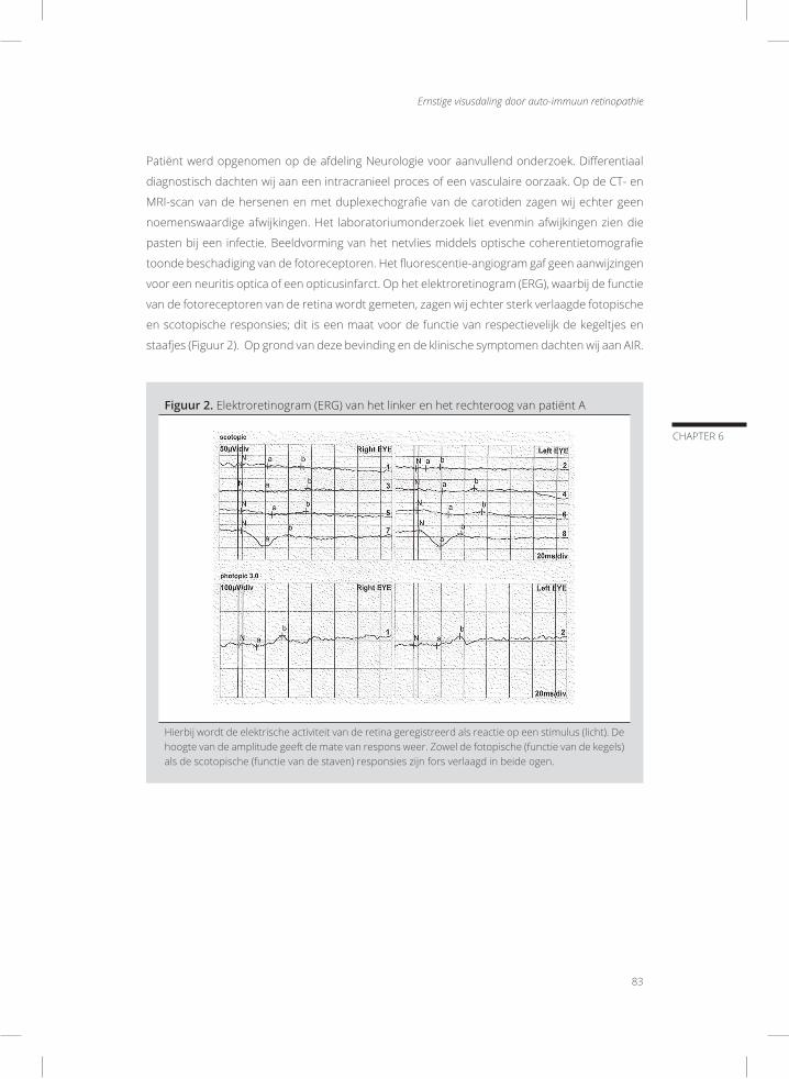
Ernstige visusdaling door auto-immuun retinopathie
83
CHAPTER 6
Patiënt werd opgenomen op de afdeling Neurologie voor aanvullend onderzoek. Differentiaal
diagnostisch dachten wij aan een intracranieel proces of een vasculaire oorzaak. Op de CT- en
MRI-scan van de hersenen en met duplexechografie van de carotiden zagen wij echter geen
noemenswaardige afwijkingen. Het laboratoriumonderzoek liet evenmin afwijkingen zien die
pasten bij een infectie. Beeldvorming van het netvlies middels optische coherentietomografie
toonde beschadiging van de fotoreceptoren. Het fluorescentie-angiogram gaf geen aanwijzingen
voor een neuritis optica of een opticusinfarct. Op het elektroretinogram (ERG), waarbij de functie
van de fotoreceptoren van de retina wordt gemeten, zagen wij echter sterk verlaagde fotopische
en scotopische responsies; dit is een maat voor de functie van respectievelijk de kegeltjes en
staafjes (Figuur 2). Op grond van deze bevinding en de klinische symptomen dachten wij aan AIR.
Figuur 2. Elektroretinogram (ERG) van het linker en het rechteroog van patiënt A
Hierbij wordt de elektrische activiteit van de retina geregistreerd als reactie op een stimulus (licht). De hoogte van de amplitude geeft de mate van respons weer. Zowel de fotopische (functie van de kegels) als de scotopische (functie van de staven) responsies zijn fors verlaagd in beide ogen.

84
CHAPTER 6
Het serologisch onderzoek naar anti-retinale antistoffen bleek positief voor antistoffen tegen
recoverine. Hiermee werd de diagnose ‘AIR’ bevestigd (Figuur 3). Patiënt kreeg prednison oraal,
80 mg/dag gedurende 3 dagen en vervolgens 45 mg/dag, om de visus te verbeteren. Deze behan-
deling had geen resultaat. Daarnaast werd patiënt door de oogarts verwezen naar de internist
voor onderzoek naar een eventuele onderliggende maligniteit. De internist stelde vast dat patiënt
een grootcellig neuro-endocrien carcinoom in stadium IIIA had. Hij kreeg chemotherapie en
radiotherapie, waarop de tumor sterk in grootte afnam, maar dit leidde niet tot een verbetering
van zijn visus. Op de CT-scan die 7 maanden na het begin van de therapie ter controle werd
gemaakt, was slechts een resttumor zichtbaar. Er waren geen aanwijzingen voor metastases.
Vanwege de blijvende ernstige visusdaling werden patiënt visuele hulpmiddelen en begeleiding
aangeboden. Patiënt woonde 6 maanden na het stellen van de diagnose longcarcinoom thuis,
in redelijk goede conditie.
Figuur 3. Recoverine auto-antistoffen: immunoblot en immunofluorescentie
A.
B.
positieve controle
negatieve controle
patiënt A
Sample Label Controle Recoverine
(A) De immunoblot toont een sterke reactie van het serum van de patiënt op de test strip welke met het antigeen recoverine is gecoat. (B) Bij (indirecte) immunofluorescentie op een coupe van de retina (primaat) is bij het serum van de patient in het groen (FITC) een sterke fluorescentie te zien van gedeeltes van de fotoreceptoren en een gedeelte van de buitenste plexiforme laag, waarin in beide gevallen recoverine aanwezig is. De blauwe aankleuringen (DAPI) zijn de celkernen in de verschillende lagen van de retina.

Ernstige visusdaling door auto-immuun retinopathie
85
CHAPTER 6
BESCHOUWING
AIR kenmerkt zich door bilaterale, progressieve visusdaling die vaak gepaard gaat met lichtflitsen
en andere visuele sensaties, gezichtsvelddefecten, nachtblindheid, kleurzienstoornissen en para-
centrale of centrale scotomen.1-6 Bij fundoscopie worden meestal geen of geringe afwijkingen
gevonden; sommige patiënten tonen een bleke papil en dunne retinale vaten. Het ziektebeeld is
bilateraal aanwezig, maar kan zich asymmetrisch presenteren.
Volgens de huidige hypothese wordt carcinoom-geassocieerde AIR veroorzaakt door immu-
nologische kruisreactiviteit tussen tumorantigenen en retinale antigenen.1-5 De aanwezigheid
van circulerende anti-retinale antistoffen ondersteunt de diagnose ‘AIR’. Sommige anti-retinale
antistoffen kunnen ook voorkomen bij gezonde individuen, zij het in lage titer.1-5,7 Verder zijn er
patiënten met AIR zonder aantoonbare retinale auto-antistoffen. Van het gehele spectrum van
verschillende anti-retinale antistoffen zijn de bepalingen van anti-recoverine en anti-enolase het
meest gebruikelijk voor het stellen van de diagnose AIR.4
Visusklachten als paraneoplastisch verschijnsel
Visusklachten zijn meestal het eerste symptoom bij patiënten met carcinoom-geassocieerde
AIR.3-5 Pas nadat de verdenking op carcinoom-geassocieerde AIR is ontstaan, wordt de patiënt
naar de internist doorverwezen en een carcinoom ontdekt. De latentietijd tussen carcinoom-ge-
associeerde AIR en de diagnose van een carcinoom varieert van weken tot jaren.1,3-5 Bij patiënten
met melanoom-geassocieerde AIR zijn de visusklachten met fotopsieën vaak het eerste teken
van metastasering van een reeds bekend melanoom.1,3-5 Onze patiënt werd in enkele weken zo
goed als blind. Gezien zijn ernstige klachten, zonder duidelijke afwijkingen bij fundoscopie of
aanwijzingen voor andere oorzaken, was de verdenking op AIR hoog. Een afwijkend ERG en de
aanwezigheid van anti-retinale antistoffen ondersteunden deze diagnose.
AIR wordt meestal gezien als paraneoplastisch syndroom bij diverse soorten carcinomen. Het is
daarom belangrijk verder onderzoek te verrichten naar een mogelijke maligniteit. Onze patiënt
had een longcarcinoom, een van de typen carcinomen die gepaard gaan met paraneoplastische
AIR. Andere carcinomen die met carcinoom-geassocieerde AIR kunnen voorkomen, zijn gynaeco-
logische en hematologische maligniteiten, long-, mamma-, prostaat-, en coloncarcinoom.1,3-5 De
behandeling bestaat uit reductie van de tumorgrootte door middel van radiotherapie, chemo-
therapie of chirurgie.1-3 Ook immunomodulatoren zouden een positief effect kunnen hebben op
de visus.8 Door de beperkte behandelingsmogelijkheden en de blijvende schade aan de retina is
de prognose voor de visus vaak slecht.
Het ERG is cruciaal voor de diagnostiek naar AIR, maar de diagnostiek blijft lastig. Tot voor kort
was het niet mogelijk om anti-retinale antistoffen in Nederland te laten bepalen. Op de afdeling

86
CHAPTER 6
Immunologie van het Erasmus MC in Rotterdam is recent de diagnostiek naar de anti-retinale
antistof tegen recoverine geïmplementeerd; deze methode bestaat uit indirecte immunofluores-
centie en immunoblot. Tot op heden is het alleen in Duitsland mogelijk anti-retinale antistoffen
tegen enolase te bepalen; bepalingen van andere antistoffen worden alleen in het kader van
onderzoek verricht. In de nabije toekomst verwacht men meerdere anti-retinale antistoffen te
kunnen bepalen, waaronder antistoffen tegen enolase, transducine en bipolaire cellen. Deze
diagnostiek, die ook met ELISA kan worden uitgevoerd, zal naar verwachting voor routinebepa-
lingen ter beschikking komen.
CONCLUSIE
Auto-immuunretinopathie is een zeldzame, snel progressieve oogaandoening waarbij anti-reti-
nale antistoffen voorkomen die vermoedelijk destructie van de retina veroorzaken, met blijvende
visusdaling als gevolg. De verdenking op paraneoplastische AIR is hoog bij een onbegrepen visus-
daling bij patiënten met of zonder een maligniteit. Omgekeerd dient bij patiënten met AIR of de
verdenking daarop onderzoek naar een mogelijke maligniteit verricht te worden. De anti-retinale
antistof tegen recoverine kan sinds kort bepaald worden in het Erasmus MC, ter ondersteuning
van de diagnose ‘AIR’. Een visusdaling door AIR is vaak ernstig en blijvend en behandeling met
immunosuppressiva is niet altijd toereikend. Mogelijk bieden nieuwe behandelingen die de vor-
ming van antistoffen kunnen beïnvloeden, zoals rituximab, meer mogelijkheden in de toekomst.

Ernstige visusdaling door auto-immuun retinopathie
87
CHAPTER 6
LITERATUUR
1. Braithwaite T, Vugler A, Tufail A. Autoimmune Retinopathy. Opthalmologica. 2012;228(3):131-42
2. Grange L, Dalal M, Nussenblatt RB, Sen HN. Autoimmune Retinopathy. Am J Opthalmol. 2014;157(2):266-
72
3. Comlegkoglu DU, Thompson IA, Sen HN. Autoimmune retinopathy. Curr Opin Opthalmol. 2013;24(6):598-
605
4. Braithwaite T, Holder GE, Lee RWJ, Plant GT, Tufail A. Diagnostic features of the autoimmune retinopathies.
Autoimmun Rev. 2014;13(4-5):534-8
5. Adamus G. Autoantibody targets and their cancer relationship in the pathogenicity of paraneoplastic
retinopathy. Autoimmun Rev. 2009;8(5):410-4
6. Heckenlively JR, Ferreyra HA. Autoimmune retinopathy: a review and summary. Semin Immunopathol.
2008 Apr;30(2):127-34
7. Shimazaki K, Jirawuthiworavong GV, Heckenlively JR, Gordon LK. Frequency of anti-retinal antibodies in
normal human serum. J Neuroophthalmool. 2008;28:5-11
8. Ferreyra HA, Jayasundera T, Khan NW, He S, Lu Y, Heckenlively JR. Management of autoimmune retinop-
athies with immunosuppression. Arch Ophthalmol. 2009 Apr;127(4):390-7


ANTIRETINAL ANTIBODIES IN CENTRAL SEROUS CHORIORETINOPATHY: PREVALENCE AND CLINICAL IMPLICATIONS
Josianne C.E.M. ten Berge*, Elon H. C. van Dijk*, Marco W.J. Schreurs, Jacolien Vermeer, Camiel J. F. Boon, Aniki Rothova
* These authors contributed equally
Acta Ophthalmol. 2017 Apr 26, Epub ahead of print
7

90
CHAPTER 7
ABSTRACT
Purpose: To investigate the possible role of autoimmune reactions directed against retinal tissue
in central serous chorioretinopathy (CSC), by analyzing the presence of serum antiretinal anti-
bodies (ARAs) and establishing their clinical relevance.
Methods: Sixty-three CSC patients were included and clinical characteristics were collected.
Serum samples of all CSC patients, 101 uveitis patients, and 60 healthy donors were analysed
for the presence of ARAs by indirect immunofluorescence. Furthermore, all CSC serum samples
were analysed on Western blot. Correlations between laboratory findings and clinical features
of CSC were determined by logistic regression.
Results: ARAs were present in 54% of the CSC patients, in 46% of uveitis patients (p=0.153), and in
17% of healthy controls (p<0.001). The majority of ARAs in CSC were directed against photorecep-
tors (27%), which occurred significantly more often compared to uveitis patients (15%, p=0.039)
and to healthy controls (5%, p=0.003). No associations between clinical CSC characteristics and
the presence of ARAs were found.
Conclusion: Serum ARAs are present in more than half of the CSC patients, and especially ARAs
directed against photoreceptors were detected more frequently compared to both healthy
controls and uveitis patients. Further research is warranted to unravel the role of ARAs in the
pathogenesis of CSC.
Antiretinal antibodies in central serous chorioretinopathy
91
CHAPTER 7
INTRODUCTION
Central serous chorioretinopathy (CSC) is a specific and relatively common early-onset chorio-
retinal disease that primarily affects the macula. In CSC, a subretinal fluid (SRF) leakage through
a dysfunctional retinal pigment epithelium (RPE) leads to detachment of the neuroretina.1-3 A
prolonged neuroretinal detachment in the macula causes permanent central visual loss due to
photoreceptor atrophy.1,2 Such a loss of visual acuity, sometimes associated with image distor-
tion and loss of colour and contrast vision, may have a high impact on a patient’s personal and
professional life. Early diagnosis and treatment is desirable to try to improve the visual outcome
and quality of life.1,3-12
The exact pathogenesis of CSC is currently obscure; presumably CSC occurs due to dysfunction of
the RPE with hyperpermeability, swelling, and leakage of the underlying choroid.3,13 Moreover, the
optimal treatment for CSC is unknown.14 CSC is up to six times more common in men (estimated
mean annual age-adjusted incidence: 9.9 per 100,000) compared to women (estimated incidence:
1.7 per 100,000).15 CSC is associated with the use of steroid containing medication, with odds ratios
up to 37.1, as well as with endogenous hypercortisolism.16-19 Familial occurrence of CSC has been
described and recent studies have found evidence of genetic associations in CSC patients, including
genetic polymorphrisms in the CFH gene, the ARMS2 gene, the C4b gene, and the CD5 gene.20-27
Although little is known about the exact cause of CSC, a role of the immune system via the
complement system has been suggested based on associations found in complement-related
genes. Recent evidence suggests a role for antiretinal antibodies (ARAs) in uveitis and age-related
macular degeneration.28-31 A systematic study on presence and the possible role of ARAs in CSC
is currently lacking.
We hypothesize that damage to the RPE outer blood-retinal barrier in CSC may result in a sec-
ondary formation of ARAs, which may affect the clinical course of CSC. In this study, we set out
to investigate the presence of serum ARAs in CSC patients and to analyse a possible correlation
of ARAs with the clinical characteristics of CSC.
METHODS
Patient and data selection
In this study, we included 63 Caucasian patients with chronic CSC who visited the outpatient
clinic of the Department of Ophthalmology of the Leiden University Medical Center, Leiden,
the Netherlands. The diagnosis of chronic CSC was based on ophthalmic examination and mul-
timodal imaging, including fundoscopy, optical coherence tomography (OCT) using either the
92
CHAPTER 7
spectral-domain OCT or the Cirrus OCT device, fundus autofluorescence, fluorescein angiography
(FA), and indocyanine green angiography. The diagnosis of chronic CSC was based on the pres-
ence of all of the following criteria: serous SRF on OCT, ≥1 area of multifocal diffuse leakage or
irregular RPE window defects on FA, and corresponding hyperfluorescent areas on indocyanine
green angiography. Patients diagnosed with acute CSC were excluded from this study, since it is
currently unclear if acute CSC and chronic CSC represent a continuum or are separate disease
entities. Patients with evidence of other retinal diagnoses, choroidal neovascularisation, and/or
polypoidal choroidal vasculopathy were also excluded. Clinical data of CSC patients were collected
from medical data files and included patient demographics (age, gender, and (family) history) and
ocular characteristics comprising stage, duration, activity and laterality of CSC, use of steroids
or immunosuppressive medications, presence of intraretinal edema, treatment for CSC, and
central retinal thickness (distance from the outer part of the ellipsoid zone to the inner part of
the internal limiting membrane; CRT). The CRT of the affected eye was selected; if both eyes were
affected the right eye was included. Diffuse CSC was characterized by the presence of >5 disc
areas of hyperfluorescent RPE changes or leakage on FA. Serum samples of 101 patients with
uveitis (intermediate uveitis, posterior uveitis or panuveitis) were used as disease controls, since
a higher prevalence of ARAs in serum in this cohort has been described previously.30 Serum of
60 blood bank donors (gender and age unknown) was used as (presumed) healthy controls. The
study adhered to the tenets of the Declaration of Helsinki. Approval of the ethics committee and
institutional review board was obtained.
Detection of ARAs using indirect immunofluorescence (IIF)
Initial screening of sera for ARAs was performed as described previously.30 In short, cryosections
of primate retinal tissue generated by Euroimmun (Lubeck, Germany) were left unfixed and incu-
bated with 1:100 diluted serum for 30 minutes at room temperature. Sections were washed in
stagnant phosphate-buffered saline (PBS) and incubated with goat-anti-human IgG conjugated
with fluorescein isothiocyanate (FITC) for 30 minutes at room temperature. Thereafter, sections
were washed in stagnant PBS and embedded. The positive control consisted of retinal tissue
incubated with 1:100 diluted serum of an antinuclear antibody (ANA) positive patient; for the
negative controls we used incubation with PBS and 1:100 diluted serum of a healthy control.
Detection of ANA using IIF
All sera showing staining of the retinal nuclear layers (outer/inner nuclear layer, ganglion cell layer)
on IIF were subsequently analysed for the presence of ANAs. ANA detection was performed by
IIF using HEp-2 cells (Inova, San Diego, California, United States), as described before.30 In sum-
mary, HEp-2 cells were incubated with 1:80 diluted serum for 30 minutes at room temperature.
After washing in PBS with continuous stirring, slides were incubated for another 30 minutes with
FITC-conjugated goat anti-human IgG with propidium iodide (Inova, San Diego, California, United
States). Subsequently, slides were washed and embedded.
Antiretinal antibodies in central serous chorioretinopathy
93
CHAPTER 7
Evaluation of IIF results
All slides were evaluated with a fluorescence microscope (20x magnification) by two independent
observers. Specific retinal layers (ganglion cell layer, inner plexiform layer, inner nuclear layer,
outer plexiform layer, outer nuclear layer, and rods and cones layer) were evaluated for the pres-
ence of fluorescent staining including intensity of the staining. When both ANA (on HEp-2-cells)
and ARA (on primate retinal tissue) had an equivalent intensity of staining, a sample was scored
as ‘unknown’ because this combination does not allow a proper discrimination between the
presence and absence of ARAs, by possible masking due to ANAs. These samples were excluded
from the final analyses.
Western blot analysis
All CSC patients were evaluated for the presence of ARAs using Western blot analysis. Healthy
human retinal protein extract was obtained after retina tissue homogenization in PBS. Tissue
fragments were removed by centrifugation and the supernatant was frozen at -80°C until use.
Retinal tissue extract was fractionated by SDS-polyacrylamide gel electrophoresis and separated
proteins were subsequently transferred to nitrocellulose membranes. The membranes were
blocked by incubation with 5% non-fat dry milk and incubated with serum of CSC patients or
with appropriate control serum at a dilution of 1:100 in 5% non-fat dry milk in tris-buffered
saline (TBS) overnight. After multiple washes with 0.1% TBS, membrane bound human IgG was
identified by horseradish peroxidase-conjugated F(ab’) 2 goat anti-human IgG (Thermo Fisher
Scientific, Waltham, Massachusetts, United States) at a dilution of 1:5000 in 5% non-fat dry milk
in TBS-Tween. Reactivity was visualized using enhanced chemiluminescence.
Statistics
First, descriptive analyses were performed to obtain information on the characteristics of the CSC
patients. Second, logistic regression with correction for age and gender was employed to evaluate
if the presence of ARAs is associated to CSC. Finally, logistic regression with correction for age
and gender was used to identify any clinical characteristics that were possibly associated with
the presence of ARAs. P<0.05 was considered statistically significant. All analyses were performed
with IBM SPSS Statistics version 21.
RESULTS
Patient characteristics
All patient characteristics are shown in Table 1. A total of 63 chronic CSC patients (56 male, 7
female), with a mean age of 51 ± 9 years (range, 31-72 years) was included in this study. The
median duration of CSC at the time of blood collection was 585 days (range, 3-7832 days). Diffuse
CSC was present in 11/62 patients (18%; data not available for one patient). In 38/48 CSC patients

94
CHAPTER 7
(79%; data not available for 15 patients) SRF was present at the moment of blood collection,
indicating active CSC. The mean CRT was 134 ± 37 µm (range, 57-226 µm). Previous to blood
collection, no treatment for CSC was given in 35/63 patients (56%), whereas 13/63 patients (21%)
had received either micropulse or focal laser treatment and 10/63 patients (16%) had received
photodynamic therapy.
Table 1. Clinical characteristics of 63 patients with central serous chorioretinopathy at the moment of blood collection
Patient characteristics
Male-to-female ratio 8:1
Mean age (SD), in years 51 (9)
Median duration of CSC (min-max), in days* 585 (3-7832)
CSC characteristics Number (%)
Stage **Focal CSCDiffuse CSC
51/62 (82%)11/62 (18%)
Active CSC **,‡ 38/48 (79%)
Bilateral CSC during follow-up ‡ 32/63 (51%)
Recurrent CSC **, ‡ 26/62 (42%)
Familial CSC 2/63 (3%)
Presence of intraretinal edema during follow-up 10/63 (16%)
Mean central retinal thickness (SD), in micrometers ** 134 (37) (46 patients)
Previous treatment for CSCPhotodynamic therapyMicropulse or focal laser treatmentOther treatments or combinations of treatmentNo treatment
10/63 (16%)13/63 (21%)5/63 (8%)35/63 (56%)
Systemic corticosteroids medicationNeverDuring diagnosis of CSC< 3 months before the diagnosis of CSCAfter or > 3 months before the diagnosis of CSC
33/63 (52%)30/63 (16%)7/63 (11%)13/63 (21%)
Use of systemic corticosteroids at the moment of blood collection 9/63 (14%)
Use of systemic immunosuppressive medication (excluding steroids) ** 1/49 (2%)
Comorbidities **Autoimmune diseasesMalignancies
3/59 (5%)4/59 (7%)
* Duration of CSC: interval between the initial diagnosis of CSC and the moment of blood collection** Data not available for all patients‡ Presenceofsubretinalfluidisconsideredtobetheindicationof‘active’CSCAbbreviations: CSC = central serous chorioretinopathy, SD = standard deviation

Antiretinal antibodies in central serous chorioretinopathy
95
CHAPTER 7
ARAs on IIF in CSC patients
The IIF results are summarized in Table 2. Positive staining of the retina on IIF was present in 35/63
CSC patients (56%). After adjustment for retinal staining due to ANA, the presence of ARAs was
confirmed in 32/59 CSC patients (54%). Among different staining patterns observed, staining of
the photoreceptors was the most prevalent pattern in CSC patients (17/63; 27%; Figure 1). Other
observed staining patterns in serum of CSC patients included staining of nuclear layers (N=8),
fluorescence between the inner nuclear layer and the outer plexiform layer (N=7), fluorescence
between the outer nuclear layer and the photoreceptors (N=3), and/or staining of the inner
plexiform layer (N=2). Nine CSC patients had a combination of different staining patterns on IIF.
Figure 1. Staining patterns on indirect immunofluorescence from serum of patients with central serous chorioretinopathy
Serum of central serous chorioretinopathy patients was tested for the presence of antiretinal antibod-ies using indirect immunofluorescence with primate retinal tissue. Presence of antiretinal antibodies was visualized by labelling with fluorescein isothiocyanate (FITC). Panel A shows staining of the photo-receptors (arrow); panel B shows no retinal staining (absence of antiretinal antibodies).
The prevalence of ARAs in CSC patients was higher than in healthy controls (10/59; 17%; p<0.001).
In addition, staining of the photoreceptors occurred in a lower percentage of healthy controls (5%)
compared to CSC patients (p=0.003). In patients with uveitis, any positive staining of the retina
(after correction for ANA presence) was observed in 39/84 patients (46%), which was not signifi-
cantly different from CSC patients (p=0.153). In contrast, specific staining of the photoreceptors
in uveitis patients (15%) was less prevalent compared to CSC patients (p=0.039). No significant
differences in the prevalence of staining of other specific retinal layers between CSC patients and
the two control groups were observed.
96
CHAPTER 7
Clinical characteristics of CSC in relation to presence of ARAs on IIF
The presence of ARAs on IIF was higher in the six female CSC patients (100%) compared to the
57 male CSC patients (46%; p=0.024; Table 3). Staining of the photoreceptors in the samples of
female and male CSC patients (57% versus 23%) did not differ (p=0.078). All other clinical char-
acteristics of CSC (including age at onset, duration of CSC, stage of CSC, activity of CSC, unilateral
versus bilateral CSC, recurrence of CSC, familial occurrence of CSC, CRT, presence of intraretinal
edema, previous CSC treatment, and systemic steroid use during confirmation of the diagnosis of
CSC) were not significantly associated with the presence of ARAs or specific staining of the pho-
toreceptors. Masked assessment of the OCT images of the 17 CSC patients with photoreceptor
staining on IIF and of 17 CSC patients without photoreceptor staining on IIF (randomly selected)
showed no remarkable differences in signs of retinal damage.
ARAs on Western blot in patients with CSC
Multiple ARAs were observed in the majority of CSC patients (Figure 2). The most abundant ARA
reactivity on Western blot was observed with those CSC patients showing staining of the photore-
ceptors on IIF, when compared to patients without retinal staining patterns as well as to patients
with other staining patterns on IIF. The most prevalent ARAs on Western blot in CSC patients
showing staining of the photoreceptors on IIF had molecular weights of approximately 24 kDa
and 45 kDa. However, these antibodies were not entirely specific for samples with photoreceptor
staining, since they were also present in CSC patients showing no staining of the photoreceptors
on IIF, and even in patients with no staining at all.
DISCUSSION
We show that serum ARAs were present in 54% of patients with chronic CSC and in 17% of healthy
controls. No significant difference in the prevalence of overall ARAs in serum was found between
CSC patients and uveitis patients (46%), although staining of the photoreceptors occurred more
often in CSC patients (27%) than in uveitis patients (15%). No differences in the presence of ARAs
were observed when CSC patients were subdivided into specific subgroups based on clinical
characteristics.
The multiple staining patterns on IIF in individual CSC patients indicate the presence of diverse
ARAs, which was confirmed by Western blot analysis. Also, within patients who were solely
showing staining of the photoreceptors on IIF, Western blot indicated the presence of diverse
antibodies directed against the retina. This suggests that diverse retinal antibodies are associated
with photoreceptor staining.
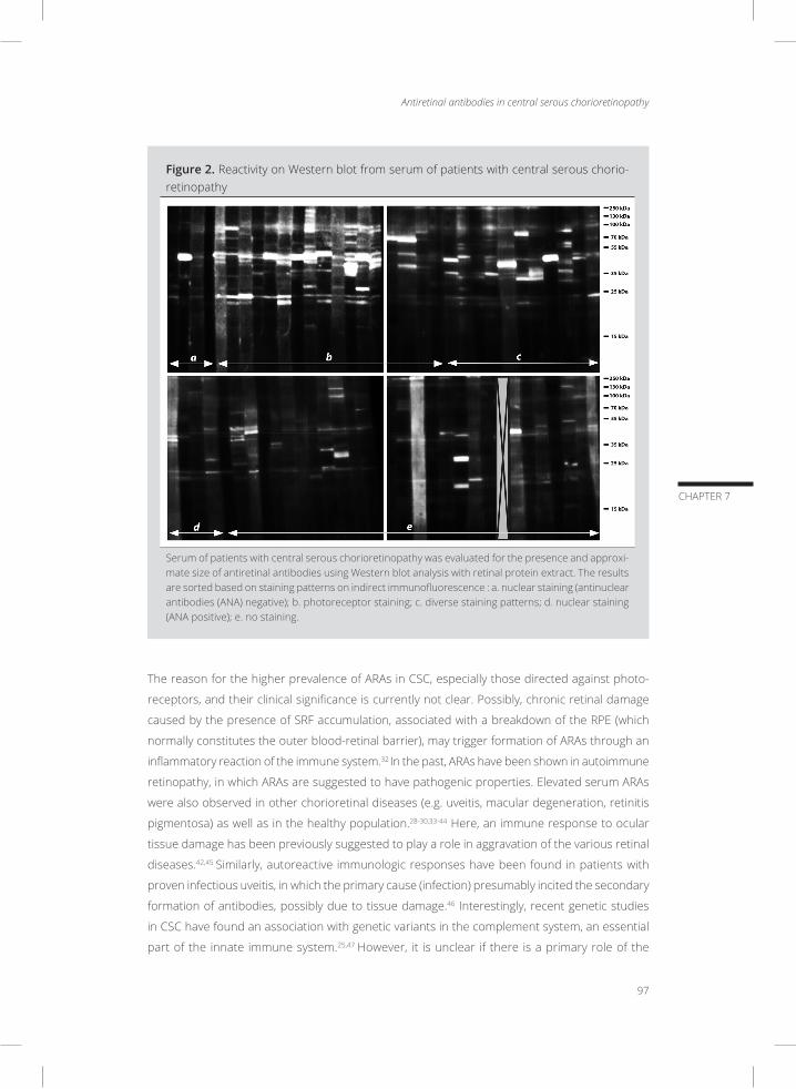
Antiretinal antibodies in central serous chorioretinopathy
97
CHAPTER 7
Figure 2. Reactivity on Western blot from serum of patients with central serous chorio-retinopathy
Serum of patients with central serous chorioretinopathy was evaluated for the presence and approxi-mate size of antiretinal antibodies using Western blot analysis with retinal protein extract. The results are sorted based on staining patterns on indirect immunofluorescence : a. nuclear staining (antinuclear antibodies (ANA) negative); b. photoreceptor staining; c. diverse staining patterns; d. nuclear staining (ANA positive); e. no staining.
The reason for the higher prevalence of ARAs in CSC, especially those directed against photo-
receptors, and their clinical significance is currently not clear. Possibly, chronic retinal damage
caused by the presence of SRF accumulation, associated with a breakdown of the RPE (which
normally constitutes the outer blood-retinal barrier), may trigger formation of ARAs through an
inflammatory reaction of the immune system.32 In the past, ARAs have been shown in autoimmune
retinopathy, in which ARAs are suggested to have pathogenic properties. Elevated serum ARAs
were also observed in other chorioretinal diseases (e.g. uveitis, macular degeneration, retinitis
pigmentosa) as well as in the healthy population.28-30,33-44 Here, an immune response to ocular
tissue damage has been previously suggested to play a role in aggravation of the various retinal
diseases.42,45 Similarly, autoreactive immunologic responses have been found in patients with
proven infectious uveitis, in which the primary cause (infection) presumably incited the secondary
formation of antibodies, possibly due to tissue damage.46 Interestingly, recent genetic studies
in CSC have found an association with genetic variants in the complement system, an essential
part of the innate immune system.25,47 However, it is unclear if there is a primary role of the
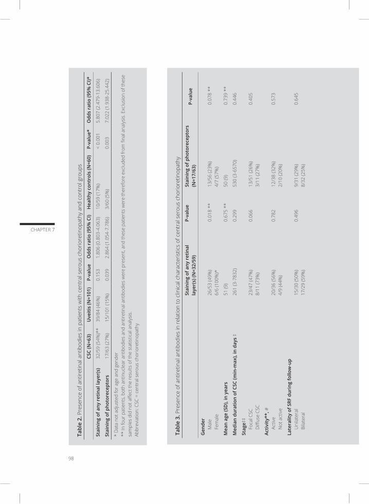
98
CHAPTER 7
Tabl
e 3.
Pre
senc
e of
ant
iretin
al a
ntib
odie
s in
rela
tion
to c
linic
al c
hara
cter
istic
s of
cen
tral
ser
ous
chor
iore
tinop
athy
Stai
ning
of a
ny r
etin
al
laye
r(s)
(N=3
2/59
)P-
valu
eSt
aini
ng o
f pho
tore
cept
ors
(N=1
7/63
)P-
valu
e
Gen
der
Mal
eFe
mal
e 26
/53
(49%
)6/
6 (1
00%
)*0.
018
**13
/56
(23%
)4/
7 (5
7%)
0.07
8 **
Mea
n ag
e (S
D),
in y
ears
51 (9
)0.
675
**50
(9)
0.73
9 **
Med
ian
dura
tion
of C
SC (m
in-m
ax),
in d
ays
‡26
1 (3
-783
2)0.
299
530
(3-6
570)
0.44
6
Stag
e‡‡
Foca
l CSC
Diff
use
CSC
23/4
7 (4
7%)
8/11
(73%
)0.
066
13/5
1 (2
6%)
3/11
(27%
)0.
405
Acti
vity
**, #
Activ
eN
ot a
ctiv
e20
/36
(56%
)4/
9 (4
4%)
0.78
212
/38
(32%
)2/
10 (2
0%)
0.57
3
Late
ralit
y of
SRF
dur
ing
follo
w-u
pU
nila
tera
lBi
late
ral
15/3
0 (5
0%)
17/2
9 (5
9%)
0.49
69/
31 (2
9%)
8/32
(25%
)0.
645
Recu
rren
ce‡‡
Recu
rren
t CSC
Firs
t CSC
epi
sode
11
/24
(46%
)21
/34
(62%
)0.
393
5/26
(19%
)12
/36
(33%
)0.
379
Fam
ilial
Fa
mili
al C
SCN
ot fa
mili
al C
SC1/
2 (5
0%)
31/5
7 (5
4%)
0.94
91/
2 (5
0%)
16/6
1 (2
6%)
0.45
4
Mea
n ce
ntra
l ret
inal
thic
knes
s (S
D),
in m
icro
met
ers‡
‡12
5 (3
2) (2
4 pa
tient
s)0.
091
121
(39)
(14
patie
nts)
0.08
0
Intr
aret
inal
ede
ma
duri
ng fo
llow
-up
Pres
ent
Abse
nt7/
9 (7
8%)
25/5
0 (5
0%)
0.22
24/
10 (4
0%)
13/5
3 (2
5%)
0.28
8
Trea
tmen
t: p
hoto
dyna
mic
ther
apy
(PD
T)PD
TN
o(t s
olel
y) P
DT
6/10
(60%
)26
/49
(53%
)0.
665
3/10
(30%
)14
/53
(26%
)0.
804
Trea
tmen
t: (f
ocal
and
/or
mic
ropu
lse)
lase
rLa
ser t
reat
men
tN
o(t s
olel
y) la
ser t
reat
men
t7/
13 (5
4%)
25/4
6 (5
4%)
0.95
66/
13 (4
6%)
11/5
0 (2
2%)
0.05
3
Use
of c
orti
cost
eroi
dsD
urin
g or
< 3
mon
ths
befo
re C
SC d
iagn
osis
N
o cu
rren
t use
25/4
9 (5
1%)
7/10
(70%
)0.
674
12/5
3 (2
3%)
5/10
(50%
)0.
138
* Af
ter
the
stud
y w
as c
ompl
eted
, thr
ee a
dditi
onal
sam
ples
from
fem
ale
patie
nts
with
CSC
wer
e an
alyz
ed a
nd a
ll sh
owed
abs
ence
of A
RAs
on II
F, r
esul
ting
in a
n ul
timat
e pe
rcen
tage
pos
itive
ARA
s am
ong
fem
ales
of 6
7% (6
/9),
whi
ch is
not
dis
tinct
from
mal
e CS
C pa
tient
s (2
6/53
; 49%
).**
Det
erm
ined
by
χ2 te
st (g
ende
r) o
r Man
n W
hitn
ey U
test
(age
)‡
Dur
atio
n of
CSC
: fro
m C
SC d
iagn
osis
to th
e m
omen
t of b
lood
col
lect
ion
‡‡ D
ata
not a
vaila
ble
for a
ll pa
tient
s#
Pres
ence
of s
ubre
tinal
flui
d is
con
side
red
to b
e th
e in
dica
tion
of ‘a
ctiv
e’ C
SCAb
brev
iatio
ns: C
SC =
cen
tral
ser
ous
chor
iore
tinop
athy
, SD
= s
tand
ard
devi
atio
n
Tabl
e 2.
Pre
senc
e of
ant
iretin
al a
ntib
odie
s in
pat
ient
s w
ith c
entr
al s
erou
s ch
orio
retin
opat
hy a
nd c
ontr
ol g
roup
s
CSC
(N=6
3)U
veit
is (N
=101
)P-
valu
eO
dds
rati
o (9
5% C
I)H
ealt
hy c
ontr
ols
(N=6
0)P-
valu
e*O
dds
rati
o (9
5% C
I)*
Stai
ning
of a
ny r
etin
al la
yer(
s)32
/59
(54%
)**
39/8
4 (4
6%)
0.15
31.
806
(0.8
03-4
.063
)10
/59
(17%
)<
0.00
15.
807
(2.4
79-1
3.60
6)
Stai
ning
of p
hoto
rece
ptor
s 17
/63
(27%
)15
/101
(15%
)0.
039
2.86
4 (1
.054
-7.7
86)
3/60
(5%
)0.
003
7.02
2 (1
.938
-25.
442)
* D
ata
not a
djus
ted
for a
ge a
nd g
ende
r**
In fo
ur p
atie
nts,
bot
h an
tinuc
lear
ant
ibod
ies
and
antir
etin
al a
ntib
odie
s w
ere
pres
ent,
and
thes
e pa
tient
s w
ere
ther
efor
e ex
clud
ed fr
om fi
nal a
naly
sis.
Exc
lusi
on o
f the
se
sam
ples
did
not
affe
ct th
e re
sults
of t
he s
tatis
tical
ana
lysi
s.Ab
brev
iatio
n: C
SC =
cen
tral
ser
ous
chor
iore
tinop
athy
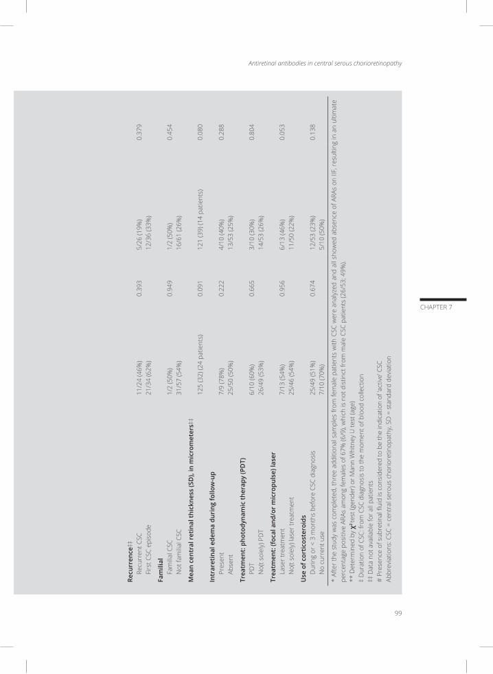
Antiretinal antibodies in central serous chorioretinopathy
99
CHAPTER 7
Tabl
e 3.
Pre
senc
e of
ant
iretin
al a
ntib
odie
s in
rela
tion
to c
linic
al c
hara
cter
istic
s of
cen
tral
ser
ous
chor
iore
tinop
athy
Stai
ning
of a
ny r
etin
al
laye
r(s)
(N=3
2/59
)P-
valu
eSt
aini
ng o
f pho
tore
cept
ors
(N=1
7/63
)P-
valu
e
Gen
der
Mal
eFe
mal
e 26
/53
(49%
)6/
6 (1
00%
)*0.
018
**13
/56
(23%
)4/
7 (5
7%)
0.07
8 **
Mea
n ag
e (S
D),
in y
ears
51 (9
)0.
675
**50
(9)
0.73
9 **
Med
ian
dura
tion
of C
SC (m
in-m
ax),
in d
ays
‡26
1 (3
-783
2)0.
299
530
(3-6
570)
0.44
6
Stag
e‡‡
Foca
l CSC
Diff
use
CSC
23/4
7 (4
7%)
8/11
(73%
)0.
066
13/5
1 (2
6%)
3/11
(27%
)0.
405
Acti
vity
**, #
Activ
eN
ot a
ctiv
e20
/36
(56%
)4/
9 (4
4%)
0.78
212
/38
(32%
)2/
10 (2
0%)
0.57
3
Late
ralit
y of
SRF
dur
ing
follo
w-u
pU
nila
tera
lBi
late
ral
15/3
0 (5
0%)
17/2
9 (5
9%)
0.49
69/
31 (2
9%)
8/32
(25%
)0.
645
Recu
rren
ce‡‡
Recu
rren
t CSC
Firs
t CSC
epi
sode
11
/24
(46%
)21
/34
(62%
)0.
393
5/26
(19%
)12
/36
(33%
)0.
379
Fam
ilial
Fa
mili
al C
SCN
ot fa
mili
al C
SC1/
2 (5
0%)
31/5
7 (5
4%)
0.94
91/
2 (5
0%)
16/6
1 (2
6%)
0.45
4
Mea
n ce
ntra
l ret
inal
thic
knes
s (S
D),
in m
icro
met
ers‡
‡12
5 (3
2) (2
4 pa
tient
s)0.
091
121
(39)
(14
patie
nts)
0.08
0
Intr
aret
inal
ede
ma
duri
ng fo
llow
-up
Pres
ent
Abse
nt7/
9 (7
8%)
25/5
0 (5
0%)
0.22
24/
10 (4
0%)
13/5
3 (2
5%)
0.28
8
Trea
tmen
t: p
hoto
dyna
mic
ther
apy
(PD
T)PD
TN
o(t s
olel
y) P
DT
6/10
(60%
)26
/49
(53%
)0.
665
3/10
(30%
)14
/53
(26%
)0.
804
Trea
tmen
t: (f
ocal
and
/or
mic
ropu
lse)
lase
rLa
ser t
reat
men
tN
o(t s
olel
y) la
ser t
reat
men
t7/
13 (5
4%)
25/4
6 (5
4%)
0.95
66/
13 (4
6%)
11/5
0 (2
2%)
0.05
3
Use
of c
orti
cost
eroi
dsD
urin
g or
< 3
mon
ths
befo
re C
SC d
iagn
osis
N
o cu
rren
t use
25/4
9 (5
1%)
7/10
(70%
)0.
674
12/5
3 (2
3%)
5/10
(50%
)0.
138
* Af
ter
the
stud
y w
as c
ompl
eted
, thr
ee a
dditi
onal
sam
ples
from
fem
ale
patie
nts
with
CSC
wer
e an
alyz
ed a
nd a
ll sh
owed
abs
ence
of A
RAs
on II
F, r
esul
ting
in a
n ul
timat
e pe
rcen
tage
pos
itive
ARA
s am
ong
fem
ales
of 6
7% (6
/9),
whi
ch is
not
dis
tinct
from
mal
e CS
C pa
tient
s (2
6/53
; 49%
).**
Det
erm
ined
by
χ2 te
st (g
ende
r) o
r Man
n W
hitn
ey U
test
(age
)‡
Dur
atio
n of
CSC
: fro
m C
SC d
iagn
osis
to th
e m
omen
t of b
lood
col
lect
ion
‡‡ D
ata
not a
vaila
ble
for a
ll pa
tient
s#
Pres
ence
of s
ubre
tinal
flui
d is
con
side
red
to b
e th
e in
dica
tion
of ‘a
ctiv
e’ C
SCAb
brev
iatio
ns: C
SC =
cen
tral
ser
ous
chor
iore
tinop
athy
, SD
= s
tand
ard
devi
atio
n
100
CHAPTER 7
immune system in the pathogenesis of CSC. The mere presence of serum autoantibodies does
not necessarily indicate an autoimmune basis of the disease. Moreover, ARAs were not found in
all patients and exhibited a wide variety.
The presence of ARAs in serum of CSC patients might be hypothetically explained by their immune
predisposition. CSC can occur in patients with various immune-mediated diseases including
membranoproliferative glomerulonephritis and systemic lupus erythematosus.48,49 It is unclear
whether CSC in these patients is only caused by glucocorticoid treatment prescribed for these
conditions or if it is also influenced by the presence of inflammatory disease.22 Another potential
systemic mechanism in the development of ARAs could be the mimicry between ocular antigens
and microbial proteins, brought up by a previous systemic infection.50 However, the presence of
antecedent infectious disorders in CSC has not previously been assigned as its cause. A higher
prevalence of Helicobacter pylori infections in CSC patients has been described, although a firm
association has never been proven.14,51,52 Moreover, autoimmune responses would be rather
expected in a female-dominated disease. In CSC, particularly men between the age of 25 and 55
are affected, analogous to the phase of life in which highest androgen levels can be detected,
suggesting a possible role of this hormone within the development of CSC. In our study, the role
of the immune system in CSC seemed to be most important in female CSC patients, as ARAs
occurred in all six initially included female CSC patients. However, after our study was completed,
we had the possibility to analyse three extra samples from female patients with CSC. All three
samples showed absence of ARAs on IIF, resulting in similar occurrences of ARAs in male and
female CSC patients (Table 3).
So far it is unclear whether ARAs are involved in the pathogenesis of CSC or whether their pres-
ence represents a secondary epiphenomenon. As CSC is mainly a disease of the choroid and RPE,
experiments assessing antibodies directed against the choroid and RPE could give more insight
to the pathogenesis.1-3,13 The assessment of intraocular ARAs might help to clarify the possible
pathogenic role of ARAs.30
The prevalence of ARAs in serum of blood bank donors of 17% is similar to the prevalences of
ARAs in healthy controls in previous studies.29,30,43,45,46,53 One could argue that some of the blood
donors might have ocular conditions which could also lead to the formation of ARAs. However,
this would not affect our results as we found a difference in the prevalences of ARAs even with
our blood bank controls.
In conclusion, serum ARAs are more common in CSC patients than in healthy controls. No clear
association between the presence of ARAs and clinical CSC characteristics could be identified in
the current cohort. To unravel the possible involvement of autoimmune reactivity in the patho-
genesis of CSC, further research is warranted.
Antiretinal antibodies in central serous chorioretinopathy
101
CHAPTER 7
REFERENCES
1. Gemenetzi M, De Salvo G, Lotery AJ. Central serous chorioretinopathy: an update on pathogenesis and
treatment. Eye (London, England). 2010;24(12):1743-1756.
2. Yannuzzi LA. Central serous chorioretinopathy: a personal perspective. American journal of ophthalmol-
ogy. 2010;149(3):361-363.
3. Daruich A, Matet A, Dirani A, et al. Central serous chorioretinopathy: Recent findings and new physiopa-
thology hypothesis. Progress in retinal and eye research. 2015;48:82-118.
4. Bujarborua D. Long-term follow-up of idiopathic central serous chorioretinopathy without laser. Acta
ophthalmologica Scandinavica. 2001;79(4):417-421.
5. Gilbert CM, Owens SL, Smith PD, Fine SL. Long-term follow-up of central serous chorioretinopathy. The
British journal of ophthalmology. 1984;68(11):815-820.
6. Stewart JM. Half dose verteporfin PDT for central serous chorioretinopathy. The British journal of oph-
thalmology. 2006;90(7):805-806.
7. Brancato R, Scialdone A, Pece A, Coscas G, Binaghi M. Eight-year follow-up of central serous chorioreti-
nopathy with and without laser treatment. Graefe’s archive for clinical and experimental ophthalmology
= Albrecht von Graefes Archiv fur klinische und experimentelle Ophthalmologie. 1987;225(3):166-168.
8. Levine R, Brucker AJ, Robinson F. Long-term follow-up of idiopathic central serous chorioretinopathy by
fluorescein angiography. Ophthalmology. 1989;96(6):854-859.
9. Loo RH, Scott IU, Flynn HW, Jr., et al. Factors associated with reduced visual acuity during long-term
follow-up of patients with idiopathic central serous chorioretinopathy. Retina (Philadelphia, Pa.).
2002;22(1):19-24.
10. Otsuka S, Ohba N, Nakao K. A long-term follow-up study of severe variant of central serous chorioreti-
nopathy. Retina (Philadelphia, Pa.). 2002;22(1):25-32.
11. Wang MS, Sander B, Larsen M. Retinal atrophy in idiopathic central serous chorioretinopathy. American
journal of ophthalmology. 2002;133(6):787-793.
12. Wong R, Chopdar A, Brown M. Five to 15 year follow-up of resolved idiopathic central serous chorioreti-
nopathy. Eye (Lond). 2004;18(3):262-268.
13. Liew G, Quin G, Gillies M, Fraser-Bell S. Central serous chorioretinopathy: a review of epidemiology and
pathophysiology. Clinical & experimental ophthalmology. 2013;41(2):201-214.
14. Wong KH, Lau KP, Chhablani J, Tao Y, Li Q, Wong IY. Central serous chorioretinopathy: what we have learnt
so far. Acta Ophthalmol. 2016;94(4):321-325.
15. Kitzmann AS, Pulido JS, Diehl NN, Hodge DO, Burke JP. The incidence of central serous chorioretinopathy
in Olmsted County, Minnesota, 1980-2002. Ophthalmology. 2008;115(1):169-173.
16. Bouzas EA, Scott MH, Mastorakos G, Chrousos GP, Kaiser-Kupfer MI. Central serous chorioretinopathy
in endogenous hypercortisolism. Archives of ophthalmology. 1993;111(9):1229-1233.
17. Carvalho-Recchia CA, Yannuzzi LA, Negrao S, et al. Corticosteroids and central serous chorioretinopathy.
Ophthalmology. 2002;109(10):1834-1837.
18. Jonas JB, Kamppeter BA. Intravitreal triamcinolone acetonide and central serous chorioretinopathy. Br J
Ophthalmol. 2005;89(3):386-387.
19. Haimovici R, Koh S, Gagnon DR, Lehrfeld T, Wellik S. Risk factors for central serous chorioretinopathy: a
case-control study. Ophthalmology. 2004;111(2):244-249.
20. Jenkins CD, Rosenman RH, Friedman M. Development of an objective psychological test for the deter-
mination of the coronary-prone behavior pattern in employed men. Journal of chronic diseases.
1967;20(6):371-379.
102
CHAPTER 7
21. Yannuzzi LA. Type-A behavior and central serous chorioretinopathy. Retina (Philadelphia, Pa.).
1987;7(2):111-131.
22. Wang M, Munch IC, Hasler PW, Prunte C, Larsen M. Central serous chorioretinopathy. Acta ophthalmo-
logica. 2008;86(2):126-145.
23. Weenink AC, Borsje RA, Oosterhuis JA. Familial chronic central serous chorioretinopathy. Ophthalmo-
logica. Journal international d’ophtalmologie. International journal of ophthalmology. Zeitschrift fur
Augenheilkunde. 2001;215(3):183-187.
24. Park DW, Schatz H, Gaffney MM, McDonald HR, Johnson RN, Schaeffer D. Central serous chorioretinopathy
in two families. European journal of ophthalmology. 1998;8(1):42-47.
25. Miki A, Kondo N, Yanagisawa S, Bessho H, Honda S, Negi A. Common variants in the complement factor H
gene confer genetic susceptibility to central serous chorioretinopathy. Ophthalmology. 2014;121(5):1067-
1072.
26. Schubert C, Pryds A, Zeng S, et al. Cadherin 5 is regulated by corticosteroids and associated with central
serous chorioretinopathy. Human mutation. 2014;35(7):859-867.
27. Breukink MB, Schellevis RL, Boon CJ, et al. Genomic Copy Number Variations of the Complement Com-
ponent C4B Gene Are Associated With Chronic Central Serous Chorioretinopathy. Invest Ophthalmol Vis
Sci. 2015;56(9):5608-5613.
28. Morohoshi K, Ohbayashi M, Patel N, Chong V, Bird AC, Ono SJ. Identification of anti-retinal antibodies in
patients with age-related macular degeneration. Exp Mol Pathol. 2012;93(2):193-199.
29. Patel N, Ohbayashi M, Nugent AK, et al. Circulating anti-retinal antibodies as immune markers in age-re-
lated macular degeneration. Immunology. 2005;115(3):422-430.
30. Ten Berge JC, Schreurs MW, Vermeer J, Meester-Smoor MA, Rothova A. Prevalence and clinical impact of
antiretinal antibodies in uveitis. Acta Ophthalmol. 2016;94(3):282-288.
31. Kubicka-Trzaska A, Wilanska J, Romanowska-Dixon B, Sanak M. Serum anti-endothelial cell antibodies in
patients with age-related macular degeneration treated with intravitreal bevacizumab. Acta Ophthalmol.
2016;94(7):e617-e623.
32. Caspi RR. A look at autoimmunity and inflammation in the eye. J Clin Invest. 2010;120(9):3073-3083.
33. Stanford MR, Graham E, Kasp E, Sanders MD, Dumonde DC. A longitudinal study of clinical and immuno-
logical findings in 52 patients with relapsing retinal vasculitis. Br J Ophthalmol. 1988;72(6):442-447.
34. Kijlstra A, La Heij E, Hendrikse F. Immunological factors in the pathogenesis and treatment of age-related
macular degeneration. Ocul Immunol Inflamm. 2005;13(1):3-11.
35. Kumar M, Gupta RM, Nema HV. Role of autoimmunity in retinitis pigmentosa. Ann Ophthalmol.
1983;15(9):838-840.
36. Nussenblatt RB, Liu B, Li Z. Age-related macular degeneration: an immunologically driven disease. Curr
Opin Investig Drugs. 2009;10(5):434-442.
37. Nussenblatt RB, Liu B, Wei L, Sen HN. The immunological basis of degenerative diseases of the eye. Int
Rev Immunol. 2013;32(1):97-112.
38. Patel M, Chan CC. Immunopathological aspects of age-related macular degeneration. Semin Immuno-
pathol. 2008;30(2):97-110.
39. Morohoshi K, Goodwin AM, Ohbayashi M, Ono SJ. Autoimmunity in retinal degeneration: autoimmune
retinopathy and age-related macular degeneration. J Autoimmun. 2009;33(3-4):247-254.
40. Shimazaki K, Jirawuthiworavong GV, Heckenlively JR, Gordon LK. Frequency of anti-retinal antibodies in
normal human serum. J Neuro-Ophthalmol. 2008;28(1):5-11.
41. Gurne DH, Tso MO, Edward DP, Ripps H. Antiretinal antibodies in serum of patients with age-related
macular degeneration. Ophthalmology. 1991;98(5):602-607.
42. Heckenlively JR, Aptsiauri N, Nusinowitz S, Peng C, Hargrave PA. Investigations of antiretinal antibodies in

Antiretinal antibodies in central serous chorioretinopathy
103
CHAPTER 7
pigmentary retinopathy and other retinal degenerations. Trans Am Ophthalmol Soc. 1996;94:179-200;
discussion 200-176.
43. Kubicka-Trzaska A. Typy przeciwcial przeciwsiatkowkowych (PPS) u chorych na samoistne zapalenie
tylnego odcinka blony naczyniowej w tescie immunofluorescencji posredniej. Klin Oczna. 2004;106(1-
2):45-49.
44. Kubicka-Trzaska A, Wilanska J, Romanowska-Dixon B, Sanak M. Circulating anti-retinal antibodies in
response to anti-angiogenic therapy in exudative age-related macular degeneration. Acta Ophthalmol.
2014;92(8):e610-614.
45. Chant SM, Heckenlively J, Meyers-Elliott RH. Autoimmunity in hereditary retinal degeneration. I. Basic
studies. Br J Ophthalmol. 1985;69(1):19-24.
46. Whittle RM, Wallace GR, Whiston RA, Dumonde DC, Stanford MR. Human antiretinal antibodies in toxo-
plasma retinochoroiditis. Br J Ophthalmol. 1998;82(9):1017-1021.
47. de Jong EK, Breukink MB, Schellevis RL, et al. Chronic central serous chorioretinopathy is associated with
genetic variants implicated in age-related macular degeneration. Ophthalmology. 2015;122(3):562-570.
48. Shimura M, Tatehana Y, Yasuda K, Saito S, Sasaki T, Tamai M. Choroiditis in systemic lupus erythematosus:
systemic steroid therapy and focal laser treatment. Japanese journal of ophthalmology. 2003;47(3):312-
315.
49. Awan MA, Grierson DJ, Walker S. Bilateral macular sub-retinal fluid and retinal pigment epithelial
detachment associated with type 2 membrano-proliferative glomerulonephritis. Clinical & experimental
optometry : journal of the Australian Optometrical Association. 2008;91(5):476-479.
50. Adamus G, Chan CC. Experimental autoimmune uveitides: multiple antigens, diverse diseases. Int Rev
Immunol. 2002;21(2-3):209-229.
51. Mauget-Faysse M, Kodjikian L, Quaranta M, et al. Role de l’Helicobacter pylori dans la chorioretinopathie
sereuse centrale et l’epitheliopathie retinienne diffuse. Resultats de la premiere etude prospective pilote.
J Fr Ophtalmol. 2002;25(10):1021-1025.
52. Cotticelli L, Borrelli M, D’Alessio AC, et al. Central serous chorioretinopathy and Helicobacter pylori. Eur J
Ophthalmol. 2006;16(2):274-278.
53. Heckenlively JR, Jordan BL, Aptsiauri N. Association of antiretinal antibodies and cystoid macular edema
in patients with retinitis pigmentosa. Am J Ophthalmol. 1999;127(5):565-573.


8ANTIRETINAL AND ANTINUCLEAR ANTIBODIES IN UVEITIS WITH LATENT AND ACTIVE TUBERCULOSIS
Rina La Distia Nora, Josianne C.E.M. ten Berge, Aniki Rothova, Marco W.J. Schreurs
Accepted for publication in Acta Ophthalmologica

Antibodies in uveitis with latent and active tuberculosis
107
CHAPTER 8
The pathogenesis of uveitis in the setting of active and latent tuberculosis (TB) is not entirely clari-
fied. Next to genuine infection, an important part of pathogenesis was attributed to (auto)immune
reactions initiated by Mycobacterium tuberculosis (Mtb).1 Infection with Mtb can be associated
with the production of diverse serum autoantibodies.2 Herein, we investigate the influence of
(latent) TB on the presence of serum antinuclear and antiretinal autoantibodies (ANA and ARA)
in Indonesian patients with uveitis.
Blood samples from patients with uveitis associated with active (not yet treated) pulmonary TB
(N=10) and uveitis of unknown cause (N=85) were collected from June 2014 until May 2015.
Classification of patients was performed according to SUN classification, and specific diagnoses
were determined after the basic work-up for uveitis as indicated in our previous publication.3 The
diagnosis of active pulmonary TB was based on clinical and/or microbiological and radiological
findings.3 This study was performed with the approval of the local medical ethical committee.
All patients underwent QuantiFERON-Tb Gold (QFT) (Cellestis Inc., Carnegie, Australia). Screening
for the presence of ANA using HEp-2 cells (Inova, San Diego, California) and the presence of ARA
using primate retinal tissue (Euroimmun, Lubeck, Germany) was performed by indirect immu-
nofluorescence as described before.4 Logistic regressions with correction for age and gender
were performed using SPSS to analyse differences in the presence of ANA and ARA between the
diagnosis groups.
Patients with uveitis of unknown cause were divided according to their QFT results in 58 patients
with latent TB (QFT positive) and 27 patients without evidence of prior TB exposure (QFT negative).
All QFT positive patients were assessed by the pulmonologist and examined for the possible
presence of pulmonary and extrapulmonary TB, but no cases of extrapulmonary involvement
were found. The group with an unknown cause of uveitis and latent TB consisted of more female
patients compared to the other groups (72% vs 30% in uveitis in the setting of active pulmonary
TB group and 41% in the uveitis of unknown cause and QFT negative group, p-value 0.044) and
older age patients (mean age 46 years vs 40 years in uveitis in the setting of active pulmonary
TB group and 39 years in uveitis of unknown cause and QFT negative group, p-value 0.003). The
median QFT value in patients with uveitis of unknown cause and latent TB was 5.0 IU/ml, and in
patients with known tuberculosis induced uveitis 1.7 IU/ml.
Patients’ serum ANA and ARA results are shown in Table 1. Patients with uveitis and either active
or latent TB were characterized by high prevalence of systemic autoreactivity (ANA positive). In
contrast, a higher proportion of organ-specific autoreactivity (ARA positive) was found in uveitis
patients without evidence of any previous contact with Mtb. Induction of ANA, reported previ-
ously for active TB, apparently also occurs in latent TB.5 Interestingly, organ-specific autoreactivity
(ARA) appears to be suppressed in both active and latent TB, however, the local production of
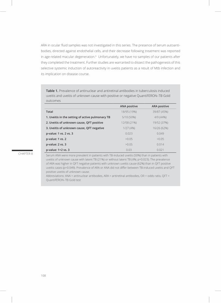
108
CHAPTER 8
ARA in ocular fluid samples was not investigated in this series. The presence of serum autoanti-
bodies, directed against endothelial cells, and their decrease following treatment was reported
in age-related macular degeneration.6 Unfortunately, we have no samples of our patients after
they completed the treatment. Further studies are warranted to dissect the pathogenesis of this
selective systemic induction of autoreactivity in uveitis patients as a result of Mtb infection and
its implication on disease course.
Table 1. Prevalence of antinuclear and antiretinal antibodies in tuberculosis induced uveitis and uveitis of unknown cause with positive or negative QuantiFERON–TB Gold outcomes
ANA positive ARA positive
Total 18/95 (19%) 39/87 (45%)
1. Uveitis in the setting of active pulmonary TB 5/10 (50%) 4/9 (44%)
2. Uveitis of unknown cause, QFT positive 12/58 (21%) 19/52 (37%)
3. Uveitis of unknown cause, QFT negative 1/27 (4%) 16/26 (62%)
p-value: 1 vs. 2 vs. 3 0.023 0.049
p-value: 1 vs. 2 >0.05 >0.05
p-value: 2 vs. 3 >0.05 0.014
p-value: 1+2 vs. 3 0.03 0.021
Serum ANA were more prevalent in patients with TB-induced uveitis (50%) than in patients with uveitis of unknown cause with latent TB (21%) or without latent TB (4%; p=0.023). The prevalence of ARA was higher in QFT negative patients with unknown uveitis cause (62%) than in QFT positive uveitis cases (p=0.049). Prevalence of ARA or ANA did not differ between TB-induced uveitis and QFT positive uveitis of unknown cause. Abbreviations: ANA = antinuclear antibodies, ARA = antiretinal antibodies, OR = odds ratio, QFT = QuantiFERON–TB Gold test

Antibodies in uveitis with latent and active tuberculosis
109
CHAPTER 8
REFERENCES
1. Wroblewski KJ, Hidayat AA, Neafie RC, Rao NA, Zapor M. Ocular tuberculosis: A clinicopathologic and
molecular study. Ophthalmology. 2011;118(4):772-777.
2. Shen CY, Hsieh SC, Yu CL, Wang JY, Lee LN, Yu CJ. Autoantibody prevalence in active tuberculosis: reactive
or pathognomonic? BMJ Open. 2013;3(7).
3. La Distia Nora R, Sitompul R, Bakker M, et al. Tuberculosis and other causes of uveitis in Indonesia. Eye
(Lond). 2017.
4. Ten Berge JC, Schreurs MW, Vermeer J, Meester-Smoor MA, Rothova A. Prevalence and clinical impact of
antiretinal antibodies in uveitis. Acta Ophthalmol. 2016;94(3):282-288.
5. Elkon K, Casali P. Nature and functions of autoantibodies. Nat Clin Pract Rheumatol. 2008;4(9):491-498.
6. Kubicka-Trzaska A, Wilanska J, Romanowska-Dixon B, Sanak M. Serum anti-endothelial cell antibodies in
patients with age-related macular degeneration treated with intravitreal bevacizumab. Acta Ophthalmol.
2016;94(7):e617-e623.

ANTIRETINAL ANTIBODIES IN MEXICAN CHILDREN WITH SEVERE PARS PLANITIS
Luz Elena Concha-del Río, Maria de Lourdes Arellanes-García, Marco W.J. Schreurs, Aniki Rothova, Josianne C.E.M. ten Berge
Submitted for publication
9
112
CHAPTER 9
ABSTRACT
Pars planitis (PP) is a subtype of intermediate uveitis and a particularly severe form of PP has
been described in pediatric Mexican patients. Although the pathogenesis of PP is not clarified,
autoimmune reactions have been suggested to play a role. This study investigates the presence
and possible clinical role of serum antiretinal antibodies (ARAs) of Mexican children with PP (N=16)
and age-matched controls with strabismus (N= 19). All samples were tested for the presence of
ARAs by indirect immunofluorescence using primate retinal tissue. Serum ARAs were noted in
57% (8/14) of PP patients and in 40% (6/15) of controls (p=0.356), and no correlation was found
between the presence of ARAs and clinical characteristics of PP. Insight in the cellular component
of the immune system and / or analyses of intraocular fluids of PP eyes might provide further
insight in the pathogenesis of PP.
Antiretinal antibodies in pediatric pars planitis
113
CHAPTER 9
INTRODUCTION
Pars planitis (PP) is characterized predominantly by vitreous inflammation, with or without vit-
reous condensations (“snowballs”), pars plana exudates (“snowbanks”) in at least one eye, and
peripheral retinal vasculitis. An exceptionally severe form of PP has been reported in children with
uveitis from Mexico.1-3 The clinical course of PP is usually chronic with exacerbations and has great
on patients’ well-being and quality of life. Despite the numerous reports on intermediate uveitis,
the specific cause and pathogenesis of PP are unknown.4 PP is considered to be of autoimmune
origin, but possible inciting antigens are so far entirely unknown. In this short report, we assess
the presence of antiretinal antibodies (ARAs) in serum of in young patients with PP from Mexico
and evaluate their possible clinical role.
METHODS
We prospectively collected serum samples from 16 patients with PP and 19 age-matched children
with strabismus who served as controls, from the Eye Inflammatory Disease Clinic at Hospital ”Dr.
Luis Sánchez Bulnes”, Asociación Para Evitar la Ceguera in Mexico. Clinical data were collected
from medical data files (Table 1). The study was performed in accordance with the Declaration
of Helsinki and in agreement with the institutional regulations and approval of local institutional
review boards.
Table 1. Clinical characteristics of patients with pars planitis
Characteristic Number (%)
Age onset (median; interquartile range) 8; 6-10
Gender (male:female) 9:7
Bilateral pars planitis 13/15 (87%)1
Anterior chamber inflammation 13/16 (81%)
Anterior synechiae 2/16 (13%)
Posterior synechiae 6/16 (38%)
Cyclitic membrane 6/16 (38%)
Snowballs 16/16 (100%)
Snowbanking 7/15 (47%)*
Phlebitis 15/15 (100%)*
Systemic treatment** 14/16 (88%)
* Data not available for all patients ** Prednisone and/or systemic immunosuppressive therapy
114
CHAPTER 9
Screening of serum samples for the presence of ARAs was performed by indirect immunoflu-
orescence using primate retinal tissue (Euroimmun, Lubeck, Germany), as described before.5
The determination of ARAs was not possible in 2 patients and 4 controls, due to positivity of
antinuclear antibodies which mask the possible presence of ARAs. Mann Whitney U tests for
continuous data and Chi-square tests for categorical data were performed to analyse differences
on the presence of ARAs between the diagnosis groups and specific clinical characteristics of PP.
RESULTS AND DISCUSSION
Serum ARAs were found in 57% (8/14) of PP patients and in 40% (6/15) of controls (p=0.356).
Staining of photoreceptors was the most frequently observed pattern with similar prevalence in
both groups. The presence of ARAs was not associated to any clinical characteristic of PP. The
high prevalence of ARAs in our young control patients without retinal disease (40%) is remark-
able and much higher than the 17% observed in the general population in Europe.6 The possible
explanation might be a higher exposure to exogenous antigens such as infections in Mexican
children. Despite the high prevalence of 57% of serum in young patients with PP, our results
suggest that ARAs are not the primary cause of the ocular disease, but possibly represent a
secondary phenomenon.
In conclusion, the prevalence of serum ARAs is similar in pediatric PP and controls from Mexico.
Further assessment of potential infectious agents and insight in the cellular component of the
immune system and/or assessment of intraocular ARAs of PP eyes might provide further insight
in the pathogenesis of PP.

Antiretinal antibodies in pediatric pars planitis
115
CHAPTER 9
REFERENCES
1. Arellanes-Garcia L, Navarro-Lopez P, Concha-Del Rio LE, Unzueta-Medina JA. Idiopathic intermediate
uveitis in childhood. Int Ophthalmol Clin. 2008;48(3):61-74.
2. Arellanes-Garcia L, Navarro-Lopez L, Recillas-Gispert C. Pars planitis in the Mexican Mestizo population:
ocular findings, treatment, and visual outcome. Ocul Immunol Inflamm. 2003;11(1):53-60.
3. Ortega-Larrocea G, Arellanes-Garcia L. Pars planitis: epidemiology and clinical outcome in a large com-
munity hospital in Mexico City. Int Ophthalmol. 1995;19(2):117-120.
4. Przezdziecka-Dolyk J, Wegrzyn A, Turno-Krecicka A, Misiuk-Hojlo M. Immunopathogenic Background of
Pars Planitis. Arch Immunol Ther Exp (Warsz). 2016;64(2):127-137.
5. Ten Berge JC, Schreurs MW, Vermeer J, Meester-Smoor MA, Rothova A. Prevalence and clinical impact of
antiretinal antibodies in uveitis. Acta Ophthalmol. 2016;94(3):282-288.
6. Alaez C, Arellanes L, Vazquez A, et al. Classic Pars Planitis: strong correlation of class II genes with gender
and some clinical features in Mexican Mestizos. Hum Immunol. 2003;64(10):965-972.

10AUTOANTIBODY PROFILING IN INTRAOCULAR FLUID OF PATIENTS WITH UVEITIS
Josianne C.E.M. ten Berge, Marco W.J. Schreurs, Joost van Rosmalen, Aniki Rothova
Submitted for publication
118
CHAPTER 10
ABSTRACT
A high prevalence of serum antiretinal antibodies (ARAs) in patients with uveitis has been previ-
ously described, though their clinical role remains elusive. Assessment of intraocular ARAs may
provide further insight into the pathogenesis of diverse uveitis entities. In this study we investigate
the prevalence of multiple specific anti-ocular antibodies (AOcAs), including ARAs, in intraocular
fluid of patients with uveitis. Autoantibody profiling with 188 different ocular antigens was per-
formed by a multiplex immunoassay with intraocular fluid samples of 76 patients with uveitis.
Clinical data from uveitis patients were collected and statistical analyses were executed to evalu-
ate associations between intraocular AOcAs and clinical characteristics. Controls consisted of 19
intraocular fluid samples from cataract patients. A spectrum of 22 different AOcAs was present
in higher levels in patients with uveitis than in controls (p≤0.05), but in moderately elevated titers
(<2x). High elevations of intraocular AOcAs in uveitis (>5x compared to cataract) were observed
in varicella zoster virus-induced uveitis, multiple sclerosis-associated uveitis and patients with
unexplained uveitis but positive quantiferon test. Presence of macular edema was associated
with increased intraocular levels of tyrosinase antibodies. Our results show that patients with
uveitis are characterized by the presence of a broad spectrum of moderately elevated levels of
intraocular AOcAs, and high intraocular AOcA levels were found in several specific uveitis entities.
This study favors secondary production of AOcAs and not their inciting role.

Autoantibody profiling in intraocular fluid of patients with uveitis
119
CHAPTER 10
INTRODUCTION
Uveitis is a severe ocular disease, which can result in permanent blindness. Uveitis has multiple
causes including infections and shows strong associations with various systemic immune-medi-
ated diseases. The pathogenesis of most uveitis entities is not fully understood, but the immune
system plays a crucial role; especially the development of autoimmune intraocular reactions in
non-infectious uveitis has been repeatedly proposed, but scarcely proven. Serum antibodies
directed against retinal tissue were more prevalent in patients with uveitis compared to healthy
controls.1 It has been hypothesized that the antiretinal antibodies (ARAs) in uveitis might either
incite the ocular disease or represent a secondary epiphenomenon induced by retinal damage.
The possible pathogenic role of these ARAs is unknown, although it has been suggested that ARAs
might aggravate and/or prolong the ocular disease.2-4
The eye is an immune privileged organ and intraocular ARAs might show an entirely different
profile than ARAs found in the peripheral blood. This phenomenon has been shown previously
in infectious uveitis, in which local production of specific antibodies is regarded as indirect proof
of the intraocular infection.5 Similarly, in neurological diseases including autoimmune encepha-
litis and multiple sclerosis the importance of analysis of cerebrospinal fluid has been previously
proven.6, 7 Currently, antigen bead arrays are successfully being used for the analysis of autoanti-
bodies. This technique enables analysis of very small volumes (such as intraocular fluids) towards
hundreds of different antigens and its potential for ARA detection has been suggested.8
Herein, we perform an autoantibody profiling of intraocular fluid samples of patients with diverse
uveitis entities and assess the prevalence of antibodies directed against 188 different ocular
antigens as potential targets, and relate the results to clinical manifestations of uveitis.
METHODS
Remainders of diagnostic intraocular fluid samples from 76 patients with uveitis were collected
from the Laboratory of Virology of the Erasmus MC, University Medical Center Rotterdam
between February 2009 and April 2015. Intraocular fluid samples of 19 patients with cataract
stored in the biobank in the same time period were used as controls. All intraocular fluid samples
were stored at -80°C. This study was approved by the local ethical committee from the Erasmus
University Medical Center (Medical Ethics Committee Erasmus MC) and adhered to the tenets of
the Declaration of Helsinki.
All uveitis patients were classified according to the localization of uveitis using the Standardization
of Uveitis Nomenclature SUN criteria.9,10 Patients underwent a standardized diagnostic protocol
120
CHAPTER 10
based on this anatomical site of inflammation. The protocol included chest radiography, erythro-
cyte sedimentation rate, blood counts, serum angiotensin-converting enzyme levels, serology for
syphilis and Lyme disease and interferon gamma release assay (IGRA) test (QuantiFERON–TB Gold
In-Tube test). In patients with anterior uveitis or panuveitis Human Leukocyte Antigen (HLA)-B27
testing was also performed. According to the clinical uveitis manifestations, further examinations
were added (tailored approach). Specific diagnoses were determined after the various diagnostic
procedures were completed. The diagnosis of intraocular infections was always confirmed by
polymerase chain reaction (PCR) and/or Goldmann-Wittmer coefficient in intraocular fluid. The
diagnosis of Fuchs uveitis syndrome (FUS) was based on clinical characteristics. The diagnosis of
sarcoidosis was either histologically proven or based on chest imaging in patients with otherwise
unexplained uveitis . All other specific diagnoses were made according to current diagnostic
criteria.
Clinical data of patients with uveitis were collected from medical data files. We registered age,
gender, location of uveitis and specific cause of uveitis. Further, we registered the following char-
acteristics at the moment of sample collection: duration of uveitis, presence of cystoid macular
edema (CME), presence of vasculitis, use of systemic immunosuppressive medication and activity
of uveitis.
Autoantibody profiling with 188 different ocular antigens, representing 97 unique ocular proteins,
was performed on intraocular fluid samples from patients with uveitis (N=76) and cataract (N=19;
Supplementary Table). Antigens were selected based on potential relevance to ocular diseases
according to literature or previous retinal immunohistochemistry staining. The used antigens
were protein fragments produced within the Human Protein Atlas, designed to have low homol-
ogy to other human proteins and expressed in Escherichia coli with an affinity tag consisting
of six histidines and an albumin-binding domain from streptococcal protein G (His6ABP).11,12 A
multiplex assay, previously validated by immunoblot and immunohistochemistry, was performed
as described before with minor alterations.13 In short, the antigens were covalently coupled to
color-coded magnetic beads to create a bead array. The samples were diluted 1:10 in assay
buffer (0.1% PBS-Tween20, 3% BSA, 160 µg/ml His6ABP), let to pre-block potential antibodies
towards the ABP-domain for 1 hour in room temperature, and subsequently incubated O/N in
room temperature with the bead array. Interactions were fixated with 0.2% PFA for 10 min before
incubation for 30 min with a fluorophore conjugated anti-human IgG Fab fragment. A FlexMap3D
instrument (Luminex Corp.) was used to acquire a read-out. The autoantibody profiling was per-
formed at the SciLifeLab Autoimmunity Profiling Facility in Stockholm, Sweden.
For the statistical analyses, continuous variables were summarized using medians and ranges,
and categorical variables were summarized using percentages. Patient demographics were com-
pared between diagnosis groups using Mann Whitney U tests for continuous data and Fisher’s
Autoantibody profiling in intraocular fluid of patients with uveitis
121
CHAPTER 10
exact tests for categorical data. Linear regressions with correction for age and gender were per-
formed to analyse differences between the diagnosis groups (uveitis or specific uveitis entities
versus cataract). In addition subgroup analyses of these linear regressions were performed to
compare specific uveitis entities with cataract. To analyse differences within the uveitis group we
performed linear regressions with age, gender, location of uveitis (reference category: anterior
uveitis), uveitis entity (reference category: uveitis associated to sarcoidosis), uveitis activity, sys-
temic immunosuppressive therapy, presence of CME and presence of vasculitis as independent
variables. Some specific uveitis entities were not analysed as separate groups (uveitis associated
with herpes simplex virus (HSV), Sjögren syndrome, HLA B27-associated uveitis and birdshot
chorioretinopathy), because their numbers were too small. All regression analyses were per-
formed using the natural logarithm of the measured median fluorescent intensities as dependent
variable. Relative (fold) increases of differences in levels of AOcAs were calculated by exponenti-
ating the estimated regression coefficients. The linear regressions were performed with a robust
MM-type estimation method, to account for the fact that the model residuals were not normally
distributed.14 We used the signal intensities as an indication for the levels of intraocular AOcAs. To
adjust for the multiple comparisons of the different antigens, a Bonferroni correction was applied
(all p-values were multiplied by 188). P-values of ≤0.05 were considered statistically significant. All
statistical tests were two-sided. The analyses were performed using SPSS and R (version 3.3.1),
with the robustbase package for the robust linear regressions.15
RESULTS
The median age of the 76 included uveitis patients was 48 years, and 46% of patients were
males. Median duration of uveitis was two years (ranging from zero to 36 years), and 51 (67%)
cases were active during sample collection. Patients’ characteristics are shown in Table 1. The
most prevalent cause of uveitis in our cohort was sarcoidosis (14/76, 18%). Nine patients with
sarcoidosis-associated uveitis were biopsy proven and in five patients the diagnosis was based
on radiologic criteria. Varicella zoster virus (VZV)-induced uveitis included 4 patients with retinitis
and 3 with anterior uveitis. Cataract patients were older than the uveitis patients (median age of
69 years, p=0.0001) and 42% were males (p>0.05).
The levels of 22 different intraocular AOcAs were higher in uveitis than in controls (all p≤0.05 as
determined by linear regression), but with moderately increased titers (up to 2x compared to
controls). The most significant results were observed in the AOcA levels specific for RPE-retinal G
protein-coupled receptor (number 169; p=0.0031) and retinol dehydrogenase 8 (number 160; p=
0.0042). Levels of intraocular AOcAs varied greatly between different uveitis entities. Intraocular
AOcAs with at least 5-fold increased titers (compared to cataract controls) were observed in
three specific entities: VZV-induced uveitis, multiple sclerosis (MS)-associated uveitis and patients

122
CHAPTER 10
with unexplained uveitis but with a positive quantiferon test (all p<0.05; Table 2 and Figure 1).
Intraocular levels of all AOcAs in FUS (with or without an intraocular rubella virus infection) were
similar to cataract controls (all p>0.05).
Table 1. Characteristics of uveitis patients (N=76)
Characteristic Number (%)
Male-to-female ratio 35 - 41 (46% - 54%)
Median age in years (range) 48 (17 - 86)
Median duration uveitis in years (range) 2 (0 – 36)
LocationAnteriorIntermediatePosteriorPanuveitis
26 (34%)10 (13%)23 (30%)17 (22%)
Specific cause or associationInfectious
RubellaToxoplasma gondiiVaricella zoster virusCytomegalovirusHerpes simplex virus
Systemic diseaseSarcoidosisMultiple sclerosisSjögren syndromeHLA B27 positive
Ocular lymphomaClinical entity
Birdshot chorioretinopathyFuchs uveitis syndrome
UnknownQuantiferon positive
36 (47%)12 (16%)8 (11%)7 (9%)7 (9%)2 (3%)
24 (32%)14 (18%)8 (11%)1 (1%)1 (1%)
5 (7%) 3 (4%)
2 (3%)1 (1%)
8 (11%)6 (8%)
Active uveitis 51 (67%)
Presence of cystoid macular edema 19 (25%)
Presence of vasculitis 16 (21%)
Systemic immunosuppressive treatment 16 (21%)
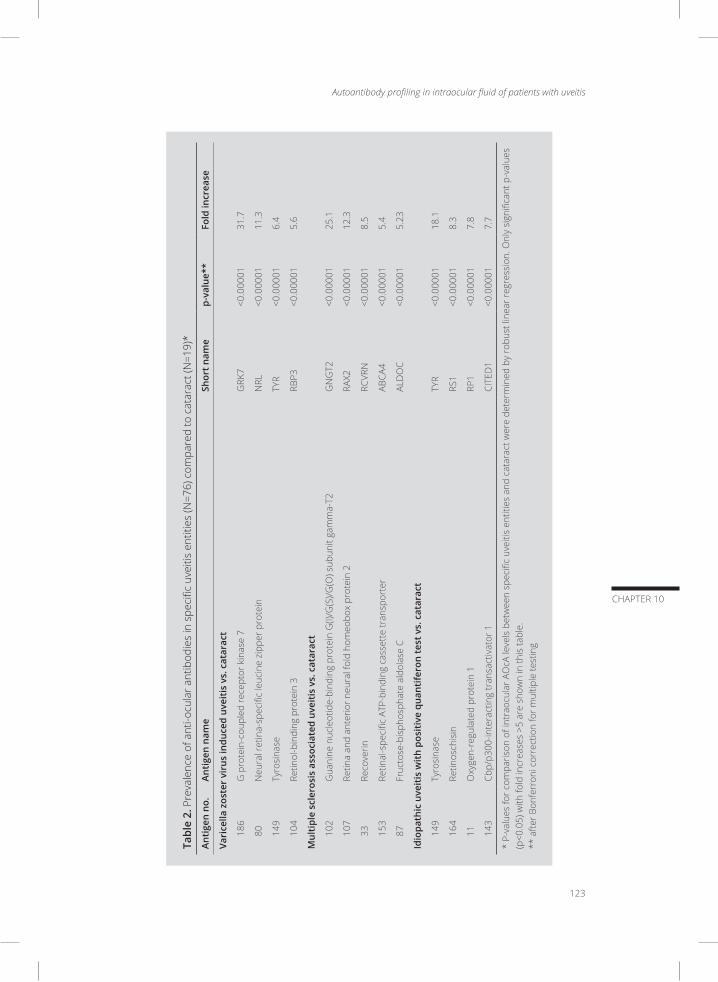
Autoantibody profiling in intraocular fluid of patients with uveitis
123
CHAPTER 10
Tabl
e 2.
Pre
vale
nce
of a
nti-o
cula
r ant
ibod
ies
in s
peci
fic u
veiti
s en
titie
s (N
=76)
com
pare
d to
cat
arac
t (N
=19)
*
Anti
gen
no.
Anti
gen
nam
eSh
ort n
ame
p-va
lue*
*Fo
ld in
crea
se
Vari
cella
zos
ter
viru
s in
duce
d uv
eiti
s vs
. cat
arac
t
186
G p
rote
in-c
oupl
ed re
cept
or k
inas
e 7
GRK
7<0
.000
0131
.7
80N
eura
l ret
ina-
spec
ific
leuc
ine
zipp
er p
rote
inN
RL<0
.000
0111
.3
149
Tyro
sina
seTY
R<0
.000
016.
4
104
Retin
ol-b
indi
ng p
rote
in 3
RBP3
<0.0
0001
5.6
Mul
tipl
e sc
lero
sis
asso
ciat
ed u
veit
is v
s. c
atar
act
102
Gua
nine
nuc
leot
ide-
bind
ing
prot
ein
G(I)
/G(S
)/G(O
) sub
unit
gam
ma-
T2G
NG
T2<0
.000
0125
.1
107
Retin
a an
d an
terio
r neu
ral f
old
hom
eobo
x pr
otei
n 2
RAX2
<0.0
0001
12.3
33Re
cove
rinRC
VRN
<0.0
0001
8.5
153
Retin
al-s
peci
fic A
TP-b
indi
ng c
asse
tte
tran
spor
ter
ABCA
4<0
.000
015.
4
87Fr
ucto
se-b
isph
osph
ate
aldo
lase
CAL
DO
C<0
.000
015.
23
Idio
path
ic u
veit
is w
ith
posi
tive
qua
ntife
ron
test
vs.
cat
arac
t
149
Tyro
sina
seTY
R<0
.000
0118
.1
164
Retin
osch
isin
RS1
<0.0
0001
8.3
11O
xyge
n-re
gula
ted
prot
ein
1RP
1<0
.000
017.
8
143
Cbp/
p300
-inte
ract
ing
tran
sact
ivat
or 1
CITE
D1
<0.0
0001
7.7
* P-
valu
es fo
r com
paris
on o
f int
raoc
ular
AO
cA le
vels
bet
wee
n sp
ecifi
c uv
eitis
ent
ities
and
cat
arac
t wer
e de
term
ined
by
robu
st li
near
regr
essi
on. O
nly
sign
ifica
nt p
-val
ues
(p<0
.05)
with
fold
incr
ease
s >5
are
sho
wn
in th
is ta
ble.
**
afte
r Bon
ferr
oni c
orre
ctio
n fo
r mul
tiple
test
ing

124
CHAPTER 10
Figure 1. Levels of intraocular tyrosinase (number 149) in different uveitis entities and in cataract
* Extreme outliers: <(25th percentile -/- 3x interquartile range) or >(75th percentile +/+ 3x interquartile range)
The AOcA levels within the uveitis population and their associations with clinical characteristics are
shown in Table 3. Significant associations between high levels of specific AOcAs and VZV-induced
uveitis, MS-associated uveitis or unexplained uveitis with positive quantiferon test were identified.
Presence of CME was associated with higher titers of tyrosinase (number 79, p=0.0026), although
with a moderate increased titer compared to uveitis patients without CME (1.4x). Active uveitis
had lower levels of most AOcAs, but these results did not reach significance after Bonferroni
correction. No differences in levels of AOcAs were found between all other clinical characteristics
of uveitis.

Autoantibody profiling in intraocular fluid of patients with uveitis
125
CHAPTER 10
Tabl
e 3.
Pre
senc
e of
ant
i-ocu
lar a
ntib
odie
s co
mpa
red
with
in u
veiti
s (N
=76)
in re
latio
n to
uve
itis
char
acte
ristic
s*
Anti
gen
no.
Anti
gen
nam
eSh
ort n
ame
p-va
lue*
*Fo
ld in
crea
se
Spec
ific
uvei
tis
enti
ties
Vari
cella
zos
ter
viru
s in
duce
d uv
eiti
s
104
Retin
ol-b
indi
ng p
rote
in 3
RBP3
0.00
803.
8
134
Best
roph
in-2
BEST
20.
0054
2.2
Mul
tipl
e sc
lero
sis
asso
ciat
ed u
veit
is
107
Retin
a an
d an
terio
r neu
ral f
old
hom
eobo
x pr
otei
n 2
RAX2
0.00
014.
6
86Ag
e-re
late
d m
acul
opat
hy s
usce
ptib
ility
pro
tein
2AR
MS2
0.00
432.
9
34Vi
sual
pig
men
t-lik
e re
cept
or p
erop
sin
RRH
0.03
722.
4
185
Rod
cGM
P-sp
ecifi
c 3’
,5’-c
yclic
pho
spho
dies
tera
se s
ubun
it al
pha
PDE6
A0.
0006
2.2
Idio
path
ic u
veit
is w
ith
posi
tive
qua
ntife
ron
test
139
Cbp/
p300
-inte
ract
ing
tran
sact
ivat
or 1
CITE
D1
0.00
083.
7
143
Cbp/
p300
-inte
ract
ing
tran
sact
ivat
or 1
CITE
D1
0.02
203.
4
Clin
ical
cha
ract
eris
tics
Pres
ence
of c
ysto
id m
acul
ar e
dem
a
79Ty
rosi
nase
TYR
0.00
261.
4
* P-
valu
es fo
r co
mpa
rison
of i
ntra
ocul
ar A
OcA
leve
ls w
ithin
the
uvei
tis c
ohor
t wer
e de
term
ined
by
robu
st li
near
reg
ress
ion.
Spe
cific
uve
itis
entit
ies
wer
e co
mpa
red
with
sa
rcoi
dosi
s as
soci
ated
uve
itis
as re
fere
nce
cate
gory
. Onl
y si
gnifi
cant
p-v
alue
s w
ith th
e hi
ghes
t fol
d in
crea
ses
are
show
n in
this
tabl
e.
** a
fter B
onfe
rron
i cor
rect
ion
for m
ultip
le te
stin
g
126
CHAPTER 10
DISCUSSION
Our study documents the presence of a broad spectrum of ocular autoantibodies in intraocular
fluids of patients with uveitis patients. Significant differences between the levels of intraocular
AOcAs in uveitis and cataract were identified. However, levels of intraocular AOcAs in uveitis were
only moderately increased (up to 2x compared to cataract), which may have been caused by the
merger of heterogeneous autoantibody profiles of different uveitis entities. Comparison of spe-
cific uveitis entities with cataract showed various high intraocular AOcA levels (>5x) in VZV-induced
uveitis, MS-associated uveitis and idiopathic uveitis with positive quantiferon test. Patients with
CME exhibited moderately increased titers of anti-tyrosinase.
VZV-induced uveitis was one the entities which was associated with high levels of AOcAs. Spe-
cifically, antibodies directed against retinol-binding protein 3 (RBP-3) were present in high levels
(compared to cataract, and other uveitis entities). RBP-3, also known as interphotoreceptor ret-
inoid-binding protein (IRBP) is found primarily in the interphotoreceptor matrix of the retina
and it is thought to transport retinoids between the retinal pigment epithelium (RPE) and the
photoreceptors. RBP-3 antibodies have been previously observed in some ocular diseases,
including autoimmune retinopathy, macular telangiectasia type 2 and age related macular
degeneration.16-18 The RBP-3 antigen has been found to be highly uveitogenic in multiple animal
models.19,20 RBP-3 might represent a non-specific target involved in the pathogenesis different
ocular diseases. Another highly prevalent antibody in VZV-induced uveitis was directed against
bestrophin-2 (Best-2). Within the bestrophin family, bestrophin-1 (Best-1) is the most investigated
and described ocular antigen. Mutations in the BEST gene may cause bestrophinopathies (such
as Best’s disease), and antibodies directed against the Best-1 protein are involved in vitelliform
paraneoplastic retinopathy.21,22 The Best-2 antigen is, in contrast to Best-1, not found in the RPE,
but in the ciliary body.23 The Best-2 antigen is involved in regulation of intra-ocular pressure by
antagonizing the formation of aqueous humour.24,25 The relationship between Best-2 antigen
and uveitis has not yet been reported. Interestingly, all four patients with retinitis as well as one
patient with VZV-induced anterior uveitis exhibited high titres of Best-2. These patients had no
eye pressure associated problems.
In patients with MS (with and without uveitis), the presence of AOcAs in serum and cerebrospinal
fluid has been repeatedly noted.26-29 In our series, we identified several specific AOcAs in high
titres in intraocular fluids in MS-associated uveitis, namely recoverin and fructose-bisphosphate
aldolase C. These two AOcAs were previously related to cancer associated retinopathy, and aldo-
lase C was also observed in patients with diabetic retinopathy.30,31 Another target found in high
titres in MS-associated uveitis was surprisingly age-related maculopathy susceptibility protein 2
(ARMS2). ARMS2 is involved in complement activation and its gene was repeatedly reported in
AMD.32,33 An over-activated complement system has also been noted in MS lesions by immuno-
Autoantibody profiling in intraocular fluid of patients with uveitis
127
CHAPTER 10
histochemistry.34-36 It has been suggested that the complement may play a secondary role in the
pathogenesis of MS by aggravating the course of disease.
Multiple reports propose the involvement of the immune system in uveitis associated with latent
tuberculosis infection (LTBI).37 The current hypotheses include a low-grade infection, an immu-
nological reaction induced by the tuberculosis bacilli, or a combination of both. High levels of
intraocular AOcAs in uveitis with LTBI favour the involvement of immune reactions in the patho-
genesis of LTBI associated uveitis.
Interestingly, CME was associated with antibodies directed against tyrosinase. Tyrosinase is a
peptide of the melanocytes, a protein that within the eye is mainly located in the RPE and was
previously described in serum of patients with Vogt-Koyanagi-Harada (VKH) disease.38 It is thought
that the pathogenesis of VKH disease involves autoreactive T-cells directed against tyrosinase,
possibly triggered by molecular mimicry.39,40 Tyrosinase involvement in CME might be in part due
to damage of RPE, which plays a crucial role in the pathogenesis of CME. Previously, it has been
noted that use of eye drops toxic for melanocytes also causes increased activity of tyrosinase.41
It might be that an immune response directed against tyrosinase is indicative for ocular diseases
that affect the pigmented layers within the eye.
The spectrum of multiple AOcAs found in different uveitis entities suggest a secondary production
of AOcAs in uveitis rather than its inciting role. The presence of AOcAs in these patients might
be explained by a secondary formation induced by ocular tissue damage. Ocular antigen release
by leakage through the blood-retina-barrier might cause exposure to the immune system and
activate the production of systemic AOcAs, which then diffuse back into the eye. Intraocular
AOcAs may contribute to the aggravation of uveitis and have a modulating role in the course of
the disease. In contrast, an inciting role of intraocular AOcAs has been proposed in the patho-
genesis of autoimmune retinopathies (including cancer-associated- and melanoma-associated
retinopathies), but the possible intraocular production of AOcAs in patients with AIR was not yet
investigated.42 However, details on the pathogenicity of most AOcAs are lacking and the presence
of natural intraocular autoantibodies is unknown.43,44 The mere presence of AOcAs does not
automatically relate to a disease. The absence of a standardized and validated method for AOcA
testing complicates the determination of the possible role of specific AOcAs.8,45
In the present study, we have chosen for the use of continuous values of the signal intensities
from the multiplex assay for our statistical analyses, since this approach is more sensitive than
the use of dichotomous values. One could argue that by using continuous data, outcomes might
have been influenced by background signals, which cannot be distinguished from signals from
measured antibodies bound to the beads. However, the use of dichotomous data has similar
limitations as this approach demands a specific cut-off determination, which similarly is influenced

128
CHAPTER 10
by the background signal and would be in consequence arbitrary. Despite this drawback of our
laboratory method, it has given us the opportunity to analyze a great number of antigens in a
very small amount of material and indicates the potentially relevant ocular targets. Our study
includes measurements of intraocular fluids without simultaneous analyses of AOcAs in serum.
Comparison of intraocular AOcAs levels with the levels of AOcAs in serum might indicate which
AOcAs are indicative of intraocular production and possibly clinically relevant.5 Exploration of
simultaneously collected serum and intraocular fluid for potential interesting ocular autoanti-
bodies that were observed in this study could provide valuable information. The multiplex assay,
which we used for the antibody profiling, however does not allow direct comparison of AOcA
levels between serum and intraocular fluid.
In conclusion, our results show moderately increased levels of a broad spectrum of AOcAs in
multiple uveitis entities. High levels of specific AOcAs were observed in intraocular fluid of patients
with VZV-induced uveitis, MS-associated uveitis and idiopathic uveitis with positive quantiferon
test. The results of this study may serve as a platform for future exploration in the pathogenesis
of specific uveitis entities.
ACKNOWLEDGEMENTS
We would like to thank Cecilia Hellström and the rest of the SciLifeLab Autoimmunity Profiling
Facility in Stockholm, Sweden for the autoantibody profiling.
SUPPLEMENTARY DATA
Supplementary Table. Amino acid sequence and uniprot ID of ocular antigens used for the auto-
antibody profiling

Autoantibody profiling in intraocular fluid of patients with uveitis
129
CHAPTER 10
REFERENCES
1. Ten Berge JC, Schreurs MW, Vermeer J, Meester-Smoor MA, Rothova A. Prevalence and clinical impact of
antiretinal antibodies in uveitis. Acta Ophthalmol. 2016;94(3):282-288.
2. Chant SM, Heckenlively J, Meyers-Elliott RH. Autoimmunity in hereditary retinal degeneration. I. Basic
studies. Br J Ophthalmol. 1985;69(1):19-24.
3. Heckenlively JR, Aptsiauri N, Nusinowitz S, Peng C, Hargrave PA. Investigations of antiretinal antibodies in
pigmentary retinopathy and other retinal degenerations. Trans Am Ophthalmol Soc. 1996;94:179-200;
discussion 200-176.
4. Ten Berge J, van Dijk EH, Schreurs MW, Vermeer J, Boon CJ, Rothova A. Antiretinal antibodies in central
serous chorioretinopathy: prevalence and clinical implications. Acta Ophthalmol. 2017.
5. Rothova A, de Boer JH, Ten Dam-van Loon NH, et al. Usefulness of aqueous humor analysis for the diag-
nosis of posterior uveitis. Ophthalmology. 2008;115(2):306-311.
6. Dalmau J, Lancaster E, Martinez-Hernandez E, Rosenfeld MR, Balice-Gordon R. Clinical experience and
laboratory investigations in patients with anti-NMDAR encephalitis. Lancet Neurol. 2011;10(1):63-74.
7. Quintana FJ, Farez MF, Izquierdo G, Lucas M, Cohen IR, Weiner HL. Antigen microarrays identify CNS-pro-
duced autoantibodies in RRMS. Neurology. 2012;78(8):532-539.
8. Bazhin AV. The need for standardization of antiretinal antibody detection and measurement. Am J Oph-
thalmol. 2009;147(2):374; author reply 374-375.
9. Trusko B, Thorne J, Jabs D, et al. The Standardization of Uveitis Nomenclature (SUN) Project. Development
of a clinical evidence base utilizing informatics tools and techniques. Methods Inf Med. 2013;52(3):259-
265, S251-256.
10. Jabs DA, Nussenblatt RB, Rosenbaum JT, Standardization of Uveitis Nomenclature Working G. Standard-
ization of uveitis nomenclature for reporting clinical data. Results of the First International Workshop. Am
J Ophthalmol. 2005;140(3):509-516.
11. Persson A, Hober S, Uhlen M. A human protein atlas based on antibody proteomics. Curr Opin Mol Ther.
2006;8(3):185-190.
12. Uhlen M, Bjorling E, Agaton C, et al. A human protein atlas for normal and cancer tissues based on anti-
body proteomics. Mol Cell Proteomics. 2005;4(12):1920-1932.
13. Ten Berge JC, van Rosmalen J, Vermeer J, et al. Serum Autoantibody Profiling of Patients with Paraneo-
plastic and Non-Paraneoplastic Autoimmune Retinopathy. PLoS One. 2016;11(12):e0167909.
14. Koller M, Stahel WA. Sharpening Wald-type inference in robust regression for small samples. Comput
Stat Data An. 2011;55(8):2504-2515.
15. R Core Team. R: A Language and Environment for Statistical Computing. 2013:Vienna, Austria.
16. Bianciotto C, Shields CL, Thirkill CE, Materin MA, Shields JA. Paraneoplastic retinopathy with multiple
detachments of the neurosensory retina and autoantibodies against interphotoreceptor retinoid binding
protein (IRBP) in cutaneous melanoma. Br J Ophthalmol. 2010;94(12):1684-1685, 1696.
17. Morohoshi K, Ohbayashi M, Patel N, Chong V, Bird AC, Ono SJ. Identification of anti-retinal antibodies in
patients with age-related macular degeneration. Exp Mol Pathol. 2012;93(2):193-199.
18. Zhu L, Shen W, Zhu M, et al. Anti-retinal antibodies in patients with macular telangiectasia type 2. Invest
Ophthalmol Vis Sci. 2013;54(8):5675-5683.
19. Deeg CA, Thurau SR, Gerhards H, Ehrenhofer M, Wildner G, Kaspers B. Uveitis in horses induced by
interphotoreceptor retinoid-binding protein is similar to the spontaneous disease. Eur J Immunol.
2002;32(9):2598-2606.
20. Hirose S, Kuwabara T, Nussenblatt RB, Wiggert B, Redmond TM, Gery I. Uveitis induced in primates by

130
CHAPTER 10
interphotoreceptor retinoid-binding protein. Arch Ophthalmol. 1986;104(11):1698-1702.
21. Eksandh L, Adamus G, Mosgrove L, Andreasson S. Autoantibodies against bestrophin in a patient with
vitelliform paraneoplastic retinopathy and a metastatic choroidal malignant melanoma. Arch Ophthalmol.
2008;126(3):432-435.
22. Johnson AA, Guziewicz KE, Lee CJ, et al. Bestrophin 1 and retinal disease. Prog Retin Eye Res. 2017;58:45-
69.
23. Zhang Y, Patil RV, Marmorstein AD. Bestrophin 2 is expressed in human non-pigmented ciliary epithelium
but not retinal pigment epithelium. Mol Vis. 2010;16:200-206.
24. Bakall B, McLaughlin P, Stanton JB, et al. Bestrophin-2 is involved in the generation of intraocular pressure.
Invest Ophthalmol Vis Sci. 2008;49(4):1563-1570.
25. Zhang Y, Davidson BR, Stamer WD, Barton JK, Marmorstein LY, Marmorstein AD. Enhanced inflow and
outflow rates despite lower IOP in bestrophin-2-deficient mice. Invest Ophthalmol Vis Sci. 2009;50(2):765-
770.
26. Forooghian F, Adamus G, Sproule M, Westall C, O’Connor P. Enolase autoantibodies and retinal function
in multiple sclerosis patients. Graefes Arch Clin Exp Ophthalmol. 2007;245(8):1077-1084.
27. Gorczyca WA, Ejma M, Witkowska D, et al. Retinal antigens are recognized by antibodies present in sera
of patients with multiple sclerosis. Ophthalmic Res. 2004;36(2):120-123.
28. Ohguro H, Chiba S, Igarashi Y, Matsumoto H, Akino T, Palczewski K. Beta-arrestin and arrestin are
recognized by autoantibodies in sera from multiple sclerosis patients. Proc Natl Acad Sci U S A.
1993;90(8):3241-3245.
29. Somers V, Govarts C, Somers K, Hupperts R, Medaer R, Stinissen P. Autoantibody profiling in multiple
sclerosis reveals novel antigenic candidates. J Immunol. 2008;180(6):3957-3963.
30. Ahn BY, Song ES, Cho YJ, Kwon OW, Kim JK, Lee NG. Identification of an anti-aldolase autoantibody as a
diagnostic marker for diabetic retinopathy by immunoproteomic analysis. Proteomics. 2006;6(4):1200-
1209.
31. Adamus G, Choi D, Raghunath A, Schiffman J. Significance of Anti-retinal Autoantibodies in Cancer-asso-
ciated Retinopathy with Gynecological Cancers. J Clin Exp Ophthalmol. 2013;4(6):307.
32. Zeng F, Zhang M, Xu Y, Xu H. ARMS2 interference leads to decrease of proinflammatory mediators. Graefes
Arch Clin Exp Ophthalmol. 2013;251(11):2539-2544.
33. Smailhodzic D, Klaver CC, Klevering BJ, et al. Risk alleles in CFH and ARMS2 are independently asso-
ciated with systemic complement activation in age-related macular degeneration. Ophthalmology.
2012;119(2):339-346.
34. Ingram G, Loveless S, Howell OW, et al. Complement activation in multiple sclerosis plaques: an immu-
nohistochemical analysis. Acta Neuropathol Commun. 2014;2:53.
35. Loveless S, Neal JW, Howell OW, et al. Tissue microarray methodology identifies complement pathway
activation and dysregulation in progressive multiple sclerosis. Brain Pathol. 2017.
36. Watkins LM, Neal JW, Loveless S, et al. Complement is activated in progressive multiple sclerosis cortical
grey matter lesions. J Neuroinflammation. 2016;13(1):161.
37. Bansal R, Gupta A, Gupta V, Dogra MR, Bambery P, Arora SK. Role of anti-tubercular therapy in uveitis
with latent/manifest tuberculosis. Am J Ophthalmol. 2008;146(5):772-779.
38. Read RW, Rao NA, Cunningham ET. Vogt-Koyanagi-Harada disease. Curr Opin Ophthalmol. 2000;11(6):437-
442.
39. Gocho K, Kondo I, Yamaki K. Identification of autoreactive T cells in Vogt-Koyanagi-Harada disease. Invest
Ophthalmol Vis Sci. 2001;42(9):2004-2009.
40. Sugita S, Takase H, Kawaguchi T, Taguchi C, Mochizuki M. Cross-reaction between tyrosinase peptides
and cytomegalovirus antigen by T cells from patients with Vogt-Koyanagi-Harada disease. Int Ophthalmol.
Autoantibody profiling in intraocular fluid of patients with uveitis
131
CHAPTER 10
2007;27(2-3):87-95.
41. Mahanty S, Kawali AA, Dakappa SS, et al. Aqueous humor tyrosinase activity is indicative of iris melanocyte
toxicity. Exp Eye Res. 2017.
42. Ohguro H, Maruyama I, Nakazawa M, Oohira A. Antirecoverin antibody in the aqueous humor of a patient
with cancer-associated retinopathy. Am J Ophthalmol. 2002;134(4):605-607.
43. Fox AR, Gordon LK, Heckenlively JR, et al. Reply. Am J Ophthalmol. 2016;170:242-243.
44. Adamus G, Wilson DJ. The need for standardization of antiretinal antibody detection and measurement.
Am J Ophthalmol. 2009;147(3):557, author reply 557-558.
45. Forooghian F, Macdonald IM, Heckenlively JR, et al. The need for standardization of antiretinal antibody
detection and measurement. Am J Ophthalmol. 2008;146(4):489-495.

INTRAOCULAR CYTOKINE PROFILE AND AUTOIMMUNE REACTIONS IN RETINITIS PIGMENTOSA, AGE-RELATED MACULAR DEGENERATION, GLAUCOMA AND CATARACT
Josianne C.E.M. ten Berge, Zainab Fazil, L. Ingeborgh van den Born, Roger C.W. Wolfs, Marco W.J. Schreurs, Willem A. Dik*, Aniki Rothova*
These authors contributed equally as last authors
Submitted for publication
11
134
CHAPTER 11
ABSTRACT
Purpose: To analyze intraocular cytokine levels and prevalence of intraocular antiretinal anti-
bodies (ARAs) in patients with retinitis pigmentosa (RP), age-related macular degeneration (AMD),
glaucoma and cataract, and correlate the results to clinical manifestations.
Methods: We collected intraocular fluid samples from patients with RP (N=25), AMD (N=12),
glaucoma (N=28) and cataract (N=22), and serum samples paired with the intraocular fluids from
patients with RP (N=7) and cataract (N=10). Interleukin(IL)-1β, IL-1ra, IL-2, IL-6, IL-6rα, IL-7, IL-8,
IL-10, IL-17A, IL-23, thymus- and activation-regulated chemokine (TARC), monocyte chemoattrac-
tant protein-1 (MCP-1), tumor necrosis factor-alpha (TNF-α), placental growth factor (PlGF) and
vascular endothelial growth factor (VEGF) were measured using a multiplex assay. ARA detection
was performed by indirect immunofluorescence.
Results: Increasing age was associated with increasing levels of IL-6, IL-8, TNF-α and VEGF. All
patient groups exhibited distinct profiles of intraocular cytokines. Intraocular levels of IL-8 were
highest in patients with AMD and glaucoma. Cataract patients exhibited high intraocular levels of
IL-23. Intraocular levels of IL-2, IL-6, MCP-1 and PlGF in RP patients exceeded the levels of serum,
indicating intraocular production. Intraocular ARAs were found in only one patient with AMD.
Conclusions: Increased levels of inflammatory cytokines in intraocular fluid of patients with
originally non-inflammatory ocular diseases show that intraocular inflammation is involved in
their pathogenesis of these entities. Moreover, we show that increasing age is associated with
increasing levels of intraocular cytokines, and conclude that future studies on intraocular medi-
ators should be corrected for age of patients.

Intraocular cytokines in non-inflammatory ocular diseases
135
CHAPTER 11
INTRODUCTION
Retinitis pigmentosa (RP), age related macular degeneration (AMD) and glaucoma are ocular
diseases that can cause irreversible damage, resulting in reduced visual acuity and blindness.
Treatment options are often limited; so far, no cure is available for these diseases and only some
of their complications can be prevented or treated. The pathogenesis of RP, AMD and glaucoma
is not fully clarified, but a growing body of evidence documents the involvement of the immune
system. Further insights into the role of immune activation could lead to potential new therapeutic
modalities for these blinding diseases.
Recent studies identified autoantibodies directed against retinal tissue in serum of patients
with RP, AMD or glaucoma.1,2 An association between antiretinal antibodies (ARAs) in serum and
macular edema has been observed in RP.3 Decrease of serum ARA levels were reported after
intravitreal anti-vascular endothelial growth factor (VEGF) therapy in AMD.4 Also, a variant of the
complement factor H (CFH) gene, which causes uncontrolled complement activation, has been
linked to AMD.2 Presence of ARAs in aqueous humor and serum have been observed in patients
with glaucoma and in neurodegenerative damage of the optic nerve.5 Further, elevated levels of
different chemokines, including monocyte chemotactic protein 1 (MCP-1) and interleukin IL-8,
as well as the pro-inflammatory cytokine IL-6, have been described in aqueous humor of RP,
AMD and glaucoma.6-11
Autoimmune reactions against retina, choroid and/or retinal pigment epithelium (RPE) might
contribute to continuation and/or aggravation of some of these initially non-inflammatory ocular
diseases.2,12 However, the role of autoimmune reactions within the eye and comparison of inflam-
matory reactions between different degenerative ocular diseases has been scarcely addressed.
Herein, we investigate specific cytokine-, chemokine- and growth factor levels, and presence of
ARAs in intraocular fluid samples in patients with RP, AMD, glaucoma and cataract, and relate the
laboratory outcomes to clinical manifestations.
METHODS
Sample collection
In this cross-sectional study, we obtained intraocular fluid samples from 87 patients with RP, AMD,
glaucoma and cataract (controls) from the biobank at the Erasmus University Medical Center and
the biobank of the Rotterdam Eye Hospital. Ocular fluids within the biobank were collected during
the beginning of a cataract extraction. The study was approved by the local ethics committee
from the Erasmus University Medical Center (Medical Ethics Committee Erasmus MC) and the

136
CHAPTER 11
ethics committee from the institutional research board from the Rotterdam Eye Hospital, and
adhered to the tenets of the Declaration of Helsinki. All intraocular fluid samples were stored at
-80°C. Serum samples paired with intraocular fluids of 7 patients with RP and 10 patients with
age related cataract were also obtained from the biobank.
Patient and data collection
The diagnosis of RP was based on clinical characteristics such as night blindness, visual field
constriction, retinal abnormalities observed through fundoscopy, and/or electroretinographic
changes confirming the presence of RP-related photoreceptor damage. The diagnosis of AMD
was carried out through clinical examination and optical coherence tomography. Diagnosis of
glaucoma was based on the clinical presentation with high intraocular pressure, optic nerve
damage and/or on characteristic visual field loss. Participants suffering from ocular comorbidity
or from a combination of included ocular diseases were excluded.
Clinical characteristics of all patients were collected. For RP patients, the presence of cystoid mac-
ulopathy (CM) was assessed as: 1). no CM, 2). any prior CM, and/or 3). current CM (<4 weeks prior
to sample collection). For patients with AMD, differentiation between exudative and dry AMD was
made and treatment with anti-VEGF was noted: 1). no anti-VEGF medication, 2). any prior use of
anti-VEGF medication and/or 3). current use of anti-VEGF medication (<4 weeks prior to sample
collection). Patients with glaucoma were classified by the type of their glaucoma. Prescription
of antihypertensive eye drops and filtrating surgery prior to sample collection were registered.
Cytokine analysis
Measurement of interleukins (IL-1 β, IL-1ra, IL-2, IL-6, IL-6rα, IL-7, IL-8, IL-10, IL-17A, IL-23), thymus-
and activation-regulated chemokine (TARC), MCP-1, tumor necrosis factor-alpha (TNF-α), placental
growth factor (PlGF) and VEGF was performed with a Luminex multiplex bead immunoassay
system (R&D Systems Europe, Ltd; UK). The selection of the cytokine panel was based on potential
relevance according to previous reports and/or possible targets for treatment options.7,8,11,13,14
The assays were performed according to the manufacturer’s instructions with exception of one
additional dilution step within the standards (in total 7 standard dilutions). Fifty µL of undiluted
intraocular fluid samples were transferred to the plate, with exception of intraocular fluid samples
with insufficient amount of material (N =16), which were diluted to a total volume of 50 µL. Serum
samples were diluted two-fold according to the manufacturer’s standard protocol. Measurements
were performed on a Bio-Plex MAGPIX machine and data was analyzed using Bio-Plex Manager
MP software.
Antiretinal antibody analysis
Presence of ARAs was assessed by indirect immunofluorescence (IIF) using primate retinal tissue
(Euroimmun) and evaluated as described before in ten Berge et al.15 IIF was conducted with

Intraocular cytokines in non-inflammatory ocular diseases
137
CHAPTER 11
intraocular fluids samples with sufficient volume available, and on all serum samples. Samples
that displayed nuclear staining on retinal tissue were also analyzed in a routine IIF antinuclear
antibody (ANA) screening test using HEp-2 cells (Inova).
Statistical analysis
Data from the Luminex immunoassay were analyzed both as continuous data as well as categor-
ical data. For the continuous analyses, values below the lower limit of detection were replaced
by the lowest value of the reference curve. For categorical analyses, we used the lowest value
of the reference curve as cut-off point. Continuous variables were summarized using medians
and ranges and categorical variables were summarized using percentages. Logistic regression
for categorical data and linear regressions for continuous data with age, gender and diagnosis
in the model were assessed to compare laboratory outcomes between diagnosis groups. Statis-
tical analyses were performed using IBM SPSS Statistics, version 21 and a p-value of <0.05 was
considered as statistically significant.
RESULTS
Patient characteristics
Aqueous humor samples were obtained from a total of 87 patients: RP (N =25), AMD (N =12),
glaucoma (N =28) and cataract (N =22). Serum samples obtained simultaneously with intraocular
fluids samples were available from 17 patients: RP (N =7) and cataract patients (N =10). Gender
distribution did not differ between groups, but age differed significantly (p<0.001); specifically
patients with AMD were older and patients with RP were younger (Table 1). The AMD group con-
sisted of 10 patients with dry and 2 patients with exudative AMD. The classification of glaucoma
included primary open angle glaucoma (POAG, N =22), narrow-angle glaucoma (N =4), normal
tension glaucoma (N =1) and glaucoma secondary to pigment dispersion syndrome (N =1).
Table 1. Patient characteristics
Retinitis pigmentosa
Age-related macular degeneration
Glaucoma Cataract p-value
Total number of patients N=25 N=12 N=28 N=22
Median age in years (range) 51 (25-86) 84 (68-94) 73 (50-88) 66 (17-80) < 0.001
Gender (male) 12/25 (48%) 3/12 (25%) 13/28 (46%) 8/22 (36%) n.s.
Abbreviation: n.s.= not significant (p>0.05)
138
CHAPTER 11
Prevalence of intraocular cytokines
The prevalence of cytokines, chemokines and growth factors in intraocular fluid is summarized in
the Supplementary Table. Gender did not influence the prevalence of cytokines. The prevalence
of intraocular IL-6 and TNF-α increased with age (p=0.012 and p=0.002, respectively). IL-2 and
MCP-1 were present in all intraocular fluid samples, while IL-1 β and IL-17A were undetectable in
all intraocular fluid samples. TARC was detected in intraocular fluid of patients with RP, AMD and
glaucoma, but not in cataract patients. Differences in presence of cytokines in intraocular fluids
between ocular diseases were however not significant. No associations were found between
clinical characteristics (such as duration of disease or treatment) and the mere presence of
cytokines, chemokines or growth factors in intraocular fluid.
Prevalence of cytokines in paired intraocular and serum samples
A similar cytokine profile was found in serum and intraocular fluid in RP patients, except for IL-2
and IL-6, which were more often present in the intraocular fluid samples, and IL-1ra and TARC,
which were more frequently observed in serum. Presence of serum cytokines was not different
between RP patients and cataract patients.
Levels of intraocular cytokines
Linear regressions using specific diagnosis, age and gender in the model showed that intraocular
levels of IL-6, IL-8, TNF-α and VEGF correlated positively with age (p=0.009, p=0.049, p=0.019
and p<0.001, respectively; Figure 1). Gender showed no association with intraocular cytokine
levels. Different cytokine profiles were observed for RP, AMD, glaucoma and cataract, specifically
intraocular levels of IL-6rα (p=0.019), IL-8 (p=0.032), and IL-23 (p<0.004) differed between the
studied ocular diseases (Figure 2). RP patients were characterized by low levels of intraocular
IL-8 and IL-23. Intraocular IL-8 levels were highest in patients with AMD and glaucoma. Cataract
patients had high levels of IL-23. Intraocular levels of IL-6rα were higher in patients with RP or
glaucoma than in patients with AMD or cataract. VEGF levels were highest in intraocular fluids of
AMD patients and lowest in RP, though the differences did not reach significance after correction
for age (Table 2).
RP patients who ever had CM during their disease course, exhibited lower intraocular IL-2 levels
than RP patients without CM (p=0.042). Glaucoma patients treated with antihypertensive eye
drops displayed lower intraocular IL-6 levels compared to glaucoma without this treatment
modality (p<0.001). Intraocular IL-8 levels were higher in glaucoma patients who underwent
surgical treatment (p=0.035).
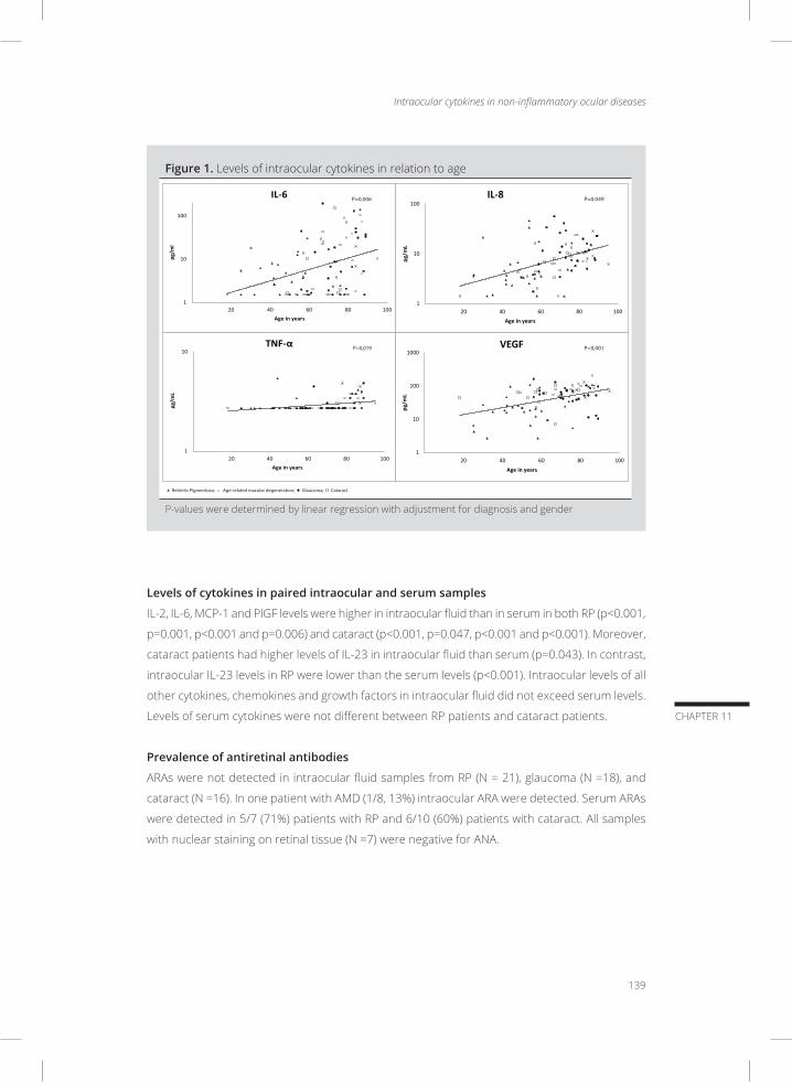
Intraocular cytokines in non-inflammatory ocular diseases
139
CHAPTER 11
Figure 1. Levels of intraocular cytokines in relation to age
1
10
100
20 40 60 80 100
pg/m
l
Age in years
IL-6
1
10
100
20 40 60 80 100
pg/m
L
Age in years
IL-8
1
10
20 40 60 80 100
pg/m
L
Age in years
TNF-α
1
10
100
1000
20 40 60 80 100
pg/m
L
Age in years
VEGF
Retinitis Pigmentosa; Age-related macular degeneration; Glaucoma; Cataract
P=0,006 P=0,049
P=0,019 P<0,001
P-values were determined by linear regression with adjustment for diagnosis and gender
Levels of cytokines in paired intraocular and serum samples
IL-2, IL-6, MCP-1 and PlGF levels were higher in intraocular fluid than in serum in both RP (p<0.001,
p=0.001, p<0.001 and p=0.006) and cataract (p<0.001, p=0.047, p<0.001 and p<0.001). Moreover,
cataract patients had higher levels of IL-23 in intraocular fluid than serum (p=0.043). In contrast,
intraocular IL-23 levels in RP were lower than the serum levels (p<0.001). Intraocular levels of all
other cytokines, chemokines and growth factors in intraocular fluid did not exceed serum levels.
Levels of serum cytokines were not different between RP patients and cataract patients.
Prevalence of antiretinal antibodies
ARAs were not detected in intraocular fluid samples from RP (N = 21), glaucoma (N =18), and
cataract (N =16). In one patient with AMD (1/8, 13%) intraocular ARA were detected. Serum ARAs
were detected in 5/7 (71%) patients with RP and 6/10 (60%) patients with cataract. All samples
with nuclear staining on retinal tissue (N =7) were negative for ANA.
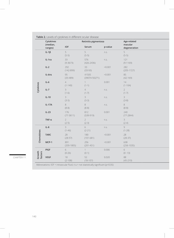
140
CHAPTER 11
Table 2. Levels of cytokines in different ocular disease
Cytokines (median, ranges)
Retinitis pigmentosa Age-related macular degeneration
Glaucoma Cataract p-value (comparison of IOF)IOF Serum p-value IOF Serum p-value
Cyto
kine
s
IL-1β 5 (5-5)
5 (5-5)
n.s. 5 (5-5)
5 (5-5)
5 (5-5)
5 (5-5)
n.s. n.s.
IL-1ra 33 (9-3673)
576 (426-2436)
n.s. 121 (9-1169)
65 (9-876)
124 (9-2357)
608 (328-1043)
n.s. n.s.
IL-2 292 (142-899)
33 (33-93)
<0.001 282 (205-1727)
271 (151-1371)
275 (212-1101)
136 (15-262)
<0.001 n.s.
IL-6rα 95 (33-489)
41505 (39074-50271)
<0.001 85 (42-169)
99 (22-502)
87 (24-263)
44327 (34714-58859)
<0.0010.019
IL-6 4 (1-140)
1(1-1)
0.001 14 (1-104)
5 (1-192)
2 (1-151)
1 (1-3)
0.047 n.s.
IL-7 3 (1-6)
4 (1-7)
n.s. 2 (1-7)
3 (1-12)
1 (1-9)
5 (2-7)
0.004 n.s.
IL-10 3 (3-5)
3 (3-3)
n.s. 3 (3-8)
3 (3-7)
3 (3-8)
5 (3-24)
n.s. n.s.
IL-17A 8 (8-8)
8 (8-8)
n.s. 8 (8-8)
8 (8-8)
8 (8-8)
10 (8-25)
n.s. n.s.
IL-23 176 (77-3611)
812 (539-919)
0.001 240 (77-2844)
315 (77-2828)
2592 (144- 3493)
750 (510-945)
0.043 0.004
TNF-α 2 (2-5)
2 (2-3)
n.s. 3 (2-4)
2 (2-4)
2 (2-3)
2 (2-4)
n.s. n.s.
Chem
okin
es
IL-8 3 (1-46)
6 (2-21)
n.s. 9 (1-28)
9 (1-56)
6 (1-16)
7 (1-12)
n.s. 0.032
TARC 28 (28-57)
180 (101-681)
<0.001 28 (28-37)
28 (28-57)
28(28-28)
262 (62-470)
<0.001 n.s.
MCP-1 891 (339-1805)
256 (201-451)
<0.001 638 (256-1035)
731 (264-3495)
618 (249-1920)
320 (120-478)
<0.001 n.s.
Gro
wth
fa
ctor
s
PlGF 6 (0-26)
1 (0-1)
0.006 6 (0 -13)
5 (0-13)
5 (2-12)
1 (0-2)
<0.001 n.s.
VEGF 18 (2-108)
52 (18-107)
0.020 88 (45-210)
54 (9-159)
63 (7-101)
73 (18-126)
n.s. n.s.
Abbrevations: IOF = intraocular fluid, n.s.= not statistically significant (p>0.05)

Intraocular cytokines in non-inflammatory ocular diseases
141
CHAPTER 11
Table 2. Levels of cytokines in different ocular disease
Cytokines (median, ranges)
Retinitis pigmentosa Age-related macular degeneration
Glaucoma Cataract p-value (comparison of IOF)IOF Serum p-value IOF Serum p-value
Cyto
kine
s
IL-1β 5 (5-5)
5 (5-5)
n.s. 5 (5-5)
5 (5-5)
5 (5-5)
5 (5-5)
n.s. n.s.
IL-1ra 33 (9-3673)
576 (426-2436)
n.s. 121 (9-1169)
65 (9-876)
124 (9-2357)
608 (328-1043)
n.s. n.s.
IL-2 292 (142-899)
33 (33-93)
<0.001 282 (205-1727)
271 (151-1371)
275 (212-1101)
136 (15-262)
<0.001 n.s.
IL-6rα 95 (33-489)
41505 (39074-50271)
<0.001 85 (42-169)
99 (22-502)
87 (24-263)
44327 (34714-58859)
<0.0010.019
IL-6 4 (1-140)
1(1-1)
0.001 14 (1-104)
5 (1-192)
2 (1-151)
1 (1-3)
0.047 n.s.
IL-7 3 (1-6)
4 (1-7)
n.s. 2 (1-7)
3 (1-12)
1 (1-9)
5 (2-7)
0.004 n.s.
IL-10 3 (3-5)
3 (3-3)
n.s. 3 (3-8)
3 (3-7)
3 (3-8)
5 (3-24)
n.s. n.s.
IL-17A 8 (8-8)
8 (8-8)
n.s. 8 (8-8)
8 (8-8)
8 (8-8)
10 (8-25)
n.s. n.s.
IL-23 176 (77-3611)
812 (539-919)
0.001 240 (77-2844)
315 (77-2828)
2592 (144- 3493)
750 (510-945)
0.043 0.004
TNF-α 2 (2-5)
2 (2-3)
n.s. 3 (2-4)
2 (2-4)
2 (2-3)
2 (2-4)
n.s. n.s.
Chem
okin
es
IL-8 3 (1-46)
6 (2-21)
n.s. 9 (1-28)
9 (1-56)
6 (1-16)
7 (1-12)
n.s. 0.032
TARC 28 (28-57)
180 (101-681)
<0.001 28 (28-37)
28 (28-57)
28(28-28)
262 (62-470)
<0.001 n.s.
MCP-1 891 (339-1805)
256 (201-451)
<0.001 638 (256-1035)
731 (264-3495)
618 (249-1920)
320 (120-478)
<0.001 n.s.
Gro
wth
fa
ctor
s
PlGF 6 (0-26)
1 (0-1)
0.006 6 (0 -13)
5 (0-13)
5 (2-12)
1 (0-2)
<0.001 n.s.
VEGF 18 (2-108)
52 (18-107)
0.020 88 (45-210)
54 (9-159)
63 (7-101)
73 (18-126)
n.s. n.s.
Abbrevations: IOF = intraocular fluid, n.s.= not statistically significant (p>0.05)

142
CHAPTER 11
Figure 2. Levels of intraocular cytokines in relation to ocular diagnosis
10
100
1000pg
/mL
Diagnosis
IL-6rα
1
10
100
pg/m
L
Diagnosis
IL-8
10
100
1000
10000
pg/m
L
Diagnosis
IL-23
Re�ni�s Pigmentosa Age related macular degera�on Glaucoma Cataract N=25 N=12 N=28 N=22
Re�ni�s Pigmentosa Age related macular degera�on Glaucoma Cataract N=25 N=12 N=28 N=22
Re�ni�s Pigmentosa Age related macular degera�on Glaucoma Cataract N=25 N=12 N=28 N=22
P=0.019
P=0.032
P=0.004
10
P-values were determined by linear regression with adjustment for age and gender
Intraocular cytokines in non-inflammatory ocular diseases
143
CHAPTER 11
DISCUSSION
Our study describes different pro-inflammatory intraocular cytokine profiles in RP, AMD, glaucoma
and cataract, and reveals a positive correlation between intraocular cytokine levels and increasing
age. Interestingly, intraocular fluid samples from cataract patients displayed the highest levels of
IL-23. Though intraocular levels of VEGF were highest in AMD patients, after age correction no
significant differences were found between the various diagnostic groups. Comparison of paired
serum and intraocular fluid samples of RP patients showed that intraocular levels of IL-2, IL-6,
MCP-1 and PlGF exceeded the serum levels, suggesting a local production. Intraocular ARAs were
absent in nearly all samples.
Although a genetic mutation is the cause of RP, inflammation was suggested to have a (secondary)
role in the disease pathogenesis.2,16,17 It has been reported that RP patients with CM exhibited
more often ARAs in their peripheral blood.18 Conform these findings, in our series we observed
ARAs in all RP patients with CM. However, in contrast to serum, ARAs were not at all detected in
intraocular fluids of patients with RP.
Previous reports on intraocular cytokines in RP show higher levels of IL-6, IL-8, MCP-1 and TARC
in RP compared to cataract.11,19 We observed also intraocular presence of these mediators in
RP patients, but their levels were not elevated compared to other groups. This discrepancy may
be explained by the low number of included patients in our study, or possible differences in
disease stage and/or extent of degeneration. Yet RP patients, like glaucoma patients, had higher
intraocular levels of soluble IL-6rα (sIL-6rα) compared to cataract. sIL-6Rα interacts with IL-6,
forming the IL-6/sIL-6Rα complex, which subsequently induces IL-6 trans-signaling by binding
cell membrane expressed gp130.20 IL-6 trans-signaling is recognized to enhance IL-6 activity
under inflammatory conditions and moreover to inhibit intra-ocular T-cell apoptosis in uveitis,
which likely exacerbates or prolongs the disease process.21-23 Further we observed a significant
association between lower levels of intraocular IL-2 (a growth factor for regulatory T-cells) in RP
patients who had CM. This may indicate a deregulated immune function, such as loss of tolerance,
affecting the clinical manifestation of the disease and the formation of serum ARAs as observed
in this and other studies. Intraocular VEGF levels were lowest in the RP group, which is in line with
the rare presence of retinal neovascularization in RP.
Inflammation was implicated in the development and progression of AMD.24 So far, most previous
studies investigated intraocular fluids of exudative AMD, demonstrating high levels of inflamma-
tory mediators, including IL-6, IL-8, MCP-1 and VEGF.6,25 However, it is still unknown whether these
cytokines play a role in the primary pathogenesis of AMD or represent a secondary result of the
disease process. We investigated patients with mainly dry AMD and observed higher intraocular
IL-8 compared to cataract and RP. Previous studies revealed that elevated (intraocular) levels of
144
CHAPTER 11
IL-8 and IL-8 gene polymorphisms were associated with angiogenesis.26,27 IL-6 and VEGF reached
highest levels in the AMD group, though not significantly different compared to other diagnosis
groups. According to previous studies, these mediators have been implicated in angiogenesis,
and decrease during treatment with anti-VEGF agents.8,28 Retinal neuroprotective effects of VEGF
have also been described, yet data on this matter are inconclusive and may be dependent on
the VEGF variant, disease (stage) or experimental model used.29,30
In glaucoma the role of immune reactions is not known and could be either pathogenic or
neuroprotective. In our study patients with glaucoma, who consisted mainly of POAG, were
characterized by high intraocular levels of IL-8, consistently with previous findings.31-34 IL-8 is a
main chemoattractant for neutrophils which have been found to accumulate in the trabecular
meshwork in POAG.35 The highest levels of IL-8 were found in glaucoma patients who underwent
surgical treatment prior to the surgical procedure during intraocular sample collection. This
suggest that higher levels of IL-8 might be explained by immune activation in response to (sur-
gically inflicted) tissue damage. Increased TNF-α levels have been reported in intraocular fluid,
the trabecular meshwork, optic head and the retina of glaucoma patients, however in our study
intraocular TNF-α appeared undetectable in most cases.33,35-39 This discrepancy may have resulted
from differences in laboratory techniques and specific patient groups. So far, to our knowledge,
only sporadic studies are available on the effect of anti-TNF medication in glaucoma, which show
an increase of fibrosis after surgical treatment.40
In our study, cataract was generally characterized by lower levels of pro-inflammatory mediators
compared to other studied diseases, with the exception of IL-23. IL-23, produced by dendritic
cells/myeloid cells, is well known for its key role in several autoimmune diseases via the IL-23/
IL-17axis and associated pathological Th17 development.41,42 Despite the presence of IL-23 in
most intraocular fluids analyzed in our study, IL-17A was never detected. Interestingly, some
studies report immunosuppressive effects of IL-23 within tumor microenvironments by suppress-
ing lymphocyte effector function and enhanced production of immune-regulatory cytokines.43-45
Also in a model of experimental autoimmune uveitis it was found that IL-23 receptor expressing
gd T cells can exert immune suppressive effects due to their ability to bind IL-23.46 Although
the mechanism by which IL-23 can mediate immune suppressive effects clearly requires further
study, it is tempting to speculate that the low intraocular IL-23 levels observed in patients with
RP, AMD and glaucoma may reflect diminished immune protection of the eye. Intraocular IL-23
levels revealed no association with serum levels nor age of patients.
Aging is associated with development of a chronic state of low-grade tissue inflammation that also
involves the retina, and is associated with increased susceptibility to multiple diseases, including
glaucoma and AMD.47-50 In support of this, we observed a gradual increase of the intraocular
levels of IL-6, IL-8, TNF-α and VEGF with increasing age. A positive correlation between intraocular
Intraocular cytokines in non-inflammatory ocular diseases
145
CHAPTER 11
cytokine levels and age has been reported in two previous studies.31,34 As a consequence of this
correlation, all of our results are adjusted for the age of patients. However, systematic corrections
for age have not been performed in previous studies, which may have affected the interpretation
of these findings. It should thus be kept in mind that studies on intraocular cytokine profiling
without age adjustment (and without age matched control groups) may show age-related bias
rather than disease-associated differences.
Intraocular levels of IL-2, IL-6, MCP-1 and PlGF were higher than serum levels of patients with
RP and cataract. The higher intraocular levels of inflammatory components may suggest local
production, possibly by infiltrated immune cells or resident cells, such as retinal pigment epithe-
lial cells.51,52 These findings may contribute to the understanding of the pathogenesis of RP and
development of new treatment possibilities. In contrast, the low occurrence of intraocular ARAs
suggests a negligible role of such antibodies in disease pathogenesis of RP. ARAs detected in
serum from RP and cataract patients may represent a reflection of altered blood retinal barrier
properties.
In conclusion, the expression of inflammatory cytokines within the eye was strongly influenced
by the age of patients, which shows that the correction for age is necessary in future studies
on intraocular mediators. Differences in intraocular cytokines profiles were observed between
originally non-inflammatory ocular diseases, suggesting involvement of inflammation, however
complex pathways with multiple signaling functions make a diagnostic role rather impossible.
The role of immune reactions in basically non-inflammatory ocular diseases might influence the
clinical manifestations and severity of ocular changes.
SUPPLEMENTARY DATA
Supplementary Table. Prevalence of cytokines in different ocular disease
146
CHAPTER 11
REFERENCES
1. Bell K, Gramlich OW, Von Thun Und Hohenstein-Blaul N, et al. Does autoimmunity play a part in the
pathogenesis of glaucoma? Prog. Retin. Eye Res. 2013;36:199-216.
2. Nussenblatt RB, Liu B, Wei L, Sen HN. The immunological basis of degenerative diseases of the eye. Int
Rev Immunol. 2013;32(1):97-112.
3. Heckenlively JR, Jordan BL, Aptsiauri N. Association of antiretinal antibodies and cystoid macular edema
in patients with retinitis pigmentosa. Am. J. Ophthalmol. 1999;127(5):565-573.
4. Kubicka-Trzaska A, Wilanska J, Romanowska-Dixon B, Sanak M. Serum anti-endothelial cell antibodies in
patients with age-related macular degeneration treated with intravitreal bevacizumab. Acta Ophthalmol.
2016;94(7):e617-e623.
5. Joachim SC, Bruns K, Lackner KJ, Pfeiffer N, Grus FH. Antibodies to alpha B-crystallin, vimentin, and heat
shock protein 70 in aqueous humor of patients with normal tension glaucoma and IgG antibody patterns
against retinal antigen in aqueous humor. Curr Eye Res. 2007;32(6):501-509.
6. Jonas JB, Tao Y, Neumaier M, Findeisen P. Cytokine concentration in aqueous humour of eyes with exu-
dative age-related macular degeneration. Acta Ophthalmol. 2012;90(5):e381-388.
7. Rezar-Dreindl S, Sacu S, Eibenberger K, et al. The Intraocular Cytokine Profile and Therapeutic Response in
Persistent Neovascular Age-Related Macular Degeneration. Invest Ophthalmol Vis Sci. 2016;57(10):4144-
4150.
8. Chalam KV, Grover S, Sambhav K, Balaiya S, Murthy RK. Aqueous interleukin-6 levels are superior to
vascular endothelial growth factor in predicting therapeutic response to bevacizumab in age-related
macular degeneration. J Ophthalmol. 2014;2014:502174.
9. Chiu K, Yeung SC, So KF, Chang RC. Modulation of morphological changes of microglia and neuroprotection
by monocyte chemoattractant protein-1 in experimental glaucoma. Cell. Mol. Immunol. 2010;7(1):61-68.
10. Freedman J, Iserovich P. Pro-inflammatory cytokines in glaucomatous aqueous and encysted Molteno
implant blebs and their relationship to pressure. Invest. Ophthalmol. Vis. Sci. 2013;54(7):4851-4855.
11. Yoshida N, Ikeda Y, Notomi S, et al. Clinical evidence of sustained chronic inflammatory reaction in retinitis
pigmentosa. Ophthalmology. 2013;120(1):100-105.
12. Kauppinen A, Paterno JJ, Blasiak J, Salminen A, Kaarniranta K. Inflammation and its role in age-related
macular degeneration. Cell Mol Life Sci. 2016;73(9):1765-1786.
13. Huang W, Chen S, Gao X, et al. Inflammation-related cytokines of aqueous humor in acute primary
angle-closure eyes. Invest Ophthalmol Vis Sci. 2014;55(2):1088-1094.
14. Du S, Huang W, Zhang X, Wang J, Wang W, Lam DS. Multiplex cytokine levels of aqueous humor in acute
primary angle-closure patients: fellow eye comparison. BMC Ophthalmol. 2016;16:6.
15. Ten Berge JC, Schreurs MW, Vermeer J, Meester-Smoor MA, Rothova A. Prevalence and clinical impact of
antiretinal antibodies in uveitis. Acta Ophthalmol. 2016;94(3):282-288.
16. Chant SM, Heckenlively J, Meyers-Elliott RH. Autoimmunity in hereditary retinal degeneration. I. Basic
studies. Br J Ophthalmol. 1985;69(1):19-24.
17. Hettinga YM, van Genderen MM, Wieringa W, Ossewaarde-van Norel J, de Boer JH. Retinal Dystrophy in
6 Young Patients Who Presented with Intermediate Uveitis. Ophthalmology. 2016;123(9):2043-2046.
18. Heckenlively JR, Jordan BL, Aptsiauri N. Association of antiretinal antibodies and cystoid macular edema
in patients with retinitis pigmentosa. Am J Ophthalmol. 1999;127(5):565-573.
19. Salom D, Diaz-Llopis M, Garcia-Delpech S, Udaondo P, Sancho-Tello M, Romero FJ. Aqueous humor levels
of vascular endothelial growth factor in retinitis pigmentosa. Invest Ophthalmol Vis Sci. 2008;49(8):3499-
3502.
Intraocular cytokines in non-inflammatory ocular diseases
147
CHAPTER 11
20. Rose-John S. IL-6 trans-signaling via the soluble IL-6 receptor: importance for the pro-inflammatory activ-
ities of IL-6. Int J Biol Sci. 2012;8(9):1237-1247.
21. Nowell MA, Richards PJ, Horiuchi S, et al. Soluble IL-6 receptor governs IL-6 activity in experimental arthri-
tis: blockade of arthritis severity by soluble glycoprotein 130. J Immunol. 2003;171(6):3202-3209.
22. Barkhausen T, Tschernig T, Rosenstiel P, et al. Selective blockade of interleukin-6 trans-signaling improves
survival in a murine polymicrobial sepsis model. Crit Care Med. 2011;39(6):1407-1413.
23. Curnow SJ, Scheel-Toellner D, Jenkinson W, et al. Inhibition of T cell apoptosis in the aqueous humor of
patients with uveitis by IL-6/soluble IL-6 receptor trans-signaling. J Immunol. 2004;173(8):5290-5297.
24. Adamus G. Can innate and autoimmune reactivity forecast early and advance stages of age-related
macular degeneration? Autoimmun Rev. 2017;16(3):231-236.
25. Knickelbein JE, Chan CC, Sen HN, Ferris FL, Nussenblatt RB. Inflammatory Mechanisms of Age-related
Macular Degeneration. Int Ophthalmol Clin. 2015;55(3):63-78.
26. Forooghian F, Kertes PJ, Eng KT, et al. Alterations in intraocular cytokine levels following intravitreal ran-
ibizumab. Can J Ophthalmol. 2016;51(2):87-90.
27. Ghasemi H, Ghazanfari T, Yaraee R, Faghihzadeh S, Hassan ZM. Roles of IL-8 in ocular inflammations: a
review. Ocul Immunol Inflamm. 2011;19(6):401-412.
28. Agawa T, Usui Y, Wakabayashi Y, et al. Profile of intraocular immune mediators in patients with age-related
macular degeneration and the effect of intravitreal bevacizumab injection. Retina. 2014;34(9):1811-1818.
29. Nishijima K, Ng YS, Zhong L, et al. Vascular endothelial growth factor-A is a survival factor for retinal
neurons and a critical neuroprotectant during the adaptive response to ischemic injury. Am J Pathol.
2007;171(1):53-67.
30. Saint-Geniez M, Maharaj AS, Walshe TE, et al. Endogenous VEGF is required for visual function: evidence
for a survival role on muller cells and photoreceptors. PLoS One. 2008;3(11):e3554.
31. Kokubun T, Tsuda S, Kunikata H, et al. Characteristic Profiles of Inflammatory Cytokines in the Aqueous
Humor of Glaucomatous Eyes. Ocul Immunol Inflamm. 2017:1-12.
32. Kuchtey J, Rezaei KA, Jaru-Ampornpan P, Sternberg P, Jr., Kuchtey RW. Multiplex cytokine analysis reveals
elevated concentration of interleukin-8 in glaucomatous aqueous humor. Invest Ophthalmol Vis Sci.
2010;51(12):6441-6447.
33. Khalef N, Labib H, Helmy H, El Hamid MA, Moemen L, Fahmy I. Levels of cytokines in the aqueous humor
of eyes with primary open angle glaucoma, pseudoexfoliation glaucoma and cataract. Electron Physician.
2017;9(2):3833-3837.
34. Takai Y, Tanito M, Ohira A. Multiplex cytokine analysis of aqueous humor in eyes with primary open-angle
glaucoma, exfoliation glaucoma, and cataract. Invest Ophthalmol Vis Sci. 2012;53(1):241-247.
35. Taurone S, Ripandelli G, Pacella E, et al. Potential regulatory molecules in the human trabecular meshwork
of patients with glaucoma: immunohistochemical profile of a number of inflammatory cytokines. Mol Med
Rep. 2015;11(2):1384-1390.
36. Balaiya S, Edwards J, Tillis T, Khetpal V, Chalam KV. Tumor necrosis factor-alpha (TNF-alpha) levels in
aqueous humor of primary open angle glaucoma. Clin Ophthalmol. 2011;5:553-556.
37. Sawada H, Fukuchi T, Tanaka T, Abe H. Tumor necrosis factor-alpha concentrations in the aqueous humor
of patients with glaucoma. Invest Ophthalmol Vis Sci. 2010;51(2):903-906.
38. Tezel G, Li LY, Patil RV, Wax MB. TNF-alpha and TNF-alpha receptor-1 in the retina of normal and glauco-
matous eyes. Invest Ophthalmol Vis Sci. 2001;42(8):1787-1794.
39. Yuan L, Neufeld AH. Tumor necrosis factor-alpha: a potentially neurodestructive cytokine produced by
glia in the human glaucomatous optic nerve head. Glia. 2000;32(1):42-50.
40. Nikita E, Moulin A, Vergados I, Brouzas D, Theodossiadis PG. A Pilot Study on Ocular Safety and Efficacy
of Infliximab as an Antifibrotic Agent After Experimental Glaucoma Filtration Surgery. Ophthalmol Ther.
148
CHAPTER 11
2017.
41. Lubberts E. The IL-23-IL-17 axis in inflammatory arthritis. Nat Rev Rheumatol. 2015;11(7):415-429.
42. D’Elios MM, Del Prete G, Amedei A. Targeting IL-23 in human diseases. Expert Opin Ther Targets.
2010;14(7):759-774.
43. Langowski JL, Zhang X, Wu L, et al. IL-23 promotes tumour incidence and growth. Nature.
2006;442(7101):461-465.
44. Teng MW, Andrews DM, McLaughlin N, et al. IL-23 suppresses innate immune response independently
of IL-17A during carcinogenesis and metastasis. Proc Natl Acad Sci U S A. 2010;107(18):8328-8333.
45. Nie W, Yu T, Sang Y, Gao X. Tumor-promoting effect of IL-23 in mammary cancer mediated by infiltra-
tion of M2 macrophages and neutrophils in tumor microenvironment. Biochem Biophys Res Commun.
2017;482(4):1400-1406.
46. Liang D, Zuo A, Shao H, et al. IL-23 receptor expression on gammadelta T cells correlates with their
enhancing or suppressive effects on autoreactive T cells in experimental autoimmune uveitis. J Immunol.
2013;191(3):1118-1125.
47. Castelo-Branco C, Soveral I. The immune system and aging: a review. Gynecol Endocrinol. 2014;30(1):16-
22.
48. Xu H, Chen M, Forrester JV. Para-inflammation in the aging retina. Prog Retin Eye Res. 2009;28(5):348-368.
49. Chakravarthy U, Wong TY, Fletcher A, et al. Clinical risk factors for age-related macular degeneration: a
systematic review and meta-analysis. BMC Ophthalmol. 2010;10:31.
50. Leske MC, Wu SY, Hennis A, Honkanen R, Nemesure B, Group BES. Risk factors for incident open-angle
glaucoma: the Barbados Eye Studies. Ophthalmology. 2008;115(1):85-93.
51. Holtkamp GM, Van Rossem M, de Vos AF, Willekens B, Peek R, Kijlstra A. Polarized secretion of IL-6 and
IL-8 by human retinal pigment epithelial cells. Clin Exp Immunol. 1998;112(1):34-43.
52. Elner VM, Scales W, Elner SG, Danforth J, Kunkel SL, Strieter RM. Interleukin-6 (IL-6) gene expression and
secretion by cytokine-stimulated human retinal pigment epithelial cells. Exp Eye Res. 1992;54(3):361-368.


12SUMMARY AND CONCLUSIONS


Summary and conclusions
153
CHAPTER 12
SUMMARY
Autoimmune reactions have been implicated in various ocular diseases but their presence and
pathogenic role have been scarcely proven. Autoimmunity was especially linked to uveitis (a collec-
tive term for any intraocular inflammation) and autoimmune retinopathy (AIR), but also to retinitis
pigmentosa (RP), age-related macular degeneration (AMD) and glaucoma. Obviously, if autoimmu-
nity plays a principal role in these diseases, the downregulation of autoimmune reactions might
bring a beneficial effect to affected patients. The work presented in this thesis aims to assess the
presence of humoral autoimmunity in uveitis and other chorioretinal diseases, including AIR, and to
clarify its potential role in their pathogenesis. This summary presents the main findings of this thesis.
Chapter 1 provides a general introduction addressing the presence and the role of humoral
autoimmunity in uveitis and other chorioretinal diseases. Autoimmunity is characterized by activ-
ity of the immune system directed against the body’s own cells and tissues. The immune system
stops tolerating ‘self’ antigens, and autoreactive antibodies and/or T-cells attack the body’s own
antigens. Convincing evidence exists that our immune system is able to react against our own
retina and can produce antiretinal antibodies (ARAs). These ARAs can presumably attack and
destroy retinal cells, and rapidly leading to visual loss or even blindness. Such reactions of the
immune system have long been considered to play a major pathogenic role in uveitis. In other
ocular diseases, such as RP, AMD and glaucoma, the formation of ARAs has also been observed
and reported. However, the exact pathogenic role of antiretinal humoral autoimmunity in uveitis
and other ocular diseases is not yet known. One current hypothesis includes the primary role of
ARAs in the initiation of the chorioretinal diseases; another possibility addresses the secondary
role of ARAs and proposes that the formation of ARAs in any ocular disease might cause a mild
inflammation in the retina, that might negatively influence the course of the disease (e.g. by
development of chronicity or enhancement of progression of retinal damage).
In Chapter 2 we determine the prevalence of systemic immune-mediated diseases in a large
cohort of 1327 patients with uveitis and/or scleritis, and put a special emphasis on prevalence of
autoimmune and auto-inflammatory diseases according to novel findings in immune mediated
diseases. In contrast to the widespread belief, it appeared that autoimmune uveitis is a rare
diagnosis, which comprises only 5% (62/1327) of the total uveitis/scleritis population. The most
common autoimmune diseases observed in patients with uveitis were multiple sclerosis (1.8%,
24/1327) and Vogt Koyanagi Harada disease (1.1%, 14/1327). Patients with classical autoimmune
connective tissue disease (N=17) suffered mostly from rheumatoid arthritis and granulomatosis
with polyangiitis and exhibited predominantly scleritis (53%, 9/17). Optic neuropathy was the most
frequent complication of autoimmune uveitis (44%, 27/62), and was predominantly observed in
MS and Vogt Koyanagi Harada disease. Interestingly, the visual acuity in autoimmune diseases
with uveitis remained stable during 5-year follow-up, possibly as a result of adequate treatment
154
CHAPTER 12
with systemic immunomodulatory medication. None of the patients with autoimmune uveitis/
scleritis developed permanent bilateral visual acuity of less than 0.1. In this study we emphasize
that the term “autoimmune uveitis” should be reserved exclusively for intraocular inflammations
of confirmed autoimmune origin and should not be used as a synonym for all non-infectious
uveitis cases.
In Chapter 3 we prospectively evaluate the presence of antinuclear antibody (ANA) and the
diagnostic value of ANA profiling in 105 adult patients with uveitis. ANAs are antibodies directed
against a variety of nuclear antigens, and can be detected in elevated levels in serum of patients
with genuine autoimmune diseases such as systemic lupus erythematosus, but can also be prev-
alent in other disease types and in the general population, predominantly in women and elderly.
Our study showed that 28% of patients with uveitis is positive for ANA whilst only 8-17% of the
healthy population is considered ANA positive. ANA positivity was more prevalent in patients with
infectious uveitis (31%) than patients with systemic immune mediated diseases (20%). We did not
observe any relationship between the presence of ANA, ANA titer, or specific ANA patterns and
specific diagnoses or clinical characteristics of uveitis. The discrimination of autoimmune systemic
diseases in uveitis by ANA profiling seems not possible, however this cannot be entirely excluded
since we included a limited number of patients in the specific diagnostic groups. Based on our
findings, we concluded that ANA profiling in the adult uveitis population has limited diagnostic
value and that ANAs are probably not involved in the pathogenesis of uveitis.
Chapter 4 describes the prevalence and possible clinical relevance of ARAs in serum in a pro-
spective study of 126 patients with different uveitis entities and compares it with 60 blood bank
donors. Screening for the presence of ARAs was performed by indirect immunofluorescence
(IIF) using primate retinal tissue. Although IIF does not allow antigenic specification of ARAs, this
technique is very sensitive and therefore represents a useful screening assay. In our study, serum
ARAs were observed in 47% of patients with uveitis and were more frequent in uveitis when com-
pared with healthy controls (17%, p<0.001). Specific associations between the presence of serum
ARAs or its subtypes (based on typical staining patterns on IIF) and clinical ocular characteristics of
uveitis were not observed. These results suggest that serum ARAs reflect a secondary response
of the immune system to damaged ocular tissue, and probably have no inciting role in uveitis.
In Chapter 5 we attempt to determine the possible diagnostic role of both known and unknown
ARAs in patients with AIR. AIR encompasses a group of rare diseases most commonly presenting
as paraneoplastic syndromes in combination with various malignancies. Multiple serum ARAs
have been reported in AIR, and it was assumed that ARAs are directly involved in retinal destruc-
tion and rapidly progressive loss of vision. So far, however, only few retinal antigens in AIR were
specified and studied. To investigate this more extensively, we performed a multicenter study
in cooperation with the SciLifeLab Autoimmunity Profiling Facility in Stockholm (Sweden). Serum
Summary and conclusions
155
CHAPTER 12
samples of 24 patients with presumed AIR were examined for the presence of antibodies against
multiple retinal antigens by autoantibody profiling and compared to 151 uveitis patients and 21
cataract controls. Antigens used for the autoantibody profiling consisted of 188 peptide frag-
ments derived from 97 unique ocular proteins produced within the Human Protein Atlas of the
Swedish laboratory and profiling was performed by a multiplex bead array based immunoassay.
We found that patients with presumed AIR were characterized by the presence of multiple serum
ARAs, and that the spectrum of ARAs differed widely among individual patients with AIR. Autoan-
tibodies directed against the retinal antigen recoverin were the most prevalent ARAs in patients
with presumed AIR (13%). Despite the high levels of multiple ARAs in AIR, our findings clearly
show that serum ARAs are also present in patients with cataract and uveitis, and consequently
the specificity of ARAs as markers for AIR was low. We concluded that the diagnosis of AIR cannot
be based on the mere presence of (multiple) serum ARAs.
In Chapter 6 we demonstrate the medical history of a patient with paraneoplastic AIR. The patient
presented with progressive severe visual loss in both eyes. He also suffered from loss of his
peripheral vision, and in his serum high levels of ARAs directed against recoverin were detected.
After referral to an internist he was diagnosed with lung carcinoma. The patient was treated with
chemo-/radiotherapy and high dosage of corticosteroids, unfortunately without any effect on the
visual performance of the patient. A lung carcinoma is the most commonly associated malignancy
in paraneoplastic AIR, however any type of malignancy can be involved in this disease. Usually both
eyes are affected, but unilateral cases also have been described. Visual complaints may precede
detection of the malignancy, which makes the role of the ophthalmologist vital. Treatment options
in cancer-associated AIR are limited and visual prognosis is often very poor.
In Chapter 7 we investigate the presence and possible role of ARAs directed against retinal tissue
in central serous chorioretinopathy (CSC). Although little is known about the exact cause of CSC,
genetic variants in the complement system of patients with CSC indicate a possible role of the
immune system. We retrospectively analysed serum of 63 patients with CSC for the presence
of ARAs by IIF and Western blot with retinal protein extract and compared those to 101 uveitis
patients, and 60 healthy donors. The Western blot allowed us to approximate the antigen size
of observed ARAs, which indicates the presence of specific ARAs. We detected ARAs in 54% of
patients with CSC compared to 46% of patients with uveitis (p>0.05) and 17% of healthy controls
(p<0.001). Interestingly, the ARAs in CSC were more often directed against the photoreceptors
(27%) than in uveitis (15%, p=0.039), however staining patterns on retinal tissue were divers, indi-
cating the presence of various ARAs. Western blot analysis showed multiple ARA reactive antigens
in retinal extract in the majority of patients with CSC. This heterogeneity and variable presence
of ARAs in CSC make a primary role of ARAs in CSC unlikely. Furthermore, the mere presence of
serum autoantibodies does not necessarily indicate an autoimmune basis of the disease and
shows that, similar to uveitis, ARAs might be secondary and induced by the retinal damage.
156
CHAPTER 12
In Chapter 8 we retrospectively explore the prevalence of systemic and retinal autoantibodies
in patients from Indonesia with uveitis and either active or latent tuberculosis (TB). In Indonesia,
uveitis associated with active systemic and latent TB is highly prevalent. The pathogenesis of
uveitis in the setting of latent TB is not clarified. One of the hypotheses presumes the immune
reaction to mycobacterial antigens and their molecular mimicry with retinal antigens. We inves-
tigated the presence of ARAs and ANAs in 95 Indonesian patients with uveitis classified into 3
groups: uveitis associated with active TB (N=10), with latent TB (N=58) and without evidence for
prior or current TB (N=85). We found that patients with uveitis and either active or latent TB were
characterized by a low proportion of ocular-specific auto-reactivity (ARAs) and a high prevalence
of systemic auto-reactivity (ANA), compared to uveitis patients who had no evidence of previous
contact with M. tuberculosis (p=0.03 and p=0.021). The lower prevalence of ARAs in serum does
not point toward an autoimmune pathogenesis in uveitis associated with latent or active tuber-
culosis, however assessment of intraocular ARAs might show an entirely different pattern and
clinical relevance of autoimmune processes in uveitis with active or latent TB.
In Chapter 9, we perform a retrospective study and assess the presence of ARAs in young Mex-
ican patients with severe pars planitis (N=16) and compare the results to Mexican age-matched
controls (N=19). An exceptionally severe form of pars planitis of unknown origin is observed in
pediatric Mexican patients. Despite the extensive examinations, the cause of this disorder is not
clarified. Pars planitis is considered to be of autoimmune origin, but the exact pathogenesis is not
known. Investigation of serum samples of pars planitis patients for the presence of ARAs showed a
prevalence of 57%, however a comparable prevalence (40%) was observed in Mexican control chil-
dren with strabismus. This high prevalence of ARAs in young control patients could be the result
of a higher exposure to exogenous antigens (e.g. infections). Similar to the Netherlands, these
results suggest that ARAs are not the primary cause of uveitis, but possibly represent a secondary
phenomenon. Nevertheless, a cellular autoimmune component or investigation of intraocular
material might still point in the direction of an autoimmune pathogenesis in pars planitis.
In the last chapters we left the serological analysis and explore the determination of humoral
autoimmune reactions in intraocular fluid. Chapter 10 is a retrospective study that investigates
intraocular fluid samples from 76 patients with uveitis and 19 cataract controls for the presence
of ARAs. In collaboration the SciLifeLab Autoimmunity Profiling Facility in Stockholm (Sweden), we
performed a multiplex immunoassay using the same 188 specific ocular antigens as in chapter
5. We found a spectrum of 22 different ARAs in higher levels in patients with uveitis compared
to cataract controls (p≤0.05), but in moderately elevated titers. High elevations (5-fold increased
titers compared to cataract controls) of multiple ARAs were observed in three specific uveitis
entities: varicella zoster virus-induced uveitis, multiple sclerosis-associated uveitis and patients
with unexplained uveitis but positive quantiferon test (all p<0.05). Presence of macular edema
was associated with high intraocular levels of tyrosinase antibodies (p=0.0026). The spectrum of
Summary and conclusions
157
CHAPTER 12
multiple ARAs in intraocular fluid of patients with uveitis favors a secondary production rather
than their inciting role and implicate that uveitis in the setting of latent TB infection might be joint
with intraocular autoimmune reactions. The results of this study may provide a platform for future
exploration of specific ocular antigens in the pathogenesis of uveitis.
Inflammatory autoimmune processes were not only assumed to play a role in the pathogenesis
of uveitis, but also in various originally non-inflammatory ocular diseases such as RP, AMD and
glaucoma. Therefore, in Chapter 11 we retrospectively determine the levels of 15 intraocular
cytokines and the prevalence of intraocular ARAs in patients with RP (N=25), AMD (N=12), glau-
coma (N=28) and cataract (N=22), and correlate the laboratory results to clinical manifestations.
In addition, we studied serum samples paired with the intraocular fluids from patients with RP
(N=7) and cataract (N=10). We observed that increasing age was associated with increasing levels
of multiple intraocular cytokines and other mediators, and detected distinct cytokine profiles for
each group of diagnoses. Specifically, intraocular levels of IL-6rα (p=0.003), IL-8 (p=0.032), and
IL-23 (p<0.004) differed between the studied ocular diseases. In RP patients, intraocular levels
of IL-2, IL-6, MCP-1 and PlGF exceeded the serum levels, which indicates an active intraocular
production and involvement of these mediators in RP. Intraocular ARAs were found in only one
patient with AMD, in contrast to serum ARAs that were detected in a majority of studied entities.
The differences in cytokine profiles between ocular diseases, suggest a role of immune activity
in the pathogenesis of these originally non-inflammatory ocular diseases. The involvement of
immune reactions might contribute to continuation and/or aggravation of the disease.
CONCLUSIONS
Our results do not show any evidence for a primary role of retina specific autoantibodies in the ini-
tiation of uveitis and other chorioretinal diseases. The presence of serum ARAs in ocular diseases
might be explained by a secondary formation induced by ocular tissue damage. Though higher
levels of serum ARAs were found in patients with ocular disorders than in control population, we
found no associations with specific disorders or clinical manifestations of studied ocular diseases.
Moreover, our findings indicate that serum ARAs probably do not have an inciting role in AIR, and
that the diagnosis of AIR cannot be based on the mere presence of serum ARAs.
The spectrum of intraocular ARAs in uveitis indicate some involvement of humoral autoimmu-
nity, predominantly in three specific disorders: varicella zoster virus-induced uveitis, multiple
sclerosis-associated uveitis and patients with unexplained uveitis but positive quantiferon test.
In addition, intraocular autoantibodies directed against tyrosinase have been associated with
the presence of cystoid macular edema. Intraocular ARA production in AIR, especially in cancer
associated retinopathies, would be an interesting topic for future research and could help clarify
its pathogenesis and provide novel treatment opportunities.


SAMENVATTING DANKWOORDABOUT THE AUTHORPHD PORTFOLIOLIST OF PUBLICATIONS
APPENDICES

Samenvatting
161
APPENDICES
SAMENVATTING
Het immuunsysteem lijkt een belangrijke rol te spelen in de pathogenese van verschillende oog-
ziekten. Met name bij uveïtis, een inwendige oogontsteking, lijkt het immuunsysteem betrokken
te zijn, maar ook bij andere oogaandoeningen zoals retinitis pigmentosa (RP), leeftijdsgebonden
maculadegeneratie (AMD) en glaucoom wordt een rol van het immuunsysteem verondersteld.
Verder inzicht in de rol van het immuunsysteem bij netvliesaandoeningen kan in de toekomst als
mogelijk aangrijpingspunt dienen voor gerichtere behandelingen van diverse oogaandoeningen.
Het werk dat in dit proefschrift wordt gepresenteerd, heeft als doel de potentiële pathogene rol
van (humorale) auto-immuun reacties bij uveïtis en enkele andere netvliesaandoeningen verder
op te helderen. In deze samenvatting worden de belangrijkste bevindingen van dit proefschrift
beschreven.
In hoofdstuk 1 wordt een algemene introductie gegeven over auto-immuniteit in uveïtis en
andere oogaandoeningen. Auto-immuniteit wordt gekenmerkt door activiteit van het immuunsy-
steem gericht tegen eigen cellen en weefsels van het lichaam. Het immuunsysteem stopt met het
tolereren van lichaamseigen antigenen, en auto-antistoffen (humorale immuniteit) en/of auto-re-
actieve T-cellen (cellulaire immuniteit) vallen het eigen lichaam aan. Er zijn aanwijzingen dat het
immuunsysteem zich onder bepaalde omstandigheden tegen het eigen netvlies kan richten.
Hierbij worden zogenaamde anti-retinale antistoffen (ARA’s) geproduceerd die mogelijk schade
aan het netvlies kunnen aanbrengen. Tot op heden zijn de bewijzen voor de aanwezigheid van
ARA’s bij oogaandoeningen schaars en is de rol van humorale auto-immuun reacties in de patho-
genese van uveïtis en andere oogaandoeningen onbekend. Diverse theorieën over ARA’s en hun
mogelijke effect bij oogaandoeningen zijn ontwikkeld. Een van de hypotheses veronderstelt dat
ARA’s specifieke oogaandoeningen initiëren; een andere hypothese suggereert dat ARA’s slechts
een bijproduct zijn en de reeds bestaande ziekte nadelig kunnen beïnvloeden, bijvoorbeeld door
het ontwikkelen van een chronisch of progressief beloop van de oogaandoening.
In hoofdstuk 2 bepalen we de aanwezigheid van systemische immuun-gemedieerde ziektebeel-
den in een cohort van 1327 patiënten met uveïtis en/of scleritis (ontsteking van de harde oogrok).
We leggen hierbij nadruk op de prevalentie van auto-immuniteit en auto-inflammatie in uveïtis.
Het bleek dat auto-immuun uveïtis een zeer zeldzame diagnose is, welke slechts bij 5% (62/1327)
van de totale uveïtis-/scleritis-populatie voorkomt. De meest voorkomende auto-immuunziekten
bij patiënten met uveïtis waren multiple sclerose (MS; 1,8%, 24/1327) en de ziekte van Vogt-Koya-
nagi-Harada (VKH; 1,1%, 14/1327). Bij patiënten met systemische auto-immuun collageenziekten
zoals reumatoïde artritis en granulomatose met polyangiitis kwam scleritis relatief veel voor (53%,
9/17). Optische neuropathie (een beschadiging van de oogzenuw) was de meest voorkomende
complicatie van auto-immuun uveïtis (44%, 27/62), en werd voornamelijk gezien bij MS en de
ziekte van VKH. De visus van patiënten met auto-immuun uveïtis bleef gedurende 5 jaar follow-up
162
APPENDICES
stabiel en niemand ontwikkelde een permanente bilaterale gezichtscherpte van minder dan 0.1,
mogelijk door de adequate behandeling met systemische immunomodulerende medicatie. In dit
onderzoek benadrukken wij dat de term ‘auto-immuun uveïtis’ uitsluitend gebruikt mag worden
voor inwendige ontstekingen met bewezen karakteristieken van auto-immuun reacties, en het
geen synoniem is voor een niet-infectieuze uveïtis.
In hoofdstuk 3 evalueren we de aanwezigheid en diagnostische waarde van antinucleair anti-
stoffen (ANA) bij 105 volwassen patiënten met uveïtis. ANA’s zijn antilichamen gericht tegen
verschillende delen van de eigen celkern (nucleus). Deze kunnen worden gedetecteerd in een
verhoogde titer in het bloed van patiënten met auto-immuunziekten, zoals systemische lupus
erythematosus. Daarnaast kunnen ANA’s ook voorkomen bij de algemene bevolking, met name bij
vrouwen en ouderen. Uit onze studie bleek dat 28% van de patiënten met uveïtis positief is voor
ANA, wat hoger is dan de in de literatuur beschreven prevalentie van de ANA positieve gezonde
bevolking (8-17%). Een positieve ANA in het bloed werd vaker geobserveerd bij patiënten met
een infectieuze uveïtis (31%) dan bij patiënten met systemische immuun-gemedieerde ziekte
(20%). We hebben geen verband gevonden tussen de aanwezigheid van ANA, de ANA titer of
een specifiek ANA patroon en specifieke diagnoses of klinische kenmerken van uveïtis. Hierdoor
lijkt het onderscheiden van de verschillende soorten uveïtis door middel van ANA-analyse niet
mogelijk, maar dit kan niet volledig worden uitgesloten gezien het beperkt aantal patiënten in de
subgroepen. Op basis van onze bevindingen concludeerden we dat ANA-analyse in de volwas-
sen uveïtis populatie een beperkte diagnostische waarde heeft en dat ANA’s waarschijnlijk niet
betrokken zijn bij de pathogenese van uveïtis.
Hoofdstuk 4 is een prospectieve studie waarin de prevalentie en mogelijke rol van ARA’s in bloed
van 126 patiënten met uveïtis wordt vergeleken met 60 bloedbankdonoren. Middels indirecte
immunofluorescentie (IIF) met primaat retinaal weefsel werd serum getest op de aanwezigheid
van ARA’s. Hoewel het met IIF niet mogelijk is om ARA’s te specificeren, is het een zeer gevoelige
techniek, wat het een bruikbare screeningsassay maakt. In onze studie werden ARA’s aangetoond
bij 47% van de patiënten met uveïtis, wat significant vaker was dan bij de gezonde controles (17%,
p <0,001). Specifieke verbanden tussen de aanwezigheid van ARA’s en klinische oculaire kenmer-
ken van uveïtis werden niet waargenomen. Onze resultaten suggereren dat ARA’s in het bloed
geen primaire rol spelen in het ontstaan van uveïtis, en waarschijnlijk een secundaire reactie zijn
van het immuunsysteem op het reeds beschadigde oogweefsel.
In hoofdstuk 5 bepalen we de mogelijke diagnostische rol van verschillende specifieke ARA’s bij
patiënten met auto-immuun retinopathie (AIR). AIR omvat een zeldzame groep immuun-gemedi-
eerde oogziektes, die meestal als een paraneoplastische aandoening voorkomen in combinatie
met een maligniteit. AIR is geassocieerd met de aanwezigheid van verschillende ARA’s, waarbij
wordt verondersteld dat deze antistoffen schade aan het netvlies veroorzaken en tot progressief

Samenvatting
163
APPENDICES
visusverlies leiden. Tot nu toe zijn slechts enkele ARA’s bij AIR gespecificeerd en is de rol van
deze ARA’s bij AIR niet volledig opgehelderd. In dit onderzoek hebben wij in samenwerking met
SciLifeLab Autoimmunity Profiling Facility in Stockholm (Zweden) serum van 24 patiënten met AIR
onderzocht de op aanwezigheid van 188 verschillende ARA’s. Middels een multiplex immunoassay
werden bloedmonsters getest en vergeleken met serum van 151 patiënten met uveïtis en 21
patiënten met cataract. Uit de resultaten bleek dat individuele patiënten met AIR zeer verschil-
lende ARA’s in hun serum hadden. Er werden ARA’s gevonden die reeds vanuit de literatuur
bekend waren (zoals anti-recoverine), maar er werden ook een aantal nieuwe ARA’s geïdentifi-
ceerd. Tevens bleek uit onze resultaten dat ARA’s ook voorkomen bij patiënten met uveïtis en
cataract, waardoor de specificiteit van ARA’s voor de diagnose AIR laag was. We concludeerden dat
de diagnose van AIR niet kan worden gesteld op enkel de aanwezigheid van ARA’s in het serum.
In hoofdstuk 6 tonen we een casus van een patiënt met een paraneoplastische AIR. De patiënt
presenteerde zich op de polikliniek oogheelkunde met ernstig visusverlies in beide ogen. In
het serum van de patiënt werden anti-retinale antistoffen tegen recoverine aangetoond. Na
doorverwijzing naar een internist werd hij gediagnosticeerd met longcarcinoom, waarmee de
diagnose carcinoom geassocieerde AIR werd bevestigd. De patiënt werd behandeld met chemo-
en radiotherapie en een hoge dosis corticosteroïden, helaas zonder enige verbetering van zijn
visus. Een longcarcinoom is de meest voorkomende maligniteit die is geassocieerd met een
paraneoplastische AIR, maar elke vorm van een maligniteit kan bij deze ziekte betrokken zijn.
Vaak zijn bij AIR beide ogen aangedaan, maar in zeldzamere gevallen presenteert de ziekte zich
asymmetrisch of zelfs unilateraal. Visusklachten kunnen het eerste symptoom zijn van een para-
neoplastische AIR, waardoor de rol van de oogarts van groot belang is bij het diagnosticeren van
de maligniteit. De behandelingsmogelijkheden bij een paraneoplastische AIR zijn beperkt en de
visuele prognose is vaak erg slecht.
In hoofdstuk 7 onderzoeken we de aanwezigheid en mogelijke rol van ARA’s in centrale sereuze
chorioretinopathie (CSC). Bij CSC is er sprake van vochtlekkage in het netvlies. Hoewel weinig
bekend is over de exacte oorzaak van CSC, wordt een mogelijke rol van het immuunsysteem
verondersteld. We hebben retrospectief serum onderzocht van 63 patiënten met CSC op de
aanwezigheid van ARA’s middels IIF en Western blot, en de resultaten vergeleken met 101
uveïtis patiënten en 60 bloedbankdonoren. ARA’s werden bij 54% van de patiënten met CSC
gedetecteerd, in vergelijking met 46% van de uveïtis patiënten (p>0.05) en 17% van de bloed-
bankdonoren (p <0.001). ARA’s in CSC waren vaker gericht tegen de fotoreceptoren dan ARA’s bij
uveïtis (p=0.039). Zowel de IIF als Western blot analyse toonde multipele ARA’s bij de meerderheid
van de patiënten met CSC aan, die tussen de patiënten zeer divers waren. De heterogeniteit van
ARA’s in CSC maakt dat het onwaarschijnlijk is dat ARA’s een primaire rol spelen bij CSC, eveneens
het geval is met ARA’s in serum bij uveïtis.
164
APPENDICES
In hoofdstuk 8 onderzoeken we de prevalentie van systemische en retinale auto-antistoffen (ANA
en ARA) bij patiënten uit Indonesië met uveïtis en actieve of latente tuberculose (TB). In Indonesië
is uveïtis geassocieerd met actieve systemische of latente TB een veel voorkomende oogaan-
doening. De pathogenese van uveïtis en een latent TB is nog niet opgehelderd en verondersteld
wordt dat een auto-immuun reacties geïnitieerd door M. Tuberculosis hierin een belangrijke rol
spelen. We hebben de prevalentie van ARA’s en ANA’s onderzocht bij 95 Indonesische patiënten
met 3 verschillende soorten uveïtiden: uveïtis geassocieerd met actieve TB (N = 10), met latente
TB (N = 58) en zonder (latente) TB (N = 85). Het bleek dat patiënten met uveïtis en een actieve of
latente TB vaker systemische antistoffen (ANA’s) en minder vaak retinale autoantistoffen (ARA’s)
hadden, ten opzichte van patiënten met uveïtis zonder (latente) TB (p = 0,03 en p = 0,021). De
lagere prevalentie van ARA’s in het serum bij patiënten met uveïtis en een (latente) TB wijst niet
in de richting van een auto-immuun pathogenese van dit ziektebeeld, echter zou onderzoek van
intra-oculair materiaal een totaal ander inzicht kunnen bieden.
In hoofdstuk 9 voeren we een retrospectieve studie uit naar de prevalentie van ARA’s bij jonge
Mexicaanse patiënten met ernstige pars planitis (N=16) ten opzichte van Mexicaanse leeftijds-
genoten (N=19). Pars planitis is een idiopathische vorm van uveïtis, die gekarakteriseerd wordt
door voornamelijk ontsteking van het glasvocht. Een uitzonderlijk ernstige vorm van pars plani-
tis wordt waargenomen bij kinderen in Mexico. Ondanks eerder uitgebreid onderzoek naar de
pathogenese van pars planitis, waarbij wordt verondersteld dat het ziektebeeld auto-immuun
zou zijn, is de exacte oorzaak van deze aandoening niet opgehelderd. In ons onderzoek naar de
prevalentie van auto-immuun reacties vonden we ARA’s bij 57% van de kinderen met pars plani-
tis, maar een vergelijkbare prevalentie (40%) werd waargenomen bij Mexicaanse kinderen met
strabismus. De hoge prevalentie van ARA’s bij de controle patiënten kan het gevolg zijn van een
hogere blootstelling aan exogene antigenen (bijvoorbeeld infecties). Deze resultaten suggereren
dat ARA’s niet de primaire oorzaak zijn van pars planitis, evenals de resultaten in Nederland met
uveïtis patiënten laten zien. Een rol van auto-immuniteit bij het ontstaan van pars planitis kan
echter niet worden uitgesloten, aangezien cellulaire auto-immuun reacties en het van intra-oculair
materiaal niet onderzocht zijn. Deze zouden alsnog op een auto-immuun pathogenese van pars
planitis kunnen duiden.
In de laatste hoofdstukken van dit proefschrift hebben we ons geconcentreerd op de bepaling van
humorale auto-immuun reacties in intra-oculair vocht. Hoofdstuk 10 is een retrospectieve studie
waarin intra-oculair vocht van 76 patiënten met uveïtis en 19 cataractcontroles wordt onderzocht
op de aanwezigheid van 188 verschillende ARA’s. In samenwerking met de SciLifeLab Autoimmu-
nity Profiling Facility in Stockholm (Zweden), hebben we een multiplex immunoassay uitgevoerd.
We vonden 22 verschillende ARA’s in hogere titers bij patiënten met uveïtis in vergelijking met
controles met cataract (p ≤0,05), maar de titers waren zwak verhoogd. Hoge titers van verschil-
lende ARA’s (5-voudige verhoogde titers in uveïtis in vergelijking met cataractcontroles) werden
Samenvatting
165
APPENDICES
waargenomen bij drie specifieke uveïtis-entiteiten: varicella zoster-virus geïnduceerde uveïtis,
multiple sclerose geassocieerde uveïtis en patiënten met onverklaarde uveïtis en een positieve
quantiferon test (alle p <0,05 ). De aanwezigheid van cystoïd macula oedeem was geassocieerd
met hoge intra-oculaire titers van tyrosinase antistoffen (p = 0,0026). Het brede spectrum van
verschillende intra-oculaire ARA’s in patiënten met uveïtis suggereert eerder een secundaire
productie dan een initiërende rol bij uveïtis. De resultaten van deze studie kunnen als platform
dienen voor toekomstige onderzoek naar specifieke oculaire antigenen in de pathogenese van
uveïtis.
Inflammatoire auto-immuun processen werden niet alleen verondersteld een rol te spelen bij
de pathogenese van uveïtis, maar ook bij verschillende oorspronkelijk niet-inflammatoire oog-
ziekten zoals RP, AMD en glaucoom. In hoofdstuk 11 bepalen we de prevalentie en titers van 15
verschillende intra-oculaire cytokines en de prevalentie van intra-oculaire ARA’s bij patiënten met
RP (N = 25), AMD (N = 12), glaucoom (N = 28) en cataract (N = 22) en trachten we de laboratori-
umresultaten aan de klinische manifestaties te correleren. Daarnaast wordt er serum onderzocht
dat gepaard met intra-oculair vocht werd afgenomen van patiënten met RP (N = 7) en cataract (n
= 10). We hebben geconstateerd dat een toenemende leeftijd geassocieerd was met stijgende
concentraties van meerdere intra-oculaire cytokines, en we detecteerden dat de concentraties
van een aantal cytokines tussen de onderzochte groepen verschilden, zijnde: IL-6rα (p = 0,003),
IL-8 (p = 0,032) en IL-23 (p <0,004). Bij patiënten met RP waren de titers van IL-2, IL-6, MCP-1 en
PlGF hoger in het oog dan in het bloed, wat een aanwijzing kan zijn voor een actieve intra-oculaire
productie van deze mediatoren. Intra-oculaire ARA’s werden gevonden bij slechts één patiënt
met AMD, in tegenstelling tot serum ARA’s, welke werden gedetecteerd in de meerderheid van
de onderzochte patiënten. De verschillen in cytokine profielen tussen de onderzochte patiënten-
groepen suggereren een rol van het immuunsysteem bij de pathogenese van deze oorspronkelijk
niet-inflammatoire oogziekten. Mogelijk dragen deze immuunreacties bij aan een verergering van
een reeds bestaande aandoening.
Concluderend, onze resultaten geven geen aanwijzingen voor een primaire rol van ARA’s bij het
initiëren van uveïtis en andere chorioretinale aandoeningen. De aanwezigheid van serum ARA’s
bij oogaandoeningen kan worden verklaard door een secundaire vorming van antistoffen welke
wordt geïnduceerd door oogweefselschade. Hoewel bij patiënten met oogaandoeningen hogere
titers van ARA’s in serum werden gevonden dan bij de gezonde populatie, vonden we geen asso-
ciaties met specifieke oogheelkundige klinische manifestaties. Tevens wijzen onze bevindingen
erop dat ARA’s in serum bij AIR zeer waarschijnlijk geen initiërende rol hebben, en dat de diagnose
van AIR niet kan worden gebaseerd op enkel de aanwezigheid van ARA’s in het bloed.
Het spectrum intra-oculaire ARA’s bij uveïtis duidt op een rol van het humorale auto-immuun
systeem bij de pathogenese bij uveïtis, voornamelijk bij 3 specifieke entiteiten: varicella zoster-vi-

166
APPENDICES
rus geïnduceerde uveïtis, multiple sclerose geassocieerde uveïtis en patiënten met onverklaarde
uveïtis en een positieve quantiferon test. Daarnaast zijn autoantistoffen gericht tegen tyrosinase
geassocieerd met de aanwezigheid van cystoïd macula oedeem. Intra-oculaire ARA’s bij AIR, in
het bijzonder bij paraneoplastische retinopathie, zouden een interessant onderwerp zijn voor
toekomstig onderzoek en zouden kunnen bijdragen aan het verduidelijken van de pathogenese
AIR en het ontwikkelen van nieuwe behandelingsmogelijkheden.
Dankwoord
167
APPENDICES
DANKWOORD
Dit proefschrift is in de afgelopen vier jaar tot stand gekomen mede dankzij de steun en inspan-
ning van vele anderen. Ik ben ontzettend dankbaar voor de kans die ik heb gekregen om dit
proefschrift te mogen schrijven en het plezier dat ik hieraan heb beleefd. Ik ben iedereen die
betrokken is geweest bij het ontstaan van dit proefschrift zeer erkentelijk. Allereerst en in het
bijzonder wil ik de patiënten die bereid waren mee te werken aan de onderzoeken bedanken
voor hun deelname aan de verschillende wetenschappelijke studies.
Dit proefschrift zou zonder de onuitputtelijke inzet van mijn promotor prof. dr. Aniki Rothova
niet tot stand zijn gekomen. Lieve Aniki, vanaf mijn eerste dag als onderzoeker heb je je volledig
toegewijd aan het begeleiden en aansturen van ons project. Je leerde me de basis van het
doen van wetenschappelijk onderzoek, en tegelijk heel veel over uveïtis. Jouw toewijding, snelle
werkvermogen, kritische blik en geniale ideeën waardeer ik enorm en daar heb ik heel veel van
geleerd. Ik bewonder je kennis en kunde over uveïtis; maar ook jouw empathie voor patiënten,
en de passie en het enthousiasme waarmee je je vak uitoefent maken diepe indruk op mij. Ik
ervaar onze hechte samenwerking als heel fijn en bijzonder, en ik hoop dit nog lang met je te
kunnen voorzetten. Dank je wel voor je vertrouwen en betrokkenheid, bij zowel het onderzoek
als op persoonlijk gebied. Ik heb veel waardering voor wie je bent, en kon me geen betere pro-
motor wensen.
Beste dr. Marco Schreurs, als copromotor ben je nauw betrokken geweest bij het onderzoeks-
project en heb je mij op het gebied van immunologie heel veel bijgebracht. Jouw deur stond altijd
voor mij open. Je denkt snel en bent oplossingsgericht: bij lastige vraagstukken of problemen kon
je me altijd verder helpen. Daarnaast heb je me de kans geboden om op het laboratorium te
werken, iets waar ik heel veel van heb geleerd en wat ik met ontzettend veel plezier heb gedaan.
Ik wil je bedanken voor de intensieve begeleiding, je kritische beoordelingen van de verschillende
artikelen en de fijne samenwerking van de afgelopen jaren.
Beste prof. dr. Hans Vingerling, mijn promotor, veel dank voor de tussentijdse besprekingen gedu-
rende het onderzoekstraject. Jouw heldere en overzichtelijke blik op de manuscripten plaatsten
voor mij het geheel in een breder perspectief. Ik ben je zeer erkentelijk voor je adviezen en steun.
Daarnaast wil ik je bedanken voor de mogelijkheid die ik heb gekregen om zowel te promoveren
als met de opleiding tot oogarts te mogen starten.
De leden van de leescommissie - prof. dr. Martin van Hagen, prof. dr. Joke de Boer en prof. dr.
Camiel Boon - wil ik bedanken voor het beoordelen van het manuscript. Martin en Joke, bedankt
voor de fijne en leerzame tijd tijdens de diverse uveïtis congressen en besprekingen. Camiel,
bedankt voor je begeleiding bij een van de artikelen uit dit proefschrift. Prof. dr. Carolien Klaver,
168
APPENDICES
dr. Mary van Schooneveld, dr. Emine Kiliç, en dr. Wim Dik: hartelijk dank voor het plaatsnemen
in de grote commissie. Wim en Mary, bedankt voor jullie hulp en expertise bij het schrijven van
diverse hoofdstukken uit dit proefschrift.
Kamergenootjes in het HS-gebouw - prof. dr. Aniki Rothova, drs. Joeri de Hoog, drs. Fahriye
Hakan-Groen en studenten Zainab Fazil, Daniël de Bruyn en Laura Eurelings - bedankt voor jullie
gezelligheid, hulp, en luisterende oren. Lieve Joeri, dank voor je humor en de goudvissen. Lieve
Faffi, heel veel succes met het afronden van je proefschrift, ik kijk uit naar onze tijd samen als
AIOSsen! Nicole van Basten, bedankt voor de organisatie van al het papierwerk.
Alle medewerkers van de afdeling Oogheelkunde wil ik hartelijk bedanken voor de fijne samen-
werking en jullie interesse in de voortgang van het onderzoek. Bedankt aan iedereen de mij
geadopteerd heeft bij de diverse spreekuren op de polikliniek Oogheelkunde: prof. dr. Aniki
Rothova, drs. Joeri de Hoog, dr. Alberta Thiadens, Sharon Lie-A-Njoek, drs. Annelie Tan, drs.
Leontien van Vliet, dr. Barbara Wensing, drs. King Wong, drs. Sylvie Steenmeijer, AIOSsen en
stafartsen. Jullie hebben mij heel veel over oogheelkunde en uveïtis bijgebracht, deze ervaringen
zijn van onschatbare waarde.
Balie 2 en dr. Magda Meester-Smoor, ontzettend bedankt voor de hulp bij het includeren van
patiënten in de biobank. Dr. Roger Wolfs, bedankt voor de grote aantallen voorste oogkamer-
puncties die je voor dit onderzoek hebt verzameld. Promovendi van de afdeling Oogheelkunde,
bedankt voor de gezelligheid en interessante gesprekken tijdens de verschillende congressen.
Graag wil ik de leden van de uveïtis werkgroep bedanken voor hun interesse en betrokkenheid
bij het onderzoek. De uveïtis congressen met jullie waren fantastisch! Alle oogartsen uit de rest
van Nederland ben ik zeer erkentelijk voor het toesturen van samples voor het onderzoek.
Bedankt aan de collega’s van de werkgroepen AID, IRD en de front service van het Laboratorium
Medische Immunologie voor de prettige werksfeer, de bereidheid om altijd te helpen en het
meedenken in oplossingen. Mijn dank is groot! Lieve Jacolien Vermeer en Nicole Nagtzaam, dank
jullie wel dat jullie met alle geduld mij de basiswerkzaamheden op het laboratorium hebben
bijgebracht. Ik waardeer het enorm dat jullie altijd voor me klaar hebben gestaan, en ik hoop dat
we nog met regelmaat samen sushi zullen gaan eten. Lieve Jacolien, ik ben verheugd dat jij als
paranimf aan mijn zijde wilt staan.
Tevens wil ik de collega’s van de Klinische Immunologie - prof. dr. Martin van Hagen, dr. Virgil Dalm,
dr. Paul van Daele, dr. Jan van Laar en de AIOSsen Immunologie - bedanken voor de wekelijkse
leerzame ophthalmo-immunologie besprekingen en jullie collegialiteit. De nauwe samenwerking
tussen de afdelingen Oogheelkunde en Immunologie heb ik als zeer prettig ervaren en heeft een
grote bijdrage geleverd aan het tot stand komen van dit manuscript.
Dankwoord
169
APPENDICES
Bedankt aan alle coauteurs voor jullie fijne en vruchtbare samenwerkingen, in het bijzonder
dank aan dr. Joost van Rosmalen voor al je hulp bij de statistiek. Thank you to all co-authors for
your contributions and fruitful collaborations. Dear Rina la Distia Nora, I would like to thank you
for our pleasant teamwork and all the ‘spekkoek’. I wish you good luck with finishing your PhD!
Mijn lieve vrienden en vriendinnen, dank jullie wel voor de noodzakelijke afleiding en gezelligheid.
Vanaf de zijlijn hebben jullie mij gestimuleerd, jullie vriendschap betekent veel voor mij. Lieve
Abby, dank je voor je altijd luisterend oor, je sprankelende persoonlijkheid en alle lol die we
samen hebben.
Lieve familie en schoonfamilie, dank jullie wel voor jullie steun en interesse in het onderzoeks-
project. Oom Marcel, bedankt voor alles wat je me meegegeven hebt.
Lieve tante Henny, bedankt voor alles wat je me hebt geleerd vanaf de allereerste dag. Je hebt
me gevormd tot wie ik nu ben, jouw bijdrage ligt aan de basis van dit proefschrift.
Lieve tante Ghyslaine, ook al woon je wat verder weg, zo voelt dat niet. Dank je wel dat je altijd
betrokken bent. Ik weet dat ik onvoorwaardelijk op je kan rekenen, jij bent mijn joker!
Lieve Elaine, ik voel me bevoorrecht dat je mijn zus bent. Je staat altijd voor me klaar en ik ben
enorm vereerd dat je als paranimf aan mijn zijde wilt staan. Dank je wel voor je hulp bij de afron-
ding van dit boekje. Ik wens jou en Miguel heel veel geluk samen.
Lieve papa en mama, van jongs af aan hebben jullie mij gestimuleerd en met beide benen op
de grond gezet; dit proefschrift is een van de mooie resultaten. Ik ben jullie heel dankbaar voor
jullie vertrouwen en dat jullie er altijd voor me zijn. Ik prijs me enorm gelukkig met zulke lieve en
betrokken ouders.
Lieve Rutger, dank je wel voor wie je bent, en voor alle mooie momenten die we samen beleven.
Ik ben dankbaar voor de ruimte die je me geeft en je onvoorwaardelijke steun en liefde. Ik kijk
ontzettend uit naar 18 mei 2018 en alle andere mooie momenten die voor ons liggen!


About the author
171
APPENDICES
ABOUT THE AUTHOR
Josianne Carina Elvire Maria ten Berge was born on the 8th of March, 1988 in Delft, the Neth-
erlands. She grew up in Hoek van Holland and graduated from secondary school at the ISW
Gasthuislaan, ‘s-Gravenzande in 2006. After studying one year Liberal Arts and Sciences at the
University of Utrecht, she transferred to the Erasmus University Rotterdam to study Medicine.
She obtained her medical degree in February 2014, and subsequently began her PhD study
described in this thesis under supervision of Prof. dr. A. Rothova (Ophthalmology) and dr. M.W.J.
Schreurs (Clinical Immunology). In February 2018, she will start her residency in Ophthalmology
at the Department of Ophthalmology at the Erasmus Medical Centre, headed by Prof. dr. J.R.
Vingerling. Since 2007 Josianne runs together with her sister wine shop ‘Vini Sardi’ located in the
center of The Hague.

PhD portfolio
173
APPENDICES
PHD PORTFOLIO
Name PhD student: J.C.E.M. ten Berge
Erasmus MC Department: Ophthalmology
PhD period: March 2014 – January 2018
Promotors: Prof. dr. J.R. Vingerling, Prof. dr. A. Rothova
Courses and workshops
2014 Work with endnote
2014 Systematic Literature Retrieval in PubMed
2014 Research Integrity
2014 PhD Day Erasmus Medical Center 2014
2014 The Basic Introduction Course on SPSS
2015 Biomedical English Writing and Communication
2015 PhD Day Erasmus Medical Center 2015
2015 Fundus Auto Fluorescence in Practice
2015 LVAO Training Day on Intra-ocular Inflammation and Uveitis
2016 Diagnostics and Treatment of Uveitis
2017 Coaching medical students (as part of University Teaching Qualification)
Presentations
2014 Department of Ophthalmology, Erasmus MC: ‘Autoimmuneretinopathy’
2014 Department of Immunology, Erasmus MC: ‘Autoimmuneretinopathy’
2014 Uveitis workgroup: ‘Autoimmuneretinopathy’
2016 Department of Immunology, Erasmus MC: ‘The eye under attack: Impact of autoimmu-
nityonchorioretinaldisorders’
2017 Department of Ophthalmology, Erasmus MC: ‘Uveitis:autoimmuneornot?’
2017 Department of Ophthalmology, Erasmus MC: ‘Autoimmunity in uveitis and other cho-
rioretinaldisorders’
2016 Department of Immunology, Erasmus MC: ‘Autoimmunity in uveitis and other chorio-
retinaldisorders’
(Inter)national conferences
2014 Dutch Ophthalmology PhD Students 3rd conference (DOPS), Nijmegen, The Nether-
lands
2014 ARVO-Ned, Utrecht, The Netherlands: ‘Antibodies in malignancy-associated eye diseases
(oral presentation)
2014 ‘Eilanddagen Oogheelkunde’ Congress, Schiermonnikoog, The Netherlands
2015 Dutch Ophthalmology PhD Students (DOPS) 4th conference, Nijmegen, The Nether-
174
APPENDICES
lands: ‘Theeyeunderattack: Impactofautoimmunityonchorioretinaldisorders’ (oral
presentation)
2015 Nederlands Oogheelkundig Gezelschap (NOG) jaarvergadering, Groningen,
The Netherlands: ‘Antiretinal antibodies in uveitis, autoimmune retinopathy and
thehealthypopulation’(oral presentation)
2015 ‘Eilanddagen Oogheelkunde’ Congress, Schiermonnikoog, The Netherlands
2015 Nederlandse Vereniging voor Immunologie (NVvI) Annual Meeting, Noordwijkerhout,
The Netherlands: ‘Prevalenceandclinical impactofanti-retinalantibodies inuveitis’
(poster presentation)
2016 Dutch Ophthalmology PhD Students (DOPS) 5th conference, Nijmegen, The Neth-
erlands: Serumantibodyprofilingofpatientswithautoimmune retinopathy (poster
presentation)
2016 Nederlands Oogheelkundig Gezelschap (NOG) jaarvergadering, Maastricht, The
Netherlands: Serumantibodyprofilingofpatientswithautoimmuneretinopathy’(oral
presentation)
2016 ‘Eilanddagen Oogheelkunde’ Congress, Schiermonnikoog, The Netherlands
2016 9th International Symposium on Uveitis (ISU), Dublin, Ireland: ‘Serumantibodyprofiling
ofpatientswithparaneoplasticandnon-paraneoplasticautoimmuneretinopathy’ (oral
presentation)
2017 Dutch Ophthalmology PhD Students (DOPS) 6th conference, Nijmegen, The Netherlands
2017 Nederlands Oogheelkundig Gezelschap (NOG) jaarvergadering, Maastricht, The
Netherlands: Uveitis: autoimmune or not? (oral presentation)
2017 ‘Eilanddagen Oogheelkunde’ Congress, Schiermonnikoog, The Netherlands
2017 14th Congress of the International Ocular Inflammation Society (IOIS), Lausanne,
Switzerland: ‘Autoantibodyprofilinginintraocularfluidofpatientswithuveitis’ (oral pre-
sentation)
Teaching activities
2017 Supervising research master 4th year medical student
2017 – current Coaching of medical bachelor students
2017 Lecture for 2nd year medical students (in association with immunologist):
‘Autoimmunediseasesoftheeye’
Other academic activities
2014 – current Weekly clinical work at uveitis outpatient department of ophthalmology, Eras-
mus MC
2014 – current Uveitis workgroup meetings, Utrecht, The Netherlands
2014 – current Weekly clinical ophthalmology-immunology seminars, Erasmus MC
2014 – current Bimonthly clinical ophthalmology seminars, Erasmus MC
List of publications
175
APPENDICES
LIST OF PUBLICATIONS
Ten Berge JC, Schreurs MW, Van Daele PL, Rothova A. Autoimmunity in uveitis. Accepted for
publication in Acta Ophthalmologica
La Distia Nora R, Ten Berge JC, Rothova A, Schreurs MW. Antiretinal and antinuclear antibodies
in uveitis with latent and active tuberculosis. Accepted for publication in Acta Ophthalmologica
Fazil Z, Ten Berge JC, Langerak AW, Rothova A, Dik WA. Intraocular inflammatory profile of rubella
associated uveitis. Accepted for publication in Ocular Immunology and Inflammation
Ten Berge JC, Groen-Hakan F, Rothova A, Schreurs MW. Antinuclear antibody profiling in uveitis.
Accepted for publication in Acta Ophthalmologica
Groen-Hakan F, Eurelings L, Ten Berge JC, Van Laar J, Ramakers CR, Dik WA, Rothova A. Diagnostic
value of serum soluble interleukin-2 receptor for sarcodoisis-associated uveitis. JAMA Ophthalmol.
2017;135(12):1352-1358
De Hoog J, Ten Berge JC, Groen F, Rothova A. Rhegmatogenous retinal detachment in uveitis. J
Ophthalmic Inflamm Infect. 2017;7(1):22
Marquez A, Cordero-Coma M, Martin-Villa JM, Gorrono-Echebarria MB, Blanco R, Diaz Valle D, Del
Rio MJ, Blanco A, Olea JL, Cordero Y, Capella MJ, Diaz-Llopis M, Ortego-Centeno N, Ruiz-Arruza I,
Llorenc V, Adan A, Fonollosa A, Ten Berge JC, Atan D, Dick AD, De Boer JH, Kuiper J, Rothova A,
Martin J. New insights into the genetic component of non-infectious uveitis through an Immuno-
chip strategy. J Med Genet. 2017;54(1):38-46
Ten Berge JC, van Dijk EH, Schreurs MW, Vermeer J, Boon CJ, Rothova A. Antiretinal antibodies
in central serous chorioretinopathy: prevalence and clinical implications. Acta Ophthalmol. 2017
Apr 26, Epub ahead of print
Ten Berge JC, van Rosmalen J, Vermeer J, Hellstrom C, Lindskog C, Nilsson P, Qundos U, Rothova
A, Schreurs MW. Serum Autoantibody Profiling of Patients with Paraneoplastic and Non-Parane-
oplastic Autoimmune Retinopathy. PLoS One. 2016;11(12):e0167909
Ten Berge JC, Schreurs MW, Vermeer J, Meester-Smoor MA, Rothova A. Prevalence and clinical
impact of antiretinal antibodies in uveitis. Acta Ophthalmol. 2016;94(3):282-288
Ten Berge JC, Schreurs MW, Dufour-van den Goorbergh BC, de Witte PM, van Schooneveld
176
APPENDICES
MJ, Rothova A. Ernstige visusdaling door auto-immuunretinopathie. Ned Tijdschr Geneeskd.
2015;159:A8039
Ten Berge JC, van Daele PL, Rothova A. Rubella Virus-associated Anterior Uveitis in a Vaccinated
Patient: A Case Report. Ocul Immunol Inflamm. 2015:1-2
De Jong EA, Ten Berge JC, Dwarkasing RS, Rijkers AP, Eijck CH. The accuracy of MRI, endorectal
ultrasonography, and computed tomography in predicting the response of locally advanced rectal
cancer after preoperative therapy: A metaanalysis. Surgery. 2015;159(3):688-99
Ten Berge JC, Suker M, Bruno MJ, Poley JW, Dwarkasing R, Biermann K, van Eijck CH. Are a Double
Duct Sign or Endoscopic Biopsies Reliable Predictors of Malignancy in Periampullary Lesions. Dig
Surg. 2015;32(4):306-311