Embed Size (px)
Citation preview
JOURNAL OF RESEARCH of the National Bureau of Standards —A. Physics and ChemistryVol. 70A, No. 1, January-February 1966
The Spherulitic Crystallization of Isotactic Polypropylene FromSolution: On the Evolution of Monoclinic Spherulites FromDendritic Chain-Folded Crystal Precursors1
F. Khoury
(October 19, 1965)
Polypropylene can be crystallized in the form of monoclinic (Natta) spherulites from moderatelyconcentrated solutions of the polymer in different solvents. A study is presented involving both opticaland electron microscopy which has led to the characterization of the unusual structure and morphologyof the dendritic precursor crystals from which such spherulites evolve, as well as the manner in whichthese precursors degenerate progressively into spherulites.
The overall shape of the above mentioned polypropylene dendrites approximates that of a rec-tangular parallelepiped (reference axes x, y, z, where xjy ~ 1.1 and y > 2z). These dendrites consistof a dense but not compact network of monolayer chain-folded lamellar branches which traverse thedendrite diagonally with respect to its rectangular x, y cross section, the fold surfaces of the individuallamellar branches (i.e., those faces between which the constituent molecules in each branch fold backand forth) being normal to the x, y cross section. Electron diffraction data indicate that the orienta-tion of the b-crystallographic axis is unique throughout the cross-hatched array of lamellar branchesand is parallel to z, the latter axis corresponding to the direction of slowest growth of the dendrite as awhole as well as its constituent branches. It has also been deduced on the basis of the above featurescoupled with electron diffraction data and consideration of two different but plausible model twinneddendrites that the fold surfaces of the lamellar branches are (001) and that the angle between the c-crystallographic axis in intercrossing branches is circa 80°. The possible origin of this unusual modeof twinning, which involves an 80° change in the orientation of the chain axes in offspring branches fromthat in parent branches, is briefly presented.
The process of evolution of monoclinic polypropylene spherulites from the unusually twinneddendritic crystal precursors is contrasted with the evolution of spherulites in other polymers; further-more, the relevance of the observations presented in this study to an understanding of the origin of thepreviously reported atypical fine structures exhibited by monoclinic spherulites of polypropylenecrystallized from the melt, is discussed.
Key Words: Chain-folded, crystal, dendritic, electron microscopy, isotactic, monoclinic, mor-phology, optical microscopy, polymer, polypropylene, precursor, spherulite.
1. Introduction The spherulitic crystallization of polypropylene inrpi r ., . . . thin films of the polymer crystallized from the melt
ene when the polymer is crystallized trom moderately j . ^ * *• i ^ r J • *u- •* * j i :• /• *u iiA + in l\ different optical properties are formed in this tern-
concentrated solutions (in the range 3/4 to 10 percent) A v A I * * J U T ^ - * U
. l * c •£ n »u i c perature range. A complementary study by Keithin some solvents. Specifically, the relevance of v ^ r o l c f, , * r *u A-CC * / c
, ,. j • j r *u- • A «. *u et al., [2] of the structure of these different types ofvarious observations derived from this study to the h e r ' J j t e s u s i m i c r o b e a m x . r a y diffraction tech-further elucidation of the mechanism of growth and P ^ ^ ^ ^ h e r u l £ e 8 t h a t a r e f o r m e dhitherto little understood origin of the atypical fine H peratures above 132 °C and the majority of thestructures of spherulites of the monoclinic crystaUine h e J i t e g f o r m e d b e l o w m oC S
J thef m o n o .
modification of polypropylene will be discussed in ^ c r y s t a l | i n e s t r u c t u r e proposed by Natta et al.,a i * [3] for isotactic polypropylene.3
1 Portions of the work described in this paper were presented before the Division of HighPolymer Physics of the American Physical Society in March 1963, March 1964, and March1965. The observations described in section 3, involving the crystallization of polypropyl- 2 Figures in brackets indicate the literature references at the end of this paper.ene from xylene and mineral oil, were made during the author's association with the Avisun 3 The monoclinic unit cell parameters are: a = 6.65 A, b = 20.% A, c = 6.50 A, fi = 99°20'.Corp., Marcus Hook, Pa. All other work was carried out at NBS under partial financial Four chains in a 3i helical configuration pass through the unit cell which contains 12 mon-support from the Advanced Research Projects Agency. omer units. The axes of the helical chains are oriented parallel to the c-axis of the cell [3].
29

The optical characteristics (birefringence) of thesemonoclinic spherulites were found to depend onthe temperature at which they are formed. Briefly,Padden and Keith [1] point out that the majority ofthe monoclinic spherulites grown below 132 °C andabove 138 °C are respectively positively birefringentand negatively birefringent. While these are thepredominant types of monoclinic spherulites formedin these temperature ranges, an appreciable number of"mixed" 4 spherulites of the same crystalline modifi-cation are also formed in these temperature ranges.Further, most of the monoclinic spherulites formedin the range 134 to 138 °C are of the mixed type.
The exact origin of the differences in the opticalproperties of the various types of monoclinic spheru-lites is not known. The preferred radial crystal-lographic orientations in both the positive and negativespherulites, as deduced from microbeam x-ray diffrac-tion data, are indistinguishable. According to Keithet al., [2] in both these types of spherulites the c-crystallographic axis, and hence the axes of the helicalchains, are oriented at an angle of 65 to 70° to theradius. The b-axis orientation is tangential.
In addition to the predominantly positive and fewermixed monoclinic spherulites which grow at tempera-tures below 132 °C, Padden and Keith [1] observedthat a few negatively birefringent spherulites areformed also in the same temperature range (110 to132 °C). These rarer spherulites possess an ap-parently pseudohexagonal metastable crystallinestructure [2, 4, 5]. The axes of the chain moleculesare normal to the radial direction in such spherulites[1, 2].
Spherulites of the more common monoclinic crystal-line modification and the rarer hexagonal crystallinemodification of polypropylene exhibit highly contrast-ing fine structures. The morphology of the rarerhexagonal spherulites is similar to that commonlyexhibited by spherulites in numerous other polymers[6]. Surface replicas of hexagonal spherulites formedin thin films of polymer reveal, as described by Geil[6], a radiating fine structure consisting of thin lamellarstructural units which extend radially outwards fromthe central regions of the spherulites. The crystal-lographic orientation in these spherulites as derivedfrom both their optical properties as well as themicrobeam x-ray diffraction experiments referred toabove (which indicate that the chain molecules areoriented normal to the radius), coupled with theuniformity in thickness of the radiating lamellaenormal to the radius, corroborate that these lamellaeare most probably chain-folded single crystal struc-tural units. The fold surfaces (i.e., the surfacesbetween which the molecules are folded regularlyback and forth) of the lamellae are parallel to the radialdirection as has been established in spherulites ofother polymers, e.g., polyethylene, polyoxymethylene,and poly(4-methylpentene-l) [7].
4 These spherulites do not exhibit symmetrical extinction characteristics between crossedpolarizers; individual spherulites exhibit, randomly, positive birefringence, negative bire-fringence, or irregular transitions from one sign of birefringence to another along differentradial paths.
In contrast with the radiating chain-foldedlamellar morphology which, as indicated above, ischaracteristic of spherulites of numerous polymers,as well as those of the pseudohexagonal crystallinemodification of polypropylene, spherulites of themonoclinic crystalline modification of polypropyleneexhibit highly complex morphologies the nature andorigin of which have hitherto been little understood.Brief descriptions of the morphology of such spheru-lites have been presented by Bailey [8] (see also Keller[9]) and Geil [10]. Bailey [8] has described spheruliteswhich exhibit a cross-hatched morphology consistingapparently of interwoven or intercrossing radiallyand tangentially oriented "fibrils." Geil [10] describespositive and negative monoclinic spherulites as con-sisting of radially oriented ribbonlike or ropelikestructural units which exhibit a transverse ribbedor corrugated 'morphology; apparently no specificmorphological differences between the positively andnegatively birefringent types of monoclinic spheruliteshave been observed.
Additional evidence of the unusual morphologiesassociated with the crystallization of polypropylenein its monoclinic crystalline modification has beenfurther reported by Geil [11]. This evidence hasbeen derived from a study of the morphology of ex-tremely thin films of polypropylene crystallized byslow cooling from the melt on a substrate. Examina-tion under the electron microscope revealed that thoseportions of the film which crystallized in the pseudo-hexagonal crystal modification consisted of thin(~ 150 A thick) lamellar chain-folded crystal layerslying parallel to the substrate. Frequent instances ofspiral development of superposed layers initiated byscrew dislocations in basal lamellae were observed.In contrast, those regions of the polymer film whichcrystallized in the monoclinic modification were foundto consist of "incipient spherulites" various portionsof which exhibited differing morphologies. In someregions of these structures a distinctly cross-hatchedfine structure akin to that reported by Bailey [8] wasobserved. Geil points out that the narrow structuralunits which may be seen to intercross at right angleswithin limited areas appear to be thicker normal tothe substrate on which they grew than they are wide.The possibility is indicated by Geil [11] that they maybe chain-folded single crystals the fold surfaces ofwhich are oriented normal to the plane of the film.In other regions of these "incipient spherulites"individual broad ribbonlike structures were discernedwhich exhibited in their thicker parts a transversestriated fine structure, reminiscent of that exhibitedby the constituent radiating units in positive and nega-tive monoclinic spherulites [10].
The unorthodox morphologies of isotactic poly-propylene in which the polymer crystallizes in itsmore common monoclinic crystalline modificationclearly suggested that some fundamental differencesmust exist between the mechanism of evolution ofmonoclinic spherulites of that polymer and the mech-anism leading to the formation of polymer spherulitesthat exhibit the more conventional radiating lamellar
30
fine structures. The original objectives of the presentstudy were to (a) investigate the nature of the habitsand fine structures of crystalline aggregates that hotmoderately concentrated solutions of polypropyleneyield in suspension in solvent when they are cooled tolower temperatures, and (b) determine whether anyinformation relevant to the elucidation of the originof the complex morphologies exhibited by the rela-tively intractable melt-grown monoclinic spherulitescould be derived from the structural characterizationof crystalline aggregates grown from solution (underdifferent conditions) that are more accessible to de-tailed morphological investigations.
It will first be shown in what ensues that poly-propylene can be readily crystallized from solutionin the form of monoclinic spherulites in suspension.Experiments will subsequently be described, the outcome of which has been to provide a detailed insightinto the unorthodox nature of the crystal precursorsfrom which monoclinic spherulites evolve whenformed from solution. The direct relevance of theobservations derived from the latter experiments toan understanding of the origin of the atypical finestructures of melt-grown monoclinic spherulites willbe pointed out. Finally, the nature of an underlyingfundamental phenomenological difference betweenthe mechanism of formation of spherulites of themonoclinic crystalline modification of polypropyleneand that of polymer spherulites which exhibit themore typical radiating lamellar or fibrillar morphol-ogies will be discussed.
2. Experimental Procedures
2.1. Materials
Isotactic polypropylene in powder form having anumber average (osmotic) molecular weight of 98000[12] was used in all the experiments. The solventsused were: mineral oil (medicinal grade, specificgravity according to manufacturer 0.880-0.900 at15.6 °C), xylene (A.C.S. reagent grade, boiling range139 to 139.3 °C) and amyl acetate (purified). Thexylene and amyl acetate were distilled before use.
2.2. Solutions
Unless otherwise stated, solution concentrationsare expressed throughout as weight (grams) of polymerper volume (100 cm3) of solvent at room temperature.In general polymer solutions were prepared by addingthe polypropylene powder to solvent at room tem-perature in a Pyrex flask and then immersing the flaskand its contents for half an hour in a bath held iso-thermally at an appropriate elevated temperature,i.e., at 180 °C for the mineral oil preparations, and at140 °C for both the xylene and amyl acetate prepara-tions. The resulting solutions were then either cooledcontinuously to room temperature (sec. 3.1) or trans-ferred to constant temperature baths held at lowertemperatures (sec. 3.2a; 4). Details pertaining spe-cifically to some individual experiments are given inappropriate parts of the paper.
2.3. Optical MicroscopyWith the exception of some experiments described
in section 3.2b, the various structural entities pre-cipitated from solution under different conditions wereexamined under the optical microscope at room tem-perature while still in suspension in liquid using eitherpolarizing or phase contrast optics. In the latter casecontrast was often enhanced by either exchangingor highly diluting the original solvent in which thevarious structures were in suspension at room tem-perature with a liquid of appropriate refractive index.Cargille Refractive Index Liquids were used in suchinstances. Unless otherwise stated, all opticalmicrographs shown in the paper represent the typesof structures that will be discussed as they variouslyappear when seen in different perspectives (we referspecifically here to structures that are not spherulites)while in suspension in a liquid medium.
2.4. Electron Microscopy
A JEM 6A 5 electron microscope was used in thestudy described in section 4. The precautions re-quired in examining the crystals described therein, intransmission, and the technique used in replicatingtheir surfaces are presented in the selfsame section.
3. Results and Discussion
3.1. Preliminary Evidence of the Spherulitic Crystal-lization of Polypropylene From Solution in Its Mono-clinic Crystalline Modification
Exploratory experiments revealed that when hot3/4-percent and 10 percent solutions of polypro-pylene in mineral oil or xylene are cooled continuouslyto room temperature at ~ 0.5 °C/min, the polymercrystallizes in the form of spherulites in suspensionin solvent.
The mineral oil solutions yielded only p6sitivelybirefringent monoclinic spherulites, examples of whichas seen under the polarizing microscope while stillin suspension in solvent are shown in figures 1 and 2.
In contrast, the xylene solutions yielded spherulitesexhibiting various birefringent properties dependingon the initial polymer concentration. Thus, whereasall the spherulites grown from the 3/4 percent xylenesolution were negatively birefringent (fig. 3), the 10percent xylene solution yielded on cooling a prepon-derance of spherulites of different sizes exhibiting"mixed" birefringent characteristics as well as a fewsmaller distinctly negatively birefringent spherulites.The mixed spherulites were essentially of two generaltypes, namely, some which exhibit an overall irregularchange from positive birefringence in the central re-gions to negative birefringence nearer the periphery,
5 Instrument identified by trade name in order to adequately specify equipment used.Such identification in no way implies endorsement or recommendation by the NationalBureau of Standards.
31

FIGURE 1. Positively birefringent spherulites grown on cooling a 0.75 percent solution ofpolypropylene in mineral oil.
Crossed polarizers, X 600.
and others which exhibit asymmetric birefringentcharacteristics along different radial paths (see foot-note 3). Examples of spherulites grown from a 10percent xylene solution are shown in figure 4.
Wide-angle x-ray diffraction patterns (powder)obtained from samples consisting of spherulitesformed from the 3/4 percent and 10 percent xylenesolutions were characteristic of the monoclinic crys-talline modification of polypropylene proposed byNatta et al., [3]. An x-ray powder pattern obtainedfrom a sample of the negatively birefringent spheru-lites formed from the more dilute xylene solution isshown in figure 5 in which the four prominent 110,040, 130, and composite 111, 131, 041 rings charac-teristic of monoclinic polypropylene may be readilydetected.
The various solution-grown spherulites describedbriefly above are by no means rigid structures. Theyundergo varying degrees of deformation after elimina-tion of the solvent by direct drying (xylene prepara-tions) or by suitable extraction (mineral oilpreparations). The extent of deformation to irregularshapes was pronounced in the less compact spherulites
grown from the dilute polymer solutions. The spher-ulites grown from the 10 percent xylene solution,however, underwent only some radial shrinkage ondrying while still retaining their spherical shape.Examples of spherulites removed from the latter prep-aration as they appear under oblique incident illumina-tion after drying are shown in figure 6.
Further study aimed specifically at determiningthe origin of the different birefringent characteristicsof the various types of spherulites referred to abovewas not attempted. It is clear, however, that thebirefringent characteristics of individual spherulitesgrown from the 10 percent xylene solution depend onthe temperature range within which they developduring the cooling cycle. The available observationsindicate that lower crystallization temperatures leadto the formation of negatively birefringent spherulites,as evidenced by the fact that the smallest spherulitesformed in this preparation were negatively birefringent.This is further substantiated by the, albeit irregular,change in sign of birefringence (from positive in theinterior to negative nearer the periphery) exhibited bymany of the larger "mixed" spherulites. That lower
32
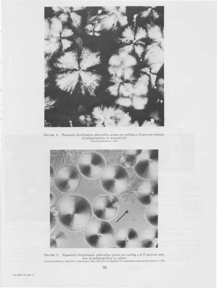
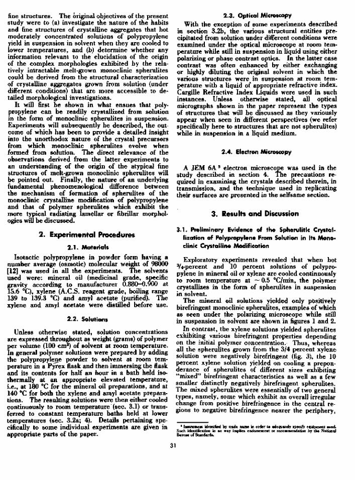
FIGURE 2. Positively birefringent spheruUtes grown on cooling a 10 percent solutionof polypropylene in mineral oil.
Crossed polarizers,X 220.
FIGURE 3. Negatively birefringent spheruUtes grown on cooling a 0.75 percent solu-tion of polypropylene in xylene.
Crowed polariien and 1/4 A compensator (slow direction of vibration of compensator indicated by arrow), X370.
33
792-867 O-66-3

FIGURE 4. Spherulites exhibiting "mixed" birefringent characteristics grown on cool-ing a 10 percent solution of polypropylene in xylene.
Crossed polarizers. X 470.
FIGURE 5. Wide angle x-ray powder pattern obtained from a sampleoj negatively birefringent spherulites formed on cooling a 0.75percent solution of polypropylene in xylene. _
V e r t i c a l b a r s i d e n t i f y i n n e r 1 1 0 , 0 4 0 , 1 3 0 a n d c o m p o s i t e I I I , L S I , 0 4 ] r i n g s c h a r a c t e r -istie of ihe monoclinic (Naiia) crystalline modification of polypropylene.
temperatures of crystallization lead to the formation ofnegatively birefringent monoclinic spherulites froma 10 percent xylene solution, was further substan-tiated by the fact that when such a solution is rapidlyquenched in liquid nitrogen (rate of cooling to roomtemperature ~ 44 °C/min) only negatively birefringentspherulites are formed.
3.2. On the Nature of the Early Stages of Formation of FIGURE 6. Spherulites grown on coo/ing a 10 percent solution of
Monoclinic Spherulites From Solution They ar(, depicted as leen^nde?oT£«^Dol«i^dfinddenl illuminatio, after bavin.,,,.y M , ? ± 2 Edried in air at room temperature. X 270.a. The Habits of Polypropylene Crystallized From 3/4 percent Solu-
tions of the Polymer in Mineral Oil Under Isothermal Conditions(80 to 11 2 °C) clinic spherulites similar to those shown in figure 1.
At higher temperatures, however, in the range 105 toAt 80 and 90 °C the polymer crystallizes very rap- 112 °C individual solutions yield in addition to rela-
idly. The solutions yield positively birelringenl mono- lively more compact positively birefringent monoclinic
34

\
FIGURE 7. Positively birefringent spherulites and smaller aspher-ical aggregates grown from a 0.75 percent solution of polypropy-lene in mineral oil at 112 °C.
Polarizers crossed at 80°, x .T70.
FIGURE 8. Crystalline aggregate grown from a 0.75 percent solu-tion of polypropylene in mineral oil at 112 °C.
Note elliptical profile of aggregate when seen as depicted here along an axis (z) at rightangles to its plane U,y) of preferential development. Unpolarized light. X 1.930.
spherulites of different sizes (fig. 7), numerous smallerrigid nonspherulitic monoclinic crystalline aggregates 6
which exhibit differing but related habits. The re-spective shapes of these various aggregates werereadily deduced by observing them (after crystalliza-tion was completed and the solutions cooled to roomtemperature) as they tumbled, or were induced totumble slowly, in suspension in liquid under the opticalmicroscope in transmission either in unpolarized lightor more appropriately using phase contrast optics.
An example is shown in figure 8 of an aggregatetypical of many that are formed in the temperaturerange 105 to 112 °C. These aggregates exhibitcharacteristically contrasting profiles when viewedalong different directions. Observing such structuresas they tumbled in a liquid medium revealed that theypossess a plane of preferential development. Whenthey are oriented so as to occupy a maximum surfacearea in the field of view of the microscope they exhibit,as may be seen in figure 8, an essentially elliptic allyshaped profile. For the sake of convenience in whatfollows, the major and minor axes of the aggregatecross section depicted in figure 8 will be referred tobelow as the (x) and (y) axes, respectively, and the axisnormal to the [x,y) plane (the plane of preferentialdevelopment) as the (z) axis. When such an aggregateis viewed along any axis lying in the {x, y) plane (i.e.,if the aggregate shown in figure 8 is rotated through90° from the position depicted therein about any axisnormal to the microscope axis), it exhibits an essen-
Tli, i, aggregate is used throughout ihs l r u r l m . i l entity which has grown from lolutlOlterized as he ing an individual coherent c rys ta lgales will be differentiated by suitable adjunchedral, etc.) refer sly to disti
paper in a generic sense lo denote an\.ml which could nol he oulriglilly charac-.1 a splierulile. Different types of aggie(e.g., elliptical, apparent •« pseudopoly-shape characteristics.
FIGURE l). Aggregate similar to that shown in figure H but viewededge-on along an axis parallel to its plane (x.y) of preferentialdevelopment.
Unpolarized light, X 1.930.
tially incipient sheaflike appearance as shown in figure9. Overall growth along the (z) direction is substan-tially slower than in the (x,y) plane, the relative dis-parity in growth being roughly characterized by thefollowing overall (x, y, 2) dimensions: —30 /x X 20 fxX5 fx (i.e., x:y:z— 6:4:1). In actual fact these ag-gregates are somewhat thicker along the (z)-direetionin the peripheral regions than at the (-enter. Thisis more clearly revealed by examining these struc-tures under the phase contrast microscope, as may be
35
!
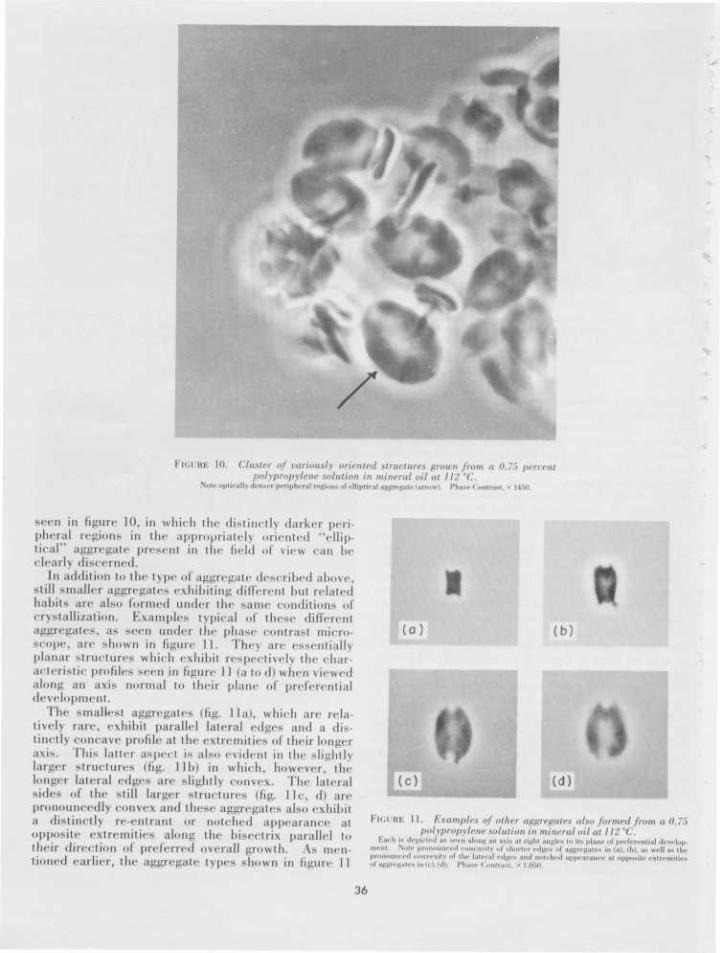
FIGURE 10. Cluster of variously oriented structures grown from a 0.75 percentpolypropylene solution in mineral oil at 112 °C.
Note optically denser peripheral regions of elliptical aggregate (arrow). Phase (Contrast, x 145(1.
seen in figure 10, in which the distinctly darker peri-pheral regions in the appropriately oriented "ellip-tical" aggregate present in the field of view can beclearly discerned.
It) addition to the type of aggregate described above,still smaller aggregates exhibiting different but relatedhabits are also formed under the same conditions ofcrystallization. Examples typical of these differentaggregates, as seen under the phase contrast micro-scope', are shown in figure 11. They are essentiallyplanar structures which exhibit respectively the char-acteristic profiles seen in figure I I (a to (I) when viewedalong an axis normal to their plane of preferentialdevelopment.
The smallest aggregates (fig. lla), which are rela-tively rare, exhibit parallel lateral edges and a dis-tinctly concave profile at the extremities of their longeraxis. This latter aspect is also evident in the slightlylarger structures (fig. lib) in which, however, thelonger lateral edges are slightly convex. The lateralsides of the still larger structures (fig. l ie , d) arepronouncedly convex and these aggregates also exhibita distinctly re-entrant or notched appearance atopposite extremities along the bisectrix parallel totheir direction of preferred overall growth. As men-tioned earlier, the aggregate types shown in figure 11
Q(b)
(c)
FIGURE 11. Examples of other aggregates also formed from a 0.75polypropylene solution in mineral oil at 112 °C.
Each ia depicted as seen along an axis ai right angles to its plane of preferential develop-ment . Note pronounced concavity ..I shorter edges of aggregates in (a), (b), as well as thepronounced convexity ..I the l a i c a l edges and notched a p p e a r a n c e at opposite extremitiesof aggregates in (c), (d). Phase Contrast. « 1.050.
36

are thinner (< 5 /A) than they are long or broad. They,too, however, as in the case of the elliptical structuresdescribed above, are slightly thicker in the peripheralregions than at the center. They all exhibit incipientsheaflike appearances when viewed on edge (i.e.,along any axis in the plane of the respective crosssections depicted in figure 11). A view is shown infigure 12 including a few "notched" aggregates of thetype shown in figure l i e , d lying flat on a substrateand several other nonspherulitic aggregates similarto those described above, oriented with their planeof preferential growth parallel to the axis of the phasecontrast microscope.
A comparison of the relative sizes and correspond-ing habit features of the types of aggregates shown infigure 11 which had all formed under the same con-ditions as the elliptical structures shown in figures8 and 10, indicates an evident size-habit trend (in-creased convexity of the lateral sides and progressivefilling in of the re-entrant extremities) which stronglysuggested that these structures might well representearly successive stages in the formation of the larger"elliptical" structures (figs. 8 to 10), but further de-velopment of which had been arrested due to depletionof the polymer from the solution. By the same token,since the spherulites (fig. 7) that are formed underthe same conditions as the types of aggregates de-scribed above were larger than the elliptical structuresformed at the same temperatures in individual solu-
tions, the possibility was envisaged that the ellipticaland other aggregates are representative of the earlysuccessive transient structures that develop pro-gressively during the initial stages of growth of in-dividual monoclinic spherulites. This was confirmedby some experiments described below.
b. Direct Microscopic Observations of the Growth of MonoclinicSpherulites From Concentrated Solutions of Polypropylene inMineral Oil
The procedure adopted in these experiments wasfacilitated by the low volatility of the mineral oil atthe temperatures at which the experiments werecarried out. Appropriate weights of powderedpolypropylene and mineral oil were deposited on amicroscope slide at room temperature. The polymer-solvent mixture was then covered with a thin cover-glass and placed in the heating chamber of a Leitzheating stage (Model 350) mounted on the rotatingstage of a phase contrast microscope. The tempera-ture of the heating stage was then raised to 200 °Cand maintained at that temperature for approximately10 min. The temperature was then lowered by suitableadjustment of the current supply to the hot stage andmaintained within ± 1 °C of a predetermined tempera-ture at which the rate of spherulite formation was slowenough to be recorded with a conventional photographic
I'H.i RE 12. General view including numerous randomly oriented aggregatesf r o m the s a m e p r e p a r a t i o n as t h o s e s h o w n i n figures 7 to II.
Phase Contrast, x 660.
37
camera. The vertical size of the heating stage andthe mechanically restricted vertical travel of the phasecontrast condenser system used were such that suit-able illumination could be obtained only for low andmedium power phase contrast objectives. All directobservations and photographic recordings were madeusing a X 20 phase contrast objective. The sequencesdescribed below were recorded within a period of 30to 45 min.
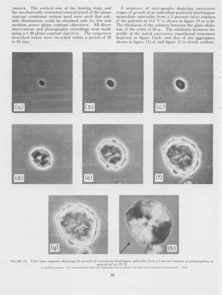
A sequence of micrographs depicting successivestages of growth of an individual positively birefringentmonoclinic spherulite from a 5 percent (w/w) solutionof the polymer at 113 °C is shown in figure 13 (a to h).The thickness of the solution between the glass slideswas of the order of 50 fx. The similarity between theprofile of the initial successive transitional structuresdepicted in figure 13a,b, and that of the aggregatesshown in figure llc,d, and figure 12 is clearly evident.
FIGURE 13. Time lapse sequence depicting the growth of a positively birefringent spherulite from a 5 percent solution of polypropylene inmineral oil at 113 °C.
(a g) Pha»e contrast. (h)Crotied polarizers Ami 1/1 A compensator (arrow indicates slow direction of vibration of compensator). X670.
38
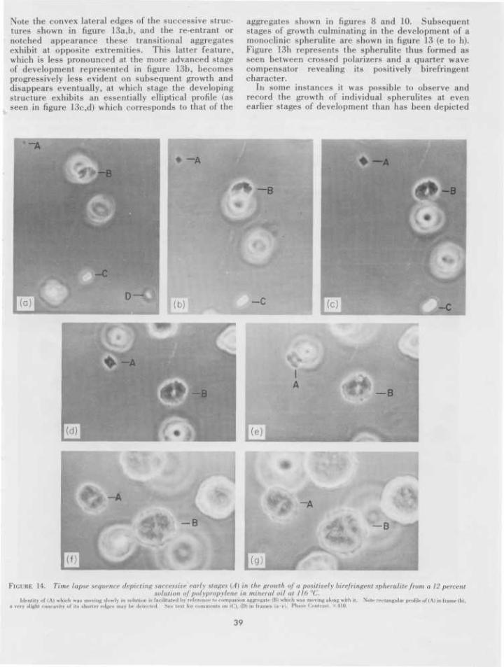
Note the convex lateral edges of the successive struc-tures shown in figure 13a,b, and the re-entrant ornotched appearance these transitional aggregatesexhibit at opposite extremities. This latter feature,which is less pronounced at the more advanced stageof development represented in figure 13b, becomesprogressively less evident on subsequent growth anddisappears eventually, at which stage the developingstructure exhibits an essentially elliptical profile (asseen in figure 13c,d) which corresponds to that of the
aggregates shown in figures 8 and 10. Subsequentstages of growth culminating in the development of amonoclinic spherulite are shown in figure 13 (e to h).Figure 13h represents the spherulite thus formed asseen between crossed polarizers and a quarter wavecompensator revealing its positively birefringentcharacter.
In some instances it was possible to observe andrecord the growth of individual spherulites at evenearlier stages of development than has been depicted
FIGURE 14. Time lapse sequence depicting successive early stages (A) in the growth of a positively birefringent spherulite from a 12 percentsolution of polypropylene in mineral oil at 11(> "('..
I d e n t i t y o f I A ) w h i c h w a s m o v i n g s l o w l y in s o l u t i o n i s f a c i l i t a t e d b y r e f e r e n c e i<> c o m p a n i o n a g g r e g a t e ( B ) w h i c h w a s m o v i n g a l o n g w i t h i t . N o t e r e c t a n g u l a r p r o f i l e o f ( A ) in f r a m e (l>),a v e r y a l i g h t c o n c a v i t y o f i t s s h o r t e r r e l i c s m a y b e d e t e c t e d . S e e t e x l f o r c o m m e n U o n ( C ) , ( D ) in f r a m e i (a c ) . P h a s e C o n t r a » l • H O .
39
in the sequence shown above. Correspondingly, thevery initial resolvable precursor structure was foundto exhibit (when appropriately oriented) a rectangular(almost square) profile. Early stages of developmentof an individual monoclinic spherulite from a 12 per-cent (w/w) solution of polypropylene in mineral oilat 116 °C are depicted in the sequence of micro-graphs shown in figure 14. The particular structure(A) the growth of which was followed in this sequencewas moving very slowly in the solution; its identity isconfirmed in figure 14 (a to g) by a reference aggregate(B), at a more advanced stage of growth, which wasmoving along with it (note the elliptical profile of B).
The apparently rectangular (almost square) profileof the first clearly resolvable precursor seen in figure14b is evident. At the growth stages depicted infigure 14c,d the profile of the growing structure canbe seen to correspond with that of the smallest andrarer aggregates (of the type shown in fig. lla,b)described in the previous subsection. Note thelonger lateral edges and the distinctly cusped appear-ance (concave with respect to the interior) of theshorter edges of the growing aggregate (A) in figure14c,d. The aggregate subsequently assumes on fur-ther growth a profile similar to that of the structures
shown in figure llc,d. At the more advanced stagesof growth depicted in figure 14 (fto g) the aggregatemay be seen to exhibit an essentially elliptical pro-file—it eventually developed into a positively bire-fringent spherulite.
On the basis of the evident similarity between thetransient precursors seen in the sequences shown infigures 13, 14, and the aggregates described in theprevious subsection (3.2a), it is to be expected thattheir appearance would vary significantly dependingon their orientation with respect to the direction ofviewing, hence the contrasting appearance of pre-cursors C (fig. 14 a to c) and D (fig. 14a). It maybe readily deduced that they are respectively orientedwith their plane of preferential development essen-tially normal to the plane of the field of view photo-graphed in figure 14.
In summary, the observations described in thissection provide a gross insight into the nature of theearly stages of development of monoclinic spherulitesof polypropylene. Further opportunity to characterizein finer detail the early stages of development of suchspherulites is clearly provided by the ready accessi-bility of the representative precursors of the spheru-lites formed from the 3/4 percent mineral oil solutions.
r
* * * * *
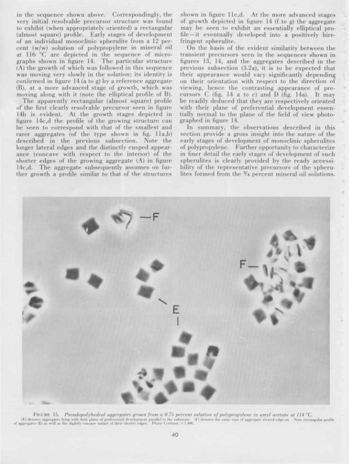
FIGURE 15. Pseudopolyhedral aggregates grown jrom a 0.75 percent solution of polypropylene in amyl acetate at 111 "C.(E) denotes aggregates lying wiili ilicir plane of preferential development parallel to the substrate. (F) denotes il»' same type of aggregate viewed c d g e o n . Nm<- r ec t angu la r profile
of aggregates (E) as well as the slightly concave nature of their shorter edges . Phase Contrast, x 1.400.
40
Clearly these structures can'be readily isolated fromthe latter preparations and subjected to an electronmicroscopic study using replicating techniques inorder to characterize the nature of their fine structure.
The apparently polyhedral earliest precursors whichdevelop during the initial stages of formation of thespherulites are of particular initial interest, however.Similar essentially planar objects exhibiting a squareor slightly rectangular profile have been respectivelyobserved by Leugering [13] and Geil [14] to form duringthe initial stages of growth of monoclinic spherulitesfrom the melt. In short, the formation of such initialstructures is not a peculiar characteristic of the modeof growth of monoclinic spherulites under the specificconditions of crystallization described in this section —it is evidently a phenomenon of more general occur-rence. The discovery that ZU percent solutions ofpolypropylene in amyl acetate yield, at temperaturesbetween 110 to 115 °C, monoclinic crystalline aggre-gates the shape of which (see fig. 15) coincides withthat of the above-mentioned initial apparently poly-hedral precursors of monoclinic polypropylenespherulites, was therefore particularly gratifying [15].The outcome of an optical and electron microscopicstudy, which has led to the characterization of thehighly unorthodox structure of these model counter-parts of the earliest microscopically resolvablespherulite precursors, is discussed in the ensuingsection. It will be shown that they are not polyhedralobjects, but possess instead a complex fine structurewhich is distinctly dendritic in character.
4. On the Morphology of Model Counterpartsof the Pseudopolyhec'ral Precursors of Mono-clinic Spherulites of Polypropylene
The observations presented in this section werederived from an optical and electron microscopicstudy of crystalline aggregates grown from 3/4 percentsolutions of polypropylene in amyl acetate at 114 °C.Within 6 hr at that temperature the solutions exhibita very faint cloudiness indicating the onset of crys-tallization. The particular solution from which theaggregates shown in figure 15 were formed was heldat 114 °C for 10 days. At the end of this period, thesolvent and suspended aggregates were poured intoa special filtration vessel maintained at 114 °C. Thetype of vessel used was essentially similar in designto a filtering apparatus previously described by Bassettet al., [16]. The preparation was filtered isothermallyand washed several times with fresh amyl acetatepreheated to 114 °C. Care was taken not to allowthe filtrate to dry between successive washings. Aftera final addition of hot fresh solvent, filtration wasstopped, and the vessel allowed to cool to roomtemperature.
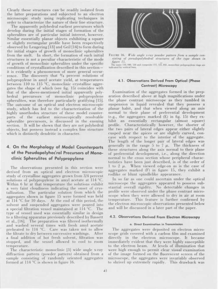
The characteristic' monoclinic | 3 | wide angle x-raydiffraction pattern (powder pattern) obtained from asample consisting of randomly oriented aggregatesformed at 114 °C is shown in figure 16.
FIGURE 16. Wide angle x-ray powder pattern from a sample con-sisting of pseudopolyhedral structures of the type shown infigure 15. _The inner 110, 040, 130 and composite 111, 131,041, monoclinic polypropylene rings are
indicated by vertical bars.
4.1. Observations Derived From Optical (PhaseContrast) Microscopy
Examination of the aggregates formed in the prep-aration described above at high magnifications underthe phase contrast microscope as they tumbled insuspension in liquid revealed that they possess aplanar habit, and that when viewed along an axisnormal to their plane of preferential development(e.g., the aggregates marked (E) in fig. 15) they ex-hibit an essentially rectangular (almost square)profile. Characteristically however, the shorter ofthe two pairs of lateral edges appear either slightlycusped near the apices or are slightly curved, con-cave with respect to the interior of the aggregate.The length of the longer pair of lateral edges wasgenerally in the range 5 to 7 fx. The thickness ofthese structures along the axis normal to their planeof preferential development, i.e., along the directionnormal to the cross section whose peripheral charac-teristics have been just described, is of the order of1 to 2 ix. When viewed edge-on as is the case ofaggregates marked (F) in figure 15, they exhibit arodlike or blunt spindlelike appearance.
In so far as one could ascertain under the opticalmicroscope the aggregates appeared to possess sub-stantial overall rigidity. No detectable changes inprofile were observed under the phase contrast micro-scope when they were allowed to dry in air at roomtemperature. This feature is further confirmed bythe electron microscopic observations presented belowand will be discussed in a later part of the paper.
4.2. Observations Derived From Electron Microscopy
a. Direct Examination in Transmission
The aggregates were deposited on electron micro-scope grids covered with a carbon film and examineddirectly in the electron microscope. It becameimmediately evident that they were highly susceptibleto the electron beam. At levels of illumination thatwere high enough to permit clear visual examinationof the image formed on the fluorescent screen of themicroscope, the aggregates were invariably observedto shrivel up extremely rapidly. Phis drawback was
41

overcome by scanning the grids and photographingthe aggregates at very low intensities and by limitingthe area of the specimen grid exposed to the electronbeam at any given time to approximately one gridsquare. This was accomplished by the conventionalmethod of using the double condenser system of themicroscope and appropriately defocusing the secondcondenser lens.
Under such conditions of low illumination it waspossible to discern only faintly the outline of individualaggregates on the microscope screen. Consecutivemicrographs recorded within 30 sec of exposure ofindividual aggregates to the electron beam under suchconditions of low "illumination" revealed no detect-able morphological changes in that period of time.Within two minutes, however, substantial changes inoverall shape were discerned, even visually.
Electron micrographs depicting three individualaggregates photographed immediately after beingexposed to the electron beam are shown in figures17, 18, and 19a. These aggregates originate from thesame preparation as the structures shown in figure 15.Two characteristic features detected under the opticalmicroscope are clearly revealed in these electronmicrographs, namely, the overall essentially rec-tangular profile of each aggregate and the slightly
cusped or concave nature of the shorter "sides."The length to breadth ratio of the aggregates (asdetermined from the length of the central axes bisect-ing opposite sides) was generally found to be of theorder of 1.1:1.
As would be expected from the 1 to 2 fx thicknessof these structures they are essentially opaque tothe electron beam, particularly so in the somewhatthicker central regions. Nevertheless, transmissionelectron micrographs generally revealed two char-acteristic morphological features. First, what ap-peared to be narrow diagonally oriented spikelikeoutgrowths were frequently seen protruding from theedges of the aggregates, as may be seen in particularin figures 17 and 19a. Second, a weakly discerniblediagonally oriented crOss-hatched appearance whichcould be traced from the peripheral regions for alimited distance into the interior, as may be seen forexample in figure 18, was frequently detected in theelectron micrographs.
In addition to the above-mentioned features ap-parently spurious lamellar single crystallike out-growths, as may be seen for example near the apex(G) of the aggregate partially in view in figure 18, wereoccasionally observed in the peripheral regions of someaggregates.
FIGURE 17. Electron micrograph of a pseudopolyhedral polypropylene aggregate of the type shown in figure IS.N o t e o v e r a l l e s s e n t i a l l y r e c t a n g u l a r prof i l e o f t h e a g g r e g a t e a s w e l l ;is t h e d i a g o n a l l y o r i e n t e d s p i k e l i k e o u t g r o w t h s p r o t r u d i n g f rom i ts p e r i p h e r y . A
n o t e s l i f i l i i lv c o n c a v e prof i l e »l o n e <>l i ts i h o r t e r - ' s i d e s . " S h a d o w e d ( P t . - P d ) . x 1 6 . 8 0 0 .
42

The consequence of prolonged exposure to the elec-tron beam is shown in figure 19b. The aggregateswere always observed to undergo preferential overallshrinkage along a direction normal to their longeraxis as may be readily ascertained by comparing figure19a and figure 19b. No further change in aspectwas observed beyond the stage of shriveling depictedin the, latter electron micrograph even when the in-tensity of illumination is substantially increased. Thespecificity of the mode in which the aggregates de-form remains unexplained. The change they undergowhen exposed to the electron beam is primarilyattributable to melting or partial melting resultingfrom the heating effects of the electron beam and thepoor dissipation of heat generated by the impingingelectrons due to the low thermal conductivity of the ag-gregates and their supporting carbon film. It is tobe expected that the polypropylene chains undergocross-linking on exposure to the electron beam [17].On the basis of established experience, for examplecontrasting the behavior of these aggregates with that
of polyethylene single crystals, it may be readilyassumed that the occurrence of cross-linking (whichleads to a loss of crystallinity in polyethylene crystalswithout their undergoing a change in shape) is not theprime factor leading to the drastic deformation whichthe aggregates undergo when irradiated.
b. Electron Microscopic Examination of Surface Replicas
In order further to elucidate the nature of the finestructure of the aggregates, replicas of their largesurfaces were prepared using the method of shadowtransfer. For this purpose, the aggregates were ini-tially deposited on a glass slide and allowed to dryin air at room temperature; they were then shadowedwith metal (80 percent Pt-20 percent Pd alloy). Abacking film of carbon was subsequently evaporatedonto the shadowed aggregates on the slide. Theresulting composite metal-carbon "film" thus formedwas floated over distilled water. The aggregateswere found to adhere to the film, portions of which
the arShado
FIGURE IH. Electron micrograph of a pseudopolyhedral polypropylene aggregate.c the faintly discernible diagonally oriented cross-hatched appearance extending inwards fron • of the shorter edget of the aggregate indicated by
near the left-hand coiner of the figure. Also note apparent lamellar character of outgrowths (('.) near the ai><\ of another neighboring aggregate,d I ' I - IM x 16,400.
43

FIGURE l(>. Electron micrographs depicting effect of prolongedexposure to the electron beam.
(a) Micrograph recorded al low intensity immediately after exposure i<> the elect ion beam.(I)) Micrograph of same aggregate after 2 tnin exposure to the electron beam. Note char-acteristic preferential shrinkage al ri^ln angles to the longer edges ol the aggregate.Shadowed (I'i-P.1) x L3.300.
were then picked up on electron microscope gridsand allowed to dry in air at room temperature. Somedifficulty was encountered in dissolving the aggregatesfrom the metal-carbon replicating medium but theywere effectively dissolved by immersing the replicassupported on the grids in excess distilled decalin,heating the solvent to 160 °C, and maintaining it al
thai temperature under a blanket of nitrogen for Itf hr.Replicas of the large aggregate surfaces prepared
in the manner described above revealed, as may beseen in figure 20, a distinct cross-hatched surfacetexture consisting of a network of narrow (ISO to 200 A)p
apparently intercrossing structural units which tra-verse the surface for varying distances diagonally
44
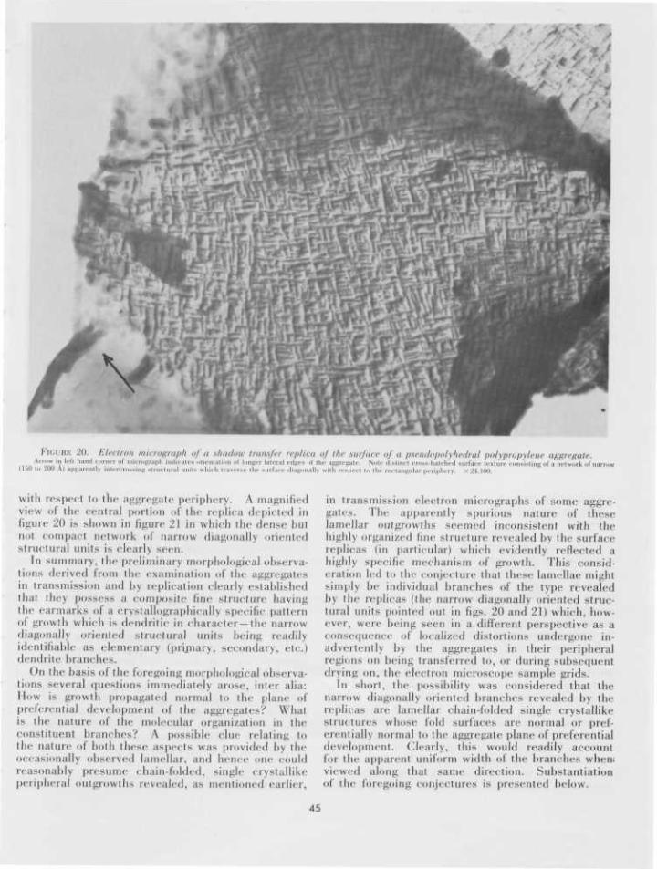
FIGURE 20. Electron micrograph of a shadow transfer replica of the surface of a pseudopolyhedral polypropylene aggregate.__ Anow in lefl hand corner of micrograph indicates urientation of longer lateral edges of the aggregate. Note distinct cross-hatched surface texture consisting of a network ofnar
(150 to 200 A) apparently intercrossing structural units which traverse the Burface diagonally with reaped to the rectangular periphery. X24.100.
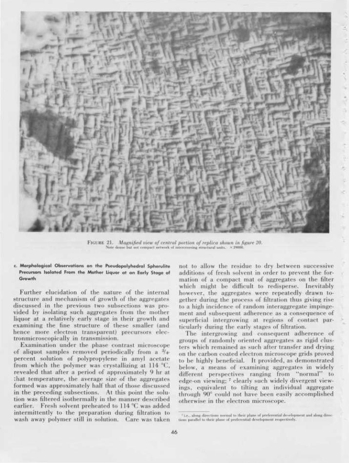
with respect to the aggregate periphery. A magnifiedview of the central portion of the replica depicted infigure 20 is shown in figure 21 in which the dense hutnot compact network of narrow diagonally orientedstructural units is clearly seen.
In summary, the preliminary morphological observa-tions derived from the examination of the aggregatesin transmission and by replication clearly establishedthat they possess a composite fine structure havingthe earmarks of a erystallographieally specific patternof growth which is dendritic in character —the narrowdiagonally oriented structural units being readilyidentifiable as elementary (primary, secondary, etc.)dendrite branches.
On the basis of the foregoing morphological observa-tions several questions immediately arose, inter alia:How is growth propagated normal to the plane ofpreferential development of the aggregates? Whatis the nature of the molecular organization in theconstituent branches? A possible clue relating tothe nature of both these aspects was provided by theoccasionally observed lamellar, and hence one couldreasonably presume chain-folded, single crystallikeperipheral outgrowths revealed, as mentioned earlier,
in transmission electron micrographs of some aggre-gates. The apparently spurious nature of theselamellar outgrowths seemed inconsistent with thehighly organized fine structure revealed by the surfacereplicas (in particular) which evidently reflected ahighly specific mechanism of growth. This consid-eration led to the conjecture that these lamellae mightsimply be individual branches of the type revealedby the replicas (the narrow diagonally oriented struc-tural units pointed out in figs. 20 and 21) which, how-ever, were being seen in a different perspective as aconsequence of localized distortions undergone in-advertently by the aggregates in their peripheralregions on being transferred to, or during subsequentdrying on, the electron microscope sample grids.
In short, the possibility was considered that thenarrow diagonally oriented branches revealed by thereplicas are lamellar chain-folded single erystallikestructures whose fold surfaces are normal or pref-erentially normal to the aggregate plane of preferentialdevelopment. Clearly, this would readily accountfor the apparent uniform width of the branches when;viewed along that same direction. Substantiationof the foregoing conjectures is presented below.
45
f '••
I ; ; : •
,
i) . •*
FIGURE 21. Magnified view of central portion of replica shown in figure 20.Note dense hut not compact network of intercrossing structural units. X39000.
c. Morphological Observations on the Pseudopolyhedral SpherulitePrecursors Isolated From the Mother Liquor at an Early Stage ofGrowth
Further elucidation of the nature of the internalstructure and mechanism of growth of the aggregatesdiscussed in the previous two subsections was pro-vided by isolating such aggregates from the motherliquor at a relatively early stage in their growth andexamining the fine structure of these smaller (andhence more electron transparent) precursors elec-tronmicroscopically in transmission.
Examination under the phase contrast microscopeof aliquot samples removed periodically from a :J/4-percent solution of polypropylene in amyl acetatefrom which the polymer was crystallizing at 114 °C,revealed that after a period of approximately 9 hr atthat temperature, the average size of the aggregatesformed was approximately half that of those discussedin the preceding subsections. At this point the solu-tion was filtered isothermally in the manner describedearlier. Fresh solvent preheated to 114°C was addedintermittently to the preparation during filtration towash away polymer still in solution, ("are was taken
not to allow the residue to dry between successiveadditions of fresh solvent in order to prevent the for-mation of a compact mat of aggregates on the filterwhich might be difficult to redisperse. Inevitablyhowever, the aggregates were repeatedly drawn to-gether during the process of filtration thus giving riseto a high incidence of random interaggregate impinge-ment and subsequent adherence as a consequence olsuperficial intergrowing at regions of contact par-ticularly during the early stages ol filtration.
The intergrowing and consequent adherence ofgroups of randomly oriented aggregates as rigid clus-ters which remained as such after transfer and dryingon the carbon coated electron microscope grids provedto be highly beneficial. It provided, as demonstratedbelow, a means of examining aggregates in widelydifferent perspectives ranging from "normal" toedge-on viewing; 7 clearly such widely divergent view-ings, equivalent to tilting an individual aggregatethrough 90° could not have been easily accomplishedotherwise in the electron microscope.
<•., along direcparallel to tl>< plai
nal to tin,1 prefer.
r planenial dev
ntialdereaped
and along dire
46

• H i
\ H
FIGURE 22. Electron micrograph oj a cluster oj variously oriented "halj-grown" pseudopolyhedral polypropylene aggregates.(H) is oriented with its plane (if preferential development at right angles to the microscope axis, (J) and (K) are viewed edge-on, and (L) is an aggregate at an inclination intermediate
between the extreme orientations represented by (H), (.1) and (K). X 12,800.
An electron micrograph depicting a cluster of"half-grown" aggregates isolated from the motherliqour at 114 °C in the manner described above, isshown in figure 22. Characteristically the aggre-gates are variously oriented; in particular, aggregate(H) (note its rectangular profile) is oriented "normally"with respect to the microscope axis, whereas aggre-gates (J) and (K) are oriented on edge. The plane ofaggregate (L) is inclined to the substrate. It shouldbe pointed out that these "hall-grown" aggregateswere found also to be susceptible to the heating effectsof the electron beam, hence the precautions describedearlier (i.e., scanning and photographing at low beamintensities and suitably limiting the area of a specimengrid exposed to the electron beam at any time) hadto be resorted to. The characteristic morphologicalfeatures exhibited by the aggregates when viewed"normally" and edge-on are considered in detailbelow.
An examination of electron micrographs of aggre-gates oriented with their plane at right angles to theelectron beam revealed the following structural char-acteristics which may be readily discerned in the rep-resentative aggregate shown in figure 23. First, they
exhibit an overall rectangular (almost square) profile,their length to breadth ratio being of the order of1.1:1. On average they were found to be about 2.5 fxlong. Second, they exhibit a highly regular fine struc-ture consisting of a dense but not compact "network"of narrow diagonally oriented branches. This evi-dently dendritic cross-hatched fine structure could inall instances be traced throughout each aggregatethus oriented from the thicker central regions to theperiphery. The width of the constituent branches(based on the measurement of those branches thatare individually resolvable in the peripheral regions)is of the order of 150 to 200 A.
On the basis of geometric considerations (length/breadth ratio), the acute angle subtended at the centerof the crystals by the diagonally oriented branches(i.e., the angle subtended at the center by the shorteraggregate edges) is estimated to be — 85°. Accu-rate direct interbranch angular measurements arefrustrated by the overall opacity of the crystals whichare 0.5 /u~0.75 fjL thick.
These observations further confirmed that the cross-hatched surface structure revealed by the replicas ofthe larger precursors described earlier is character-
47
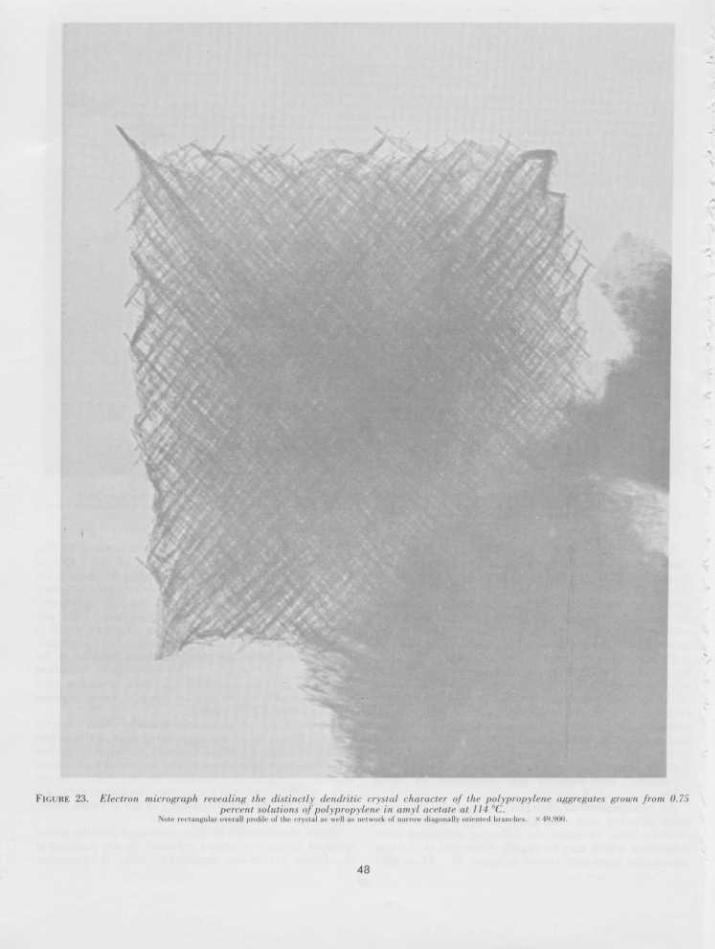
FIGURE 23. Electron micrograph revealing the distinctly dendritic crystal character of the polypropylene aggregates grown from 0.7!)percent solutions of polypropylene in amyl acetate at 114 °C.
Nnic rectangular overall profile of the crystal as well as network of narrow diagonally oriented branches. X49,900.
43
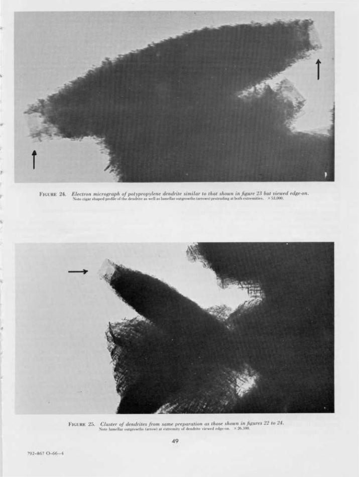
FIGURE 24. Electron micrograph of polypropylene dendrite similar to that shown in figure 23 but viewed edge-on.Note cigar shaped profile of the dendrite as well as lamellar outgrowths (arrows) protruding at both extremities. X 53,000.
•
FIGURE 2.r>. Cluster of dendrites from same preparation as those shown in figures 22 to 24.Nole lamellar outgrowths (arrow) el extremity of dendrite viewed edge-OD. X 26,100.
49
792-867 O-66-4
istic of their internal structure. The continuity ofthe same dendritic pattern of branch propagationparallel to the plane of preferential development ofthe smaller precursors just described and the largeraggregates discussed in the previous subsections isevident. Some loss of specificity in the mode ofpropagation of the branches as these dendrites becomelarger is however suggested by regional deviations inparallelism of the branches revealed by the replicasof the bigger dendrites and by the apparent concavityof their shorter "sides."
We turn now to a consideration of how these evi-dently dendritic crystals propagate along the directionnormal to their plane of preferential development.In this respect the structural features exhibited by thesmaller "half-grown" dendrites when viewed edge-on(as exemplified in fig. 22 (structures J, K) and figs.24, 25) were examined. When viewed thus, i.e., alongdirections parallel to their plane of preferential de-velopment, they exhibit a cigar-shaped profile; theywere on average found to be 0.75 /x wide at the centerand somewhat narrower at the extremities, which isconsistent with the observed decrease in overallopacity to the electron beam from the central to theperipheral regions when they are viewed as shown infigure 22 (H) and figure 23. In all instances in whichthe dendrites were examined edge-on, distinctly lamel-lar single crystallike outgrowths were observedprotruding from their extremities as may be seen infigures 24 and 25; further the profile of these lamellaealways formed a natural extension of the overallcigar-shaped contour of the dendrites (see both ex-tremities of the dendrite shown in fig. 24 and the onevisible extremity of the dendrite shown in fig. 25).This latter feature, coupled with the general observa-tion that no such lamellae were seen protruding else-where in dendrites examined in edge-on perspectiveindicated that the lamellae are not spurious outgrowthsbut are the extremities of constituent diagonallyoriented branches which extend from the interior tothe rectangular periphery of the crystals.
The evidence presented in the preceding paragraphsubstantiates the proposition advanced earlier thatthe uniformly narrow diagonally oriented dendritebranches are chain-folded crystal entities whose foldsurfaces are oriented at right angles to the dendriteplane of preferential development. These branchesonce formed can be readily visualized to develop in acontinuous fashion at right angles to that plane aswell as diagonally during the growth process. Asimplified and essentially self-explanatory schematicrepresentation of the mode of growth of these poly-propylene dendrites as presently envisaged is shownin figure 26. For the sake of simplicity only fourprimary and two secondary branches are depicted;their respective fold surfaces (f) (i.e., the faces be-tween which the molecules fold back and forth) arealso identified. Clearly, profuse crystallographicallyspecific branching involving a dihedral angle of ~ 85°between the fold surfaces in parent and offspringbranches is envisaged.
The apparent overall rigidity of the dendrites may
be attributed to interlocking at points where branchesimpinge and grow "around" one another as depictedin figure 26.
It should be emphasized at this stage that the pat-tern of development exhibited by these dendriticcrystals is highly unusual. It is common experiencethat solution-grown chain-folded polymer crystals(lamellae) generally thicken along a direction normalto their fold surfaces through the agency of screwdislocations (with Burger's vectors parallel to the chainsegments). These dislocations give rise to the for-mation of additional chain-folded terraces which arecoplanar with the basal lamella.8 As far as this authoris aware, no previous instances have been reported,in the context of chain-folded crystals of other poly-mers, of the occurrence of the repeated formationof lamellar outgrowths the fold surfaces of which areoriented at a substantial and specific angle (—85° inthe present instance) to the fold surfaces of the parentlamellae from which they have grown, as exemplifiedby the polypropylene crystals described herein.
Further elucidation of the structure and mecha-nism of growth of these unusual dendritic crystalsclearly required their characterization on a crystal-lographic basis. Some observations pertaining tothe nature of the molecular orientation in the dendritesare presented below.
d. On the Crystallography of the Dendrites
The birefringent properties of the dendrites weredetermined by examining the crystals orthoscopicallyunder a polarizing microscope while they were insuspension in amyl acetate and other liquid media atroom temperature. The resulting observations aredescribed below and their significance in so far asproviding a rough indication of the nature of themolecular orientation in the dendrite branches isdiscussed in conjunction with some limited electrondiffraction data. Electron diffraction experimentshave been somewhat frustrated by the instability ofthe crystals in the electron beam, as well as by theoverall opacity of the crystals to the impinging elec-trons. In a few instances the crystals were preservedlong enough at low illuminations that a selected areaelectron diffraction pattern as well as the correspond-ing selected area including a crystal were recordedphotographically. The few diffraction patterns whichhave so far been recorded, and which will be discussedpresently, were extremely weak; they could not bediscerned visually on the fluorescent screen of theelectron microscope. Evidently only the peripheralregions of the dendrites contributed to the diffractionpatterns.
The room temperature extinction characteristics ofthe larger dendrites (such as those shown in figs. 15,17 to 21), when variously oriented with respect tocrossed polarizers as well as the corresponding inter-
8 The situation is somewhat more complex in the case of hollow pyramidal crystals. Insuch instances the additional layers are also hollow pyramidal and coaxial with the "basal"layer [18].
50
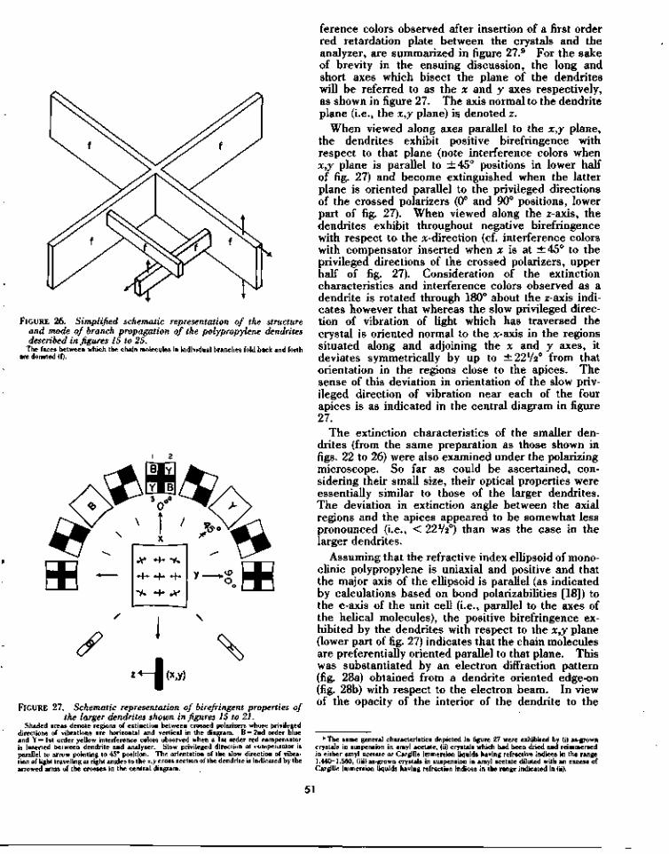
FIGURE 26. Simplified schematic representation of the structureand mode of branch propagation of the polypropylene dendritesdescribed in figures 15 to 25.
The faces between which the chain molecules in individual branches fold back and forthare denoted (f).
FIGURE 27. Schematic representation of birefringent properties ofthe larger dendrites shown in figures 15 to 21.
Shaded areas denote regions of extinction between crossed polarizers whose privilegeddirections of vibrations are horizontal and vertical in the diagram. B = 2nd order blueand Y=lst order yellow interference colors observed when a 1st order red compensatoris inserted between dendrite and analyser. Slow privileged direction of compensator isparallel to arrow pointing to 45° position. The orientation of the slow direction of vibra-tion of light traveling at right angles to the x,y cross section of the dendrite is indicated by thearrowed arms of the crosses in the central diagram.
ference colors observed after insertion of a first orderred retardation plate between the crystals and theanalyzer, are summarized in figure 27.9 For the sakeof brevity in the ensuing discussion, the long andshort axes which bisect the plane of the dendriteswill be referred to as the x and y axes respectively,as shown in figure 27. The axis normal to the dendriteplane (i.e., the x,y plane) is denoted z.
When viewed along axes parallel to the x,y plane,the dendrites exhibit positive birefringence withrespect to that plane (note interference colors whenx,y plane is parallel to ±45° positions in lower halfof fig. 27) and become extinguished when the latterplane is oriented parallel to the privileged directionsof the crossed polarizers (0° and 90° positions, lowerpart of fig. 27). When viewed along the z-axis, thedendrites exhibit throughout negative birefringencewith respect to the x-direction (cf. interference colorswith compensator inserted when x is at ±45° to theprivileged directions of the crossed polarizers, upperhalf of fig. 27). Consideration of the extinctioncharacteristics and interference colors observed as adendrite is rotated through 180° about the z-axis indi-cates however that whereas the slow privileged direc-tion of vibration of light which has traversed thecrystal is oriented normal to the x-axis in the regionssituated along and adjoining the x and y axes, itdeviates symmetrically by up to ±22V2° from thatorientation in the regions close to the apices. Thesense of this deviation in orientation of the slow priv-ileged direction of vibration near each of the fourapices is as indicated in the central diagram in figure27.
The extinction characteristics of the smaller den-drites (from the same preparation as those shown infigs. 22 to 26) were also examined under the polarizingmicroscope. So far as could be ascertained, con-sidering their small size, their optical properties wereessentially similar to those of the larger dendrites.The deviation in extinction angle between the axialregions and the apices appeared to be somewhat lesspronounced (i.e., < 22V20) than was the case in thelarger dendrites.
Assuming that the refractive index ellipsoid of mono-clinic polypropylene is uniaxial and positive and thatthe major axis of the ellipsoid is parallel (as indicatedby calculations based on bond polarizabilities [18]) tothe c-axis of the unit cell (i.e., parallel to the axes ofthe helical molecules), the positive birefringence ex-hibited by the dendrites with respect to the x,y plane(lower part of fig. 27) indicates that the chain moleculesare preferentially oriented parallel to that plane. Thiswas substantiated by an electron diffraction pattern(fig. 28a) obtained from a dendrite oriented edge-on(fig. 28b) with respect to the electron beam. In viewof the opacity of the interior of the dendrite to the
9 The same general characteristics depicted in figure 27 were exhibited by (i) as-growncrystals in suspension in amyl acetate, (ii) crystals which had been dried and reimmersedin either amyl acetate or Cargille immersion liquids having refractive indices in the range1.440—1.560, (iii) as-grown crystals in suspension in amyl acetate diluted with an excess ofCargille immersion liquids having refraction indices in the range indicated in (ii).
51
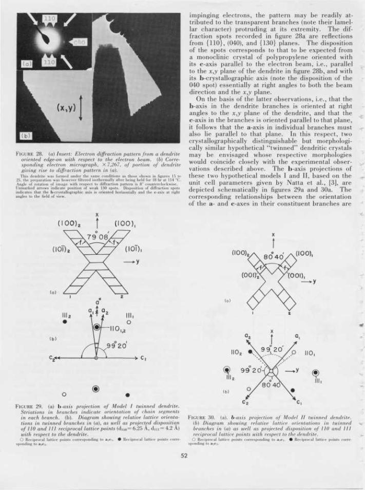
FIGURE 28. (a) Insert: Electron diffraction pattern from a dendriteoriented edge-on with respect to the electron beam, (b) Corre-sponding electron micrograph, X 7,267, of portion of dendritegiving rise to diffraction pattern in (a).
This dendrite was formed under the same conditions as those shown in figures 15 to25, the preparation was however filtered isothermally after being held for 18 hr at 114 °C.Angle of rotation of image with respect to diffraction pattern is 8° counterclockwise.Unmarked arrows indicate position of weak 130 spots. Disposition of diffraction spotsindicates that the b-crystallographic axis is oriented horizontally and the c-axis at rightangles to the field of view.
impinging electrons, the pattern may be readily at-tributed to the transparent branches (note their lamel-lar character) protruding at its extremity. The dif-fraction spots recorded in figure 28a are reflectionsfrom {110}, (040), and {130} planes. The dispositionof the spots corresponds to that to be expected froma monoclinic crystal of polypropylene oriented withits c-axis parallel to the electron beam, i.e., parallelto the x,y plane of the dendrite in figure 28b, and withits b-crystallographic axis (note the disposition of the040 spot) essentially at right angles to both the beamdirection and the x,y plane.
On the basis of the latter observations, i.e., that theb-axis in the dendrite branches is oriented at rightangles to the x,y plane of the dendrite, and that thec-axis in the branches is oriented parallel to that plane,it follows that the a-axis in individual branches mustalso lie parallel to that plane. In this respect, twocrystallographically distinguishable but morphologi-cally similar hypothetical "twinned" dendritic crystalsmay be envisaged whose respective morphologieswould coincide closely with the experimental obser-vations described above. The b-axis projections ofthese two hypothetical models I and II, based on theunit cell parameters given by Natta et al., [3], aredepicted schematically in figures 29a and 30a. Thecorresponding relationships between the orientationof the a- and c-axes in their constituent branches are
(IOO)2 (100),
f T £ 0 8 ^
x\// ("'
FIGURE 29. (a) h-axis projection of Model I twinned dendrite.Striations in branches indicate orientation of chain segmentsin each branch, (b). Diagram showing relative lattice orienta-tions in twinned branches in (a), as well as projected dispositionof 110 and III reciprocal lattice points (dn()= 6.2S A, d n i = 4.2 A)with respect to the dendrite.
O Reciprocal lattice point* corresponding to a,<-,. • Reciprocal lattice points corre-
(100)
(a)
(001
(h)
FIGURE 30. (a), b-axis projection of Model II twinned dendrite.(b) Diagram showing relative lattice orientations in twinnedbranches in (a) OS well as projected disposition of III) and IIIreciprocal lattice points with respect to the dendrite.
O Reciprocal lattice points corresponding to aiCi. • Reciprocal lattice points cone
52
shown in figures 29b and 30b. The fold surfaces (f)of individual branches are viewed edge-on in bothmodels as depicted in figures 29a and 30a. In bothmodels the acute dihedral angle between the foldsurfaces of the intercrossing branches is circa 80°(cf. experimentally estimated angle ~ 85°). In orderto compare the essential features of these modelswith respect to experimental observations we identifythe axes bisecting the acute and obtuse angles betweenthe dendrite branches as the x and y reference axesrespectively (cf. the x- and y-axes which have beenreferred to previously in the discussion of the experi-mentally observed optical properties of the dendrites(fig. 27)).
The essential and contrasting features of models Iand I t are summarized below:
Model I. The dominant feature of this model isthat the axes of the constituent helical molecules,and hence the c-axis, in all the branches are orientedthroughout the whole structure parallel to y. Thea* and [001] axes in the "intercrossing" (twinned)branches in this model are parallel to x and y respec-tively. The respective lattice orientations in thetwinned branches are such as to be brought into coin-cidence by rotation through 180° either about x(i.e., a*) as shown in figure 32 or about y (i.e., [001]).It may be noted that a* which is normal to the b-axisin the monoclinic unit cell of polypropylene, is veryclosely parallel to the [601] axis in the cell, and thatthe (106) planes are very closely normal to the (100)planes._ Individual branches in this model are boundby (101), (100) and (010) faces (the latter which areparallel to the jc,y cross section are not shown in fig.29a). The (101) faces, which are the fold surfaces,are denoted (f) in figure 29a.
Model II. In this model (fig. 30) it is assumed thatthe fold surfaces are parallel to the (001) planes ineach branch whose other faces are parallel to the (100)and (010) planes. It is also assumed that the (100)planes in individual branches are parallel to the (001)planes in branches that "cross" them. The chainmolecules in intercrossing branches are symmetricallyinclinded at 49° 40' to y in the x,y plane, and thetwinned lattice orientations are such as to be broughtinto coincidence by rotation_ through 180° about x.It may be noted that the [101] and [101] axes in thebranches are very nearly parallel to x and y respec-tively.
Both Models I and II conform with the experimentalobservation that the b-axis in the dendrites is orientedat right angles to their x9y plane. That the overalldevelopment of the dendrites is slowest at right anglesto the latter plane may be accounted for in bothmodels if it is envisaged that the growth of individualbranches occurs more rapidly along the directionnormal to their (100) faces (i.e., along a*) than along[010].
In summary, therefore, on the basis of the twomodels discussed above, the possibility was envis-aged, as an initial guide, that the mechanism of pro-pagation of the dendritic monoclinic polypropylenecrystals may involve either repeated twinning through
180° lattice rotations about a* or [001] as in figure 29a,or about an axis at right angles to the b-crystallographicaxis and almost coincident to [101] as in figure 30a.
Using the two model twinned crystals I and II as abasis of reference, we turn now to a consideration ofthe experimentally observed optical properties of thedendrites about their x,y cross section (upper half offig. 27), in conjunction with some electron diffrac-tion data obtained from their peripheral regions withthe crystals oriented with their x,y cross section atright angles to the electron beam.
The optical feature of the dendrites which initiallystrikes attention is the overall negative birefrigencethey exhibit with respect to their x-axis since, a priori,this feature is consistent with the orientation of thechain molecules in Model I. The birefringence withrespect to the x-axis was measured however, and foundto be of the order of —0.007 birefringence units.10
This value is rather lower than would be expectedwhen considered in the light of birefringence-drawratio data obtained by Keedy et al., [19] from drawnfilaments of isotactic polypropylene. A value closerto —0.03 birefringence units with respect to x wouldbe expected of a structure corresponding to Model I.
While the low birefringence of the crystals withrespect to x argues against Model I this feature alonecould not be used to discriminate unambiguouslyagainst this model in view of the uncertainty involvedin the determination of the overall thickness of thedendrites at right angles to the x,y cross section,coupled with the fact that these structures are notsolid objects and that consequently their effectivethickness is most probably lower than the overallthickness which was used to calculate their bire-fringence.
A further factor adding to the uncertainty in inter-preting the low value of the observed birefringenceunambiguously in terms of a precursor model is thefact that the extinction behavior of the dendrites,which is not uniform throughout their x,y crosssection (fig. 27), indicates deviations (albeit sym-metrical) in average molecular orientation about thatcross section. We turn, therefore, to the outcome ofelectron diffraction experiments which, althoughsubject to complications associated with the latterfactor, nevertheless provide a more direct meansof crystallographic characterization.
Two typically weak electron diffraction patternsobtained from smaller dendrites (same preparation asthe crystals shown in figs. 22 to 25) are shown infigures 31 and 32. Interpretation of these patternsis provided by consideration of figure 33 in whichthe b-axis projection of the monoclinic unit cell ofpolypropylene is depicted together with the corre-sponding projection of the reciprocal lattice. Theprojected dispositions of the 110 and 111 reciprocal
10 These measurements were made on the larger dendrites from the same preparation asthose shown in figure 15. The dendrites in suspension in amyl acetate were deposited ona glass slide. Partial evaporation of excess solvent was allowed to occur in air after whichan excess of a Cargille immersion liquid whose refractive index (1.510) corresponded tothe isotropic refractive index of highly crystalline unoriented polypropylene [2] was addedto the deposit. Possible contributions of form birefrigence were thus minimized. Thebirefrigence measurements were carried out with a Berek compensator.
53
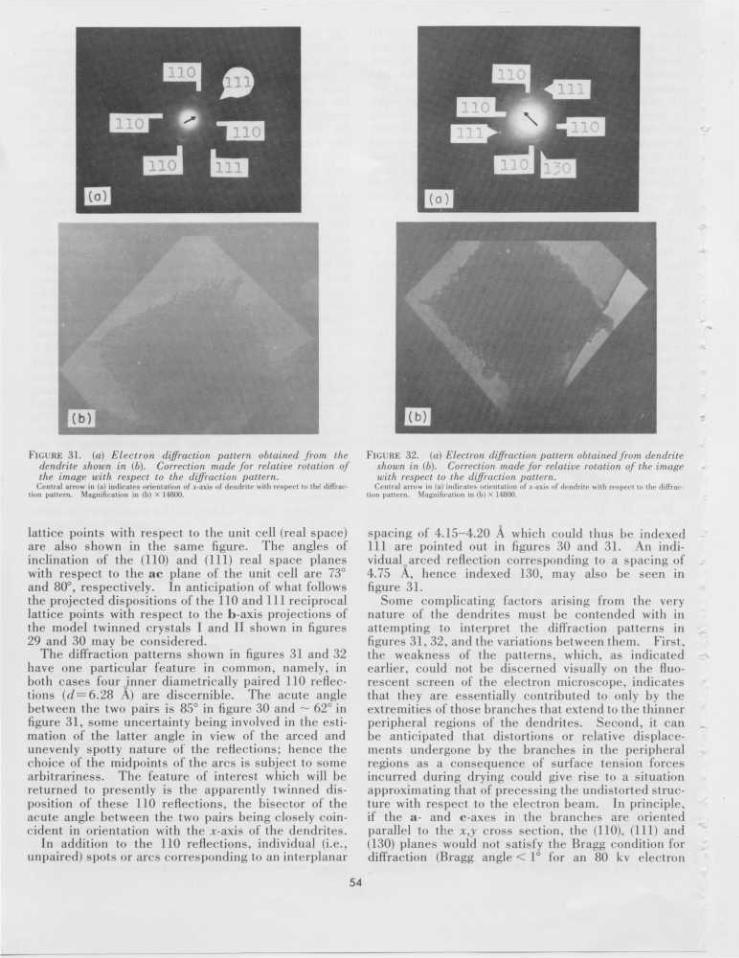
FIGURE 31. (a) Electron diffraction pattern obtained from thedendrite shown in (b). Correction made for relative rotation ofthe image with respect to the diffraction pattern.
Central arrow in (a) indicates orientation of x-axis of dendrite with respect to the diffrac-tion pattern. Magnification in (b) X 14800.
FIGURE 32. (a) Electron diffraction pattern obtained from dendriteshown in (b). Correction made for relative rotation of the imagewith respect to the diffraction pattern.
Central arrow in (a) indicates orientation of a;-axis of dendrite with respect to the diffrac-tion pattern. Magnification in (b) X 14800.
lattice points with respect to the unit cell (real space)are also shown in the same figure. The angles ofinclination of the (110) and (111) real space planeswith respect to the ac plane of the unit cell are 73°and 80°, respectively. In anticipation of what followsthe projected dispositions of the 110 and 111 reciprocallattice points with respect to the b-axis projections ofthe model twinned crystals I and II shown in figures29 and 30 may be considered.
The diffraction patterns shown in figures 31 and 32have one particular feature in common, namely, inboth cases four inner diametrically paired 110 reflec-tions (of =6.28 A) are discernible. The acute anglebetween the two pairs is 85° in figure 30 and ~ 62° infigure 31, some uncertainty being involved in the esti-mation of the latter angle in view of the arced andunevenly spotty nature of the reflections; hence thechoice of the midpoints of the arcs is subject to somearbitrariness. The feature of interest which will bereturned to presently is the apparently twinned dis-position of these 110 reflections, the bisector of theacute angle between the two pairs being closely coin-cident in orientation with the *-axis of the dendrites.
In addition to the 110 reflections, individual (i.e.,unpaired) spots or arcs corresponding to an interplanar
spacing of 4.15-4.20 A which could thus be indexed111 are pointed out in figures 30 and 31. An indi-vidual arced reflection corresponding to a spacing of4.75 A, hence indexed 130, may also be seen infigure 31.
Some complicating factors arising from the verynature of the dendrites must be contended with inattempting to interpret the diffraction patterns infigures 31, 32, and the variations between them. First,the weakness of the patterns, which, as indicatedearlier, could not be discerned visually on the fluo-rescent screen of the electron microscope, indicatesthat they are essentially contributed to only by theextremities of those branches that extend to the thinnerperipheral regions of the dendrites. Second, it canbe anticipated that distortions or relative displace-ments undergone by the branches in the peripheralregions as a consequence of surface tension forcesincurred during drying could give rise to a situationapproximating that of precessing the undistorted struc-ture with respect to the electron beam. In principle,if the a- and c-axes in the branches are orientedparallel to the x,y cross section, the (110), (111) and(130) planes would not satisfy the Bragg condition fordiffraction (Bragg angle < 1° for an 80 kv electron
54
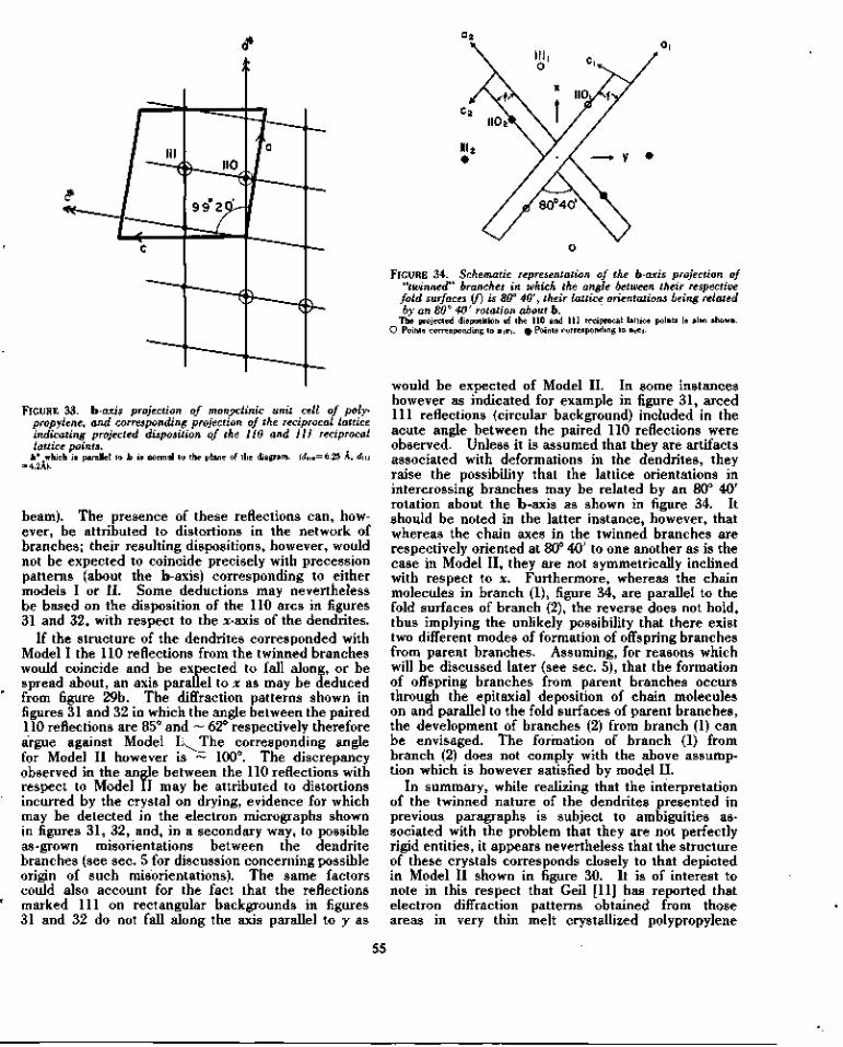
FIGURE 33. h-axis projection of monpclinic unit cell of poly-propylene, and corresponding projection of the reciprocal latticeindicating projected disposition of the 110 and 111 reciprocallattice points.
b* which is parallel to b is normal to the plane of the diagram. (dno—6.25 A, dm= 4.2A).
beam). The presence of these reflections can, how-ever, be attributed to distortions in the network ofbranches; their resulting dispositions, however, wouldnot be expected to coincide precisely with precessionpatterns (about the b-axis) corresponding to eithermodels I or II. Some deductions may neverthelessbe based on the disposition of the 110 arcs in figures31 and 32, with respect to the jc-axis of the dendrites.
If the structure of the dendrites corresponded withModel I the 110 reflections from the twinned brancheswould coincide and be expected to fall along, or bespread about, an axis parallel to x as may be deducedfrom figure 29b. The diffraction patterns shown infigures 31 and 32 in which the angle between the paired110 reflections are 85° and ~ 62° respectively thereforeargue against Model I.^The corresponding anglefor Model II however is ~ 100°. The discrepancyobserved in the angle between the 110 reflections withrespect to Model II may be attributed to distortionsincurred by the crystal on drying, evidence for whichmay be detected in the electron micrographs shownin figures 31, 32, and, in a secondary way, to possibleas-grown misorientations between the dendritebranches (see sec. 5 for discussion concerning possibleorigin of such misorientations). The same factorscould also account for the fact that the reflectionsmarked 111 on rectangular backgrounds in figures31 and 32 do not fall along the axis parallel to y as
FIGURE 34. Schematic representation of the b-axis projection of"twinned" branches in which the angle between their respective
fold surfaces if) is 80° 40', their lattice orientations being relatedby an 80° 40' rotation about b.
The projected disposition of the 110 and 111 reciprocal lattice points is also shown.O Points corresponding to aiCi. £ Points corresponding to a>c>.
would be expected of Model II. In some instanceshowever as indicated for example in figure 31, arced111 reflections (circular background) included in theacute angle between the paired 110 reflections wereobserved. Unless it is assumed that they are artifactsassociated with deformations in the dendrites, theyraise the possibility that the lattice orientations inintercrossing branches may be related by an 80° 40'rotation about the b-axis as shown in figure 34. Itshould be noted in the latter instance, however, thatwhereas the chain axes in the twinned branches arerespectively oriented at 80° 40' to one another as is thecase in Model II, they are not symmetrically inclinedwith respect to x. Furthermore, whereas the chainmolecules in branch (1), figure 34, are parallel to thefold surfaces of branch (2), the reverse does not hold,thus implying the unlikely possibility that there existtwo different modes of formation of offspring branchesfrom parent branches. Assuming, for reasons whichwill be discussed later (see sec. 5), that the formationof offspring branches from parent branches occursthrough the epitaxial deposition of chain moleculeson and parallel to the fold surfaces of parent branches,the development of branches (2) from branch (1) canbe envisaged. The formation of branch (1) frombranch (2) does not comply with the above assump-tion which is however satisfied by model II.
In summary, while realizing that the interpretationof the twinned nature of the dendrites presented inprevious paragraphs is subject to ambiguities as-sociated with the problem that they are not perfectlyrigid entities, it appears nevertheless that the structureof these crystals corresponds closely to that depictedin Model II shown in figure 30. It is of interest tonote in this respect that Geil [11] has reported thatelectron diffraction patterns obtained from thoseareas in very thin melt crystallized polypropylene
55
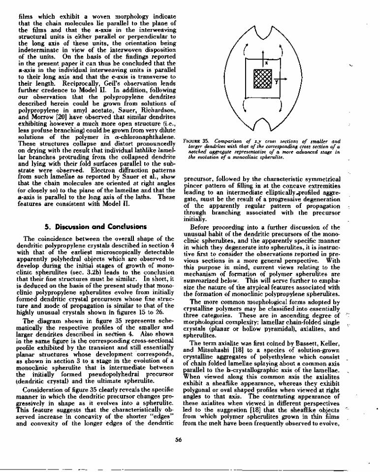
films which exhibit a woven morphology indicatethat the chain molecules lie parallel to the plane ofthe films and that the a-axis in the interweavingstructural units is either parallel or perpendicular tothe long axis of these units, the orientation beingindeterminate in view of the interwoven dispositionof the units. On the basis of the findings reportedin the present paper it can thus be concluded that thea-axis in the individual interweaving units is parallelto their long axis and that the c-axis is transverse totheir length. Reciprocally, Geil's observation lendsfurther credence to Model II. In addition, followingour observation that the polypropylene dendritesdescribed herein could be grown from solutions ofpolypropylene in amyl acetate, Sauer, Richardson,and Morrow [20] have observed that similar dendritesexhibiting however a much more open structure (i.e.,less profuse branching) could be grown from very dilutesolutions of the polymer in aj-chloronaphthalene.These structures collapse and distort pronouncedlyon drying with the result that individual lathlike lamel-lar branches protruding from the collapsed dendriteand lying with their fold surfaces parallel to the sub-strate were observed. Electron diffraction patternsfrom such lamellae as reported by Sauer et al., showthat the chain molecules are oriented at right angles(or closely so) to the plane of the lamellae and that thea-axis is parallel to the long axis of the laths. Thesefeatures are consistent with Model II.
5. Discussion and Conclusions
The coincidence between the overall shape of thedendritic polypropylene crystals described in section 4with that of the earliest microscopically detectableapparently polyhedral objects which are observed todevelop during the initial stages of growth of mono-clinic spherulites (sec. 3.2b) leads to the conclusionthat their fine structures must be similar. In short, itis deduced on the basis of the present study that mono-clinic polypropylene spherulites evolve from initiallyformed dendritic crystal precursors whose fine struc-ture and mode of propagation is similar to that of thehighly unusual crystals shown in figures 15 to 26.
The diagram shown in figure 35 represents sche-matically the respective profiles of the smaller andlarger dendrites described in section 4. Also shownin the same figure is the corresponding cross-sectionalprofile exhibited by the transient and still essentiallyplanar structures whose development corresponds,as shown in section 3 to a stage in the evolution of amonoclinic spherulite that is intermediate betweenthe initially formed pseudopolyhedral precursor(dendritic crystal) and the ultimate spherulite.
Consideration of figure 35 clearly reveals the specificmanner in which the dendritic precursor changes pro-gressively in shape as it evolves into a spherulite.This feature suggests that the characteristically ob-served increase in concavity of the shorter "edges"and convexity of the longer edges of the dendritic
FIGURE 35. Comparison of x,y cross sections of smaller andlarger dendrites with that of the corresponding cross section of anotched aggregate representative of a more advanced stage inthe evolution of a monoclinic spherulite.
precursor, followed by the characteristic symmetricalpincer pattern of filling in at the concave extremitiesleading to an intermediate elliptic ally-profiled aggre-gate, must be the result of a progressive degenerationof the apparently regular pattern of propagationthrough branching associated with the precursorinitially.
Before proceeding into a further discussion of theunusual habit of the dendritic precursors of the mono-clinic spherulites, and the apparently specific mannerin which they degenerate into spherulites, it is instruc-tive first to consider the observations reported in pre-vious sections in a more general perspective. Withthis purpose in mind, current views relating to themechanism of formation of polymer spherulites aresummarized below. This will serve further to empha-size the nature of the atypical features associated withthe formation of monoclinic polypropylene spherulites.
The more common morphological forms adopted bycrystalline polymers may be classified into essentiallythree categories. These are in ascending degree ofmorphological complexity: lamellar chain-folded singlecrystals (planar or hollow pyramidal), axialites, andspherulites.
The term axialite was first coined by Bassett, Keller,and Mitsuhashi [18] to a species of solution-growncrystalline aggregates of polyethylene which consistof chain folded lamellae splaying about a common axisparallel to the b-crystallographic axis of the lamellae.When viewed along this common axis the axialitesexhibit a sheaflike appearance, whereas they exhibitpolygonal or oval shaped profiles when viewed at rightangles to that axis. The contrasting appearance ofthese axialites when viewed in different perspectivesled to the suggestion [18] that the sheaflike objectsfrom which polymer spherulites grown in thin filmsfrom the melt have been frequently observed to evolve,
56
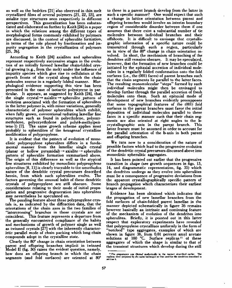
as well as the hedrites [21] also observed in thin meltcrystallized films of several polymers [21, 22, 23], areaxialite type structures seen respectively in differentperspectives. This generalization has been substan-tiated and elaborated on further by Keith [24] in a paperin which the relations among the different types ofmorphological forms commonly exhibited by polymersare discussed, in the context of spherulite formation,in terms of the role played by fractionation and im-purity segregation in the crystallization of polymers[25, 26].
According to Keith [24] axialites and spherulitesrepresent respectively successive stages in the evolu-tion of an initially formed lamellar chain-folded crys-tal growing (e.g., from the melt) under the influence ofimpurity species which give rise to cellulation at thegrowth fronts of the crystal along which the chainmolecules accrue in a regularly folded manner. Mor-phological evidence supporting this view has beenpresented in the case of isotactic polystyrene in par-ticular. It appears, as suggested by Keith [24], thatthe single crystal —> axialite —> spherulite pattern ofevolution associated with the formation of spherulitesin the latter polymer is, with minor variations, generallycharacteristic of polymer spherulites which exhibit,when fully grown, conventional radiating lamellar finestructures such as found in polyethylene, polyoxy-methylene, polyoxyethylene and poly(4-methylpen-tene-1) [7]. The same growth scheme applies mostprobably to spherulites of the hexagonal crystallinemodification of polypropylene.
It is evident that the pattern of evolution of mono-clinic polypropylene spherulites differs in a funda-mental manner from the lamellar single crystal(planar or hollow pyramidal) —> axialite —> spherulitepattern of spherulite formation summarized above.The origin of this difference as well as the atypicalfine structures exhibited by monoclinic polypropylenespherulites [8, 11] is clearly traceable to the unorthodoxnature of the dendritic crystal precursors describedherein, from which such spherulites evolve. Thefactors governing the unusual habit of these dendriticcrystals of polypropylene are still obscure. Someconsiderations relating to their mode of initial propa-gation and progressive degeneration into spherulitesmay nevertheless be speculated on.
The puzzling feature about these polypropylene crys-tals is, as indicated by the diffraction data, that theorientations of the chain axes in the two families of"intercrossing" branches in these crystals are notcoincident. This feature represents a departure fromthe generally encountered compliance of the habitsand mechanisms of growth of polymer single as wellas twinned crystals [27] with the inherently character-istic parallel mode of chain packing which long chaincompounds adopt in the crystalline state.
Clearly the 80° change in chain orientation betweenparent and offspring branches implicit in twinnedModel II (fig. 30) raises the evident question, namely,how does an offspring branch in which the chainsegments (and fold surfaces) are oriented at 80°
to those in a parent branch develop from the latter insuch a specific manner? One would expect that sucha change in lattice orientation between parent andoffspring branches would involve an interim boundaryregion of considerable disorder between them if oneassumes that there exist a substantial number of tiemolecules between individual branches and theiroffshoots. It is difficult to envisage that crystallo-graphic information of a specific nature could betransmitted through such a region, particularlyso in view of the 80° change in chain orientation in-volved. In short, the mechanism of branching of thedendrites still remains obscure. It may be speculated,however, that the formation of new branches could beinitiated by the epitaxial accretion of individual mole-cules in a regularly folded conformation onto the foldsurfaces (i.e., the (001) faces) of parent branches suchthat the chain segments lie parallel to the latter faces.The resulting monomolecular "steps" formed by suchindividual molecules might then be envisaged todevelop further through the parallel accretion of freshmolecules onto them. Such an epitaxial mode ofdevelopment of new branches evidently presupposesthat some topographical features of the (001) foldsurfaces in the parent branches must favor the initialdeposition of individual molecules parallel to thesefaces in a specific manner such that their chain seg-ments are also oriented at right angles to the b-crystallographic axis in the parent branch. Thislatter feature must be assumed in order to account forthe parallel orientation of the b-axis in both parentand offspring branches.
We turn now to a consideration of the nature ofpossible factors which lead to the progressive evolutionof the dendritic crystal precursors discussed above intomonoclinic spherulitic aggregates.
It has been pointed out earlier that the progressivetransition in shape (see growth sequences in figs. 13,14, and diagrammatic representation, fig. 35) whichthe dendrites undergo as they evolve into spherulitesmust be a consequence of progressive deviations fromthe apparent crystallographically specific pattern ofbranch propagation which characterizes their earliestStages of development.
Evidence has been obtained which indicates thatthe propagation of new lamellar branches from thefold surfaces of chain-folded parent lamellae in themanner depicted schematically in figure 26 remainshowever basically an intrinsic and continuing featureof the mechanism of evolution of the dendrites intospherulites. Briefly, it is pointed out in this latterrespect that exploratory experiments have revealedthat polypropylene crystallizes uniformly in the form of"notched" type aggregates, examples of which areshown in figure 36, from 0.01 percent amyl acetatesolutions at 100 °C. Surface replicasn of theseaggregates of which the shape is similar to that ofthe transient structures which develop during the still
11 The preparation was filtered isothermally in the manner described earlier. Thereplicas were prepared by the same technique as that used for the dendrites described insection 4.
57
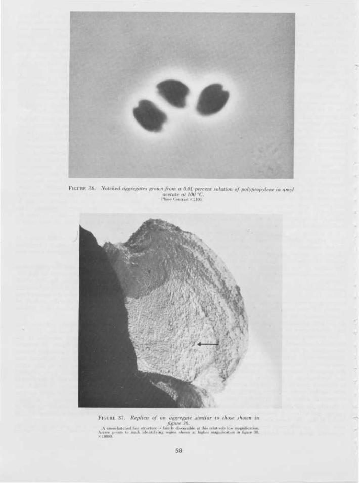
FIGURE 36. Notched aggregates grown from a 0.01 percent solution of polypropylene in amylacetate at 100 °C.
Phase Contrast X 2100.
FIGURE 37. Replica of an aggregate similar to those shown infigure 36.
A cross-hatched fine structure is faintly discernible at this relatively low magnification.Arrow points to mark identifying region shown at higher magnification in figure 38.X 10800.
58

• ¥ • i J* \ ft •» T •* ,« if •, • *
FIGURE 38. ,4 view a/ higher magnification of a portion (note arrow)of the replica shown in figure 37, revealing the cross-hatched surfacetexture of the replicated aggregate.
x 33200.
relatively early stages of evolution of the initial den-dritic crystal precursors into spherulites revealed,as shown in figures 37 and 38, a distinctly cross-hatchedfine structure clearly related to that of the dendritesdescribed in section 4.
Two factors, one possibly intrinsically associatedwith the folded conformation of the chain moleculesin the lamellar branches, and the other environmental,may be considered, that contribute to the progressivedegeneration of the dendritic crystal precursors intospherulites.
With respect to the first of the two factors indicatedabove, recent quantitative measurements of latticedistortions arising from the chain folds in polymercrystals reported by Bassett [28] may be pointed out.These measurements have been derived from an analy-sis of Moire patterns exhibited under the electronmicroscope by bilayer crystals of polyoxymethyleneand poly(4-methylpentene-l) in particular. It hasbeen shown that the spacing of a family of planesparallel to the chain molecules in crystals of these twopolymers differs depending on whether or not theplanes contain the chain folds. Such lattice distor-tions are attributed to the bulkiness of the folds whichgive rise to a slight increase in spacing between planesalong which the molecules accrue in a regularly foldedconformation. Bassett et al., [29] present evidencesuggesting that the resulting distortion of the tetra-gonal subcell in simple (four-sectored) lamellar crys-tals of poly(4-methylpentene-l) causes the lamellaeto adopt a very slightly curved ("conicalness"), asdistinct from planar overall shape. It would appearthat the highly curved bowl-shaped tetragonal crystals
of poly(4-methylpentene-l) consisting of multisectoredchain folded layers observed recently by Khoury andBarnes [30], as well as the orthorhombic crystals ofpolybutene-1 which, as reported by Holland and Miller[31], are curled up in the form of tubelike structures,owe their distinctly curved habits to lattice distortionsassociated with the chain folds in their constituentlamellae. It also seems very likely that the wellestablished twisting of the consti tuent radiatinglamellae in certain species of polymer spherulites[32, 33, 34] is also a consequence of lattice distortionsassociated with chain folds [35]. In short, thereexists a widespread body of evidence of the for-mation of chain-folded lamellae that are curved insome manner, as well as some direct and circum-stantial evidence indicating that this phenomenon maybe associated with lattice distortions due to the bulk-iness of the folds in their constituent molecules.
On the basis of the phenomena indicated in the fore-going paragraph the possibility may be envisaged thatthe lamellar branches in the polypropylene dendritesmay develop as very slightly curved rather than planarlaths. Such an effect would clearly lead cumulativelyto progressively more pronounced misorientations be-tween newly formed branches throughout the dendriteand hence to a slow change in overall shape of thedeveloping structure.
Direct detection of a possible very slight curvature ofindividual branches in the dendrites described insection 4 is clearly hindered by the impossibility oftracing individual branches unequivocally for appreci-able distances. Deviations in the relative orientationsof the dendrite branches may however be seen in the
59

replicas of the larger dendrites shown in figures 20 and21. In addition, the slight change in profile of thedendrites between the stages of growth correspondingto the smaller and larger crystals described in section 4,as evidenced by the more pronounced concavity ofthe shorter edges of the latter, coupled with thesymmetrical deviations in extinction (about the x,ycross section) between crossed polarizers, distinctlyexhibited by the larger dendrites, suggests the con-tribution of a factor inherent in the very nature of thestructure and mode of propagation of the dendritesto their progressive change in shape. It is merelypointed out in this respect, that a tendency of the fourprimary branches (i.e., those lying along the diagonalsof the rectangular x,y cross section of the dendrites)which play a major role in the propagation of thecrystals, to curve inwards towards the interior of thecrystal in the manner indicated in exaggerated fashionby the arrows in figure 35 would account for the char-acteristic manner in which the overall shape of thedendrites changes progressively during their earlystages of evolution into spherulites.
We turn now to a brief discussion of the role playedby environmental factors on the mode of propagationof the dendritic crystal precursors and their progressiveevolution into spherulites under isothermal conditions.The influence of temperature, and hence rate ofgrowth, as well as rejected impurity species (e.g.,atactic molecules and low molecular weight chains)on the mode of propagation of the dendritic crystalprecursors needs to be considered in this respect.Some indication of the effect of these factors may beobtained from a consideration of their influence on thedevelopment of individual dendrite branches. Wemay borrow in this respect from the views advancedby Keith [24] relating to the influence of impuritysegregation at the growth fronts of a chain-foldedpolymer crystal.
It may be anticipated that whether growth occursfrom solution or from the melt, an inpurity-rich layersurrounds the dendrite, thus leading to cellulation inthe form of pronounced reentrances developing atthose faces of each branch at which growth occursthrough the parallel accretion of chain molecules (i.e.,the (100) and (010) faces). Lattice misorientationsbetween neighboring "cells" may then be expectedto lead to the progressive acquisition of a polycrystal-line character by each branch, as has been observedfor example in the case of individual polystyrene chain-folded lamellae growing from the melt. Intrabranchlattice misorientations would evidently lead to mis-orientations between offspring branches growing fromdifferent regions of the fold surfaces of a parent branchand hence would contribute cumulatively to the pro-gressive degeneration of the initial dendritic crystalinto a spherulite.
Recapitulating, therefore, it is considered that thecharacteristic development of monoclinic spherulitesfrom the particular species of dendritic crystalsdescribed in this paper is brought about cumulativelyas a consequence of (a) the curving of individual den-drite branches due to lattice distortions associated
with chain folds, and (b) the progressive acquisitionof a polycrystalline character by individual branchesgrowing under the influence of rejected impurityspecies. At low undercoolings and hence slow growthrates which favor the diffusion of impurities awayfrom the growth fronts of the constituent dendritebranches the disruptive effects of cellulation are mini-mized in particular. Under such conditions the pro-gressive transition from dendritic crystal to spheru-lite will be gradual and the initial precursor dendritemay attain, as has been observed, microscopic (optical)proportions before exhibiting pronounced signs ofdegeneration. At high undercoolings, however, thedendrite to spherulite transition would occur muchearlier in the growth sequence at the submicroscopiclevel of resolution with the result that no initial pseudo-polyhedral precursor will be observed.
The considerations presented in the preceedingthree paragraphs have been only concerned with theinfluence of rejected impurity species on the degenera-tion of the dendritic precursors. An importantconsideration arises however regarding the influenceof impurity rejected species on the frequency withwhich offspring branches develop from a parent branchin these dendritic precursors. In this respect thepossibility cannot be discounted that the developmentof discrete offspring branches from the fold surfaces[(001) faces] of a parent lamella may actually resultfrom the favored epitaxial accretion of molecules(assuming the epitaxial mode of development ofbranches suggested earlier holds) only on specificasperities on the fold surfaces of parent lamellae,and not necessarily due to a cellular pattern of mono-molecular deposition of chains parallel to fold surfaceshaving an initial uniform topography. Carrying thislatter conjecture further, it would follow that the fre-quency of branching will be governed by the number ofbranch-initiating asperities per unit area of the (001)faces, such as for example localized steps formed onthose faces due to irregularities or fluctuations in thefold period of the constituent chain molecules in theparent lamella, which in turn may well be governed bythe environmental conditions.
In conclusion, while the present study provides somedetailed insight into the nature of the early stages ofevolution of monoclinic polypropylene spherulites fromsolution as well as from the melt, a final analysis willhave to account for the preferred molecular orientationwith respect to the radial direction in the fully grownspherulites, information on which is, as indicated inthe introduction, at present limited to monoclinicspherulites grown from the melt in the range of 110 to148 °C [2]. While such a detailed analysis is beyondthe scope of the present study, it may be pointed outat this stage that the preferred tangential orientationof the b-crystallographic axis in melt grown spherulites[2] correlates with the observation that growth isslowest parallel to the b-crystallographic axis in thebranches of the dendritic precursor crystals. Theaverage 65° to 70° inclination of the chain moleculeswith respect to the rach'd direction in these spherulites[2] still remains unexplained at this stage.
60

The author thanks E. Passaglia for the benefit ofmany helpful and critical discussions. Thanks aredue to P. H. Geil for the loan of unpublished electrondiffraction patterns.
6. References
[1] F. J. Padden, Jr., and H. D. Keith, J. Appl. Phys. 30 , 1479(1959).
[2] H. D. Keith, F. J. Padden, Jr., N. M. Walter, and H. W. Wyckoff,J. Appl. Phys. 30 , 1485 (1959).
[3] G. Natta, P. Corradini, M. Cesari, Atti, accad. nazl. Lincei.Rend. Classe sci. fis. mat. e nat. 2 1 , 365 (1956).
[4] E. J. Addink and J. Beintema, Polymer (London) 2 , 185 (1961).[5] A. Turner-Jones, Jean M. Aizlewood, and D. R. Beckett,
Makromolekulare Chem. 75, 134 (1964).[61 P. H. Geil, Polymer Single Crystals, ch. IV, p. 269 (Interscience,
New York, 1963).[7] P. H. Geil, Polymer Single Crystals, ch. IV, p. 223 (Interscience,
New York, 1963).[8] G. W. Bailey, J. Polymer Sci. 62 , 241 (1962).[9] A. Keller, KoUoid-Z. 165, 15 (1959).
[10] P. H. Geil, Polymer Single Crystals, ch. IV, p. 270 (Interscience,New York, 1963).
[11] P. H. Geil, Polymer Single Crystals, ch. Ill, p. 214-220 (Inter-science, New York, 1963).
[12] A. Hiltz. Private communication.[13] H. J. Leugering, Makromolekulare Chem. 6 8 , 223 (1963).[14] P. H. Geil, Polymer Single Crystals (Interscience New York,
1963, ch. HI, p. 211).[15] F. Khoury, Bull. Am. Phys. Soc. Series 11, 9, 275 (1964), Paper
JA4.[16] D. C. Bassett and A. Keller, Phil. Mag. 7, 1553 (1962).
[17] R. Salovey and F. R. Dammont, J. Polymer Sci. A l , 2155(1963).
[18] D. C. Bassett, A. Keller and S. Mitsuhashi, J. Polymer Sci.A l , 763 (1963).
[19] D. A. Keedy, J. Powers and R. S. Stein, J. Appl. Phys. 3 1 ,1911 (1960).
[20] J. A. Sauer, G. C. Richardson and D. R. Morrow, Bull. Am.Phys. Soc. Series 11, 10, 354 (1965) Paper EG8.
[21] P. H. Geil in Growth and Perfection of Crystals, p. 579 (JohnWiley & Sons, Inc., New York, N.Y., 1958).
[22] H. J. Leugering, Kolloid-Z 172, 184 (1960).[23] F. Danusso and F. Sabbioni, Rend. Inst. Lombardo Sci. Lettere
A92 435 (1958).[24] H. D. Keith, J. Polymer Sci. 2A, 4339 (1964).[25] H. D. Keith and F. J. Padden, Jr., J. Appl. Phys. 34 , 2409
(1963).[26] H. D. Keith and F. J. Padden, Jr., J. Appl. Phys. 3 5 , 1270,
1286 (1964).[27] P. H. Geil, Polymer Single Crystals, ch. II (Interscience New
York, 1963).[28] D. C. Bassett, Phil. Mag. 10, 595 (1964).[29] D. C. Bassett, F. R. Dammont and R. Salovey, Polymer (London)
5, 579 (1964).[30] F. Khoury and J. D. Barnes, Bull. Am. Phys. Soc. Series 11
10, 366 (1965) Papers HG3, HG4.[31] V. F. Holland and R. L. Miller, J. Appl. Phys. 3 5 , 3241 (1964).[32] H. D. Keith and F. J. Padden, Jr., J. Polymer Sci. 39 , 101, 123
(1959). .[33] F. P. Price, J. Polymer Sci. 39 , 139 (1959).[34] A. Keller, J. Polymer Sci. 39 , 151 (1959).[35] J. D. Hoffman and J. L. Lauritzen, Jr., J. Res. NBS 65A (Phys.
and Chem.) No. 4, 297 (1961).
(Paper 70A1-385)
61


![Crystallization of isotactic polypropylene: the effect of fiber ...X =1–exp[–k(T)tn] (1) where: X is the extent of crystallization, k(T) is a rate constant, n is the Avrami ex-ponent](https://img.pdfslide.us/doc/110x75/5fe6893d69cde76c8771df9a/crystallization-of-isotactic-polypropylene-the-effect-of-fiber-x-1aexpakttn.jpg)