Embed Size (px)
Citation preview
THE SPECTRUM OF CONTINUOUS AIRWAY ASSESSEMENT AND MANAGEMENT
2
Learning Objectives:
The following module covers:• Review of airway anatomy including pediatric differences.• Acquiring and maintaining AW patency including manual and
adjunct techniques.• AW assessment and introduction to LEMON.• Risk/benefit considerations.• Problems and Rescue Procedures.
3
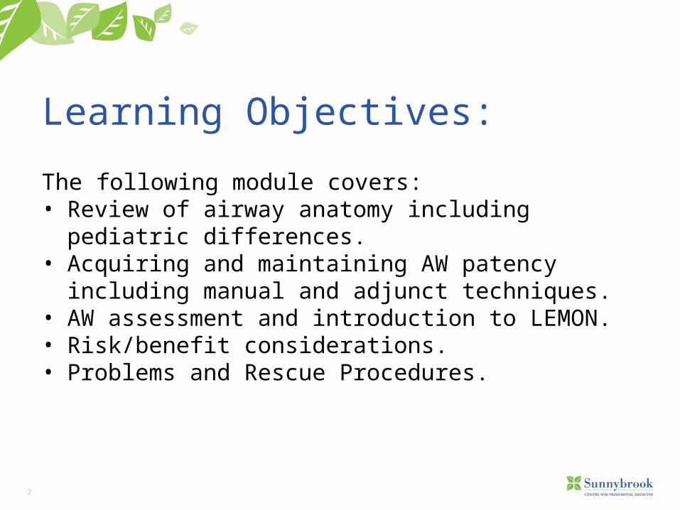
Anterior View of the Larynx
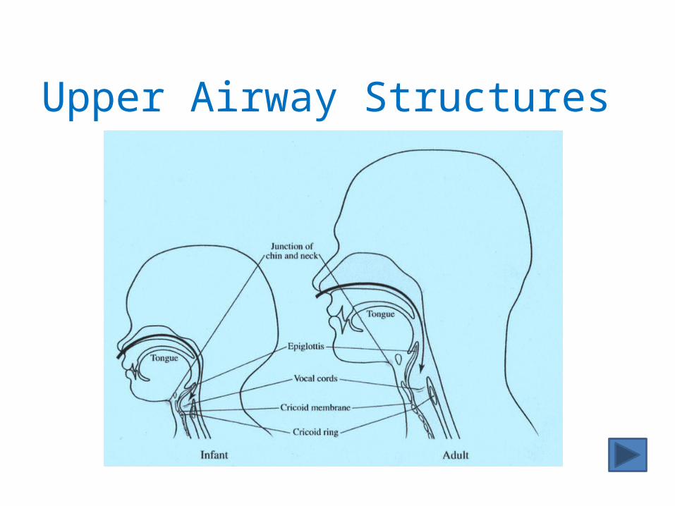
Upper Airway Structures
5
The Spectrum of Airway Management
6
Objectives
• Appreciate the spectrum of airway management from simple maneuvers to complex interventions
• Appreciate proper monitoring and need for further management during and after airway intervention
7
Airway spectrum
• Alert/patent airway • Simple manual maneuvers
• Head-tilt, chin-lift, suction, left lateral recumbent, cricoid pressure (Sellick’s), chest compressions (FBOA)
• Simple and advanced adjuncts• OPA, NPA, Bougie
• Supraglottic airways• King-LT, LMA
8
Airway Spectrum
Advanced airway• Direct laryngoscopy/intubation• BURP (not Sellick’s)
Failed airway , can’t oxygenate/can’t ventilate• Needle cricothyrotomy• McGill forceps
9
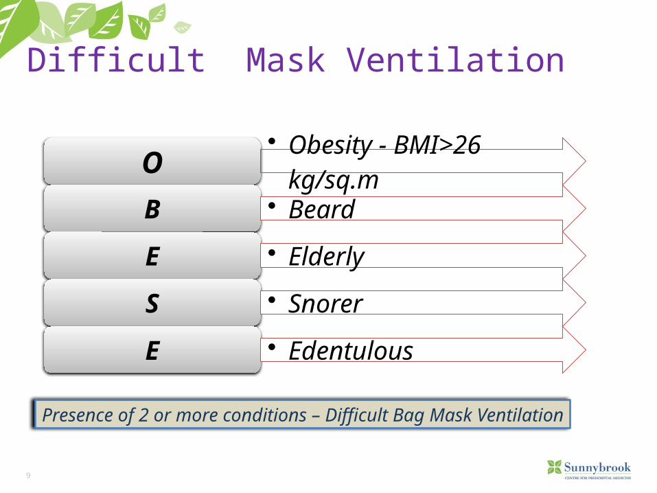
O • Obesity - BMI>26 kg/sq.m
B • Beard
E • Elderly
S • Snorer
E • Edentulous
Difficult Mask Ventilation
Presence of 2 or more conditions – Difficult Bag Mask Ventilation
10
Difficult Face Mask VentilationSigns of inadequate face mask ventilation include
– absent or inadequate chest movement– absent or inadequate breath sounds– auscultatory signs of severe obstruction– gastric air entry or dilatation– decreasing or inadequate oxygen saturation (SpO2)– absent or inadequate exhaled carbon dioxide– hemodynamic changes associated with hypoxia, hypoxemia or
hypercarbia (hypertension, tachycardia and arrhythmias, cyanosis)• Compliance vs. resistance?
– The ease which lung and thorax expand vs. any mechanical factor that limits inspired air to reach the alveoli
11
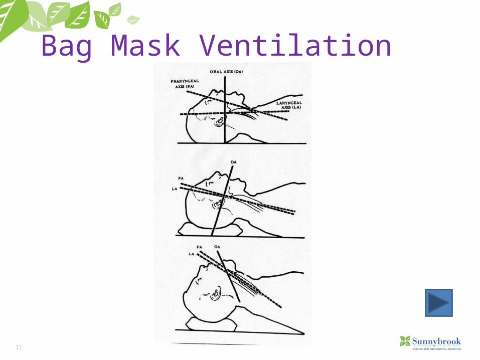
Bag Mask Ventilation
12
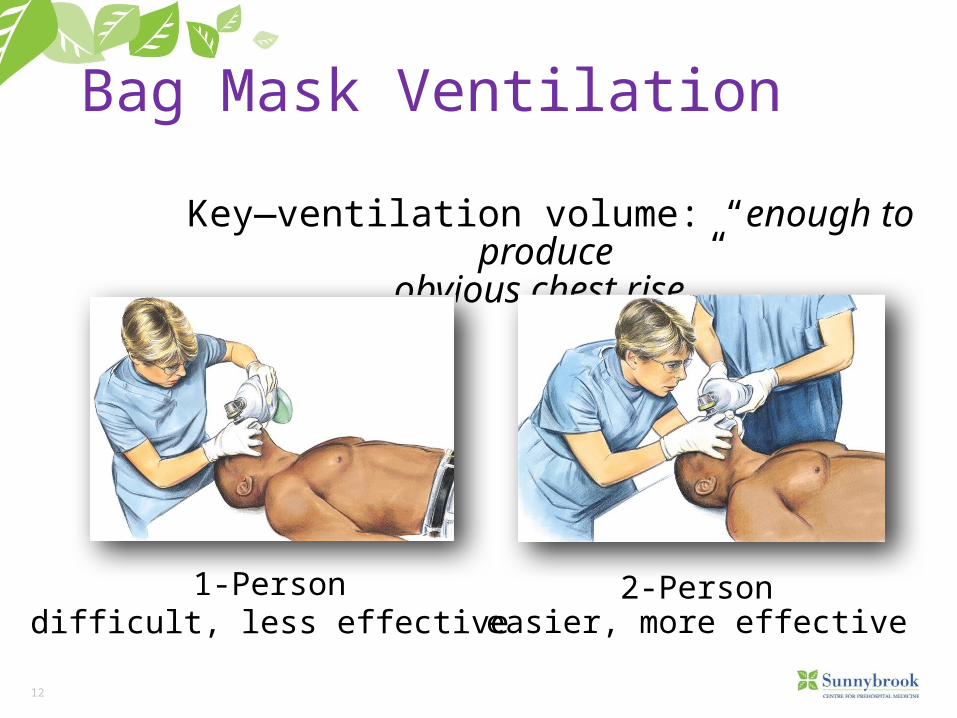
Bag Mask Ventilation
Key—ventilation volume: “enough to produce obvious chest rise”
1-Persondifficult, less effective
2-Personeasier, more effective
13
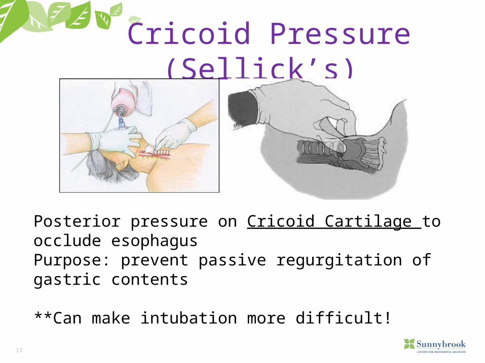
Cricoid Pressure (Sellick’s)
Posterior pressure on Cricoid Cartilage to occlude esophagusPurpose: prevent passive regurgitation of gastric contents
**Can make intubation more difficult!
14
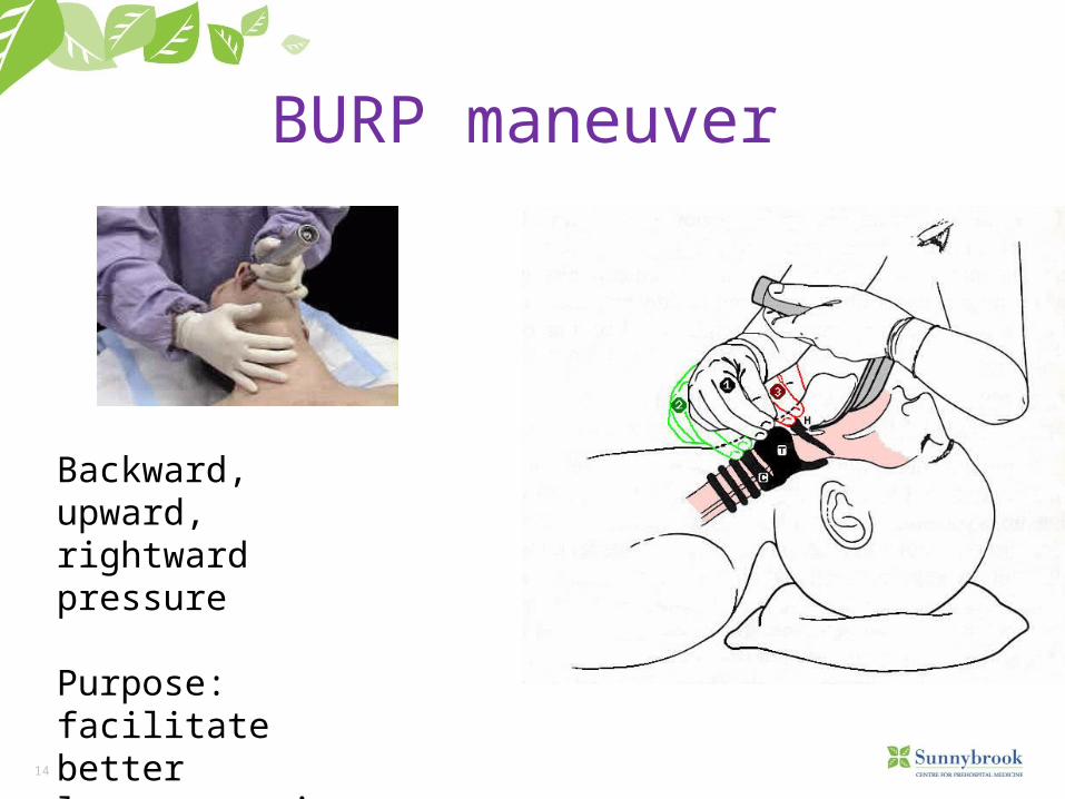
BURP maneuver
Backward, upward, rightward pressure
Purpose: facilitate better laryngoscopic view
15
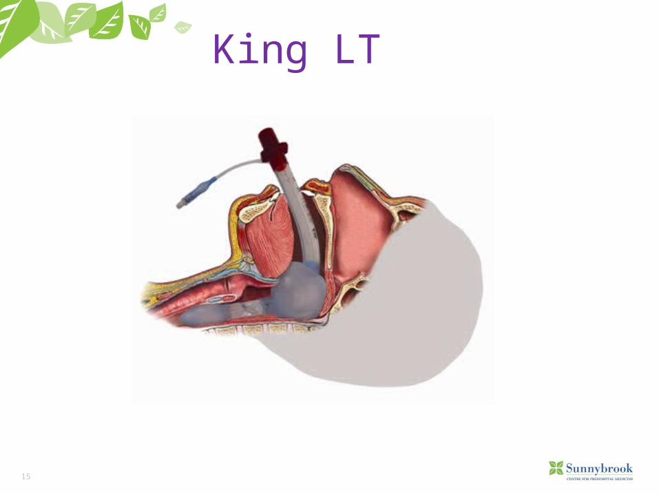
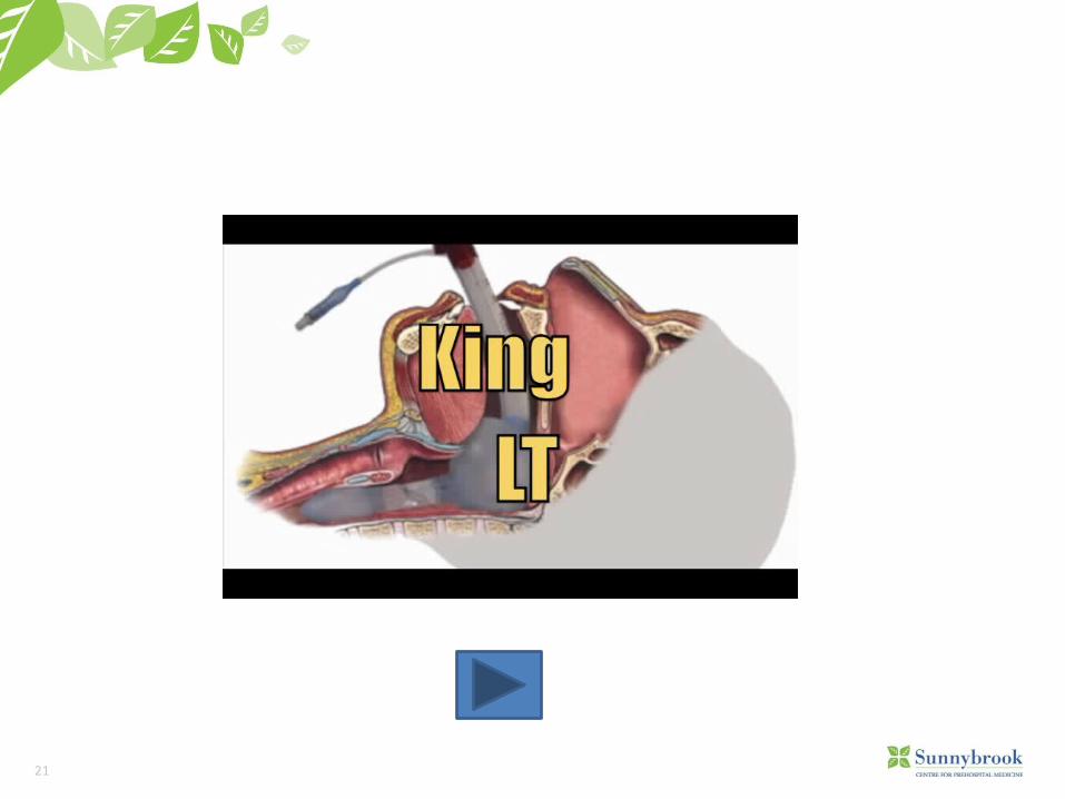
King LT
16
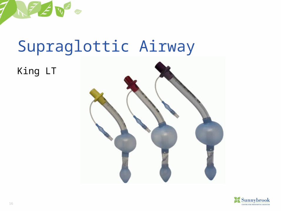
Supraglottic Airway King LT
17
Features of King SG Airways• The KING LTD is supplied clean, but is a non-sterile device• It consists of a curved tube with ventilation apertures located
between two inflatable cuffs.• The distal cuff is designed to seal the esophagus• Proximal cuff is intended to seal the oropharynx (King System,
2009).• Cardiac arrest (PCP and ACP)• Respiratory distress/arrest with GCS 3 (ACP only)
18
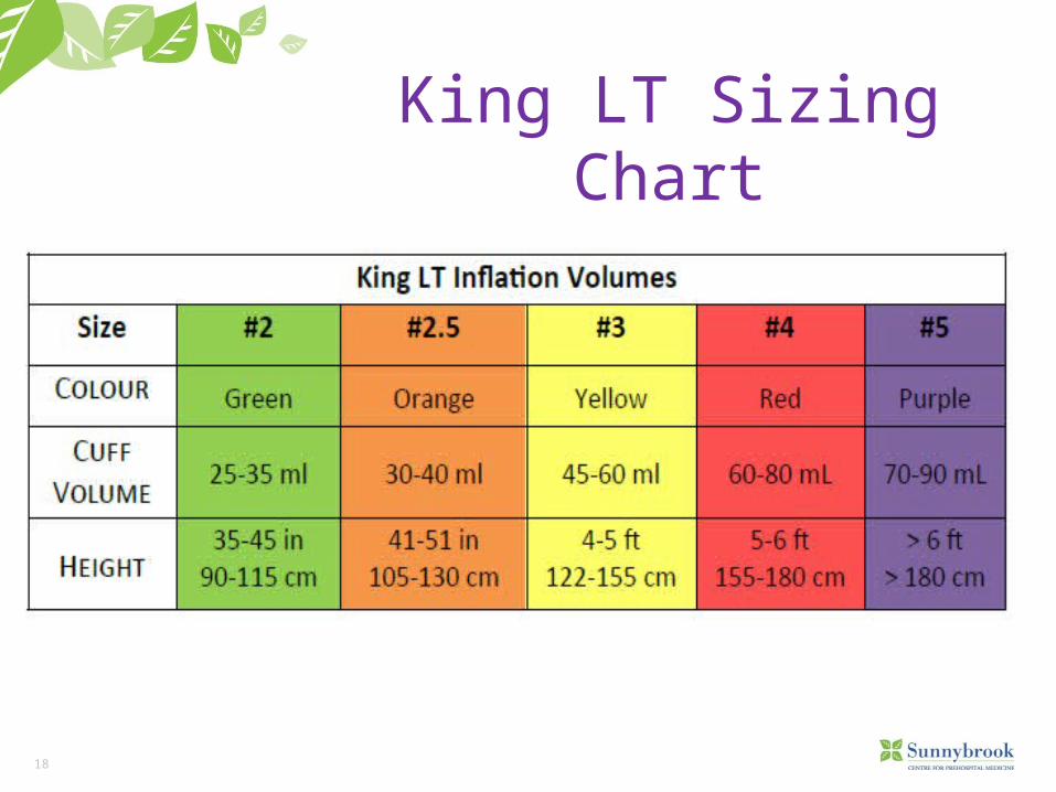
King LT Sizing Chart
19
Advantages• Emergency ventilation can take place within 15 seconds without a
laryngoscope.• Requires minimal movement of patient head.• Requires minimal education to insert.• The King laryngeal airway is designed to be inserted without direct
visualization.• Minimal risk of aspiration.• The KING LTD provides a more secure, non-intubating emergency
airway when direct laryngoscopy is not feasible (King System, 2009).
20
Disadvantages• Only make limited sizes.• The KING LTD can be used in routine procedures only up to 8
hours.• Unable to place medication down the tube.• Trauma related to balloon in trachea (King System, 2009).• Only minimizes risk of aspiration (aspiration can still occur)
21
22
Monitoring Airway Interventions
Ventilation: • Chest wall excursion, ease of BVM vents, ETCO2
Oxygenation: • SpO2, skin colour, heart rate
23
Management of the Difficult Airway
24
DEFINITION
Difficult airway: • ANYTHING that interferes with ventilation or intubation
• Anatomic• Traumatic• Infectious (airway edema)• Allergic• Behavioral
25
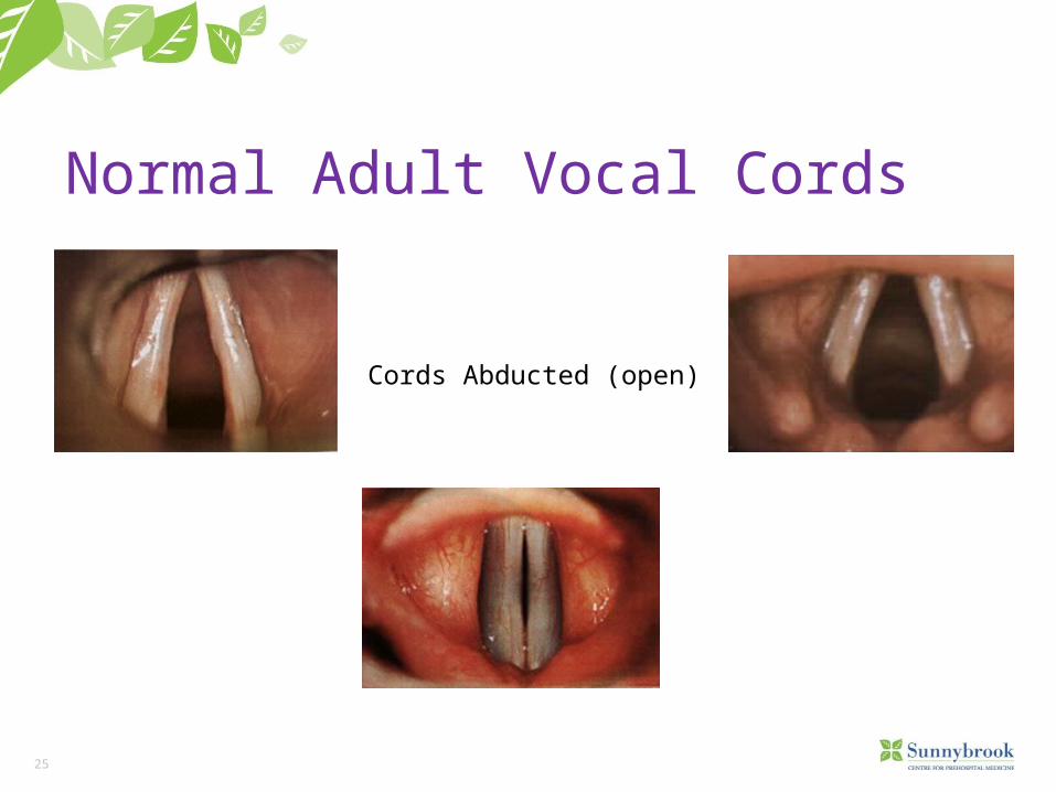
Normal Adult Vocal Cords
Cords Abducted (open)
26
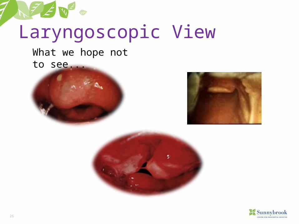
Laryngoscopic ViewWhat we hope not to see...
27
LEMON Law
Look at anatomyExamine the airwayMallampati ScaleObstructionsNeck Mobility
28
Look at Anatomy
• Obesity: rapid desaturation, short or thick neck-difficult intubation.
• Facial hair: hides small chin, can make BVM ventilation difficult.• Teeth: hide airway, obscure tube passage, may lacerate balloon,
dentures.• Poor Neck Mobility: surgery, kyphosis• Large Tongue:
29
Evaluate the 3-3-2 Rule
The 3 – 3 – 2 rule:Mouth open: 3 fingers
• Allows insertion of tube, laryngoscopeMentum (chin) to hyoid: 3 fingers
• Predicts ability to lift tongue into mandibleFloor of mouth to thyroid cartilage: 2 fingers
• If high larynx, airway tucked under base of tongue, hard to visualize
30
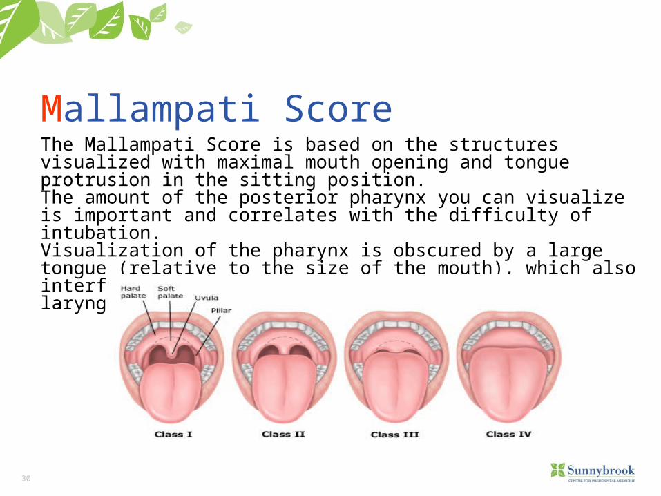
Mallampati ScoreThe Mallampati Score is based on the structures visualized with maximal mouth opening and tongue protrusion in the sitting position.The amount of the posterior pharynx you can visualize is important and correlates with the difficulty of intubation. Visualization of the pharynx is obscured by a large tongue (relative to the size of the mouth), which also interferes with visualization of the larynx on laryngoscopy.
31
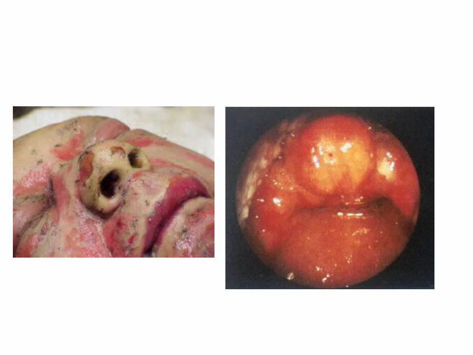
Obstruction
Evaluation for stridor, foreign bodies, and other forms of sub- and supraglottic obstruction should be performed in every patient prior to laryngoscopy
33
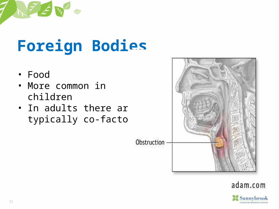
Foreign Bodies
• Food• More common in
children• In adults there are
typically co-factors
34
Causes of Obstruction
• Foreign Body (food, toys)• Trauma• Edema• Neoplasm• Blood
35
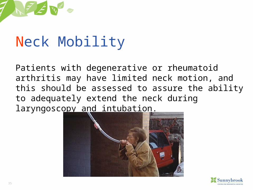
Neck Mobility
Patients with degenerative or rheumatoid arthritis may have limited neck motion, and this should be assessed to assure the ability to adequately extend the neck during laryngoscopy and intubation.
36
Indications for Intubation
Based on three fundamental clinical assessments:
1. Is there a failure of airway maintenance or protection?2. Is there a failure of ventilation or oxygenation?
3. What is the anticipated clinical course?
37
Failure of Airway Maintenance or Protection
Upper airway muscles and protective reflexes normally protect the airway from aspiration.
Patent airway helps with ensuring adequate oxygenation. What devices do we carry to help maintain a patent airway?
Not all airway devices protect the airway.
In absence of immediately reversible condition, intubation should be considered.
What is the most reliable method to assess protective reflexes?
38
Failure of Oxygenation/Ventilation
Oxygenation/Ventilation provide oxygen to vital organs, remove waste carbon dioxide and help regulate pH.
If oxygenation still does not improve despite efforts to improve ventilation, intubation should be considered.
Eg. Asthma/CHF-these patients have patent airways and can protect them, but ventilatory failure will ultimately lead to inadequate oxygenation and death.
39
What is Anticipated Clinical Course?
Although airway and ventilation are adequate at this time, conditions may change in future require airway control or protection: stab wound to neck for example.Other examples?
40
Tracheal Tube Introducer (Bougie)
GEB or “Bougie” is a device utilized to increase success when securing an airway.
EMS and hospital trials have shown increased success rate, and decreased time to intubate with the use of the bougie.
First used by Robert Macintosh in 1943 during a difficult intubation.
41
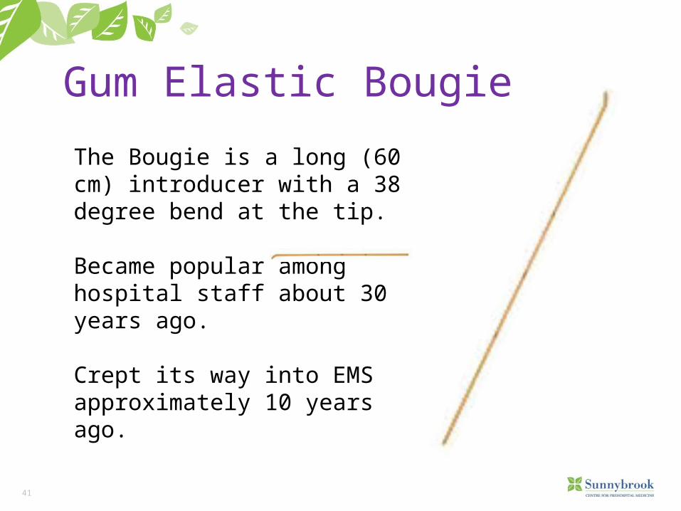
Gum Elastic Bougie
The Bougie is a long (60 cm) introducer with a 38 degree bend at the tip.
Became popular among hospital staff about 30 years ago.
Crept its way into EMS approximately 10 years ago.
42
Logistics to Consider
It can be a challenge to store the device. It’s shape has a lot to do with its advantage as an airway adjunct so it must be stored in a way to not compromise that.
It is a single use, disposable device.
Applicable to ACP scope of practice.
Does not require a specific Medical Directive as it is considered an adjunct under the ETI Medical Directive.
43
Inserting the Bougie
You may choose to lubricate the tip of the Bougie with a water based solution, however it is generally not required.
Perform laryngoscopy as per your normal technique.
At this point instead of the usual ET tube with stylet, grasp the Bougie.
Pass the Bougie through the glottic opening to the point of the 40 cm marking on the device, or until slight resistance is met. The resistance indicates the device contacting the carina or entering the bronchial tree.
At this point, ideally it becomes a two person procedure.
44
Bougie Insertion
45
Confirming the Tube
As you advance the device, you get two additional indications of correct placement, both tactile in nature.
i) Clicking sensation as the device passes over the tracheal rings. Studies indicate these are felt up to 90% of the time.
ii) Slight resistance as the device advances to the carina or into the bronchi (generally around 40 cm in an adult). The device is marked at this point.
Other methods of tube confirmation should still be utilized and remain unchanged i.e.) ETCO2, chest rise and fall, auscultation, misting etc.
46
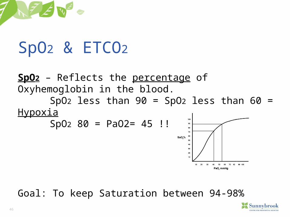
SpO2 & ETCO2
SpO2 – Reflects the percentage of Oxyhemoglobin in the blood. SpO2 less than 90 = SpO2 less than 60 = Hypoxia SpO2 80 = PaO2= 45 !!
Goal: To keep Saturation between 94-98%

47
SpO2 & ETCO2
ETCO2: The highest measurable point of CO2 concentration during patient exhalation cycle. Always reflects the current amount of exhaled CO2:
Provides the most reliable sign of patients cardiopulmonary performance.
Normal physiological range: 35-45 >45 Hypercarbia (R. Acidosis) <35 Hypocarbia (R. Alkalosis)
48
SpO2 & ETCO2
Points to remember:• There is no correlation between SpO2 and ETCOs2 (Pt. can present high O2SAT and low ETCO2 and vise versa).
• SpO2 can’t be measured accurately in hemodynamicaly compromised pt’s (levels lower than 90 may not be accurate).
• ETCO2 will reflect the patients current cardiopulmonary physiological condition.
49
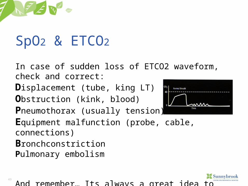
SpO2 & ETCO2
In case of sudden loss of ETCO2 waveform, check and correct:Displacement (tube, king LT)Obstruction (kink, blood)Pneumothorax (usually tension)Equipment malfunction (probe, cable, connections)BronchconstrictionPulmonary embolism
And remember… Its always a great idea to check Pt’s pulse
50
Needle Cricothyrotomy
Replacing Portex as of May 4th
• Only 13 have been done in a 5 year period (PRPS and TEMS)• 1 outright successful • 1 was a tracheal tube replacement• 50% were successfully intubated in the field post surgical
airway attempt• Extremely expensive for training / stocking (for the number of
uses)
51
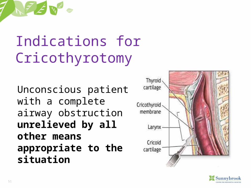
Indications for Cricothyrotomy
Unconscious patient with a complete airway obstruction unrelieved by all other means appropriate to the situation
Unconscious patient with a complete airway obstruction unrelieved by all other means appropriate to the situation
53
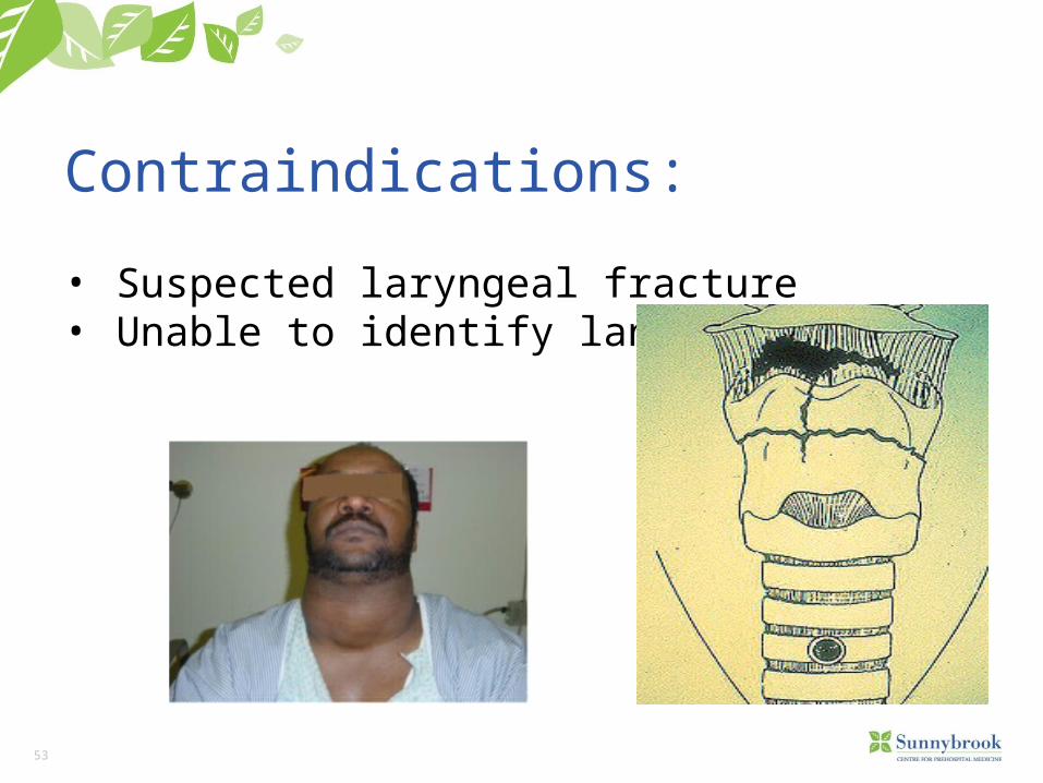
Contraindications:
• Suspected laryngeal fracture• Unable to identify landmarks
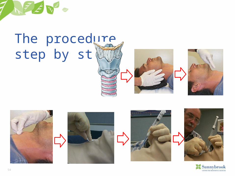
54
The procedurestep by step
55
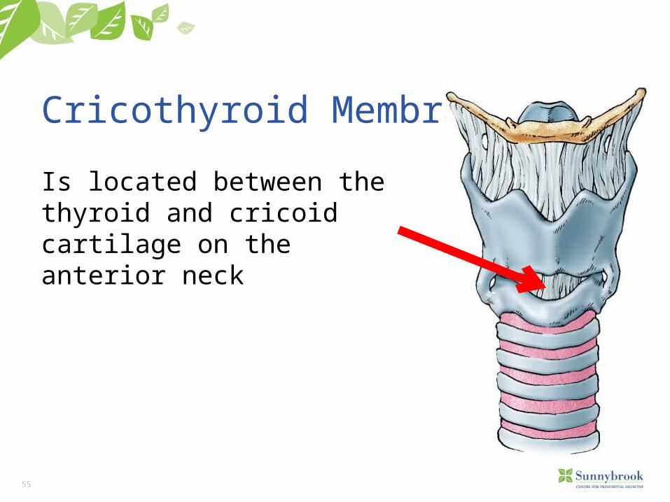
Cricothyroid Membrane
Is located between the thyroid and cricoid cartilage on the anterior neck
56
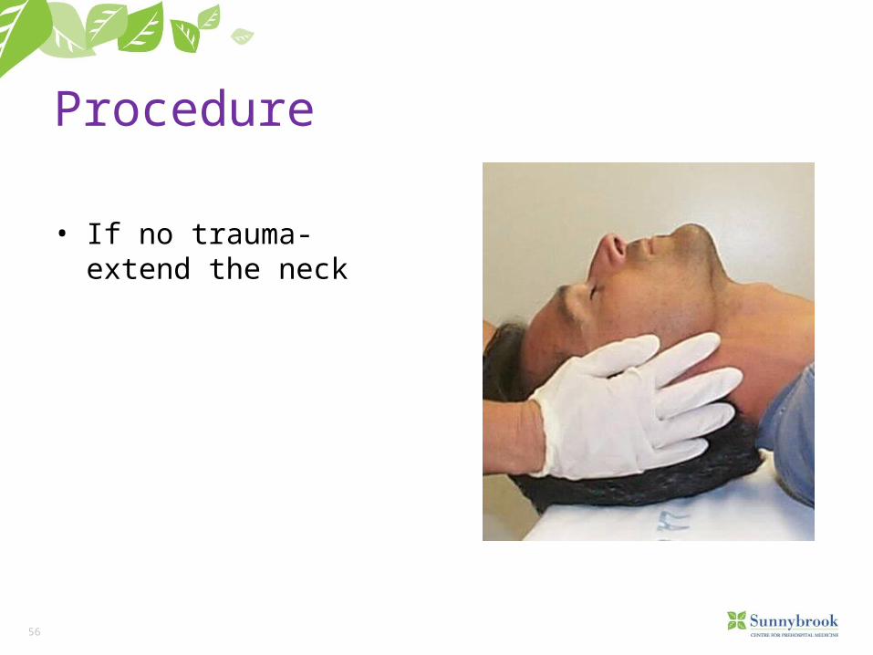
Procedure
• If no trauma- extend the neck
57
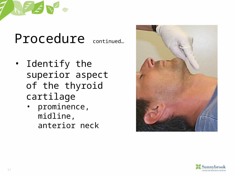
Procedure continued…
• Identify the superior aspect of the thyroid cartilage• prominence, midline,
anterior neck
58
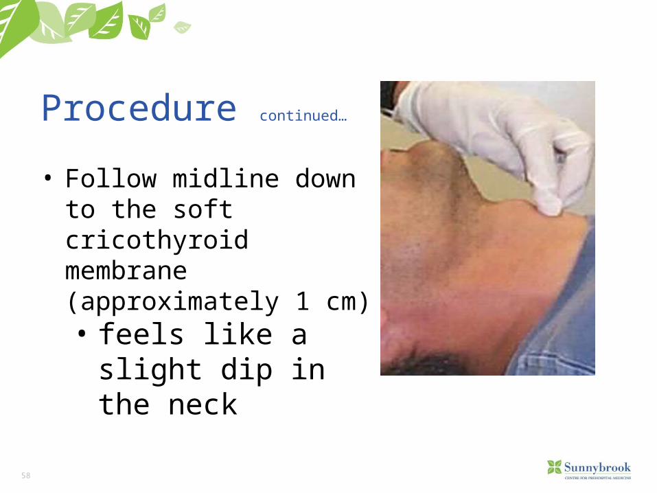
Procedure continued…
• Follow midline down to the soft cricothyroid membrane (approximately 1 cm)• feels like a slight dip in
the neck
59
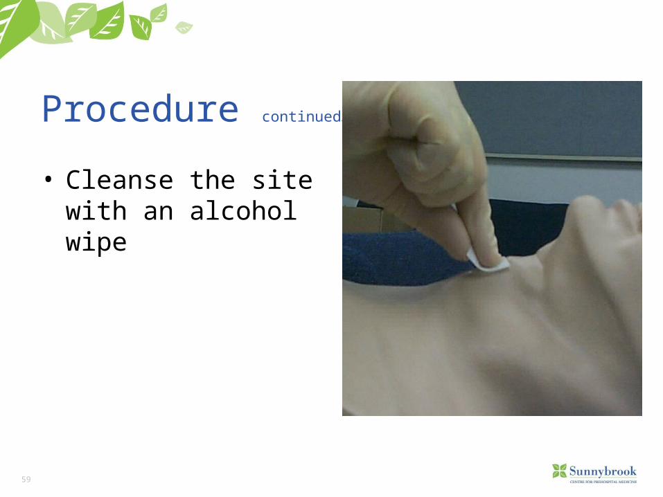
Procedure continued…
• Cleanse the site with an alcohol wipe
60
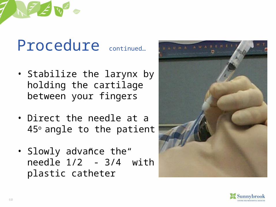
Procedure continued…
• Stabilize the larynx by holding the cartilage between your fingers
• Direct the needle at a 45o angle to the patient
• Slowly advance the needle 1/2” - 3/4” with plastic catheter
61
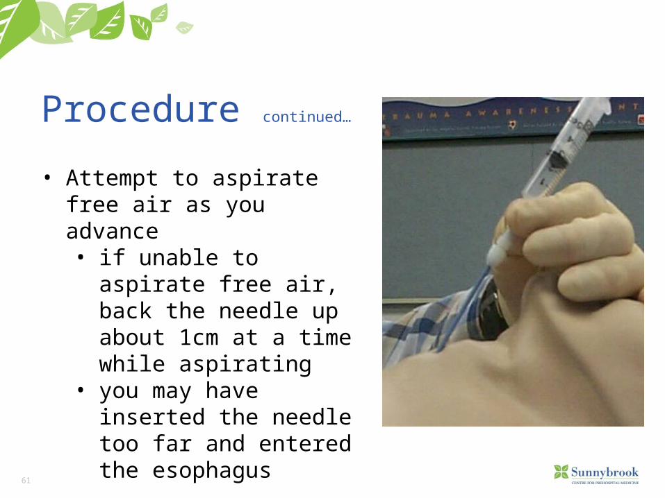
Procedure continued…
• Attempt to aspirate free air as you advance• if unable to aspirate free air,
back the needle up about 1cm at a time while aspirating
• you may have inserted the needle too far and entered the esophagus
62
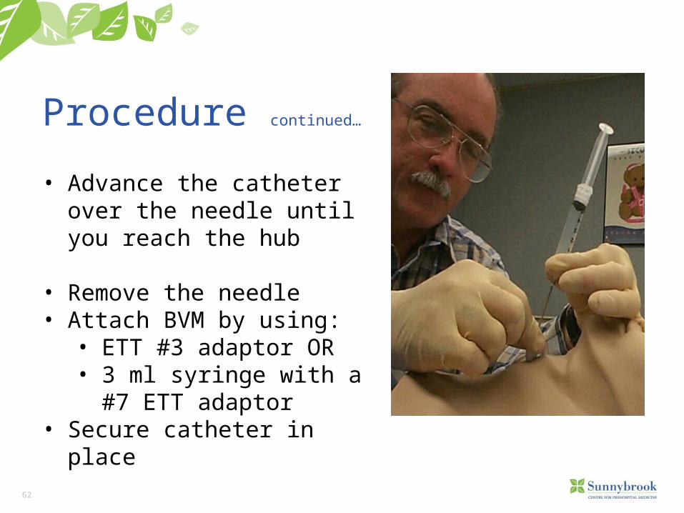
Procedure continued…
• Advance the catheter over the needle until you reach the hub
• Remove the needle• Attach BVM by using:
• ETT #3 adaptor OR• 3 ml syringe with a #7 ETT
adaptor• Secure catheter in place
63
Procedure continued…
• Assess the patient’s ABCs
• do not expect to see significant rise and fall of the chest wall
• if the patient begins spontaneous breathing, time your oxygenation with inhalation
64
Final thoughts and considerations:• Every AW is a difficult AW in the prehospital environment.• Numerous tools in the arsenal for comprehensive, effective AW
management.• No one adjunct tool does it all.• Resistance is not futile and it is often correctable. • Airway management requires constant surveillance and frequent
reassessment.• Most management techniques are applied concurrently to achieve
maximum airway access for improved oxygenation and ventilation.• Always consider risk/benefit for the patient and how to maximize the
talents at the scene.