Embed Size (px)
Citation preview

ChemicalScience
EDGE ARTICLE
Ope
n A
cces
s A
rtic
le. P
ublis
hed
on 0
9 Se
ptem
ber
2015
. Dow
nloa
ded
on 1
/9/2
022
3:42
:47
PM.
Thi
s ar
ticle
is li
cens
ed u
nder
a C
reat
ive
Com
mon
s A
ttrib
utio
n-N
onC
omm
erci
al 3
.0 U
npor
ted
Lic
ence
.
View Article OnlineView Journal | View Issue
The specificity of
aFrom the Department of Neurology, Medi
Merowingerplatz 1a, 40225 Dusseldorf, GerbBiochemistry of Plants, Ruhr-Universitat
Bochum, GermanycInstitute for Medical Biochemistry and
Greifswald, Ernst-Moritz-Arndt Universitat,
Greifswald, Germany. E-mail: horst@lillig
3834 86 5407
Cite this: Chem. Sci., 2015, 6, 7049
Received 24th April 2015Accepted 8th September 2015
DOI: 10.1039/c5sc01501d
www.rsc.org/chemicalscience
This journal is © The Royal Society of C
thioredoxins and glutaredoxins isdetermined by electrostatic and geometriccomplementarity
Carsten Berndt,a Jens-Dirk Schwennb and Christopher Horst Lillig*c
Thiol–disulfide oxidoreductases from the thioredoxin (Trx) family of proteins have a broad range of well
documented functions and possess distinct substrate specificities. The mechanisms and characteristics
that control these specificities are key to the understanding of both the reduction of catalytic disulfides
as well as allosteric disulfides (thiol switches). Here, we have used the catalytic disulfide of E. coli 30-phosphoadenosine 50-phosphosulfate (PAPS) reductase (PR) that forms between the single active site
thiols of two monomers during the reaction cycle as a model system to investigate the mechanisms of
Trx and Grx protein specificity. Enzyme kinetics, DE00 determination, and structural analysis of various Trx
and Grx family members suggested that the redox potential does not determine specificity nor efficiency
of the redoxins as reductant for PR. Instead, the efficiency of PR with various redoxins correlated
strongly to the extent of a negative electric field of the redoxins reaching into the solvent outside the
active site, and electrostatic and geometric complementary contact surfaces. These data suggest that, in
contrast to common assumption, the composition of the active site motif is less important for substrate
specificity than other amino acids in or even outside the immediate contact area.
Introduction
During the past decade the concept of cellular redox homeo-stasis shied more and more from the idea of a redox balancebetween oxidants and antioxidants towards the concept ofspatio-temporally controlled redox signalling events. Theseevents are specic with respect to the cysteinyl residues modi-ed and the redox compounds and enzymes involved. Thio-redoxins (Trxs) and glutaredoxins (Grxs) are the masterregulators of the redox state of the thiol groups of the proteomewith numerous well documented functions in essentially allcellular processes, including metabolism and cell signalling,see for instance refs. 1–4. In most cases, Trxs and Grxs reducecatalytic and allosteric disuldes, a classication introduced byHogg and coworkers.5 Although Trxs and Grxs have a broadrange of functions, each member of the family has distinctsubstrate specicities. Themechanisms and characteristics thatcontrol these specicities are unclear and have hardly beenaddressed before.
cal Faculty, Heinrich-Heine Universitat,
many
Bochum, Universitatsstraße 150, 44780
Molecular Biology, Universitatsmedizin
Ferdinand Sauerbruch Straße, DE-17475
.de; Fax: +49 3834 8605402; Tel: +49
hemistry 2015
Both Trxs and Grxs are part of the Trx family of proteins,characterized by a common structure, the Trx fold.6,7 This fold isdened by a central four to ve-stranded b-sheet, surrounded bythree to four a-helices. Trxs and Grxs catalyze thiol–disuldeexchange reactions. Two thiols in the characteristic Cys–X–X–Cysactive site reduced a target disulde in a reversible two-stepreaction. The more N-terminal thiolate cysteinyl residue, locatedat the surface of the protein in a loop connecting b1 and a1 (inGrxs) or b2 and a2 (in Trxs), attacks the target disulde resultingin an intermediate mixed disulde between the redoxin and thetarget protein. This is directly attacked by the more C-terminalactive site thiol, normally buried in the protein at the beginningof the a-helix (1 or 2), yielding a disulde in the active site and areduced target protein. Oxidized Trx is reduced by a Trx reduc-tase and Grx by two molecules of glutathione. This reactionsequence was named the dithiol mechanism to distinguish itfrom the monothiol mechanism. The latter is used by Grxs toreduce protein–glutathione mixed disuldes and requires onlyone, the more N-terminal, active site cysteinyl residue.
Both Trxs and Grxs were originally identied as electrondonors for ribonucleotide reductase from E. coli.8,9 Therequirement for Trx in sulfate assimilation was originallydescribed by Gonzalez-Porque et al.10 for yeast. Reduction ofsulfate to sulte requires two electrons with a DE0
0 of �517 mV.Adenylation and phosphorylation to 30-phosphoadenonsine-50-phosphosulfate (PAPS) lowers this redox potential to�60mV. InE. coli, Trx1 and Grx1 were identied as alternative electrondonors for the catalytic disulde in PAPS reductase (PR).11,12
Chem. Sci., 2015, 6, 7049–7058 | 7049
Chemical Science Edge Article
Ope
n A
cces
s A
rtic
le. P
ublis
hed
on 0
9 Se
ptem
ber
2015
. Dow
nloa
ded
on 1
/9/2
022
3:42
:47
PM.
Thi
s ar
ticle
is li
cens
ed u
nder
a C
reat
ive
Com
mon
s A
ttrib
utio
n-N
onC
omm
erci
al 3
.0 U
npor
ted
Lic
ence
.View Article Online
Enzymatically active PR (EC 1.8.99.4) forms a homo-dimer(2 � 28 kDa). It is devoid of redox-active chromophores butcontains a single cysteine in a strictly conserved ECGLH motifthat is located at the C-terminus.13 Steady state analysis of thereaction and mutagenesis analyses demonstrated that PRfollows a ping-pong mechanism in the reduction of PAPS tosulte and 30-50-adenosine diphosphate (PAP).13–17 In the rststep, the PR dimer reduces PAPS directly to sulte without anydetectable sulfate or sulte-bound intermediates. Oxidation ofPR yields an intermolecular disulde between the ECGLH cys-teinyl residues of the two monomers. In the second step, thisdisulde is the substrate for Trx or Grx and requires the dithiolreaction mechanism of the redoxins for reduction. Although E.coli PR is a rather promiscuous enzyme regarding its choice ofelectron donors, it cannot be reduced by any Trx or Grx. Thehigh cross-reactivity of E. coli PR was, for instance, useful in theidentication and purication of heterologous Trxs fromspinach, Synechococcus, and yeast.18 However, the two addi-tional dithiol Grxs of E. coli itself, Grx2 and Grx3,19 cannotreduce PR, neither in vivo20 nor in vitro.16
In this study, we have used E. coli PR as a model for theanalysis of the specicity of Trx family proteins addressing twohypotheses. First, is the functionality of the redoxins deter-mined by the redox potential of their active site dithiol–disul-de redox pair? Or, second, is their functionality determined byspecic molecular interactions next to the thiol–disuldeexchange reaction?
ResultsHomologous and heterologous thioredoxins andglutaredoxins as electron donors for PAPS reductase
The functionality of various thioredoxins and glutaredoxinsfrom different species with E. coli PR was analyzed in kineticassays. In addition to the well established E. coli Trx1 and Grx1,
Table 1 Redox potentials and kinetic parameters of various thioredoxin
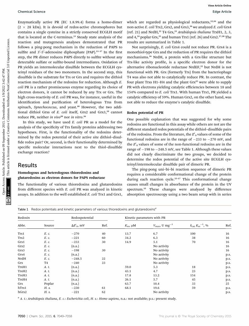
Redoxin Redoxpotential K
Abbr. Source DE00, mV Ref. K
Trx1 E. c. �270 40 1Trx2 E. c. �221 60 3Grx1 E. c. �233 30 1Grx2 E. c. (n.a.)Grx3 E. c. �198 30Grx4 E. c. (n.a.)NrdH E. c. �248.5 22Grx T4 �240 23TrxH1 A. t. (n.a.) 5TrxH2 A. t. (n.a.) 4TrxH3 A. t. (n.a.) 1TrxH4 A. t. (n.a.) 2Grx Poplar (n.a.) 6hTrx1 H. s. �230 61 6hGrx2 H. s. �221 62
a A. t.: Arabidopsis thaliana, E. c.: Escherichia coli, H. s.: Homo sapiens, n.a.
7050 | Chem. Sci., 2015, 6, 7049–7058
which are regarded as physiological reductants,12,16 and thenon-active E. coli Trx2, Grx2, and Grx3,16 we analyzed E. coli Grx4(ref. 21) and NrdH,22 T4 Grx,23 Arabidopsis thaliana TrxH1, 2, 3,and 4,24 poplar Grx,25 and human Trx1 (ref. 26) and Grx2.27,28 Theresults are summarized in Table 1.
Not surprisingly, E. coli Grx4 could not reduce PR. Grx4 is amonothiol-type Grx and the reduction of PR requires the dithiolmechanisms.16 NrdH, a protein with a Grx-like structure butTrx-like activity prole, is a specic electron donor for thealternative ribonucleotide reductase NrdEF,22 but NrdH is notfunctional with PR. Grx (formerly Trx) from the bacteriophageT4 was also not able to catalytically reduce PR. In contrast, thefour plant Trxs H1–H4 and the plant Grx25 were able to supplyPR with electrons yielding catalytic efficiencies between 18 and154% compared to E. coli Trx1. With human Trx1, PR yielded acatalytic efficiency of 59%. Human Grx2, on the other hand, wasnot able to reduce the enzyme's catalytic disulde.
Redox potential of PR
One possible explanation that was suggested for why someredoxins are functional in this assay while others are not are thedifferent standard redox potentials of the dithiol–disulde pairsof the redoxins. From the literature, the E0
0 values of some of thefunctional redoxins are in the range of �233 to �270 mV, andthe E0
0 values of some of the non-functional redoxins are in therange of �198 to �248.5 mV, see Table 1. Although these valuesdid not clearly discriminate the two groups, we decided todetermine the redox potential of the active site ECGLH cys-teinyl/intermolecular disulde pair of dimeric PR.
The ping-pong uni–bi–bi reaction sequence of dimeric PRrequires a considerable conformational change of the proteinduring each reaction cycle.15–17 This conformational changecauses small changes in absorbance of the protein in the UVspectrum.15 These changes were analyzed by differenceabsorption spectroscopy using a two beam setup with in series
s and glutaredoxinsa
inetic parameters with PR
m, mM Vmax, U mg�1 kcat Km�1, % Ref.
3.7 6.7 100 164.2 6.3 38 164.9 5.1 70 16
No activity 16No activity 16No activity p.s.No activity p.s.No activity p.s.
9.0 5.3 18 p.s.3.1 4.7 23 p.s.7.8 13.2 154 p.s.6.1 5.7 45 p.s.3.7 10.4 33 258.1 19.6 59 p.s.
No activity p.s.
: not available; p.s.: present study.
This journal is © The Royal Society of Chemistry 2015
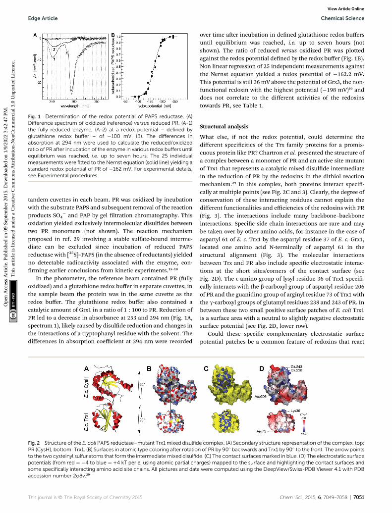
Fig. 1 Determination of the redox potential of PAPS reductase. (A)Difference spectrum of oxidized (reference) versus reduced PR, (A-1)the fully reduced enzyme, (A-2) at a redox potential – defined byglutathione redox buffer – of �100 mV. (B). The differences inabsorption at 294 nm were used to calculate the reduced/oxidizedratio of PR after incubation of the enzyme in various redox buffers untilequilibrium was reached, i.e. up to seven hours. The 25 individualmeasurements were fitted to the Nernst equation (solid line) yielding astandard redox potential of PR of �162 mV. For experimental details,see Experimental procedures.
Edge Article Chemical Science
Ope
n A
cces
s A
rtic
le. P
ublis
hed
on 0
9 Se
ptem
ber
2015
. Dow
nloa
ded
on 1
/9/2
022
3:42
:47
PM.
Thi
s ar
ticle
is li
cens
ed u
nder
a C
reat
ive
Com
mon
s A
ttrib
utio
n-N
onC
omm
erci
al 3
.0 U
npor
ted
Lic
ence
.View Article Online
tandem cuvettes in each beam. PR was oxidized by incubationwith the substrate PAPS and subsequent removal of the reactionproducts SO4
� and PAP by gel ltration chromatography. Thisoxidation yielded exclusively intermolecular disuldes betweentwo PR monomers (not shown). The reaction mechanismproposed in ref. 29 involving a stable sulfate-bound interme-diate can be excluded since incubation of reduced PAPSreductase with [35S]–PAPS (in the absence of reductants) yieldedno detectable radioactivity associated with the enzyme, con-rming earlier conclusions from kinetic experiments.15–18
In the photometer, the reference beam contained PR (fullyoxidized) and a glutathione redox buffer in separate cuvettes; inthe sample beam the protein was in the same cuvette as theredox buffer. The glutathione redox buffer also contained acatalytic amount of Grx1 in a ratio of 1 : 100 to PR. Reduction ofPR led to a decrease in absorbance at 253 and 294 nm (Fig. 1A,spectrum 1), likely caused by disulde reduction and changes inthe interactions of a tryptophanyl residue with the solvent. Thedifferences in absorption coefficient at 294 nm were recorded
Fig. 2 Structure of the E. coli PAPS reductase–mutant Trx1 mixed disulfidPR (CysH), bottom: Trx1. (B) Surfaces in atomic type coloring after rotationto the two cysteinyl sulfur atoms that form the intermediatemixed disulfidpotentials (from red ¼ �4 to blue ¼ +4 kT per e, using atomic partial chasome specifically interacting amino acid site chains. All pictures and dataaccession number 2o8v.29
This journal is © The Royal Society of Chemistry 2015
over time aer incubation in dened glutathione redox buffersuntil equilibrium was reached, i.e. up to seven hours (notshown). The ratio of reduced versus oxidized PR was plottedagainst the redox potential dened by the redox buffer (Fig. 1B).Non linear regression of 25 independent measurements againstthe Nernst equation yielded a redox potential of �162.2 mV.This potential is still 36 mV above the potential of Grx3, the non-functional redoxin with the highest potential (�198 mV)30 anddoes not correlate to the different activities of the redoxinstowards PR, see Table 1.
Structural analysis
What else, if not the redox potential, could determine thedifferent specicities of the Trx family proteins for a promis-cuous protein like PR? Chartron et al. presented the structure ofa complex between a monomer of PR and an active site mutantof Trx1 that represents a catalytic mixed disulde intermediatein the reduction of PR by the redoxins in the dithiol reactionmechanism.29 In this complex, both proteins interact speci-cally at multiple points (see Fig. 2C and 3). Clearly, the degree ofconservation of these interacting residues cannot explain thedifferent functionalities and efficiencies of the redoxins with PR(Fig. 3). The interactions include many backbone–backboneinteractions. Specic side chain interactions are rare and maybe taken over by other amino acids, for instance in the case ofaspartyl 61 of E. c. Trx1 by the aspartyl residue 37 of E. c. Grx1,located one amino acid N-terminally of aspartyl 61 in thestructural alignment (Fig. 3). The molecular interactionsbetween Trx and PR also include specic electrostatic interac-tions at the short sites/corners of the contact surface (seeFig. 2D). The 3-amino group of lysyl residue 36 of Trx1 speci-cally interacts with the b-carboxyl group of aspartyl residue 206of PR and the guanidino group of arginyl residue 73 of Trx1 withthe g-carboxyl groups of glutamyl residues 238 and 243 of PR. Inbetween these two small positive surface patches of E. coli Trx1is a surface area with a neutral to slightly negative electrostaticsurface potential (see Fig. 2D, lower row).
Could these specic complementary electrostatic surfacepotential patches be a common feature of redoxins that react
e complex. (A) Secondary structure representation of the complex, top:of PR by 90� backwards and Trx1 by 90� to the front. The arrow pointse. (C) The contact surfacesmarked in blue. (D) The electrostatic surfacerges) mapped to the surface and highlighting the contact surfaces andwere computed using the DeepView/Swiss-PDB Viewer 4.1 with PDB
Chem. Sci., 2015, 6, 7049–7058 | 7051

Fig. 3 Structural alignment of the thioredoxin family proteins inves-tigated in this study. The structures of the various redoxins werepairwise aligned to the structure of E. coli Trx 1 (PDB code 1xob) usingPyMOL and thereafter manually arranged in this alignment. The resi-dues directly interacting in the E. c. Trx1–PAPS reductase complexwere highlighted with a green background. The residues were alsospecified below the sequence, as well as the interacting residues in
7052 | Chem. Sci., 2015, 6, 7049–7058
Chemical Science Edge Article
Ope
n A
cces
s A
rtic
le. P
ublis
hed
on 0
9 Se
ptem
ber
2015
. Dow
nloa
ded
on 1
/9/2
022
3:42
:47
PM.
Thi
s ar
ticle
is li
cens
ed u
nder
a C
reat
ive
Com
mon
s A
ttrib
utio
n-N
onC
omm
erci
al 3
.0 U
npor
ted
Lic
ence
.View Article Online
kinetically with PR and be different in those that don't? Toaddress this hypothesis, we analyzed the available structures ofthe redoxins analyzed kinetically, i.e. E. coli Grx1 (pdb code:1egr),31 Grx2 (1g7o),32 Grx3 (3grx),33 Trx1 (1xob),34 and NrdH(1h75),35 bacteriophage T4 Grx (1de2),36 A. thaliana TrxH1(1x),37 as well as human Grx2 (2s) and Trx1 (1ert).38 Fromthese structures, T4 Grx and E. coli Grx2 differ the most, sincethese redoxins contain elongated loops on the interactionsurface (T4 Grx, Fig. 4A) or an additional domain that partlycovers the contact area (E. coli Grx2, Fig. 4B). These featuresobviously explain their lack of activity with PR. The remainingstructures fall into two groups with respect to their electrostaticcharacteristics (Fig. 5). The rst group consists of E. coli Grx1(Fig. 5A) and Trx1 (Fig. 5B), human Trx1 (Fig. 5C), and A.thaliana TrxH1 (Fig. 5D). This group is characterized by aneutral to slightly negative contact area, marked by a few smallpositive patches (Fig. 5A–D, 5th column). And, moreover, arather prominent negative electric eld protruding into thesurrounding solvent outside the contact surface characterizedabove (Fig. 6A–G). The second group includes E. coli Grx3(Fig. 5E and 6I), human Grx2 (Fig. 5F and 6H), and E. coli NrdH(Fig. 5G and 6J). This group lacks prominent electric eldsreaching into the solvent and displays a pronounced positivesurface potential in the area corresponding to the contactsurface between E. coli Trx1 and PR (Fig. 2C and D). Astonish-ingly, these two groups exactly correspond to the redoxins activewith PR (rst group) and the redoxins inactive with PR (secondgroup). In fact, the strength and extent of the negative electriceld of the redoxins outside the contact area directly correlatesto the catalytic efficiency of PR with these redoxins as electrondonors (Fig. 6).
Discussion
The specicity of distinct Trx and Grx proteins for any givencatalytic or allosteric disulde, see ref. 5, is a key element for thecontrolled ow of metabolites and the operation of thiolswitches in redox signalling.39 Using E. coli PR as model, ourstudy suggests that geometric and electrostatic complemen-tarity as well as electric elds and thus long-distance electro-static interactions between the redoxin and their target proteinsare the key elements for Trx family proteins' specicity andefficiency.
The differences in standard redox potentials between tworedox pairs, such as a Trx (red/ox) and a metabolic enzyme suchas PR (red/ox), are a measure of the free energy of the reactionand thus how thermodynamically favorable this reaction is.
PAPS reductase and the parts of the amino acids involved in theseinteractions (bb: backbone, sc: side chain). The interactions werecalculated with ‘contact’ of the CCP4 suite.58 Conserved residues werehighlighted with a yellow, positively charged residues with a blue, andnegatively charged residues with a red background. The positions ofthe active site as well as the Trx-fold specific cis-Pro residues werealso marked above the sequences. The secondary structures of E. c.Grx1 was also included above the sequences, the secondary structureof E. c. Trx1 below the sequences.
This journal is © The Royal Society of Chemistry 2015

Fig. 4 Structures of Grxs non-functional with PAPS reductase withaltered active site geometry. Secondary structure representations of(A) T4 Grx and (B) E. coli Grx2. The loops and domain protruding fromthe area corresponding to the contact surface in Trx1 are marked. Thepictures were computed using the DeepView/Swiss-PDB Viewer 4.1with PDB accession numbers 1de2 (T4)36 and 1g7o (Grx2).32
Edge Article Chemical Science
Ope
n A
cces
s A
rtic
le. P
ublis
hed
on 0
9 Se
ptem
ber
2015
. Dow
nloa
ded
on 1
/9/2
022
3:42
:47
PM.
Thi
s ar
ticle
is li
cens
ed u
nder
a C
reat
ive
Com
mon
s A
ttrib
utio
n-N
onC
omm
erci
al 3
.0 U
npor
ted
Lic
ence
.View Article Online
Thermodynamics, however, do not determine physiologicalreactions. Rate constants are determined by, most of all, themagnitude of the activation energy barrier. This is whatenzymes facilitate – increasing reaction rates by loweringactivation energies. In vivo, the ow of metabolites is controlledby metabolite concentrations, enzymes' specicities, theregulation of enzyme activity, and compartmentalization. Aslong as a reaction is thermodynamically favorable (or madefavorable by, for instance, detracting a reaction product)reaction rates are controlled by proteins. This was nicelyconrmed here. The differences in redox potentials of theredoxins and PR did not correlate to the efficiency of theenzyme with these redoxins as electron donors. Similarconclusions were drawn before. The standard redox potentialof Trx1 and Grx1 are �270 mV (ref. 40) and �233 mV,30
respectively, making Trx the thermodynamically more favor-able reductant. E. coli ribonucleotide reductase, however, iscatalytically more efficient with Grx1 as electron donor.41
Moreover, the redoxins which are able or unable to reduceribonucleotide reductase correspond to the same two groups asthe redoxins investigated as reductants for PR here. E. coli Trxs1, Trx2, and Grx1 as well as poplar Grx are able to reduce E. coliribonucleotide reductase,8,9,25,42 whereas E. coli Grx2, Grx4, andNrdH display marginal or no activity.22,43,44
This journal is © The Royal Society of Chemistry 2015
The importance of geometric and electrostatic complemen-tary surfaces has been highlighted before in a study that aimedat the determination of the structure of the peroxiredoxin–glu-taredoxin (Prx–Grx) hybrid protein from Haemophilus inu-enza.45 This study revealed two interaction sites on the surface ofthe Prx domain, depending on the reaction cycle of the perox-idase that involves a conformational change of the active siteperoxidatic cysteinyl residue. These areas interact with essen-tially the same contact surface on the Grx domain of a secondhybrid protein in the homo tetrameric quaternary structure.Both modes of interaction involve specic electrostatic inter-actions of two small positive patches on the Grx domain denedby lysyl residue 177 and arginyl residue 212, with two negativepatches on the Prx domain, dened by glutamyl 59 and aspar-tyl–glutamyl residues 89–90 or aspartyl residues 148, 154, and156.45 This interaction is quite similar to the interaction of Trx1with PR, see Fig. 2D. Another example for the importance ofcomplementary surfaces was provided by the crystal structuresof the two barley TrxH isoforms 1 and 2 and a complex of TrxH2with the a-amylase/subtilisin inhibitor BASI.46 This study alsoconcluded that substrate specicity and reaction efficiency maybe mainly based on complementary contact areas and specicmolecular interactions between the Trxs and their target, andnot on differences in redox potentials, that are almost identicalfor these two Trxs.47
Our conclusions are further supported by an analysis of threemore complexes between Trx family proteins and their targetsthat are available in the protein data base. The complexes ofhuman Trx1 and thioredoxin interacting protein (TXNIP),48
namely TrxH2 and BASI, and between yeast Trx1 and methio-nine sulfoxide reductase A (MsrA)49 are depicted in Fig. 7. Theproteins do not only demonstrate surface complementarity but,when separated, also a perfect complementarity between theirelectrostatic eld maps when the interacting surfaces face eachother (Fig. 7, rst column).
Long-range electrostatic interactions are thought to be adriving force for facilitating and enhancing the rate of specicbinding of a protein to a target.50,51 Not surprisingly, the beststudied mechanisms of signalling and regulation – reversiblephosphorylation – primarily affect electrostatic interactions.Here, we have seen a strong correlation between the efficiencyof PR with various Trx and Grx proteins as electron donor andthe extent and strength of the negative electric elds of theredoxins protruding into the solvent, mostly outside theimmediate contact area (Fig. 5). The only study addressing thispoint so far by Bunik et al.52 analyzed the activation of a-ketoglutarate dehydrogenase by various Trxs. Supporting theconclusions drawn here, this study identied that the length ofthe a-helix 1 (where part of the active site is located) and thesurrounding charges correlate with the inuence of the Trxs onthe a-ketoglutarate dehydrogenase complex. The efficiency ofthe Trxs tested directly correlated to the strength of the, in thiscase positive, electric elds/polarization and the highest dipolevector. Bunik et al. concluded that the ‘selective action of athioredoxin should stem from specic recognition uponformation of the thioredoxin–target complex [.] before thehighly reactive catalytic groups are brought together’.52 This
Chem. Sci., 2015, 6, 7049–7058 | 7053
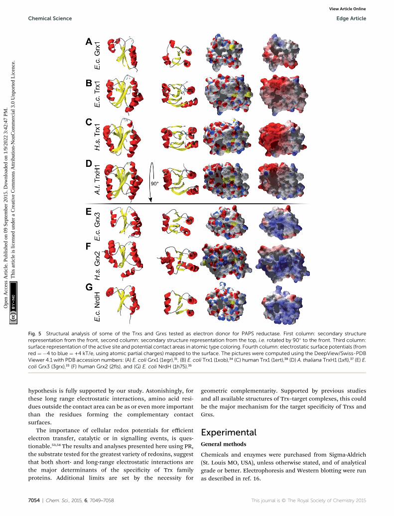
Fig. 5 Structural analysis of some of the Trxs and Grxs tested as electron donor for PAPS reductase. First column: secondary structurerepresentation from the front, second column: secondary structure representation from the top, i.e. rotated by 90� to the front. Third column:surface representation of the active site and potential contact areas in atomic type coloring. Fourth column: electrostatic surface potentials (fromred ¼ �4 to blue ¼ +4 kT/e, using atomic partial charges) mapped to the surface. The pictures were computed using the DeepView/Swiss-PDBViewer 4.1 with PDB accession numbers: (A) E. coli Grx1 (1egr),31, (B) E. coli Trx1 (1xob),34 (C) human Trx1 (1ert),38 (D) A. thaliana TrxH1 (1xfl),37 (E) E.coli Grx3 (3grx),33 (F) human Grx2 (2fls), and (G) E. coli NrdH (1h75).35
Chemical Science Edge Article
Ope
n A
cces
s A
rtic
le. P
ublis
hed
on 0
9 Se
ptem
ber
2015
. Dow
nloa
ded
on 1
/9/2
022
3:42
:47
PM.
Thi
s ar
ticle
is li
cens
ed u
nder
a C
reat
ive
Com
mon
s A
ttrib
utio
n-N
onC
omm
erci
al 3
.0 U
npor
ted
Lic
ence
.View Article Online
hypothesis is fully supported by our study. Astonishingly, forthese long range electrostatic interactions, amino acid resi-dues outside the contact area can be as or even more importantthan the residues forming the complementary contactsurfaces.
The importance of cellular redox potentials for efficientelectron transfer, catalytic or in signalling events, is ques-tionable.53,54 The results and analyses presented here using PR,the substrate tested for the greatest variety of redoxins, suggestthat both short- and long-range electrostatic interactions arethe major determinants of the specicity of Trx familyproteins. Additional limits are set by the necessity for
7054 | Chem. Sci., 2015, 6, 7049–7058
geometric complementarity. Supported by previous studiesand all available structures of Trx–target complexes, this couldbe the major mechanism for the target specicity of Trxs andGrxs.
ExperimentalGeneral methods
Chemicals and enzymes were purchased from Sigma-Aldrich(St. Louis MO, USA), unless otherwise stated, and of analyticalgrade or better. Electrophoresis and Western blotting were runas described in ref. 16.
This journal is © The Royal Society of Chemistry 2015
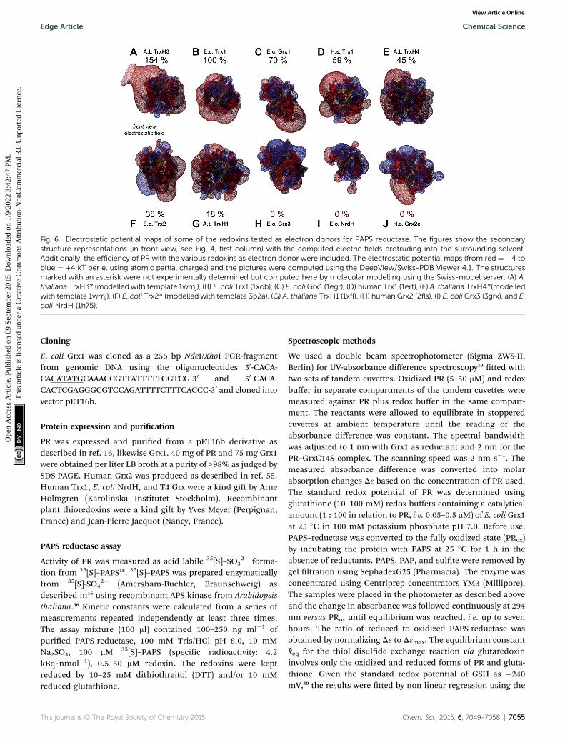
Fig. 6 Electrostatic potential maps of some of the redoxins tested as electron donors for PAPS reductase. The figures show the secondarystructure representations (in front view, see Fig. 4, first column) with the computed electric fields protruding into the surrounding solvent.Additionally, the efficiency of PR with the various redoxins as electron donor were included. The electrostatic potential maps (from red ¼ �4 toblue ¼ +4 kT per e, using atomic partial charges) and the pictures were computed using the DeepView/Swiss-PDB Viewer 4.1. The structuresmarked with an asterisk were not experimentally determined but computed here by molecular modelling using the Swiss-model server. (A) A.thaliana TrxH3* (modelled with template 1wmj), (B) E. coli Trx1 (1xob), (C) E. coliGrx1 (1egr), (D) human Trx1 (1ert), (E) A. thaliana TrxH4*(modelledwith template 1wmj), (F) E. coli Trx2* (modelled with template 3p2a), (G) A. thaliana TrxH1 (1xfl), (H) human Grx2 (2fls), (I) E. coliGrx3 (3grx), and E.coli NrdH (1h75).
Edge Article Chemical Science
Ope
n A
cces
s A
rtic
le. P
ublis
hed
on 0
9 Se
ptem
ber
2015
. Dow
nloa
ded
on 1
/9/2
022
3:42
:47
PM.
Thi
s ar
ticle
is li
cens
ed u
nder
a C
reat
ive
Com
mon
s A
ttrib
utio
n-N
onC
omm
erci
al 3
.0 U
npor
ted
Lic
ence
.View Article Online
Cloning
E. coli Grx1 was cloned as a 256 bp NdeI/XhoI PCR-fragmentfrom genomic DNA using the oligonucleotides 50-CACA-CACATATGCAAACCGTTATTTTTGGTCG-30 and 50-CACA-CACTCGAGGGCGTCCAGATTTTCTTTCACCC-30 and cloned intovector pET16b.
Protein expression and purication
PR was expressed and puried from a pET16b derivative asdescribed in ref. 16, likewise Grx1. 40 mg of PR and 75 mg Grx1were obtained per liter LB broth at a purity of >98% as judged bySDS-PAGE. Human Grx2 was produced as described in ref. 55.Human Trx1, E. coli NrdH, and T4 Grx were a kind gi by ArneHolmgren (Karolinska Institutet Stockholm). Recombinantplant thioredoxins were a kind gi by Yves Meyer (Perpignan,France) and Jean-Pierre Jacquot (Nancy, France).
PAPS reductase assay
Activity of PR was measured as acid labile 35[S]–SO32� forma-
tion from 35[S]–PAPS18. 35[S]–PAPS was prepared enzymaticallyfrom 35[S]-SO4
2� (Amersham-Buchler, Braunschweig) asdescribed in16 using recombinant APS kinase from Arabidopsisthaliana.56 Kinetic constants were calculated from a series ofmeasurements repeated independently at least three times.The assay mixture (100 ml) contained 100–250 ng ml�1 ofpuried PAPS-reductase, 100 mM Tris/HCl pH 8.0, 10 mMNa2SO3, 100 mM 35[S]–PAPS (specic radioactivity: 4.2kBq$nmol�1), 0.5–50 mM redoxin. The redoxins were keptreduced by 10–25 mM dithiothreitol (DTT) and/or 10 mMreduced glutathione.
This journal is © The Royal Society of Chemistry 2015
Spectroscopic methods
We used a double beam spectrophotometer (Sigma ZWS-II,Berlin) for UV-absorbance difference spectroscopy19 tted withtwo sets of tandem cuvettes. Oxidized PR (5–50 mM) and redoxbuffer in separate compartments of the tandem cuvettes weremeasured against PR plus redox buffer in the same compart-ment. The reactants were allowed to equilibrate in stopperedcuvettes at ambient temperature until the reading of theabsorbance difference was constant. The spectral bandwidthwas adjusted to 1 nm with Grx1 as reductant and 2 nm for thePR–GrxC14S complex. The scanning speed was 2 nm s�1. Themeasured absorbance difference was converted into molarabsorption changes D3 based on the concentration of PR used.The standard redox potential of PR was determined usingglutathione (10–100 mM) redox buffers containing a catalyticalamount (1 : 100 in relation to PR, i.e. 0.05–0.5 mM) of E. coli Grx1at 25 �C in 100 mM potassium phosphate pH 7.0. Before use,PAPS–reductase was converted to the fully oxidized state (PRox)by incubating the protein with PAPS at 25 �C for 1 h in theabsence of reductants. PAPS, PAP, and sulte were removed bygel ltration using SephadexG25 (Pharmacia). The enzyme wasconcentrated using Centriprep concentrators YM3 (Millipore).The samples were placed in the photometer as described aboveand the change in absorbance was followed continuously at 294nm versus PRox until equilibrium was reached, i.e. up to sevenhours. The ratio of reduced to oxidized PAPS-reductase wasobtained by normalizing D3 to D3max. The equilibrium constantkeq for the thiol disulde exchange reaction via glutaredoxininvolves only the oxidized and reduced forms of PR and gluta-thione. Given the standard redox potential of GSH as �240mV,30 the results were tted by non linear regression using the
Chem. Sci., 2015, 6, 7049–7058 | 7055
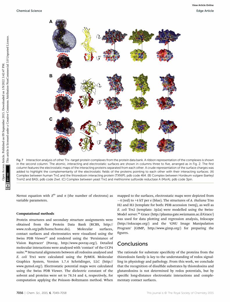
Fig. 7 Interaction analysis of other Trx–target protein complexes from the protein data bank. A ribbon representation of the complexes is shownin the second column. The atomic, interacting and electrostatic surfaces are shown in columns three to five, arranged as in Fig. 2. The firstcolumn features the electrostatic maps of the interacting proteins separated from each other. A crude representation of the surface charges wasadded to highlight the complementarity of the electrostatic fields of the proteins pointing to each other with their interacting surfaces. (A)Complex between human Trx1 and the thioredoxin interacting protein (TXNIP), pdb code 4ll4. (B) Complex between Hordeum vulgare (barley)TrxH2 and BASI, pdb code 2iwt. (C) Complex between yeast Trx1 and methionine sulfoxide reductase A (MsrA), pdb code 3pin.
Chemical Science Edge Article
Ope
n A
cces
s A
rtic
le. P
ublis
hed
on 0
9 Se
ptem
ber
2015
. Dow
nloa
ded
on 1
/9/2
022
3:42
:47
PM.
Thi
s ar
ticle
is li
cens
ed u
nder
a C
reat
ive
Com
mon
s A
ttrib
utio
n-N
onC
omm
erci
al 3
.0 U
npor
ted
Lic
ence
.View Article Online
Nernst equation with E00 and n (the number of electrons) asvariable parameters.
Computational methods
Protein structures and secondary structure assignments wereobtained from the Protein Data Bank (RCBS, http://www.rcsb.org/pdb/home/home.do). Molecular surfaces,contact surfaces and electrostatics were visualized using theSwiss PDB Viewer57 and rendered using the ‘Persistance ofVision Raytracer’ (Povray, http://www.povray.org/). Detailedmolecular interactions were analyzed with ‘contact’ of the CCP4suite.58 Structural alignments between all redoxins analysed andE. coli Trx1 were calculated using the PyMOL MolecularGraphics System, Version 1.7.4 Schrodinger, LLC (http://www.pymol.org/). Electrostatic potential maps were calculatedusing the Swiss PDB Viewer. The dielectric constant of thesolvent and proteins were set to 78.54 and 4, respectively, forcomputation applying the Poisson–Boltzmann method. When
7056 | Chem. Sci., 2015, 6, 7049–7058
mapped to the surfaces, electrostatic maps were depicted from�4 (red) to +4 kT per e (blue). The structures of A. thaliana TrxsH2 and H3 (template for both: PDB accession 1wmj), as well asE. coli Trx2 (template: 3p2a) were modelled using the Swiss-Model server.59 Grace (http://plasma-gate.weizmann.ac.il/Grace/)was used for data plotting and regression analysis, Inkscape(http://inkscape.org/) and the ‘GNU Image ManipulationProgram’ (GIMP, http://www.gimp.org/) for preparing thegures.
Conclusions
The rationale for substrate specicity of the proteins from thethioredoxin family is key to the understanding of redox signal-ling in physiology and pathology. From this work, we concludethat the recognition of disulde substrates by thioredoxins andglutaredoxins is not determined by redox potentials, but byspecic long-distance electrostatic interactions and comple-mentary contact surfaces.
This journal is © The Royal Society of Chemistry 2015
Edge Article Chemical Science
Ope
n A
cces
s A
rtic
le. P
ublis
hed
on 0
9 Se
ptem
ber
2015
. Dow
nloa
ded
on 1
/9/2
022
3:42
:47
PM.
Thi
s ar
ticle
is li
cens
ed u
nder
a C
reat
ive
Com
mon
s A
ttrib
utio
n-N
onC
omm
erci
al 3
.0 U
npor
ted
Lic
ence
.View Article Online
Acknowledgements
The Authors wish to thank Arne Holmgren (Stockholm, Swe-den), Jean-Pierre Jacquot (Nancy, France), and Yves Meyer(Perpignan, France) for providing proteins for analyses. Weexpress our gratitude to Marcel Deponte (Heidelberg, Germany)for discussing the manuscript and inspiring this work. Thiswork was supported by grants from the Deutsche For-schungsgemeinscha (Li 984/3-1 and Be 3259/5-1).
References
1 C. H. Lillig and A. Holmgren, Antioxid. Redox Signaling, 2007,9, 25–47.
2 M. M. Gallogly and J. J. Mieyal, Curr. Opin. Pharmacol., 2007,7, 381–391.
3 C. H. Lillig, C. Berndt and A. Holmgren, Biochim. Biophys.Acta, Gen. Subj., 2008, 1780, 1304–1317.
4 E.-M. Hanschmann, J. R. Godoy, C. Berndt, C. Hudemannand C. H. Lillig, Antioxid. Redox Signaling, 2013, 19, 1539–1605.
5 B. Schmidt, L. Ho and P. J. Hogg, Biochemistry, 2006, 45,7429–7433.
6 J. L. Martin, Struct. Lond. Engl. 1993, 1995, 3, 245–250.7 Y. Qi and N. V. Grishin, Proteins, 2005, 58, 376–388.8 T. C. Laurent, E. C. Moore and P. Reichard, J. Biol. Chem.,1964, 239, 3436–3444.
9 A. Holmgren, J. Biol. Chem., 1979, 254, 3664–3671.10 P. Gonzalez-Porque, A. Baldesten and P. Reichard, J. Biol.
Chem., 1970, 245, 2371–2374.11 M. L. Tsang and J. A. Schiff, J. Bacteriol., 1978, 134, 131–138.12 M. L. Tsang, J. Bacteriol., 1981, 146, 1059–1066.13 F. A. Krone, G. Westphal and J. D. Schwenn, Mol. Genet.
Genomics, 1991, 225, 314–319.14 J. D. Schwenn, F. A. Krone and K. Husmann, Arch. Microbiol.,
1988, 150, 313–319.15 U. Berendt, T. Haverkamp, A. Prior and J. D. Schwenn, Eur. J.
Biochem., 1995, 233, 347–356.16 C. H. Lillig, A. Prior, J. D. Schwenn, F. Aslund, D. Ritz,
A. Vlamis-Gardikas and A. Holmgren, J. Biol. Chem., 1999,274, 7695–7698.
17 C. H. Lillig, A. Potamitou, J.-D. Schwenn, A. Vlamis-Gardikasand A. Holmgren, J. Biol. Chem., 2003, 278, 22325–22330.
18 J. D. Schwenn and U. Schriek, Z. fur Naturforschung, 1987, 42,93–102.
19 F. Aslund, B. Ehn, A. Miranda-Vizuete, C. Pueyo andA. Holmgren, Proc. Natl. Acad. Sci. U. S. A., 1994, 91, 9813–9817.
20 M. Russel, P. Model and A. Holmgren, J. Bacteriol., 1990, 172,1923–1929.
21 A. P. Fernandes, M. Fladvad, C. Berndt, C. Andresen,C. H. Lillig, P. Neubauer, M. Sunnerhagen, A. Holmgrenand A. Vlamis-Gardikas, J. Biol. Chem., 2005, 280, 24544–24552.
22 A. Jordan, F. Aslund, E. Pontis, P. Reichard and A. Holmgren,J. Biol. Chem., 1997, 272, 18044–18050.
This journal is © The Royal Society of Chemistry 2015
23 O. Berglund and B. M. Sjoberg, J. Biol. Chem., 1970, 245,6030–6035.
24 Y. Meyer, J. P. Reichheld and F. Vignols, Photosynth. Res.,2005, 86, 419–433.
25 N. Rouhier, A. Vlamis-Gardikas, C. H. Lillig, C. Berndt,J.-D. Schwenn, A. Holmgren and J.-P. Jacquot, Antioxid.Redox Signaling, 2003, 5, 15–22.
26 Y. Tagaya, Y. Maeda, A. Mitsui, N. Kondo, H. Matsui,J. Hamuro, N. Brown, K. Arai, T. Yokota and H. Wakasugi,EMBO J., 1989, 8, 757–764.
27 M. Lundberg, C. Johansson, J. Chandra, M. Enoksson,G. Jacobsson, J. Ljung, M. Johansson and A. Holmgren, J.Biol. Chem., 2001, 276, 26269–26275.
28 V. N. Gladyshev, A. Liu, S. V. Novoselov, K. Krysan, Q. A. Sun,V. M. Kryukov, G. V. Kryukov and M. F. Lou, J. Biol. Chem.,2001, 276, 30374–30380.
29 J. Chartron, C. Shiau, C. D. Stout and K. S. Carroll,Biochemistry, 2007, 46, 3942–3951.
30 F. Aslund, K. D. Berndt and A. Holmgren, J. Biol. Chem.,1997, 272, 30780–30786.
31 P. Sodano, T. H. Xia, J. H. Bushweller, O. Bjornberg,A. Holmgren, M. Billeter and K. Wuthrich, J. Mol. Biol.,1991, 221, 1311–1324.
32 B. Xia, A. Vlamis-Gardikas, A. Holmgren, P. E. Wright andH. J. Dyson, J. Mol. Biol., 2001, 310, 907–918.
33 K. Nordstrand, F. slund, A. Holmgren, G. Otting andK. D. Berndt, J. Mol. Biol., 1999, 286, 541–552.
34 M. F. Jeng, A. P. Campbell, T. Begley, A. Holmgren,D. A. Case, P. E. Wright and H. J. Dyson, Struct. Lond. Engl.1993, 1994, 2, 853–868.
35 M. Stehr, G. Schneider, F. Aslund, A. Holmgren andY. Lindqvist, J. Biol. Chem., 2001, 276, 35836–35841.
36 Y. Wang, G. Amegbey and D. S. Wishart, J. Biomol. NMR,2004, 29, 85–90.
37 F. C. Peterson, B. L. Lytle, S. Sampath, D. Vinarov, E. Tyler,M. Shahan, J. L. Markley and B. F. Volkman, Protein Sci.,2005, 14, 2195–2200.
38 A. Weichsel, J. R. Gasdaska, G. Powis and W. R. Montfort,Struct. Lond. Engl. 1993, 1996, 4, 735–751.
39 M. Deponte and C. H. Lillig, Biol. Chem., 2015.40 G. Krause, J. Lundstrom, J. L. Barea, C. Pueyo de la Cuesta
and A. Holmgren, J. Biol. Chem., 1991, 266, 9494–9500.41 A. P. Fernandes and A. Holmgren, Antioxid. Redox Signaling,
2004, 6, 63–74.42 A. Miranda-Vizuete, A. E. Damdimopoulos, J. Gustafsson
and G. Spyrou, J. Biol. Chem., 1997, 272, 30841–30847.43 A. Vlamis-Gardikas, F. Aslund, G. Spyrou, T. Bergman and
A. Holmgren, J. Biol. Chem., 1997, 272, 11236–11243.44 R. Ortenberg, S. Gon, A. Porat and J. Beckwith, Proc. Natl.
Acad. Sci. U. S. A., 2004, 101, 7439–7444.45 S. J. Kim, J. R. Woo, Y. S. Hwang, D. G. Jeong, D. H. Shin,
K. Kim and S. E. Ryu, J. Biol. Chem., 2003, 278, 10790–10798.46 K. Maeda, P. Hagglund, C. Finnie, B. Svensson and
A. Henriksen, Protein Sci., 2008, 17, 1015–1024.47 K. Maeda, P. Hagglund, O. Bjornberg, J. R. Winther and
B. Svensson, FEBS Lett., 2010, 584, 3376–3380.
Chem. Sci., 2015, 6, 7049–7058 | 7057
Chemical Science Edge Article
Ope
n A
cces
s A
rtic
le. P
ublis
hed
on 0
9 Se
ptem
ber
2015
. Dow
nloa
ded
on 1
/9/2
022
3:42
:47
PM.
Thi
s ar
ticle
is li
cens
ed u
nder
a C
reat
ive
Com
mon
s A
ttrib
utio
n-N
onC
omm
erci
al 3
.0 U
npor
ted
Lic
ence
.View Article Online
48 J. Hwang, H.-W. Suh, Y. H. Jeon, E. Hwang, L. T. Nguyen,J. Yeom, S.-G. Lee, C. Lee, K. J. Kim, B. S. Kang, J.-O. Jeong,T.-K. Oh, I. Choi, J.-O. Lee and M. H. Kim, Nat. Commun.,2014, 5, 2958.
49 X.-X. Ma, P.-C. Guo, W.-W. Shi, M. Luo, X.-F. Tan, Y. Chenand C.-Z. Zhou, J. Biol. Chem., 2011, 286, 13430–13437.
50 M. K. Gilson, Curr. Opin. Struct. Biol., 1995, 5, 216–223.51 H.-X. Zhou, Phys. Biol., 2005, 2, R1–R25.52 V. Bunik, G. Raddatz, S. Lemaire, Y. Meyer, J. P. Jacquot and
H. Bisswanger, Protein Sci., 1999, 8, 65–74.53 L. Flohe, Biochim. Biophys. Acta, 2013, 1830, 3139–3142.54 C. Berndt, C. H. Lillig and L. Flohe, Front. Pharmacol., 2014, 5.55 C. H. Lillig, C. Berndt, O. Vergnolle, M. E. Lonn,
C. Hudemann, E. Bill and A. Holmgren, Proc. Natl. Acad.Sci. U. S. A., 2005, 102, 8168–8173.
56 C. H. Lillig, S. Schiffmann, C. Berndt, A. Berken, R. Tischkaand J. D. Schwenn, Arch. Biochem. Biophys., 2001, 392, 303–310.
7058 | Chem. Sci., 2015, 6, 7049–7058
57 N. Guex and M. C. Peitsch, Electrophoresis, 1997, 18, 2714–2723.
58 M. D. Winn, C. C. Ballard, K. D. Cowtan, E. J. Dodson,P. Emsley, P. R. Evans, R. M. Keegan, E. B. Krissinel,A. G. W. Leslie, A. McCoy, S. J. McNicholas,G. N. Murshudov, N. S. Pannu, E. A. Potterton,H. R. Powell, R. J. Read, A. Vagin and K. S. Wilson, ActaCrystallogr., Sect. D: Biol. Crystallogr., 2011, 67, 235–242.
59 T. Schwede, J. Kopp, N. Guex and M. C. Peitsch, Nucleic AcidsRes., 2003, 31, 3381–3385.
60 H. El Hajjaji, M. Dumoulin, A. Matagne, D. Colau, G. Roos,J. Messens and J.-F. Collet, J. Mol. Biol., 2009, 386, 60–71.
61 W. H. Watson, J. Pohl, W. R. Montfort, O. Stuchlik,M. S. Reed, G. Powis and D. P. Jones, J. Biol. Chem., 2003,278, 33408–33415.
62 J. Sagemark, T. H. Elgan, T. R. Burglin, C. Johansson,A. Holmgren and K. D. Berndt, Proteins, 2007, 68, 879–892.
This journal is © The Royal Society of Chemistry 2015