Embed Size (px)
Citation preview

The Source of Enteric Nervous
System Progenitor Cells Present in
Aganglionic Gut in Hirschsprung’s
Disease
Thesis submitted in accordance with the requirements
of the University of Liverpool for the degree of Master
of Philosophy by
George Stephen Bethell
August 2014

Acknowledgments
Firstly, I would like to thank my supervisors Mr Simon Kenny and Professor David
Edgar for their help and support over the year and for giving me this opportunity in
the first place. I would also like to thank Dr David Fawkner-Corbett and Mr David
Wilkinson for their support at the beginning of the year in the lab and their
continued mentoring throughout the year. Additionally, Angelica Mesa has been a
useful collaborator in the lab.
This work would not have been possible without the efforts of the paediatric
surgeons, pathologists, theatre staff, parents and children of Alder Hey Children’s
hospital who allowed and assisted in the collection of human tissue samples.
I am also very grateful to the Royal College of Surgeons of England and Champs
Appeal Hirschsprung’s charity for funding both this project and me personally this
year.
Finally, I would like to thank my parents for allowing me to undertake another year
of study and supporting me financially throughout it.

Abstract
The source of Enteric Nervous System Progenitor Cells present in
Aganglionic Gut in Hirschsprung’s disease – George Stephen Bethell
The enteric nervous system develops predominantly from vagal neural crest cells
which proliferate and differentiate into enteric neurons and glia whilst migrating to
the distal gut. Failure of this process for unknown reasons results in Hirschsprung’s
disease (HSCR) which is characterised by an absence of enteric ganglia in a variable
length of distal gut. This causes life threatening bowel obstruction and requires
surgical intervention to remove the affected bowel. Following surgery 10-30% of
patients suffer from long term constipation or faecal incontinence which can
worsen with time.
Our group has previously shown that enteric nervous system progenitor cells
(ENSPC) can be isolated from ganglionic HSCR gut which then differentiate into
enteric neurons and glia in vitro. Most importantly, these ENSPC form clusters of
cells known as neurospheres which when implanted into ex vivo aganglionic mouse
gut, restore normal patterns of contractility. More recently and quite surprisingly,
ENSPC have also been obtained from aganglionic HSCR gut and behave similarly to
ganglionic ENSPC in vitro. The aim of this thesis is to add understanding to this
finding by determining the source of ENSPC in aganglionic HSCR gut.

The first results chapter focuses on the optimum medium which should be used to
culture neurospheres. It was found that a horse serum based medium promotes the
formation of neurospheres under adherent conditions more effectively than a more
complex medium containing various growth factors.
The next section tests the hypothesis that ENSPC are located within thickened nerve
trunks in aganglionic gut. Using samples of aganglionic HSCR gut from patients with
all variants of HSCR it was possible to correlate the presence of thickened nerve
trunks and the ability to obtain ENSPC as aganglionic gut from total colonic and total
intestinal HSCR doesn’t contain thickened nerve trunks. It was found that it was not
possible to obtain ENSPC from aganglionic HSCR gut where thickened nerve trunks
were absent. This suggests that ENSPC are associated with the thickened nerve
trunks in aganglionic gut.
Subsequently, experiments were aimed at determining whether aganglionic ENSPC
are of neural crest lineage. Fluorescence activated cell sorting was used to obtain a
sub-population of P75 positive neural crest derived cells from freshly dissociated
aganglionic HSCR gut. After 6 days in culture these cells differentiated into neurons
whereas this was not the case in the P75 negative sub-population. Finally
immunohistochemistry was used to look at the structure of thickened nerve trunks
in aganglionic gut and the expression of P75 is correlated to the possible cellular
sources of aganglionic ENSPC.

When combined, work in this thesis narrows down the possibilities of the cellular
origin of ENSPC of which the most likely source is cells of Schwann cell lineage
located within thickened nerve trunks. These findings direct further work and make
the potential of in vivo ENSPC stimulation and a medical treatment for HSCR more
possible.

Table of contents
Acknowledgments .................................................................... ii
Table of contents ..................................................................... vi
Abbreviations ..........................................................................xiii
List of tables, diagrams and figures ......................................... xv
Chapter 1 – Background and introduction ................................ 1
1.1 Overview ............................................................................................................. 1
1.2 Hirschsprung’s Disease ....................................................................................... 2
1.2.1 Introduction ................................................................................................. 2
1.2.2 Familial and sporadic HSCR.......................................................................... 3
1.2.3 Syndromic HSCR ........................................................................................... 7
1.2.4 Classification ................................................................................................ 8
1.2.5 History .......................................................................................................... 9
1.2.6 Pathology ................................................................................................... 11
1.2.7 Clinical presentation .................................................................................. 13
1.2.8 Investigations ............................................................................................. 14
1.2.9 Treatment .................................................................................................. 15
1.2.9.1 Swenson procedure ............................................................................ 16
1.2.9.2 Duhamel procedure ............................................................................ 16
1.2.9.3 Soave procedure ................................................................................. 17
1.2.9.4 Current practice .................................................................................. 19
1.2.10 Complications .......................................................................................... 20

1.3 The Enteric Nervous System ............................................................................. 22
1.3.1 Introduction ............................................................................................... 22
1.3.2 Structure of the ENS .................................................................................. 22
1.3.3 Function of the ENS ................................................................................... 22
1.3.4 Development of the Enteric Nervous System ........................................... 24
1.3.4.1 Migration and proliferation of Neural Crest Cells ............................... 24
1.3.4.2 GDNF-RET pathway ............................................................................. 28
1.3.4.3 Differentiation of enteric neural crest cells ........................................ 30
1.3.4.4 EDN3-EDNRB pathway ........................................................................ 32
1.4 A stem cell adjunctive therapy to HSCR ........................................................... 33
1.4.1 Introduction ............................................................................................... 33
1.4.2 Characteristics of ENSPC obtained from embryonic mouse ..................... 34
1.4.3 Characteristics of ENSPC obtained from human neonatal gut .................. 34
1.4.4 Human ENSPC from aganglionic HSCR gut ................................................ 36
1.5 Aims of the thesis ............................................................................................. 39
Chapter 2 – Comparison of neurosphere development in
different culture media ........................................................... 40
2.1 Overview ........................................................................................................... 40
2.2 Introduction ...................................................................................................... 41
2.3 Aims .................................................................................................................. 42
2.4 Methods ........................................................................................................... 43
2.4.1 Buffers and media ...................................................................................... 43
2.4.1.1 Horse serum medium .......................................................................... 43
2.4.1.2 Neurosphere medium ......................................................................... 43
2.4.1.3 Immunofluorescence Blocking Buffer ................................................. 43

2.4.2 Cell isolation, culture and immunofluorescence ....................................... 44
2.4.2.1 Post natal human samples .................................................................. 44
2.4.2.2 Details of samples used ....................................................................... 44
2.4.2.3 Isolation of cells................................................................................... 45
2.4.2.4 Refreshing culture media .................................................................... 47
2.4.2.5 Immunofluorescence of cells in culture using chamber well slides ... 47
2.4.2.6 Primary antibodies .............................................................................. 49
2.4.2.7 Secondary antibodies .......................................................................... 49
2.4.2.8 Immunofluorescence of neurosphere sections .................................. 50
2.4.2.9 Analysis of immunofluorescence ........................................................ 50
2.4.2.10 Phase contrast images of cultures .................................................... 51
2.4.2.11 Statistical analysis ............................................................................. 51
2.5 Results .............................................................................................................. 52
2.5.1 Morphology of cells in culture ................................................................... 52
2.5.2 P75 expression by cells in both media ....................................................... 54
2.5.3 Percentage of cells expressing P75 in culture in both media .................... 56
2.5.4 Number of cells present in culture at different time points ..................... 57
2.5.5 P75 expression within neurospheres in horse serum medium ................. 58
2.5.6 ENSPC differentiation in both media ......................................................... 59
2.6 Discussion ......................................................................................................... 61
2.6.1 Both media produce a similar number of ENSPC ...................................... 61
2.6.2 Horse serum medium promotes neurosphere formation ......................... 62
2.6.3 Presence of non neural crest derived cells within neurospheres ............. 63
2.6.4 Further optimisation of media .................................................................. 64
2.7 Conclusion ........................................................................................................ 66

Chapter 3 – Thickened nerve trunks as a source of ENSPC in
aganglionic HSCR gut .............................................................. 67
3.1 Overview ........................................................................................................... 67
3.2 Introduction ...................................................................................................... 68
3.3 Aim .................................................................................................................... 70
3.4 Methods ........................................................................................................... 71
3.4.1 Hospital histology reports.......................................................................... 71
3.4.2 Details of aganglionic samples used .......................................................... 72
3.4.3 Immunofluorescence using chamber well slides ....................................... 73
3.5 Results .............................................................................................................. 74
3.5.1 Histopathology reports on aganglionic samples obtained ........................ 74
3.5.2 P75 expression by cells obtained from aganglionic HSCR gut after 0.5 days
in culture ............................................................................................................. 75
3.5.3 P75 expression by cells obtained from aganglionic HSCR gut after 10 days
in culture ............................................................................................................. 77
3.5.4 Percentage of P75 expression in aganglionic samples .............................. 78
3.6 Discussion ......................................................................................................... 80
3.6.1 Aganglionic ENSPC are associated with thickened nerve trunks .............. 80
3.6.2 P75 expression increases with time in culture .......................................... 81
3.6.3 Limitations ................................................................................................. 82
3.6.4 Aganglionic short and long segment HSCR gut can be used for stem cell
retrieval ............................................................................................................... 82
3.6.5 Revisiting the source of ENSPC obtainable from ganglionic HSCR gut ...... 83
3.6.6 Revisiting the aetiology of HSCR ................................................................ 84
3.6.7 ENSPC from aganglionic HSCR may be of Schwann cell lineage................ 84
3.6.8 The potential of a medical treatment for HSCR ........................................ 86

3.7 Conclusion ........................................................................................................ 86
Chapter 4 – Are ENSPC obtained from aganglionic HSCR gut of
neural crest lineage? ............................................................... 87
4.1 Overview ........................................................................................................... 87
4.2 Introduction ...................................................................................................... 88
4.3 Aims .................................................................................................................. 90
4.4 Methods ........................................................................................................... 91
4.4.1 Details of samples used (in chronological order) ...................................... 91
4.4.2 Obtaining cultured cells ............................................................................. 92
4.4.3 Tissue dissociation ..................................................................................... 92
4.4.4 Labelling of the NCC sub-population ......................................................... 93
4.4.5 Immunocytochemistry ............................................................................... 93
4.4.6 Labelling of dead cells ................................................................................ 94
4.4.7 Florescence activated cell analysis ............................................................ 94
4.4.8 Florescence activated cell sorting.............................................................. 94
4.4.9 Culture of sorted cells ................................................................................ 95
4.4.10 FACS software .......................................................................................... 95
4.5 Results .............................................................................................................. 96
4.5.1 Overview of samples used to develop method and test hypothesis ........ 96
4.5.2 Identification of P75 expression amongst cells taken from culture using
FACS .................................................................................................................... 98
4.5.3 Evaluation of the P75 antibody when used with FACS .............................. 99
4.5.4 Attempts to identify freshly dissociated cells expressing P75 using FACS
.......................................................................................................................... 100
4.5.5 Identification and sorting of freshly dissociated cells expressing P75 using
FACS with culture and characterisation of sub-populations ............................ 101

4.6 Discussion ....................................................................................................... 106
4.6.1 ENSPC obtained from aganglionic HSCR gut are of neural crest lineage 106
4.6.2 The source of ENSPC in ganglionic HSCR gut ........................................... 106
4.6.3 The cellular origin of ENSPC in HSCR gut ................................................. 107
4.6.4 Differences in FACS histograms between ganglionic and aganglionic HSCR
gut cells labelled with P75 ................................................................................ 108
4.6.5 Implications of findings in this chapter ................................................... 109
4.7 Conclusion ...................................................................................................... 111
Chapter 5 – Characterisation of ganglionic and aganglionic
HSCR gut ............................................................................... 112
5.1 Overview ......................................................................................................... 112
5.2 Introduction .................................................................................................... 113
5.3 Aim .................................................................................................................. 115
5.4 Methods ......................................................................................................... 116
5.4.1 Immunohistochemistry ............................................................................ 116
5.4.2 Primary antibodies ................................................................................... 117
5.4.3 Secondary antibodies .............................................................................. 117
5.5 Results ............................................................................................................ 118
5.5.1 Expression of P75 and calretinin in aganglionic HSCR gut ...................... 118
5.5.2 Expression of P75 and calretinin in ganglionic HSCR gut ........................ 120
5.5.3 Expression of P75 and S100 in aganglionic HSCR gut .............................. 122
5.5.4 Expression of P75 and calretinin in ganglionic HSCR gut ........................ 124
5.5.5 Expression of P75 and tyrosine hydroxylase in HSCR gut ....................... 126
5.5.6 Expression of P75 and laminin in HSCR gut ............................................. 127
5.5.7 Expression of smooth muscle actin and S100 in HSCR gut ...................... 128
5.6 Discussion ....................................................................................................... 129

5.6.1 A ring of intense P75 expression surrounds thickened nerve trunks ...... 129
5.6.2 The cellular source of aganglionic ENSPC ................................................ 130
5.6.3 The significance of thickened nerve trunks ............................................. 131
5.6.4 Nerve fibres are present in muscular layers of ganglionic and aganglionic
HSCR gut ........................................................................................................... 131
5.6.5 Further work with more extensive HSCR variants ................................... 132
5.7 Conclusion ...................................................................................................... 133
Chapter 6 – Overall discussion, areas for further work and
conclusion ............................................................................. 134
6.1 Overview ......................................................................................................... 134
6.2 Introduction .................................................................................................... 135
6.3 The cellular source of ENSPC in aganglionic HSCR gut ................................... 136
6.4 Appearance of thickened nerve trunks .......................................................... 137
6.5 Aetiology of HSCR ........................................................................................... 139
6.6 Final conclusion .............................................................................................. 140
6.7 Future work .................................................................................................... 142
6.7.1 Are cells of Schwann cell lineage the source of ENSPC in aganglionic HSCR
gut? ................................................................................................................... 142
6.7.2 Which chemical factors stimulate the differentiation of ENSPC? ........... 142
6.7.3 Suicide gene incorporation into ENSPC and other cells within
neurospheres .................................................................................................... 143
6.7.4 Implantation of human aganglionic neurospheres into a mouse model of
HSCR .................................................................................................................. 144
References ............................................................................ 145
Appendix 1 – Patient information sheets .............................. 159

Abbreviations
DAPI 4',6-diamidino-2-phenylindole
ASCL1 Achaete scute homolog 1
ANOVA Analysis of variance
BMP4 Bone morphogenic factor 4
DNA Deoxyribonucleic acid
DMEM Dulbecco’s modified Eagle medium
EDNRB Endothelin B receptor
ECE1 Endothelin converting enzyme
EDN3 Endothelin-3
ENS Enteric nervous system
ENSPC Enteric nervous system progenitor cells
ENCC Enteric neural crest cells
FGF-10 Fibroblast growth factor 10
FACS Fluorescence activated cell sorting
GI Gastrointestinal
GDNF Glial cell line derived neurotrophic factor
GFRα1-4 Glycosyl-phosphatidyl-inositol linked cell surface glycoproteins

HSCR Hirschsprung's disease
IBB Immunofluorescence blocking buffer
KIF26A Kinesin superfamily protein 26A
NCC Neural crest cells
Tuj Neuron specific class III ß tubulin
NTN Neurturin
NOS Nitric oxide synthase
PBS Phosphate buffered saline
PHOX2B Paired-like homeobox 2b
PTEN Phosphatase and tensin homolog
PGP9.5 Protein gene product 9.5
RET Receptor tyrosine kinase
S100 S100 Calcium binding protein B
SMA Smooth muscle actin
SOX10 SRY-related HMG-box
SEM Standard error of mean
ZFHX1B Zinc finger homeobox 1b

List of tables, diagrams and figures
Diagram 1.1. Schematic showing gut resected in definitive surgery for HSCR. ........ 15
Table 2.1. Details of samples used in experiments in this chapter ........................... 44
Diagram 2.2. Structure of gut tissue. ......................................................................... 46
Table 2.3. Primary antibodies used in experiments in this chapter .......................... 49
Table 2.4. Secondary antibodies used in experiments in this chapter ...................... 49
Figure 2.5. Representative phase contrast images of human ENS and smooth muscle
cells cultured in neurosphere medium or horse serum medium. ............................. 52
Figure 2.6. Representative immunocytochemistry images of human ENS and smooth
muscle cells cultured in neurosphere medium or horse serum medium labelled with
the neural crest cell marker P75. ............................................................................... 54
Figure 2.7. Time course of expression of the neural crest marker P75. .................... 56
Figure 2.8. Percentage change in total cell numbers in culture between passages. 57
Figure 2.9. Immunohistochemistry of a representative section of a neurosphere
removed from horse serum media culture stained with the NCC marker P75. ........ 58
Figure 2.10. Representative immunocytochemistry images of human ENS and
smooth muscle cells cultured for 45 days in neurosphere medium or horse serum
medium labelled with the immature and mature neuronal markers Tuj and NOS
respectively. ............................................................................................................... 59
Diagram 3.1. Structure of a typical extrinsic nerve. ................................................... 69
Figure 3.2. Thickened nerve trunk expressing P75 in aganglionic short segment HSCR
gut. ............................................................................................................................. 69
Table 3.3. Details of samples used in experiments in this chapter ........................... 72
Table 3.4. Summary of samples used in experiments in this chapter ....................... 72
Table 3.5. Details of the extent of HSCR and presence of thickened nerve trunks in
the aganglionic gut sample as determined by a consultant histopathologist for each
patient ........................................................................................................................ 74
Figure 3.6. Phase contrast and immunocytochemistry of cells from aganglionic HSCR
gut after overnight culture stained with P75 antibodies. .......................................... 75

Figure 3.7. Phase contrast and immunocytochemistry of cells from aganglionic gut
after 10 days in culture stained with P75 antibodies. ............................................... 77
Figure 3.8. Mean expression of P75, thickened nerve trunk presence and number of
samples tested for each variant of HSCR within the aganglionic gut. ....................... 78
Table 4.1. Details of samples used for experiments in this chapter. ......................... 91
Table 4.2. Table showing the results of experiments on each sample, underlining
whether it was possible to identify a P75 positive population and whether it was
possible to culture the sub-populations. ................................................................... 97
Figure 4.3. FACS analysis of cells cultured from short segment aganglionic HSCR gut
labelled with P75. ....................................................................................................... 98
Figure 4.4. FACS analysis of cells cultured from total colonic ganglionic HSCR gut
labelled with P75 and Sytox orange. .......................................................................... 99
Figure 4.5. FACS analysis of cells freshly dissociated from short segment aganglionic
HSCR gut labelled with P75. ..................................................................................... 100
Figure 4.6. Fluorescence activated cell sorting of cells obtained from ganglionic and
aganglionic HSCR gut immediately after tissue dissociation and characterisation of
each population following 6 days in culture. ........................................................... 102
Figure 4.7. FACS analysis of the P75 negative sub-population of cells immediately
after sorting from freshly dissociated cells from short segment aganglionic HSCR gut
labelled with Sytox orange, dead cell stain.............................................................. 104
Table 5.1. Primary antibodies used in experiments in this chapter ........................ 117
Table 5.2. Secondary antibodies used in experiments in this chapter .................... 117
Figure 5.3. Immunohistochemistry of full thickness aganglionic short segment HSCR
gut labelled with P75 and Calretinin. ....................................................................... 118
Figure 5.4. Immunohistochemistry of full thickness ganglionic short segment HSCR
gut labelled with P75 and Calretinin. ....................................................................... 120
Figure 5.5. Immunohistochemistry of full thickness aganglionic short segment HSCR
gut labelled with P75 and S100. ............................................................................... 122
Figure 5.6. Immunohistochemistry of full thickness ganglionic short segment HSCR
gut labelled with P75 and S100. ............................................................................... 124
Figure 5.7. Immunohistochemistry of full thickness ganglionic and aganglionic short
segment HSCR gut labelled with P75 and Tyrosine Hydroxylase. ........................... 126

Figure 5.8. Immunohistochemistry of full thickness ganglionic and aganglionic short
segment HSCR gut labelled with P75 and Laminin. ................................................. 127
Figure 5.9. Immunohistochemistry of full thickness ganglionic and aganglionic short
segment HSCR gut labelled with Smooth Muscle Actin and S100. .......................... 128

Chapter 1 – Background and introduction
1.1 Overview
This chapter starts by introducing Hirschsprung’s disease (HSCR). HSCR is a
congenital disorder where the enteric nervous system (ENS) has failed to form in a
variable length of distal gut resulting in tonic contraction in the affected, otherwise
known as aganglionic, gut. This results in a functional obstruction of the gut and
dilation of the proximal section, known as megacolon. Most commonly, HSCR is
suspected if a neonate fails to pass meconium within 24 hours of birth or has
distension of the abdomen. Diagnosis is confirmed by taking suction biopsies from
the rectum and the absence of ganglia is looked for. If HSCR is diagnosed the
definitive treatment is surgical intervention to remove the aganglionic gut in early
life. Many patients experience lifelong morbidity.
As well as an overview of HSCR, this introduction also covers the function and
development of the ENS. Finally, work to date, exploring stem cell adjunctive
therapies for HSCR, is described leading onto the aims of this thesis.
1.2 Hirschsprung’s Disease
1.2.1 Introduction
HSCR is a congenital disorder resulting in aganglionosis of a variable length of distal
gut with an incidence of 1 in 5000 live births and occurs with a male to female ratio
of 4:1. Incidence varies between ethnic groups with HSCR being rarer in Hispanics (1
in 10,000) and relatively more common in Asians with an incidence of 2.8 per
10,000 live births.(Amiel et al. 2008)
It was initially suspected that genetics had a role in HSCR for a few reasons. Firstly,
there was an increased incidence in siblings than observed in the general
population; secondly, the unbalanced sex ratio; thirdly, the association of HSCR with
other congenital abnormalities and also many reports of mouse models of colonic
aganglionosis showing Mendelian patterns of inheritance.(Tam & Garcia-Barcelo
2009)
HSCR is an isolated disorder in 70% of cases however it is associated with other
congenital abnormalities and termed syndromic HSCR in the remaining 30% of
cases.(Borrego et al. 2013) In instances where HSCR occurs as an isolated disorder it
can be split into sporadic disease and familial disease.

1.2.2 Familial and sporadic HSCR
Familial HSCR accounts for 20% of cases, has a complex pattern of inheritance and
displays low, sex dependent penetrance. Sporadic HSCR accounts for the remaining
patients who have the isolated disorder. It is well reported that there is variability in
the HSCR phenotype despite similar genotypes. In total there are nine different
genes that are associated with the HSCR phenotype.(Tam & Garcia-Barcelo 2009)
These are the genes for receptor tyrosine kinase (RET), glial cell line derived
neurotrophic factor (GDNF), neurturin (NTN), paired-like homeobox 2b (PHOX2B),
endothelin B receptor (EDNRB), endothelin-3 (EDN3), endothelin converting enzyme
(ECE1), SRY-related HMG-box (SOX10) and zinc finger homeobox 1b
(ZFHX1B).(Kenny, Tam & Garcia-Barcelo 2010)
The first gene that is linked with HSCR was initially discovered in the late 1980’s.
This discovery came about through mouse work and through genotyping patients
with HSCR. It was found that the HSCR phenotype was present in mice with
disruption of the RET gene on chromosome 10, it was also found that the RET gene
mutations were present in families of HSCR patients. Mutations to the RET gene
account for 50% of familial HSCR cases and 35% of sporadic HSCR.(Tam & Garcia-
Barcelo 2009) RET is a receptor found on enteric nervous system progenitor cells
(ENSPC), it is vital for normal development of the ENS and is discussed in more
detail later in this chapter.

The GDNF gene was first suspected as being involved in HSCR as it is the factor that
is detected by the RET receptor in the ENS. GDNF has several co-ligands one of
which is NTN. Work with mice found a similar phenotype between mice with
knockout of RET and other mice with a knockout of GDNF. There have however
been no reports of HSCR patients with an isolated mutation to GDNF or NTN. There
have been very small numbers of HSCR patients reported with a GDNF mutation
alongside a RET mutation and just one case report of a NTN mutation alongside a
RET mutation; this was in a familial case.(Kenny, Tam & Garcia-Barcelo 2010; Tam &
Garcia-Barcelo 2009) There has also been a study where genotyping of patients with
lower gastrointestinal tract motility disorders such as slow transit constipation
however there were no mutations to GDNF found in these patients.(Chen et al.
2002)
PHOX2B encodes for a surface protein expressed by cells of the ENS which is
essential for RET expression. When homozygous disruption of the PHOX2B gene
occurs in mice the ENS fails to develop.(Tam & Garcia-Barcelo 2009) In a separate
study it was reported that mouse embryos with mutation to the PHOX2B gene fail
to express RET, this is expected due to what is known about the significance of
RET.(Pattyn et al. 1999) There have been reports of the presence of PHOX2B
mutation in syndromic HSCR in humans and there is one study which has found the
association of this gene to non-syndromic HSCR. This study found that a single
nucleotide polymorphism of PHOX2B is associated with HSCR in a study of 91
patients. It is thought that this is in the presence of a RET mutation too however
this was not looked into and is required to give definitive evidence as to whether an
isolated PHOX2B mutation can cause HSCR.(Garcia-Barcelo et al. 2003)
The involvement of the EDNRB gene was first demonstrated in mice. Knock outs of
this gene gave rise to the HSCR phenotype. In humans, mutations of the EDNRB
gene are found in both isolated and syndromic HSCR, with the syndrome being
Shah-Waardenburg syndrome. (Kenny, Tam & Garcia-Barcelo 2010) The mutation
was found to be more penetrant when it was homozygous however this was sex
dependent. It has also been shown that a heterozygous disruption gives rise to
isolated HSCR whereas homozygous disruption results in Shah-Waardenburg
syndrome. The EDNRB gene mutation is usually inherited from unaffected parents
and results in short segment HSCR. This mutation is only responsible for about 5%
of cases of HSCR.(Tam & Garcia-Barcelo 2009)
Disruption of the EDN3 gene in mice results in a similar phenotype to disruption of
the EDNRB gene. This can be expected due to what is known about the signalling
pathway which includes the receptor and peptide encoded for by these genes and is
discussed further on in this chapter. Very few mutations to the EDN3 gene have
been found in patients with HSCR. When they have been found heterozygous
mutations have given rise to isolated disease whereas homozygous mutations have
resulted in Shah-Waardenburg syndrome. This is similar to what is seen in
mutations to the EDNRB gene. A gene closely associated with EDN3 is ECE1 as this

encodes for an enzyme which converts EDN3 into its active form. Evidence from
mouse models show that a knockout of this gene results in the HSCR phenotype
however this has only ever been associated in HSCR once. This patient had
syndromic HSCR.(Tam & Garcia-Barcelo 2009)
SOX10 is a transcriptional regulator involved in the development of cell lineage, the
functions of SOX10 are discussed further on in this chapter. In a mouse model with
heterozygous knock out of the SOX10 gene the phenotype was very similar to Shah-
Waardenburg syndrome in humans. In humans a heterozygous mutation results in
Shah-Waardenburg syndrome, showing that this gene is associated with syndromic
HSCR. There have however been no reports of SOX10 mutations in isolated
HSCR.(Kenny, Tam & Garcia-Barcelo 2010; Tam & Garcia-Barcelo 2009)
ZFHX1B encodes for a protein that is essential for formation of vagal neural crest
cells. There are no known mouse studies of this gene. It has been shown that
mutations to the ZFHX1B gene are present in Mowat Wilson syndrome, a syndrome
resulting in craniofacial deformities, epilepsy, mental retardation and HSCR. (Kenny,
Tam & Garcia-Barcelo 2010) There have been no reports of ZFHX1B being involved
in isolated HSCR.

1.2.3 Syndromic HSCR
HSCR is associated with chromosomal anomalies or congenital abnormality in 30%
of cases. Of the chromosomal abnormalities trisomy 21, also known as Down
syndrome, is most commonly associated with HSCR. This has led to work looking at
the over expression of genes on chromosome 21, related to the development of the
ENS however there have been limited data produced from these studies so far. It is
known however that the RET hypomorphic allele, which causes HSCR, is more
common in patients with trisomy 21.(Amiel et al. 2008)
HSCR is associated with other neurocristopathies, this is congenital disease due to
failure of migration, proliferation, differentiation or survival of neural crest cells
(NCC). This is predictable due to the development of the ENS and will be discussed
later in this chapter. It is particularly important to promptly identify multiple
endocrine neoplasia syndrome type 2B associated with HSCR, which is one of the
neurocristopathies, by genetic screening due to the risk that medullary thyroid
carcinoma poses.(Kenny, Tam & Garcia-Barcelo 2010)

1.2.4 Classification
HSCR is classified on the length of gut affected by the disease. Short segment
disease does not extend proximal to the rectosigmoid junction of the large
intestine, this accounts for over 80% of cases.(Obermayr et al. 2013) Long segment
disease extends proximally beyond the rectosigmoid junction and has an incidence
of 15-20%. In total colonic disease the whole of the large intestine is affected, this
accounts for 5% of HSCR. A very rare variant of HSCR is total intestinal aganglionosis
which extends at least to the duodenum and has even been reported to extend to
the oesophagus, this is seen in around 1 in 500,000 live births.(Bergeron, Silversides
& Pilon 2013; Ruttenstock & Puri 2009) Interestingly, the male to female gender
bias in short segment disease is 4:1, in long segment disease it is 2:1 and it is lost in
total colonic and total intestinal aganglionosis.(Borrego et al. 2013; Kenny, Tam &
Garcia-Barcelo 2010)

1.2.5 History
The first observation of the disease may have been reported in 1691 by a Dutch
anatomist called Frederick Ruysh.(Cass 1986) He described a case where a five year
old presented with abdominal pain and did not respond to the “usual treatment” of
the time which was not documented. She subsequently died and on post mortem a
megacolon was seen. Although there a many unknowns regarding this case there is
a suspicion that this is the first documented case of HSCR.
There were various similar reports over the years until in 1901 Tittel, an Austrian
physician reported the first histological study of one of these patients. He reported
“sparse development of the plexuses throughout the colon, but normal findings in
the ileum”.(Tittel 1901)
In 1904 Harald Hirschsprung wrote a chapter in a textbook dedicated to congenital
dilation of the colon and described 10 patients who had “congenital dilation of the
colon”. Five of the ten patients died before 12 months of life. In all of the patients
who underwent post mortem, dilated and hypertrophied gut was described. It was
also confirmed that there was no obvious mechanical obstruction however there
was little thought at this point into the aetiology of the disease. It was however
recognised as a congenital abnormality.(Holschneider 2008)

In 1920 a case report was produced detailing the absence of nerve ganglia in the
sigmoid colon but presence of ganglia in more proximal colon in two brothers. This
triggered thought into the aetiology of the disease and many trials using rodents
took place to show that the megacolon was in fact due to the absence of the ENS in
distal colon, giving the first understanding of the pathological process which caused
HSCR.(Holschneider 2008)
In 1948 Swenson discovered, by use of a barium enema and fluoroscopy in patients
with HSCR, that there was spasm in a section of gut in these patients. This defined
the level of obstruction and added further information to the process causing
megacolon in HSCR patients.(Holschneider 2008; Swenson 1999) In the same year
the abnormal histological findings were correlated to the abnormal physiological
findings in the affected gut and HSCR was recognised histopathologically as the
absence of ENS ganglia. (Zuelzer & Wilson 1948)
The subsequent key events in the history of HSCR involve the evolution of surgical
techniques used to treat the disease. This is discussed in detail in the management
section further on in this chapter.

1.2.6 Pathology
HSCR is caused by failure of development of the ENS, causing aganglionosis, in a
variable length of distal gut. There may be one pathophysiology for all cases of
HSCR or different pathophysiologies depending on the length of aganglionosis
present. Current thought is that functional enteric neural crest cells (ENCC) have
failed to migrate along the full length of the gut. Another theory has come about
from previous work from our group looking at aganglionic gut from HSCR
patients.(Wilkinson et al. 2013) This is that the ENCC have migrated but have failed
to differentiate into functional ganglia. This is discussed in more detail later on in
this chapter.
In HSCR, the affected length of gut causes a functional obstruction of the contents
of the gastrointestinal (GI) tract as the lack of inhibitory innervation results in tonic
contraction of the smooth muscle in the two muscular layers of the gut. The result
of this is visible at surgery as the proximal section of gut is dilated due to the
obstruction and is sometimes described as ‘megacolon’.
In the aganglionic segment of gut in HSCR there is an absence of myenteric and
submucosal ganglia and presence of hypertrophied extrinsic nerve trunks which
originate from the pelvic plexus in short and long segment HSCR.(Okamoto, Satani
& Kuwata 1982) In long segment HSCR there are a reduced number of thickened
trunks observed compared to short segment HSCR on histology for reasons

unknown at this point in time.(Watanabe et al. 1998) In total colonic disease there
is a total absence of thickened nerve trunks in aganglionic gut, this is also the case
in total intestinal aganglionosis. Subsequently, interstitial cells of Cajal are also
usually absent from the muscular layers in aganglionic total colonic and total
intestinal HSCR gut but are present in the same layers in short and long segment
HSCR gut.(Solari, Piotrowska & Puri 2003)
This raises questions on the pathophysiology behind total colonic and total
intestinal disease as there are major histological differences in these patients
compared to short and long segment disease in regards to the thickened nerve
trunks.
Very little is known about the formation, presence and function of thickened nerve
trunks. One unpublished theory that might explain the presence of these thickened
trunks in aganglionic gut in short and long segment HSCR is that the ENS normally
inhibits the growth of the extrinsic nerve trunks within the myenteric plexus and
submucosal plexus. Therefore when the ENS is not present there is overgrowth of
these nerves.

1.2.7 Clinical presentation
The typical presentation of HSCR is when a neonate fails to pass meconium within
the first 24 hours of life. The neonate can also develop life threatening bowel
obstruction with necrotising enterocolitis due to the build of faeces and bacterial
overgrowth within the gut. It is also possible, particularly with short segment
disease, for a patient to present later on in childhood or very rarely in their early
teenage years with chronic constipation.(Arshad, Powell & Tighe 2012)
Antenatal screening is currently not used to screen for HSCR. It would be possible to
genotype a foetus antenatally however this involves invasive tests which would only
be carried out on suspicion of some other congenital abnormality. It is not possible
to detect HSCR on a foetal ultrasound scan. Even if it was possible to identify HSCR
genetically the extent of aganglionosis and prognostic factors such as likely
response to surgery would be unknown and hence there is little argument regarding
a benefit to discovering HSCR antenatally.

1.2.8 Investigations
If HSCR is suspected rectal suction biopsies from 2cm above the dentate line are
taken, an adequate biopsy must include the submucosal tissue layer, which in
normally innervated gut would include the submucosal plexus. Histopathological
staining then takes place to attempt to identify an increased activity of
acetylcholinesterase which is present within the thickened nerve trunks within the
muscular layers in HSCR.(de Arruda Lourencao et al. 2013) The presence or absence
of ganglia is also assessed using haematoxylin and eosin staining. This is particularly
important as it will differentiate total colonic and total intestinal HSCR from
normally innervated gut.(De Lorijn et al. 2005) The limitation of this investigation is
that it is superficial and does not take tissue from below the submucosal layer of
the rectum.
To confirm the diagnosis several full thickness biopsies, including both muscular
layers of the gut, are taken under general anaesthesia at the time of surgery for
HSCR. The biopsy is sent fresh to the laboratory during the procedure and
acetylcholinesterase staining takes place, as with the suction biopsies. Once a
definite diagnosis is given the level of the aganglionosis can be determined as the
several biopsies at various lengths along the dilated gut will reveal this information.
Once this is known surgery can continue as the aganglionic gut to be resected has
been determined histologically.(De Lorijn et al. 2005)

1.2.9 Treatment
Treatment for short and long segment disease consists of removal of the affected
length of gut by one of three techniques. In total colonic disease there are various
approaches to treatment however most commonly treatment involves a total
colectomy with the formation of a distal ileostomy or the formation of an ileal
pouch. Outcomes are poor for these patients and the majority experience lifelong
morbidity.(Moore 2012) In more extensive aganglionosis a jejunostomy is usually
formed and the patient must rely on a small gut transplant for any chance of
survival beyond the neonatal period.(Bond & Reyes 2004) The aim of any definitive
surgical intervention is to remove the dysfunctional gut which includes the
aganglionic gut and the transition zone, leaving only ganglionic gut (Diagram 1.1).
Diagram 1.1. Schematic showing gut resected in definitive surgery for HSCR. Areas
shaded in grey show gut resected and areas in green shows gut spared during
surgery.

The three surgical techniques for HSCR are the Swenson, the Duhamel and the
Soave procedures.
1.2.9.1 Swenson procedure
The Swenson procedure was the first definitive treatment for HSCR and was first
described in 1948.(Swenson & Bill 1948) After the level of aganglionosis is
determined, the distal colon is mobilised by laparotomy and resection down to the
anal canal takes place. The aganglionic section of colon is then removed and the
ganglionic colon is anastomosed to the anal canal. This procedure is considered as
very invasive and carries a large risk to nearby urogenital structures including the
autonomic plexus. The internal anal sphincter is also removed in this procedure.
Due to the risks that this procedure carried other techniques were developed.
Recently however this technique has been revisited and adapted so that a full-
thickness dissection occurs via the anus. This isn’t common practice but the
outcomes from one centre report that the technique is as desirable as the other
more modern techniques.(Levitt et al. 2013)
1.2.9.2 Duhamel procedure
The Duhamel technique was originally described in 1956.(Duhamel 1956) The
advantages of this procedure over the Swenson procedure was that the internal

anal sphincter was left intact which was thought to give a lower risk of long term
complications. This was accomplished by commencing the full thickness dissection a
few centimetres above the dentate line. The resection via the abdominal route was
also only into the rectal pouch hence avoiding trauma to the urogenital organs. The
aganglionic gut is resected and an anastomosis takes place between the aganglionic
colon and the remaining few centimetres of rectum.
Since 1956 the main modification to this technique has been the introduction of a
stapler to form a longitudinal anastomosis via the anus at operation.(Steichen,
Spigland & Nunez 1987)
1.2.9.3 Soave procedure
In 1964 Soave developed a technique for treating HSCR that involved a more
superficial dissection and was undertaken by a transanal approach.(Soave 1964) The
dissection started a few centimetres above the dentate line and was only as deep as
the submucosal layer of the rectum. The dissection then continues moving
proximally for another 3-4cm before the dissection becomes full thickness. The
aganglionic section of gut is then removed via the transabdominal route and
resected. This technique has the advantage of leaving an aganglionic muscular cuff
in which the pulled through ganglionic gut sits on top of. Therefore there is less risk
of damage to the organs of the pelvis. Originally the anastomosis between the

rectum and the ganglionic colon was delayed for 10 days before taking place via a
subsequent laparotomy.
Later on in 1964 Boley modified the Soave technique and reported the use of a
primary anastomosis instead of delaying it as Soave had originally described.(Boley
1964) This has led to the Soave procedure becoming commonly known as the
Soave-Boley procedure.
In 1996 Saltzman reported a transanal pull through of the aganglionic gut with
primary anastomosis.(Saltzman et al. 1996) The major advantage of this
modification was a shorter operating time as the transanal dissection can occur at
the same time as the biopsies are obtained with laparotomy. A shorter stay
postoperatively was also reported, although there were no suggestions as to why
this was the case.
Finally in 1999 a laparoscopic assisted transanal pull through with primary
anastomosis was described.(Georgeson et al. 1999) This carried the same
advantages as Saltzman had reported but with the additional benefits that
laparoscopic surgery brings.

1.2.9.4 Current practice
In 2011 every paediatric surgeon in the UK and Ireland who operated on children
with HSCR was surveyed.(Bradnock & Walker 2011) There were 64 respondents in
total from 21 of the 26 tertiary paediatric surgery centres. Only 40 of the
respondents routinely performed definitive surgery for HSCR giving a median of 5
(range 1.5-16) procedures per year per consultant. 35 out of the 40 consultants aim
to operate on a neonate at less than 3 months of age. The Duhamel procedure was
the technique of choice for 18 of the 40 surgeons with the Soave-Boley technique
being favoured for the other 22 surgeons. No surgeons favoured using the historical
Swenson technique. Remarkably only 18 of the 40 surgeons used a laparoscopic
assisted approach.
The authors of the paper reporting this survey and compared their work to a similar
study(Huddart 1998) which was published eleven years before theirs. The major
changes that occurred over the eleven year time period was the increase in the
number of surgeons willing to operate on neonates younger than 3 months (46% v
88%). The amount of paediatric surgeons operating on HSCR patients also increased
from 41% in 1998 to 85% in 2011. The use of laparoscopy and the Soave-Boley
technique has also increased over this time period.
Some surgeons are only seeing a handful of these patients per year as there are a
large proportion of surgeons operating on patients with HSCR. There have therefore
been suggestions that fewer surgeons should operate on these patients and that
subspecialisation should occur in all units. The main thought behind this is that
there are bound to be better outcomes for the patients however there is currently
no long term outcome data available to support this.(Bradnock & Walker 2011)
1.2.10 Complications
Short term complications for all surgical HSCR procedures include enterocolitis,
anastomotic leak and strictures. Most worryingly however are the long term
complications that can result in lifelong morbidity.
The limitation of current surgery is that the internal anal sphincter, which is
supplied by the ENS, is not removed and is anastomosed to the ganglionic gut. This
results in the aganglionic internal anal sphincter failing to relax and contract as it
should do in normally innervated gut. Therefore, between 10% and 30% of patients
suffer from lifelong morbidity due to either faecal incontinence or chronic
constipation.(Wilkinson, Edgar & Kenny 2012) There is also evidence which shows
that complications worsen as the patient grows older.(Jarvi et al. 2010) Chronic
constipation can be improved with regular Botulinum toxin injections targeted at
the internal anal sphincter. Unfortunately, results vary from patient to patient and
more work is needed to understand who benefits best from this
treatment.(Koivusalo, Pakarinen & Rintala 2009) For the patients who suffer from
faecal incontinence there are various strategies which are used. These include

changes to diet to avoid certain foods such as fatty foods, the usage of laxatives (to
treat pseudo diarrhoea), rectal washouts and also anti-diarrhoeal
medication.(Catto-Smith, Trajanovska & Taylor 2007) Some patients, particularly
those with anastomotic strictures require reoperation on the muscular cuff left with
the Soave procedure.(Dickie et al. 2014) Despite attempts to treat these
complications, 10% of these patients require a permanent colostomy due to the
severity of their symptoms.(Baillie et al. 1999)

1.3 The Enteric Nervous System
1.3.1 Introduction
The ENS is an extensive network of neurons and glia which are present within the GI
tract and innervate the gut. Its main function is to control the motility of ingested
food through the gut along with the control of absorption and secretion, of
nutrients and electrolytes.
1.3.2 Structure of the ENS
In humans there are approximately 400-600 million neurons within the ENS which
group together forming ganglia. These ganglia are located and confined to two
separate layers of the gut wall, the myenteric plexus and the submucosal
plexus.(Obermayr et al. 2013) Along with connections between the ganglia in each
plexus enteric nerves extend into and through the muscular layers to connect the
two nervous layers. In addition to this nerves also extend to the mucosal
layer.(Gulbransen & Sharkey 2012)
1.3.3 Function of the ENS
The ENS has several important functions. Firstly, it innervates the smooth, circular
and longitudinal muscle of the GI tract from the oesophagus through to the internal
anal sphincter therefore controlling, through reflex pathways, peristalsis of

digesting food along the GI tract. The ENS is also responsible for controlling both
water and electrolyte secretion and absorption, along with blood flow, throughout
the GI tract.
Smooth muscle sphincters along the GI tract are also controlled by the ENS,
including the internal anal sphincter which is vital for continence and normal
defecation. The ENS has both sympathetic and parasympathetic innervation from
extrinsic pelvic nerves. When the sympathetic pathway is activated passage of the
contents of the GI tract is restricted by inhibition of the muscular layers. Blood
supply, fluid secretion and electrolyte secretion is also reduced. With increased
sympathetic innervation the sphincters of the tract which are controlled by the ENS
contract. Parasympathetic innervation produces opposite consequences to
sympathetic innervation.(Furness 2008)
In terms of differing functions between the two layers it is thought that the
myenteric plexus (located as a layer between the longitudinal and circular muscle)
has a larger role in motility than the submucosal plexus which oversees absorption
and secretion.(Hall & Guyton 2011; Jiang, Liu & Gershon 2003)

1.3.4 Development of the Enteric Nervous System
The ENS is formed from NCC which have migrated into the foregut, as they are
migrating they are also proliferating and to some extent differentiating. Migration
continues to the distal hindgut in order to colonise the entire gut with ENSPC which
give rise to the mature ENS.
1.3.4.1 Migration and proliferation of Neural Crest Cells
There are five different populations of NCC within the developing embryo; these are
the cranial, trunk, cardiac, vagal and sacral NCC. The populations relevant to the
ENS are the vagal and sacral NCC. Ablation studies of NCC in chicks have shown that
the majority of the contribution to the ENS is by the vagal crest, which is formed
adjacent to somites one to seven.(Bergeron, Silversides & Pilon 2013; Wallace &
Burns 2005) In addition to the colonisation by vagal NCC, there is also migration in
the caudal to rostral direction of sacral NCC, which are found adjacent to somites 28
onwards. This has been shown using a quail-chick chimera system and sacral NCC
were found to only colonise the gut beyond the umbilical portion. (Obermayr et al.
2013) It has been shown that these sacral NCC rely on contact with the vagal NCC
before the sacral NCC spread rostrally within the gut.(Sasselli, Pachnis & Burns
2012) Therefore sacral cells cannot contribute to the development of the ENS
without the presence of vagal NCC in the distal gut. Furthermore, the axons of
extrinsic neurons migrate into the distal hindgut from the pelvic plexus with the

sacral NCC but the signalling pathways involved with this are unknown.(Erickson et
al. 2012; Wang et al. 2011)
Once the vagal and sacral NCC migrate from the hindbrain into the foregut they are
termed ENCC. These ENCC colonise the gut as far as and including the internal anal
sphincter travelling in the rostral caudal direction within the
mesenchyme.(Bergeron, Silversides & Pilon 2013) These ENCC migrate through the
gut in cords of connected cells forming a migratory wave front.(Conner et al. 2003)
Studies using mice embryos have shown that the migration to the caecum is a
continuous process of advancing cells arranged in cords provided there is a
sufficient number of ENCC present. If only a small number of these cells are in the
gut then there is an arrest in migration.(Druckenbrod & Epstein 2005) At the
caecum a different pattern of migration is seen, the wave front becomes static for
around 8-12 hours during which some ENCC advance as isolated cells into the
proximal colon. The isolated cells then form groups so that there are a number of
isolated small populations of cells. Strands then form between these groups and
migration continues as a continuous wave to the distal colon, colonising the full
length of the gut.
In mouse studies, this unique process in the caecum has been shown to be due to
specific signalling of the caecal mesenchyme. This signalling occurs by the release of
various factors which act upon the ENCC. Fibroblast growth factor 10 (FGF-10) is

responsible for the growth of caecal epithelium, the superficially layer of the
caecum, which is the section of gut between the ileum and ascending colon. It is
unique to the caecum.(Burns, Pasricha & Young 2004; Druckenbrod & Epstein 2005)
Bone morphogenic factor 4 (BMP4) has been shown to stimulate the development
of enteric neurons as well as reduce the size of the progenitor pool.(Chalazonitis
2004; Druckenbrod & Epstein 2005) There are also increased levels of GDNF and
EDN3 released from the caecal mesenchymal cells. GDNF is a chemo-attractant for
ENCC whilst EDN3 inhibits the actions of GDNF. This may explain how the
population is arrested in their migration within the caecum. The presence of GDNF
prevents ENCC continuing their migration as they have done up to the caecum. The
increase of EDN3 signalling, 8-12 hours later, may inhibit the stasis of these cells
within the caecum and hence allow the continuation of migration into and beyond
the proximal colon.(Druckenbrod & Epstein 2005)
Another source of isolated advancing cells in the colon is from ENCC that have
crossed the mesentery from the small intestine.(Druckenbrod & Epstein 2005) In
humans and mice the mid small intestine and proximal colon are transiently
juxtaposed during normal gut development. This happens during the rotation and
physiological herniation of the mid gut and takes place between weeks 4 and 8 of
gestation in humans. The mid gut herniates into the proximal umbilical coelom and
then rotates 90o anticlockwise around the vitelline duct. The herniated mid gut then
returns to the abdominal cavity whilst rotating a further 180o anticlockwise. The gut
now lies in the correct orientation after a total of 270o rotation. Following this

embryological process in mice it has been found that ENCC are present in the
mesentery between these two sections of gut.(Coventry et al. 1994; Obermayr et al.
2013) It has been shown that a substantial proportion of ENCC in the colon are from
this anatomical shortcut from the small gut. It has been shows that the GDNF-RET
signalling pathway has a role to play in this process.(Nishiyama et al. 2012) This
work suggests that the rotation of the mid gut is not only required to give rise to
normal, mature anatomy in this region but to also allow colonisation of the distal
gut tube with ENCC in order to give rise to the ENS.
In addition to longitudinal migration there must also be movement of ENCC
centripetally. In mice, rats and chicks ENCC migrate once they are present in the
superficial layer (which will become the myenteric plexus) into the deeper,
submucosal plexus so that both layers are innervated. This is shown to be regulated
by Netrin which is a protein which guides axon development.(Jiang, Liu & Gershon
2003)
The colonisation of the entire gut is a relatively lengthy process and takes seven
weeks in humans. NCC enter the proximal gut at week five of gestation, the now
termed ENCC reach the terminal ileum at week seven and finally reach the distal
rectum at week twelve of gestation.(Fu et al. 2004) In mice this process takes
around 5 days and starts with NCC entering the foregut at day 9 of gestation, the
ENCC reach the caecal region at day 11.5 and reach the distal rectum by day 14.5 of

gestation.(Young et al. 1998) The average gestation of a mouse is 20 days. As
discussed, the whole process is regulated by cell surface molecules expressed by the
migrating ENCC along with factors secreted by the mesenchymal cells.(Obermayr et
al. 2013) As well as migrating, the cells are also proliferating and differentiating into
neurons and glial cells. This is a continuous process, which starts as soon as NCC
enter the foregut.(Anderson et al. 2007)
1.3.4.2 GDNF-RET pathway
The most important signalling pathway within the development of the ENS is the
GDNF-RET pathway. GDNF is produced by mesenchymal cells within the developing
gut and RET is a tyrosine kinase receptor which is expressed by ENCC.(Obermayr et
al. 2013) The interaction between the RET receptors and GDNF is regulated by
glycosyl-phosphatidyl-inositol linked cell surface glycoproteins (GFRα1-4).(Barlow,
de Graaff & Pachnis 2003) In studies using mouse models where knockout of the
RET, GDNF and GFRα1 have occurred total aganglionosis of the gut results. Whereas
hypomorphic mutation results in partial aganglionosis in mice, which can be used as
a model of HSCR.(Barlow, de Graaff & Pachnis 2003; Cacalano et al. 1998; Moore et
al. 1996; Pichel et al. 1996)
Signalling between GDNF and RET is essential for survival, proliferation, migration
and differentiation of ENCC. GDNF is expressed by the mesenchyme from the
foregut to the caecum. The concentration of GDNF within different sections along

the gut vary over time producing a concentration gradient in the caudal rostral
direction. This keeps the ENCC moving towards the distal gut and prevents stasis
except for in the caecum as mentioned previously. Despite this it is thought that
GDNF does not have a role in attracting NCC to the foregut in the first place as
experiments in RET knockout mice found that these cells die as they reach the
foregut instead of before. Therefore GDNF-RET interaction is exclusive to
ENCC.(Natarajan et al. 2002; Young et al. 2001)
There are numerous transcription factors but most notably SOX10 and PHOX2B that
are needed for the expression of RET, therefore failure of production of these
factors results in failure of the GDNF-RET pathway.(Southard-Smith, Kos & Pavan
1998)
As well as positive regulators of RET there are two negative regulators which are
required to avoid over expression of RET and the resultant hyperganglionosis. These
regulators, Sprouty 2 and the Kinesin superfamily protein 26A (KIF26A), have an
important role in determining the total number of neurons.(Taketomi et al. 2005;
Zhou et al. 2009) This has been shown in a mouse model. Mice with knock outs of
the genes responsible for these factors develop up to 50% more neurons in the gut.
This hyperganglionosis also results in megacolon, as already mentioned a dilation of
the gut due to the accumulation of gut contents proximal to an obstruction, and
oesophageal achalasia. Oesophageal achalasia is a disorder of motility where the

primary problem in that the lower oesophageal sphincter fails to relax along with
absence of peristalsis. This is thought to be caused by an imbalance to the complex
excitatory and inhibitory mechanisms within the ENS due to the
hyperganglionosis.(Taketomi et al. 2005)
Another cause of hyperganglionosis is the deletion of the phosphatase and tensin
homolog (PTEN) gene which is responsible for a phosphatase which controls cell
growth, proliferation and death. In a mouse model involving the deletion of this
gene, megacolon and intestinal pseudo obstruction occurred which resulted in
death at around two to three weeks after birth.(Puig et al. 2009) This is most likely
due to failure of normal neural cell apoptosis during development and hence the
result is hyperganglionosis. It is also known that there is crosstalk between the PTEN
and the RET pathway.(Zbuk & Eng 2007)
It is clear that pathology occurs with hyperganglionosis as well aganglionosis, hence
this has to be considered when developing any ENS stem cell therapy to ensure that
hyperganglionosis does not occur.
1.3.4.3 Differentiation of enteric neural crest cells
It has been shown in mouse models that neurogenesis occurs within 24 hours of
ENCC entering the foregut. This process of differentiation into neurons and glial

cells continues into the postnatal period. In mice, this process stops at around 3
months of life and does not normally continue into the adult period. Neurogenesis
has however been shown to continue in the adult period as a response to ENS injury
and is regulated by 5-hydroxytryptamine 4 receptors.(Laranjeira et al. 2011;
Obermayr et al. 2013)
Many of the chemical factors which influence ENCC migration also affect cellular
differentiation. There are various molecules produced by the gut mesenchyme that
promote the differentiation of the ENCC, to note these are GDNF, neurotrophin 3
and bone morphogenetic proteins including BMP4.(Obermayr et al. 2013) There is
also one factor that inhibits the differentiation of ENCC as shown in mouse studies.
This is EDN3 and its function is to prevent premature differentiation of ENCC whilst
they are still migrating in order to preserve the progenitor pool.(Wu et al. 1999)
There are also transcription factors expressed by ENCC that have a role in the
differentiation of ENCC within the primitive gut. SOX10 inhibits ENCC differentiation
and is expressed by all NCC as they migrate towards the foregut. Once the cells
enter the foregut PHOX2B and Achaete scute homolog 1 (ASCL1) are also expressed.
The expression of these two factors once the NCC have entered the gut causes
inhibition of SOX10. Therefore an enhanced expression of SOX10 prevents the
differentiation of NCC and a reduced expression of SOX10 results in premature

neurogenesis and gliogeneis.(Obermayr et al. 2013; Sasselli, Pachnis & Burns 2012)
To note, SOX10 is also expressed by mature Schwann cells.
1.3.4.4 EDN3-EDNRB pathway
As stated, NCC start to differentiate into mature neurons and glial cells within 24
hours of entering the foregut in mice. This is regulated by EDN3 which is a signalling
factor produced by gut mesenchyme that interacts directly with the EDNRB on
ENCC. In mice with a knock-out of the gene coding for EDNRB an increased rate of
differentiation of the ENCC is seen. This reduces the amount of immature
progenitor cells present in the gut resulting in arrest in migration and hence
aganglionosis is seen in the distal gut.(Obermayr et al. 2013) The distal gut is only
affected as this pathway is required for terminal ENCC migration and not the initial
attraction or migration within the foregut and midgut.(Lee, Levorse & Shin 2003)
1.4 A stem cell adjunctive therapy to HSCR
1.4.1 Introduction
Despite the development of many surgical options for patients with HSCR, current
treatment is associated with lifelong morbidity for many patients.(Baillie et al. 1999;
Jarvi et al. 2010; Wilkinson, Edgar & Kenny 2012) Therefore, research is needed
which looks at the prevention of long term constipation and faecal incontinence
which many HSCR patients suffer from.
Work undertaken by our group previously has explored the feasibility of generating
an ENS in vivo with the use of ENSPC to reinstate normal patterns of contractility
within the gut of patients with HSCR. It is thought that his would prevent long term
postoperative morbidity. Mouse and human tissue has been used previously and is
detailed in this section.
To avoid immunological rejection issues autologous implantation is considered the
best approach. It is also thought that a stem cell therapy would best function as an
adjunctive therapy to surgery for the reason that it would be very difficult to
colonise the entire gut with progenitor cells however it would be possible to
colonise a small but functionally important area, such as the internal anal sphincter.

1.4.2 Characteristics of ENSPC obtained from embryonic mouse
In recent years, ENSPC were isolated from embryonic mouse gut. These cells were
shown to differentiate in vitro into enteric neurons and glia and form neurospheres.
A neurosphere is a self-adherent cluster of neural stem cells, progenitor cells and
their progeny which were originally cultured from cells derived from the central
nervous system before groups such as ours have been able to culture enteric
neurospheres.(Uchida et al. 2000) In addition, these enteric neurospheres were
implanted into ex vivo mouse gut. Following time in culture proliferation, migration
and differentiation of ENSPC into neurons and glia was observed within the ex vivo
gut.(Almond et al. 2007) Most importantly, when these neurospheres were
implanted into ex vivo aganglionic mouse gut, high frequency contractions were
restored which showed the ability of ENSPC to develop a functional ENS in
vitro.(Lindley et al. 2008)
The next step was to translate this work to experiments using human enteric
neurospheres containing ENSPC.
1.4.3 Characteristics of ENSPC obtained from human neonatal gut
Cells were obtained from human neonatal gut and neurospheres were generated in
culture. As with the embryonic mouse neurospheres, human neurosphere
transplantation into ex vivo mouse gut allowed ENSPC migration and differentiation

into neurons and glia.(Almond et al. 2007) Most importantly human neurospheres
were also shown to restore normal patterns of smooth muscle contractility in
aganglionic ex vivo mouse gut by forming functional synapses to the native smooth
muscle.(Lindley et al. 2008) These findings were also identical in neurospheres
formed from cells obtained from ganglionic HSCR gut.
The next step involved developing a sufficient quantity of neurospheres as the
number of primary neurospheres that were cultured was relatively low. This
involved dissociating primary neurospheres and allowing secondary neurospheres
to form. This was then repeated to produce tertiary neurospheres. This resulted in
exponential growth from ten primary neurospheres to around 300 tertiary
neurospheres. Each generation of neurospheres were found to have similar
characteristics in terms of size and neuronal marker immunoreactivity using PGP9.5.
This is important as it shows that it is possible to generate a large number of
neurospheres which is required for a stem cell therapy for HSCR using neurosphere
implantation.(Lindley et al. 2009)
It was assumed from previous experiments that neurospheres contained progenitor
cells and provided an environment for progenitor cell proliferation. However it was
not known how the cells behaved within the neurosphere formation. Further
experiments found that ENSPC rapidly proliferate on the surface of the neurosphere
before moving towards the centre. After 8 days in the centre these progenitor cells

started to show a neural phenotype. Notch signalling was inhibited in cells within
neurospheres by inhibiting a notch signalling peptide which resulted in a reduction
in proliferation to 55% compared to the controls. Differentiation subsequently
increased, this was identified by increased expression of the neuron markers Tuj
and NOS. It was concluded that Notch signalling was responsible for progenitor cell
proliferation and inhibition of progenitor cell differentiation in the periphery of the
neurosphere.(Theocharatos et al. 2013) This is significant as a failure of notch
signalling in an embryo may be result in a reduced progenitor pool and premature
development of the ENS before cells have migrated to the distal hindgut causing
HSCR. Another implication of this finding is that it may be possible to control ENSPC
within neurospheres once implanted into aganglionic gut by inhibiting Notch
signalling. This would stimulate the differentiation of cells and limit proliferation
which is required for development of the ENS. Most importantly, differentiated cells
are safer compared to un-differentiated progenitor cells in terms of their ability to
form tumours. This is a concern with any stem cell therapy and could be
marginalised with in vivo Notch signalling inhibition.
1.4.4 Human ENSPC from aganglionic HSCR gut
Most recently, and quite remarkably, neurospheres have been cultured from cells
obtained from aganglionic short and long segment HSCR gut. These aganglionic
neurospheres were then compared to neurospheres formed from cells obtained
from ganglionic HSCR gut. It was found that there is a similar proportion of smooth
muscle, neural crest derived cells and mature neurons in both types of neurosphere

over four time points. Neurospheres from aganglionic gut were also implanted into
aganglionic, ex vivo mouse gut and normal patterns of contractility were
restored.(Wilkinson et al. 2013)
This finding has important implications. Firstly, aganglionic gut obtained at pull
through procedure can be used as a source of ENSPC which can be cultured into
neurospheres and implanted into the remaining aganglionic internal anal sphincter.
This avoids the requirement to resect ganglionic gut in HSCR patients to use as a
source of ENSPC for stem cell therapy. However, most interestingly this work raises
a new question which is whether ENSPC can be stimulated within the aganglionic
HSCR gut in vivo to proliferate and differentiate into a functional ENS. In order to
explore this further the molecular cues which trigger ENSPC proliferation and
differentiation must be understood. Ultimately, further work in this area could lead
to a therapeutic treatment for HSCR and surgical intervention could be avoided all
together.
In order to explore the use of aganglionic ENSPC for stem cell therapy for HSCR the
source of these cells needs to be understood, predominantly for safety reasons and
also to improve the efficacy of a stem cell therapy. There are three likely
possibilities. Firstly, it may be that ENCC have migrated to the distal gut during
development but have failed to differentiate into a functional ENS. Hence, they
have remained in an undifferentiated state until exposed to culture conditions.
Secondly, that a cell type located within the thickened nerve trunks which is the
only neuronal tissue in aganglionic HSCR gut, acts as ENSPC. Lastly, it may be
possible that ENSPC obtained from aganglionic HSCR gut are of non neural crest
lineage and when these cells are exposed to culture conditions they are able to
differentiate into enteric neurons and glia.
As well as understanding the source and cellular origin of ENSPC in aganglionic gut
the reason why these cells have failed to form an ENS when they are present in the
tissue must also be answered in order to understand how these cells can be
stimulated in vivo.

1.5 Aims of the thesis
Overall the aim of this thesis is to confirm the finding that it is possible to obtain
ENSPC from aganglionic HSCR gut as well as determining the source of these cells. This
can be further broken down into the aims of each results chapter:
1. To determine which culture media is optimal for culturing human enteric
neurospheres in terms of neurosphere yield and ENSPC yield. This work is
required as the group has previously used two different types of media which
are both capable of culturing neurospheres. There has however been no
formal work to determine which media is best suited for this duty.
2. To determine whether ENSPC are located within the thickened nerve trunks in
aganglionic HSCR gut. This is a possible source of ENSPC and therefore these
trunks should be investigated further.
3. To determine whether ENSPC from aganglionic HSCR gut are of neural crest
origin. If this can be determined then it will be possible to rule out a non neural
source of ENSPC such as interstitial cells of Cajal which may have the ability to
reprogram and act as ENSPC.
4. To understand the structure and cell types associated with thickened nerve
trunks within aganglionic HSCR gut compared to ganglia in ganglionic HSCR gut.
This is required to determine which cell types within thickened nerve trunks
may be the source of ENSPC in aganglionic HSCR gut.
Chapter 2 – Comparison of neurosphere development
in different culture media
2.1 Overview
Our group has used two different media to culture neurospheres from ganglionic
and aganglionic HSCR gut samples. However, there have been no formal
experiments to determine which medium is optimal. It was found in this chapter
that both media give rise to a similar number of ENSPC, however the horse serum
based medium favoured the formation of neurospheres. Therefore, only horse
serum medium will be used to culture neurospheres from human gut in subsequent
experiments. This information will facilitate progress towards future neurosphere
preparation for implantation into the gut of HSCR patients.

2.2 Introduction
Previous neurosphere culture by our group has relied on a relatively complex
medium known as neurosphere medium. The contents of this are detailed in the
methods section of this chapter although to note this contains epidermal growth
factor, fibroblast growth factor and fetal bovine serum. This medium, used together
with non adherent conditions was successful at culturing neurospheres containing
ENSPC and their enteric neurons and glia progeny.(Almond et al. 2007; Lindley et al.
2008; Lindley et al. 2009) Alongside the work with neurospheres, cells from
aganglionic gut were also cultured with the aim of growing smooth muscle cells to
produce artificial sphincters for neurosphere implantation to test for restoration of
function. These cells were grown in medium without added growth factors but
containing horse serum rather than fetal bovine serum and using adherent dishes.
Remarkably, in this horse serum medium neurospheres developed in cultures of
aganglionic HSCR gut as discussed in Chapter 1. The subsequent experiments to
compare the characteristics of neurospheres from ganglionic and aganglionic HSCR
gut also used the horse serum medium however no formal experiments took place
to determine whether there was any difference between horse serum medium and
neurosphere medium in their abilities to culture neurospheres from cells isolated
from human gut.

2.3 Aims
We wanted to determine whether there was a difference between neurosphere
yield, the proportion of ENSPC to other cells and also neuronal development in
cultures using either the horse serum medium or neurosphere medium. This will
achieved by culturing cells from the same patients in the two different media and
performing immunocytochemistry staining with NCC and neuronal markers at four
different time points.

2.4 Methods
2.4.1 Buffers and media
2.4.1.1 Horse serum medium
DMEM high glucose (4.5% w/v) was supplemented with 20% v/v medium 199
(Gibco®), 7% v/v heat inactivated horse serum (Gibco®), 100 Units/ml penicillin and
100 μg/ml streptomycin.
2.4.1.2 Neurosphere medium
DMEM low (1% w/v) glucose (Gibco®, Life Technologies, Paisley, UK) was
supplemented with 2% v/v chicken embryo extract (Sera Laboratories, West Sussex,
UK), 1% v/v fetal calf serum (Sigma-Aldrich, Dorset, UK), 2mM final concentration L-
glutamine (Gibco®), 100 Units/ml penicillin, 100 μg/ml streptomycin, 0.05mM final
concentration mercaptoethanol (Gibco®), 20ng/ml final concentration fibroblast
growth factor II (Source Bioscience, Nottinghamshire, UK) and 20ng/ml final
concentration epidermal growth factor (Sigma-Aldrich).
2.4.1.3 Immunofluorescence Blocking Buffer
PBS (CaCl2 / MgCl2 free, Gibco®) was supplemented with 20% v/v Goat serum
(Gibco®), 0.1% w/v NaN3 (Sigma-Aldrich) and 1% w/v bovine serum albumin (Sigma-
Aldrich).

2.4.2 Cell isolation, culture and immunofluorescence
2.4.2.1 Post natal human samples
Ethical approval from North West 3 Research Ethics Committee (Ref:10/H1002/77)
was obtained to remove 1cm2 of gut from paediatric patients undergoing stoma
closure. The author obtained consent himself for the majority of samples from the
patients’ parents and senior surgeons obtained consent where the author was not
available. Parents were provided with information leaflets about the research which
were individualised to those with HSCR and then those without HSCR (Appendix 1).
The sample was transported under sterile conditions, wrapped in saline soaked
gauze, to the universities stem cell laboratory at room temperature.
2.4.2.2 Details of samples used
Breakdown of samples
Patient ID Pathology Age at operation Gestation at birth
C011 Necrotising enterocolitis 2 months 32 weeks
C012 Anorectal malformation 9 months 38 weeks
C013 Necrotising enterocolitis 11 months 40 weeks
C014 Anorectal malformation 10 months 34 weeks
Table 2.1. Details of samples used in experiments in this chapter

2.4.2.3 Isolation of cells
The sample was removed from the saline gauze and washed twice in 10ml PBS
under sterile conditions in a 90mm petri dish (Sterilin, Newport, UK). A final wash of
PBS supplemented with Gentamycin at a concentration of 50 µg/ml took place. A
dissecting microscope (M165FC, Leica Microsystems, Bucks, UK) was then used to
separate the mucosa and sub mucosa from the specimen along with removal of any
blood vessels and any adipose tissue from the serosal surface (Diagram 2.2). The
remaining tissue, which is the muscular layers, was then manually chopped into
1mm2 pieces and transferred to a 15ml centrifuge tube (E1415-0200, Starlab,
Hertfordshire, UK). The sample was centrifuged at 150g for 5 minutes in order to
remove the PBS/Gentamycin solution. 2ml of Dispase (Gibco®) and 2ml of
Collagenase IV (Gibco®), both at a concentration of 1% (w/v), were added to the
sample and left in a water bath for 1 hour at 37oC. Every 15 minutes the sample was
gently triturated using a 5ml glass pipette. After 1 hour the sample is centrifuged at
150g for 5 minutes and fresh enzymes were added. This cycle would be repeated 2-
3 times depending on the appearance of the sample when 10µl was taken and
viewed under an inverted microscope, (Nikon TMS-F, Nikon UK, Surrey, UK). Once
there are no visible pieces remaining, centrifugation produced a pellet of cells which
was re-suspended in PBS and passed through a 100µm cell strainer (Falcon®, BD
Biosciences, UK). Following centrifugation a final pellet was re-suspended in Horse
serum medium. 10µl of the sample was then taken and added to 10µl 0.4% (w/v)
Trypan blue, before counting cells using a haemocytometer. Between 2-4 x 10⁷ cells
were then plated on 60mm adherent dishes (Nalge Nunc, New York, USA) with 6ml

of Horse serum medium or Neurosphere medium supplemented with 0.5µg/ml
amphotericin B (Fungizone®, Gibco®) and 50µg/ml Gentamicin (Gibco®). The
cultures were kept in an incubator at 37oC with 5% CO2 added to air.
Diagram 2.2. Structure of gut tissue. The ganglia of the ENS are shown as green
circles located in the submucosal plexus and the myenteric plexus. Nerve fibres
connect ganglia within each layer, connect to ganglia in the other layer, innervate
the circular and longitudinal muscle layers and innervate the mucosal layer. Blood
vessels are depicted as red circles in the serosal layer. The layers marked in grey on
the right of the diagram are dissected and discarded from human samples and the
layers marked in green are kept and dissociated for tissue culture in this thesis.
Adapted from Gulbransen and Sharkley 2012.

2.4.2.4 Refreshing culture media
Twice a week the cultures were removed from the incubator in order to refresh the
media. Firstly, the dish was swirled to move any debris into the centre of the dish,
3ml of media was then removed from the sides of the dish using a 1ml pipette. This
media was then replaced with 3ml of fresh media at 37oC. Amphotericin B and
Gentamycin, in the concentrations above, were added to the media when feeding
occurred during the first 7 days.
2.4.2.5 Immunofluorescence of cells in culture using chamber well slides
At isolation, before a cell suspension has been cultured, the volume of the
suspension to contain 20,000 cells, was calculated. Alternatively, cells in culture had
their supernatant removed by centrifugation at 150g for 5 minutes in a 15ml
centrifuge tube. The pellet produced was then re-suspended in 1ml of 0.05% (w/v)
Trypsin (Sigma-Aldrich) and added to the culture dish along with a further 2ml of
Trypsin. After 10 minutes at 37oC in an incubator gentle manual dissociation
occurred using a 1ml pipette. The suspension was then centrifuged to produce a
pellet which was then re-suspended in 1ml of media. A cell count took place and the
volume needed to give 20,000 cells was calculated. The remaining cells were re-
cultured on a fresh 60mm adherent dish.
200µl of PBS containing 5% (v/v) Fibronectin (Merck Millipore) was added to each
chamber of the adherent chamber well slide (Thermo Scientific). This was then

incubated at 37oC for 1 hour before the PBS containing Fibronectin was removed
and a brief PBS wash took place. 20,000 cells were then re-suspended in 500µl of
the same medium as they were in when in culture and added to each well. The cells
were then incubated overnight at 37oC with 5% CO2. The following day a PBS wash
took place and 200µl of 4% (w/v) paraformaldehyde was added to each well to fix
the cells. Following a PBS wash, 200µl of immunofluorescence blocking buffer (IBB)
was added to each well and left for 1 hour. Primary antibodies were then diluted in
IBB to a concentration specified in table 2.3 and added to each well. At least one
well for a control received IBB alone. The slide was then left overnight refrigerated
at 2-4oC. A PBS wash took place and secondary antibodies also diluted in IBB then
added to the concentration specified in table 2.4. The slide was then left at room
temperature in the dark for 1 hour. A final PBS wash took place and the chamber
was separated from the glass slide using the tool supplied with the chamber well
slides. The slide was left until dry and a drop of ProLong® Gold antifade reagent
with DAPI (Life Technologies) was placed over each well on the slide before covering
with a cover slip (Thermo scientific). The slide was then left at room temperature in
the dark overnight to dry before viewing and imaging.

2.4.2.6 Primary antibodies
Primary antibodies
Antibody Supplier Concentration Host
P75 (ab3125) Abcam 1:1000 Mouse Monoclonal IgG1
NOS Abcam 1:200 Rabbit Polyclonal IgG
Tuj (MMS-435P) Convance 1:500 Mouse Monoclonal IgG2a
Table 2.3. Primary antibodies used in experiments in this chapter
P75 was used as a neural crest marker as this binds to nerve growth factor
receptors on neural crest cells and their differentiated progeny. This will therefore
identify ENSPC, enteric neurons and glial in the cells obtained from human gut. This
has been used previously by our group and others and dual stains with both
neuronal and glial markers. (Almond et al. 2007; Lindley et al. 2009)
2.4.2.7 Secondary antibodies
Secondary antibodies
Antibody Supplier Concentration Host
Alexa Fluor 488 - Anti-mouse Life Technologies 1:1000 Goat IgG
Alexa Fluor 594 - Anti-rabbit Life Technologies 1:1000 Goat IgG
Table 2.4. Secondary antibodies used in experiments in this chapter

2.4.2.8 Immunofluorescence of neurosphere sections
Neurospheres were removed from culture and placed in a Peel-A-Way® mould
(Polysciences Europe, Eppelheim, Germany). The neurospheres were then washed
using PBS and fixed using 4% (w/v) paraformaldehyde for 15 minutes. The
neurospheres are then embedded in 500µl of Shandon Cryomatrix (Thermo
Scientific) and frozen to -80oC. After a minimum of 3 hours at -80oC sections were
cut at 7µm with a MX35 microtome blade and a HM505N cryostat (both Thermo
Scientific). Sections were mounted onto Superfrost® Plus microscope slides (Thermo
Scientific) and washed once with PBS. A hydrophobic PAP marker pen (DAKO,
Cambridgeshire, UK) was then used to delineate the neurospheres from unused
sections of the slide. IBB was then added as described in the immunofluorescence
of cells in culture using chamber well slides section and the same protocol was used
from this point on to produce neurosphere section slides.
2.4.2.9 Analysis of immunofluorescence
Slides were viewed using a Leica DM IRB Inverted microscope (Leica microsystems).
Images were captured using Leica Application Suite V4.3 (Leica Microsystems).
Manual random sampling of rows of cells occurred using the 20x objective and the
number of fluorescing cells and nuclei identified by DAPI were counted. Each row
contained between 100-300 cells and the mean of the three rows was taken to give
a percentage of cells expressing the antigen in the sample. A limitation of this
approach is that it is vulnerable to observer bias however this was favoured over
the use of computer imaging software due to the cost and complexity of these
systems. Adobe Photoshop CS3 (Adobe Inc., California, USA) was used for merging
images.
2.4.2.10 Phase contrast images of cultures
Images of cultures were obtained using a Leica DMRB Upright microscope with a
Leica DFC420C camera and captured using Leica Application Suite V4.3.
2.4.2.11 Statistical analysis
GraphPad Prism version 5 (GraphPad Software Corporation, California, USA) was
used to create graphs and perform statistical analyses. Two-way ANOVA was
performed unless otherwise specified.
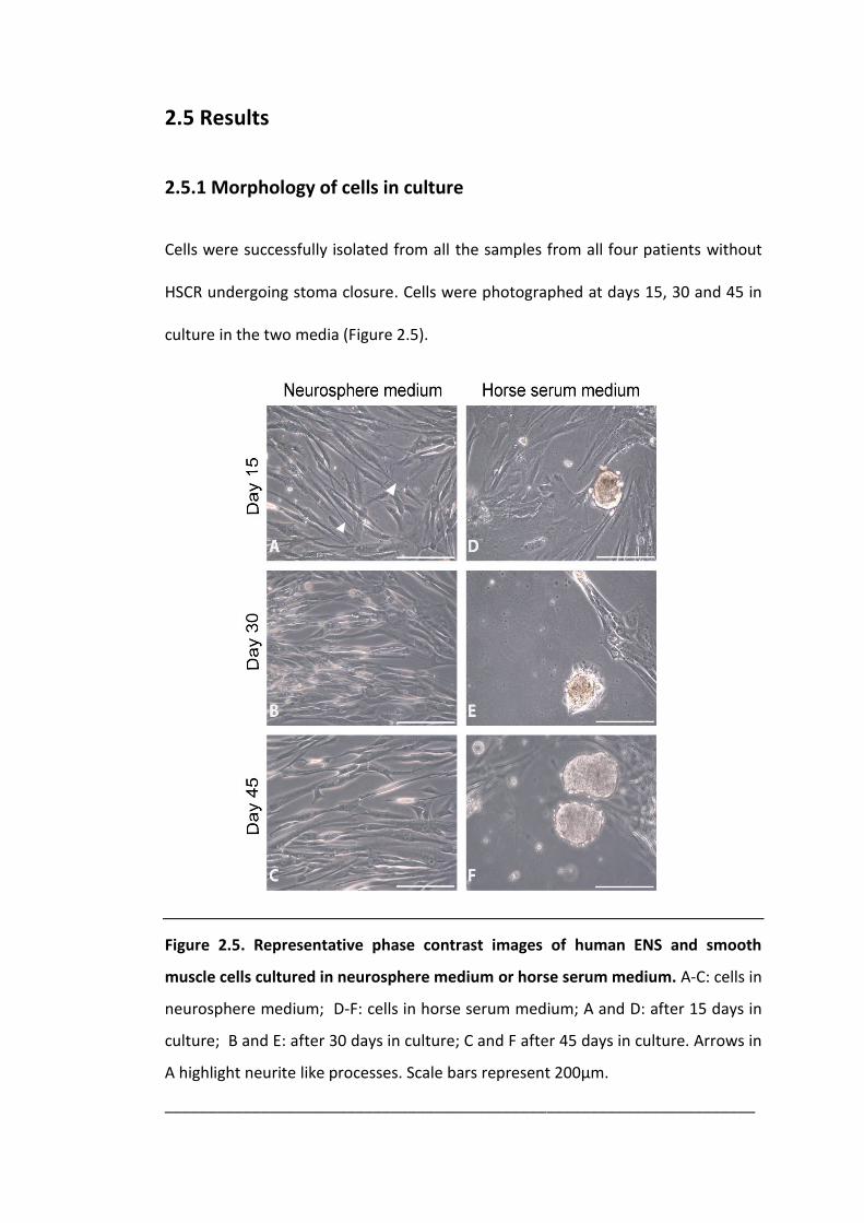
2.5 Results
2.5.1 Morphology of cells in culture
Cells were successfully isolated from all the samples from all four patients without
HSCR undergoing stoma closure. Cells were photographed at days 15, 30 and 45 in
culture in the two media (Figure 2.5).
Figure 2.5. Representative phase contrast images of human ENS and smooth
muscle cells cultured in neurosphere medium or horse serum medium. A-C: cells in
neurosphere medium; D-F: cells in horse serum medium; A and D: after 15 days in
culture; B and E: after 30 days in culture; C and F after 45 days in culture. Arrows in
A highlight neurite like processes. Scale bars represent 200µm.
____________________________________________________________________

It is evident in the neurosphere medium that the morphology of the cells in culture
do not vary between each time point (Figure 2.5 A-C). In the horse serum medium,
the size of the neurosphere like structures appears to increase along with the
number of them as time in culture increases (Figure 2.5 D-F). This is consistent with
previous work completed by our group.(Almond et al. 2007)
There are obvious differences between the morphologies of the cells in the
neurosphere medium compared to the horse serum media. In the neurosphere
medium, cells are flat and elongated or spindle-shaped and there are also some
neurite like processes present in the culture (see arrows in Figure 2.5 A). However,
in the horse serum medium there are neurosphere like structures that have budded
off from the flat cells on the surface of the dish. Initially they are attached to the flat
cells on the surface of the dish (Figure 2.5 D) but then detach and are suspended in
the medium (Figure 2.5 F). It should also be noted that the numbers of cells visible
on the surface of the dish decreases as the number of neurospheres increases.

2.5.2 P75 expression by cells in both media
Figure 2.6. Representative immunocytochemistry images of human ENS and
smooth muscle cells cultured in neurosphere medium or horse serum medium
labelled with the neural crest cell marker P75. Cell nuclei are labelled blue with
DAPI and P75 cell surface receptors are labelled green. A-D: cells in neurosphere
medium; E-H: cells in horse serum medium; A and E: after overnight culture; B and
F: after 15 days in culture; C and G: after 30 days in culture; D and H: after 45 days
in culture. Scale bars represent 50µm.

After overnight culture cells expressing P75 within the isolated cells have not
formed neurosphere like structures or elongated cells bearing the morphology of
neurons (Figure 2.6 A and E). The cells cultured in the neurosphere medium
expressing P75 are flat with long neurite projections to other expressing cells
(Figure 2.6 B-D). After 45 days in neurosphere medium the cells appear more
concentrated despite being seeded in equal number on the chamber slide, the cells
expressing P75 have not formed neurospheres and long neurites are visible (Figure
2.6 D). The cells in horse serum media have formed dense clusters containing cells
expressing P75, these appear to be early neurospheres (Figure 2.6 E-G).
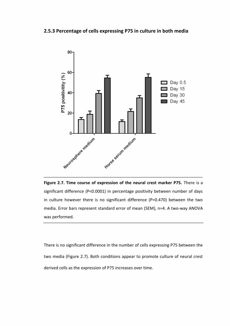
2.5.3 Percentage of cells expressing P75 in culture in both media
Figure 2.7. Time course of expression of the neural crest marker P75. There is a
significant difference (P<0.0001) in percentage positivity between number of days
in culture however there is no significant difference (P=0.470) between the two
media. Error bars represent standard error of mean (SEM), n=4. A two-way ANOVA
was performed.
There is no significant difference in the number of cells expressing P75 between the
two media (Figure 2.7). Both conditions appear to promote culture of neural crest
derived cells as the expression of P75 increases over time.

2.5.4 Number of cells present in culture at different time points
Figure 2.8. Percentage change in total cell numbers in culture between passages.
Each passage occurred every 15 days. Note a percentage change is shown rather
than total numbers as the initial number of cells dissociated varies substantially.
There is a significant difference (P <0.0001) in the change in total number of cells in
culture over time however there is no significant difference (P=0.275) between the
two media. Error bars represent SEM, n=4. A two-way ANOVA was performed.
The change in number of cells since the last passage was calculated instead of total
numbers as there was large variation in the number of cells isolated mainly due to
the quality of sample obtained. Note the day 30 data relates to the change from day
15. Data for the change in numbers between day 30 and day 45 have not been
included as unfortunately culture contamination meant that some of the cultures

were discarded. During the first 15 days in culture there is a similar reduction in the
number of living cells present in the two culture conditions however between day
15 and day 30 there was a mean increase in the number of cells present in both
media (Figure 2.8). There is a moderately large SEM however there is no statistical
significance between the two media. Combined with the data on percentage of cells
expressing P75 (Figure 2.7) this shows that the mean number of these cells present
is similar in each culture.
2.5.5 P75 expression within neurospheres in horse serum medium
Figure 2.9. Immunohistochemistry of a representative section of a neurosphere
removed from horse serum media culture stained with the NCC marker P75. Cell
nuclei are labelled blue with DAPI and P75 cell surface receptors are labelled green.
Arrows highlight neurite like processes. Scale bar represents 100µm.

The neurosphere like structures seen in horse serum media are neurospheres as
they are shown to contain cells which label with P75 which has been previously
shown to identify ENSPC and their neuronal and glial progeny (Figure 2.9). There are
fine neurite processes present emerging from the periphery of the neurosphere and
also within the neurosphere too (see arrows in Figure 2.9).
2.5.6 ENSPC differentiation in both media
Figure 2.10. Representative immunocytochemistry images of human ENS and
smooth muscle cells cultured for 45 days in neurosphere medium or horse serum
medium labelled with the immature and mature neuronal markers Tuj and NOS
respectively. Cell nuclei are labelled blue with DAPI, cells expressing Tuj are green
and cells expressing NOS are red. A: neurosphere medium; B horse serum medium.
Scale bars represent 50µm.
Although previous experience by our group with P75 has shown that the majority of
cells dual label with P75 and neuronal markers after time in culture, it is not clear so
far in these experiments whether cells in both conditions were differentiating as
well as proliferating. Therefore neuronal markers were also used to look for the
presence of neurons in the samples. Neuronal cells in neurosphere medium appear
elongated and fine neurites are visible (Figure 2.10 A) whereas neuronal cells in
horse serum medium are clustered into what appears to be an early neurosphere
(Figure 2.10 B). Most importantly in both media, there are not only immature
neurons labelled with Tuj but also mature neurons labelled with NOS.

2.6 Discussion
In the literature, ENS and other neurospheres have mainly been derived and
cultured under non adherent condition.(Almond et al. 2007; Lindley et al. 2008;
Reynolds & Weiss 1992) However, it has been reported that a greater yield of
neurospheres can be achieved using adherent culture dishes in CNS
neurospheres.(Konagaya et al. 2011, 2013) As the use of autologous neurospheres
as a therapy for HSCR is being explored, adherent culture conditions have been
favoured by our group over recent years.(Wilkinson, Edgar & Kenny 2012)
Additionally, an advantage of using adherent conditions is that there is no need to
repeatedly passage neurospheres to prevent them adhering to the dish surface
which has been required in previous work by our group. (Almond et al. 2007) The
work in this chapter has therefore sought the optimal medium to culture ENS
neurospheres under adherent conditions by comparing the two media previously
used by our group.
2.6.1 Both media produce a similar number of ENSPC
It is clear from the mean percentage of P75 expression and the mean number of
cells in both culture conditions at different time points that each medium produces
a similar proportion and total number of neural crest derived cells. It has been well
documented that ENS neurons and glia both express the NCC marker P75,
consistent with the notion that this NCC marker also identifies ENSPC.(Joseph et al.
2011; Lindley et al. 2009) Here it is evident that both media promote differentiation

of ENSPC into neurons which is an essential property of any medium to culture cells
for autologous stem cell therapies in HSCR.(Wilkinson, Edgar & Kenny 2012)
Therefore, in terms of culture of ENSPC it would seem that there is no difference
between the two media.
2.6.2 Horse serum medium promotes neurosphere formation
The main difference between the two media is the morphology of the cells cultured
in them. After 12 hours in culture the appearance of cells expressing P75 is similar
suggesting that either these cells are yet to recover from the stresses of tissue
dissociation or these cells are ENSPC prior to differentiation. Following 15 days in
culture it is clear that horse serum medium promotes the formation of
neurospheres containing ENSPC whereas neurosphere medium results in a flat
appearance of the cells with long neurite projections. These findings sound
paradoxical as neurosphere medium does optimally promote neurosphere
development, however our work has used adherent conditions as opposed to non
adherent conditions where neurosphere generation has been successful.(Almond et
al. 2007; Lindley et al. 2009) As mentioned, there is evidence for the use of
adherent conditions to generate neurospheres but in order to confirm these
findings with ENS neurospheres further study could compare the two media under
adherent and non adherent conditions.(Konagaya et al. 2011, 2013)

In the horse serum medium where neurospheres formed, phase contrast imaging
showed that neurospheres began to form on the surface of the dish before
enlarging in size and finally budding off to become suspended in the medium. This
raises the question of whether one single ENSPC proliferates to form the
neurosphere or if multiple cells adhere to form the structure together. Some study
has already taken place to answer this question which found that ENS neurospheres
are multipotent but this doesn’t address whether all neural crest derived cells in a
neurosphere originate from one ENSPC.(Almond et al. 2007) In order to understand
the formation of ENS neurospheres further work should look at these neurospheres
forming possibly by utilising live cell imaging with immunofluorescence of ENSPC
with P75 antibodies. It was also noticed that neurospheres were greater in size after
a longer time in horse serum medium cultures, this would suggest that more cells
are present however it is not clear as to whether there is any advantage in having
larger neurospheres when considering autologous implantation. It is logical that an
increased number of cells will have greater potential in the number of ENS neurons
and ganglia that are formed however as a neurosphere becomes larger questions
are raised about the ability of oxygen and nutrients to diffuse to the centre of these
structures. If diffusion is not possible past a certain size then it is possible that
central cells apoptose leaving a hollow structure.
2.6.3 Presence of non neural crest derived cells within neurospheres
In both media it is clear that there are non neural crest derived cells present. The
majority of these cells in primary ENS neurospheres have been shown to be smooth
muscle cells in previous work from our group.(Wilkinson et al. 2013) The ratio of
neural crest derived cells to smooth muscle cells increases from generation to
generation of neurosphere because the culture conditions favour cells of the neural
crest lineage.(Lindley et al. 2009) It is not clear however whether smooth muscle
cells apoptose or neural crest derived cells proliferate at a much greater rate than
other cells. It is also unknown what role, if any, these smooth muscle cells have
within the ENS neurosphere. A clonal NCC can proliferate and differentiate into cells
expressing smooth muscle actin as well as cells expressing neuronal markers, which
may explain the origin of smooth muscles cells in ENS neurospheres.(Morrison et al.
2000) A possibility for future work would be to attempt to culture purified
neurospheres as it would be safer to implant one cell type instead of many.
2.6.4 Further optimisation of media
The major differences in the horse serum medium is the larger amount of serum
and glucose present however it is not clear why this promotes the formation of
neurospheres. It is clear that the components of the neurosphere medium are
effective at culturing ENSPC. The question has to be asked as to whether some of
these components should be added to the horse serum medium. However to
undertake experiments to determine whether there is an advantage to developing a
new medium a large number of cells and time will be required as many components
at multiple concentrations will have to be tried.

It should also be noted that neurosphere medium has more components meaning
that it is more expensive and leaves more room for variability between batches
depending on where the components have been sourced from and when, which is
another reason why horse serum medium is favourable. Before human implantation
of ENS neurospheres can take place a serum free medium will have to be developed
due to cross species infection risk which represents a challenge for the future.
2.7 Conclusion
It is clear that horse serum medium with adherent conditions should be used for all
future work involving the culture of human ENS neurospheres before human trials.
There may be some advantage in adding components such as growth factors to the
horse serum medium to create a new medium however these are areas for future
work and are outside of the work achievable in this thesis. It must also be
considered that there are advantages to using a simpler medium which will be
easier to adapt to serum free, as there is hope that this work will culminate in
clinical trials in the future.

Chapter 3 – Thickened nerve trunks as a source of
ENSPC in aganglionic HSCR gut
3.1 Overview
This chapter confirms the previous observation that it is possible to isolate ENSPC
from aganglionic short and long segment HSCR gut where there are thickened nerve
trunks present. However it is demonstrated here that it is not possible to obtain
ENSPC from aganglionic total colonic and total intestinal HSCR gut in which there
are no thickened nerve trunks. The implication of this finding is discussed.
3.2 Introduction
As described in chapter one, preliminary work from our group has shown that
ENSPC can be isolated from aganglionic gut in short and long segment HSCR. These
ENSPC then form mature neurons and ganglia of the ENS. There is however no data
from patients with the rarer total colonic and total intestinal variants of HSCR. This
information would be interesting as it is well documented that thickened nerve
trunks are present within aganglionic gut in patients with short and long segment
disease but these trunks are absent in total colonic and total intestinal
disease.(Kobayashi, O'Briain & Puri 1994b; Solari, Piotrowska & Puri 2003). Thus,
observations on aganglionic total colonic and total intestinal HSCR gut tissue may
aid our identification of the source of ENSPC in aganglionic gut. Thickened nerve
trunks are hypertrophied extrinsic nerves originating from the pelvic plexus.
Normally, extrinsic nerve fibres consist of bundles of axons which are encased by
Schwann cells. There are many of these bundles in nerves which are contained
within fibrous layers, which are the endoneurium, epineurium and perineurium
(Diagram 3.1). Immunohistochemistry of tissue sections of aganglionic HSCR gut
using the NCC marker P75 by others and by our group have demonstrated high
levels of immunoreactivity in cells at the periphery of the nerve fibre, which in a
normal nerve would be the fibrous perineurium (Figure 3.2).(Kobayashi, O'Briain &
Puri 1994b; Wilkinson et al. 2013)

Diagram 3.1. Structure of a typical extrinsic nerve. The nerve is encased by the
perineurium (P) consisting of perineurial cells. Axons (grey circles) are wrapped by
myelinated Schwann cells (S) and non-myelinated Schwann cells (N). Within the
endoneurium (E) are endoneurial fibroblasts (F). Adapted from Joseph et al 2004.
Figure 3.2. Thickened nerve trunk expressing P75 in aganglionic short segment
HSCR gut. The most intense expression is at the periphery of the nerve trunk but
there is lower levels of expression within the trunk corresponding to the location of
endoneurial compartments. Scale bar represents 50µm.

The hypothesis in this chapter is that the source of ENSPC obtained from
aganglionic gut are the thickened nerve trunks. This is proposed as ENSPC in vitro
express the NCC marker along with the thickened nerve trunks in vivo. As it is
known that total colonic and total intestinal aganglionic gut do not have thickened
nerve trunks observations on this tissue presents the possibility of testing this
hypothesis.(Kobayashi, O'Briain & Puri 1994b; Solari, Piotrowska & Puri 2003) To
this end, aganglionic gut samples were obtained from patients with different
variants of HSCR, as confirmed by a consultant histopathologist, in order to attempt
to isolate and culture ENSPC. The success or failure of this is then correlated with
the presence or absence of thickened nerve trunks.
3.3 Aim
To establish whether thickened nerve trunks are the source of ENSPC expressing the
NCC marker P75 in aganglionic HSCR gut.

3.4 Methods
Cells from the muscular layers of aganglionic HSCR gut were dissociated and
cultured with horse serum medium as described in chapter 2. It should be noted
that samples from patients with total colonic and total intestinal disease were
obtained at the site of stoma formation which was proximal jejunum in total
intestinal disease and distal ileum in total colonic disease.
3.4.1 Hospital histology reports
Histology reports were produced by the Histopathology department at Alder Hey
Children’s Hospital where the samples were obtained and accessed electronically.
The presence or absence of thickened nerve trunks, along with the extent of
disease, was noted.

3.4.2 Details of aganglionic samples used
Breakdown of samples
Patient ID Pathology
H020 HSCR – total colonic
H021 HSCR – short segment
H022 HSCR – short segment
H023 HSCR – short segment
H024 HSCR – long segment
H025 HSCR – total intestinal
H027 HSCR – short segment
H028 HSCR – short segment
H029 HSCR – short segment
H030 HSCR – short segment
H031 HSCR – short segment
H032 HSCR – short segment
H033 HSCR – short segment
H034 HSCR – short segment
H035 HSCR – short segment
Table 3.3. Details of samples used in experiments in this chapter
Summary of samples obtained
Pathology Number of samples
HSCR – short segment 12
HSCR – long segment 1
HSCR – total colonic 1
HSCR – total intestinal 1
Table 3.4. Summary of samples used in experiments in this chapter

3.4.3 Immunofluorescence using chamber well slides
This took place as described in section 2.4.2.5. The primary antibody used was P75
(Abcam) and the secondary was Alexa Fluor goat anti-mouse 488 (Life Technologies)
at the concentration specified in section 2.4.2.6 and 2.4.2.7. Negative controls
consisting of immunocytochemistry with Alexa Fluor 488 secondary antibodies but
no primary antibodies was performed for all samples.

3.5 Results
3.5.1 Histopathology reports on aganglionic samples obtained
Histopathology reports found aganglionosis in all samples along with a presence or
absence of thickened nerve trunks as detailed in table 3.5. Results from all levels of
aganglionic biopsies was looked at to determine thickened nerve trunk presence.
Pathology reports for samples used
Patient ID Pathology Thickened nerve trunks?
H020 HSCR – total colonic No
H021 HSCR – short segment Yes
H022 HSCR – short segment Yes
H023 HSCR – short segment Yes
H024 HSCR – long segment Few
H025 HSCR – total intestinal No
H027 HSCR – short segment Yes
H028 HSCR – short segment Yes
H029 HSCR – short segment Yes
H030 HSCR – short segment Yes
H031 HSCR – short segment Yes
H032 HSCR – short segment Yes
H033 HSCR – short segment Yes
H034 HSCR – short segment Yes
H035 HSCR – short segment Yes
Table 3.5. Details of the extent of HSCR and presence of thickened nerve trunks in
the aganglionic gut sample as determined by a consultant histopathologist for
each patient

3.5.2 P75 expression by cells obtained from aganglionic HSCR gut after
0.5 days in culture
Cells were successfully obtained from all samples detailed above and
immunocytochemistry was performed after overnight culture (Figure 3.6). In terms
of cell yield, there was no difference between samples noticed.
Figure 3.6. Phase contrast and immunocytochemistry of cells from aganglionic
HSCR gut after overnight culture stained with P75 antibodies. A-C: short segment
HSCR; D-F: total colonic HSCR; G-I: negative control consisting of short segment
HSCR gut with only secondary antibodies. A, D, G: phase contrast; B, E, H: cell nuclei
identified by DAPI (blue); C, F, I; neural crest derived cells expressing P75 (green).
Scale bars represent 50µm.

Cells were present in both samples as shown by phase contrast imaging and
identification of cell nuclei with DAPI (Figure 3.6 A and D, B and E). However P75
positive neural crest derived cells were only present in aganglionic gut from short
and long segment HSCR (Figure 3.6 C) but such cells were not detected from the
samples from patients with total colonic or total intestinal HSCR (Figure 3.6 F). Also,
the specificity of the secondary antibody is shown confirming the positive finding
(Figure 3.6 I). The percentage of P75 expression and the number of samples tested
in shown in Figure 3.8.

3.5.3 P75 expression by cells obtained from aganglionic HSCR gut after
10 days in culture
Figure 3.7. Phase contrast and immunocytochemistry of cells from aganglionic gut
after 10 days in culture stained with P75 antibodies. A-C: short segment HSCR; D-F:
total colonic HSCR; G-I: negative control consisting of short segment HSCR gut with
only secondary antibodies. A, D, G: phase contrast; B, E, H: cell nuclei identified by
DAPI (blue); C, F, I; neural crest derived cells expressing P75 (green). Scale bars
represent 50µm.
After 10 days culture the presence of cells was again confirmed with phase contrast
imaging and identification of cell nuclei (Figure 3.7 A and D, B and E). Cells
expressing P75 were only identified in aganglionic short and long segment HSCR gut

(Figure 3.7 C) and there was no expression of this marker detected amongst cells
obtained from aganglionic total colonic or total intestinal HSCR gut (Figure 3.7 F). As
at day 0, a negative control was performed which confirms the reliability of the
method and the results (Figure 3.7 I). It should also be noted that the percentage of
cells expressing P75 increased after time in culture, this is quantified in Figure 3.8.
3.5.4 Percentage of P75 expression in aganglionic samples
The percentage of cells expressing P75 for each variant of HSCR at each time point
was calculated (Table 3.8).
Mean P75 expression at day 0.5 and day 10 amongst the different extents of HSCR
Pathology Number of
patients
Thickened
nerve trunks?
P75
expression
day 0.5
P75
expression
day 10
Short segment 12 Yes 10.0% +/- 1.9
SEM
22.7% +/- 2.9
SEM
Long segment 1 Few 11.1% 23.1%
Total colonic 1 No 0% 0%
Total intestinal 1 No 0% 0%
Figure 3.8. Mean expression of P75, thickened nerve trunk presence and number
of samples tested for each variant of HSCR within the aganglionic gut. The mean is
shown along with the SEM for short segment samples. In the other variants of HSCR
there is one sample of each so a mean cannot be calculated.

Figure 3.6, 3.7 and 3.8 show that it was possible to isolate and culture cells
expressing P75 without fail from aganglionic short and long segment HSCR gut
whereas it was not possible to do this from aganglionic total colonic or total
intestinal HSCR gut. As well as observing this link the percentage of cells expressing
P75 in culture from aganglionic short and long segment gut increased over time in
keeping with previous work by the group.(Wilkinson et al. 2013)

3.6 Discussion
3.6.1 Aganglionic ENSPC are associated with thickened nerve trunks
It was possible to identify P75 positive neural crest derived cells in the dissociates
obtained from aganglionic gut in short and long segment HSCR where also
thickened nerve trunks were present. However this was not achievable in samples
from the 2 patients with total colonic and total intestinal HSCR where these
thickened nerve trunks were absent. As our group has previously shown that
mature enteric neurons and glia in vitro express P75, this fits with the hypothesis
that the ENSPC which can be isolated from aganglionic gut in short and long
segment disease are derived from the thickened nerve trunks.(Wilkinson et al.
2013) The correlation between the presence of thickened nerve trunks in short and
long segment HSCR and the absence of these in total colonic and total intestinal
HSCR in our patients is consistent with reports from other groups.(Kobayashi,
O'Briain & Puri 1994b; Solari, Piotrowska & Puri 2003)
In the aganglionic gut obtained from the patient with long segment HSCR, few
thickened nerve trunks were seen on histology. There is an obvious limitation with
this classification which relies on the observations of the histopathologist and no
quantitative evidence. However, in this sample a similar percentage of cells
expressing P75 compared to aganglionic gut from short segment HSCR was
observed at both time points. With only one patient it is not possible to draw a
conclusion from this result and repeats with more samples are required. A method

of quantifying the number of thickened nerve trunks present should also be
considered as this would allow the observation of a dose response relationship
between the number of thickened nerve trunks and the percentage of P75
expression in the tissue. This would ultimately strengthen the results in this chapter.
3.6.2 P75 expression increases with time in culture
It has also been shown in this experiment that if ENSPC were not identified in the
single cell suspensions at isolation they are also not present after up to 10 days in
culture. This may dismiss the possibility that when ENSPC are present in culture
they are derived from non neural crest derived cells, such as reprogrammed
mesenchymal cells, which are displaying a neural phenotype after time in
culture.(Mareschi et al. 2006) It should also be noted that where cells expressing
P75 were isolated, the percentage of cells expressing this marker increased after 10
days in culture. This is likely due to the conditions used favouring the survival and
proliferation of cells of NCC lineage whereas cells of non NCC lineage fail to survive
in these conditions. This is in keeping with previous work by our group and is
promising as it shows that when these cells are placed in the culture conditions
used the percentage of these cells increases.(Almond et al. 2007; Lindley et al.
2008; Lindley et al. 2009) This is important when considering the culture and
implantation of human ENSPC into HSCR patients.(Wilkinson, Edgar & Kenny 2012)

3.6.3 Limitations
The limitation of this work is that there was only one sample from each of the total
colonic and total intestinal variants of HSCR which was investigated. This problem
comes with any work looking at very rare disease and in the paediatric centre where
our samples are obtained around one case of this disease is seen per year. Future
work should therefore aim to repeat this work to confirm these findings. One way
to increase the availability of these rare samples would be to create a national
network where consent was applicable nationwide and human samples could be
obtained.
A further limitation of this study is that the site the aganglionic sample is taken from
differs between patients depending on the extent of disease. There is little known
about differences between proximal and distal aganglionic gut in total colonic and
total intestinal patients. However, biopsies were obtained from multiple levels of
the gut in these patients including the rectum which was reported in both cases as
aganglionic with absent thickened nerve trunks.
3.6.4 Aganglionic short and long segment HSCR gut can be used for
stem cell retrieval
Previously it was observed in six patients with short segment HSCR and one long
segment HSCR that ENSPC were obtainable from the aganglionic gut.(Wilkinson et

al. 2013) This has now been repeated successfully with a further twelve short
segment and one long segment aganglionic HSCR samples. This is consistent with
work which found that ENSPC can arise from a niche external to the ENS ganglia.(Liu
et al. 2009) This added evidence strengthens the possibility that autologous ENSPC
implantation will be achievable using the aganglionic gut removed at operation for
HSCR patients with short and long segment disease. This would of course allow any
stem cell treatment to be much more practical and involve less cell number
expansion before implantation compared to if ganglionic gut was required for
ENSPC culture. Unfortunately in the rarer variants of disease ganglionic bowel will
need to be sourced to obtain the progenitor cells.
3.6.5 Revisiting the source of ENSPC obtainable from ganglionic HSCR
gut
If in aganglionic HSCR gut ENSPC are obtained from thickened nerve trunks it is not
clear why ENSPC from ganglionic gut have similar characteristics as described
previously.(Lindley et al. 2008; Lindley et al. 2009) This may be that the ENSPC are
sourced from non-hypertrophied extrinsic nerves in ganglionic gut however the
number of cells obtained from aganglionic gut should be greater than ganglionic
gut, which is not the case. The similarities observed between ganglionic and
aganglionic ENSPC may also lead towards glial including Schwann cells as a source of
ENSPC as glia are present within ganglia of the ENS and Schwann cells are present
within extrinsic nerves.(Joseph et al. 2011; Kalcheim & Rohrer 2014; Laranjeira et al.
2011)
3.6.6 Revisiting the aetiology of HSCR
The aetiology of HSCR is unknown however from small mammal studies it can be
said that the most likely cause is failure of migration of ENCC.(Obermayr et al. 2013)
If ENSPC that can be obtained from aganglionic HSCR gut are present within the
thickened nerve trunks it seems more likely that ENCC have failed to migrate from
the vagal neural crest to the distal gut. It is less likely that ENCC have migrated to
the distal gut and failed to differentiate into enteric neurons and glia otherwise we
should be able to obtain ENSPC from aganglionic gut where thickened nerve trunks
are absent. However, this cannot be ruled out as there is work which shows that for
sacral NCC to enter the hind gut extrinsic nerve fibres must be present.(Erickson et
al. 2012) There is also the possibility that different variants of HSCR have different
aetiologies which is suggested by the histopathological differences seen in the
aganglionic tissue.
3.6.7 ENSPC from aganglionic HSCR may be of Schwann cell lineage
Cells of Schwann cell lineage are a possible source of ENSPC in aganglionic HSCR gut
from within thickened nerve trunks. It has been put forward by two groups that
ENSPC are most likely of glial origin when obtained from post natal gut and

Schwann cells are a type of glial cell in the peripheral nervous system.(Joseph et al.
2011; Laranjeira et al. 2011) In addition it has also been possible to generate
peripheral nervous system neurospheres from Schwann cells obtained for mouse
and rat sciatic nerve.(Martin et al. 2012) Most significantly migrating NCC have been
shown to dual label with SOX10 and PHOX2B showing that in the autonomic
nervous system Schwann cells precursors also have the ability to undergo
neurogenesis and hence have neuronal progenitor characteristics.(Kalcheim &
Rohrer 2014) However, it has been shown that sacral NCC migrate into the hindgut
with extrinsic nerve fibres so it is possible that ENSPC from aganglionic gut are NCC
that have failed to differentiate and remain in progenitor form.(Erickson et al. 2012)
This theory coincides with the finding that it is only possible to obtain ENSPC from
aganglionic gut where thickened nerve trunks have formed.
To fully explore the possibility of Schwann cells as a source of ENSPC further work
should look at the characterisation of aganglionic HSCR tissue sections with a glial
marker and expression of this marker should be looked for amongst cells isolated
from aganglionic gut. The hypothesis would be that P75 positive cells co stain with a
glial marker such as S100.(Hirose, Sano & Hizawa 1986) This could be evaluated
using fluorescence activated cell sorting and is an area for further work.
3.6.8 The potential of a medical treatment for HSCR
This work also raises the question as to why ENSPC fail to leave the thickened nerve
trunks in vivo and develop into ganglia of the ENS. If this can be understood there is
the possibility of therapeutic stimulation of these cells in vivo in HSCR patients to
differentiate into a ENS and restore functionality in the aganglionic gut. This would
treat HSCR without the need for surgical intervention which has substantial benefits
for patients.
3.7 Conclusion
The work in this chapter has raised several key questions that should be a focus of
further work. Most importantly the cell which is the source of ENSPC needs to be
identified in order to develop an autologous stem cell transplant for HSCR patients
which is as safe and efficacious as possible. Of equal importance the possibility of
stimulating ENSPC in vivo to differentiate into a functional ENS should be explored
to exclude the need of surgical intervention in these patients.

Chapter 4 – Are ENSPC obtained from aganglionic
HSCR gut of neural crest lineage?
4.1 Overview
This chapter utilises fluorescence activated cell sorting (FACS) to show that neural
crest derived cells obtained from aganglionic gut are the source of enteric neurons
in vitro and are therefore ENSPC. This not only adds to our understanding of the
origin of ENSPC that can be isolated from aganglionic HSCR gut but also raises
questions on whether neural crest derived cells can be stimulated in situ to
proliferate and differentiate into enteric neurons and glia in HSCR patients.

4.2 Introduction
It is clear from work by us and others that the thickened nerve trunks present in
aganglionic HSCR gut contain cells of neural crest origin.(Kobayashi et al. 1994;
Kobayashi, O'Briain & Puri 1994a; Wilkinson et al. 2013) Our group has shown that
cells expressing markers for enteric neurons and glia cultured from aganglionic
HSCR gut dual stain with the NCC marker, P75.(Wilkinson et al. 2013) Also, results in
chapter 3 show that it was not possible to obtain ENSPC from aganglionic gut where
thickened nerve trunks are absent and these results suggest that ENSPC are sourced
from the thickened nerve trunks in aganglionic gut. Taking this into account, it is
logical to assume that cells expressing the P75 NCC marker in culture are the same
cells which label with P75 in the thickened nerve trunks within tissue sections in
aganglionic gut, however there is a possibility that these cells derive from
reprogramming of other isolated cells. These cells could be reprogrammed
mesenchymal cells for example that behave as ENSPC in vitro.(Mareschi et al. 2006)
Subsequently, there is evidence which shows that mature interstitial cells of Cajal
have the ability to act as fibroblast like cell progenitors and therefore it must be
considered whether they also have the capability to function as neural
progenitors.(Huizinga & White 2008)
In regards to a therapy for HSCR, it could be argued that it is irrelevant to know
which cells are ENSPC as long as they are obtainable from aganglionic HSCR gut and
differentiate into mature enteric neurons and glia. However, if more is understood

about the origin and characteristics of ENSPC it is more likely that it will be possible
to understand how to control these cells in vivo post transplantation so that a
functional ENS is generated without uncontrolled proliferation and tumour
formation. Also, with further information about these cells, it is more likely that it
will be possible to stimulate ENSPC in vivo to proliferate and differentiate in
aganglionic gut without the need for stem cell retrieval. With this information, there
is the possibility of developing a therapeutic treatment for HSCR and hence surgical
intervention could be avoided.
It is hypothesised that a sub-population of cells expressing the P75 NCC marker
separated immediately from other cells dissociated from aganglionic gut in HSCR
patients will differentiate in culture into enteric neurons.
FACS has been used previously to sort cells obtained from rat sciatic nerve and
normoganglionic embryonic mouse gut with the neural crest marker, P75, however
to our knowledge this marker has not been used to sort human cells.(Bixby et al.
2002) It is also unclear whether the sorted cells will survive the sorting process to
be cultured when the duration between surgical resection of the sample to culture
of the sorted cells is around 24 hours. Therefore, part of the results section details
the development of the technique used.

4.3 Aims
The first aim is to develop a technique that will allow FACS of freshly dissociated
cells that will give two sub-populations of cells that can be cultured. Once this is
achieved it can be used to determine whether ENSPC are primarily of neural crest
lineage or not.

4.4 Methods
4.4.1 Details of samples used (in chronological order)
Breakdown of samples used
Patient
ID
Pathology Ganglionic, Aganglionic or Both
gut used
H029 HSCR – short segment Aganglionic
H026 HSCR – total colonic Ganglionic
H030 HSCR – short segment Both
H030 HSCR – short segment Both
C015 Anorectal malformation Ganglionic
C016 Anorectal malformation Ganglionic
C017 Anorectal malformation Ganglionic
C018 Anorectal malformation Ganglionic
C016 Anorectal malformation Ganglionic
H031 HSCR – short segment Both
H032 HSCR – short segment Both
C018 Anorectal malformation Both
H033 HSCR – short segment Both
H034 HSCR – short segment Both
H035 HSCR – short segment Both
Table 4.1. Details of samples used for experiments in this chapter. Gut samples
from 15 patients were used in total. 9 of these patients had HSCR and 6 had
anorectal malformations. Where applicable the innervation status of the samples
used, as confirmed by a consultant histopathologist, is also specified.

4.4.2 Obtaining cultured cells
To develop the labelling and sorting technique human gut cells which included
ENSPC which had already been cultured for around 10 days were used. A pellet of
cells was obtained using the technique for obtaining a cell pellet for
immunocytochemistry in section 2.4.2.5 however 0.05% (w/v) Trypsin was replaced
with 0.2% Collagenase IV (Gibco®) as P75 labelling and detection with FACS was
unsuccessful when Trypsin was used. This is likely to Trypsin damaging the cell
surface receptors.
4.4.3 Tissue dissociation
It was found that it was not possible to label and identify a separate subpopulation
of NCC using the tissue dissociation technique used in other chapters of this thesis
(section 2.4.2.3). This problem was likely due to the combination of the enzymes
and time spent in the enzymes damaging or removing the P75 cell surface receptor
from NCC. A technique published by another group was therefore modified and
tested which allowed the identification of a sub-population of NCC after tissue
dissociation.(Bixby et al. 2002) Thus the technique as described in section 2.4.2.3
was modified as follows: 1% w/v Dispase was removed from the protocol leaving 1%
w/v Collagenase IV as the only enzyme used. Tissue was left in the enzyme solution
for 90 minutes with trituration through a 5ml pipette ten times every 15 minutes.
This is a reduced duration compared to the original tissue dissociation method

where tissue was left in enzyme solution for between 2 and 3 hours depending on
dissociation progress as assessed using an inverted microscope and a 10µl sample.
4.4.4 Labelling of the NCC sub-population
The cell suspension was transferred to ice-cold FACS buffer (PBS supplemented with
1% w/v bovine serum albumin, penicillin (100units/ml) and 0.1% w/v streptomycin).
After 20 min, 200,000 cell aliquots were re-suspended in 200µl FACS buffer
containing 1:1000 mouse anti normal mouse IgG antibody (Santa Cruz) for the
negative controls or 5 million cells in 1:1000 mouse anti-P75 antibody (Abcam) for
FACS selection. After 30 minutes on ice, 800µl of ice-cold FACS buffer was added
and cell aliquots were centrifuged and re-suspended in 200µl FACS buffer with
1:1000 Alexa Fluor 488 goat anti-mouse secondary antibody (Life technologies).
After 45 minutes on ice, 800µl of ice-cold FACS buffer was added and the cells were
pelleted, re-suspended in 1ml FACS buffer and passed through a 70µm cell strainer.
4.4.5 Immunocytochemistry
The effectiveness of cell labelling was evaluated by also undertaking
immunocytochemistry on 20,000 cells taken from the sample. The technique is as
described in section 2.4.2.5 using P75 primary antibodies and Alexa Flour 488
secondary antibodies where cells are cultured overnight before fixing.
4.4.6 Labelling of dead cells
Where mentioned in the results section dual staining of cells for FACS took place
with Sytox Orange (Life Technologies) dead cell stain. Following labelling of the NCC
sub-population as detailed above 1µl of Sytox Orange was added to the labelled
cells. The cells were left for 30 minutes before they were ready for FACS.
4.4.7 Florescence activated cell analysis
Florescence activated cell analysis took place using FACScalibur (BD Biosciences).
4.4.8 Florescence activated cell sorting
Fluorescence activated cell sorting took place using FACSaria (BD Biosciences). Cells
were sorted into 5ml polystyrene round bottom tubes (BD Biosciences) that
contained 1ml horse serum medium (DMEM high glucose (4.5% w/v) supplemented
with 20% v/v medium 199 (Gibco®), 7% v/v heat inactivated horse serum (Gibco®),
100 Units/ml penicillin and 100 μg/ml streptomycin). Filled tubes were then stored
on ice until the sort had finished.

4.4.9 Culture of sorted cells
Cells were initially cultured on adherent 24 well plates (Nalge Nunc, New York, USA)
in densities of 100,000, 200,000 and 500,000 in horse serum medium. However the
cells did not survive.
Subsequently, 8 well chamber slides (Thermo Scientific) were prepared by coating
with 10µg/cm2 Poly-D Lysine and 1µg/cm2 Laminin before washing in H2O and
allowing to air dry. 100,000 cells from the P75 positive and negative sorted
subpopulations were plated separately in each chamber in 500µl horse serum
medium and incubated at 37°C with 5% CO2 for 6 days. Immunocytochemistry was
performed as previously described with Tuj (Covance, Maidenhead, UK) antibodies
diluted 1:500 in IBB.
4.4.10 FACS software
Flowing Software (Flowingsoftware.com) was used to count the number of events
and produce graphs.

4.5 Results
4.5.1 Overview of samples used to develop method and test
hypothesis
Patient
ID
Pathology FACS
immediately
post tissue
dissociation?
Ganglionic
(G),
Aganglionic
(Ag) or
Both (B)
Successful
FACS analysis
with separate
P75
population?
Successful
sub-
population
culture?
H029 HSCR – short
segment
No Ag Yes Not
attempted
H026 HSCR – total
colonic
No G Yes Not
attempted
H030 HSCR – short
segment
Yes B No Not
attempted
H030 HSCR – short
segment
No B Yes No
C015 Anorectal
malformation
Yes G Yes Not
attempted
C016 Anorectal
malformation
Yes G Yes Not
attempted
C017 Anorectal
malformation
Yes G Yes Not
attempted
C018 Anorectal
malformation
Yes G Yes Not
attempted

Patient
ID
Pathology FACS
immediately
post tissue
dissociation?
Ganglionic
(G),
Aganglionic
(Ag) or
Both (B)
Successful
FACS analysis
with separate
P75
population?
Successful
sub-
population
culture?
C016 Anorectal
malformation
No G Yes No
H031 HSCR – short
segment
Yes B Yes No
H032 HSCR – short
segment
Yes B Yes No
C018 Anorectal
malformation
No B Yes No
H033 HSCR – short
segment
Yes B Yes Yes
H034 HSCR – short
segment
Yes B Yes Yes
H035 HSCR – short
segment
Yes B Yes Yes
Table 4.2. Table showing the results of experiments on each sample, underlining
whether it was possible to identify a P75 positive population and whether it was
possible to culture the sub-populations. Green rows indicate successful P75
positive cell identification and culture of sub-populations. Amber rows indicate
successful P75 positive cell identification without an attempt to culture the sub-
populations. Red rows indicate failure of P75 positive cell identification or failure of
culture of sub-populations.

4.5.2 Identification of P75 expression amongst cells taken from culture
using FACS
Initially, cells which had been obtained from short segment aganglionic HSCR gut (as
specified in Table 4.2) were taken after 10 days in culture and prepared for FACS
then analysed (Figure 4.3).
Figure 4.3. FACS analysis of cells cultured from short segment aganglionic HSCR
gut labelled with P75. A: represents a dot plot showing the separate population of
labelled cells. B: represents a overlaid histogram showing the separate population
of labelled cells. The grey peak is the negative control using isotype primary
antibodies and the clear peak is cells labelled with the P75 antibodies. The
population of P75 labelled cells is 21.5% of the total cells present.
It was possible to identify a population of cells labelled with the NCC marker P75 in
previously cultured cells from short segment aganglionic HSCR gut (Figure 4.3 A and

B). The percentage of cells expressing P75 is consistent with the number identified
using immunocytochemistry after the same duration in culture in chapter 3.
4.5.3 Evaluation of the P75 antibody when used with FACS
Following the results of the first experiment there was concern the positive finding
could be dead cells binding to the P75 antibody due to changes in membrane
permeability of dead cells which is well documented. Therefore to check that the
P75 antibody was not binding to dead cells Sytox Orange dead cell stain was used
with cultured cells.
Figure 4.4. FACS analysis of cells cultured from total colonic ganglionic HSCR gut
labelled with P75 and Sytox orange. A: represents a histogram showing cells
labelled with P75. The red peak is the negative control using isotype primary
antibodies and the clear peak is cells labelled with the P75 antibodies. The
population of P75 labelled cells is 18.1% of the total cells present. B: represents a
histogram showing cells labelled as dead with Sytox orange. The population of cells
labelled as dead is 21.9% of the total cells present. C: represents a histogram of only
cells labelled with P75 showing cells also labelled as dead with Sytox orange. The
majority of cells are alive.

The vast majority of cells labelled with P75 were alive (Figure 4.4 C). This finding
combined with the use an isotype primary antibody control is evidence that neural
crest derived cells are being labelled specifically, and can be maintained alive for
culture.
4.5.4 Attempts to identify freshly dissociated cells expressing P75
using FACS
Identification of a P75 labelled population was then attempted on freshly
dissociated cells from short segment aganglionic HSCR gut. The original tissue
dissociation technique detailed in section 2.4.2.3 using 1% (w/v) Collagenase IV and
1% (w/v) Dispase was used.
Figure 4.5. FACS analysis of cells freshly dissociated from short segment
aganglionic HSCR gut labelled with P75. A: represents a dot plot showing no
separate population of labelled cells. B: represents a histogram showing no
separate population of labelled cells. The grey peak is the negative control using
isotype primary antibodies and the clear peak is cells exposed to P75 antibodies.

It was not possible to label and identify a population of cells expressing P75 in
freshly dissociated cells (Figure 4.5 A and B). Immunocytochemistry was performed
on a sample of the cells which did identify cells expressing P75 at a percentage
consistent with findings in chapter 3 (not shown).
4.5.5 Identification and sorting of freshly dissociated cells expressing
P75 using FACS with culture and characterisation of sub-populations
As this technique works with cultured cells it is likely that the effects of the
dissociation process are preventing the P75 antibody binding to the cell surface
receptors on neural crest derived cells. The tissue dissociation was therefore
modified as described in the methods section in an attempt to retain the structure
of the cell surface receptors and allow labelling of freshly dissociated cells. This
technique was successful and sub-populations of cells were cultured and
immunocytochemistry was performed to characterise the sub-populations.

Figure 4.6. Fluorescence activated cell sorting of cells obtained from ganglionic
and aganglionic HSCR gut immediately after tissue dissociation and
characterisation of each population following 6 days in culture. A and B:
histograms of the FACS displaying P75 positive and negative selected
subpopulations. The grey peaks are the negative control using isotype primary
antibodies and the clear peak are cells labelled with the P75 antibodies. The arrow
in B highlights a second peak of P75 expression. C and D: Tuj immunofluorescence
staining of the P75 positive cell subpopulations from ganglionic and aganglionic
bowel respectively after 6 days culture (note that the majority of cells are Tuj
positive). E and F: Tuj immunofluorescence staining of the P75 negative cell
subpopulations from ganglionic and aganglionic bowel respectively (note the
absence of Tuj positive cells).

It was possible to label, identify and sort cells expressing P75 from cells freshly
dissociated from ganglionic and aganglionic short segment HSCR gut (Figure 4.6 A
and B). The histogram from aganglionic gut differs from ganglionic gut in the way
that there is a second peak of cells expressing P75 in the aganglionic sample but not
to such an intensity in terms of florescence as the majority of positively sorted cells
which are identified on the histograms (Figure 4.6 A-B, see arrow for second peak
on B).
Initially, it was attempted to culture the two sub-populations of cells on adherent
24 well plates as described in the methods section but neither population survived.
It is thought that this was due to the limited cell population size as trophic factors
secreted from cells would be much more dilute compared to culture conditions
used previously. The failure of survival may also have been due to the trauma that
the cells experienced in the 24 hour period between tissue dissociation and culture
with this FACS method. Therefore, laminin and poly-d-lysine coated chamber slides
were used (see methods) for culturing sorted cells. The positively sorted cells
survived but there were fewer cells from the negatively sorted sub-population
which survived in culture for 6 days. This is most likely due to the culture conditions
favouring cells of neural crest lineage. In order to show this is the case and that the
negative population is not just cell debris, this population was stained with the dead
cell stain, Sytox orange, immediately after sorting (Figure 4.7).

In both the ganglionic and aganglionic HSCR gut the positively stained sub-
populations expressed the neuronal marker Tuj after 6 days in culture (Figure 4.6 C
and D). The mean percentage of cells expressing Tuj are 89.3% and 85.4% (n=3) in
ganglionic and aganglionic gut respectively. In both the ganglionic and aganglionic
HSCR gut the negatively stained sub-populations did not express the neuronal
marker Tuj after 6 days in culture (Figure 4.6 E and F). A negative control consisting
of staining with only the secondary antibody was carried out for all sub-populations
(not shown).
Figure 4.7. FACS analysis of the P75 negative sub-population of cells immediately
after sorting from freshly dissociated cells from short segment aganglionic HSCR
gut labelled with Sytox orange, dead cell stain. A: represents a dot plot showing
the separate population of cells labelled as dead. B: represents a histogram showing
the separate population of cells labelled as dead. The grey peak represents a control
consisting of unlabelled cells whereas the white peak represents cells labelled with
the dead cell stain, Sytox orange. 5.6% of cells labelled as dead.

Of the cells sorted as not expressing P75 only 5.6% of the cells were dead
immediately after sorting (Figure 4.7 A and B). Once the cells were seeded onto a 8
well chamber slide for culture the majority of cells appeared dead and did not
adhere to the slide hence the majority of cells fail to take to culture conditions. A
much larger proportion of the cells in the P75 positive sub population survive in the
culture conditions used.

4.6 Discussion
4.6.1 ENSPC obtained from aganglionic HSCR gut are of neural crest
lineage
It has been shown that cells of neural crest lineage separated by FACS from other
cells immediately after dissociation from aganglionic short segment HSCR gut can
differentiate in vitro into neurons. This shows that P75 positive neural crest derived
cells in aganglionic gut are partially or wholly responsible for ENSPC and the mature
enteric neurons and glia which have been cultured from aganglionic short segment
HSCR gut previously.(Wilkinson et al. 2013) Moreover, the fact that it was not
possible to derive neurons from the population of cells not expressing P75 on
isolation suggests that there is no non neural crest lineage source of cultured
ENSPC.
4.6.2 The source of ENSPC in ganglionic HSCR gut
This finding questions the source of ENSPC which can be obtained from ganglionic
HSCR gut. Previously it was thought that these cells are sourced from the ganglia, as
this is the main source of neural cells in ganglionic gut.(Almond et al. 2007)
However, now ENSPC have now been shown to be present in both ganglionic and
aganglionic gut and also ENSPC cultured from each segment behave the same in
vitro and upon implantation into ex vivo aganglionic mouse gut, it is entirely
possible that ENSPC obtainable from ganglionic HSCR gut are extra-

ganglionic.(Wilkinson et al. 2013) It is therefore feasible that cells expressing P75
associated with the extrinsic nerves are the source of ENSPC in ganglionic HSCR gut.
4.6.3 The cellular origin of ENSPC in HSCR gut
As cells of non neural crest lineage did not give rise to neurons we can consider the
cellular origins of ENSPC in HSCR gut. It is possible that cells of Schwann cell lineage
are the source of ENSPC in aganglionic HSCR gut and either these cells of Schwann
cell lineage, associated with extrinsic nerves or other glial cells associated with the
ganglia, are the source in ganglionic HSCR gut. As discussed in chapter 3 it has been
found that Schwann cell precursors have dual roles as functional Schwann cells and
neuronal progenitors in the developing parasympathetic nervous system, which
may also be the case in HSCR gut.(Kalcheim & Rohrer 2014) Subsequently, it is
known that glial cells have a role in neurogenesis in vitro and also described by one
group in vivo.(Joseph et al. 2011; Laranjeira et al. 2011) A possible non glial source
of ENSPC are neuron cell bodies, which are present in ganglia but absent in extrinsic
nerves which only contain the axon.(Topp & Boyd 2006) This seems less likely
though due to common characteristics of ENSPC from ganglionic and aganglionic
gut.(Wilkinson et al. 2013) The other well documented NCC derivative in extrinsic
nerve trunks are fibroblasts within the endoneurium, however little is known about
the ability of these cells to undergo neurogenesis.(Joseph et al. 2004) The migration
of sacral NCC during development into the hindgut with extrinsic fibres has been
described.(Erickson et al. 2012) This is another possibility as to a source of ENSPC in
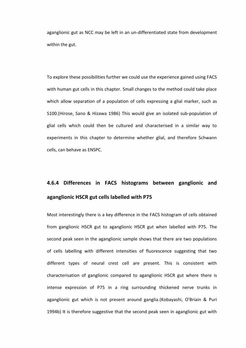
aganglionic gut as NCC may be left in an un-differentiated state from development
within the gut.
To explore these possibilities further we could use the experience gained using FACS
with human gut cells in this chapter. Small changes to the method could take place
which allow separation of a population of cells expressing a glial marker, such as
S100.(Hirose, Sano & Hizawa 1986) This would give an isolated sub-population of
glial cells which could then be cultured and characterised in a similar way to
experiments in this chapter to determine whether glial, and therefore Schwann
cells, can behave as ENSPC.
4.6.4 Differences in FACS histograms between ganglionic and
aganglionic HSCR gut cells labelled with P75
Most interestingly there is a key difference in the FACS histogram of cells obtained
from ganglionic HSCR gut to aganglionic HSCR gut when labelled with P75. The
second peak seen in the aganglionic sample shows that there are two populations
of cells labelling with different intensities of fluorescence suggesting that two
different types of neural crest cell are present. This is consistent with
characterisation of ganglionic compared to aganglionic HSCR gut where there is
intense expression of P75 in a ring surrounding thickened nerve trunks in
aganglionic gut which is not present around ganglia.(Kobayashi, O'Briain & Puri
1994b) It is therefore suggestive that the second peak seen in aganglionic gut with

FACS is from cells associated with the ring of intense P75 expression surrounding
thickened nerve trunks on immunohistochemistry. Further characterisation of these
nerve trunks is required however to confirm this finding. Further FACS could
separate the two different peaks into independent sub-populations and the ability
to differentiate into enteric neurons could then be tested. This would provide
evidence for the cell type which are acting as ENSPC in aganglionic gut. Again the
similarities between ENSPC from both ganglionic and aganglionic HSCR gut question
whether the cells with increased expression do have a role.(Wilkinson et al. 2013)
4.6.5 Implications of findings in this chapter
This work has several implications. Firstly, it has been shown that neural crest
derived cells are the source of ENSPC obtained from aganglionic HSCR gut. This will
allow further work to explore how NCC can be stimulated to differentiate and
proliferate into an ENS in vivo in HSCR patients. This information could be used to
develop a therapeutic treatment for HSCR. This would be even more promising if
the exact cell type could be established. Secondly, it should be established whether
neurospheres can be cultured from isolated cells of neural crest origin and if so
whether they have the same characteristics in terms of restoring contractility in ex
vivo aganglionic mouse gut as un-purified enteric neurospheres. Now that a cell
sorting technique has been developed this work could take place with the
hypothesis that a purified neural crest cell neurosphere will be more effective in
terms of restoring contractility as a neurosphere containing other cell types. Even, if
this is not the case there are likely to be major safety advantages to transplanted

one lineage of cells by excluding other cells such as smooth muscle cells when
considering a future stem cell therapy for HSCR.(Wilkinson, Edgar & Kenny 2012)
Finally, the finding that neural crest derived cells alone are responsible for the
ENSPC obtained from HSCR gut make it likely that in normal human development
NCC are the only progenitor for the ENS. Although this has been shown in several
mouse studies this adds further evidence to human ENS development and can be
used to direct further work exploring the aetiology of HSCR.(Obermayr et al. 2013)

4.7 Conclusion
This chapter has found that ENSPC are neural crest derived cells when obtained
from both ganglionic and aganglionic HSCR gut. This adds understanding to the
previous findings that ENSPC can be isolated from aganglionic HSCR gut and
questions the source of ENSPC in ganglionic HSCR gut. This work raises several
pertinent questions, but most importantly further work should look to identify the
cell type which behaves as ENSPC in aganglionic HSCR gut. Currently it is felt that
cells of Schwann cell lineage are responsible, however to develop a safe and
efficacious stem cell therapy, along with the possibility of a therapeutic treatment
for HSCR, this information needs to be known.
Chapter 5 – Characterisation of ganglionic and
aganglionic HSCR gut
5.1 Overview
This chapter builds on previous work characterising aganglionic HSCR gut and in
particular, the thickened nerve trunks. Full thickness gut was obtained from HSCR
patients and labelled with antibody markers for NCC, neurons, glia and surrounding
muscular tissue to detail the histology in both ganglionic and aganglionic HSCR gut.
Findings in this chapter add understanding to work carried out in chapters 3 and 4
in vitro where it was found that ENSPC from aganglionic gut are neural crest derived
cells associated with the thickened nerve trunks.

5.2 Introduction
As detailed in chapters 3 and 4 it has been shown that it is possible to isolate from
short and long segment aganglionic HSCR gut ENSPC cells which express the neural
crest maker P75. As cells in the thickened nerve trunks in the aganglionic gut also
express P75, along with the association of not being able to obtain ENSPC from
aganglionic gut where thickened nerve trunks are absent, it is hypothesised that
these cells within the trunks have progenitor properties. In order to determine
which cell has the progenitor properties of the cells present within the thickened
nerve trunks, further characterisation is needed.
Electron microscopy has previously been used to characterise thickened nerve
trunks in aganglionic gut compared to intrinsic and extrinsic nerves in ganglionic
gut.(Baumgarten, Holstein & Stelzner 1973) It was observed that Schwann cells,
axons and large collagen filled spaces were contained within the endoneurium of
these nerves. The Schwann cells seen in this study were of immature appearance,
as the cytoplasm contained few organelles, compared to mature Schwann cells. It
was also stated that the Schwann cells in thickened nerve trunks bear the
appearance of un-differentiated Schwann cell precursors in the developing
peripheral nervous system. In terms of the axons, there were more present per
Schwann cell in the thickened nerve trunks compared to extrinsic nerves in
ganglionic gut and the axons were hypertrophied as they contained large fluid filled
vesicles. The presence of these is attributed to the fact that the axons are blind

ending and fail to synapse. Finally, it was reported that the thickened nerve trunks
were encased abnormally in multiple layers of coherent perineurial cells.
There has been some more recent work published characterising histopathologically
aganglionic HSCR gut, however the majority of this work focuses on the finding of
the thickened nerve trunks and their relation to other cells such as Interstitial cells
of Cajal without focus on the internal structure of the trunks.(Matsuda et al. 2006;
Nemeth et al. 2001; Solari, Piotrowska & Puri 2003) One study has reported the
presence of a thick ring of cells surrounding the thickened nerve trunks which
labelled with P75 antibodies, possibly consisting of perineurial cells.(Kobayashi,
O'Briain & Puri 1994b) This study found a similar appearance in mesenteric nerves
and therefore concluded that thickened nerve trunks in aganglionic gut are of
extrinsic origin. This finding is consistent with previous work by our group in which
this thick ring of cells expressing P75 was also detected.(Wilkinson et al. 2013) It is
unclear why thickened nerve trunks are present in aganglionic gut and why they
differ in appearance to normal extrinsic nerves. With antibody staining, paying
particular attention to the varying intensities of P75 expression and associated
neuronal plus glia expression it is hoped that more can be understood about the
internal structures of these nerves but most importantly potential sources of ENSPC
from aganglionic gut can be identified.

ENSPC obtained from ganglionic and aganglionic HSCR gut have similar
characteristics, in that they differentiate into enteric neurons and glia in vitro and
can restore contractility in aganglionic ex vivo mouse gut when transplanted, this
therefore suggests that they are from the same source. It is hoped that by
characterising ganglionic gut as well as aganglionic gut we can come closer to
understanding the similarities that both groups of cells share. In order to do this,
ganglia and extrinsic nerves in ganglionic gut must be compared to thickened nerve
trunks within aganglionic gut.
5.3 Aim
The aim of the experiments in this chapter is to characterise using
immunofluorescence both ganglionic and aganglionic HSCR gut focussing on the
thickened nerve trunks. In order to do this a variety of markers associated with the
thickened nerve trunks which identify neurons, glia and NCC will be used. The
surrounding smooth muscle will also be identified to determine how this relates
structurally to nerve fibres.

5.4 Methods
5.4.1 Immunohistochemistry
Human full thickness gut tissue was obtained as described in section 2.4.2.1. Tissue
for immunohistochemistry was immersed in 4% (w/v) paraformaldehyde for 2-3
hours to fix the tissue. The tissue was then rinsed twice with PBS and placed
overnight in 20% (w/v) sucrose before dissection took place to orientate the tissue
planes. Tissue was then placed in Peel-A-Way moulds (Polysciences Europe,
Eppelheim, Germany), embedded in clear Shandon Cryomatrix (Thermo Scientific)
and left at -80oC overnight. 7µm sections of tissue were then cut using a MX35
microtome blade and HM505N cryostat (both Thermo Scientific) at -22oC. Tissue
was mounted on Superfrost Plus microscope slides (Thermo Scientific) and stored at
-80oC.
Antibody staining, slide preparation and imaging then took place as described for
immunocytochemistry in section 2.4.2.5. The antibodies used are described below.

5.4.2 Primary antibodies
Primary antibodies
Antibody Supplier Concen
tration
Host Target
P75 (ab3125) Abcam 1:1000 Mouse
Monoclonal
IgG1
NCC (Morrison et al.
2000)
Calretinin (ab702) Abcam 1:250 Rabbit
Polyclonal IgG
ENS cells (Lindley et
al. 2009)
S100 (ab868) Abcam 1:500 Rabbit
Polyclonal IgG
Glia (Almond et al.
2007)
Tyrosine
Hydroxylase (ab112)
Abcam 1:500 Rabbit
Polyclonal IgG
Early Dopaminergic
neurons (Almond et
al. 2007)
Laminin (l9393) Sigma-
Aldrich
1:500 Rabbit
Polyclonal IgG
Connective tissue
(Kostrominova,
2011)
SMA (ab7817) Abcam 1:200 Mouse
Monoclonal
IgG2a
Smooth muscle
(Lindley et al. 2009)
Table 5.1. Primary antibodies used in experiments in this chapter
5.4.3 Secondary antibodies
Secondary antibodies
Antibody Supplier Concentration Host
Alexa Fluor 488 - Anti-mouse Life Technologies 1:1000 Goat IgG
Alexa Fluor 594 - Anti-rabbit Life Technologies 1:1000 Goat IgG
Table 5.2. Secondary antibodies used in experiments in this chapter
5.5 Results
5.5.1 Expression of P75 and calretinin in aganglionic HSCR gut
Figure 5.3. Immunohistochemistry of full thickness aganglionic short segment
HSCR gut labelled with P75 and Calretinin. A and B: longitudinal smooth muscle
layer; C and D: extrinsic nerve fibres in the inter-muscular layers; E and F: circular
smooth muscle layer; G and H: extrinsic nerve fibres in the submucosal layer.
A,C,E,G are labelled with DAPI (blue) and P75 (green). B,D,F,H are labelled with
Calretinin (red). Scale bars represent 50µm.

It is evident that there is a ring of cells expressing high levels of P75, surrounding a
thickened nerve trunk with some expression from within the structure (Figure 5.3
C). Within the muscular layers in aganglionic HSCR tissue there is some expression
of P75 within neurite like structures (Figure 5.3 A and E). Calretinin staining, which
identifies enteric neurons confirms the innervation status of the tissue (Figure 5.3
B,D,F,H).

5.5.2 Expression of P75 and calretinin in ganglionic HSCR gut
Figure 5.4. Immunohistochemistry of full thickness ganglionic short segment HSCR
gut labelled with P75 and Calretinin. A and B: full thickness gut; C and D:
submucosal plexus; E and F: circular smooth muscle layer; G and H: myenteric
plexus. A,C,E,G are labelled with DAPI (blue) and P75 (green). B,D,F,H are labelled
with DAPI (blue) and Calretinin (red). Scale bars represent 50µm.

In ganglionic HSCR gut the ganglia express P75 however there is one intensity of
expression throughout the ganglia and certainly no surrounding ring (Figure 5.4 C
and G). Within the muscular layers there is some expression of P75 running in in the
muscular fibres (Figure 5.4 A and G). Calretinin staining, which identifies enteric
neurons confirms the innervation status of the tissue (Figure 5.4 B,D,F,H).
Ganglionic HSCR gut was also compared to gut obtained from patients without
HSCR, there was no differences observed between ganglia or nerve distribution (not
shown).
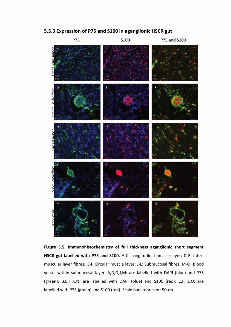
5.5.3 Expression of P75 and S100 in aganglionic HSCR gut
Figure 5.5. Immunohistochemistry of full thickness aganglionic short segment
HSCR gut labelled with P75 and S100. A-C: Longitudinal muscle layer; D-F: Inter-
muscular layer fibres; G-I: Circular muscle layer; J-L: Submucosal fibres; M-O: Blood
vessel within submucosal layer. A,D,G,J,M: are labelled with DAPI (blue) and P75
(green); B,E,H,K,N: are labelled with DAPI (blue) and S100 (red); C,F,I,L,O: are
labelled with P75 (green) and S100 (red). Scale bars represent 50µm.
P75 S100 P75 and S100

When aganglionic tissue was dual labelled with P75 and S100, a glial marker, it
appears that the centre of thickened nerve trunks dual stain however the ring
intensely expressing P75 does not express S100 (Figure 5.5 F and L). Many of the
fibres expressing P75 in the muscular layers also express S100, showing that most of
fibres are associated with glial cells (Figure 5.5 C and I). Finally, a blood vessel can
be identified in the aganglionic gut showing the clear difference in appearance
between this and the thickened nerve trunks (Figure 5.5 M-O).

5.5.4 Expression of P75 and calretinin in ganglionic HSCR gut
Figure 5.6. Immunohistochemistry of full thickness ganglionic short segment HSCR
gut labelled with P75 and S100. A-C: Longitudinal muscle layer; D-F: Myenteric
plexus; G-I: Circular muscle layer; J-L: Submucosal plexus. A,D,G,J: are labelled with
DAPI (blue) and P75 (green); B,E,H,K: are labelled with DAPI (blue) and S100 (red);
C,F,I,L: are labelled with P75 (green) and S100 (red). Arrow in L shows identifies an
extrinsic nerve like fibre. Scale bars represent 50µm.
P75 S100 P75 and S100

When ganglia are dual stained with P75 and S100 there is expression of both
markers throughout (Figure 5.6 F and L). Structures with the appearance of extrinsic
nerves can also be identified in the ganglionic gut (Figure 5.6 L see arrow). Many of
the fibres expressing P75 in the muscular layers also express S100, identifying the
presence of glial cells (Figure 5.6 C and I). Ganglionic HSCR gut was also compared
using the S100 glial marker to gut obtained from patients without HSCR, there were
no differences observed (not shown).

5.5.5 Expression of P75 and tyrosine hydroxylase in HSCR gut
Figure 5.7. Immunohistochemistry of full thickness ganglionic and aganglionic
short segment HSCR gut labelled with P75 and Tyrosine Hydroxylase. A-C:
ganglionic gut; D-F: aganglionic gut. A,D: are labelled with DAPI (blue) and P75
(green); B,E: are labelled with DAPI (blue) and TH (red); C,F: are labelled with P75
(green) and TH (red). Scale bars represent 50µm.
In ganglia the dopaminergic neuronal marker, tyrosine hydroxylase (TH), dual labels
with P75 in a similar pattern to S100 in that there is expression of both markers
throughout the ganglia (Figure 5.7 C). In contrast, when aganglionic tissue is dual
stained, the centre of the thickened nerve trunks expresses both P75 and TH
however the ring intensely expressing P75 does not label with TH (Figure 5.7 F).
P75 TH P75 and TH

5.5.6 Expression of P75 and laminin in HSCR gut
Figure 5.8. Immunohistochemistry of full thickness ganglionic and aganglionic
short segment HSCR gut labelled with P75 and Laminin. A-C: ganglionic gut; D-F:
aganglionic gut. A,D: are labelled with DAPI (blue) and P75 (green); B,E: are labelled
with DAPI (blue) and Laminin (red); C,F: are labelled with P75 (green) and Laminin
(red). Scale bars represent 50µm.
Antibodies to laminin were used to identify the basement membranes associated
with the nerve structures. In ganglionic gut there was no expression of this marker
surrounding ganglia (Figure 5.8 C). However, when thickened nerve trunks were
dual stained with P75 and laminin there was expression of both markers in close
relationship to the ring surrounding the trunks. The laminin however was expressed
very marginally externally to the P75 expression (Figure 5.8 F).
P75 Laminin P75 and Laminin

5.5.7 Expression of smooth muscle actin and S100 in HSCR gut
Figure 5.9. Immunohistochemistry of full thickness ganglionic and aganglionic
short segment HSCR gut labelled with Smooth Muscle Actin and S100. A-C:
ganglionic gut; D-F: aganglionic gut. A,D: are labelled with DAPI (blue) and SMA
(green); B,E: are labelled with DAPI (blue) and S100 (red); C,F: are labelled with SMA
(green) and S100 (red). Scale bars represent 50µm.
Lastly, to identify the relationship of the smooth muscle to neuronal structures,
tissue from both ganglionic and aganglionic gut was dual stained with S100 and
smooth muscle actin (SMA). The ganglia appeared to have a close relationship with
the smooth muscle in the ganglionic gut and there was is no layer between the two
tissue types (Figure 5.9 C). However in aganglionic gut there is a space between the
two tissue types which contains cell nuclei (Figure 5.9 E and F). When compared
with figure 5.5, where dual labelling has taken place with P75 and S100 it would
appear that this space is occupied by the ring intensely expressing P75.
SMA TH SMA and TH

5.6 Discussion
5.6.1 A ring of intense P75 expression surrounds thickened nerve
trunks
The thickened nerve trunks identified in both the submucosal and inter-muscular
layers of aganglionic gut were of a similar appearance in terms of P75 expression to
those described in the literature in that there was a ring of intense P75 expression
on the periphery of the structure.(Kobayashi, O'Briain & Puri 1994b). Additionally,
axons expressing the dopaminergic neuronal marker tyrosine hydroxylase and
endoneurial P75 positive glia are shown to be deep to the intense ring of P75
expression. There is some evidence which shows that the fibroblastic cells of the
perineurium surrounding extrinsic nerves consists of neural crest derived cells
however more recently lineage tracing has shown that this is not the case.(Hirose,
Sano & Hizawa 1986; Joseph et al. 2004) Also, using electron microscopy multiple
layers of perineurial cells can be observed surrounding thickened nerve trunks
compared to normal extrinsic nerves.(Baumgarten, Holstein & Stelzner 1973) This
evidence suggests that the thickened rings surrounding the thickened nerve trunks
are unusual. It is unlikely that these cells are perineurial cells as found in normal
extrinsic nerves as they express the neural crest marker P75, are thickened
compared to normal perineurium and express high levels of P75 compared to
endoneurial tissue within the nerve. On the contrary, the expression of laminin just
outside the thickened nerve trunks appears to identify basement membranes which
are in keeping with the structure of a surrounding perineurial layer.

It may also be possible that the increased expression is actually ENCC which
migrated into the gut from the sacral NCC contribution with extrinsic nerves during
the embryological period, but have failed to differentiate into an ENS.(Erickson et al.
2012) However it has been reported that this ring of intense P75 expression is also
seen in mesenteric nerves which are peripheral nerves and therefore this possibility
would appear less likely.(Kobayashi, O'Briain & Puri 1994b)
5.6.2 The cellular source of aganglionic ENSPC
The hypothesis that cells of Schwann cell lineage are the source of ENSPC in
aganglionic gut is supported by the expression of the glial marker, S100, and weak
P75 expression within the thickened nerve trunks. The intense ring of P75
expression does not label with S100 however and therefore does not these contain
Schwann cells. It is therefore possible that cells of this thickened surrounding layer
around the nerve trunks are the source of ENSPC. However it seems more probable
from studies showing the ability of glial and Schwann cells to undergo neurogenesis
that this is not the case.(Joseph et al. 2011; Laranjeira et al. 2011) Additionally, as
ENSPC from both ganglionic and aganglionic gut behave the same and S100 is also
expressed within ganglia it is more likely that glial cells are a source of ENSPC from
both ganglionic and aganglionic HSCR gut. It may also be possibly that ENSPC in
ganglionic gut are obtained from cells of Schwann cell lineage within the extrinsic
fibres and not the ganglia.

5.6.3 The significance of thickened nerve trunks
It has been shown that thickened nerve trunks in aganglionic HSCR gut are blind
ending extrinsic nerves but it is not clear why they are present in this gut.(Tam &
Boyd 1990) In the central nervous system it has been found that blind ending
nerves which fail to synapse atrophy instead of hypertrophy as in the aganglionic
gut.(Hefti et al. 1986) It seems logical that the extrinsic nerves increase in size as
they seek synapses with the ENS however this has not been shown and contrasts
the behaviour of central neurons in similar circumstances.
5.6.4 Nerve fibres are present in muscular layers of ganglionic and
aganglionic HSCR gut
Previous work has found, as expected, that nerve fibres are present within the
muscular layers in non HSCR gut and ganglionic HSCR gut which appear to connect
the two layers of the ENS, this was also seen in this study.(Kobayashi, O'Briain &
Puri 1994b; Matsuda et al. 2006) In terms of the aganglionic gut there was disparity
in the literature regarding the presence of nerve fibres in this tissue. One study
reported the absence of these fibres within the muscular layers(Kobayashi, O'Briain
& Puri 1994b) whereas another group observed the presence of these neurites in
the aganglionic gut.(Matsuda et al. 2006) It was also found in this study that nerve
fibres were present within the aganglionic gut in the muscular layers. These fibres
are likely of extrinsic origin and if functional are likely responsible for the tonic
smooth muscle contraction seen in HSCR. It is unclear as to whether these fibres are

identical in ganglionic and aganglionic gut however if they are it would seem that
dysfunction seen in HSCR is solely due to the absence of enteric ganglia.
5.6.5 Further work with more extensive HSCR variants
Unfortunately tissue from HSCR affecting the whole of the colon or beyond was not
available in our study. It has previously been reported that that interstitial cells of
Cajal are present in the muscular layers of aganglionic gut in short segment HSCR
however these cells are absent in the same layers of aganglionic total colonic HSCR
gut.(Solari, Piotrowska & Puri 2003) It is likely from the findings in chapter 3
detailing the absence of neural crest derived cells amongst cells isolated from
aganglionic total colonic HSCR gut that there is also an absence of neurites in the
muscular layers in this gut too. If this hypothesis is true there is further evidence to
support different aetiologies in the more extensive forms of HSCR. This is another
area where further work is required.
5.7 Conclusion
Work in this chapter has added to a limited number of studies which have
characterised aganglionic HSCR gut. Of particular importance, the presence of
Schwann cells and neurons has been identified in the thickened nerve trunks which
are of neural crest origin and support the hypothesis that cells of Schwann cell
lineage are the source of ENSPC. There are still many unanswered questions
surround the thickened nerve trunks including their development and function as
well as new questions about the aetiology of the different extensities of HSCR.

Chapter 6 – Overall discussion, areas for further work
and conclusion
6.1 Overview
This final chapter brings the work carried out in this thesis together in one final
discussion along with review of the aims of this thesis and new developments in the
field are addressed. The key areas focussed on are the cellular origin of ENSPC in
aganglionic gut, the appearance of the thickened nerve trunks and the aetiology of
HSCR. Finally, areas for further work are identified and the clinical implications of
work in this thesis when considering a stem cell therapy for HSCR are stated.

6.2 Introduction
As stated in chapter 1, the aim of work carried out in this thesis is to confirm and
add understanding to the observation that ENSPC can be isolated from short and
long segment aganglionic HSCR gut. Specifically, the objective has been to locate
the cellular origin of ENSPC within aganglionic gut so more can be understood about
how these cells can be controlled, predominantly for safety and efficacy reasons,
before work can progress to human stem cell therapy trials. Not only has the
original observation been confirmed, but it has also been shown that the ENSPC
from aganglionic HSCR gut are neural crest derived and the findings in chapter 3 and
chapter 5 suggest that these cells are located within thickened nerve trunks which
are pathognomonic of the aganglionic gut in short and long segment disease.
In addition, chapter 2 identified that horse serum medium is favourable to
neurosphere medium under adherent conditions for culturing neurospheres. In
order to explore the possibility of transplantation of neurospheres in HSCR patients
they must be first cultured, therefore horse serum medium was the only media
used beyond chapter 2 of this thesis. Furthermore, horse serum medium contains
fewer components than neurosphere medium, which has been used by our group
previously.(Almond et al. 2007; Lindley et al. 2008; Lindley et al. 2009) This is likely
to be advantageous if this work progresses to human trials, where adaption to
serum free media is required for safety reasons, as there will be fewer components
to replace.

6.3 The cellular source of ENSPC in aganglionic HSCR gut
Using samples of aganglionic HSCR gut from patients with all variants of HSCR it was
possible to correlate the presence of thickened nerve trunks and the ability to
obtain ENSPC, as aganglionic gut from total colonic and total intestinal doesn’t
contain thickened nerve trunks. However, only two patients with total colonic or
total intestinal disease were sampled. Subsequently, using cell sorting it was also
found that P75 positive neural crest derived cells obtained from aganglionic HSCR
gut differentiate into neurons indicating that ENSPC are of neural crest origin,
whereas the P75 negative cells did not do this.
In terms of the cellular origin of ENSPC, it has been shown that enteric glia can form
functional neurons, in vitro, although the ability of these cells to generate neurons
in vivo has been questioned.(Joseph et al. 2011; Laranjeira et al. 2011) Most
interestingly, it has very recently been discovered that the progenitors of Schwann
cells can also act as neuronal progenitors in the developing parasympathetic
nervous system.(Kalcheim & Rohrer 2014) With this information highlighting glia
and specifically the Schwann cell lineage as neuronal progenitors it is highly likely
that Schwann cells located in thickened nerve trunks in aganglionic HSCR gut are the
source of aganglionic ENSPC. If this is the case then further efforts in this area
should look at the control of cells of Schwann cell lineage to differentiate into
neurons which if discovered could open the doors to therapeutic treatment for
HSCR.

6.4 Appearance of thickened nerve trunks
Thickened nerve trunks present in aganglionic gut of short and long segment HSCR
are extrinsic, blind ending nerves originating from the pelvic plexus.(Tam & Boyd
1990) Another group described an intense ring of P75 neural crest cell marker
expression surrounding these nerves along with weakened expression
internally.(Kobayashi, O'Briain & Puri 1994b) This thickened ring corresponds to an
observation that thickened nerve trunks have multiple layers of perineurium
compared to other extrinsic nerves which have much fewer.(Baumgarten, Holstein
& Stelzner 1973) It was found in this thesis that there was expression of the
neuronal and glia markers, tyrosine hydroxylase and S100 respectively, from within
the trunk where there was also expression of the neural crest marker P75.
However, surrounding this core there is a ring of intense P75 expression which does
not express any of the other markers used, including neuronal and glial markers. In
normal extrinsic nerves the perineurium has been shown using lineage tracing to
not be of NCC lineage, therefore what we and others have seen is not normal
perineurium surrounding a thickened nerve trunk as normal perineurium does not
express P75.(Joseph et al. 2004) The identity of the cells expressing P75 in this
thickened ring is unclear. However, it is entirely possible that these cells are ENSPC
as we show that neural crest derived cells from aganglionic HSCR gut differentiate
into neurons.

Of particular interest, it was found that there are two populations of cells
expressing P75 identified by FACS on the aganglionic sample whereas there was just
one population in the ganglionic sample. In the aganglionic sample, one of
populations expressed P75 with more intensity than the other, which may
correspond to the cells of the ring of intense P75 expression seen in the thickened
nerve trunks. In regards to this, there was no effort made to select only one of
these populations using cell sorting and this is an area for further work in order to
narrow down further the source of ENSPC in aganglionic gut.
Using S100 and TH antibodies glia and neurons were identified in the core of the
thickened nerve trunks. As these nerve trunks are of extrinsic origin the glia
identified are Schwann cells. This corresponds to an observation of thickened nerve
trunks using electron microscopy which found Schwann cells in these trunks, but
most importantly, on ultramorphology they were of immature appearance, similar
to those found in the developing peripheral nervous system.(Baumgarten, Holstein
& Stelzner 1973) This makes it more likely when combined with the finding that
Schwann cells precursors act as neuronal progenitors in the developing
parasympathetic nervous system that these Schwann cells are the source of ENSPC
from aganglionic gut.(Kalcheim & Rohrer 2014)

6.5 Aetiology of HSCR
Work in this thesis has raised several questions about the aetiology of HSCR. It has
been shown in small mammal studies that the HSCR phenotype occurs due to
failure of migration of vagal NCC to the distal gut.(Obermayr et al. 2013) Work in
this thesis suggests that one type of ENSPC are located within thickened nerve
trunks in aganglionic HSCR gut however it is unknown whether these are ENCC or
other neural crest derived cells such as Schwann cells. There is evidence to suggest
that the sacral contribution of NCC to the ENS travel into the gut via the extrinsic
fibres.(Erickson et al. 2012) If these cells fail to differentiate and remain as
progenitors then it is entirely possible that these are the ENSPC we are able to
obtain from aganglionic HSCR gut. If this is the case, then the aetiology of HSCR may
include failure of proliferation of sacral NCC with or without failure of migration
too.
Although it is thought extrinsic nerves hypertrophy as a response to failing to
synapse it is unclear why thickened nerve trunks are present in aganglionic short
and long segment HSCR gut.(Hefti et al. 1986) Most interestingly it must be
considered why these trunks are absent in aganglionic total colonic and total
intestinal HSCR gut. This would suggest that the aetiology of the more extensive
disease differs from short and long segment disease. It is possible that the more
extensive forms are the result of more extensive NCC disease as the ENS and
extrinsic nerves in the gut have failed to form. On the other hand total colonic and

total intestinal HSCR may be due to disease of smooth muscle in the gut which is
unfavourable to neuronal development as both nervous systems are affected. It is
already known that short segment HSCR can be caused by an extrinsic defect
consisting of failure of the EDN3-EDNRB signalling pathway as EDN3 is not produced
by gut mesenchyme.(Obermayr et al. 2013) This theory is supported by an
observation that interstitial cells of Cajal are absent in aganglionic gut in total
colonic and total intestinal HSCR but present in the aganglionic gut in short and long
segment disease suggesting abnormalities in the smooth muscle in the more
extensive forms of HSCR.(Solari, Piotrowska & Puri 2003)
6.6 Final conclusion
In order to develop a stem cell therapy for HSCR the cellular identity of ENSPC
obtainable from aganglionic gut needs to be established for safety and efficacy
reasons. Results in this thesis showing that ENSPC are of neural crest origin located
within thickened nerve trunks, combined with evidence showing that Schwann cells
have an ability to act as neuronal precursors, makes it likely that cells of Schwann
cell lineage are the ENSPC we have isolated from aganglionic gut.(Joseph et al.
2011; Kalcheim & Rohrer 2014; Laranjeira et al. 2011) Other neural crest derived
cells expressing P75 in thickened nerve trunks are neurons, which can be ruled out
as ENSPC as only axons and not cell bodies are present in extrinsic nerves, and
abnormal surrounding perineurial cells which may have a role however little is
known about this abnormal tissue.

Now that it has been established that ENSPC are located within aganglionic gut in
short and long segment HSCR there are two possibilities in terms of translating this
work to human HSCR therapy. Firstly, aganglionic gut obtained at pull through
procedure could be used to obtain ENSPC. Aganglionic neurospheres containing
ENSPC and their differentiated neurons and glia can then be cultured and implanted
into the remaining aganglionic internal anal sphincter which is thought to cause the
long term complications after HSCR surgery. It is then likely that cells within
neurospheres will proliferate and differentiate to form a functional ENS and
therefore a functional sphincter. There is a risk with any stem cell therapy that
uncontrolled proliferation of immature cells will result in tumour formation, this risk
could be minimalised if cell sorting provided a population of mature neurons and
glia that form neurospheres without the addition of immature ENSPC too. However,
if the endogenous ENSPC in the thickened nerve trunks of short and long segment
HSCR aganglionic gut can be stimulated to proliferate and differentiate in situ then
it possible that HSCR patients can avoid surgery and undergo medical treatment to
stimulate development of a functional ENS.

6.7 Future work
6.7.1 Are cells of Schwann cell lineage the source of ENSPC in
aganglionic HSCR gut?
To explore this cell sorting could be used with similar methodology as used in
chapter 4. However, if a surface receptor glial marker is used which is similar to
S100 then an isolated sub-population of Schwann cells could be characterised after
time in culture and the expression of mature neuronal markers could be searched
for. This would give a definitive answer regarding the cellular origin of ENSPC in
aganglionic gut. The possible barriers to undertaking this work is the challenge of
finding a cell surface receptor marker that can be used with FACS successfully and
the challenges of culturing a small yield of primary cells that is inevitable from such
a minimal cell sub-population.
6.7.2 Which chemical factors stimulate the differentiation of ENSPC?
In order to identify therapeutic agents which may have the potential to stimulate
the development of an ENS in aganglionic gut in vivo a variety of different agents
must be tested in vitro. There are some agents that seem more likely including
those which stimulate 5HT4 receptor activation, which has been shown to allow
enteric neural regeneration in guinea pigs.(Matsuyoshi et al. 2010) Additionally,
inhibition of the Notch pathway in human ENSPC neurospheres has been shown to
decrease ENSPC proliferation whilst increasing differentiation in vitro.(Theocharatos

et al. 2013) Nevertheless, to comprehensively undertake this work high throughput
screening may be required using a library of chemical agents. This has been well
documented in the stem cell field and would efficiently identify chemicals which can
be further tested and potentially trialled in mouse models of HSCR.(Gupta et al.
2009)
6.7.3 Suicide gene incorporation into ENSPC and other cells within
neurospheres
A key concern with any stem cell therapy is the risk of losing control of a cell type
after implantation resulting in tumour formation.(Wilkinson, Edgar & Kenny 2012)
In order to reduce the risk to patients of treatment for what is a benign disease
there should be a method to control cells post implantation. To achieve this a
suicide gene could be incorporated into the DNA of all cells before implantation
which would induce apoptosis by production of the p53 protein for
example.(Malecki 2012) If there is any concern about uncontrolled cell proliferation
a known therapeutic agent which activates the gene can be administered. To
develop this technique, in vitro experimentation is required. A potential problem
with this idea is, genetic changes in cancerous cells may reduce or abolish the
effectiveness of the suicide gene. In terms of the method itself careful consideration
is required to ensure that all implanted cells within a neurosphere are incorporated
with the gene.

6.7.4 Implantation of human aganglionic neurospheres into a mouse
model of HSCR
It has been shown that florescence labelled mouse enteric neurospheres can be
transplanted into postnatal mouse gut which then form ganglia and display similar
electrophysiological functional characteristics to ENS ganglia.(Hotta et al. 2013)
However, to our knowledge, there have been no studies with a mouse model of
HSCR such as EDNRB-/- mice and transplantation of enteric neurospheres to assess
the ability to restore normal functionality in aganglionic gut.(Erickson et al. 2012) In
particular, aganglionic human neurospheres should be transplanted into the
aganglionic mouse gut and observation should take place short term, to test the
efficacy of the treatment and long term, to identify any signs of tumour formation.
If a stem cell therapy for HSCR is shown to be safe and efficacious in mice then the
logical progression will be to human clinical trials.

References
Almond, S., Lindley, R.M., Kenny, S.E., Connell, M.G. & Edgar, D.H. (2007)
'Characterisation and transplantation of enteric nervous system progenitor cells',
Gut, vol. 56, no. 4, pp. 489-496.
Amiel, J., Sproat-Emison, E., Garcia-Barcelo, M., Lantieri, F., Burzynski, G., Borrego,
S., Pelet, A., Arnold, S., Miao, X., Griseri, P., Brooks, A.S., Antinolo, G., de Pontual, L.,
Clement-Ziza, M., Munnich, A., Kashuk, C., West, K., Wong, K.K., Lyonnet, S.,
Chakravarti, A., Tam, P.K., Ceccherini, I., Hofstra, R.M. & Fernandez, R. (2008)
'Hirschsprung disease, associated syndromes and genetics: a review', Journal of
medical genetics, vol. 45, no. 1, pp. 1-14.
Anderson, R.B., Bergner, A.J., Taniguchi, M., Fujisawa, H., Forrai, A., Robb, L. &
Young, H.M. (2007) 'Effects of different regions of the developing gut on the
migration of enteric neural crest-derived cells: a role for Sema3A, but not Sema3F',
Developmental biology, vol. 305, no. 1, pp. 287-299.
Arshad, A., Powell, C. & Tighe, M.P. (2012) 'Hirschsprung's disease', BMJ, vol. 345, p.
e5521.
Baillie, C.T., Kenny, S.E., Rintala, R.J., Booth, J.M. & Lloyd, D.A. (1999) 'Long-term
outcome and colonic motility after the Duhamel procedure for Hirschsprung's
disease', Journal of pediatric surgery, vol. 34, no. 2, pp. 325-329.
Barlow, A., de Graaff, E. & Pachnis, V. (2003) 'Enteric nervous system progenitors
are coordinately controlled by the G protein-coupled receptor EDNRB and the
receptor tyrosine kinase RET', Neuron, vol. 40, no. 5, pp. 905-916.

Baumgarten, H.G., Holstein, A.F. & Stelzner, F. (1973) 'Nervous elements in the
human colon of hirschsprung's disease (with comparative remarks on neuronal
profiles in the normal human and monkey colon and sphincter ani internus)',
Virchows Archiv. A: Pathology. Pathologische Anatomie, vol. 358, no. 2, pp. 113-136.
Bergeron, K.F., Silversides, D.W. & Pilon, N. (2013) 'The developmental genetics of
Hirschsprung's disease', Clinical genetics, vol. 83, no. 1, pp. 15-22.
Bixby, S., Kruger, G.M., Mosher, J.T., Joseph, N.M. & Morrison, S.J. (2002) 'Cell-
intrinsic differences between stem cells from different regions of the peripheral
nervous system regulate the generation of neural diversity', Neuron, vol. 35, no. 4,
pp. 643-656.
Boley, S.J. (1964) 'New Modification of the Surgical Treatment of Hirschsprungs
Disease', Surgery, vol. 56, no. 5, pp. 1015-1017.
Bond, G.J. & Reyes, J.D. (2004) 'Intestinal transplantation for total/near-total
aganglionosis and intestinal pseudo-obstruction', Seminars in pediatric surgery, vol.
13, no. 4, pp. 286-292.
Borrego, S., Ruiz-Ferrer, M., Fernandez, R.M. & Antinolo, G. (2013) 'Hirschsprung's
disease as a model of complex genetic etiology', Histology and histopathology, vol.
28, no. 9, pp. 1117-1136.
Bradnock, T.J. & Walker, G.M. (2011) 'Evolution in the management of
Hirschsprung's disease in the UK and Ireland: a national survey of practice revisited',
Annals of the Royal College of Surgeons of England, vol. 93, no. 1, pp. 34-38.
Burns, A.J., Pasricha, P.J. & Young, H.M. (2004) 'Enteric neural crest-derived cells
and neural stem cells: biology and therapeutic potential', Neurogastroenterology
and motility : the official journal of the European Gastrointestinal Motility Society,
vol. 16 Suppl 1, pp. 3-7.

Cacalano, G., Farinas, I., Wang, L.C., Hagler, K., Forgie, A., Moore, M., Armanini, M.,
Phillips, H., Ryan, A.M., Reichardt, L.F., Hynes, M., Davies, A. & Rosenthal, A. (1998)
'GFRalpha1 is an essential receptor component for GDNF in the developing nervous
system and kidney', Neuron, vol. 21, no. 1, pp. 53-62.
Cass, D. (1986) 'Hirschsprung's disease: an historical review', Progress in pediatric
surgery, vol. 20, pp. 199-214.
Catto-Smith, A.G., Trajanovska, M. & Taylor, R.G. (2007) 'Long-term continence
after surgery for Hirschsprung's disease', Journal of gastroenterology and
hepatology, vol. 22, no. 12, pp. 2273-2282.
Chalazonitis, A. (2004) 'Neurotrophin-3 in the development of the enteric nervous
system', Progress in brain research, vol. 146, pp. 243-263.
Chen, B., Knowles, C.H., Scott, M., Anand, P., Williams, N.S., Milbrandt, J. & Tam,
P.K. (2002) 'Idiopathic slow transit constipation and megacolon are not associated
with neurturin mutations', Neurogastroenterology and motility : the official journal
of the European Gastrointestinal Motility Society, vol. 14, no. 5, pp. 513-517.
Conner, P.J., Focke, P.J., Noden, D.M. & Epstein, M.L. (2003) 'Appearance of
neurons and glia with respect to the wavefront during colonization of the avian gut
by neural crest cells', Developmental dynamics : an official publication of the
American Association of Anatomists, vol. 226, no. 1, pp. 91-98.
Coventry, S., Yost, C., Palmiter, R.D. & Kapur, R.P. (1994) 'Migration of ganglion cell
precursors in the ileoceca of normal and lethal spotted embryos, a murine model
for Hirschsprung disease', Laboratory investigation; a journal of technical methods
and pathology, vol. 71, no. 1, pp. 82-93.

de Arruda Lourencao, P.L., Takegawa, B.K., Ortolan, E.V., Terra, S.A. & Rodrigues,
M.A. (2013) 'A useful panel for the diagnosis of Hirschsprung disease in rectal
biopsies: calretinin immunostaining and acetylcholinesterase histochesmistry',
Annals of diagnostic pathology, vol. 17, no. 4, pp. 352-356.
De Lorijn, F., Reitsma, J.B., Voskuijl, W.P., Aronson, D.C., Ten Kate, F.J., Smets, A.M.,
Taminiau, J.A. & Benninga, M.A. (2005) 'Diagnosis of Hirschsprung's disease: a
prospective, comparative accuracy study of common tests', The Journal of
pediatrics, vol. 146, no. 6, pp. 787-792.
Dickie, B.H., Webb, K.M., Eradi, B. & Levitt, M.A. (2014) 'The problematic Soave cuff
in Hirschsprung disease: Manifestations and treatment', Journal of pediatric
surgery, vol. 49, no. 1, pp. 77-81.
Druckenbrod, N.R. & Epstein, M.L. (2005) 'The pattern of neural crest advance in the
cecum and colon', Developmental biology, vol. 287, no. 1, pp. 125-133.
Duhamel, B. (1956) '[New operation for congenital megacolon: retrorectal and
transanal lowering of the colon, and its possible application to the treatment of
various other malformations]', La Presse medicale, vol. 64, no. 95, pp. 2249-2250.
Erickson, C.S., Zaitoun, I., Haberman, K.M., Gosain, A., Druckenbrod, N.R. & Epstein,
M.L. (2012) 'Sacral neural crest-derived cells enter the aganglionic colon of Ednrb-/-
mice along extrinsic nerve fibers', The Journal of comparative neurology, vol. 520,
no. 3, pp. 620-632.
Fu, M., Tam, P.K., Sham, M.H. & Lui, V.C. (2004) 'Embryonic development of the
ganglion plexuses and the concentric layer structure of human gut: a topographical
study', Anatomy and embryology, vol. 208, no. 1, pp. 33-41.

Furness, J.B. (2008) 'The enteric nervous system: normal functions and enteric
neuropathies', Neurogastroenterology and motility : the official journal of the
European Gastrointestinal Motility Society, vol. 20 Suppl 1, pp. 32-38.
Garcia-Barcelo, M., Sham, M.H., Lui, V.C., Chen, B.L., Ott, J. & Tam, P.K. (2003)
'Association study of PHOX2B as a candidate gene for Hirschsprung's disease', Gut,
vol. 52, no. 4, pp. 563-567.
Georgeson, K.E., Cohen, R.D., Hebra, A., Jona, J.Z., Powell, D.M., Rothenberg, S.S. &
Tagge, E.P. (1999) 'Primary laparoscopic-assisted endorectal colon pull-through for
Hirschsprung's disease - A new gold standard', Annals of surgery, vol. 229, no. 5, pp.
678-683.
Gulbransen, B.D. & Sharkey, K.A. (2012) 'Novel functional roles for enteric glia in the
gastrointestinal tract', Nature reviews. Gastroenterology & hepatology, vol. 9, no.
11, pp. 625-632.
Gupta, P.B., Onder, T.T., Jiang, G., Tao, K., Kuperwasser, C., Weinberg, R.A. &
Lander, E.S. (2009) 'Identification of selective inhibitors of cancer stem cells by high-
throughput screening', Cell, vol. 138, no. 4, pp. 645-659.
Hall, J.E. & Guyton, A.C. (2011) Guyton and Hall textbook of medical physiology,
12th edn, Saunders/Elsevier, Philadelphia, PA.
Hefti, F., Hartikka, J., Salvatierra, A., Weiner, W.J. & Mash, D.C. (1986) 'Localization
of nerve growth factor receptors in cholinergic neurons of the human basal
forebrain', Neuroscience letters, vol. 69, no. 1, pp. 37-41.
Hirose, T., Sano, T. & Hizawa, K. (1986) 'Ultrastructural localization of S-100 protein
in neurofibroma', Acta neuropathologica, vol. 69, no. 1-2, pp. 103-110.
Holschneider, A.M.P., P. (2008) Hirschsprung's Disease and Allied Disorders.

Hotta, R., Stamp, L.A., Foong, J.P., McConnell, S.N., Bergner, A.J., Anderson, R.B.,
Enomoto, H., Newgreen, D.F., Obermayr, F., Furness, J.B. & Young, H.M. (2013)
'Transplanted progenitors generate functional enteric neurons in the postnatal
colon', The Journal of clinical investigation, vol. 123, no. 3, pp. 1182-1191.
Huddart, S.N. (1998) 'Hirschsprung's disease: present UK practice', Annals of the
Royal College of Surgeons of England, vol. 80, no. 1, pp. 46-48.
Huizinga, J.D. & White, E.J. (2008) 'Progenitor cells of interstitial cells of Cajal: on the
road to tissue repair', Gastroenterology, vol. 134, no. 4, pp. 1252-1254.
Jarvi, K., Laitakari, E.M., Koivusalo, A., Rintala, R.J. & Pakarinen, M.P. (2010) 'Bowel
function and gastrointestinal quality of life among adults operated for Hirschsprung
disease during childhood: a population-based study', Annals of surgery, vol. 252, no.
6, pp. 977-981.
Jiang, Y., Liu, M.T. & Gershon, M.D. (2003) 'Netrins and DCC in the guidance of
migrating neural crest-derived cells in the developing bowel and pancreas',
Developmental biology, vol. 258, no. 2, pp. 364-384.
Joseph, N.M., He, S., Quintana, E., Kim, Y.G., Nunez, G. & Morrison, S.J. (2011)
'Enteric glia are multipotent in culture but primarily form glia in the adult rodent
gut', The Journal of clinical investigation, vol. 121, no. 9, pp. 3398-3411.
Joseph, N.M., Mukouyama, Y.S., Mosher, J.T., Jaegle, M., Crone, S.A., Dormand, E.L.,
Lee, K.F., Meijer, D., Anderson, D.J. & Morrison, S.J. (2004) 'Neural crest stem cells
undergo multilineage differentiation in developing peripheral nerves to generate
endoneurial fibroblasts in addition to Schwann cells', Development, vol. 131, no. 22,
pp. 5599-5612.
Kalcheim, C. & Rohrer, H. (2014) 'Neuroscience. Following the same nerve track
toward different cell fates', Science, vol. 345, no. 6192, pp. 32-33.

Kenny, S.E., Tam, P.K. & Garcia-Barcelo, M. (2010) 'Hirschsprung's disease',
Seminars in pediatric surgery, vol. 19, no. 3, pp. 194-200.
Kobayashi, H., Hirakawa, H., O'Briain, D.S. & Puri, P. (1994) 'Nerve growth factor
receptor staining of suction biopsies in the diagnosis of Hirschsprung's disease',
Journal of pediatric surgery, vol. 29, no. 9, pp. 1224-1227.
Kobayashi, H., O'Briain, D.S. & Puri, P. (1994a) 'Lack of expression of NADPH-
diaphorase and neural cell adhesion molecule (NCAM) in colonic muscle of patients
with Hirschsprung's disease', Journal of pediatric surgery, vol. 29, no. 2, pp. 301-304.
Kobayashi, H., O'Briain, D.S. & Puri, P. (1994b) 'Nerve growth factor receptor
immunostaining suggests an extrinsic origin for hypertrophic nerves in
Hirschsprung's disease', Gut, vol. 35, no. 11, pp. 1605-1607.
Koivusalo, A.I., Pakarinen, M.P. & Rintala, R.J. (2009) 'Botox injection treatment for
anal outlet obstruction in patients with internal anal sphincter achalasia and
Hirschsprung's disease', Pediatric surgery international, vol. 25, no. 10, pp. 873-876.
Konagaya, S., Kato, K., Nakaji-Hirabayashi, T. & Iwata, H. (2011) 'Design of culture
substrates for large-scale expansion of neural stem cells', Biomaterials, vol. 32, no.
4, pp. 992-1001.
Konagaya, S., Kato, K., Nakaji-Hirabayashi, T. & Iwata, H. (2013) 'Selective and rapid
expansion of human neural progenitor cells on substrates with terminally anchored
growth factors', Biomaterials, vol. 34, no. 25, pp. 6008-6014.
Kostrominova, T. (2011). Application of WGA lectin staining for visualization of the
connective tissue in skeletal muscle, bone, and ligament/tendon studies.
Microscopy research and technique, 74(1), pp.18--22.
Laranjeira, C., Sandgren, K., Kessaris, N., Richardson, W., Potocnik, A., Vanden
Berghe, P. & Pachnis, V. (2011) 'Glial cells in the mouse enteric nervous system can
undergo neurogenesis in response to injury', The Journal of clinical investigation,
vol. 121, no. 9, pp. 3412-3424.
Lee, H.O., Levorse, J.M. & Shin, M.K. (2003) 'The endothelin receptor-B is required
for the migration of neural crest-derived melanocyte and enteric neuron
precursors', Developmental biology, vol. 259, no. 1, pp. 162-175.
Levitt, M.A., Hamrick, M.C., Eradi, B., Bischoff, A., Hall, J. & Pena, A. (2013)
'Transanal, full-thickness, Swenson-like approach for Hirschsprung disease', Journal
of pediatric surgery, vol. 48, no. 11, pp. 2289-2295.
Lindley, R.M., Hawcutt, D.B., Connell, M.G., Almond, S.L., Vannucchi, M.G.,
Faussone-Pellegrini, M.S., Edgar, D.H. & Kenny, S.E. (2008) 'Human and mouse
enteric nervous system neurosphere transplants regulate the function of
aganglionic embryonic distal colon', Gastroenterology, vol. 135, no. 1, pp. 205-216
e206.
Lindley, R.M., Hawcutt, D.B., Connell, M.G., Edgar, D.H. & Kenny, S.E. (2009)
'Properties of secondary and tertiary human enteric nervous system neurospheres',
Journal of pediatric surgery, vol. 44, no. 6, pp. 1249-1255; discussion 1255-1246.
Liu, M.T., Kuan, Y.H., Wang, J., Hen, R. & Gershon, M.D. (2009) '5-HT4 receptor-
mediated neuroprotection and neurogenesis in the enteric nervous system of adult
mice', The Journal of neuroscience : the official journal of the Society for
Neuroscience, vol. 29, no. 31, pp. 9683-9699.
Malecki, M. (2012) 'Frontiers in Suicide Gene Therapy of Cancer', Journal of genetic
syndrome & gene therapy, vol. 2012, no. 3.

Mareschi, K., Novara, M., Rustichelli, D., Ferrero, I., Guido, D., Carbone, E., Medico,
E., Madon, E., Vercelli, A. & Fagioli, F. (2006) 'Neural differentiation of human
mesenchymal stem cells: Evidence for expression of neural markers and eag K+
channel types', Experimental hematology, vol. 34, no. 11, pp. 1563-1572.
Martin, I., Nguyen, T.D., Krell, V., Greiner, J.F., Muller, J., Hauser, S., Heimann, P. &
Widera, D. (2012) 'Generation of Schwann cell-derived multipotent neurospheres
isolated from intact sciatic nerve', Stem cell reviews, vol. 8, no. 4, pp. 1178-1187.
Matsuda, H., Hirato, J., Kuroiwa, M. & Nakazato, Y. (2006) 'Histopathological and
immunohistochemical study of the enteric innervations among various types of
aganglionoses including isolated and syndromic Hirschsprung disease',
Neuropathology : official journal of the Japanese Society of Neuropathology, vol. 26,
no. 1, pp. 8-23.
Matsuyoshi, H., Kuniyasu, H., Okumura, M., Misawa, H., Katsui, R., Zhang, G.X.,
Obata, K. & Takaki, M. (2010) 'A 5-HT(4)-receptor activation-induced neural
plasticity enhances in vivo reconstructs of enteric nerve circuit insult',
Neurogastroenterology and motility : the official journal of the European
Gastrointestinal Motility Society, vol. 22, no. 7, pp. 806-813, e226.
Moore, M.W., Klein, R.D., Farinas, I., Sauer, H., Armanini, M., Phillips, H., Reichardt,
L.F., Ryan, A.M., Carver-Moore, K. & Rosenthal, A. (1996) 'Renal and neuronal
abnormalities in mice lacking GDNF', Nature, vol. 382, no. 6586, pp. 76-79.
Moore, S.W. (2012) 'Total colonic aganglionosis in Hirschsprung disease', Seminars
in pediatric surgery, vol. 21, no. 4, pp. 302-309.
Morrison, S.J., Perez, S.E., Qiao, Z., Verdi, J.M., Hicks, C., Weinmaster, G. &
Anderson, D.J. (2000) 'Transient Notch activation initiates an irreversible switch
from neurogenesis to gliogenesis by neural crest stem cells', Cell, vol. 101, no. 5, pp.
499-510.

Natarajan, D., Marcos-Gutierrez, C., Pachnis, V. & de Graaff, E. (2002) 'Requirement
of signalling by receptor tyrosine kinase RET for the directed migration of enteric
nervous system progenitor cells during mammalian embryogenesis', Development,
vol. 129, no. 22, pp. 5151-5160.
Nemeth, L., Yoneda, A., Kader, M., Devaney, D. & Puri, P. (2001) 'Three-dimensional
morphology of gut innervation in total intestinal aganglionosis using whole-mount
preparation', Journal of pediatric surgery, vol. 36, no. 2, pp. 291-295.
Nishiyama, C., Uesaka, T., Manabe, T., Yonekura, Y., Nagasawa, T., Newgreen, D.F.,
Young, H.M. & Enomoto, H. (2012) 'Trans-mesenteric neural crest cells are the
principal source of the colonic enteric nervous system', Nature neuroscience, vol.
15, no. 9, pp. 1211-1218.
Obermayr, F., Hotta, R., Enomoto, H. & Young, H.M. (2013) 'Development and
developmental disorders of the enteric nervous system', Nature reviews.
Gastroenterology & hepatology, vol. 10, no. 1, pp. 43-57.
Okamoto, E., Satani, M. & Kuwata, K. (1982) 'Histologic and embryologic studies on
the innervation of the pelvic viscera in patients with Hirschsprung's disease',
Surgery, gynecology & obstetrics, vol. 155, no. 6, pp. 823-828.
Pattyn, A., Morin, X., Cremer, H., Goridis, C. & Brunet, J.F. (1999) 'The homeobox
gene Phox2b is essential for the development of autonomic neural crest
derivatives', Nature, vol. 399, no. 6734, pp. 366-370.
Pichel, J.G., Shen, L., Sheng, H.Z., Granholm, A.C., Drago, J., Grinberg, A., Lee, E.J.,
Huang, S.P., Saarma, M., Hoffer, B.J., Sariola, H. & Westphal, H. (1996) 'Defects in
enteric innervation and kidney development in mice lacking GDNF', Nature, vol.
382, no. 6586, pp. 73-76.

Puig, I., Champeval, D., De Santa Barbara, P., Jaubert, F., Lyonnet, S. & Larue, L.
(2009) 'Deletion of Pten in the mouse enteric nervous system induces
ganglioneuromatosis and mimics intestinal pseudoobstruction', The Journal of
clinical investigation, vol. 119, no. 12, pp. 3586-3596.
Reynolds, B.A. & Weiss, S. (1992) 'Generation of neurons and astrocytes from
isolated cells of the adult mammalian central nervous system', Science, vol. 255, no.
5052, pp. 1707-1710.
Ruttenstock, E. & Puri, P. (2009) 'A meta-analysis of clinical outcome in patients
with total intestinal aganglionosis', Pediatric surgery international, vol. 25, no. 10,
pp. 833-839.
Saltzman, D.A., Telander, M.J., Brennom, W.S. & Telander, R.L. (1996) 'Transanal
mucosectomy: A modification of the Soave procedure for Hirschsprung's disease',
Journal of pediatric surgery, vol. 31, no. 9, pp. 1272-1275.
Sasselli, V., Pachnis, V. & Burns, A.J. (2012) 'The enteric nervous system',
Developmental biology, vol. 366, no. 1, pp. 64-73.
Soave, F. (1964) 'A New Surgical Technique for Treatment of Hirschsprungs Disease',
Surgery, vol. 56, no. 5, pp. 1007-1014.
Solari, V., Piotrowska, A.P. & Puri, P. (2003) 'Histopathological differences between
recto-sigmoid Hirschsprung's disease and total colonic aganglionosis', Pediatric
surgery international, vol. 19, no. 5, pp. 349-354.
Southard-Smith, E.M., Kos, L. & Pavan, W.J. (1998) 'Sox10 mutation disrupts neural
crest development in Dom Hirschsprung mouse model', Nature genetics, vol. 18, no.
1, pp. 60-64.

Steichen, F.M., Spigland, N.A. & Nunez, D. (1987) 'The modified Duhamel operation
for Hirschsprung's disease performed entirely with mechanical sutures', Journal of
pediatric surgery, vol. 22, no. 5, pp. 436-438.
Swenson, O. (1999) 'How the cause and cure of Hirschsprung's disease were
discovered', Journal of pediatric surgery, vol. 34, no. 10, pp. 1580-1581.
Swenson, O. & Bill, A.H., Jr. (1948) 'Resection of rectum and rectosigmoid with
preservation of the sphincter for benign spastic lesions producing megacolon; an
experimental study', Surgery, vol. 24, no. 2, pp. 212-220.
Taketomi, T., Yoshiga, D., Taniguchi, K., Kobayashi, T., Nonami, A., Kato, R., Sasaki,
M., Sasaki, A., Ishibashi, H., Moriyama, M., Nakamura, K., Nishimura, J. &
Yoshimura, A. (2005) 'Loss of mammalian Sprouty2 leads to enteric neuronal
hyperplasia and esophageal achalasia', Nature neuroscience, vol. 8, no. 7, pp. 855-
857.
Tam, P.K. & Boyd, G.P. (1990) 'Origin, course, and endings of abnormal enteric
nerve fibres in Hirschsprung's disease defined by whole-mount
immunohistochemistry', Journal of pediatric surgery, vol. 25, no. 4, pp. 457-461.
Tam, P.K. & Garcia-Barcelo, M. (2009) 'Genetic basis of Hirschsprung's disease',
Pediatric surgery international, vol. 25, no. 7, pp. 543-558.
Theocharatos, S., Wilkinson, D.J., Darling, S., Wilm, B., Kenny, S.E. & Edgar, D. (2013)
'Regulation of progenitor cell proliferation and neuronal differentiation in enteric
nervous system neurospheres', PloS one, vol. 8, no. 1, p. e54809.
Tittel, K. (1901) 'Uber eine angeborene Missbildung des Dickdarmes.', Wien Klin
Wochenschr, vol. 14, pp. 903-907.

Topp, K.S. & Boyd, B.S. (2006) 'Structure and biomechanics of peripheral nerves:
nerve responses to physical stresses and implications for physical therapist
practice', Physical therapy, vol. 86, no. 1, pp. 92-109.
Uchida, N., Buck, D.W., He, D., Reitsma, M.J., Masek, M., Phan, T.V., Tsukamoto,
A.S., Gage, F.H. & Weissman, I.L. (2000) 'Direct isolation of human central nervous
system stem cells', Proceedings of the National Academy of Sciences of the United
States of America, vol. 97, no. 26, pp. 14720-14725.
Wallace, A.S. & Burns, A.J. (2005) 'Development of the enteric nervous system,
smooth muscle and interstitial cells of Cajal in the human gastrointestinal tract', Cell
and tissue research, vol. 319, no. 3, pp. 367-382.
Wang, X., Chan, A.K., Sham, M.H., Burns, A.J. & Chan, W.Y. (2011) 'Analysis of the
sacral neural crest cell contribution to the hindgut enteric nervous system in the
mouse embryo', Gastroenterology, vol. 141, no. 3, pp. 992-1002 e1001-1006.
Watanabe, Y., Ito, F., Ando, H., Seo, T., Harada, T., Kaneko, K., Ishiguro, Y. &
Kobayashi, S. (1998) 'Extrinsic nerve strands in the aganglionic segment of
Hirschsprung's disease', Journal of pediatric surgery, vol. 33, no. 8, pp. 1233-1237.
Wilkinson, D.J., Edgar, D.H. & Kenny, S.E. (2012) 'Future therapies for Hirschsprung's
disease', Seminars in pediatric surgery, vol. 21, no. 4, pp. 364-370.
Wilkinson, D.J., Frain, B., Edgar, D.H. & Kenny, S.E. (2013) Neural progenitors from
aganglionic bowel - new therapeutic possibilities for Hirschsprung’s disease?, BAPS
Bournemouth.
Wu, J.J., Chen, J.X., Rothman, T.P. & Gershon, M.D. (1999) 'Inhibition of in vitro
enteric neuronal development by endothelin-3: mediation by endothelin B
receptors', Development, vol. 126, no. 6, pp. 1161-1173.

Young, H.M., Hearn, C.J., Ciampoli, D., Southwell, B.R., Brunet, J.F. & Newgreen, D.F.
(1998) 'A single rostrocaudal colonization of the rodent intestine by enteric neuron
precursors is revealed by the expression of Phox2b, Ret, and p75 and by explants
grown under the kidney capsule or in organ culture', Developmental biology, vol.
202, no. 1, pp. 67-84.
Young, H.M., Hearn, C.J., Farlie, P.G., Canty, A.J., Thomas, P.Q. & Newgreen, D.F.
(2001) 'GDNF is a chemoattractant for enteric neural cells', Developmental biology,
vol. 229, no. 2, pp. 503-516.
Zbuk, K.M. & Eng, C. (2007) 'Cancer phenomics: RET and PTEN as illustrative
models', Nature reviews. Cancer, vol. 7, no. 1, pp. 35-45.
Zhou, R., Niwa, S., Homma, N., Takei, Y. & Hirokawa, N. (2009) 'KIF26A is an
unconventional kinesin and regulates GDNF-Ret signaling in enteric neuronal
development', Cell, vol. 139, no. 4, pp. 802-813.
Zuelzer, W. W. & Wilson, J.L. (1948) Functional intestinal obstruction on a
congenital neurogenic basis in infancy. Amer. J. Dis. Child, vol. 75, no. 1, pp. 40-64.

Appendix 1 – Patient information sheets

Information for Parents of children with
Hirschsprung’s disease
Your child is being invited to take part in a research study. Before you decide it is
important for you to understand why the research is being done and what it will involve. Please take time to read the following information carefully and discuss it with others if
you wish. Ask us if there is anything that is not clear or if you would like more information. Take time to decide whether or not you wish to take part.
Thank you for reading this.
Study Title Enteric Nervous System Stem Cell Behaviour in Mature Muscle Models
What is the purpose of the study? Your child has Hirschsprung’s disease. In this condition nerve cells are missing from the rectum. Current treatment is removal of the affected segment of bowel but even after this many children have problems with constipation or soiling. We hope to understand how to grow nerve cells to allow us to replace them, so as to improve the treatment for children with Hirschsprung’s disease in future.
Why has your child been chosen? Because your child is going to have an operation involving removal of a section of bowel. This would normally be disposed of at the end of the operation but we would like to use a small piece of this tissue to study the nerves and muscle within it.
Does your child have to take part? No. It is up to you to decide whether or not you wish your child to take part. If you do decide that your child may take part you will be given this information sheet to keep and be asked to sign a consent form. Should you not want us to use the specimen in this way, there will be no effect on the way in which we treat your child. At the end of our tests, the specimen will be disposed of in the normal way.
What will happen to my child if I do take part? Your child is going to have an operation in which surgery is performed on the bowel. During this surgery a small amount of bowel tissue will be removed and routinely sent for tests. We would like you to help us by letting us use two small pieces of this tissue (approximately two square centimetres) to grow the nerve cells and muscle cells that are contained in the bowel wall. No extra tissue would be removed and this procedure would not affect our ability to do the routine tests on the bowel segment. It will have no effect on your child.

What are the possible disadvantages and risks of taking part? There are no additional disadvantages or risks of taking part.
What are the possible benefits to my child of taking part? It is likely that the main benefits of your child taking part will be to future children born with Hirschsprung’s disease rather than you directly. However, the information that we discover may have direct benefits to your child should we be able to isolate stem cells, as in future it may be possible to transplant them.
Will my child’s participation in this study be kept confidential? Yes. Your child’s name will not be disclosed outside of the hospital.
What will happen to the results of the study? We will publish the results of this work in medical and scientific journals and if you would like to know our eventual results please inform us.
Who is organising and funding the research? This study is organised and run by Mr Simon Kenny, Consultant Paediatric Surgeon, Alder Hey Children’s Hospital, Liverpool and Mr David Wilkinson, Clinical Research Fellow, University of Liverpool. The research has been kindly funded by the British Association of Paediatric Surgeons and the Royal College of Surgeons of England.
Who has reviewed the study? This study has been reviewed by a panel of experts, on behalf of the British Association of Paediatric Surgeons and the Royal College of Surgeons of England, who believe that it is of the highest standard. It has also been approved by the Liverpool Research Ethics Committee.
Contact for further information: Mr Simon E Kenny Mr David Wilkinson Consultant Surgeon Clinical Research Fellow Department of Paediatric Surgery University of Liverpool Alder Hey Children’s Hospital Cellular & Molecular Physiology Eaton Rd Sherrington Buildings Liverpool Ashton Street L12 2AP Liverpool L69 3GE Tel: 0151 252 5434 Tel: 0151 794 5433 If you have any complaints or concerns about your child’s involvement you can contact the Patient Advice Liason Service (PALS): PALS Office Alder Hey Children’s NHS Foundation Trust Eaton Road Liverpool L12 2AP Tel: 0151 252 5374/5161 Email: [email protected]

Information for Parents of children without
Hirschsprung’s disease
Your child is being invited to take part in a research study. Before you decide it is
important for you to understand why the research is being done and what it will involve. Please take time to read the following information carefully and discuss it with others if
you wish. Ask us if there is anything that is not clear or if you would like more information. Take time to decide whether or not you wish to take part.
Thank you for reading this.
What is the purpose of the study? Some children have a condition called Hirschsprung’s disease. In this condition nerve cells are missing from part of the bowel. Current treatment is removal of the affected segment of bowel but even after this many children have problems with constipation or soiling. We hope to understand how to grow nerve cells to allow us to replace them, so as to improve the treatment for children with Hirschsprung’s disease in future.
Why has your child been chosen? Because your child is going to have an operation involving removal of a section of bowel. This would normally be disposed of at the end of the operation but we would like to use a small piece of this tissue to study the nerves and muscle within it.
Does my child have to take part? No. It is up to you to decide whether or not you wish your child to take part. If you do decide that your child may take part you will be given this information sheet to keep and be asked to sign a consent form. Should you not want us to use the specimen in this way, there will be no effect on the way in which we treat your child. At the end of our tests, the specimen will be disposed of in the normal way.
What will happen to my child if they do take part? Your child is going to have an operation in which surgery is performed on the bowel. During this surgery a small amount of bowel tissue will be removed and routinely sent for tests. We would like you to help us by letting us use two small pieces of this tissue (approximately two square centimetres) to grow the nerve cells and muscle cells that are contained in the bowel wall. No extra tissue would be removed and this procedure would not affect our ability to do the routine tests on the bowel segment. It will have no effect on your child.
What are the possible disadvantages and risks of taking part? There are no additional disadvantages or risks of taking part.

What are the possible benefits of taking part? There will no direct benefits to your child in taking part other than the knowledge that they are helping others.
Will my child’s participation in this study be kept confidential? Yes. Your child’s name will not be disclosed outside of the hospital.
What will happen to the results of the study? We will publish the results of this work in medical and scientific journals and if you would like to know our eventual results please inform us.
Who is organising and funding the research? This study is organised and run by Mr Simon Kenny, Consultant Paediatric Surgeon, Alder Hey Children’s Hospital, Liverpool and Mr David Wilkinson, Clinical Research Fellow, University of Liverpool. The research has been kindly funded by the British Association of Paediatric Surgeons and the Royal College of Surgeons of England.
Who has reviewed the study? This study has been reviewed by a panel of experts, on behalf of the British Association of Paediatric Surgeons and the Royal College of Surgeons of England, who believe that it is of the highest standard. It has also been approved by the Liverpool Research Ethics Committee.
Contact for further information: Mr Simon E Kenny Mr David Wilkinson Consultant Surgeon Clinical Research Fellow Department of Paediatric Surgery University of Liverpool Alder Hey Children’s Hospital Cellular & Molecular Physiology Eaton Rd Sherrington Buildings Liverpool Ashton Street L12 2AP Liverpool L69 3GE Tel: 0151 252 5434 Tel: 0151 794 5433 If you have any complaints or concerns about your child’s involvement you can contact the Patient Advice Liason Service (PALS): PALS Office Alder Hey Children’s NHS Foundation Trust Eaton Road Liverpool L12 2AP Tel: 0151 252 5374/5161 Email: [email protected]