Embed Size (px)
Citation preview
The Snodgrass TapesFacts and Theories on the Insect Head. Part 2.
Moderator: William Bickley, Chair, Department of Entomology, University of Maryland... do you want the podium? Do you want that stand?
Transcribed, assembled and annotated by Jeffrey W. Shultz
Robert Evans SnodgrassNo...no, don't think so. Well,… we'll go back to where we left the embryo the last time… I'll put this diagram up ... <DRAWING ON CHALK BOARD>
Remember that the embryo has that rather large head. Well, that doesn't apply only to the insect head… but to the head of all crust… all of the arthropods. Well, as I say, that probably doesn't mean anything except that gives the sense organs -- the brain inside the head -- a chance to get an early start. So, I don't suppose that the ancestors of the insects and the arthropods were big-headed animals. Well, then, that's followed up, see, by this series of segments in which the appendages grow out.
Well, the question is… that I left you with the last time… are there segments up in that head lobe? Well, as I said, there are these embryologists who claim that that region is segmented, because they found in it two or three pairs of small cavities in the mesoderm. And they claim that wherever there are cavities like that in the mesoderm, called coelomic sacs, that that means… that is segmentation. But I contend that that is not necessarily segmenta-tion unless you've got evidence of segments in the body wall itself. Because we know the segments of any arthropod or insect; it's the subdivision of the body wall [that is] independently musculated and independently moveable. And, therefore, you might call them motor units [1]. Well, so, it all depends on definition, since there's no evidence externally of segmentation in that part of the head.
And another thing that's rather embarrassing to the … to the coelomic-sac interpretation is that a number of investigators have found a pair coelomic sacs in the labrum itself, and yet they do not admit that the labrum is a segment. I mean, those that base their interpretation on the coelomic sacs, they accept the two pairs correspond-ing with the antennae and the preantennal pair as evidence of segmentation, but they won't accept those in the labrum as segments, so there's an inconsistency in their own argument.
Page 1
The second of three lectures by the insect morphologist Robert Evans Snodgrass delivered to the Department of Entomology at the University of Maryland in 1960.
Figure 1
Early mantid embryoventral, diagrammatic
antenna
embryonichead lobe
labrum
limb bud
A stick insect embryo showing ventral surface (top) andcoelomic sacs of the labral and preantennal “segments”
antenna
labrum
mouth
labralcoelomicsac
preantennalcoelomicsac
stomodeum
mesoderm
mandiblemaxilla 1
The Snodgrass TapesLecture 2. Facts and Theories on the Insect Head. Part 2.
Page 2
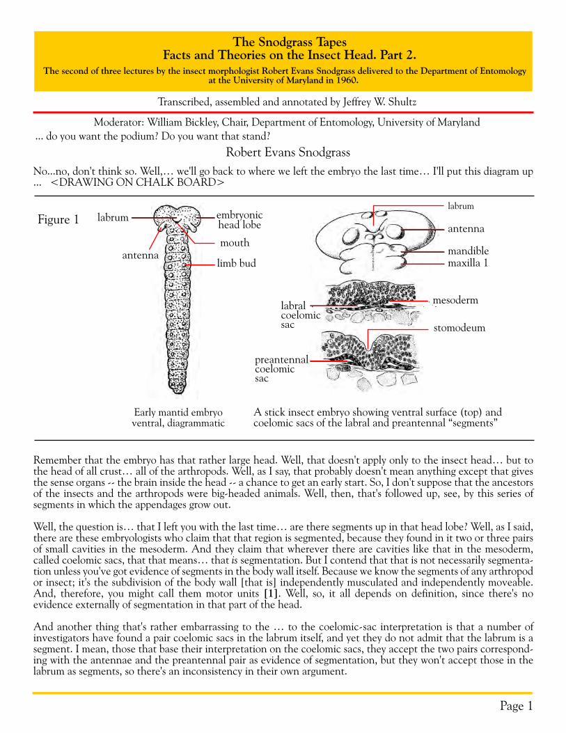
Well, today I was going to show you how some of these difficulties can be gotten around by theoretical interpreta-tions. Well, there are two European entomologists, Holmgren [2] and Hanström [3], who contend that that head lobe there corresponds with the anterior head lobe of the polychaete… or annelid worms, which is called the prostomium. You know the anterior end of segmented worms is... <drawing and talking>... Here's a lobe in front of the mouth…. that would be there and that's followed by all these segments... ??? .. Anyway, the mouth is at the base of this prostomium. Prostomium means something in front of the mouth. And that prostomium of the worm contains the brain and it may ... whatever sense organs the worm has at the anterior end of it … maybe eyes and tentacles. And the prostomium is not a segment. I mean, it doesn't correspond to the other segments. And, so, you can see, if that were translated there into that, you might very well suppose that the head lobe [of the arthropod embryo] is represented here in the worm by the prostomium [4].
Figure 2
Head and anterior segments of the polychaete Nereis virens
Dor
sal
Vent
ral
prostomium
tentacle
eye
mouth peristomium
I+II
IIIIV
palpus
IIIIIIIV
IIIIIIIV
eye
?
labrumpreantenna?
antenna 1
Diagrams of cephalization in annelids and arthropods, showing relation of the annelid prostomium to the arthropod head on the assumption that antenna 1 is a prostomial appendage
adult polychaete arthropod embryo hypothetical mandibulate arthropod
antenna 2mandiblemaxilla 1maxilla 2
labrumembryonic head lobe
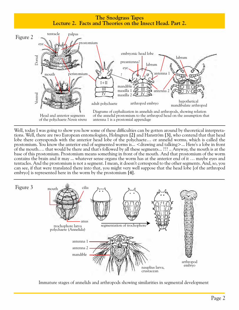
Immature stages of annelids and arthropods showing similarities in segmental development
nauplius larva,crustacean
antenna 1
antenna 2
mandible
arthropodembryo
segmentation of trochophoretrochophore larvapolychaete (Annelida)
mouth
anus
ciliaFigure 3
The Snodgrass TapesLecture 2. Facts and Theories on the Insect Head. Part 2.
Page 3
Well, that's their theory, and they back it up with a comparative study of the brain in the annelid worms and the arthropods, and they find that there's a very close similarity, or almost identity of structures, in the two. Which is a good reason for this theory that the head lobe of the arthropod is the prostomium of the worm. Well, now, some of these … take these polychaete ocean worms… and they look like this… The larva is a little thing something like that… It's divided in two here by a circle of cilia, and the mouth is right there. And the brain is formed up in that part and this part later becomes segmented. So, there's a good imitation at least of this arthropod embryo. That's called a trochophore, because of that circle of cilia around here. Trochophore literally means "wheel bear-ing", because this circle of cilia around the body is by which they move. So there's more evidence from the worm that gives a very striking likeness to the arthropod embryo. And the young crustacean is very much like that, because it ... It doesn't have much of a body. It starts off with.... like that... antenna, labrum, mandible .. and it has only three segments to begin with. And there's another thing you see all three of those things there [i.e., arthropod embryo, trochophore, nauplius] are essentially alike, except that they .. in the insect and the myriapods, the body becomes longer at once, while the crustacean has to grow. So, it seems to me that that theory is about the simplest that you can get, and the most satisfactory one that has been proposed on the relationship of the arthropods to the annelid worms. And almost everybody admits that ...or thinks that the worms and the arthropods are related organisms, but I don't believe their so closely related as some think. We'll get to that later.
So, if anybody wants to know what theory I recommend, I'd say that of Holmgren and Hanström. Hanström is the author of the large textbook I have on the comparative structure of the arthropod nervous system [5]. Because then when you consider that the ...the identical structure... the internal structure of the brain of all these forms, why it gives extra reason for accepting this theory as the most probable one we have. But if anybody wants to put ... as embryologists do ... they want to put their faith in a cavity in the mesoderm. It might be very simply be for the accumulation of waste products, even in the embryo. So, I don't think you can put too much faith in the pres-ence of a cavity in the embryo of being necessarily an indication of segments. Because otherwise you'ld have to suppose that that lobe here that once was composed of two or three moveable segments besides the prostomium.
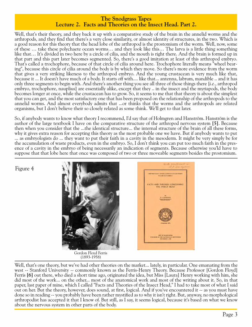
Figure 4
Gordon Floyd Ferris(1893-1958)
Well, that's one theory, but we've had other theories on the market... lately, in particular. One emanating from the west -- Stanford University -- commonly known as the Ferris-Henry Theory. Because Professor [Gordon Floyd] Ferris [6] out there, who died a short time ago, originated the idea, but Miss [Laura] Henry working with him, she did most of the work... on the other... most of the anatomical work and most of the writing about it. So, in that paper, last paper of mine, which I called "Facts and Theories of the Insect Head," I had to take most of what I said out on her. But the theory, however, does sound, at first, logical. And if you've encountered it -- as you must have done so in reading -- you probably have been rather mystified as to why it isn't right. But, anyway, no morphological arthropodist has accepted it that I know of. But still, as I say, it seems logical, because it's based on what we know about the nervous system in other parts of the body.

The Snodgrass TapesLecture 2. Facts and Theories on the Insect Head. Part 2.
Page 4
Figure 5
The segments of the thorax and abdomen in the insect ... have a ganglion in each segment.... eight in the abdomen see gives off its nerves to its own segment... ??? ... there's three segments. Well, that's the generalized pattern of the insect body, nervous system. And, as I say, in the generalized condition the nerves from each ganglion go to the segment that that ganglion belongs to. But these ganglia have a tendency to migrate forward and to unite. So that in some cases ... <drawing> There'd be a compound ganglion up here. But that ganglion will still give off nerves to all these segments down here. It's got eight ... eight of them. In other words, the ganglion ... this would be a com-pound ganglion you see formed from the last several ganglia there united with one another, but still the nerves go back to the segment from which the ganglion moved ... up. So, you can draw the conclusion, general rule or law that segments can be identified by the ganglia or ganglion from which they are innervated. It sounds logical and clear enough when you're studying the abdomen or thorax and in some cases, of course, the head. This would be ... [the] thorax has one big ganglion there where it's a combination of all the chain of ganglia, and still the nerves come back to the segment where they belong. Well, these authors, then, carried that over to the head.
Ganglia
Segments
T1 T2 T3 to A3 A4 A5 A6 A7 A8
A4
hindgut
foregut
subesophagealganglion
brain
optic lobe
T1 T2 T3 A3 A5 A6 A7 A8A2A1
T = thorax, A = abdomencompoundganglion
Dorsal view of a dissected grasshopper showing anterior migration and consolidation of segmental ganglia
ocellusoptic lobe
protocerebrum
deutocerebrum
tritocerebrum
subesophageal ganglion
mandibularnerve
maxillarynerve
labialnerve
crop
phar
ynx
aorta
frontalganglion
labral nerve
subesophagealganglion
frontalganglion
tritocerebrumdeutocerebrum
Central nervous system of a grasshopper
lateral view anterior view
Figure 6
The Snodgrass TapesLecture 2. Facts and Theories on the Insect Head. Part 2.
Page 5
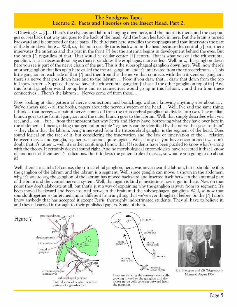
<Drawing> ...[?]... There's the clypeus and labrum hanging down here, and the mouth is there, and the esopha-gus curves back that way and goes to the back of the head. And the brain lies back in here. But the brain is turned backward and is composed of three parts. The third part here straddles the esophagus and that innervates the part of the brain down here ... Well, so, the brain usually turns backward in the head because this central [?] part there innervates the antenna and this part in the front [?] but the antenna begins in development behind the eyes. But the brain [?] regardless of that. That would be ocular center, [?] center.. That is what you call the tritocerebral ganglion. It isn't necessarily so big as that; it straddles the esophagus, more or less. Well, now, this ganglion down here you see is part of the nerve chain of the gut. This is the subesophageal ganglion down here. Well, now there's another ganglion that lies up in here called the frontal ganglion, and it's innervated from the tritocerebrum … This little ganglion on each side of that [?] and then from this the nerve that connects with the tritocerebral ganglion, there's a nerve that goes down here and to the labrum … Now, if you draw that… draw that down from the top it'll show better ... Suppose there we have the tritocerebral ganglion [it has all the other ganglia on top of it?] And this frontal ganglion would be up here and its connectives would go up in this fashion… and then from these connectives…There's the labrum ... Nerves come off from those…
Now, looking at that pattern of nerve connections and branchings without knowing anything else about it… We've always said -- all the books, papers about the nervous system of the head…. Well, I've said the same thing, I think -- that nerves ... a pair of nerves comes off from the tritocerebral ganglia and divides into two. The median branch goes to the frontal ganglion and the outer branch goes to the labrum. Well, that simply describes what you see, and ... on ... but ... from that apparent fact why Ferris and Henry have, borrowing what they have over here in the abdomen -- I mean, taking that general principle "segments can be identified by the nerve that goes to them" -- they claim that the labrum, being innervated from the tritocerebral ganglia, is the segment of the head. Does sound logical on the face of it, but considering the innervation and the law of innervation of the ... relation between nerves and ganglia, segments. it sounds quite logical. Well, if any of you have encountered it...I don't doubt that it's rather ... well, it's rather confusing. I know that [?] students have been puzzled to know what's wrong with the theory. It certainly doesn't sound right. And no morphological entomologists have accepted it that I know of, and most of them say it's ridiculous. But it follows the general rule of nerves, so what're you going to do about it?
Well, there is a catch. Of course, the tritocerebral ganglion, here, was never near the labrum, but it should be if its the ganglion of the labrum and the labrum is a segment. Well, since ganglia can move, a shown in the abdomen, why, it's safe to say, the ganglion of the labrum has moved backward and inserted itself between the antennal part of the brain and the ventral nervous system. Well, that again is kind of mysterious how it got in there. Now on that point they don't elaborate at all, but that's just a way of explaining why the ganglion is away from its segment. It's been moved backward and been inserted between the brain and the subesophageal ganglion. Well, so now that sounds altogether so farfetched and so different from anything that we've ever thought of before. So the [?] I don't know anybody that has accepted it except Ferris' thoroughly indoctrinated students. They all have to believe it, and they all carried it through to their published papers. Some of them.
Figure 7
R.E. Snodgrass and V.B. WigglesworthMontreal, August 1956
deutocerebrum
prtotocerebrum
tritocerebrum
frontalganglion
circumesophagealconnectives
subesophageal ganglionLateral view of central nervous system of a grasshopper
ganglion
sensorynerve cell
motornerve cell
muscle
externalsense organ
axon grows in
axon grows out
Diagram showing the sensory nerve cells growing inward to the ganglion and the motor nerve cells growing outward from the ganglion
The Snodgrass TapesLecture 2. Facts and Theories on the Insect Head. Part 2.
Page 6
Well, the real fallacy though is this, I think. These so-called nerves... I mean, nerves that are said to go up to the labrum don't do that at all. A lot of the descriptions of the anatomy of the nervous system in the insect have shown that those nerves are sensory nerves, and it's well known that sensory nerves begin in the epidermis and grow inward. You see, these motor nerves that we talked about here in the body have their central cells of origin in the ganglia and then grow out from the ganglia. But the sensory nerves, especially in the epidermis, ... this is a piece of epidermis... some of these epidermal cells become nerve cells and the axon of that nerve cell grows in like this ... [?] So, it's altogether wrong to say that those nerves grow out from the ganglia to the labrum. They grow just the other way. Wigglesworth [7] just recently has had two papers or publications showing just how sensory nerves do grow in from the epidermis. The real nerve cells... epidermal cells have become nerve cells. So, it's quite wrong, then, to say that those nerves grow out to the labrum. Just the opposite is true; they grow in toward the ganglion, and they unite with the connectives of the frontal ganglion in order to get into the ganglion. And, you see, sensory nerves have to connect with the motor system in the nervous centers. Because, they're insects, they don't simply don't feel or smell, the way we do, they give a reaction which has to.... The stimulus of a sensory nerve has to produce some kind of motion. It either goes after something because ... either its food or it has to avoid it, one way or the other. So, I say, with the insect, the collection of sensory nerves, especially from the... is to produce a motion of the animal that will be advantageous to it one way or another.
Figure 8
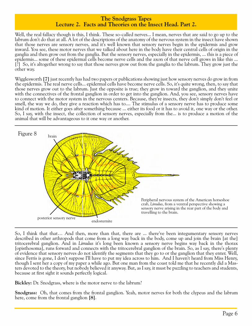
Peripheral nervous system of the American horseshoe crab, Limulus, from a ventral perspective showing a sensory nerve arising in the rear part of the body and travelling to the brain.
posterior sensory nerve
brain
endosternite
So, I think that that… And then, more than that, there are ... there've been integumentary sensory nerves described in other arthropods that come from a long way back in the body, come up and join the brain [at the] tritocerebral ganglion. And in Limulus it's long been known a sensory nerve begins way back in the thorax [opisthosoma], runs forward and connects with the tritocerebral ganglion of the brain. So, as I say, there's plenty of evidence that sensory nerves do not identify the segments that they go to or the ganglion that they enter. Well, since Ferris is gone, I don't suppose I'll have to put my idea across to him. And I haven't heard from Miss Henry, though I sent her a copy of my paper a while ago. But one man from the coast told me that he recently did a Mas-ters devoted to the theory, but nobody believed it anyway. But, as I say, it must be puzzling to teachers and students, because at first sight it sounds perfectly logical.
Bickley: Dr. Snodgrass, where is the motor nerve to the labrum?
Snodgrass: Oh, that comes from the frontal ganglion. Yeah, motor nerves for both the clypeus and the labrum here, come from the frontal ganglion [8].
The Snodgrass TapesLecture 2. Facts and Theories on the Insect Head. Part 2.
Page 7
Figure 9
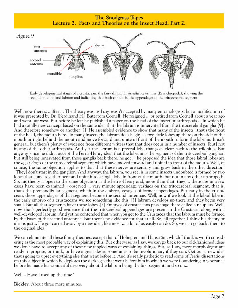
Well, now there's…after ... The theory was, as I say, wasn't accepted by many entomologists, but a modification of it was presented by Dr. [Ferdinand H.] Butt from Cornell. He resigned ... or retired from Cornell about a year ago and went out west. But before he left he published a paper on the head of the insect or arthropods ... in which he had a totally new concept based on the same idea that the labrum is innervated from the tritocerebral ganglia [9]. And therefore somehow or another [?]. He assembled evidence to show that many of the insects ..that's the front of the head, the mouth here.. in many insects the labrum does begin as two little lobes up there on the side of the mouth or right behind the mouth and move forward and unite in front of the mouth to form the labrum. It isn't general, but there's plenty of evidence from different writers that that does occur in a number of insects, [but] not in any of the other arthropods. And yet the labrum is a preoral lobe that goes clear back to the trilobites. But anyway, since he didn't accept the Ferris-Henry idea, that the labrum is the segment of the tritocerebral ganglion but still being innervated from those ganglia back there, he got ... he proposed the idea that those labral lobes are the appendages of the tritocerebral segment which have moved forward and united in front of the mouth. Well, of course, the same objection applies to that that these nerves are sensory and grow back in the other direction. [They] don't start in the ganglion. And anyway, the labrum, you see, is in some insects undoubted is formed by two lobes that come together here and unite into a single lobe in front of the mouth, but not in any other arthropods. So, his theory is open to the same objection as the Ferris-Henry and, more than that, they ... there are in a few cases have been examined... observed ... very minute appendage vestiges on the tritocerebral segment, that is, that's the premandibular segment, which in the embryo, vestiges of former appendages. But early in the crusta-cean, those appendages of that segment become the second antennae. Well, now if we look at the labral lobe in the early embryo of a crustaceans we see something like this. [?] labrum develops up there and they begin very small. But all that segments have those lobes..[?] Embryos of crustaceans pass stage there called a nauplius. Well, now, that's perfectly good evidence that the tritocerebral appendages are present in the Crustacea along with a well-developed labrum. And yet he contended that when you get to the Crustacea that the labrum must be formed by the bases of the second antennae. But there's no evidence for that at all. So, all together, I think his theory or idea is just... He got carried away by a new idea, like most ... a lot of us easily can do. So, we can go back, then, to the original idea.
We can eliminate all these funny theories, except that of Holmgren and Hanström, which I think is worth consid-ering as the most probable way of explaining this. But otherwise, as I say, we can go back to our old-fashioned ideas we don't have to accept any of these new fangled ways of explaining things. But, as I say, more morphologist are ready to propose, or think, or have a great desire sometimes to be revolutionary if they can. Get out a new idea that's going to upset everything else that went before it. And it's really pathetic to read some of Ferris' dissertations on this subject in which he deplores the dark ages that went before him in which we were floundering in ignorance before he made his wonderful discovery about the labrum being the first segment, and so on...
Well... Have I used up the time?
Bickley: About three more minutes.
Early developmental stages of a crustacean, the fairy shrimp Linderiella occidentalis (Branchiopoda), showing the second antenna and labrum and indicating that both cannot be the appendages of the tritocerebral segment
nauplius
firstantenna
secondantenna
mandible
labrum
The Snodgrass TapesLecture 2. Facts and Theories on the Insect Head. Part 2.
Page 8
Snodgrass: Three more minutes. Well...
Bickley: Do you want to answer some questions?
Snodgrass: I was going to say, I'll answer any questions. Next time I think I'll take up the subject of the evolution of the arthropods, which we can develop very nicely from this embryo. Any questions?</font>
Questioner: Did either Ferris or Henry contend that the labrum was originally a postoral structure which had migrated...
Snodgrass: No they never said anything like that at all. And, more than that, they took this as the second segment, then the ocular and the antennal region was the third segment. But here's another point I forgot to men-tion, that these labral nerves that come from the labrum. I mean, these nerves here, not only come from the labrum but they come from the overall surface of the ....??? ... of the mouthparts of the head. They're just sensory nerves that all come together ... in single strands there. So, that the group that goes to the labium ... labrum itself has no specific meaning.
Theodore Bissell: Dr. Snodgrass, aphids ... certain... they typically have one pair of setae, dorsal setae, on each abdominal segment going forward on the thorax. I think two pairs on the prothorax and then on the head it may have as many as five pairs of dorsal setae.
Snodgrass: What animal is that?
Bissell: Aphids… certain, certain aphids. I wondered if they have any indication ... make any indication of the internal segmentation of the head, ... these setae?
Snodgrass: Well, I don't believe so, because the ... Of course, we know that, as I said last time, that there are four segments up there in the composition of the head.
Bissell: Five?
Snodgrass: Four. I mean there's the premandibular, mandibular and the two maxillary segments. They undoubt-edly had …premandibular and mandibles formed here, but all that, you see, is combined in the adult head. But they are so integrated [and] consolidated that there's no trace left of the lines between them. And the muscles seem spread all over everywhere. And some muscles ... mandibular muscles are so big that they spread all over. Doesn't look as if they keep within any segmental limits. <BELL> So, I've got to use other kinds of evidence to show the segmentation of the head. Except that the embryo does show those four segments. But [the composition of that lobe is where we still have questions?]... Preoral lobe, four postoral segments all grow together in the head ... the head of the insect. As I explained last time that doesn't happen in all ... all of the arthropods. Some of the crustaceans add only one segment. Anything else? [I] heard the bell ring, so I guess I'll have to quit. Well, next time I'll go over some ideas about the ...
NOTES
APPLAUSE
1. The term “segment” often gives the impression of a distinct unit of the body with well-defined borders, like beads along a string. This occurs in some animal groups, such as tapeworms, but does not describe the condition in arthropods very well. Rather, the body shows a clear spatial periodicity that apparently results from the cyclical activation of a developmental program. Consequently, it is not really clear where one segment begins and another ends, and different workers tend to define segments and segmental borders based on their research interests or on those feature that seem obvious in some way. Thus, embryologists have tended to focus on such things as the formation of mesodermal somites and coelomic sacs; functional morphologists focus on Snodgrass's “motor units” or the appendages, molecular geneticists focus on the expression of the gene engrailed; neuroanatomists focus on the structure of ganglia and trajectory of motor nerves. Rather than using the term "segment," it is more precise to use terms like myomere for spatial periodicity in muscles, appendomere for appendages,
The Snodgrass TapesLecture 2. Facts and Theories on the Insect Head. Part 2.
Page 9
NOTES
cardiomere for the heart, etc. For example, the apparent external segmentation of most hard-bodied arthropods are scleromeres and reflect the periodicity of the sclerites, but the external segments of soft-bodied arthropods (e.g., caterpillars) reflect the sites of muscle attachment and therefore correspond to the underlying myomeres. Not all “segments” are the same.
2. Nils Fritiof Holmgren (1877–1954), Swedish zoologist, comparative neuroanatomist
Major relevant work: Holmgren, N. 1916. Zur vergleichenden Anatomie des Gehirns von Polychaeten, Onychophoren, Xiphosuren, Arachniden, Crustaceen, Myriapoden und Insekten. Vorstudien zu einer Phylogenie der Arthropoden. Svenska Vetenskapsakademien, Stockholm Handlingar. Series 2, 56(1), 303 pp, 12 plates. 3. Bertil Hanström (1891- ?), Swedish zoologist, comparative neuroanatomist, physiologist, student of Holmgren.
Major relevant work: Hanström, B. 1928. Vergleichende Anatomie Des Nervensystems der Wirbellosen Tiere. Springer, Berlin, Germany.
4. The long, complicated history of the debate on the segmental composition of the arthropod head has produced a large lexicon and terms are not always used in the same way by different people. Many workers regard the preoral “prostomium” of annelids as comparable to the somewhat hypothetical “acron” of arthropods. Snodgrass, however, postulates that the prostomium corresponds to the embryonic head lobe (= procephalon or blastocephalon) of arthropods. Some, perhaps most, workers would regard the embryonic head lobe of arthropods to be a compound structure comprising the acron, preantennal or labral segment, and the antennal segment, and, perhaps, the premandibular segment. The antennal and premandibular segments are originally postoral segments that migrate anterior to the mouth during development.
5. Since the late 18th century, biologists assumed that the similarities of annelids and arthropods (i.e., segmented bodies, mesodermally derived osmoregulatory organs, dorsal artery, ventral nerve cord, development by posterior addition of somites, etc.) reflect a close phylogenetic relationship and were combined in a group called Articulata. The morphology of onychophorans or velvet worms (e.g., Peripatus) seemed intermediate between annelids and arthropods, and thus strengthened the Articulata concept. However, recent studies of molecular sequence data, especially from 18S ribosomal nucleotides, indicates that arthropods and onychophorans are more closely related to such invertebrate phyla as Kinorhyncha, Tardigrada and Nematoda and are only distantly related to Annelida (Aguinaldo et al., 1997; Halanych, 2004) . Arthropods and their relatives were then placed in a group called Ecdy-sozoa, which is united, in part, by absence of cells with ciliary motility and by presence of a chitinous cuticle and growth through molting or ecdysis. The Ecdysozoa concept rapidly achieved wide acceptance and support for Articulata faded (but see Nielsen, 2001), such that many zoologists would now regard Snodgrass' emphasis on annelid-arthropod similarities to be outdated. However, the morphological similarities of Annelida and Arthropoda remain and require explanation, regardless of the phylogenetic proximity of the two phyla. If Ecdyso-zoa is real, then annelid-arthropod similarities are either remarkable parallelisms or reflect a shared ancient body plan that was lost many times among the ecdysozoans or both, perhaps in association with reduced size or simplifi-cations caused by altered ecology. Any scenario has significant implications for understanding evolutionary factors governing long-term changes in body plans, the origin of phyla and the role of morphology in phylogenetic analy-sis. We are not yet in a position to say that Snodgrass' and other workers were right or wrong in looking to annelids in their attempts to understand arthropod evolution.
Aguinaldo, A.M.A., J.M. Turbeville, L.S. Linford, M.C. Rivera, J.R. Garey, R.A. Raff & J.A. Lake. 1997. Evidence for a clade of nematodes, arthropods and other moulting animals. Nature, 387: 489-493.Halanych, K. 2004. The new view of animal phylogeny. Annual Review of Ecology, Evolution, and Systematics, 35: 229-256.Nielsen, C. 2003. Proposing a solution to the Articulata-Ecdysozoa controversy. Zoologica Scripta, 32: 475-82.
6. Follow these links for biographical information on Gordon Floyd Ferris (1893-1958):
Phthiraptera Central: Phthirapterists: http://www.phthiraptera.org/phthirapterists/ferris/ferris.htm Stanford University, Memorial Resolution: http://histsoc.stanford.edu/pdfmem/FerrisG.pdf Essig Museum, Univ of California, Berkeley: http://essig.berkeley.edu/collection/ferris.htm"
The Snodgrass TapesLecture 2. Facts and Theories on the Insect Head. Part 2.
Page 10
NOTES7. Sir Vincent B. Wigglesworth (1899-1994), renowned insect physiologist and author of the seminal text The Principles of Insect Physiology. Follow these links for biographical information:
Edwards, J.S. 1998. Sir Vincent Wigglesworth and the coming of age of insect development. International Journal of Developmental Biology, 42: 471-473. (http://www.ijdb.ehu.es/fulltext.9803/ft471.pdf)
Royal Entomological Society, Wigglesworth Award: http://www.royensoc.co.uk/award.html#wiggle
8. Recent work indicates that the cell bodies of the motoneurons of the labrum occur in the tritocerebrum in insects and crustaceans (Haas et al., 2001).
Haas, M.S., S.J. Brown & R.W. Beeman. 2001. Pondering the procephalon: the segmental origin of the labrum. Development, Genes & Evolution, 211: 89-95.
9. Ferdinand H. Butt (1899-?) concluded that the labrum is formed from highly modified appendages of the premandibular (= tritocerebral or intercalary) segment in insects, a view that was subsequently criticized (e.g., Matsuda, 1965) for the same reasons outlined by Snodgrass (1960) and in this lecture. Specifically, in crustaceans and chelicerates, the labrum and appendages of the tritocerebral segment are present and well developed in crus-taceans, and one cannot expect the same appendage to be in two places at once. Butt's hypothesis has been resur-rected in recent years with mounting evidence that the labrum is appendicular (e.g., Popadic et al., 1998) and has an intimate neural connection to the tritocerebrum in some insects. Haas et al. (2001) have argued that the labrum may represent the endites of the tritocerebral appendage (i.e., short branches that articulate at the base of the appendages) rather than the entire appendage, and that one element of the appendage may have migrated forward to become the labrum. This would presumably account for two appendicular elements of the premandibu-lar appendage being in different places. The debate continues.
Butt, F.H. 1960. Head development in the arthropods. Biological Reviews of the Cambridge Philosophical Society, 35: 43-91.Haas, MS, S.J. Brown & R.W. Beeman. 2001. Pondering the procephalon: the segmental origin of the labrum. Development, Genes & Evolution, 211: 89-95.Matsuda, R. 1965. Morphology and Evolution of the Insect Head. The American Entomological Institute, Ann Arbor, MI.Popadic, A., G. Panganiban, D. Rusch, W.A. Shear & T.C. Kaufman. 1998. Molecular evidence for the gnathobasic derivation of arthropod mandibles and for the appendicular origin of the labrum and other structures. Development, Genes & Evolution, 208: 142-150.Snodgrass, R.E. 1960. Facts and theories concerning the insect head. Smithsonian Miscellaneous Collections, 142(1): 1-61. Figure 1. Mantid embryo: Snodgrass, R.E. 1960. Facts and theories concerning the insect head. Smithsonian Miscellaneous Collections, 142(1): 1-61. fig. 1. Stick insect embryo: Snodgrass, R.E. 1938. Evolution of the Annelida, Onychophora, and Arthropoda. Smithsonian Miscellaneous Collections, 97(6): 1-159, fig. 42.
Figure 2. Snodgrass, R.E. 1938. Evolution of the Annelida, Onychophora, and Arthropoda. Smithsonian Miscellaneous Collections, 97(6): 1-159, figs 14, 39.
Figure 3. Trochophore, nauplius: Snodgrass, R.E. 1938. Evolution of the Annelida, Onychophora, and Arthropoda. Smithsonian Miscella-neous Collections, 97(6): 1-159, figs 4, 11. Arthropod embryo: Snodgrass, R.E. 1960. Facts and theories concerning the insect head. Smith-sonian Miscellaneous Collections, 142(1): 1-61. fig. 1.
Figure 4. Ferris: Phthiraptera Central (http://www.phthiraptera.org/phthirapterists/ferris/Gordon%20Floyd%20Ferris.jpg). Autographed cover: Original
Figure 5. Snodgrass, R.E. 1935. Principles of Insect Morphology. MacGraw-Hill Book Co., New York, fig. 247.
Figure 6. Snodgrass, R.E. 1935. Principles of Insect Morphology. MacGraw-Hill Book Co., New York, figs 248, 249.
Figure 7. Insect nervous system: Snodgrass, R.E. 1935. Principles of Insect Morphology. MacGraw-Hill Book Co., New York, figs 242, 249. Portrait of Snodgrass and Wigglesworth: University of Kansas, Photo Album of Entomologists (http://nhm.ku.edu/ksem/Assets/Images/picttourimages/073.jpg)
Figure 8. Patten, W. 1912. The Evolution of the Vertebrates and their Kin. P. Blakiston’s Sons, Philadelphia, fig. 70.
Figure 9. Snodgrass, R.E. 1956. Crustacean metamorphosis. Smithsonian Miscellaneous Collections, 131(10): 1-78, fig. 3.
Figure Credits