Embed Size (px)
Citation preview
MINI REVIEW ARTICLEpublished: 08 May 2014
doi: 10.3389/fcimb.2014.00061
The small RNA SgrS: roles in metabolism andpathogenesis of enteric bacteriaMaksym Bobrovskyy and Carin K. Vanderpool*
Department of Microbiology, University of Illinois at Urbana-Champaign, Urbana, IL, USA
Edited by:
Wyndham W. Lathem,Northwestern University FeinbergSchool of Medicine, USA
Reviewed by:
Kai Papenfort, Princeton University,USAEric Masse, University ofSherbrooke, Canada
*Correspondence:
Carin K. Vanderpool, Department ofMicrobiology, University of Illinois atUrbana-Champaign, C213 CLSL,MC-110, 601 S. Goodwin Ave.,Urbana, IL 61801, USAe-mail: [email protected]
Bacteria adapt to ever-changing habitats through specific responses to internal andexternal stimuli that result in changes in gene regulation and metabolism. One internalmetabolic cue affecting such changes in Escherichia coli and related enteric speciesis cytoplasmic accumulation of phosphorylated sugars such as glucose-6-phosphate orthe non-metabolizable analog α-methylglucoside-6-phosphate. This “glucose-phosphatestress” triggers a dedicated stress response in γ-proteobacteria including several entericpathogens. The major effector of this stress response is a small RNA (sRNA), SgrS.In E. coli and Salmonella, SgrS regulates numerous mRNA targets via base pairinginteractions that result in alterations in mRNA translation and stability. Regulation of targetmRNAs allows cells to reduce import of additional sugars and increase sugar efflux. SgrSis an unusual sRNA in that it also encodes a small protein, SgrT, which inhibits activityof the major glucose transporter. The two functions of SgrS, base pairing and productionof SgrT, reduce accumulation of phosphorylated sugars and thereby relieve stress andpromote growth. Examination of SgrS homologs in many enteric species suggests thatSgrS has evolved to regulate distinct targets in different organisms. For example, inSalmonella, SgrS base pairs with sopD mRNA and represses production of the encodedeffector protein, suggesting that SgrS may have a specific role in the pathogenesis ofsome γ-proteobacteria. In this review, we outline molecular mechanisms involved in SgrSregulation of its target mRNAs. We also discuss the response to glucose-phosphate stressin terms of its impact on metabolism, growth physiology, and pathogenesis.
Keywords: small RNA, SgrR, PtsG, glucose-phosphate stress, glycolysis, phosphoenolpyruvate
phosphotransferase system
Over the last decade, small RNAs have emerged from relativeobscurity to take their places as central regulators of gene expres-sion in organisms from all three domains of life. While hundredsof small RNAs in dozens of bacterial genomes have been iden-tified by computational or experimental methods, the functionsof the vast majority of these remain a mystery. Nevertheless, wehave learned a great deal about a small number of “model” bac-terial sRNAs, including how their production is regulated, whattargets they in turn regulate and the physiological outcomes ofsRNA-mediated regulation. In this review, we first provide a briefoverview of features of regulatory sRNAs that act on mRNAsthrough base pairing interactions. We then focus on one well-characterized sRNA, SgrS (sugar transport related sRNA) anddescribe its activities on target mRNAs and how these activitiesregulate bacterial metabolism and pathogenesis.
MECHANISMS OF REGULATION BY BACTERIAL SMALLRNAsBASIC CHARACTERISTICS OF sRNA-MEDIATED REGULATIONSeveral mechanistic classes of sRNAs have been identified indiverse bacterial species. Many characterized sRNAs act by basepairing with mRNA targets to control mRNA stability and trans-lation. Such sRNAs are often between 50 and 300 nucleotides inlength and require an RNA chaperone, Hfq, for their stability
and regulatory effects on target mRNAs (Sledjeski et al., 2001;Moller et al., 2002; Zhang et al., 2002). Hfq is a hexamericring protein with sRNA- and mRNA-binding faces (Mikuleckyet al., 2004; Link et al., 2009; Zhang et al., 2013). On sRNAs,Hfq binds to stem-loop terminator structures preceded by A/Urich sequences (Valentin-Hansen et al., 2004; Otaka et al., 2011;Ishikawa et al., 2012). Hfq-binding sites are located in 5′ untrans-lated regions (UTRs) of many mRNAs that are regulated bysRNAs (Soper and Woodson, 2008; Link et al., 2009; Salimand Feig, 2010; Salim et al., 2012). Binding of Hfq to bothsRNAs and mRNAs increases their local concentrations, stim-ulates structural remodeling to facilitate pairing and increasesannealing rates of cognate pairs (Fender et al., 2010; Maki et al.,2010; Soper et al., 2010; Hopkins et al., 2011). Other factorsinvolved in sRNA-mRNA regulatory transactions include RNaseE and components of the degradosome (Masse and Gottesman,2002; Masse et al., 2003; Morita et al., 2005). Polynucleotidephosphorylase (PNPase, a 3′–5′ exonuclease), RhlB helicaseand enolase assemble on RNase E to form a degradosomecomplex required for bulk mRNA turnover in proteobacte-ria (Carpousis, 2007). RNase E binds A/U-rich single-strandedregions of RNAs and is responsible for sRNA-mediated mRNAdegradation (Carpousis et al., 2009; Belasco, 2010; Prevost et al.,2011).
Frontiers in Cellular and Infection Microbiology www.frontiersin.org May 2014 | Volume 4 | Article 61 | 1
CELLULAR AND INFECTION MICROBIOLOGY
Bobrovskyy and Vanderpool Small RNA regulation of bacterial metabolism
REPRESSION BY sRNAsThe canonical mechanism of negative regulation by sRNAsinvolves base pairing interactions that directly inhibit transla-tion initiation because the sRNA sequesters mRNA sequencesrequired for stable ribosome binding, in the region from ∼20 ntupstream to 20 nt downstream of the start codon (Beyer et al.,1994; Huttenhofer and Noller, 1994). As more detailed studiesof sRNA regulatory mechanisms have been performed, variationson this theme have been discovered. Binding of sRNAs to mRNAsequences outside the region recognized by the ribosome can stillinhibit translation initiation (Darfeuille et al., 2007; Sharma et al.,2007; Bouvier et al., 2008; Holmqvist et al., 2010), for example, byrecruiting Hfq to bind at a site overlapping the ribosome bindingsite (RBS) (Desnoyers and Masse, 2012) or by sequestering mRNAsequences that apparently act as translational enhancer elements(Bandyra et al., 2012; Desnoyers et al., 2013).
Regardless of the mechanism, sRNA-mediated translationalrepression is often coupled to mRNA degradation by an RNaseE degradosome-dependent pathway. Translating ribosomes pro-tect mRNA from RNase E degradation (Dreyfus, 2009), thussRNA inhibition of translation unmasks RNase E recognitionsites and makes target mRNAs susceptible to degradation (Prevostet al., 2011). Though translational repression and mRNA degra-dation are typically coupled, this is usually not obligatory forgene silencing. In other words, mutations that abrogate RNaseE-dependent turnover of sRNA targets have no impact on transla-tional repression (Morita et al., 2006; Rice and Vanderpool, 2011;Rice et al., 2012). While less commonly described, in some casessRNA-mediated translational repression has no significant effecton mRNA turnover (Moller et al., 2002). Conversely, there area handful of known cases where sRNAs do not directly affecttranslation but rather specifically target mRNAs for degradation(Desnoyers et al., 2009; Pfeiffer et al., 2009).
ACTIVATION BY sRNAssRNAs can also activate gene expression post-transcriptionally.Positive regulation of target mRNAs is achieved via activation oftranslation or stabilization of the target transcript. Some mRNAshave intrinsic secondary structures in their 5′ UTRs that hin-der translation, for example, because the RBS is sequestered.Binding of sRNAs to these 5′ UTRs can prevent formation oftranslation-inhibitory secondary structures and therefore acti-vate translation (Morfeldt et al., 1995; Lease et al., 1998; Prevostet al., 2007). Another mechanism of activation by sRNAs involvessRNA-mRNA base pairing that alters accessibility of RNase Erecognition sites on mRNA targets. sRNA-mRNA binding caninduce cleavage of a target transcript, resulting in a processedmRNA with an accessible RBS (Obana et al., 2010) or alterna-tively sRNA base pairing with an intrinsically unstable mRNAcan occlude an RNase E recognition site and prevent cleavage(Papenfort et al., 2013).
THE sRNA SgrS MEDIATES THE RESPONSE TOGLUCOSE-PHOSPHATE STRESSCHARACTERISTICS OF SgrSSgrS is a 227-nt (in E. coli) Hfq-binding sRNA (Zhang et al.,2003) that is produced during “glucose-phosphate stress,” which
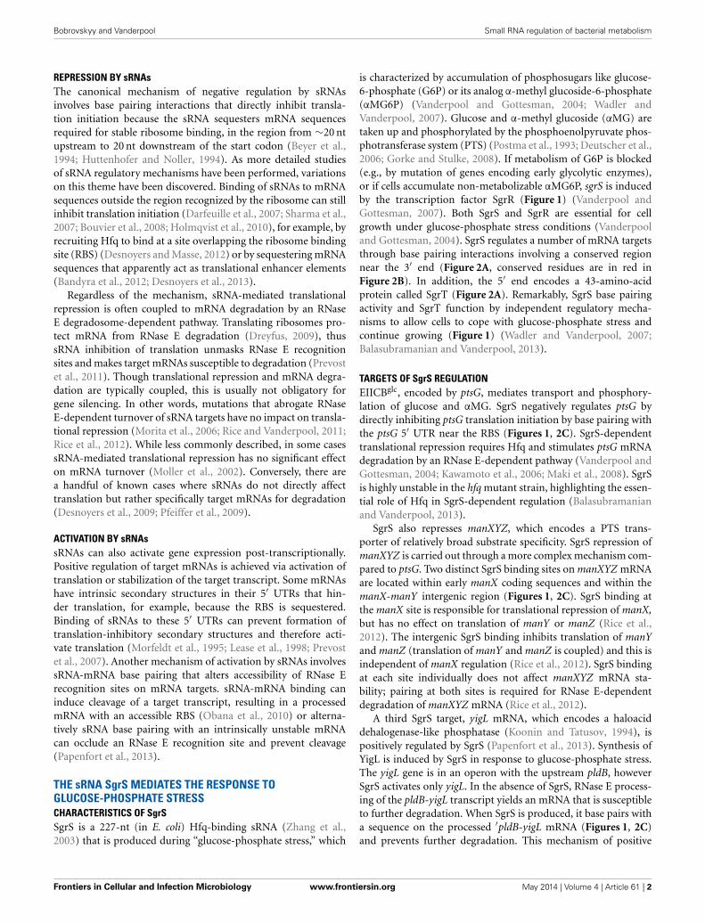
is characterized by accumulation of phosphosugars like glucose-6-phosphate (G6P) or its analog α-methyl glucoside-6-phosphate(αMG6P) (Vanderpool and Gottesman, 2004; Wadler andVanderpool, 2007). Glucose and α-methyl glucoside (αMG) aretaken up and phosphorylated by the phosphoenolpyruvate phos-photransferase system (PTS) (Postma et al., 1993; Deutscher et al.,2006; Gorke and Stulke, 2008). If metabolism of G6P is blocked(e.g., by mutation of genes encoding early glycolytic enzymes),or if cells accumulate non-metabolizable αMG6P, sgrS is inducedby the transcription factor SgrR (Figure 1) (Vanderpool andGottesman, 2007). Both SgrS and SgrR are essential for cellgrowth under glucose-phosphate stress conditions (Vanderpooland Gottesman, 2004). SgrS regulates a number of mRNA targetsthrough base pairing interactions involving a conserved regionnear the 3′ end (Figure 2A, conserved residues are in red inFigure 2B). In addition, the 5′ end encodes a 43-amino-acidprotein called SgrT (Figure 2A). Remarkably, SgrS base pairingactivity and SgrT function by independent regulatory mecha-nisms to allow cells to cope with glucose-phosphate stress andcontinue growing (Figure 1) (Wadler and Vanderpool, 2007;Balasubramanian and Vanderpool, 2013).
TARGETS OF SgrS REGULATIONEIICBglc, encoded by ptsG, mediates transport and phosphory-lation of glucose and αMG. SgrS negatively regulates ptsG bydirectly inhibiting ptsG translation initiation by base pairing withthe ptsG 5′ UTR near the RBS (Figures 1, 2C). SgrS-dependenttranslational repression requires Hfq and stimulates ptsG mRNAdegradation by an RNase E-dependent pathway (Vanderpool andGottesman, 2004; Kawamoto et al., 2006; Maki et al., 2008). SgrSis highly unstable in the hfq mutant strain, highlighting the essen-tial role of Hfq in SgrS-dependent regulation (Balasubramanianand Vanderpool, 2013).
SgrS also represses manXYZ, which encodes a PTS trans-porter of relatively broad substrate specificity. SgrS repression ofmanXYZ is carried out through a more complex mechanism com-pared to ptsG. Two distinct SgrS binding sites on manXYZ mRNAare located within early manX coding sequences and within themanX-manY intergenic region (Figures 1, 2C). SgrS binding atthe manX site is responsible for translational repression of manX,but has no effect on translation of manY or manZ (Rice et al.,2012). The intergenic SgrS binding inhibits translation of manYand manZ (translation of manY and manZ is coupled) and this isindependent of manX regulation (Rice et al., 2012). SgrS bindingat each site individually does not affect manXYZ mRNA sta-bility; pairing at both sites is required for RNase E-dependentdegradation of manXYZ mRNA (Rice et al., 2012).
A third SgrS target, yigL mRNA, which encodes a haloaciddehalogenase-like phosphatase (Koonin and Tatusov, 1994), ispositively regulated by SgrS (Papenfort et al., 2013). Synthesis ofYigL is induced by SgrS in response to glucose-phosphate stress.The yigL gene is in an operon with the upstream pldB, howeverSgrS activates only yigL. In the absence of SgrS, RNase E process-ing of the pldB-yigL transcript yields an mRNA that is susceptibleto further degradation. When SgrS is produced, it base pairs witha sequence on the processed ′pldB-yigL mRNA (Figures 1, 2C)and prevents further degradation. This mechanism of positive
Frontiers in Cellular and Infection Microbiology www.frontiersin.org May 2014 | Volume 4 | Article 61 | 2
Bobrovskyy and Vanderpool Small RNA regulation of bacterial metabolism
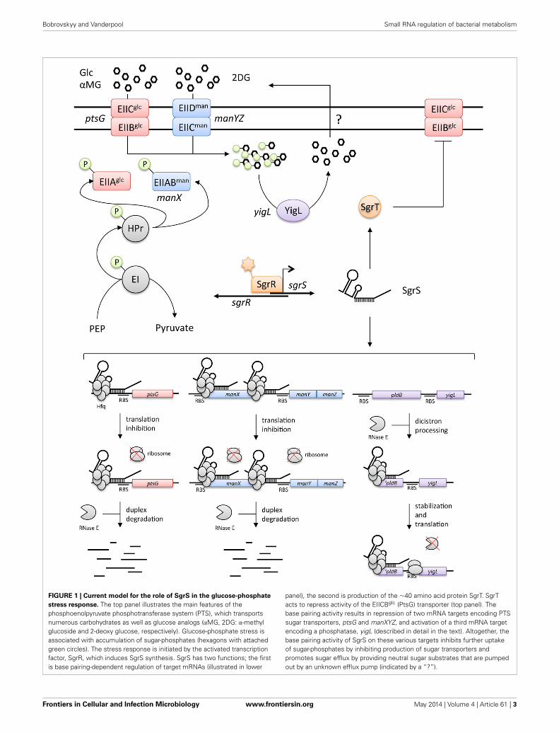
FIGURE 1 | Current model for the role of SgrS in the glucose-phosphate
stress response. The top panel illustrates the main features of thephosphoenolpyruvate phosphotransferase system (PTS), which transportsnumerous carbohydrates as well as glucose analogs (αMG, 2DG: α-methylglucoside and 2-deoxy glucose, respectively). Glucose-phosphate stress isassociated with accumulation of sugar-phosphates (hexagons with attachedgreen circles). The stress response is initiated by the activated transcriptionfactor, SgrR, which induces SgrS synthesis. SgrS has two functions; the firstis base pairing-dependent regulation of target mRNAs (illustrated in lower
panel), the second is production of the ∼40 amino acid protein SgrT. SgrTacts to repress activity of the EIICBglc (PtsG) transporter (top panel). Thebase pairing activity results in repression of two mRNA targets encoding PTSsugar transporters, ptsG and manXYZ, and activation of a third mRNA targetencoding a phosphatase, yigL (described in detail in the text). Altogether, thebase pairing activity of SgrS on these various targets inhibits further uptakeof sugar-phosphates by inhibiting production of sugar transporters andpromotes sugar efflux by providing neutral sugar substrates that are pumpedout by an unknown efflux pump (indicated by a “?”).
Frontiers in Cellular and Infection Microbiology www.frontiersin.org May 2014 | Volume 4 | Article 61 | 3
Bobrovskyy and Vanderpool Small RNA regulation of bacterial metabolism
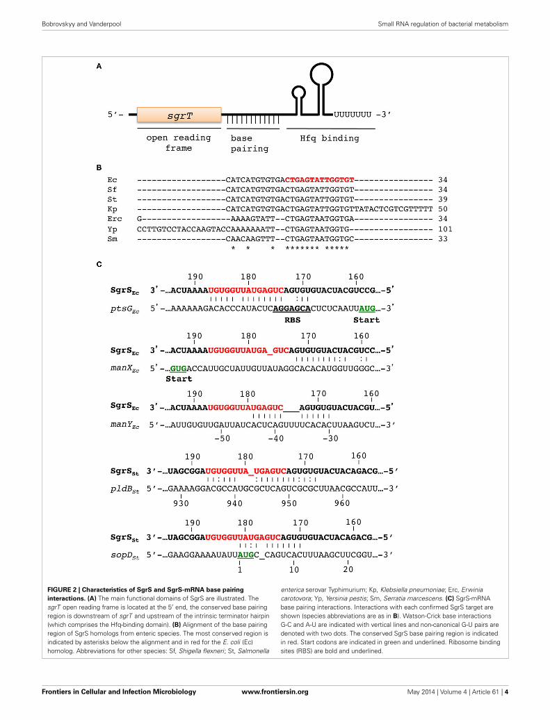
FIGURE 2 | Characteristics of SgrS and SgrS-mRNA base pairing
interactions. (A) The main functional domains of SgrS are illustrated. ThesgrT open reading frame is located at the 5′ end, the conserved base pairingregion is downstream of sgrT and upstream of the intrinsic terminator hairpin(which comprises the Hfq-binding domain). (B) Alignment of the base pairingregion of SgrS homologs from enteric species. The most conserved region isindicated by asterisks below the alignment and in red for the E. coli (Ec)homolog. Abbreviations for other species: Sf, Shigella flexneri; St, Salmonella
enterica serovar Typhimurium; Kp, Klebsiella pneumoniae; Erc, Erwiniacarotovora; Yp, Yersinia pestis; Sm, Serratia marcescens. (C) SgrS-mRNAbase pairing interactions. Interactions with each confirmed SgrS target areshown (species abbreviations are as in B). Watson-Crick base interactionsG-C and A-U are indicated with vertical lines and non-canonical G-U pairs aredenoted with two dots. The conserved SgrS base pairing region is indicatedin red. Start codons are indicated in green and underlined. Ribosome bindingsites (RBS) are bold and underlined.
Frontiers in Cellular and Infection Microbiology www.frontiersin.org May 2014 | Volume 4 | Article 61 | 4
Bobrovskyy and Vanderpool Small RNA regulation of bacterial metabolism
regulation is unusual in that an initial processing event is requiredto allow SgrS access to its binding site because cleavage withinpldB frees the pldB region from translating ribosomes (Papenfortet al., 2013). Moreover, activation of yigL by SgrS is translation-independent. SgrS stabilizes yigL mRNA by occluding a specificRNase E cleavage site upstream of the yigL coding region, notby enhancing yigL translation (Papenfort et al., 2013). A similartranslation-independent mechanism of mRNA stabilization wasrecently described for the RydC sRNA-cfa mRNA regulatory pair(Frohlich et al., 2013).
GLUCOSE-PHOSPHATE STRESS PHYSIOLOGYTargets of SgrS include sugar transporters and a sugar phos-phatase. SgrS-mediated repression of sugar transporters dimin-ishes cells’ capacity to take up sugars and therefore reducesfurther phosphosugar accumulation (Figure 1). However, thiseffect is not immediate: PtsG protein has a half-life of ∼80 min(Papenfort et al., 2013), so merely stopping new synthesis ofPtsG would not provide a fast remedy for the problem of phos-phosugar accumulation. The activation of YigL synthesis bySgrS addresses this problem since dephosphorylation of sug-ars allows their efflux (Figure 1) (Winkler, 1971; Haguenauerand Kepes, 1972; Papenfort et al., 2013). Growth competi-tion experiments between wild-type and sgrS mutants pro-vided insight into how regulation of different SgrS targetscontributes to stress resistance and growth during glucose-phosphate stress (Sun and Vanderpool, 2013). When cells arestressed while growing in rich medium, SgrS-mediated regu-lation of ptsG mRNA alone is sufficient to confer wild-typelevels of growth. In contrast, cells stressed in minimal mediaare far more growth inhibited, and repression of ptsG aloneis not sufficient to rescue growth. In minimal media stressconditions, repression of ptsG and activation of yigL are nec-essary, but not sufficient for full growth rescue (Sun andVanderpool, 2013). These findings illustrate the poorly under-stood influence of nutrient availability on the severity of glucose-phosphate stress. Moreover, these results highlight the fact thatadditional unknown SgrS targets are involved in the stressresponse.
Phosphosugar intermediates of central metabolism provideprecursors for biomass and energy, yet, as illustrated by glucose-phosphate stress, excessive accumulation of phosphosugars isdetrimental to cell growth. Other types of phosphosugar stressalso cause growth inhibition or cell lysis (Yarmolinsky et al., 1959;Englesberg et al., 1962; Irani and Maitra, 1977; Lee et al., 2009).In most cases, the mechanisms responsible for phosphosugar-associated inhibition or lysis have not been defined. However,recent work suggests that in some cases phosphosugars them-selves are not directly inhibitory. Rather, accumulation of phos-phosugars is accompanied by depletion of other metabolites,and stress is ameliorated by supplementation with the limit-ing metabolites (Lee et al., 2009, 2013; Richards et al., 2013).Glucose-phosphate stress is so far associated with accumula-tion of a few sugar-phosphate intermediates of upper glycolysis(Morita et al., 2003; Vanderpool and Gottesman, 2004; Sun andVanderpool, 2013). A recent study implicates depletion of inter-mediates of lower glycolysis, particularly phosphoenolpyruvate
(PEP) as an important cause of glucose-phosphate stress. WhenαMG is taken up and phosphorylated, it cannot be metabo-lized to replenish glycolytic intermediates. Thus, PEP utilizedto drive αMG uptake is not replaced via glycolytic metabolism.Under these conditions, SgrS regulation of target mRNAs andproduction of SgrT limits PEP consumption by reducing lev-els and activity of PtsG (Figure 1). In sgrS mutants, exposureto αMG results in strong growth inhibition (Vanderpool andGottesman, 2004; Richards et al., 2013) that is largely reversedby supplementing stressed cultures with glycolytic intermedi-ates (Richards et al., 2013). The ratios of PEP to pyruvate seemto be particularly relevant for growth during glucose-phosphatestress. Increasing pyruvate levels during stress results in lysisof sgrS mutant cells, whereas increasing PEP levels rescues cellgrowth (Richards et al., 2013). The observation that stress (andgrowth inhibition) is more severe when cells are growing in min-imal compared to rich media is also consistent with metabolitedepletion as an underlying cause of glucose-phosphate stress. Inrich media, cells do not have to synthesize many biosyntheticintermediates. In contrast, growth in minimal media requires denovo biosynthesis of amino acids. Thus, depletion of glycolyticintermediates during glucose-phosphate stress would have moresevere effects on growth under conditions where these same inter-mediates are needed as precursors for biosynthesis. Consistentwith this idea, supplementation of minimal media with aminoacids improves stress recovery in minimal medium (Sun andVanderpool, 2013).
The transcription factor SgrR also plays an important, butnot fully characterized role in glucose-phosphate stress physiol-ogy. SgrR activates expression of sgrS and at least two other genesduring glucose-phosphate stress: setA, encoding an efflux pump(Liu et al., 1999; Sun and Vanderpool, 2011), and alaC (for-merly yfdZ), a glutamate-pyruvate aminotransferase (Vanderpooland Gottesman, 2007; Kim et al., 2010). The role of alaC inhelping cells recover from glucose-phosphate stress is unknown.In contrast, setA, which is encoded just downstream of sgrS, isimportant for growth recovery under certain stress conditions(Sun and Vanderpool, 2011). Given its function as an effluxpump, the hypothesis that SetA was responsible for export ofαMG was tested, but was not supported (Sun and Vanderpool,2011). Thus, the role of SetA in glucose-phosphate stress alsoremains elusive.
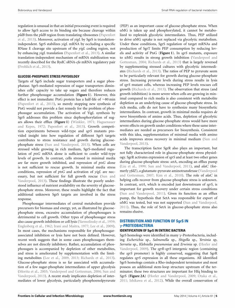
DISTRIBUTION AND FUNCTION OF SgrS INγ-PROTEOBACTERIAIDENTIFICATION OF SgrS IN ENTERIC BACTERIASgrS homologs were identified in many γ-Proteobacteria, includ-ing Escherichia sp., Salmonella sp., Shigella sp., Yersinia sp,Serratia sp., Klebsiella pneumoniae and Erwinia sp. (Horler andVanderpool, 2009). The sgrR-sgrS intergenic region (containingthe sgrS promoter) is highly conserved, suggesting that SgrRregulates sgrS expression in all these organisms. All identifiedSgrS homologs contain a Rho-independent terminator and mostpossess an additional stem-loop structure upstream of the ter-minator; these two structures are important for Hfq binding toSgrS (Figure 2A) (Horler and Vanderpool, 2009; Otaka et al.,2011; Ishikawa et al., 2012). While the overall conservation of
Frontiers in Cellular and Infection Microbiology www.frontiersin.org May 2014 | Volume 4 | Article 61 | 5
Bobrovskyy and Vanderpool Small RNA regulation of bacterial metabolism
SgrS is low, a short stretch of ∼13 nts near the SgrS 3′ endis nearly invariant (Figure 2B). This SgrS sequence is comple-mentary to the translation initiation regions of ptsG mRNAsin all species where an SgrS homolog was found (Horler andVanderpool, 2009). Mutation of residues G176 and G178 withinthe conserved region of E. coli SgrS abrogates SgrS-mediatedrepression of ptsG mRNA and prevents recovery from glucose-phosphate stress (Maki et al., 2008). Introduction of analogousmutations in the conserved regions of SgrS homologs fromSalmonella, E. carotovora, Y. pestis and K. pneumoniae similarlyprevented regulation of ptsG (Wadler and Vanderpool, 2009).Regulation of other targets is less well conserved among SgrShomologs. The SgrS sequences required for base pairing withmanX are upstream of the conserved region (Figure 2C) andare poorly conserved among SgrS homologs. SgrS homologsfrom Salmonella and K. pneumoniae have the same predictedSgrS-manX base pairing interaction and manX translation is reg-ulated as expected. In contrast, E. carotovora and Y. pestis SgrShomologs have changes in the manX pairing site resulting in lossof complementarity to their cognate manX and were accordinglyshown not to regulate manX translation (Rice and Vanderpool,2011).
CONSERVATION OF SgrTWhile the exact molecular function of SgrT has not been reported,available data strongly suggest that this small protein interactsdirectly with PtsG protein to inhibit its activity (Wadler andVanderpool, 2007). Most SgrS homologs contain open readingframes similar in size to E. coli SgrT (∼40 amino acids) (Horlerand Vanderpool, 2009). While the primary amino acid sequenceof putative SgrT homologs was not well conserved, homologsfrom Salmonella, Klebsiella, and Erwinia were functional whenexpressed in an E. coli sgrST mutant (Wadler and Vanderpool,2009). Interestingly, some species with SgrS homologs appear tolack a functional SgrT. In Yersinia sp., SgrS appears to be trun-cated at the 5′ end, and SgrS from Yersinia species ranges in sizefrom ∼85 to 140 nt and lacks the sgrT open reading frame. Inpathogenic E. coli O157:H7 strains, a point mutation in the SgrS5′ region alters the sgrT start codon, presumably abrogating SgrTproduction in these strains.
Differential presence and absence of SgrT in organisms thatpossess SgrS led to a closer comparison of E. coli K12 andSalmonella SgrS sRNAs. In E. coli K12, sgrT alone (without theregion of SgrS involved in base pairing with mRNAs) was notsufficient to allow growth rescue during glucose-phosphate stressconditions. This is in part due to very low levels of SgrT producedfrom the native sgrS allele in E. coli (Wadler and Vanderpool,2009). E. coli SgrS has a sequence in the 5′ region that formsa structure that inhibits sgrT translation. On the other hand,Salmonella SgrS does not have the same inhibitory structureand therefore produces more SgrT than E. coli SgrS (Wadlerand Vanderpool, 2009; Balasubramanian and Vanderpool, 2013).While native levels of SgrT production have not been inves-tigated in Erwinia or Klebsiella species, it was observed thatectopic production of SgrT homologs from these organisms inan E. coli sgrST mutant rescued growth during glucose-phosphatestress (Wadler and Vanderpool, 2009). Thus, SgrT is functionally
conserved when it is present, but levels of SgrT production varyamong bacteria.
SgrS REGULATION OF sopD mRNAAlthough SgrS is conserved among enteric bacteria, divergencein primary sequence has resulted in species-specific target regu-lons, exemplified by the finding that Erwinia and Yersinia SgrShomologs do not regulate their cognate manXYZ homologs (Riceand Vanderpool, 2011). Another instance of species-specific reg-ulation by SgrS is regulation of the Salmonella-specific gene sopD(Papenfort et al., 2012). SopD is an effector delivered to hostcells through the Type 3 Secretion Systems (T3SSs) encodedon Salmonella pathogenicity island (SPI)-1 and SPI-2 (Brumellet al., 2003) and it functions as a general virulence factor inmice (Jiang et al., 2004; Bakowski et al., 2007). Regulation ofsopD by SgrS involves base pairing interactions between the con-served region of SgrS and the early coding sequence of sopDmRNA (Figure 2C); the interaction inhibits translation initia-tion and stimulates sopD mRNA degradation (Papenfort et al.,2012). Interestingly, Salmonella encodes a second SopD protein,SopD2, which shares 42% identity with SopD and likely arosefrom a duplication (Jiang et al., 2004). The predicted SgrS-sopD2base pairing interaction differs from SgrS-sopD at only a singleposition, a wobble G:U base pair instead of the G:C base pair.Remarkably, this interaction that differs by only a single hydro-gen bond prevents regulation of sopD2 by SgrS (Papenfort et al.,2012).
While the biological significance of sopD regulation by SgrSis not yet clear, the inclusion of sopD in the Salmonella SgrSregulon illustrates plasticity in the evolution of sRNA regulons.The presence of sgrR-sgrS-sgrT in the same genomic context inpathogenic and non-pathogenic γ-proteobacteria (Horler andVanderpool, 2009) suggests that this is an ancestral, or “core”RNA among these organisms. Yet, this core sRNA has acquiredthe ability to regulate a gene that was horizontally acquired bySalmonella. Studies of other SgrS homologs in pathogenic andnon-pathogenic enteric bacteria will surely shed light on thebreadth of regulatory activities of this fascinating dual-functionsRNA.
REFERENCESBakowski, M. A., Cirulis, J. T., Brown, N. F., Finlay, B. B., and Brumell, J. H.
(2007). SopD acts cooperatively with SopB during Salmonella enterica serovarTyphimurium invasion. Cell. Microbiol. 9, 2839–2855. doi: 10.1111/j.1462-5822.2007.01000.x
Balasubramanian, D., and Vanderpool, C. K. (2013). Deciphering the inter-play between two independent functions of the small RNA regulator SgrS inSalmonella. J. Bacteriol. 195, 4620–4630. doi: 10.1128/JB.00586-13
Bandyra, K. J., Said, N., Pfeiffer, V., Gorna, M. W., Vogel, J., and Luisi, B. F.(2012). The seed region of a small RNA drives the controlled destruction ofthe target mRNA by the endoribonuclease RNase E. Mol. Cell 47, 943–953. doi:10.1016/j.molcel.2012.07.015
Belasco, J. G. (2010). All things must pass: contrasts and commonalities in eukary-otic and bacterial mRNA decay. Nat. Rev. Mol. Cell Biol. 11, 467–478. doi:10.1038/nrm2917
Beyer, D., Skripkin, E., Wadzack, J., and Nierhaus, K. H. (1994). How the ribo-some moves along the mRNA during protein synthesis. J. Biol. Chem. 269,30713–30717.
Bouvier, M., Sharma, C. M., Mika, F., Nierhaus, K. H., and Vogel, J. (2008). SmallRNA binding to 5′ mRNA coding region inhibits translational initiation. Mol.Cell 32, 827–837. doi: 10.1016/j.molcel.2008.10.027
Frontiers in Cellular and Infection Microbiology www.frontiersin.org May 2014 | Volume 4 | Article 61 | 6
Bobrovskyy and Vanderpool Small RNA regulation of bacterial metabolism
Brumell, J. H., Kujat-Choy, S., Brown, N. F., Vallance, B. A., Knodler, L. A., andFinlay, B. B. (2003). SopD2 is a novel type III secreted effector of Salmonellatyphimurium that targets late endocytic compartments upon delivery into hostcells. Traffic 4, 36–48. doi: 10.1034/j.1600-0854.2003.40106.x
Carpousis, A. J. (2007). The RNA degradosome of Escherichia coli: an mRNA-degrading machine assembled on RNase E. Annu. Rev. Microbiol. 61, 71–87. doi:10.1146/annurev.micro.61.080706.093440
Carpousis, A. J., Luisi, B. F., and McDowall, K. J. (2009). Endonucleolytic initiationof mRNA decay in Escherichia coli. Prog. Mol. Biol. Transl. Sci. 85, 91–135. doi:10.1016/S0079-6603(08)00803-9
Darfeuille, F., Unoson, C., Vogel, J., and Wagner, E. G. (2007). An antisenseRNA inhibits translation by competing with standby ribosomes. Mol. Cell 26,381–392. doi: 10.1016/j.molcel.2007.04.003
Desnoyers, G., Bouchard, M. P., and Masse, E. (2013). New insights into small RNA-dependent translational regulation in prokaryotes. Trends Genet. 29, 92–98. doi:10.1016/j.tig.2012.10.004
Desnoyers, G., and Masse, E. (2012). Noncanonical repression of translation initia-tion through small RNA recruitment of the RNA chaperone Hfq. Genes Dev. 26,726–739. doi: 10.1101/gad.182493.111
Desnoyers, G., Morissette, A., Prevost, K., and Masse, E. (2009). Small RNA-induced differential degradation of the polycistronic mRNA iscRSUA. EMBOJ. 28, 1551–1561. doi: 10.1038/emboj.2009.116
Deutscher, J., Francke, C., and Postma, P. W. (2006). How phosphotransferasesystem-related protein phosphorylation regulates carbohydrate metabolism inbacteria. Microbiol. Mol. Biol. Rev. 70, 939–1031. doi: 10.1128/MMBR.00024-06
Dreyfus, M. (2009). Killer and protective ribosomes. Prog. Mol. Biol. Transl. Sci. 85,423–466. doi: 10.1016/S0079-6603(08)00811-8
Englesberg, E., Anderson, R. L., Weinberg, R., Lee, N., Hoffee, P., Huttenhauer,G., et al. (1962). L-Arabinose-sensitive, L-ribulose 5-phosphate 4-epimerase-deficient mutants of Escherichia coli. J. Bacteriol. 84, 137–146.
Fender, A., Elf, J., Hampel, K., Zimmermann, B., and Wagner, E. G. (2010). RNAsactively cycle on the Sm-like protein Hfq. Genes Dev. 24, 2621–2626. doi:10.1101/gad.591310
Frohlich, K. S., Papenfort, K., Fekete, A., and Vogel, J. (2013). A small RNA activatesCFA synthase by isoform-specific mRNA stabilization. EMBO J. 32, 2963–2979.doi: 10.1038/emboj.2013.222
Gorke, B., and Stulke, J. (2008). Carbon catabolite repression in bacteria: manyways to make the most out of nutrients. Nat. Rev. Microbiol. 6, 613–624. doi:10.1038/nrmicro1932
Haguenauer, R., and Kepes, A. (1972). NaF inhibition of phosphorylation anddephosphorylation involved in -methyl-D glucoside transport in E. coli K 12.A pH dependent phenomenon sensitive to uncoupling agents. Biochimie 54,505–512. doi: 10.1016/S0300-9084(72)80235-9
Holmqvist, E., Reimegard, J., Sterk, M., Grantcharova, N., Romling, U., andWagner, E. G. (2010). Two antisense RNAs target the transcriptional reg-ulator CsgD to inhibit curli synthesis. EMBO J. 29, 1840–1850. doi:10.1038/emboj.2010.73
Hopkins, J. F., Panja, S., and Woodson, S. A. (2011). Rapid binding and releaseof Hfq from ternary complexes during RNA annealing. Nucleic Acids Res. 39,5193–5202. doi: 10.1093/nar/gkr062
Horler, R. S., and Vanderpool, C. K. (2009). Homologs of the small RNA SgrS arebroadly distributed in enteric bacteria but have diverged in size and sequence.Nucleic Acids Res. 37, 5465–5476. doi: 10.1093/nar/gkp501
Huttenhofer, A., and Noller, H. F. (1994). Footprinting mRNA-ribosome complexeswith chemical probes. EMBO J. 13, 3892–3901.
Irani, M. H., and Maitra, P. K. (1977). Properties of Escherichia coli mutantsdeficient in enzymes of glycolysis. J. Bacteriol. 132, 398–410.
Ishikawa, H., Otaka, H., Maki, K., Morita, T., and Aiba, H. (2012). The functionalHfq-binding module of bacterial sRNAs consists of a double or single hair-pin preceded by a U-rich sequence and followed by a 3′ poly(U) tail. RNA 18,1062–1074. doi: 10.1261/rna.031575.111
Jiang, X., Rossanese, O. W., Brown, N. F., Kujat-Choy, S., Galan, J. E., Finlay, B.B., et al. (2004). The related effector proteins SopD and SopD2 from Salmonellaenterica serovar Typhimurium contribute to virulence during systemic infectionof mice. Mol. Microbiol. 54, 1186–1198. doi: 10.1111/j.1365-2958.2004.04344.x
Kawamoto, H., Koide, Y., Morita, T., and Aiba, H. (2006). Base-pairing require-ment for RNA silencing by a bacterial small RNA and acceleration ofduplex formation by Hfq. Mol. Microbiol. 61, 1013–1022. doi: 10.1111/j.1365-2958.2006.05288.x
Kim, S. H., Schneider, B. L., and Reitzer, L. (2010). Genetics and regulation ofthe major enzymes of alanine synthesis in Escherichia coli. J. Bacteriol. 192,5304–5311. doi: 10.1128/JB.00738-10
Koonin, E. V., and Tatusov, R. L. (1994). Computer analysis of bacterial haloaciddehalogenases defines a large superfamily of hydrolases with diverse speci-ficity. Application of an iterative approach to database search. J. Mol. Biol. 244,125–132. doi: 10.1006/jmbi.1994.1711
Lease, R. A., Cusick, M. E., and Belfort, M. (1998). Riboregulation in Escherichiacoli: DsrA RNA acts by RNA:RNA interactions at multiple loci. Proc. Natl. Acad.Sci. U.S.A. 95, 12456–12461. doi: 10.1073/pnas.95.21.12456
Lee, S. J., Trostel, A., and Adhya, S. (2013). Metabolite changes signal genetic reg-ulatory mechanisms for robust cell behavior. MBio. 5, e00972–e00913. doi:10.1128/mBio.00972-13
Lee, S. J., Trostel, A., Le, P., Harinarayanan, R., Fitzgerald, P. C., and Adhya, S.(2009). Cellular stress created by intermediary metabolite imbalances. Proc.Natl. Acad. Sci. U.S.A. 106, 19515–19520. doi: 10.1073/pnas.0910586106
Link, T. M., Valentin-Hansen, P., and Brennan, R. G. (2009). Structure ofEscherichia coli Hfq bound to polyriboadenylate RNA. Proc. Natl. Acad. Sci.U.S.A. 106, 19292–19297. doi: 10.1073/pnas.0908744106
Liu, J. Y., Miller, P. F., Willard, J., and Olson, E. R. (1999). Functional and biochem-ical characterization of Escherichia coli sugar efflux transporters. J. Biol. Chem.274, 22977–22984. doi: 10.1074/jbc.274.33.22977
Maki, K., Morita, T., Otaka, H., and Aiba, H. (2010). A minimal base-pairing regionof a bacterial small RNA SgrS required for translational repression of ptsGmRNA. Mol. Microbiol. 76, 782–792. doi: 10.1111/j.1365-2958.2010.07141.x
Maki, K., Uno, K., Morita, T., and Aiba, H. (2008). RNA, but not proteinpartners, is directly responsible for translational silencing by a bacterial Hfq-binding small RNA. Proc. Natl. Acad. Sci. U.S.A. 105, 10332–10337. doi:10.1073/pnas.0803106105
Masse, E., Escorcia, F. E., and Gottesman, S. (2003). Coupled degradation of asmall regulatory RNA and its mRNA targets in Escherichia coli. Genes Dev. 17,2374–2383. doi: 10.1101/gad.1127103
Masse, E., and Gottesman, S. (2002). A small RNA regulates the expression of genesinvolved in iron metabolism in Escherichia coli. Proc. Natl. Acad. Sci. U.S.A. 99,4620–4625. doi: 10.1073/pnas.032066599
Mikulecky, P. J., Kaw, M. K., Brescia, C. C., Takach, J. C., Sledjeski, D. D., and Feig,A. L. (2004). Escherichia coli Hfq has distinct interaction surfaces for DsrA, rpoSand poly(A) RNAs. Nat. Struct. Mol. Biol. 11, 1206–1214. doi: 10.1038/nsmb858
Moller, T., Franch, T., Udesen, C., Gerdes, K., and Valentin-Hansen, P. (2002). Spot42 RNA mediates discoordinate expression of the E. coli galactose operon. GenesDev. 16, 1696–1706. doi: 10.1101/gad.231702
Morfeldt, E., Taylor, D., Von Gabain, A., and Arvidson, S. (1995). Activation ofalpha-toxin translation in Staphylococcus aureus by the trans-encoded anti-sense RNA, RNAIII. EMBO J. 14, 4569–4577.
Morita, T., El-Kazzaz, W., Tanaka, Y., Inada, T., and Aiba, H. (2003). Accumulationof glucose 6-phosphate or fructose 6-phosphate is responsible for destabi-lization of glucose transporter mRNA in Escherichia coli. J. Biol. Chem. 278,15608–15614. doi: 10.1074/jbc.M300177200
Morita, T., Maki, K., and Aiba, H. (2005). RNase E-based ribonucleoproteincomplexes: mechanical basis of mRNA destabilization mediated by bacterialnoncoding RNAs. Genes Dev. 19, 2176–2186. doi: 10.1101/gad.1330405
Morita, T., Mochizuki, Y., and Aiba, H. (2006). Translational repression is suf-ficient for gene silencing by bacterial small noncoding RNAs in the absenceof mRNA destruction. Proc. Natl. Acad. Sci. U.S.A. 103, 4858–4863. doi:10.1073/pnas.0509638103
Obana, N., Shirahama, Y., Abe, K., and Nakamura, K. (2010). Stabilization ofClostridium perfringens collagenase mRNA by VR-RNA-dependent cleavagein 5′ leader sequence. Mol. Microbiol. 77, 1416–1428. doi: 10.1111/j.1365-2958.2010.07258.x
Otaka, H., Ishikawa, H., Morita, T., and Aiba, H. (2011). PolyU tail of rho-independent terminator of bacterial small RNAs is essential for Hfq action. Proc.Natl. Acad. Sci. U.S.A. 108, 13059–13064. doi: 10.1073/pnas.1107050108
Papenfort, K., Podkaminski, D., Hinton, J. C., and Vogel, J. (2012). The ances-tral SgrS RNA discriminates horizontally acquired Salmonella mRNAs througha single G-U wobble pair. Proc. Natl. Acad. Sci. U.S.A. 109, E757–764. doi:10.1073/pnas.1119414109
Papenfort, K., Sun, Y., Miyakoshi, M., Vanderpool, C. K., and Vogel, J. (2013).Small RNA-mediated activation of sugar phosphatase mRNA regulates glucosehomeostasis. Cell 153, 426–437. doi: 10.1016/j.cell.2013.03.003
Frontiers in Cellular and Infection Microbiology www.frontiersin.org May 2014 | Volume 4 | Article 61 | 7
Bobrovskyy and Vanderpool Small RNA regulation of bacterial metabolism
Pfeiffer, V., Papenfort, K., Lucchini, S., Hinton, J., and Vogel, J. R. (2009). Codingsequence targeting by MicC RNA reveals bacterial mRNA silencing down-stream of translational initiation. Nat. Struct. Mol. Biol. 16, 840–846. doi:10.1038/nsmb.1631
Postma, P. W., Lengeler, J. W., and Jacobson, G. R. (1993). Phosphoenolpyruvate:carbohydrate phosphotransferase systems of bacteria. Microbiol. Rev. 57,543–594.
Prevost, K., Desnoyers, G., Jacques, J. F., Lavoie, F., and Masse, E. (2011). SmallRNA-induced mRNA degradation achieved through both translation block andactivated cleavage. Genes Dev. 25, 385–396. doi: 10.1101/gad.2001711
Prevost, K., Salvail, H., Desnoyers, G., Jacques, J. F., Phaneuf, E., and Masse, E.(2007). The small RNA RyhB activates the translation of shiA mRNA encodinga permease of shikimate, a compound involved in siderophore synthesis. Mol.Microbiol. 64, 1260–1273. doi: 10.1111/j.1365-2958.2007.05733.x
Rice, J. B., Balasubramanian, D., and Vanderpool, C. K. (2012). Small RNAbinding-site multiplicity involved in translational regulation of a polycistronicmRNA. Proc. Natl. Acad. Sci. U.S.A. 109, E2691–2698. doi: 10.1073/pnas.1207927109
Rice, J. B., and Vanderpool, C. K. (2011). The small RNA SgrS controls sugar-phosphate accumulation by regulating multiple PTS genes. Nucleic Acids Res.39, 3806–3819. doi: 10.1093/nar/gkq1219
Richards, G. R., Patel, M. V., Lloyd, C. R., and Vanderpool, C. K. (2013). Depletionof glycolytic intermediates plays a key role in glucose-phosphate stress inEscherichia coli. J. Bacteriol. 195, 4816–4825. doi: 10.1128/JB.00705-13
Salim, N. N., Faner, M. A., Philip, J. A., and Feig, A. L. (2012). Requirementof upstream Hfq-binding (ARN)x elements in glmS and the Hfq C-terminalregion for GlmS upregulation by sRNAs GlmZ and GlmY. Nucleic Acids Res. 40,8021–8032. doi: 10.1093/nar/gks392.
Salim, N. N., and Feig, A. L. (2010). An upstream Hfq binding site in the fhlAmRNA leader region facilitates the OxyS-fhlA interaction. PLoS ONE 5:e13028.doi: 10.1371/journal.pone.0013028.
Sharma, C. M., Darfeuille, F., Plantinga, T. H., and Vogel, J. (2007). A small RNAregulates multiple ABC transporter mRNAs by targeting C/A-rich elementsinside and upstream of ribosome-binding sites. Genes Dev. 21, 2804–2817. doi:10.1101/gad.447207
Sledjeski, D. D., Whitman, C., and Zhang, A. (2001). Hfq is necessary for reg-ulation by the untranslated RNA DsrA. J. Bacteriol. 183, 1997–2005. doi:10.1128/JB.183.6.1997-2005.2001
Soper, T. J., and Woodson, S. A. (2008). The rpoS mRNA leader recruitsHfq to facilitate annealing with DsrA sRNA. RNA. 14, 1907–1917. doi:10.1261/rna.1110608.
Soper, T., Mandin, P., Majdalani, N., Gottesman, S., and Woodson, S. A. (2010).Positive regulation by small RNAs and the role of Hfq. Proc. Natl. Acad. Sci.U.S.A. 107, 9602–9607. doi: 10.1073/pnas.1004435107
Sun, Y., and Vanderpool, C. K. (2011). Regulation and function of Escherichia colisugar efflux transporter A (SetA) during glucose-phosphate stress. J. Bacteriol.193, 143–153. doi: 10.1128/JB.01008-10
Sun, Y., and Vanderpool, C. K. (2013). Physiological consequences of multiple-target regulation by the small RNA SgrS in Escherichia coli. J. Bacteriol. 195,4804–4815. doi: 10.1128/JB.00722-13
Valentin-Hansen, P., Eriksen, M., and Udesen, C. (2004). The bacterial Sm-like pro-tein Hfq: a key player in RNA transactions. Mol. Microbiol. 51, 1525–1533. doi:10.1111/j.1365-2958.2003.03935.x
Vanderpool, C. K., and Gottesman, S. (2004). Involvement of a novel tran-scriptional activator and small RNA in post-transcriptional regulation of theglucose phosphoenolpyruvate phosphotransferase system. Mol. Microbiol. 54,1076–1089. doi: 10.1111/j.1365-2958.2004.04348.x
Vanderpool, C. K., and Gottesman, S. (2007). The novel transcription factorSgrR coordinates the response to glucose-phosphate stress. J. Bacteriol. 189,2238–2248. doi: 10.1128/JB.01689-06
Wadler, C. S., and Vanderpool, C. K. (2007). A dual function for a bacterialsmall RNA: SgrS performs base pairing-dependent regulation and encodes afunctional polypeptide. Proc. Natl. Acad. Sci. U.S.A. 104, 20454–20459. doi:10.1073/pnas.0708102104
Wadler, C. S., and Vanderpool, C. K. (2009). Characterization of homologs of thesmall RNA SgrS reveals diversity in function. Nucleic Acids Res. 37, 5477–5485.doi: 10.1093/nar/gkp591
Winkler, H. H. (1971). Efflux and the steady state in alpha-methylglucosidetransport in Escherichia coli. J. Bacteriol. 106, 362–368.
Yarmolinsky, M. B., Wiesmeyer, H., Kalckar, H. M., and Jordan, E. (1959).Hereditary defects in galactose metabolism in Escherichia Coli mutants, Ii.Galactose-induced sensitivity. Proc. Natl. Acad. Sci. U.S.A. 45, 1786–1791. doi:10.1073/pnas.45.12.1786
Zhang, A., Schu, D. J., Tjaden, B. C., Storz, G., and Gottesman, S. (2013).Mutations in interaction surfaces differentially impact E. coli Hfq associationwith small RNAs and their mRNA targets. J. Mol. Biol. 425, 3678–3697. doi:10.1016/j.jmb.2013.01.006
Zhang, A., Wassarman, K. M., Ortega, J., Steven, A. C., and Storz, G. (2002). TheSm-like Hfq protein increases OxyS RNA interaction with target mRNAs. Mol.Cell 9, 11–22. doi: 10.1016/S1097-2765(01)00437-3
Zhang, A., Wassarman, K. M., Rosenow, C., Tjaden, B. C., Storz, G., andGottesman, S. (2003). Global analysis of small RNA and mRNA targets of Hfq.Mol. Microbiol. 50, 1111–1124. doi: 10.1046/j.1365-2958.2003.03734.x
Conflict of Interest Statement: The authors declare that the research was con-ducted in the absence of any commercial or financial relationships that could beconstrued as a potential conflict of interest.
Received: 05 March 2014; accepted: 22 April 2014; published online: 08 May 2014.Citation: Bobrovskyy M and Vanderpool CK (2014) The small RNA SgrS: roles inmetabolism and pathogenesis of enteric bacteria. Front. Cell. Infect. Microbiol. 4:61.doi: 10.3389/fcimb.2014.00061This article was submitted to the journal Frontiers in Cellular and InfectionMicrobiology.Copyright © 2014 Bobrovskyy and Vanderpool. This is an open-access article dis-tributed under the terms of the Creative Commons Attribution License (CC BY). Theuse, distribution or reproduction in other forums is permitted, provided the originalauthor(s) or licensor are credited and that the original publication in this jour-nal is cited, in accordance with accepted academic practice. No use, distribution orreproduction is permitted which does not comply with these terms.
Frontiers in Cellular and Infection Microbiology www.frontiersin.org May 2014 | Volume 4 | Article 61 | 8