Embed Size (px)
Citation preview
The Skeleton
Dr. Ali Ebneshahidi
ebneshahidi
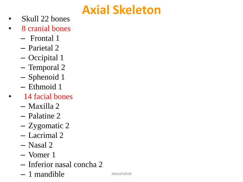
Axial Skeleton• Skull 22 bones
• 8 cranial bones
– Frontal 1
– Parietal 2
– Occipital 1
– Temporal 2
– Sphenoid 1
– Ethmoid 1
• 14 facial bones
– Maxilla 2
– Palatine 2
– Zygomatic 2
– Lacrimal 2
– Nasal 2
– Vomer 1
– Inferior nasal concha 2
– 1 mandible ebneshahidi
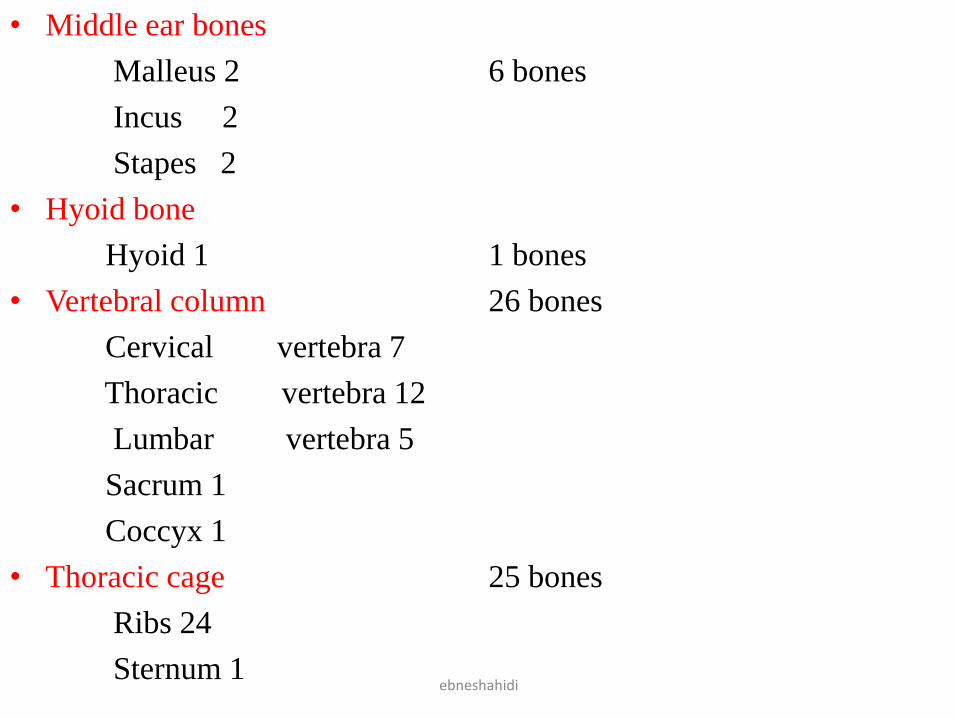
• Middle ear bones
Malleus 2 6 bones
Incus 2
Stapes 2
• Hyoid bone
Hyoid 1 1 bones
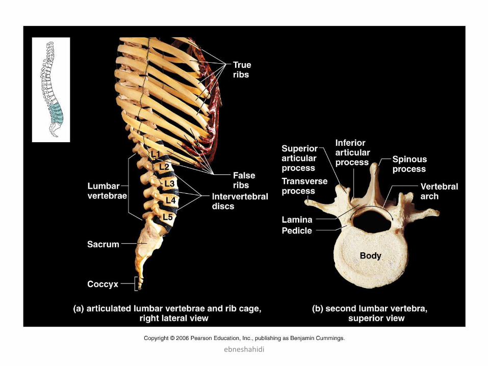
• Vertebral column 26 bones
Cervical vertebra 7
Thoracic vertebra 12
Lumbar vertebra 5
Sacrum 1
Coccyx 1
• Thoracic cage 25 bones
Ribs 24
Sternum 1ebneshahidi
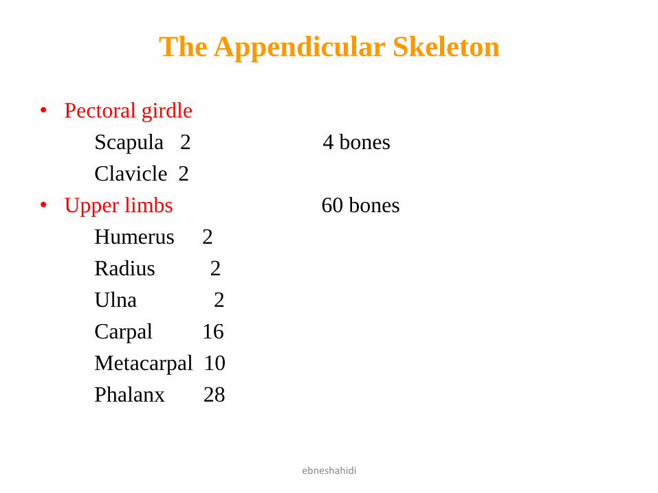
The Appendicular Skeleton
• Pectoral girdle
Scapula 2 4 bones
Clavicle 2
• Upper limbs 60 bones
Humerus 2
Radius 2
Ulna 2
Carpal 16
Metacarpal 10
Phalanx 28
ebneshahidi
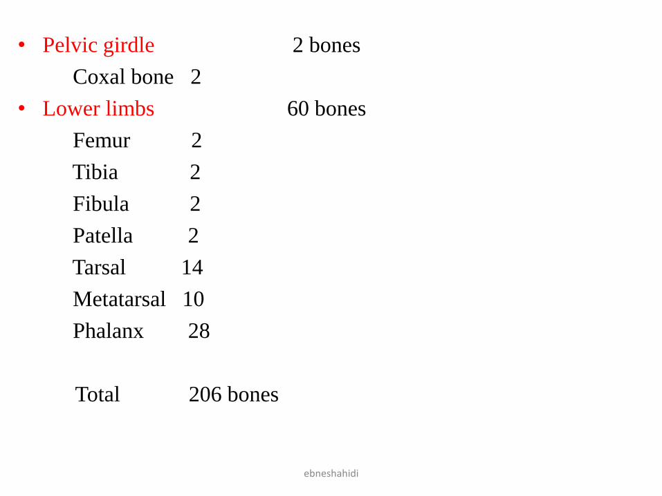
• Pelvic girdle 2 bones
Coxal bone 2
• Lower limbs 60 bones
Femur 2
Tibia 2
Fibula 2
Patella 2
Tarsal 14
Metatarsal 10
Phalanx 28
Total 206 bones
ebneshahidi
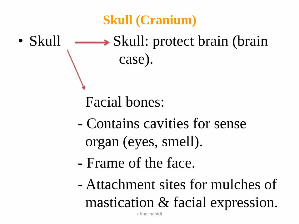
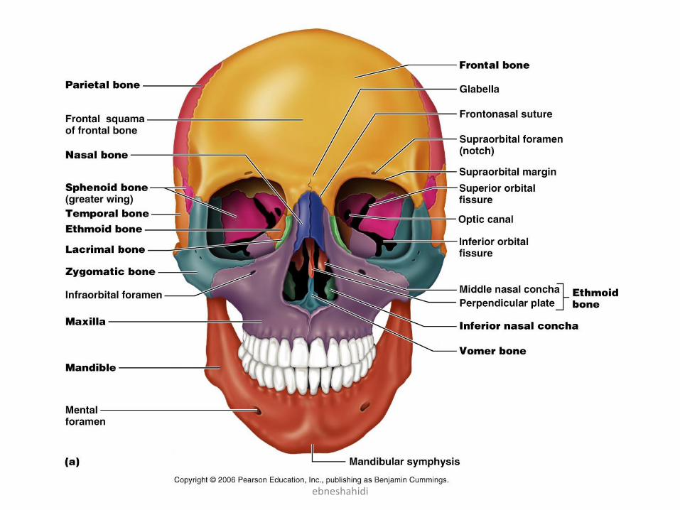
Skull (Cranium)
• Skull Skull: protect brain (brain
case).
Facial bones:
- Contains cavities for sense
organ (eyes, smell).
- Frame of the face.
- Attachment sites for mulches of
mastication & facial expression.ebneshahidi
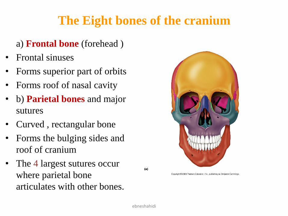
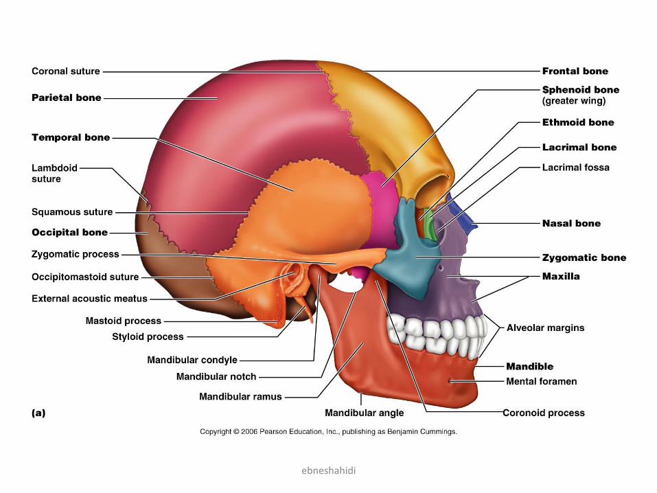
The Eight bones of the cranium
a) Frontal bone (forehead )
• Frontal sinuses
• Forms superior part of orbits
• Forms roof of nasal cavity
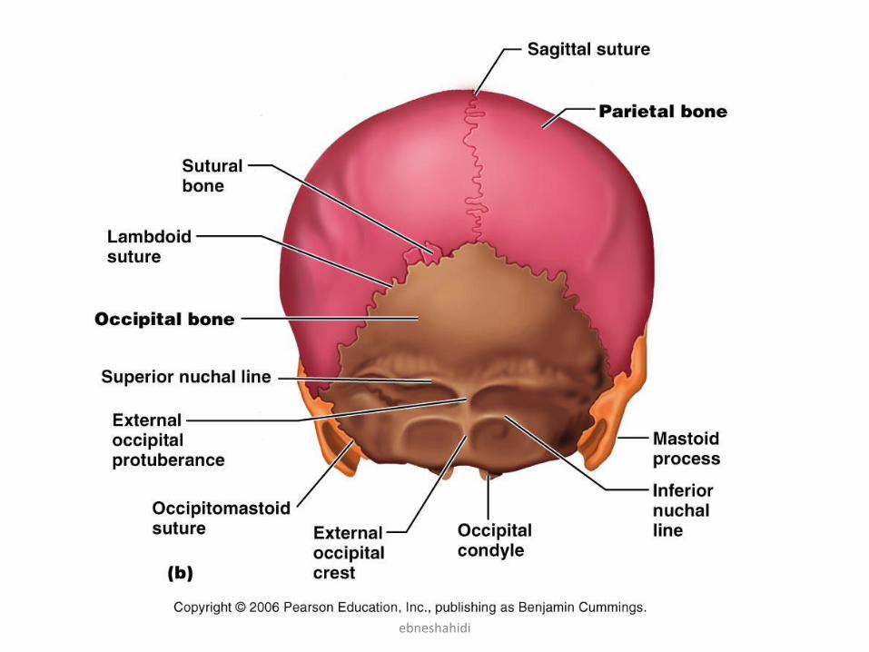
• b) Parietal bones and major
sutures
• Curved , rectangular bone
• Forms the bulging sides and
roof of cranium
• The 4 largest sutures occur
where parietal bone
articulates with other bones.
ebneshahidi
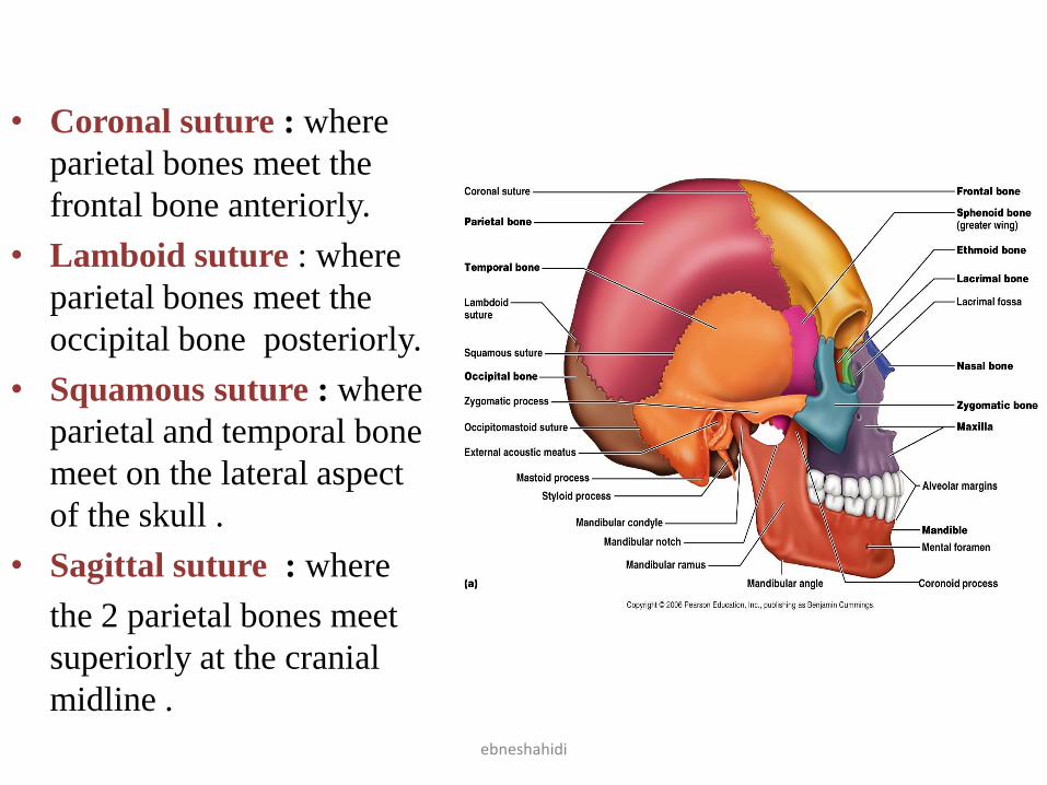
• Coronal suture : where
parietal bones meet the
frontal bone anteriorly.
• Lamboid suture : where
parietal bones meet the
occipital bone posteriorly.
• Squamous suture : where
parietal and temporal bone
meet on the lateral aspect
of the skull .
• Sagittal suture : where
the 2 parietal bones meet
superiorly at the cranial
midline .
ebneshahidi
ebneshahidi
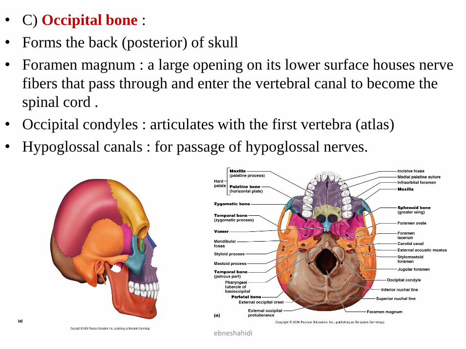
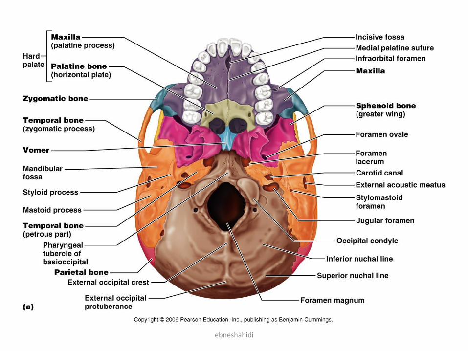
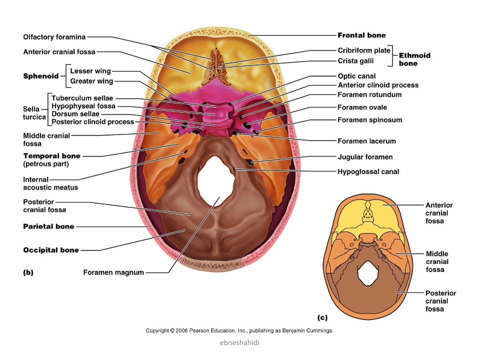
• C) Occipital bone :
• Forms the back (posterior) of skull
• Foramen magnum : a large opening on its lower surface houses nerve
fibers that pass through and enter the vertebral canal to become the
spinal cord .
• Occipital condyles : articulates with the first vertebra (atlas)
• Hypoglossal canals : for passage of hypoglossal nerves.
ebneshahidi
ebneshahidi
• d) Temporal bones :
– Lateral sides of skull.
– Contains the external auditory meatus (external ear).
– Mandibular fossa – receive condyles of mandible (lower
jaw).
– Zygomatic arch (process) – projects interiorly from the
temporal bone.
– Mastoid process – attachment for muscles of neck.
– Styloid process – attachment for muscles of tongue and
pharynx.
– Jugular fareamen – at the junction of occipital and petrous
temporal bone allows passage of the internal jugular vein.
– Carotid canal – transmit internal carotid artery.
ebneshahidi

ebneshahidi

ebneshahidi
• e) Sphenoid bone (butterfly - shaped)
– Sella turcica – indentition of a part of sphenoid,
in the depression lies the pituitary gland .
–Contains 2 sphenoid sinuses.
– It has greater and lesser wings.
–Optic foramina – allows passage of the optic
nerve.
–Superior orbital fissure – a long slit between the
greater and lesser wing allows passage of the
cranial nerves that control eye movements (III ,
IV, VI) to enter the orbit.
ebneshahidi
ebneshahidi

ebneshahidi
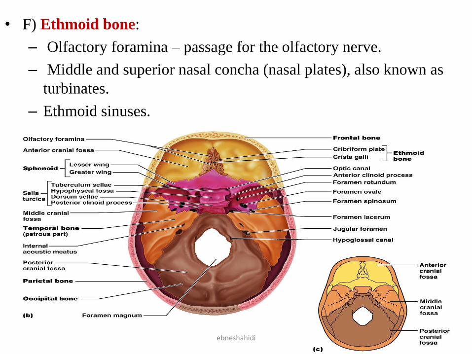
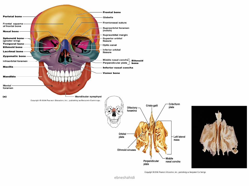
• F) Ethmoid bone:
– Olfactory foramina – passage for the olfactory nerve.
– Middle and superior nasal concha (nasal plates), also known as
turbinates.
– Ethmoid sinuses.
ebneshahidi
ebneshahidi
Facial Skeleton• Maxillary bones (upper jaw)
• All facial bones except mandible articulate with maxillae.
• Forms roof of mouth (anterior 2/3 of hard palate).
• Contains upper teeth .
• Maxillary sinuses (largest sinuses).
• Zygomatic process of maxilla articulate with zygomatic
bone.
• Infraorbital foramen – allows passage of infraorbital nerve.
• Infraorbital fissure – at junction of maxilla with the greater
wing of sphenoid allows passage of zygomatic nerve,
maxillary nerve (a branch of cranial nerve V), and blood
vessels.
ebneshahidi
ebneshahidi

ebneshahidi
• Palatine bones
– L shaped .
– Forms post 1/3 of hard plate .
• Zygomatic bones (cheek bones)
– Temporal process , which extends posteriorly to join the
zygomatic process of temporal bone. Together these
processes form the zygomatic arch .
• Lacrimal bones
– A groove in its anterior portion forms the lacrimal fossa
(tear channel).
• Nasal bones
– Forms bridge of nose medially
– Attachment of cartilaginous tissues that form the shape of
the nose .
ebneshahidi
ebneshahidi
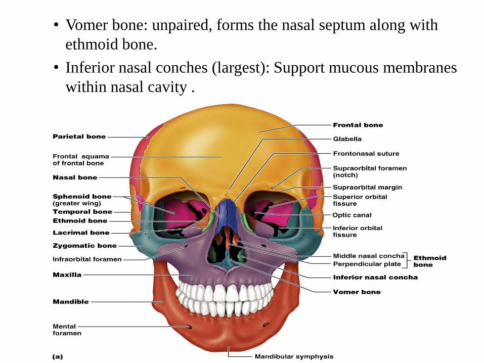
• Vomer bone: unpaired, forms the nasal septum along with
ethmoid bone.
• Inferior nasal conches (largest): Support mucous membranes
within nasal cavity .
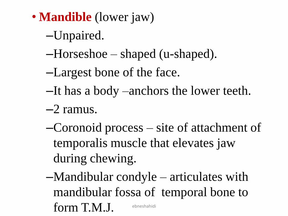
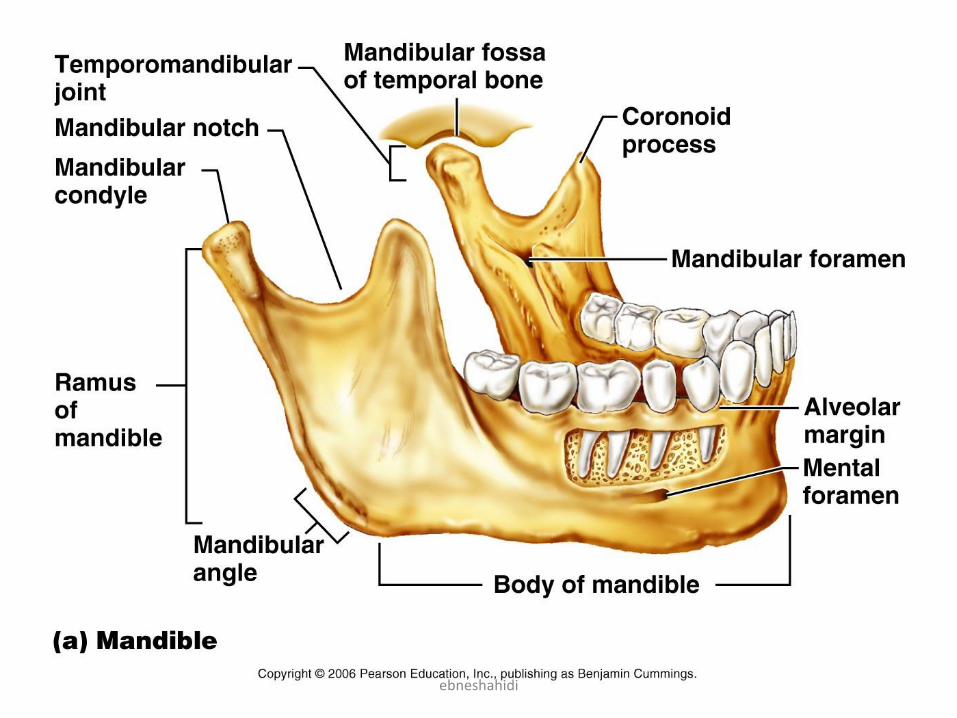
• Mandible (lower jaw)
–Unpaired.
–Horseshoe – shaped (u-shaped).
–Largest bone of the face.
–It has a body –anchors the lower teeth.
–2 ramus.
–Coronoid process – site of attachment of
temporalis muscle that elevates jaw
during chewing.
–Mandibular condyle – articulates with
mandibular fossa of temporal bone to
form T.M.J. ebneshahidi
ebneshahidi
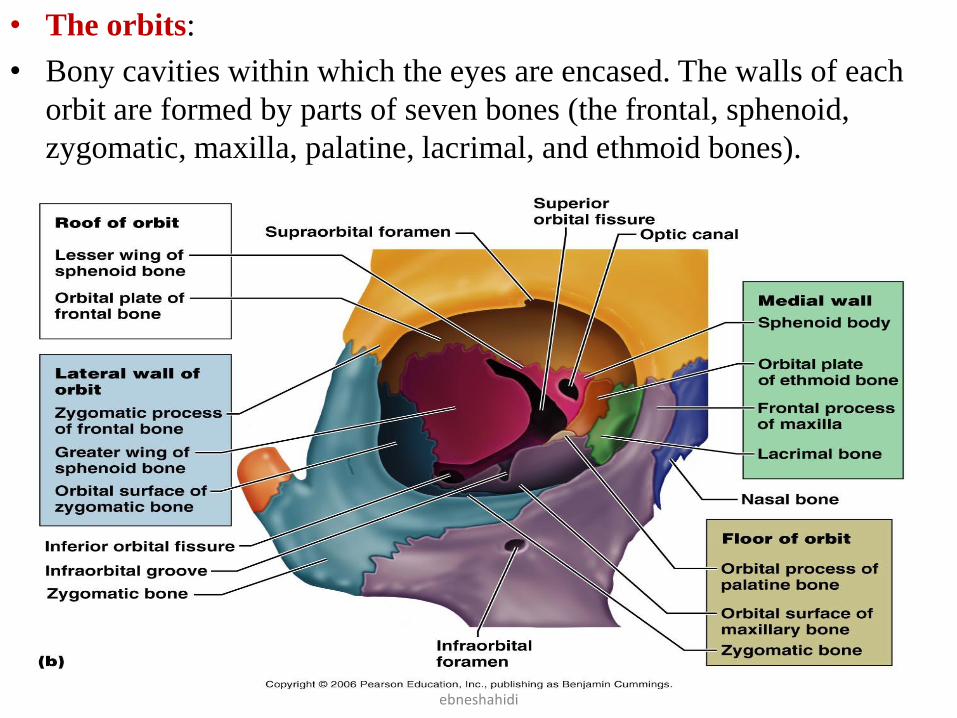
• The orbits:
• Bony cavities within which the eyes are encased. The walls of each
orbit are formed by parts of seven bones (the frontal, sphenoid,
zygomatic, maxilla, palatine, lacrimal, and ethmoid bones).
ebneshahidi
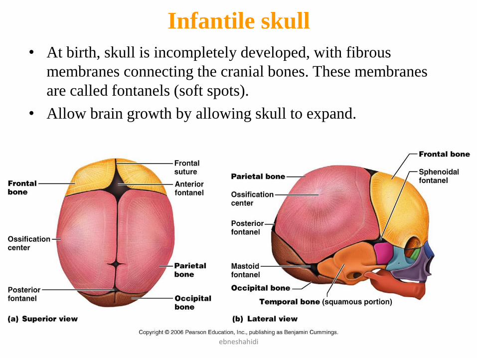
Infantile skull
• At birth, skull is incompletely developed, with fibrous
membranes connecting the cranial bones. These membranes
are called fontanels (soft spots).
• Allow brain growth by allowing skull to expand.
ebneshahidi
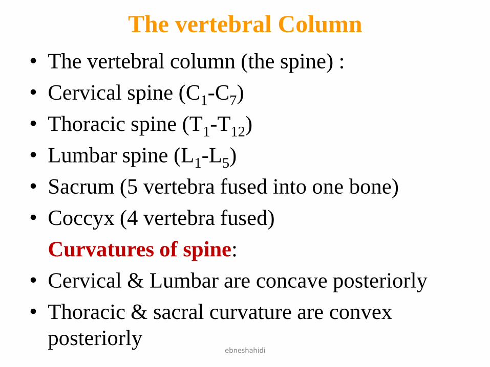
The vertebral Column
• The vertebral column (the spine) :
• Cervical spine (C1-C7)
• Thoracic spine (T1-T12)
• Lumbar spine (L1-L5)
• Sacrum (5 vertebra fused into one bone)
• Coccyx (4 vertebra fused)
Curvatures of spine:
• Cervical & Lumbar are concave posteriorly
• Thoracic & sacral curvature are convex
posteriorly ebneshahidi
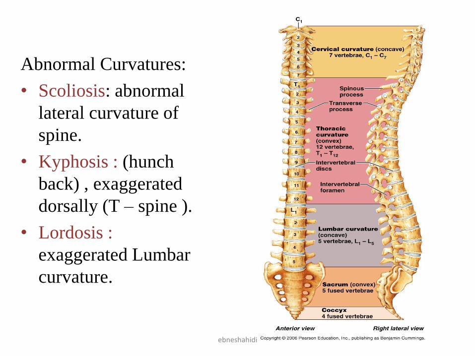
Abnormal Curvatures:
• Scoliosis: abnormal
lateral curvature of
spine.
• Kyphosis : (hunch
back) , exaggerated
dorsally (T – spine ).
• Lordosis :
exaggerated Lumbar
curvature.
ebneshahidi
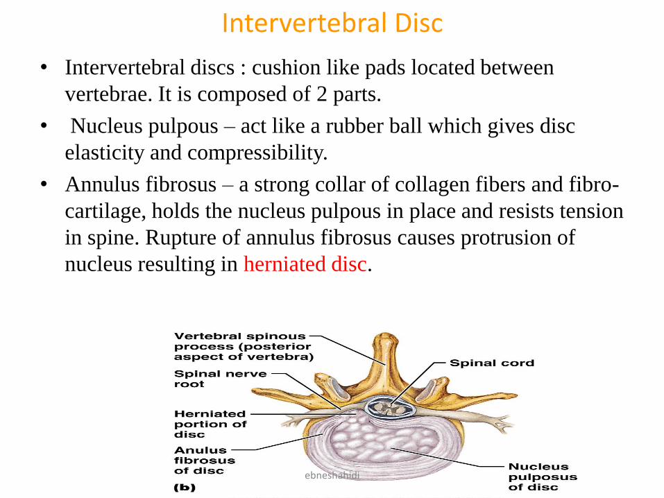
Intervertebral Disc
• Intervertebral discs : cushion like pads located between
vertebrae. It is composed of 2 parts.
• Nucleus pulpous – act like a rubber ball which gives disc
elasticity and compressibility.
• Annulus fibrosus – a strong collar of collagen fibers and fibro-
cartilage, holds the nucleus pulpous in place and resists tension
in spine. Rupture of annulus fibrosus causes protrusion of
nucleus resulting in herniated disc.
ebneshahidi
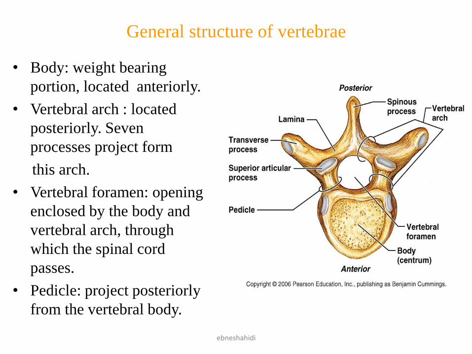
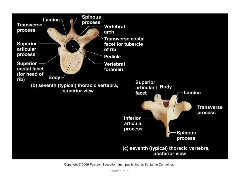
General structure of vertebrae
• Body: weight bearing
portion, located anteriorly.
• Vertebral arch : located
posteriorly. Seven
processes project form
this arch.
• Vertebral foramen: opening
enclosed by the body and
vertebral arch, through
which the spinal cord
passes.
• Pedicle: project posteriorly
from the vertebral body.
ebneshahidi
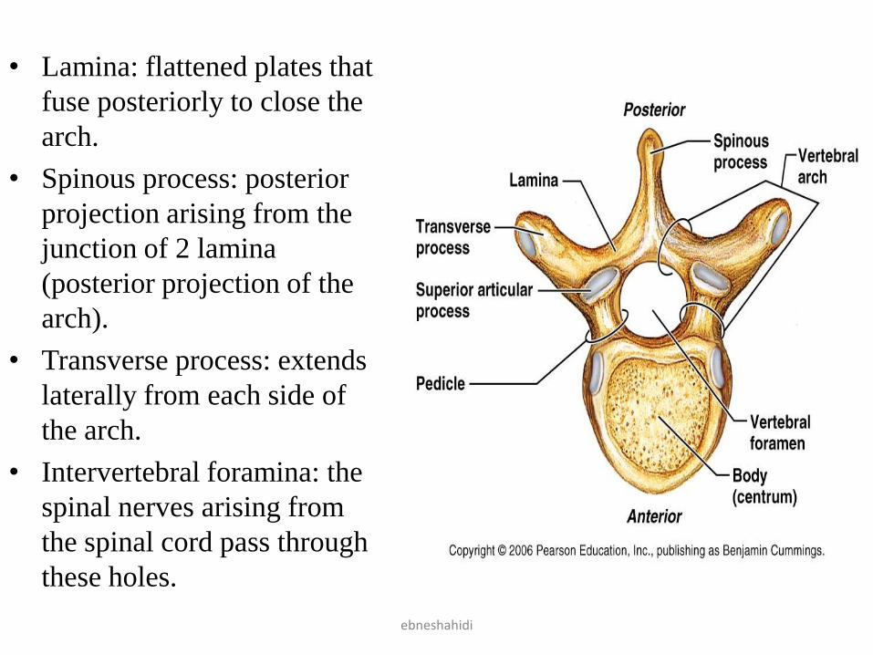
• Lamina: flattened plates that
fuse posteriorly to close the
arch.
• Spinous process: posterior
projection arising from the
junction of 2 lamina
(posterior projection of the
arch).
• Transverse process: extends
laterally from each side of
the arch.
• Intervertebral foramina: the
spinal nerves arising from
the spinal cord pass through
these holes.
ebneshahidi
Vertebral Characteristics
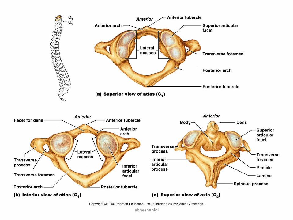
• Cervical vertebrae: (C1-C7)
• C1= known as atlas.
• C2= known as axis.
• C7= known as vertebra prominens.
• C1 – has no body and no spinous process.
• C2 – has a knob like structure called dens or odontoid process
projecting superiorly. Odontoid allows rotation of atlas.
• C3- C7 have the following characteristics:
• The body is oval shaped
• The SP's are short (except for C7 that is long).
• Vertebral foramen is triangular & large .
• Each T.P contains a transverse foramen through which the
vertebral blood vessels pass to service the brain .ebneshahidi
ebneshahidi
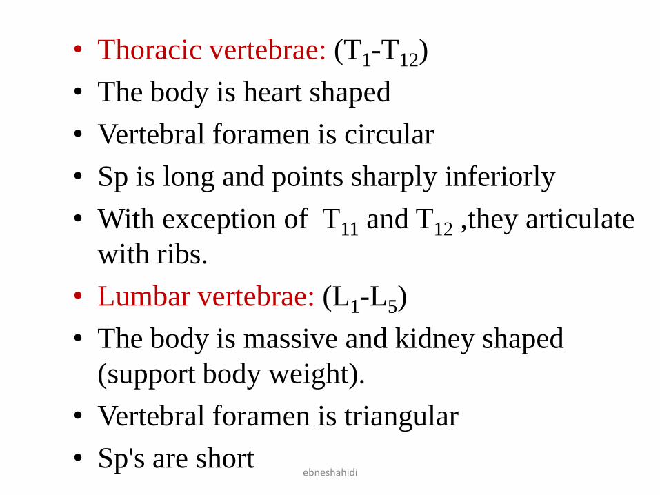
• Thoracic vertebrae: (T1-T12)
• The body is heart shaped
• Vertebral foramen is circular
• Sp is long and points sharply inferiorly
• With exception of T11 and T12 ,they articulate
with ribs.
• Lumbar vertebrae: (L1-L5)
• The body is massive and kidney shaped
(support body weight).
• Vertebral foramen is triangular
• Sp's are shortebneshahidi
ebneshahidi
ebneshahidi
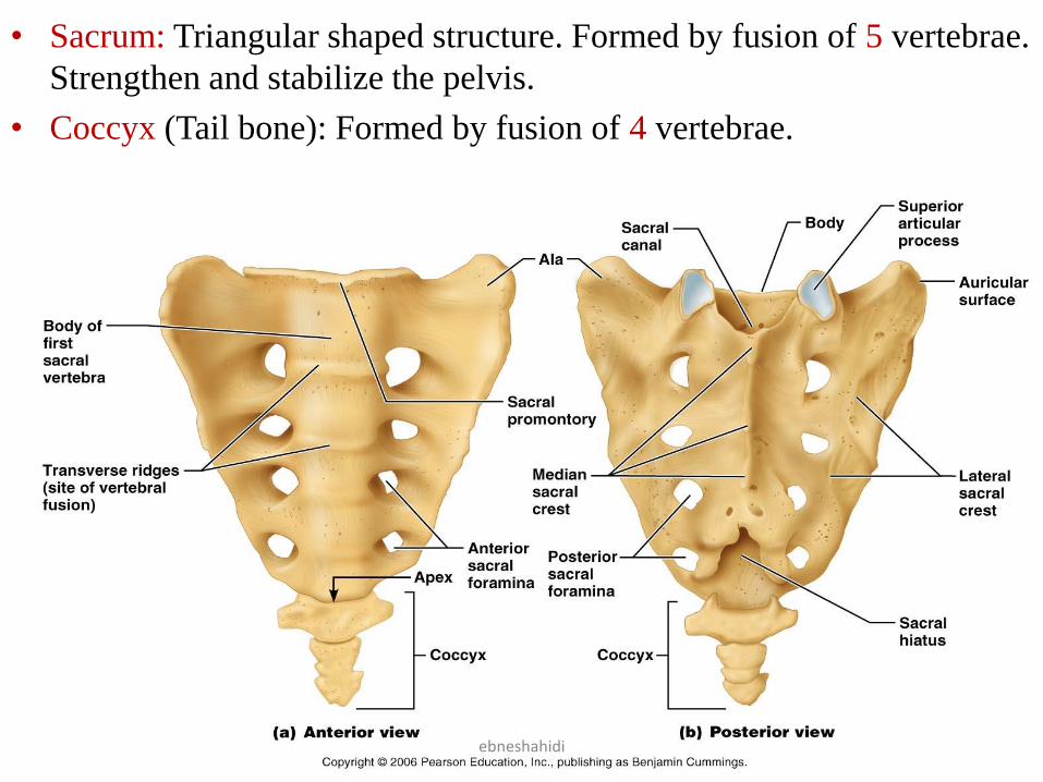
• Sacrum: Triangular shaped structure. Formed by fusion of 5 vertebrae.
Strengthen and stabilize the pelvis.
• Coccyx (Tail bone): Formed by fusion of 4 vertebrae.
ebneshahidi
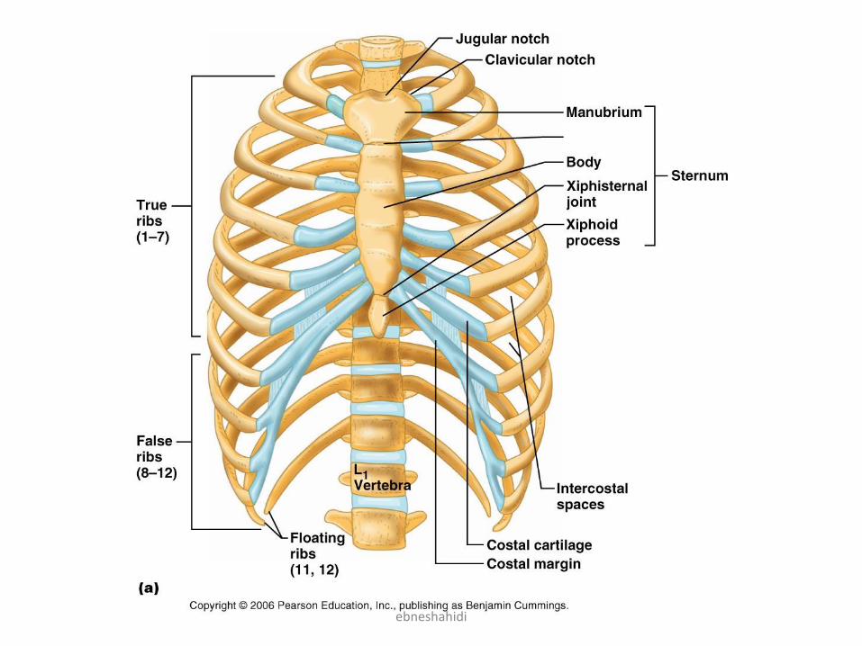
The bony Thorax (Thoracic Cage )
• Includes the ribs, thoracic vertebrae, the sternum, and the
costal cartilages.
• Sternum : (breast bone)- lies in anterior mid – line of the
thorax. It results from the fusion of 3 bones: the
Manubruin, the body and the xiphoid process.
• Ribs :
• 12 pairs
• 1-7 ribs are true ribs because they join the sternum
directly.
• Reaming 5 ribs are false ribs (8 -12) they donot have
sternal attachment, directly.
• Ribs 11-12 are floating ribs – no anterior attachment .
ebneshahidi
ebneshahidi
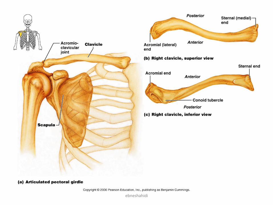
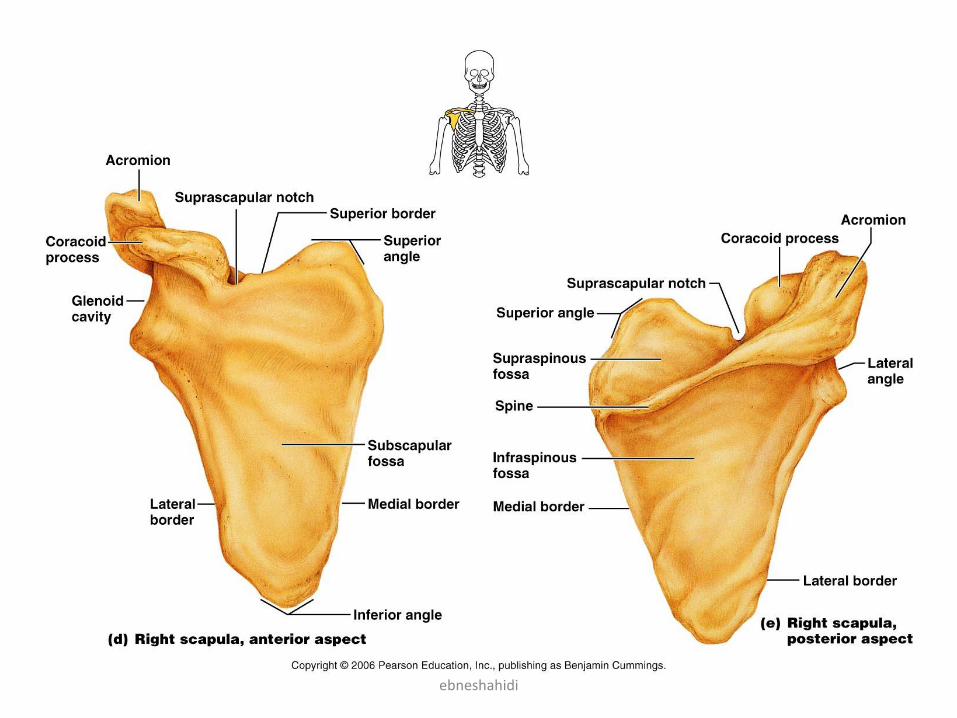
• The pectoral girdle (shoulder girdle ): 4 parts
• 2 clavicles (collar bones) & 2 scapulae (shoulder blades)
• Clavicle: has a sternal (medial end) & an acrominal (lateral end)
• Scapulae:
– has the genocide cavity that articulates with the humerus of the
arm, forming the shoulder joint.
– spine – divides the scapula into unequal portions called the supra-
spinous and infraspinous fossa.
– the Acromion: the spine ends laterally in an enlarged anterior
projection, articulates with clavicle to form A-C joint.
– coracoids process – site of attachment of biceps muscle and other
upper limb muscles .
– subscapulars fossa (cavity) – concavity of entire anterior scapular
surface .
The Appendicular Skeleton
ebneshahidi
ebneshahidi
ebneshahidi
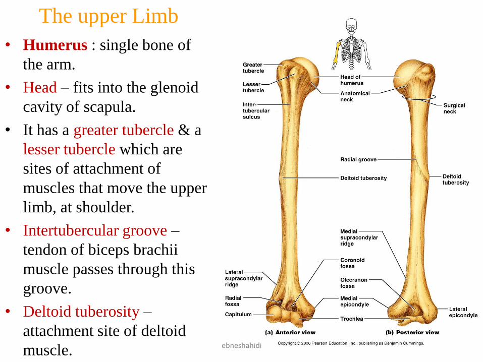
The upper Limb
• Humerus : single bone of
the arm.
• Head – fits into the glenoid
cavity of scapula.
• It has a greater tubercle & a
lesser tubercle which are
sites of attachment of
muscles that move the upper
limb, at shoulder.
• Intertubercular groove –
tendon of biceps brachii
muscle passes through this
groove.
• Deltoid tuberosity –
attachment site of deltoid
muscle. ebneshahidi
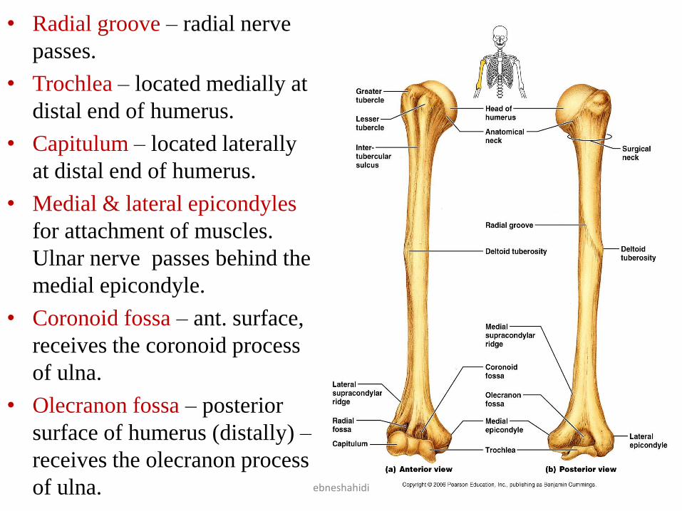
• Radial groove – radial nerve
passes.
• Trochlea – located medially at
distal end of humerus.
• Capitulum – located laterally
at distal end of humerus.
• Medial & lateral epicondyles
for attachment of muscles.
Ulnar nerve passes behind the
medial epicondyle.
• Coronoid fossa – ant. surface,
receives the coronoid process
of ulna.
• Olecranon fossa – posterior
surface of humerus (distally) –
receives the olecranon process
of ulna. ebneshahidi
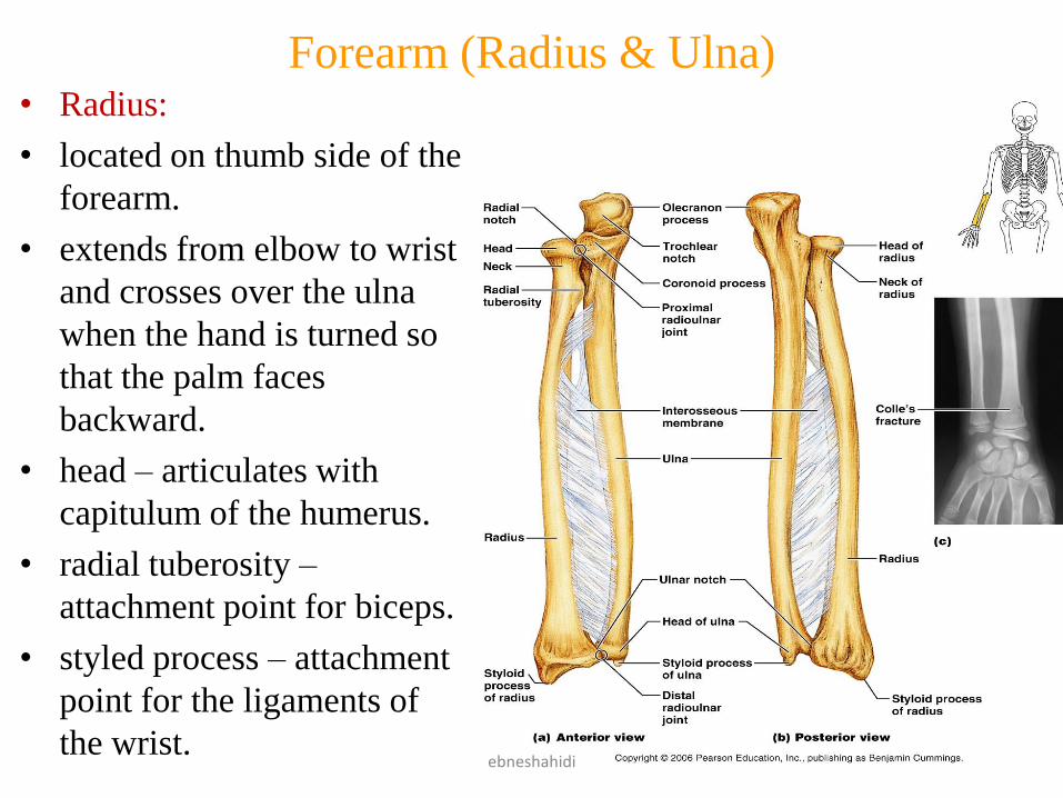
Forearm (Radius & Ulna)• Radius:
• located on thumb side of the
forearm.
• extends from elbow to wrist
and crosses over the ulna
when the hand is turned so
that the palm faces
backward.
• head – articulates with
capitulum of the humerus.
• radial tuberosity –
attachment point for biceps.
• styled process – attachment
point for the ligaments of
the wrist.ebneshahidi
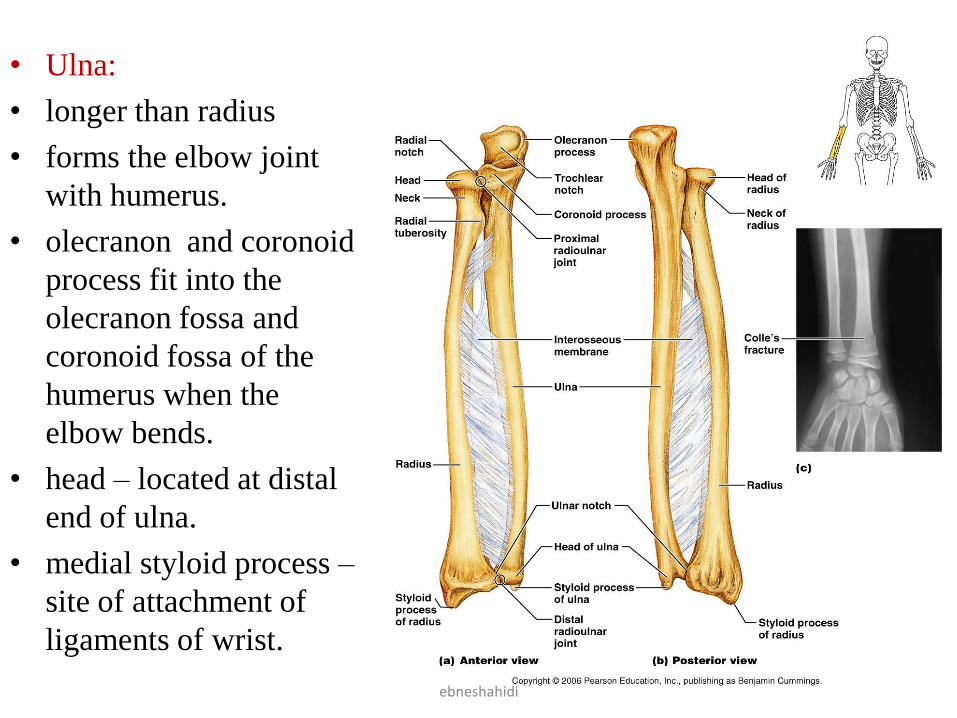
• Ulna:
• longer than radius
• forms the elbow joint
with humerus.
• olecranon and coronoid
process fit into the
olecranon fossa and
coronoid fossa of the
humerus when the
elbow bends.
• head – located at distal
end of ulna.
• medial styloid process –
site of attachment of
ligaments of wrist.
ebneshahidi
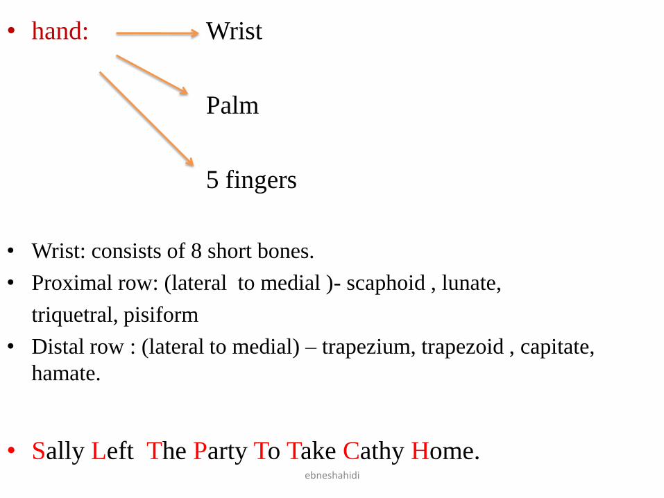
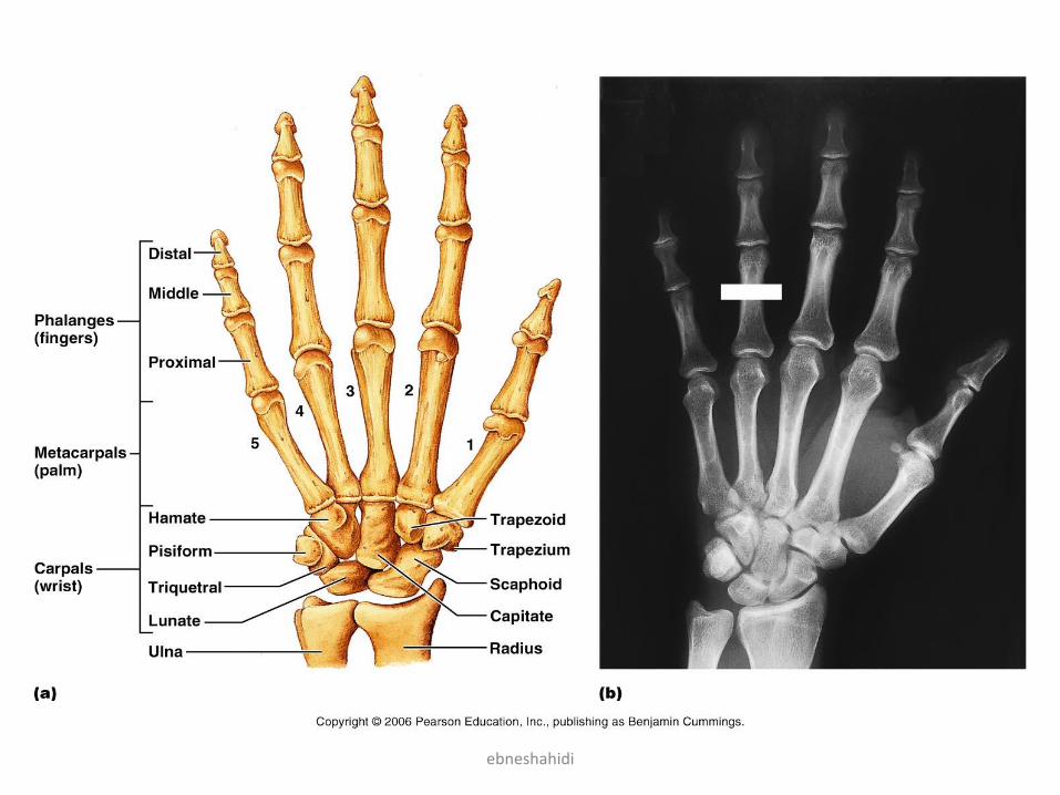
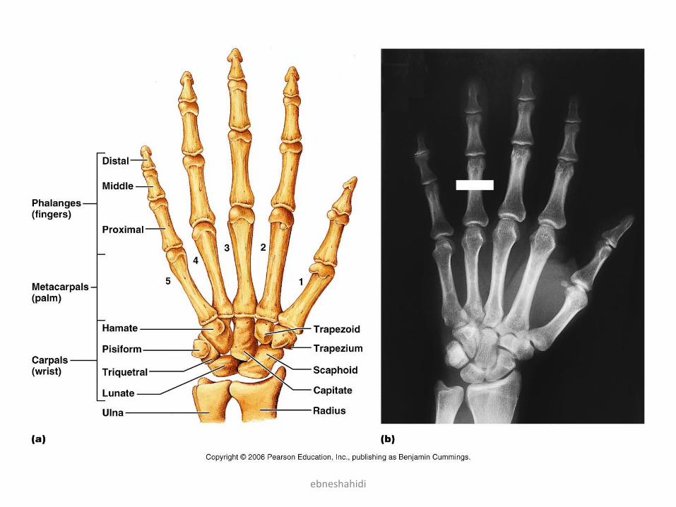
• hand: Wrist
Palm
5 fingers
• Wrist: consists of 8 short bones.
• Proximal row: (lateral to medial )- scaphoid , lunate,
triquetral, pisiform
• Distal row : (lateral to medial) – trapezium, trapezoid , capitate,
hamate.
• Sally Left The Party To Take Cathy Home.ebneshahidi
ebneshahidi
Metacarpals & Phalanges of the hand
• 5 bones, one in line with each finger,
numbered 1-5 from thumb to little finger.
• Their base articulate with carpals and their
head with phalanges (are considered long
bones even though small).
• Phalanges (Fingers):
• Numbered 1-5
• Have proximal , middle and distal ends (thumb
lacks middle phalanx).
• So each hand has 14 finger bones.ebneshahidi
ebneshahidi
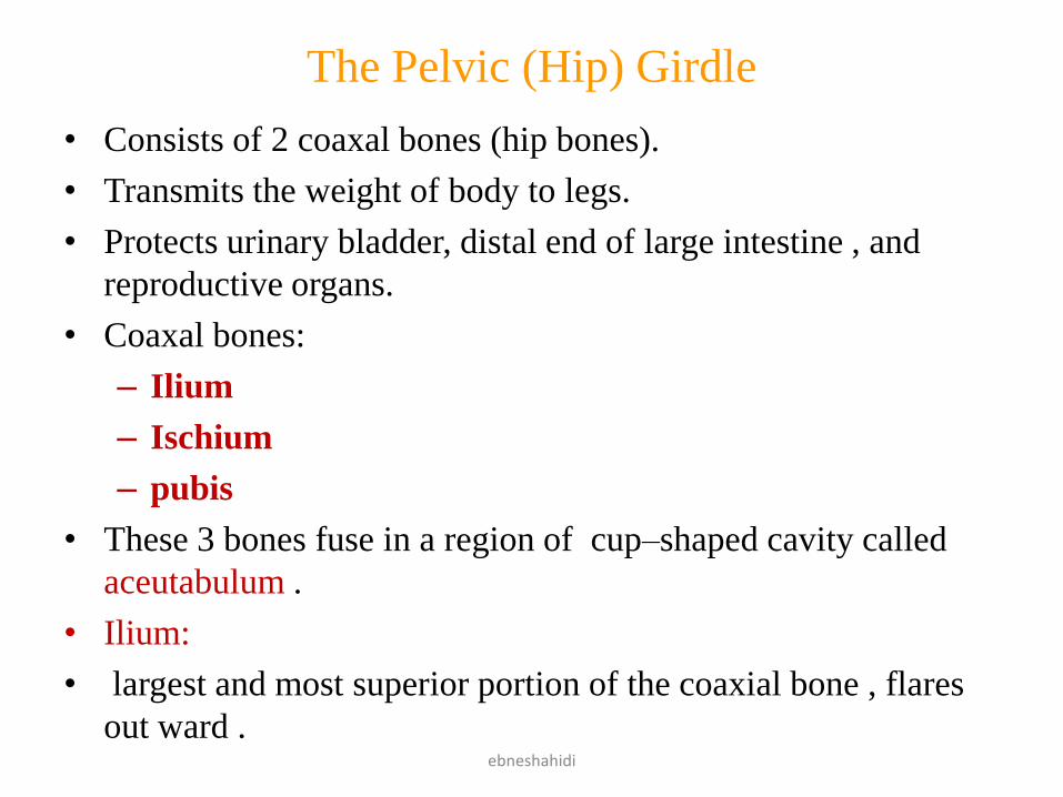
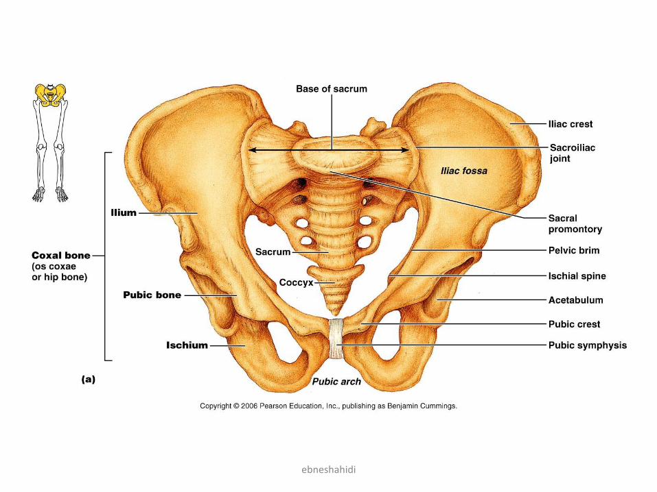
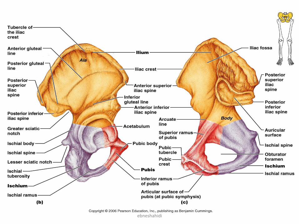
The Pelvic (Hip) Girdle
• Consists of 2 coaxal bones (hip bones).
• Transmits the weight of body to legs.
• Protects urinary bladder, distal end of large intestine , and
reproductive organs.
• Coaxal bones:
– Ilium
– Ischium
– pubis
• These 3 bones fuse in a region of cup–shaped cavity called
aceutabulum .
• Ilium:
• largest and most superior portion of the coaxial bone , flares
out ward .ebneshahidi
ebneshahidi
• posterioly it joins the sacrum to form the Sacroiliac joint.
• posterioly indents to form the greater sciatic notch.
• Ischium:
• forms the posteroinferior part of the hip bone, L – shaped.
• has an ischial tuberosity , that supports our weight when
seated, and is the strongest part of the hip bones.
• Pubis:
• forms the anterior portion of the coax bone, the 2 pubic
bones come together at midline to form the pubic symphysis
• V – shaped
• Obturator foramen – largest foramen of body (skeleton), is
formed by both ischium + pubis bones.
ebneshahidi
ebneshahidi
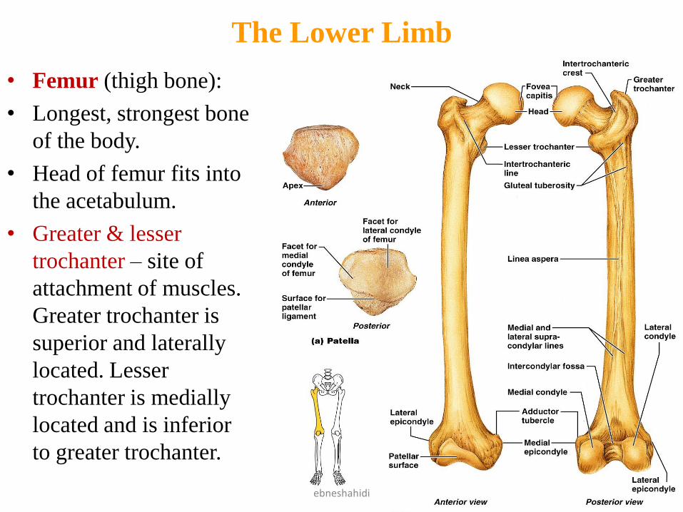
The Lower Limb
• Femur (thigh bone):
• Longest, strongest bone
of the body.
• Head of femur fits into
the acetabulum.
• Greater & lesser
trochanter – site of
attachment of muscles.
Greater trochanter is
superior and laterally
located. Lesser
trochanter is medially
located and is inferior
to greater trochanter.
ebneshahidi
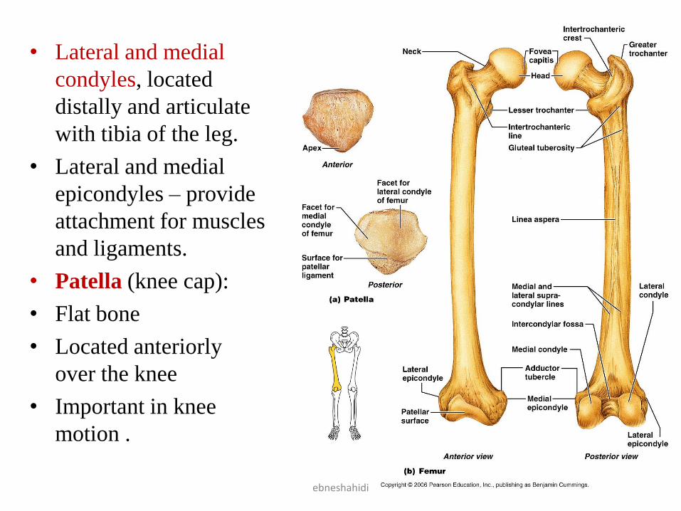
• Lateral and medial
condyles, located
distally and articulate
with tibia of the leg.
• Lateral and medial
epicondyles – provide
attachment for muscles
and ligaments.
• Patella (knee cap):
• Flat bone
• Located anteriorly
over the knee
• Important in knee
motion .
ebneshahidi
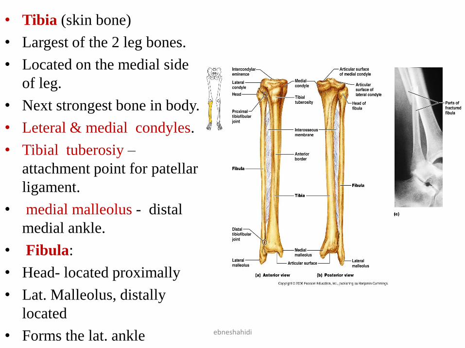
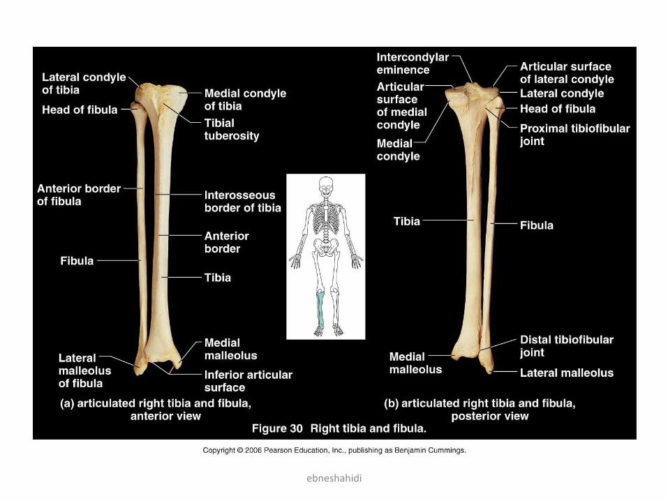
• Tibia (skin bone)
• Largest of the 2 leg bones.
• Located on the medial side
of leg.
• Next strongest bone in body.
• Leteral & medial condyles.
• Tibial tuberosiy –
attachment point for patellar
ligament.
• medial malleolus - distal
medial ankle.
• Fibula:
• Head- located proximally
• Lat. Malleolus, distally
located
• Forms the lat. ankle ebneshahidi
ebneshahidi
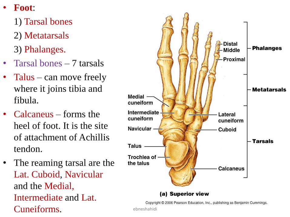
• Foot:
1) Tarsal bones
2) Metatarsals
3) Phalanges.
• Tarsal bones – 7 tarsals
• Talus – can move freely
where it joins tibia and
fibula.
• Calcaneus – forms the
heel of foot. It is the site
of attachment of Achillis
tendon.
• The reaming tarsal are the
Lat. Cuboid, Navicular
and the Medial,
Intermediate and Lat.
Cuneiforms. ebneshahidi
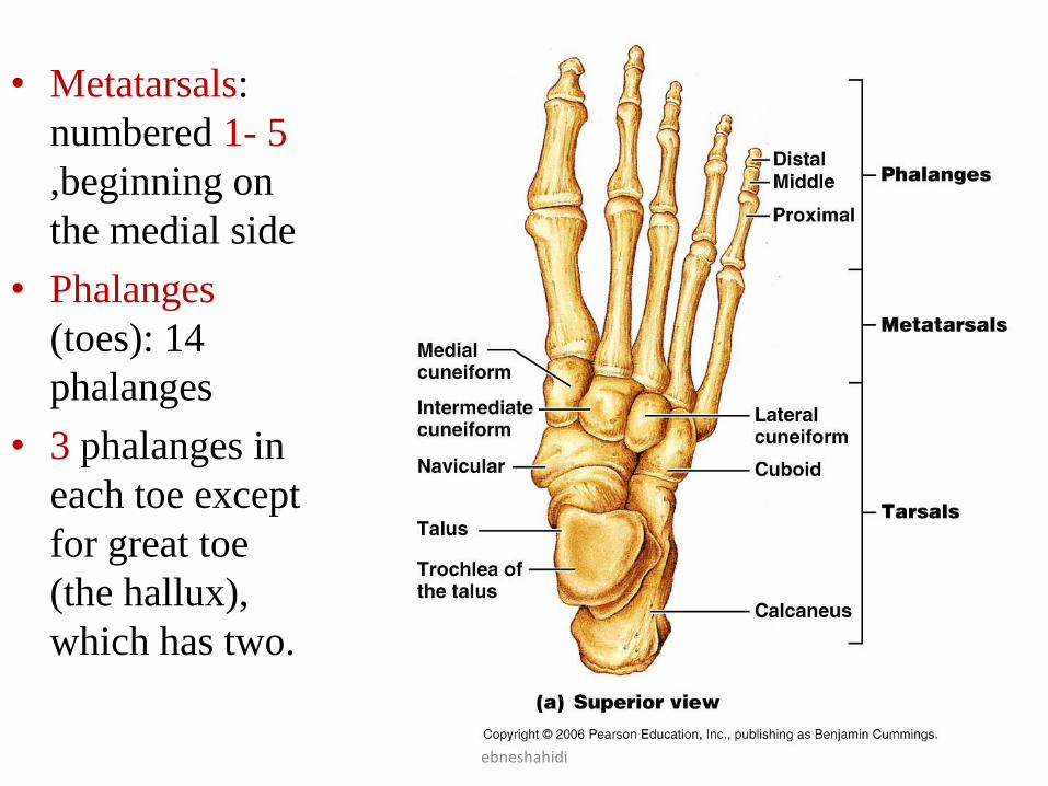
• Metatarsals:
numbered 1- 5
,beginning on
the medial side
• Phalanges
(toes): 14
phalanges
• 3 phalanges in
each toe except
for great toe
(the hallux),
which has two.
ebneshahidi
Clinical Terms
• Bunion – Deformity of great toe; lateral displacement
of great toe and medial displacement of metatarsal 1,
caused by thight shoes.
• Club foot – congenital disease in which the soles of
feet face medially and toes point inferiorly.
• Chiropractic - treating disease by manipulating the
spine.
• Podiatrists - a specialist in foot disorders .
• Orthopedists – surgeon who repair damaged bone and
joints .
• Prolapsed disc – A herniated disc.
ebneshahidi