Embed Size (px)
Citation preview
Essentials of Human Anatomy & Physiology
Copyright © 2003 Pearson Education, Inc. publishing as Benjamin Cummings
Seventh Edition Elaine N. Marieb
Chapter 5 The Skeletal System
The Skeletal System
Slide 5.1 Copyright © 2003 Pearson Education, Inc. publishing as Benjamin Cummings
• Parts of the skeletal system •Bones (skeleton)
• Joints
•Cartilages
• Ligaments (bone to bone)(tendon=bone to muscle)
• Divided into two divisions •Axial skeleton
•Appendicular skeleton – limbs and girdle
Functions of Bones
Slide 5.2 Copyright © 2003 Pearson Education, Inc. publishing as Benjamin Cummings
• Support of the body
• Protection of soft organs
• Movement due to attached skeletal muscles
• Storage of minerals and fats
• Blood cell formation
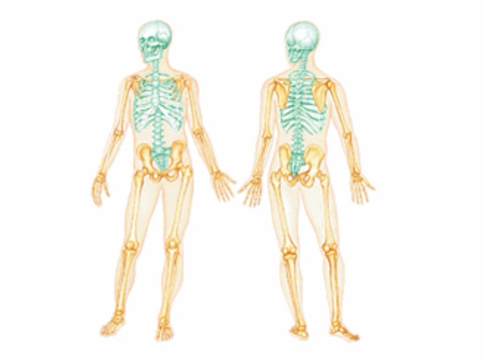
Bones of the Human Body
Slide 5.3 Copyright © 2003 Pearson Education, Inc. publishing as Benjamin Cummings
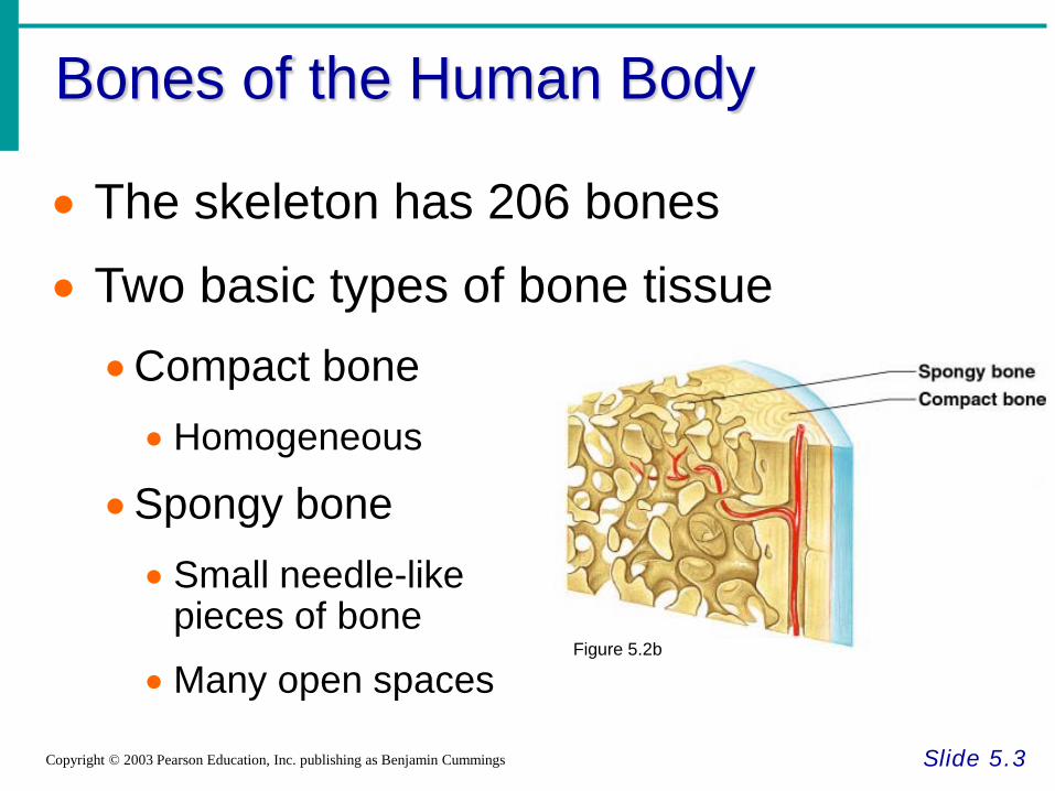
• The skeleton has 206 bones
• Two basic types of bone tissue •Compact bone • Homogeneous
•Spongy bone • Small needle-like
pieces of bone
• Many open spaces Figure 5.2b
Classification of Bones
Slide 5.4a Copyright © 2003 Pearson Education, Inc. publishing as Benjamin Cummings
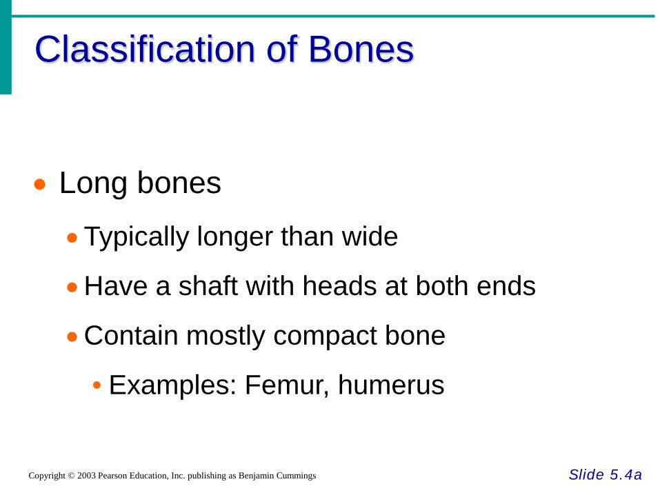
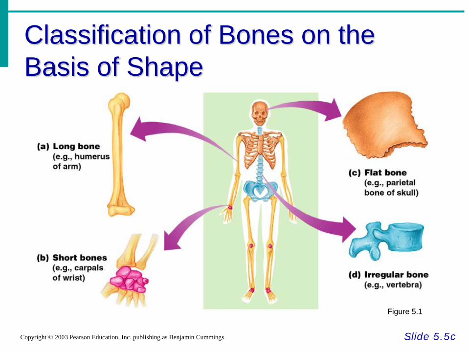
• Long bones
•Typically longer than wide
•Have a shaft with heads at both ends
•Contain mostly compact bone
• Examples: Femur, humerus
Classification of Bones
Slide 5.4b Copyright © 2003 Pearson Education, Inc. publishing as Benjamin Cummings
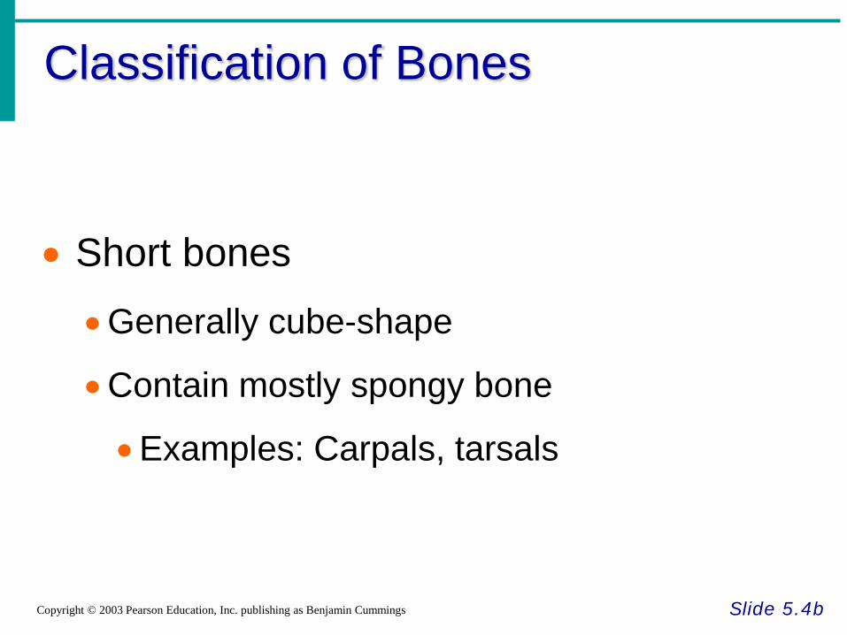
• Short bones
•Generally cube-shape
•Contain mostly spongy bone
•Examples: Carpals, tarsals
Classification of Bones
Slide 5.5a Copyright © 2003 Pearson Education, Inc. publishing as Benjamin Cummings
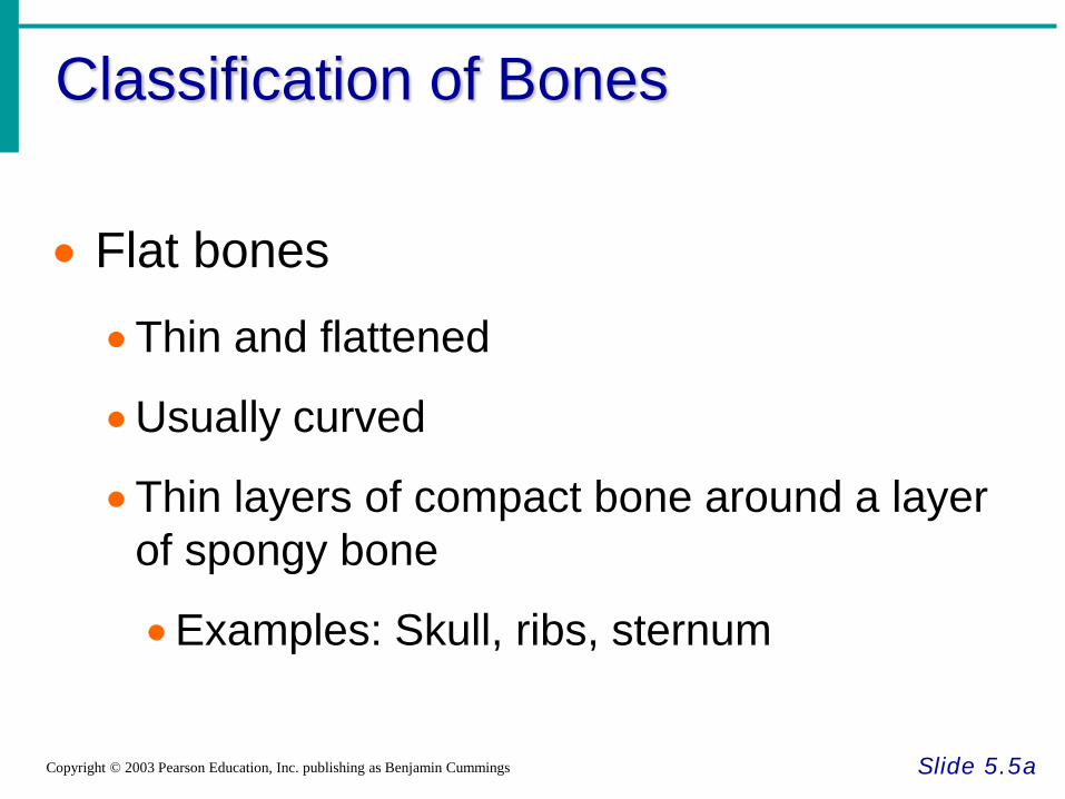
• Flat bones
•Thin and flattened
•Usually curved
•Thin layers of compact bone around a layer of spongy bone
•Examples: Skull, ribs, sternum
Classification of Bones
Slide 5.5b Copyright © 2003 Pearson Education, Inc. publishing as Benjamin Cummings
• Irregular bones
• Irregular shape
•Do not fit into other bone classification categories
•Example: Vertebrae and hip
Classification of Bones on the Basis of Shape
Slide 5.5c Copyright © 2003 Pearson Education, Inc. publishing as Benjamin Cummings
Figure 5.1
Gross Anatomy of a Long Bone
Slide 5.6 Copyright © 2003 Pearson Education, Inc. publishing as Benjamin Cummings
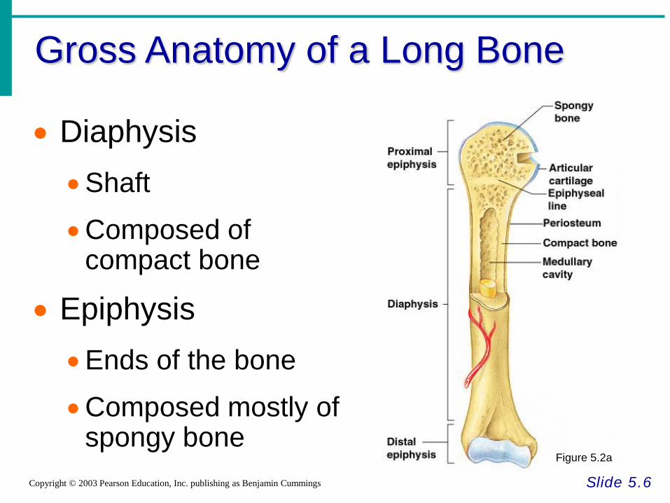
• Diaphysis •Shaft
•Composed of compact bone
• Epiphysis
•Ends of the bone
•Composed mostly of spongy bone
Figure 5.2a
Structures of a Long Bone
Slide 5.7 Copyright © 2003 Pearson Education, Inc. publishing as Benjamin Cummings
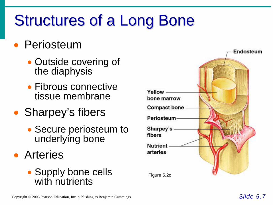
• Periosteum • Outside covering of
the diaphysis • Fibrous connective
tissue membrane
• Sharpey’s fibers • Secure periosteum to
underlying bone
• Arteries • Supply bone cells
with nutrients Figure 5.2c
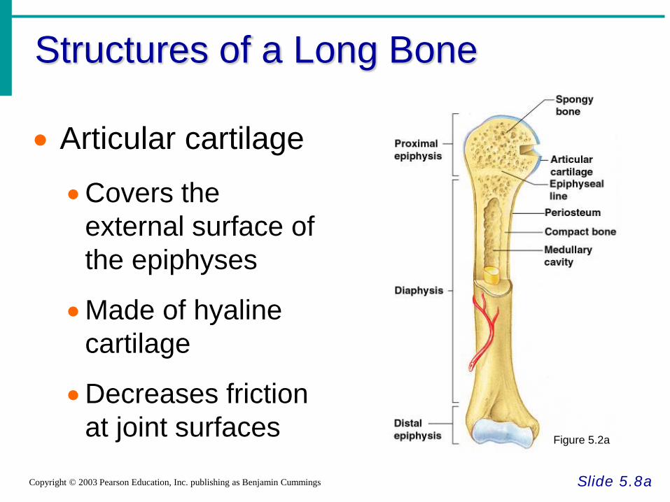
Structures of a Long Bone
Slide 5.8a Copyright © 2003 Pearson Education, Inc. publishing as Benjamin Cummings
• Articular cartilage
•Covers the external surface of the epiphyses
•Made of hyaline cartilage
•Decreases friction at joint surfaces Figure 5.2a
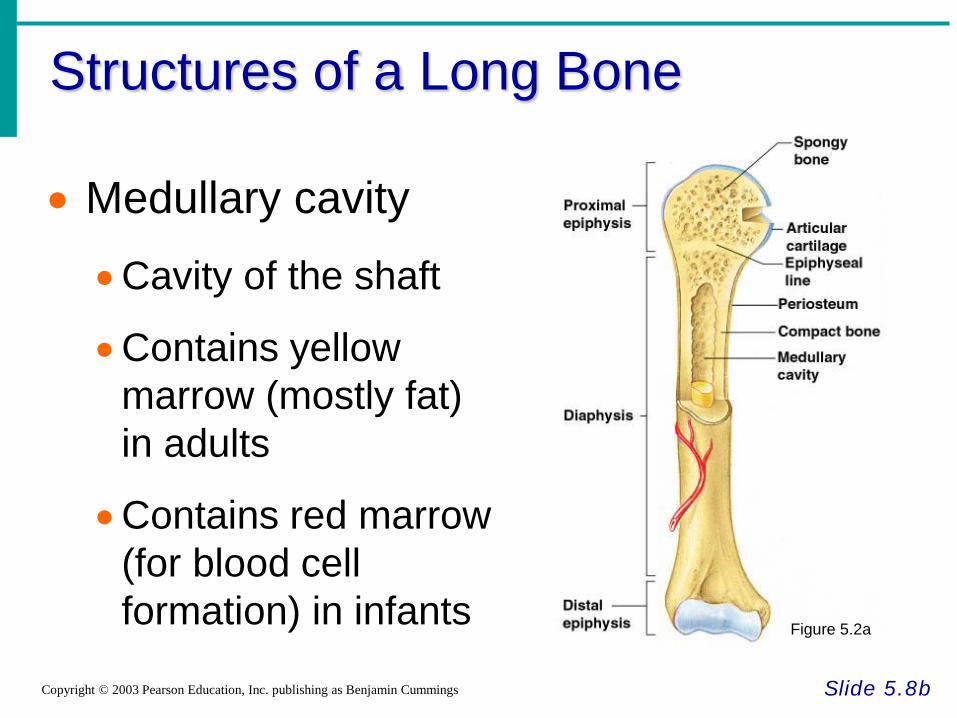
Structures of a Long Bone
Slide 5.8b Copyright © 2003 Pearson Education, Inc. publishing as Benjamin Cummings
• Medullary cavity
•Cavity of the shaft
•Contains yellow marrow (mostly fat) in adults
•Contains red marrow (for blood cell formation) in infants Figure 5.2a
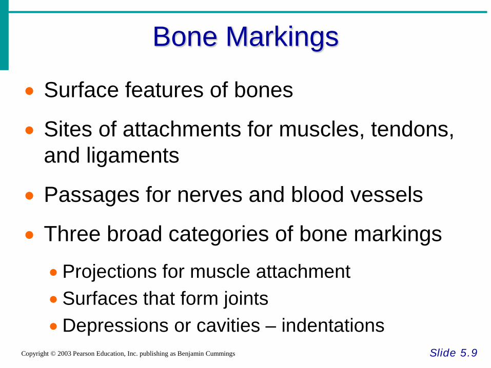
Bone Markings
Slide 5.9 Copyright © 2003 Pearson Education, Inc. publishing as Benjamin Cummings
• Surface features of bones
• Sites of attachments for muscles, tendons, and ligaments
• Passages for nerves and blood vessels
• Three broad categories of bone markings
•Projections for muscle attachment •Surfaces that form joints •Depressions or cavities – indentations
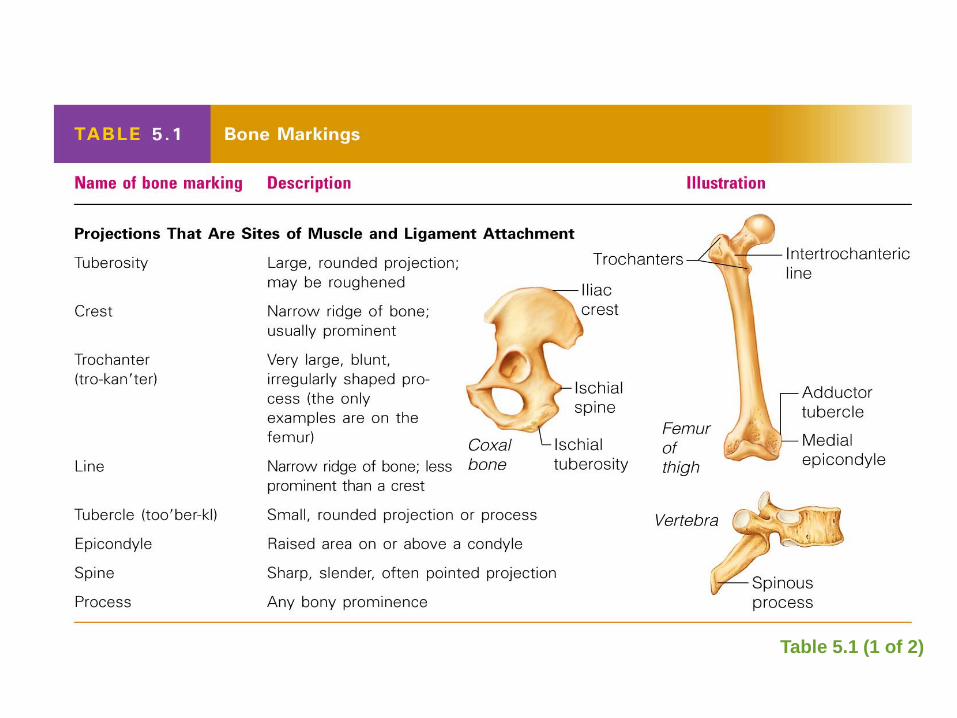
Bone Markings
Table 5.1 (1 of 2)
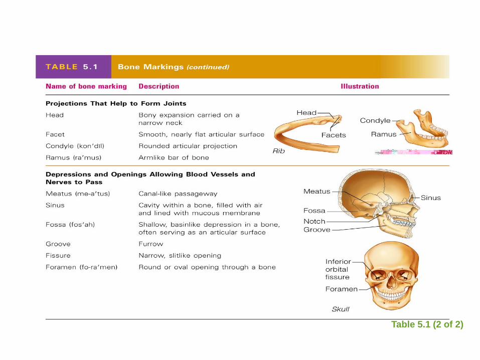
Bone Markings
Table 5.1 (2 of 2)
Microscopy Anatomy of Bone
Bone Histology • http://www.youtube.com/watch?v=PY5r_SJIG-A
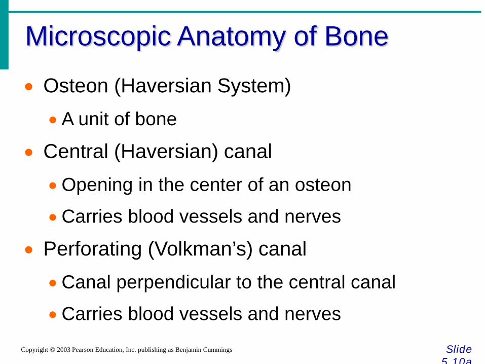
Microscopic Anatomy of Bone
Slide 5.10a
Copyright © 2003 Pearson Education, Inc. publishing as Benjamin Cummings
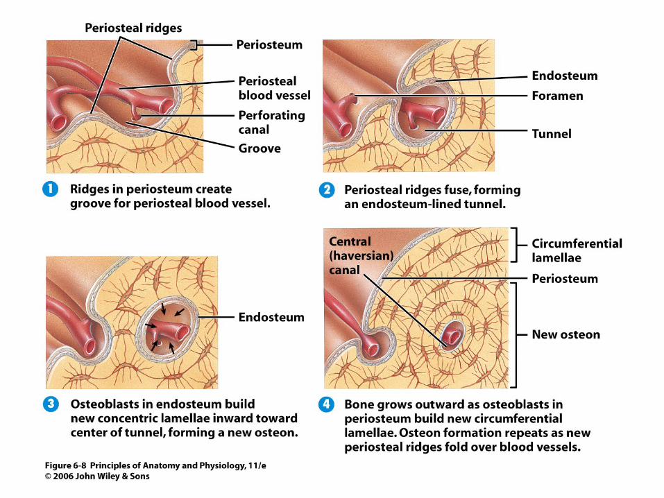
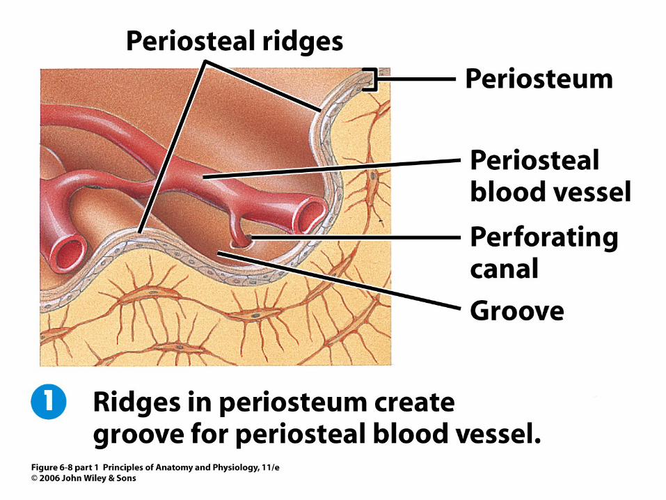
• Osteon (Haversian System) •A unit of bone
• Central (Haversian) canal •Opening in the center of an osteon
•Carries blood vessels and nerves
• Perforating (Volkman’s) canal •Canal perpendicular to the central canal
•Carries blood vessels and nerves
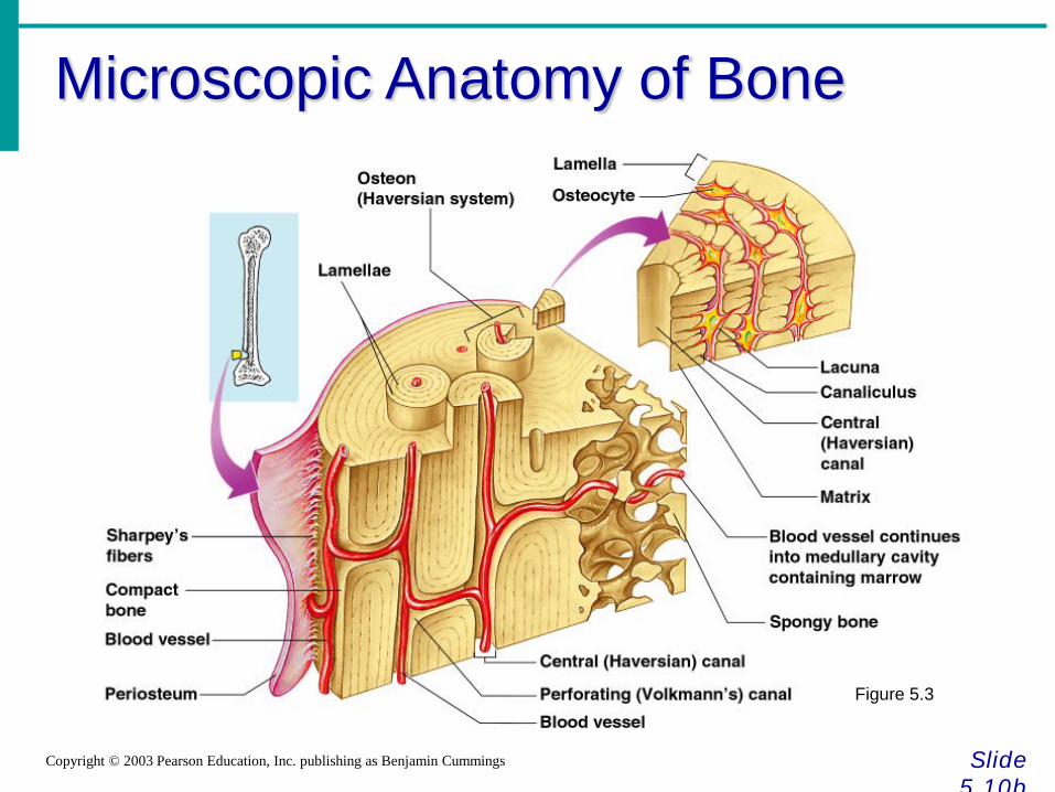
Microscopic Anatomy of Bone
Slide 5.10b
Copyright © 2003 Pearson Education, Inc. publishing as Benjamin Cummings
Figure 5.3
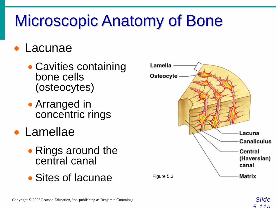
Microscopic Anatomy of Bone
Slide 5.11a
Copyright © 2003 Pearson Education, Inc. publishing as Benjamin Cummings
• Lacunae •Cavities containing
bone cells (osteocytes)
•Arranged in concentric rings
• Lamellae •Rings around the
central canal •Sites of lacunae Figure 5.3
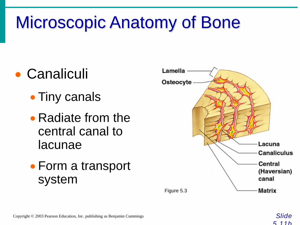
Microscopic Anatomy of Bone
Slide 5.11b
Copyright © 2003 Pearson Education, Inc. publishing as Benjamin Cummings
• Canaliculi •Tiny canals
•Radiate from the central canal to lacunae
•Form a transport system
Figure 5.3
Changes in the Human Skeleton
Slide 5.12 Copyright © 2003 Pearson Education, Inc. publishing as Benjamin Cummings
• In embryos, the skeleton is primarily hyaline cartilage
• During development, much of this cartilage is replaced by bone
• Cartilage remains in isolated areas
•Bridge of the nose
•Parts of ribs
• Joints
Bone Growth
Slide 5.13a
Copyright © 2003 Pearson Education, Inc. publishing as Benjamin Cummings
• Epiphyseal plates allow for growth of long bone during childhood
•New cartilage is continuously formed
•Older cartilage becomes ossified
•Cartilage is broken down
•Bone replaces cartilage
Bone Growth
Slide 5.13b
Copyright © 2003 Pearson Education, Inc. publishing as Benjamin Cummings
• Bones are remodeled and lengthened until growth stops
•Bones change shape somewhat
•Bones grow in width
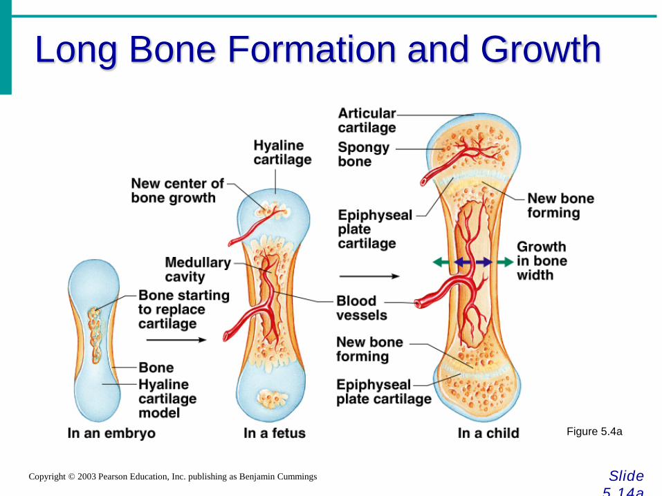
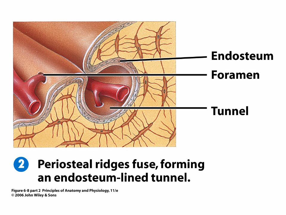
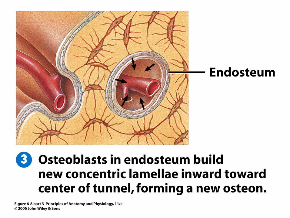
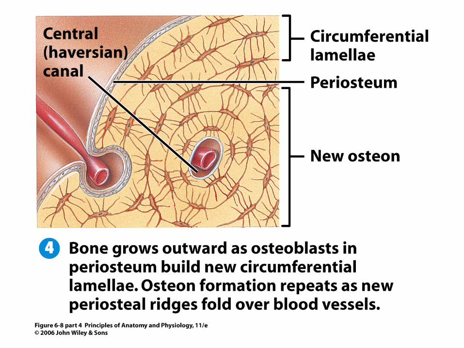
Long Bone Formation and Growth
Slide 5.14a
Copyright © 2003 Pearson Education, Inc. publishing as Benjamin Cummings
Figure 5.4a
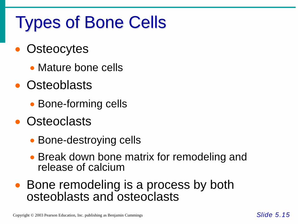
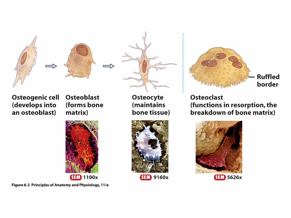
Types of Bone Cells
Slide 5.15 Copyright © 2003 Pearson Education, Inc. publishing as Benjamin Cummings
• Osteocytes • Mature bone cells
• Osteoblasts • Bone-forming cells
• Osteoclasts • Bone-destroying cells • Break down bone matrix for remodeling and
release of calcium
• Bone remodeling is a process by both osteoblasts and osteoclasts
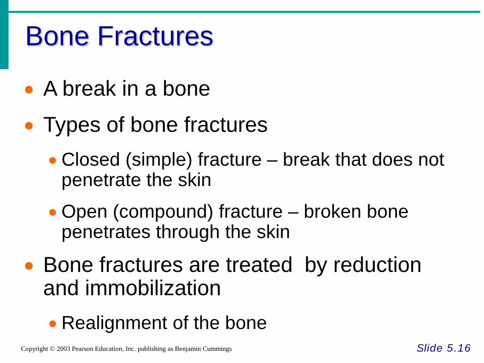
Bone Fractures
Slide 5.16 Copyright © 2003 Pearson Education, Inc. publishing as Benjamin Cummings
• A break in a bone
• Types of bone fractures •Closed (simple) fracture – break that does not
penetrate the skin
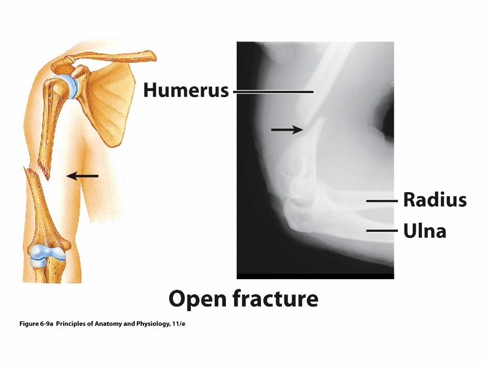
•Open (compound) fracture – broken bone penetrates through the skin
• Bone fractures are treated by reduction and immobilization •Realignment of the bone
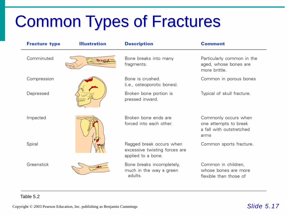
Common Types of Fractures
Slide 5.17 Copyright © 2003 Pearson Education, Inc. publishing as Benjamin Cummings
Table 5.2
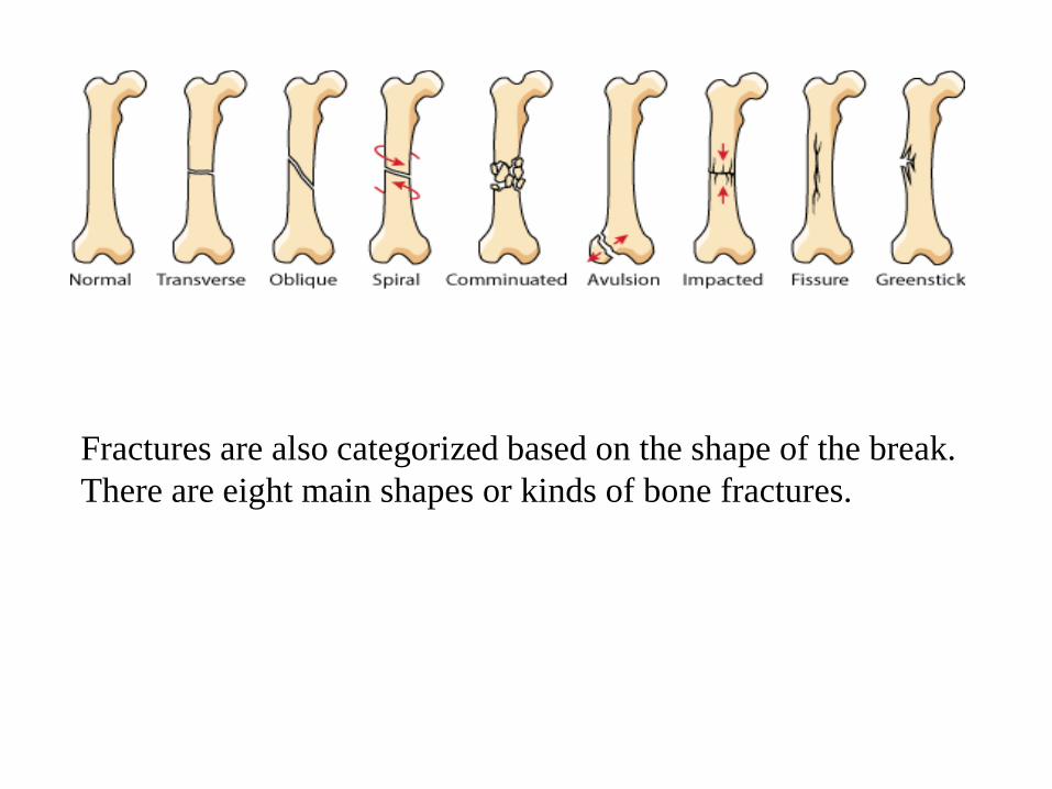
Fractures are also categorized based on the shape of the break. There are eight main shapes or kinds of bone fractures.
• Transverse fractures: go more or less straight across the bone.
• Oblique fractures: are diagonal breaks across the bone. • Spiral fractures: happen when one or both halves of the bone
are twisted. • Comminuated fractures: break the bone into more than two
pieces. • Avulsion fractures: mean pieces of the bone have been pulled
apart. • Impacted fractures: are the opposite of avulsion fractures.
These happen when a piece of bone is pushed down into another piece of bone.
• Fissure fractures: are cracks in the bone. • Greenstick fractures: happen when the bone bends and
breaks partially, but not completely.
Repair of Bone Fractures
Slide 5.18 Copyright © 2003 Pearson Education, Inc. publishing as Benjamin Cummings
• Hematoma (blood-filled swelling) is formed
• Break is splinted by fibrocartilage to form a callus
• Fibrocartilage callus is replaced by a bony callus
• Bony callus is remodeled to form a permanent patch
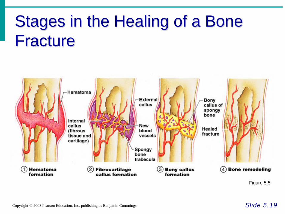
Stages in the Healing of a Bone Fracture
Slide 5.19 Copyright © 2003 Pearson Education, Inc. publishing as Benjamin Cummings
Figure 5.5
The Axial Skeleton
Slide 5.20a
Copyright © 2003 Pearson Education, Inc. publishing as Benjamin Cummings
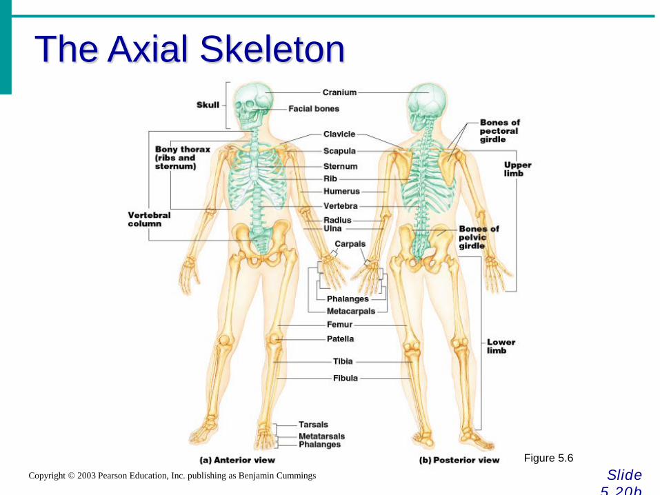
• Forms the longitudinal part of the body
• Divided into three parts •Skull
•Vertebral column
•Bony thorax
The Axial Skeleton
Slide 5.20b
Copyright © 2003 Pearson Education, Inc. publishing as Benjamin Cummings
Figure 5.6
The Skull
Slide 5.21a
Copyright © 2003 Pearson Education, Inc. publishing as Benjamin Cummings
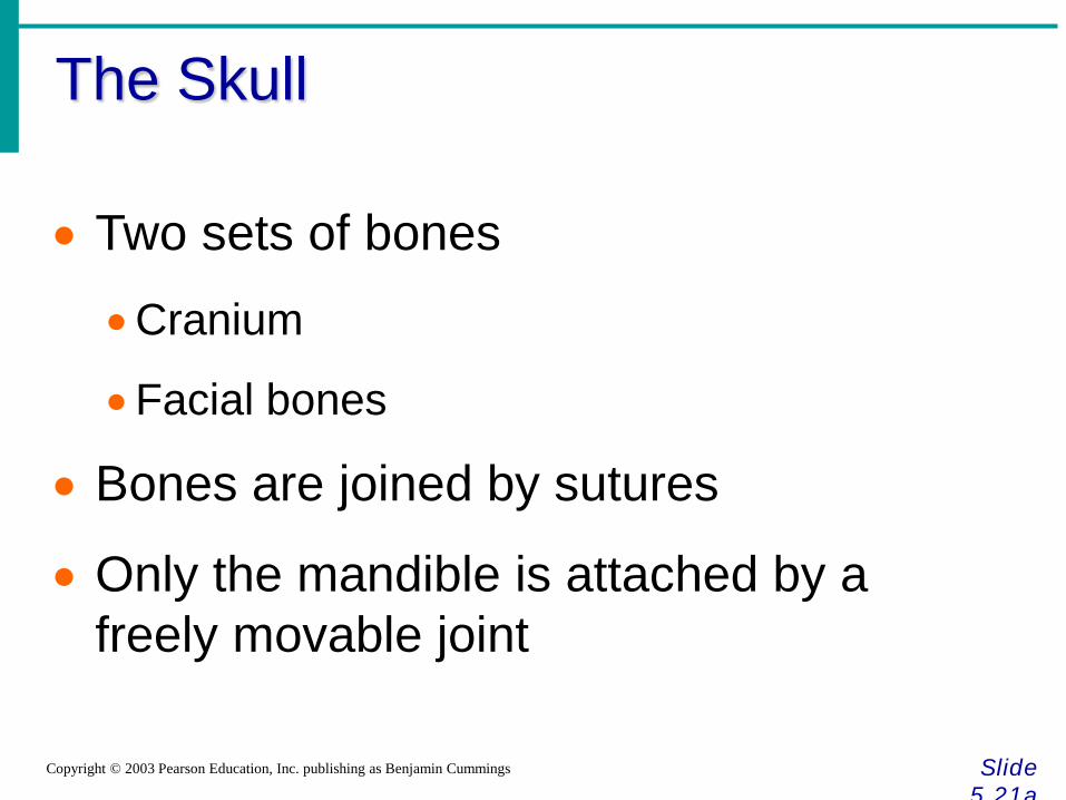
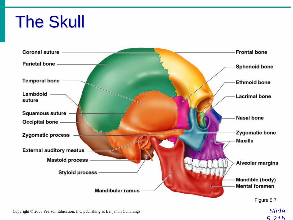
• Two sets of bones
•Cranium
•Facial bones
• Bones are joined by sutures
• Only the mandible is attached by a freely movable joint
The Skull
Slide 5.21b
Copyright © 2003 Pearson Education, Inc. publishing as Benjamin Cummings
Figure 5.7
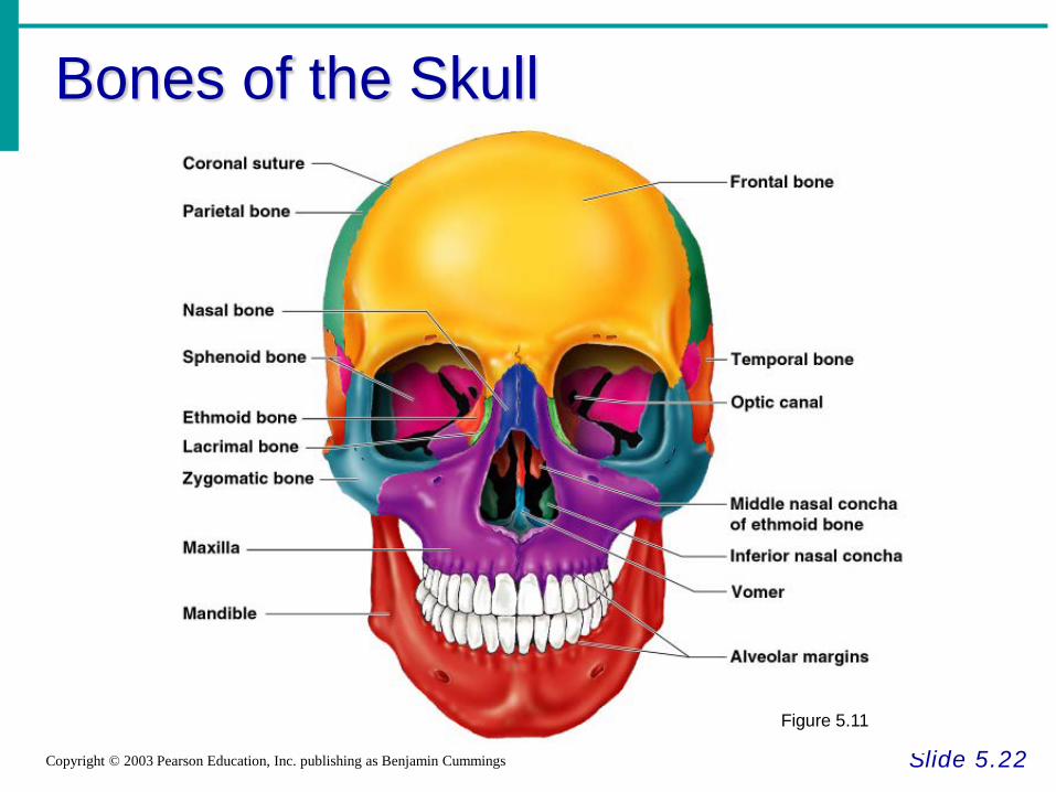
Bones of the Skull
Slide 5.22 Copyright © 2003 Pearson Education, Inc. publishing as Benjamin Cummings
Figure 5.11
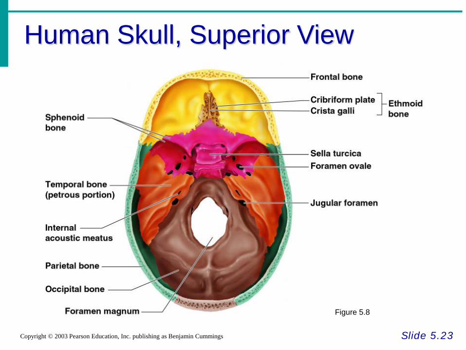
Human Skull, Superior View
Slide 5.23 Copyright © 2003 Pearson Education, Inc. publishing as Benjamin Cummings
Figure 5.8
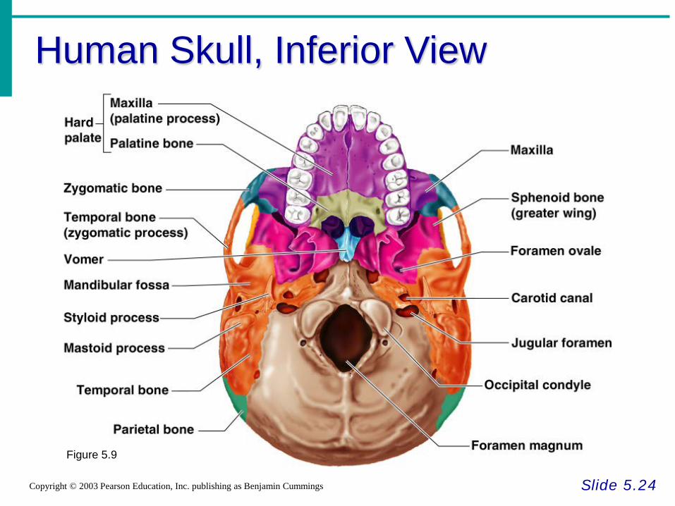
Human Skull, Inferior View
Slide 5.24 Copyright © 2003 Pearson Education, Inc. publishing as Benjamin Cummings
Figure 5.9
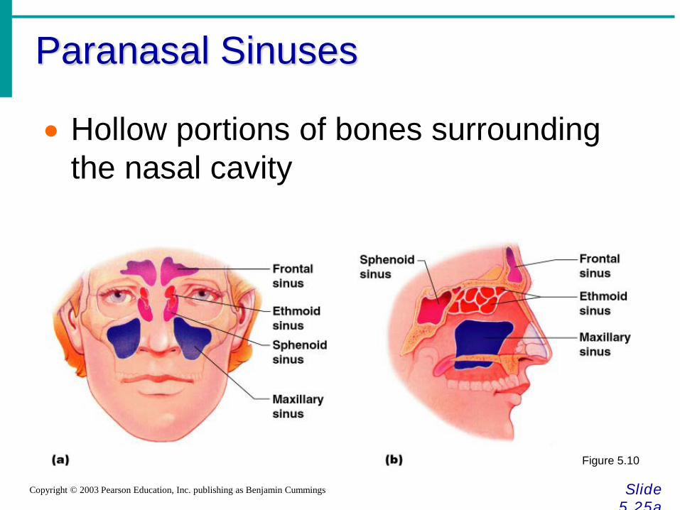
Paranasal Sinuses
Slide 5.25a
Copyright © 2003 Pearson Education, Inc. publishing as Benjamin Cummings
• Hollow portions of bones surrounding the nasal cavity
Figure 5.10
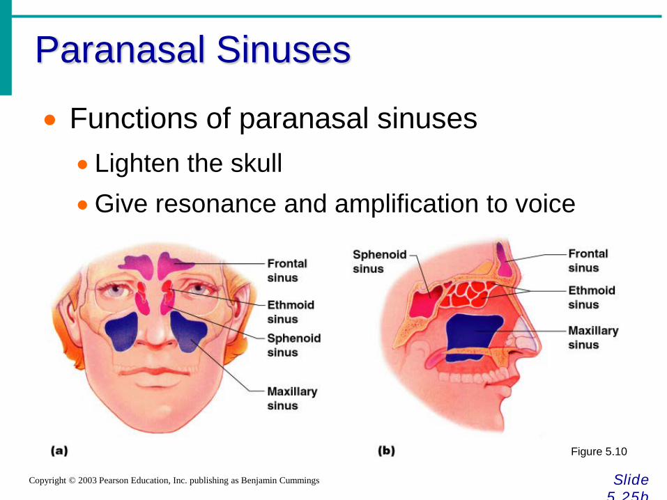
Paranasal Sinuses
Slide 5.25b
Copyright © 2003 Pearson Education, Inc. publishing as Benjamin Cummings
• Functions of paranasal sinuses • Lighten the skull •Give resonance and amplification to voice
Figure 5.10
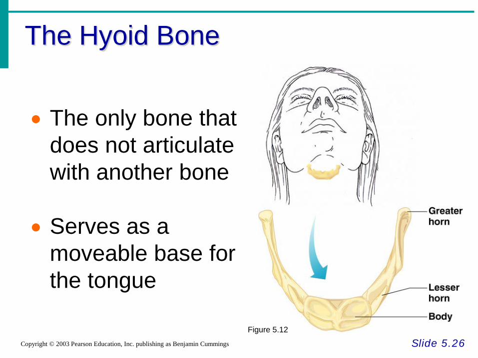
The Hyoid Bone
Slide 5.26 Copyright © 2003 Pearson Education, Inc. publishing as Benjamin Cummings
• The only bone that does not articulate with another bone
• Serves as a moveable base for the tongue
Figure 5.12
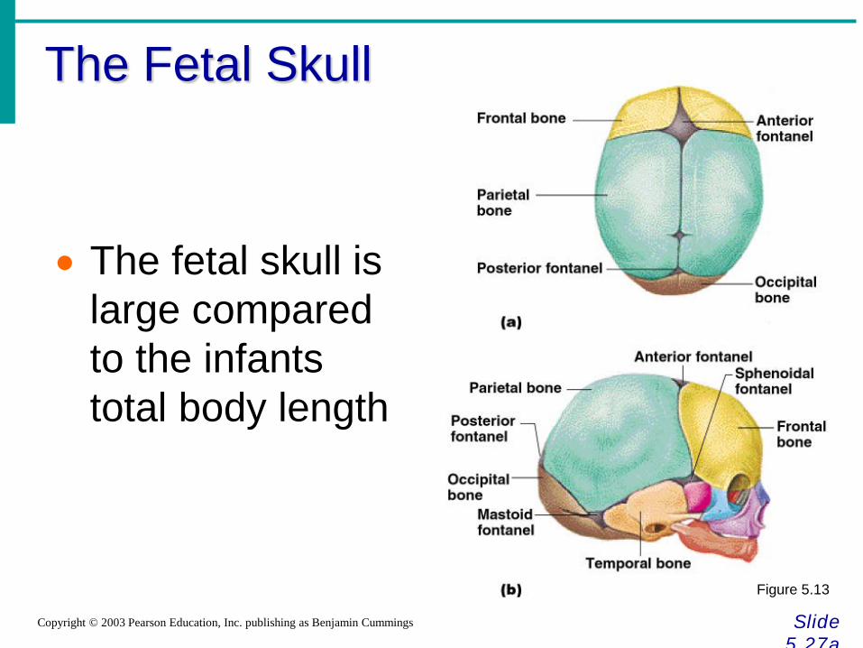
The Fetal Skull
Slide 5.27a
Copyright © 2003 Pearson Education, Inc. publishing as Benjamin Cummings
• The fetal skull is large compared to the infants total body length
Figure 5.13
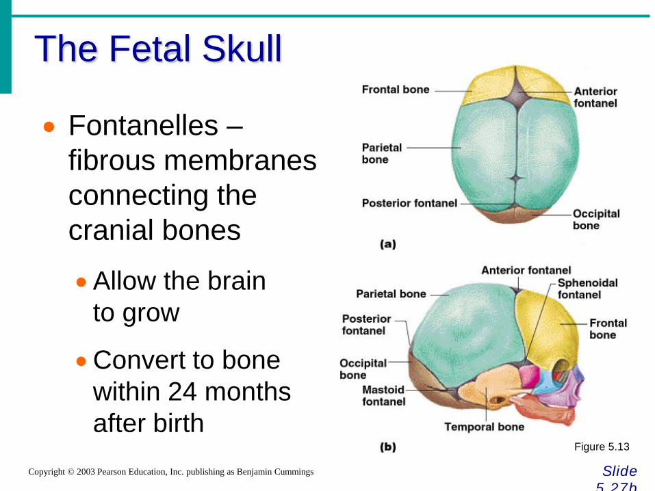
The Fetal Skull
Slide 5.27b
Copyright © 2003 Pearson Education, Inc. publishing as Benjamin Cummings
• Fontanelles – fibrous membranes connecting the cranial bones
•Allow the brain to grow
•Convert to bone within 24 months after birth
Figure 5.13
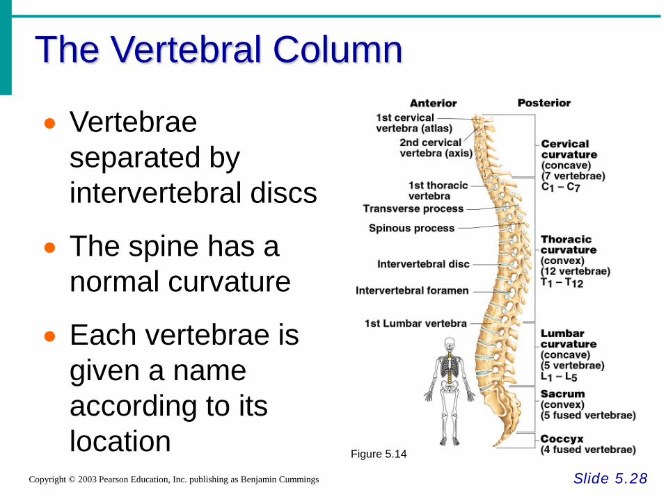
The Vertebral Column
Slide 5.28 Copyright © 2003 Pearson Education, Inc. publishing as Benjamin Cummings
• Vertebrae separated by intervertebral discs
• The spine has a normal curvature
• Each vertebrae is given a name according to its location Figure 5.14
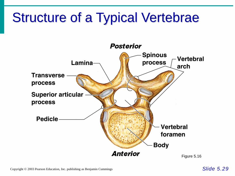
Structure of a Typical Vertebrae
Slide 5.29 Copyright © 2003 Pearson Education, Inc. publishing as Benjamin Cummings
Figure 5.16
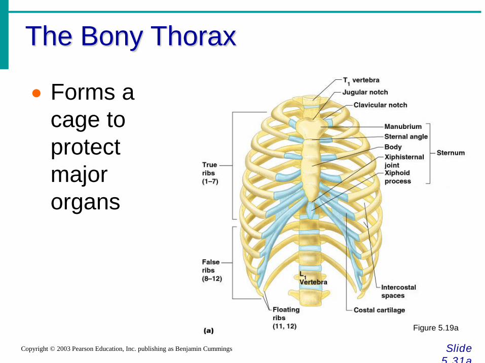
The Bony Thorax
Slide 5.31a
Copyright © 2003 Pearson Education, Inc. publishing as Benjamin Cummings
• Forms a cage to protect major organs
Figure 5.19a
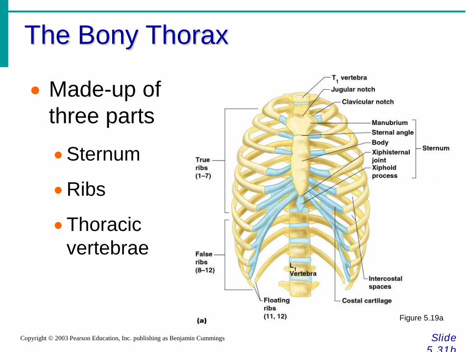
The Bony Thorax
Slide 5.31b
Copyright © 2003 Pearson Education, Inc. publishing as Benjamin Cummings
• Made-up of three parts
•Sternum
•Ribs
•Thoracic vertebrae
Figure 5.19a
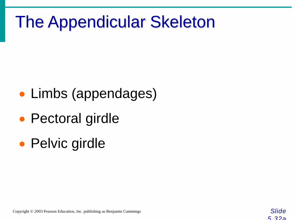
The Appendicular Skeleton
Slide 5.32a
Copyright © 2003 Pearson Education, Inc. publishing as Benjamin Cummings
• Limbs (appendages)
• Pectoral girdle
• Pelvic girdle
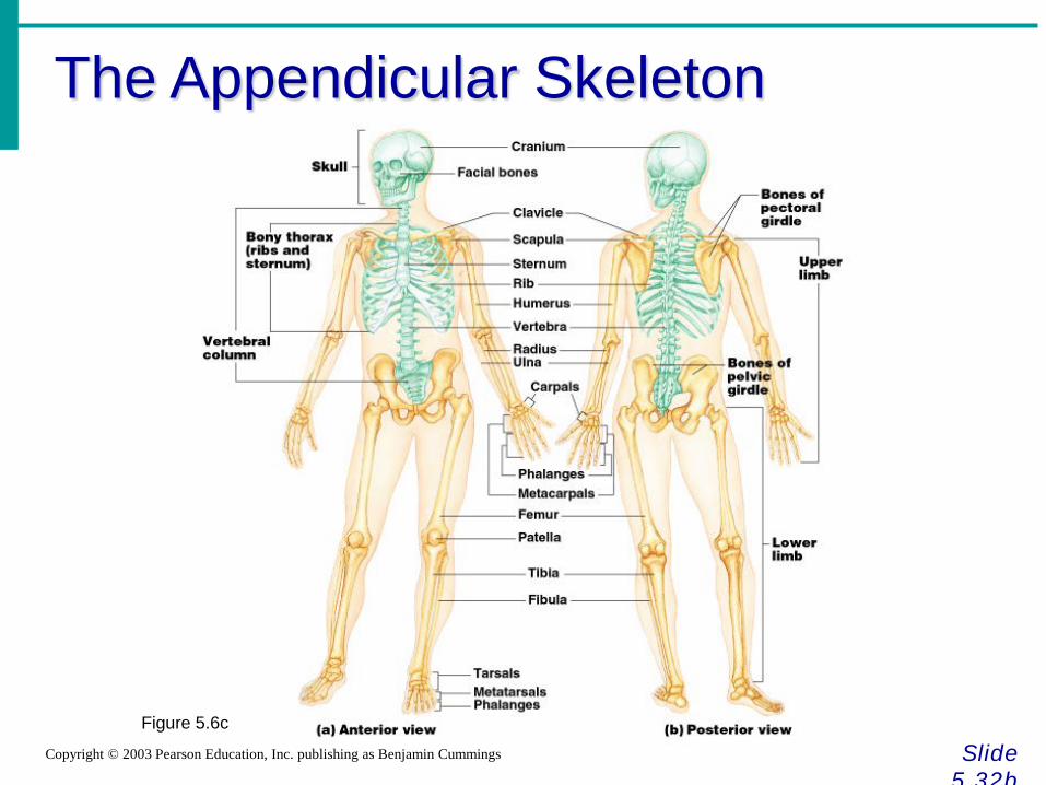
The Appendicular Skeleton
Slide 5.32b
Copyright © 2003 Pearson Education, Inc. publishing as Benjamin Cummings
Figure 5.6c
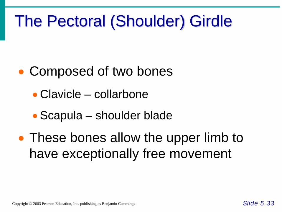
The Pectoral (Shoulder) Girdle
Slide 5.33 Copyright © 2003 Pearson Education, Inc. publishing as Benjamin Cummings
• Composed of two bones
•Clavicle – collarbone
•Scapula – shoulder blade
• These bones allow the upper limb to have exceptionally free movement
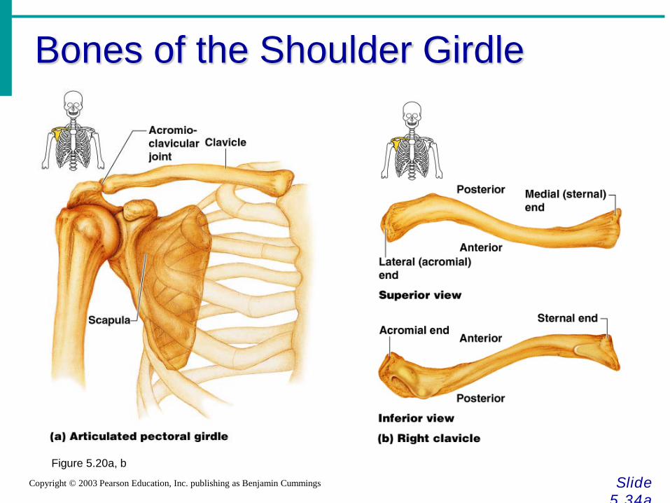
Bones of the Shoulder Girdle
Slide 5.34a
Copyright © 2003 Pearson Education, Inc. publishing as Benjamin Cummings
Figure 5.20a, b
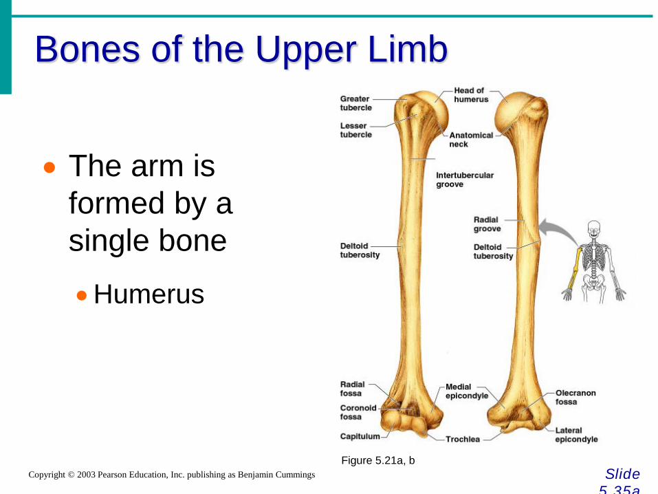
Bones of the Upper Limb
Slide 5.35a
Copyright © 2003 Pearson Education, Inc. publishing as Benjamin Cummings
• The arm is formed by a single bone
•Humerus
Figure 5.21a, b
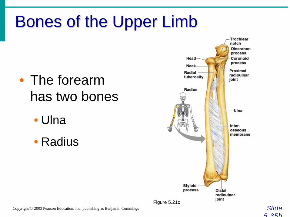
Bones of the Upper Limb
Slide 5.35b
Copyright © 2003 Pearson Education, Inc. publishing as Benjamin Cummings
• The forearm has two bones
• Ulna
• Radius
Figure 5.21c
Bones of the Upper Limb
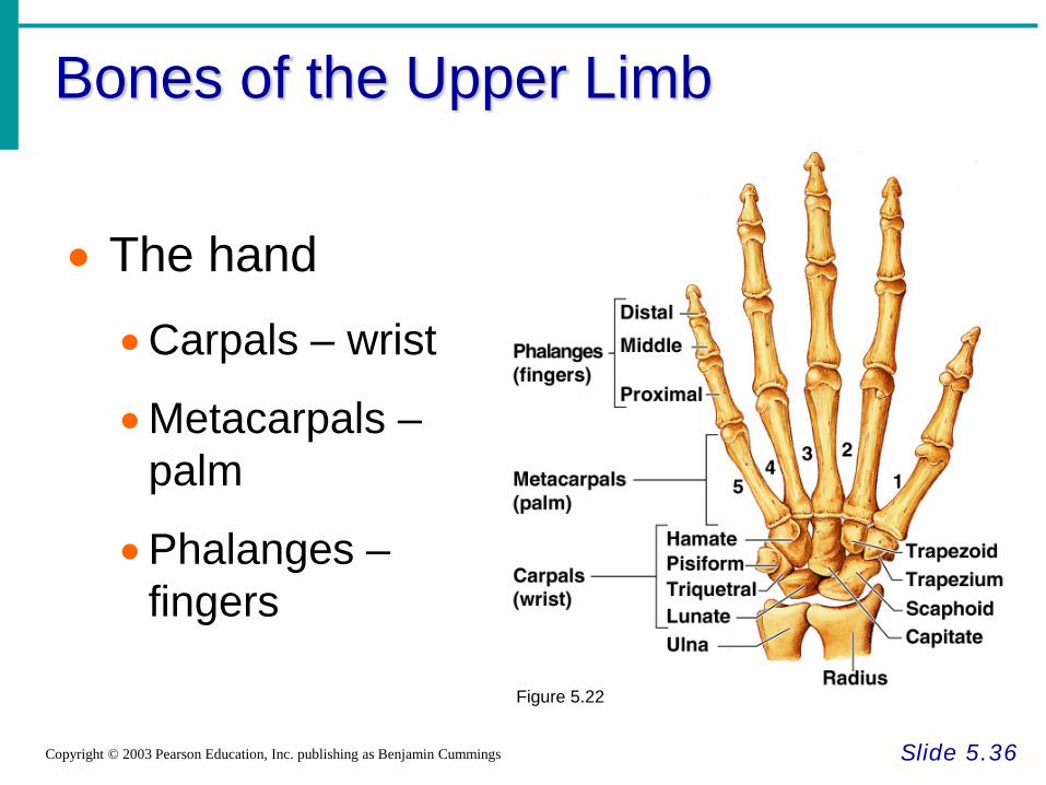
Slide 5.36 Copyright © 2003 Pearson Education, Inc. publishing as Benjamin Cummings
• The hand
•Carpals – wrist
•Metacarpals – palm
•Phalanges – fingers
Figure 5.22
Bones of the Pelvic Girdle
Slide 5.37 Copyright © 2003 Pearson Education, Inc. publishing as Benjamin Cummings
• Hip bones • Composed of three pair of fused bones
• Ilium • Ischium • Pubic bone
• The total weight of the upper body rests on the pelvis
• Protects several organs • Reproductive organs • Urinary bladder • Part of the large intestine
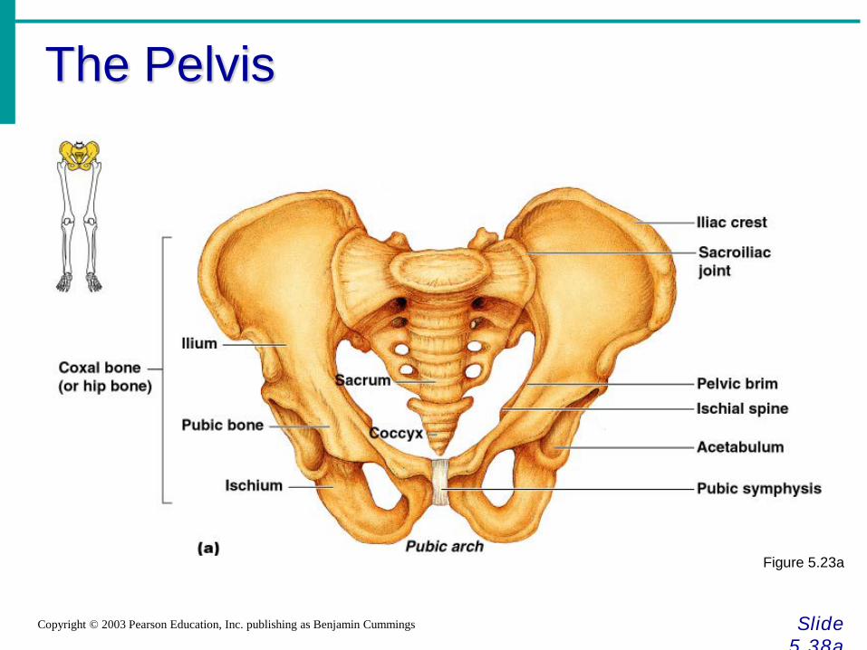
The Pelvis
Slide 5.38a
Copyright © 2003 Pearson Education, Inc. publishing as Benjamin Cummings
Figure 5.23a
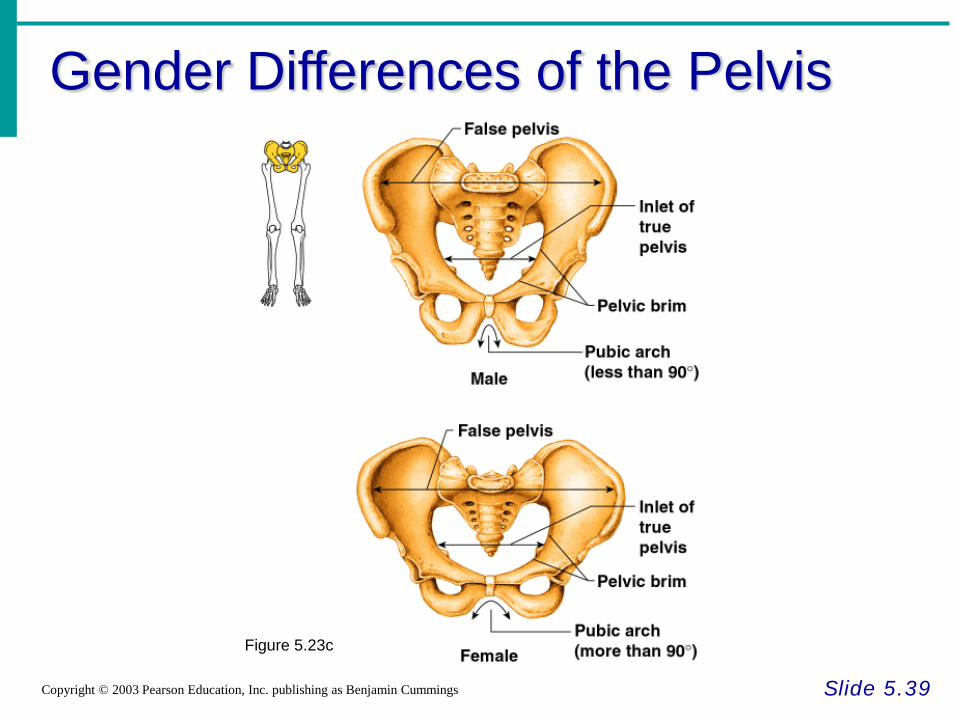
Gender Differences of the Pelvis
Slide 5.39 Copyright © 2003 Pearson Education, Inc. publishing as Benjamin Cummings
Figure 5.23c
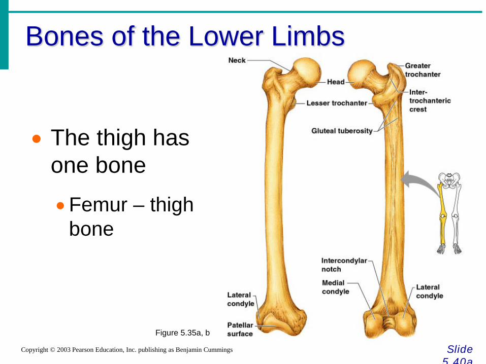
Bones of the Lower Limbs
Slide 5.40a
Copyright © 2003 Pearson Education, Inc. publishing as Benjamin Cummings
• The thigh has one bone
•Femur – thigh bone
Figure 5.35a, b
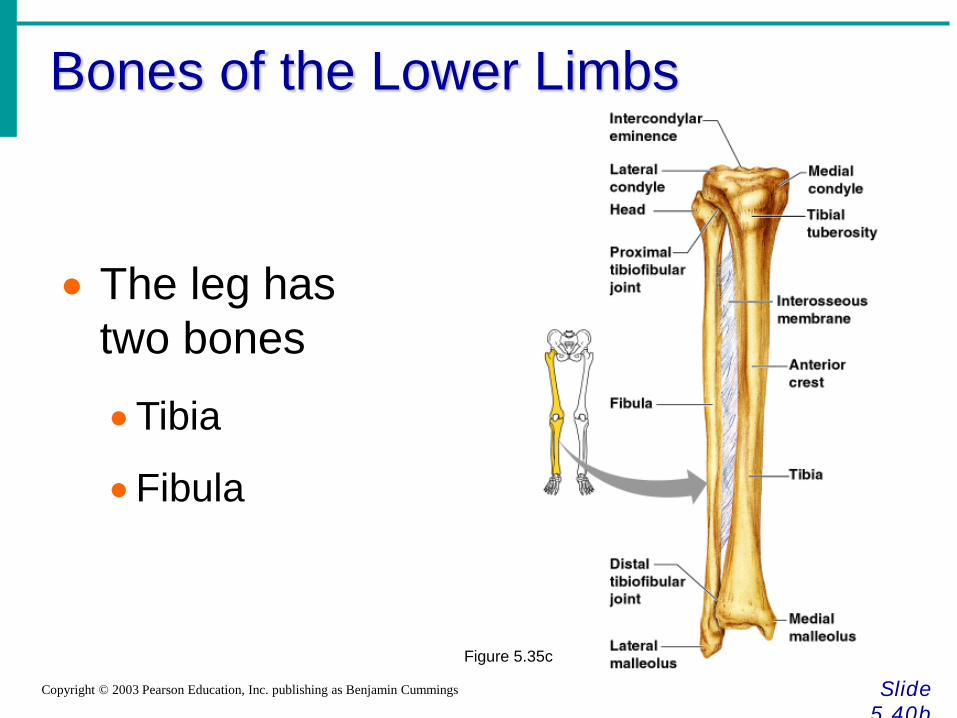
Bones of the Lower Limbs
Slide 5.40b
Copyright © 2003 Pearson Education, Inc. publishing as Benjamin Cummings
• The leg has two bones
•Tibia
•Fibula
Figure 5.35c
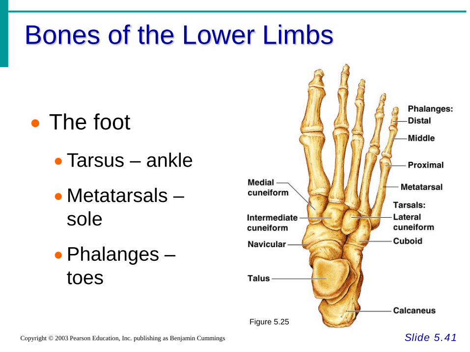
Bones of the Lower Limbs
Slide 5.41 Copyright © 2003 Pearson Education, Inc. publishing as Benjamin Cummings
• The foot
•Tarsus – ankle
•Metatarsals – sole
•Phalanges – toes
Figure 5.25
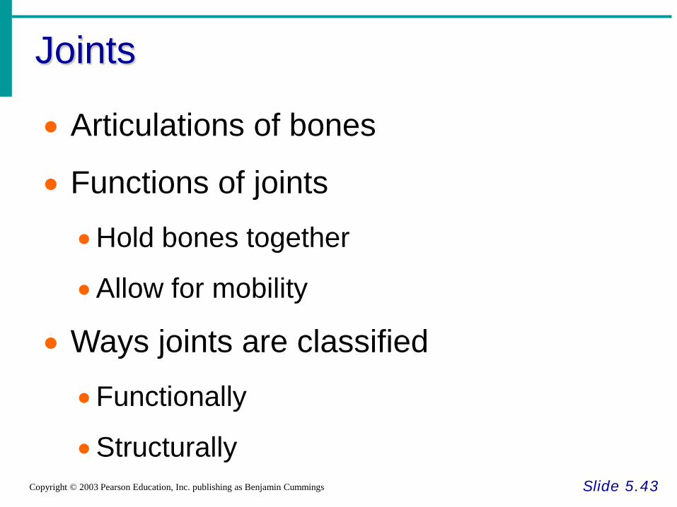
Joints
Slide 5.43 Copyright © 2003 Pearson Education, Inc. publishing as Benjamin Cummings
• Articulations of bones
• Functions of joints
•Hold bones together
•Allow for mobility
• Ways joints are classified
•Functionally
•Structurally
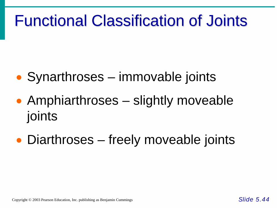
Functional Classification of Joints
Slide 5.44 Copyright © 2003 Pearson Education, Inc. publishing as Benjamin Cummings
• Synarthroses – immovable joints
• Amphiarthroses – slightly moveable joints
• Diarthroses – freely moveable joints
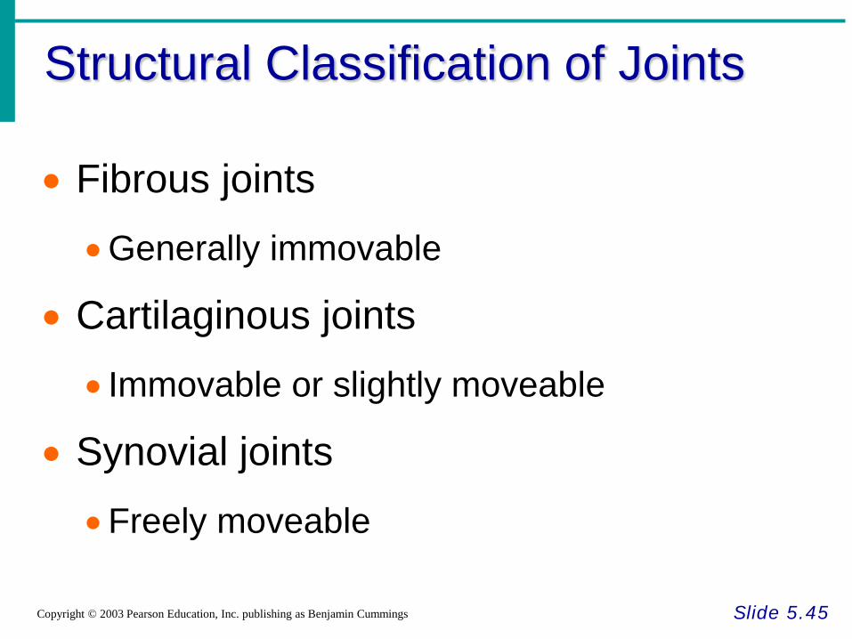
Structural Classification of Joints
Slide 5.45 Copyright © 2003 Pearson Education, Inc. publishing as Benjamin Cummings
• Fibrous joints
•Generally immovable
• Cartilaginous joints
• Immovable or slightly moveable
• Synovial joints
•Freely moveable
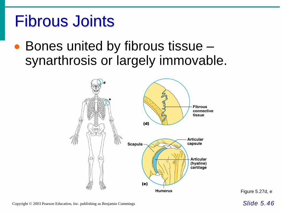
Fibrous Joints
Slide 5.46 Copyright © 2003 Pearson Education, Inc. publishing as Benjamin Cummings
• Bones united by fibrous tissue – synarthrosis or largely immovable.
Figure 5.27d, e
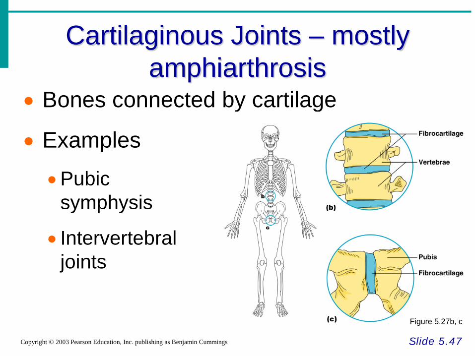
Cartilaginous Joints – mostly amphiarthrosis
Slide 5.47 Copyright © 2003 Pearson Education, Inc. publishing as Benjamin Cummings
• Bones connected by cartilage
• Examples
•Pubic symphysis
• Intervertebral joints
Figure 5.27b, c
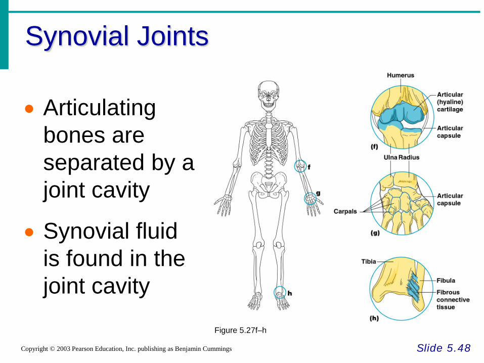
Synovial Joints
Slide 5.48 Copyright © 2003 Pearson Education, Inc. publishing as Benjamin Cummings
• Articulating bones are separated by a joint cavity
• Synovial fluid is found in the joint cavity
Figure 5.27f–h
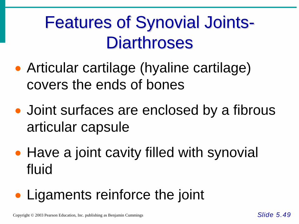
Features of Synovial Joints- Diarthroses
Slide 5.49 Copyright © 2003 Pearson Education, Inc. publishing as Benjamin Cummings
• Articular cartilage (hyaline cartilage) covers the ends of bones
• Joint surfaces are enclosed by a fibrous articular capsule
• Have a joint cavity filled with synovial fluid
• Ligaments reinforce the joint
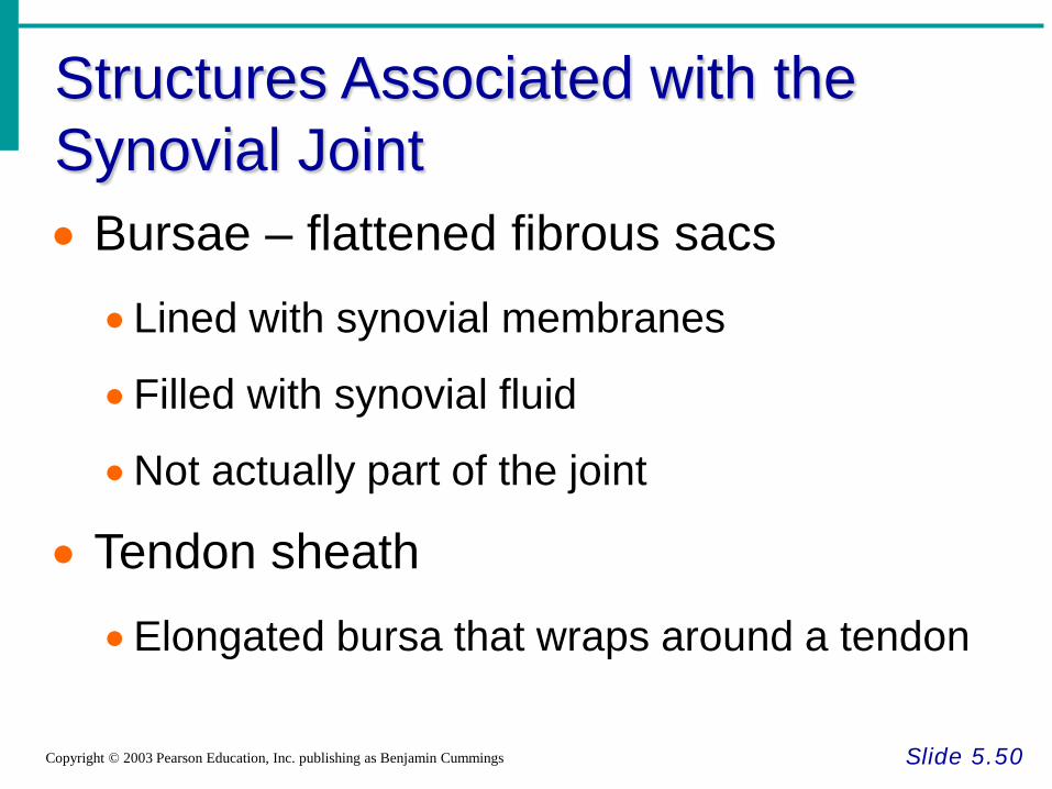
Structures Associated with the Synovial Joint
Slide 5.50 Copyright © 2003 Pearson Education, Inc. publishing as Benjamin Cummings
• Bursae – flattened fibrous sacs •Lined with synovial membranes
•Filled with synovial fluid
•Not actually part of the joint
• Tendon sheath •Elongated bursa that wraps around a tendon
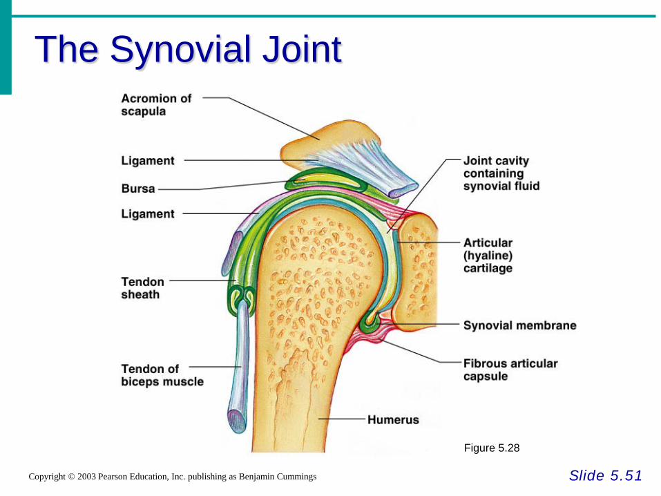
The Synovial Joint
Slide 5.51 Copyright © 2003 Pearson Education, Inc. publishing as Benjamin Cummings
Figure 5.28
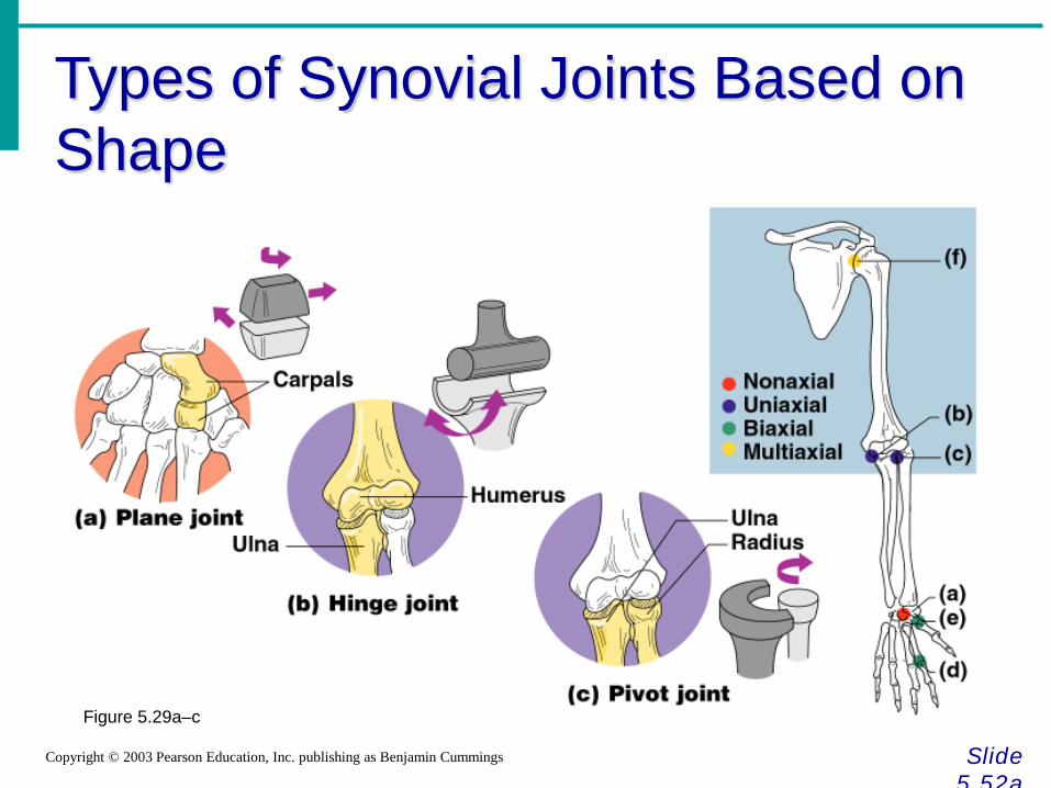
Types of Synovial Joints Based on Shape
Slide 5.52a
Copyright © 2003 Pearson Education, Inc. publishing as Benjamin Cummings
Figure 5.29a–c
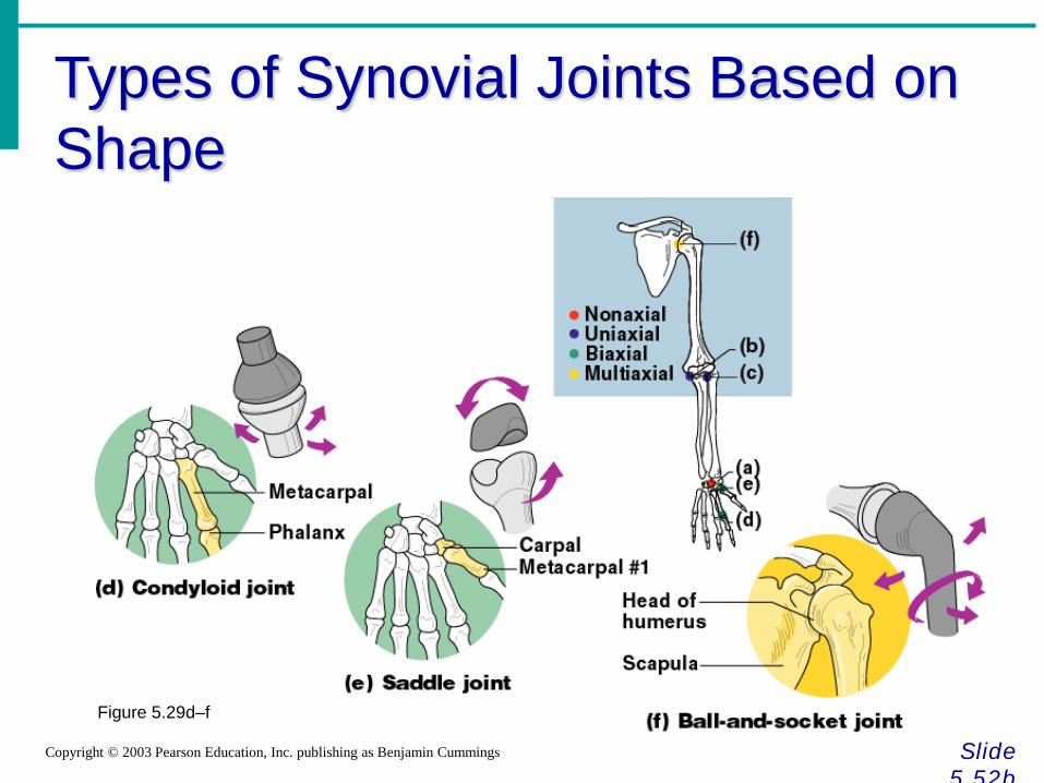
Types of Synovial Joints Based on Shape
Slide 5.52b
Copyright © 2003 Pearson Education, Inc. publishing as Benjamin Cummings
Figure 5.29d–f
Inflammatory Conditions Associated with Joints
Slide 5.53 Copyright © 2003 Pearson Education, Inc. publishing as Benjamin Cummings
• Bursitis – inflammation of a bursa usually caused by a blow or friction
• Tendonitis – inflammation of tendon sheaths
• Arthritis – inflammatory or degenerative diseases of joints •Over 100 different types
• The most widespread crippling disease in the United States
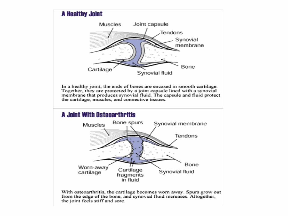
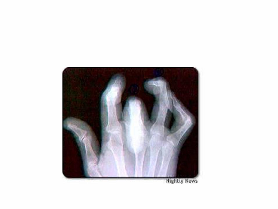
Clinical Forms of Arthritis
Slide 5.54a
Copyright © 2003 Pearson Education, Inc. publishing as Benjamin Cummings
• Osteoarthritis •Most common chronic arthritis
•Probably related to normal aging processes
• Rheumatoid arthritis •An autoimmune disease – the immune system
attacks the joints
•Symptoms begin with bilateral inflammation of certain joints
•Often leads to deformities
Clinical Forms of Arthritis
Slide 5.54b
Copyright © 2003 Pearson Education, Inc. publishing as Benjamin Cummings
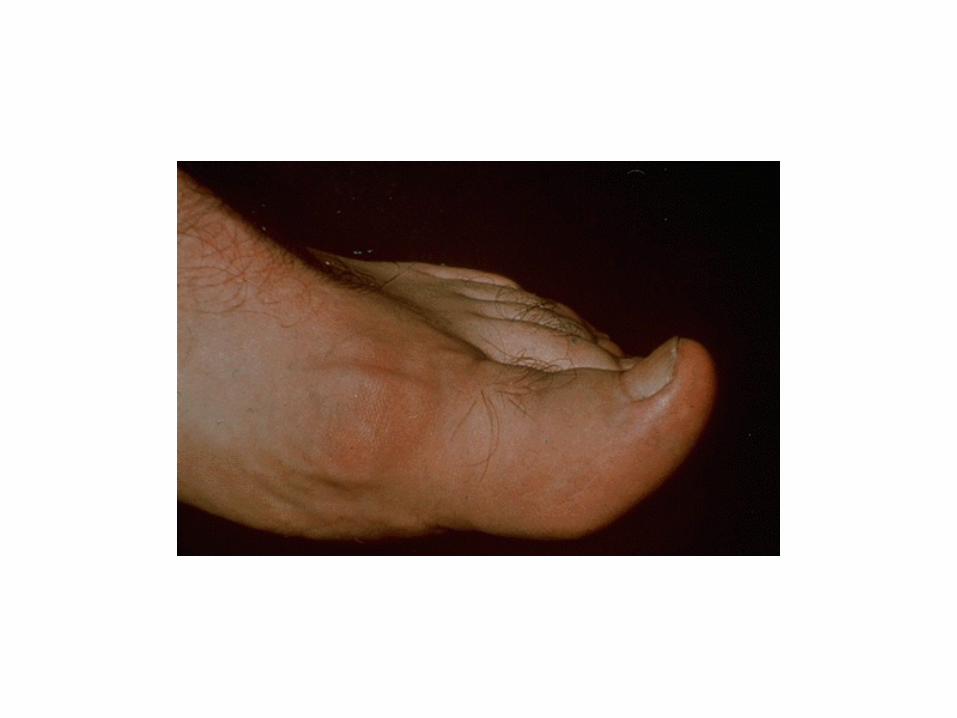
• Gouty Arthritis
• Inflammation of joints is caused by a deposition of urate crystals from the blood
•Can usually be controlled with diet


![Cartilage - Shahid Beheshti Universityfacultymembers.sbu.ac.ir/rajabi/ppt toPDF/Cartilage [Compatibility Mode].pdf · tissue and hyaline cartilage. Chondrocytes may lie singly or](https://img.pdfslide.us/doc/110x75/5e11522693c7ac3efa2277cb/cartilage-shahid-beheshti-univ-topdfcartilage-compatibility-modepdf-tissue.jpg)