Embed Size (px)
Citation preview
The Skeletal System
AP Biology
Divisions of the Skeletal System
Subdivided into two divisions: Axial Skeleton – bones that form the
longitudinal axis of the body Appendicular Skeleton – bones of the
limbs and girdles Skeletal system also includes joints,
cartilages, and ligaments The joints give the body flexibility and
allow for movement.
Functions of Bones Support
Internal framework, support and anchor soft organs
Bones of legs act as pillars to support body trunk
Rib cage supports the throacic wall Protection
Bones protect soft body organs Ex. The skull protects the brain Ex. The vertebrae surround the spinal
cord.
Functions of Bones Movement
Skeletal muscles use the bones as levers to move the body.
Remember – skeletal muscles are attached to the bones by tendons.
Storage Fat is stored in internal cavities of bones. Bones store minerals, most importantly
Calcium – important to muscles, nerves, and blood
Phosphorous
Functions of Bones
Blood Cell Formation Hematopoiesis = blood cell
formation Occurs within the marrow of certain
bones
Classification of Bones
The adult skeleton is composed of 206 bones.
2 types of bone tissue: Compact bone – dense and looks
smooth Spongy bone – composed of
needlelike pieces of bone and lots of open space
Classification of Bones Bones are classified according to shape
into 4 groups: long, short, flat, irregular Long: longer than they are wide, mostly
compact bone Short: cube-shaped, mostly spongy bone Flat: thin, flattened, and usually curved,
2 thin layers of compact bone and a layer of spongy bone in middle
most bones of the skull, ribs, and sternum Irregular: the vertebrae and the hip
bones
Structure of a Long Bone The diaphysis makes up most of the
bone’s length, composed of compact bone. Covered by a protective connective tissue
membrane called the periosteum The epiphyses are the ends of the long
bone. Mostly spongy bone
Cartilage covers this part of the bone. Provides a smooth, slippery surface that
lubricates the joints.
Structure of Bone
Epiphyseal line – thin line of bony tissue on epiphysis Remnant of the epiphyseal plate
Causes the lengthwise growth of a long bone. By the end of puberty, bones stop growing and
epiphyseal plates are completely replaced by bone.
Mark the previous location of epiphyseal plate
Interactive Web
Structure of Bone
In adults, the cavity in the shaft of the bone stores adipose tissue (fat). Yellow Marrow
In infants, this area forms blood cells, and red marrow is found there.
In adults, red marrow is only found in the cavities of spongy bone of flat bones and some long bones.
Bone Markings
Bumps, ridges, and holes in bones. Indicate where muscles, tendons,
and ligaments attach, and where blood vessels and nerves passed.
p.115
Axial Skeleton
head, neck, trunk SKULL HYOID BONE (upper neck, under jaw,
mandible) VERTEBRAL COLUMN
(spine/backbone) THORACIC CAGE (rib cage-12 pairs) STERNUM
Appendicular Skeleton
limbs and bones connecting the limbs to the:
PECTORAL GIRDLE (scapula & clavicle)
UPPER LIMBS (arms) PELVIC GIRDLE (coxal bones) LOWER LIMBS (legs)
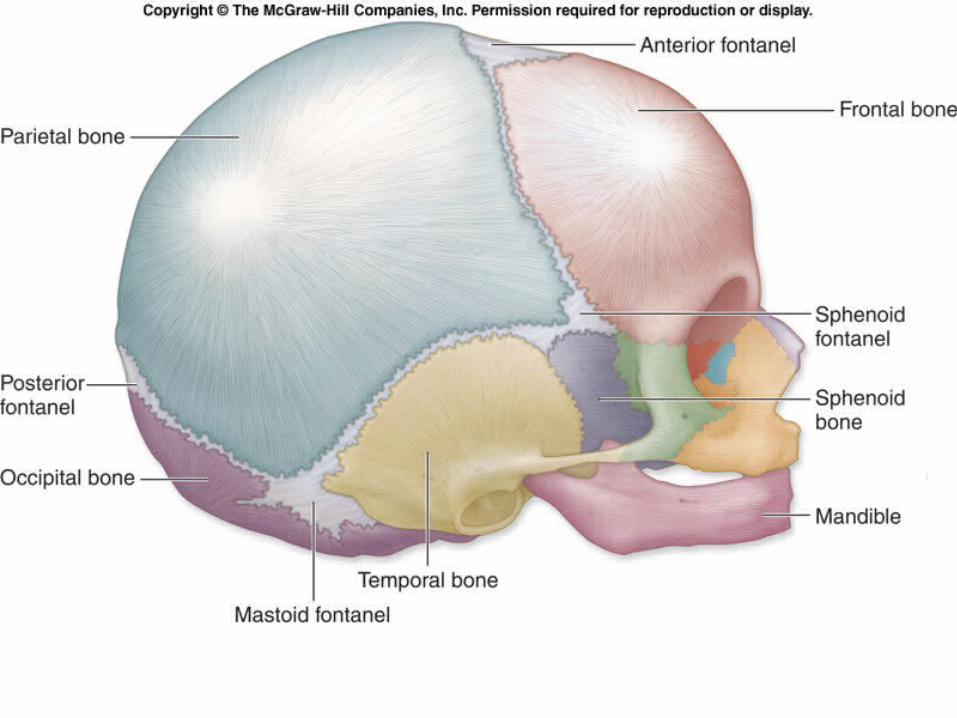
Bones of the Skull 1. Frontal - anterior portion above eyes 2. Parietal – one on each side of the skull, just
behind frontal bone 3. Occipital – forms the back of the skull and base
of the cranium 4. Temporal – forms parts of the sides and base of
cranium 5. Sphenoid – wedged between several other
bones in anterior portion of the cranium 6. Maxilla – forms upper jaws 7. Mandible – lower jaws, only moveable bone of
the skull
Vertebral Column 3 types of vertebrae:
Cervical: First 7 (neck) Thoracic: 12 vertebrae Lumbar: Last 5 (lower back)
Intervertebral disks: flexible cartilage, cushion vertebrae and absorb shock
Sacrum: fusion of 5 vertebrae Coccyx: fusion of 3-5 small, irregularly
shaped vertebrae. “tailbone”
Bones
Ribs – Thoracic Cage, 12 pairs True Ribs – first seven pairs, attach
directly to STERNUM by costal cartilage False Ribs – last five pairs Floating ribs – last two pairs
Pectoral Girdle: Shoulder. Two clavicles (collar bones) and two
scapula (shoulder blade)
Bones Arms: Upper arm – humerus. Lower arm –
radius and ulna. Wrist – 8 small bones called carpels Fingers – Metacarpels, Phalanges
Pelvic Girdle: Hips. Two large bones called COXAL BONES
Legs: Upper leg (thigh) - FEMUR. Lower leg – tibia & fibula. Ankle and Upper foot – 7 bones called TARSALS, Largest is the heel bone called the CALCANEOUS Toes – Metatarsals, Phalanges
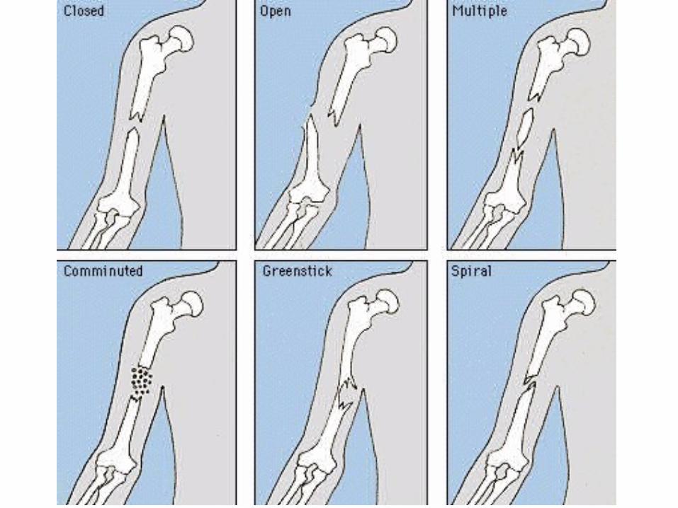
Broken Bones A simple fracture is when the bone is broken cleanly
but does not penetrate the skin. A compound/open fracture is when the bone is
sticking through the skin. A greenstick fracture is when the bone cracks on one
side only, not all the way through. A comminuted (say: kah-muh-noot-ed) fracture is
when the bone is broken into many fragments or crushed.
A compression fracture is when the bone is crushed. A depressed fracture is when the bone is broken and
pressed inward (typical of skull fracture). A spiral fracture is when a break occurs from
excessive twisting forces applied to the bone.