Embed Size (px)
Citation preview
The serine protease domain of MASP-3: enzymatic
properties and crystal structure in complex with ecotin.
Christine Gaboriaud, Rajesh Kumar Gupta, Lydie Martin, Monique Lacroix,
Laurence Serre, Florence Teillet, Gerard J Arlaud, Veronique Rossi, Nicole
Thielens
To cite this version:
Christine Gaboriaud, Rajesh Kumar Gupta, Lydie Martin, Monique Lacroix, Laurence Serre, etal.. The serine protease domain of MASP-3: enzymatic properties and crystal structure in com-plex with ecotin.. PLoS ONE, Public Library of Science, 2013, 8 (7), pp.e67962. <10.1371/jour-nal.pone.0067962>. <inserm-00868926>
HAL Id: inserm-00868926
http://www.hal.inserm.fr/inserm-00868926
Submitted on 2 Oct 2013
HAL is a multi-disciplinary open accessarchive for the deposit and dissemination of sci-entific research documents, whether they are pub-lished or not. The documents may come fromteaching and research institutions in France orabroad, or from public or private research centers.
L’archive ouverte pluridisciplinaire HAL, estdestinee au depot et a la diffusion de documentsscientifiques de niveau recherche, publies ou non,emanant des etablissements d’enseignement et derecherche francais ou etrangers, des laboratoirespublics ou prives.

The Serine Protease Domain of MASP-3: EnzymaticProperties and Crystal Structure in Complex with EcotinChristine Gaboriaud1,2,3*, Rajesh Kumar Gupta1,2,3¤a, Lydie Martin1,2,3, Monique Lacroix1,2,3,
Laurence Serre3,1¤b, Florence Teillet1,2,3, Gerard J. Arlaud1,2,3, Veronique Rossi1,2,3, Nicole M. Thielens1,2,3
1 Institut de Biologie Structurale (IBS), Direction des Sciences du Vivant, Commissariat a l’Energie Atomique et aux Energies Alternatives, Grenoble, France, 2 IBS, Centre
National de la Recherche Scientifique, Grenoble, France, 3 IBS, Universite Grenoble Alpes, Grenoble, France
Abstract
Mannan-binding lectin (MBL), ficolins and collectin-11 are known to associate with three homologous modular proteases,the MBL-Associated Serine Proteases (MASPs). The crystal structures of the catalytic domains of MASP-1 and MASP-2 havebeen solved, but the structure of the corresponding domain of MASP-3 remains unknown. A link between mutations in theMASP1/3 gene and the rare autosomal recessive 3MC (Mingarelli, Malpuech, Michels and Carnevale,) syndrome,characterized by various developmental disorders, was discovered recently, revealing an unexpected important role ofMASP-3 in early developmental processes. To gain a first insight into the enzymatic and structural properties of MASP-3, arecombinant form of its serine protease (SP) domain was produced and characterized. The amidolytic activity of this domainon fluorescent peptidyl-aminomethylcoumarin substrates was shown to be considerably lower than that of other membersof the C1r/C1s/MASP family. The E. coli protease inhibitor ecotin bound to the SP domains of MASP-3 and MASP-2, whereasno significant interaction was detected with MASP-1, C1r and C1s. A tetrameric complex comprising an ecotin dimer andtwo MASP-3 SP domains was isolated and its crystal structure was solved and refined to 3.2 A. Analysis of the ecotin/MASP-3interfaces allows a better understanding of the differential reactivity of the C1r/C1s/MASP protease family members towardsecotin, and comparison of the MASP-3 SP domain structure with those of other trypsin-like proteases yields novelhypotheses accounting for its zymogen-like properties in vitro.
Citation: Gaboriaud C, Gupta RK, Martin L, Lacroix M, Serre L, et al. (2013) The Serine Protease Domain of MASP-3: Enzymatic Properties and Crystal Structure inComplex with Ecotin. PLoS ONE 8(7): e67962. doi:10.1371/journal.pone.0067962
Editor: Claudio M. Soares, Instituto de Tecnologica Quımica e Biologica, UNL, Portugal
Received March 27, 2013; Accepted May 23, 2013; Published July 4, 2013
Copyright: � 2013 Gaboriaud et al. This is an open-access article distributed under the terms of the Creative Commons Attribution License, which permitsunrestricted use, distribution, and reproduction in any medium, provided the original author and source are credited.
Funding: This work was supported by the Commissariat a l’Energie Atomique, the Centre National de la Recherche Scientifique, the Universite Joseph Fourier(Grenoble, France) and partly by a grant from the French National Research Agency (ANR-09-PIRI-0021). RKG was supported by a post-doctoral scholarship of theEmbassy of France in India. The funders had no role in study design, data collection and analysis, decision to publish, or preparation of the manuscript.
Competing Interests: The authors have declared that no competing interests exist.
* E-mail: [email protected]
¤a Current address: Institute for Glycomics, Griffith University, Queensland, Australia¤b Current address: Institut des Neurosciences, Universite Grenoble Alpes, Grenoble, France
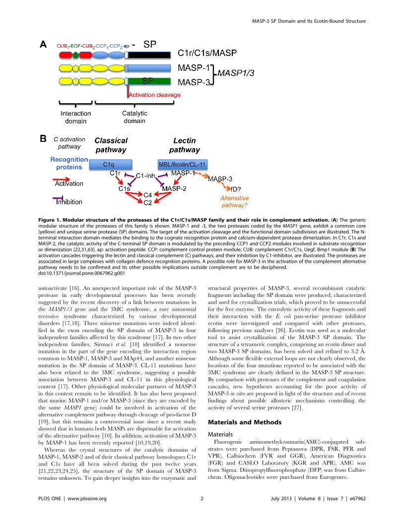
Introduction
Tightly regulated cascades of proteolytic activations control the
complement system, a key player of the host humoral defence, as
well as essential physiological processes such as coagulation and
fibrinolysis. Activation of the classical and lectin pathways of
complement is mediated by homologous modular proteases of the
C1r/C1s/MASP family (Fig. 1). This process involves large
proteolytic complexes including a recognition molecule of the
defence collagens family together with its cognate proteases. The
recognition proteins of the lectin pathway identified so far
encompass mannan-binding lectin (MBL) [1], collectin 11 (CL-
11, CL-K1) [2,3], and ficolins M, L and H (also called ficolin-1, -2
and -3) [4]. Three homologous MBL-associated serine proteases
(MASP)-1, -2 and -3, are found associated to these recognition
proteins [5], as well as two non-enzymatic components called
MAp19 (MBL-associated protein of 19 kDa) or sMAP (small
MBL-associated protein) [6,7] and MAp44 (MBL-associated
protein of 44 kDa) or MAP-1 (MBL-associated protein 1) [8,9].
MAp19 is an alternative splicing product of the MASP2 gene
whereas MASP-1, MASP-3, and MAp44 are all encoded by the
MASP1/3 gene. MASP-1 and MASP-3 only differ by their serine
protease domains and the preceding 15 amino acid residues
(Fig. 1). All MBL-associated proteins form homodimers able to
interact individually with the lectin pathway recognition proteins
through their N-terminal interaction domain.
Clear roles have been recently assigned to MASP-1 and -2 in
the activation of the complement lectin pathway, paralleling the
roles of C1r and C1s in the classical pathway (Fig. 1, [10,11]). The
classical pathway C1 complex comprises a recognition protein
C1q and a C1r2C1s2 tetrameric complex of serine proteases.
Binding of C1q to suitable targets triggers self-activation of C1r,
which in turn activates C1s, the protease responsible for cleavage
of C4 and C2, leading to assembly of the C3 convertase C4b2a
[12]. In a similar way, MASP-1 appears to be essential for the
activation of MASP-2, the latter cleaving C4 and C2 [10,11].
MASP-1 can also activate proenzyme C2 in the C4bC2 complex
[13].
A regulatory role has been suggested for MASP-3, MAp44 and
MAp19, because of their potential ability to compete with MASP-
1 and -2 for interaction with the recognition proteins
[8,9,13,14,15]. Initial analysis of recombinant MASP-3 has
revealed that it is produced in a proenzyme form, unable to
PLOS ONE | www.plosone.org 1 July 2013 | Volume 8 | Issue 7 | e67962
autoactivate [16]. An unexpected important role of the MASP-3
protease in early developmental processes has been recently
suggested by the recent discovery of a link between mutations in
the MASP1/3 gene and the 3MC syndrome, a rare autosomal
recessive syndrome characterized by various developmental
disorders [17,18]. Three missense mutations were indeed identi-
fied in the exon encoding the SP domain of MASP-3 in four
independent families affected by this syndrome [17]. In two other
independent families, Sirmaci et al. [18] identified a nonsense
mutation in the part of the gene encoding the interaction region
common to MASP-1, MASP-3 and MAp44, and another missense
mutation in the SP domain of MASP-3. CL-11 mutations have
also been related to the 3MC syndrome, suggesting a possible
association between MASP-3 and CL-11 in this physiological
context [17]. Other physiological molecular partners of MASP-3
in this context remain to be identified. It has also been proposed
that murine MASP-1 and/or MASP-3 (since they are encoded by
the same MASP1 gene) could be involved in activation of the
alternative complement pathway through cleavage of pro-factor D
[19], but this remains a controversial issue since a recent study
showed that in humans both MASPs are dispensable for activation
of the alternative pathway [10]. In addition, activation of MASP-3
by MASP-1 has been recently reported [10,19,20].
Whereas the crystal structures of the catalytic domains of
MASP-1, MASP-2 and of their classical pathway homologues C1r
and C1s have all been solved during the past twelve years
[21,22,23,24,25], the structure of the SP domain of MASP-3
remains unknown. To gain deeper insights into the enzymatic and
structural properties of MASP-3, several recombinant catalytic
fragments including the SP domain were produced, characterized
and used for crystallization trials, which proved to be unsuccessful
for the free enzyme. The esterolytic activity of these fragments and
their interaction with the E. coli pan-serine protease inhibitor
ecotin were investigated and compared with other proteases,
following previous analyses [26]. Ecotin was used as a molecular
tool to assist crystallization of the MASP-3 SP domain. The
structure of a tetrameric complex, comprising an ecotin dimer and
two MASP-3 SP domains, has been solved and refined to 3.2 A.
Although some flexible external loops are not clearly observed, the
locations of the four mutations reported to be associated with the
3MC syndrome are clearly defined in the MASP-3 SP structure.
By comparison with proteases of the complement and coagulation
cascades, new hypotheses accounting for the poor activity of
MASP-3 in vitro are proposed in light of the structure and of recent
findings about possible allosteric mechanisms controlling the
activity of several serine proteases [27].
Materials and Methods
MaterialsFluorogenic aminomethylcoumarin(AMC)-conjugated sub-
strates were purchased from Peptanova (DPR, FSR, PFR and
VPR), Calbiochem (FVR and GGR), American Diagnostica
(FGR) and CASLO Laboratory (KGR and APR). AMC was
from Sigma. Diisopropylfluorophosphate (DFP) was from Calbio-
chem. Oligonucleotides were purchased from Eurogentec.
Figure 1. Modular structure of the proteases of the C1r/C1s/MASP family and their role in complement activation. (A) The genericmodular structure of the proteases of this family is shown. MASP-1 and -3, the two proteases coded by the MASP1 gene, exhibit a common core(yellow) and unique serine protease (SP) domains. The target of the activation cleavage and the functional domain subdivision are illustrated. The N-terminal interaction domain mediates the binding to the cognate recognition protein and calcium-dependent protease dimerization. In C1r, C1s andMASP-2, the catalytic activity of the C-terminal SP domain is modulated by the preceding CCP1 and CCP2 modules involved in substrate recognitionor dimerization [22,31,63]. ap: activation peptide. CCP: complement control protein module; CUB: complement C1r/C1s, Uegf, Bmp1 module (B) Theactivation cascades triggering the lectin and classical complement (C) pathways, and their inhibition by C1-inhibitor, are illustrated. The proteases areassociated in large complexes with collagen defence recognition proteins. A possible role for MASP-3 in the activation of the complement alternativepathway needs to be confirmed and its other possible implications outside complement are to be deciphered.doi:10.1371/journal.pone.0067962.g001
MASP-3 SP Domain and Its Ecotin-Bound Structure
PLOS ONE | www.plosone.org 2 July 2013 | Volume 8 | Issue 7 | e67962
ProteinsHuman plasma thrombin and coagulation factors XIa and XIII
were obtained from Calbiochem. Bovine pancreas trypsin (TPCK
treated) was from Sigma. Ecotin was purchased from Gentaur
Molecular Products and recombinant human insulin-like growth
factor-binding protein (IGFBP)-5 from R&D Systems.
C1 inhibitor and activated C1r and C1s were purified from
human serum as described by Arlaud et al. [28,29]. Recombinant
fragments MASP-1 CCP1/2-ap-SP, MASP-2 CCP1/2-ap-SP, C1s
CCP2-ap-SP and C1r CCP2-ap-SP, and full-length MASP-3 were
produced in baculovirus-infected insect cells and purified as
described previously [16,30,31,32]. The C1s fragment CCP2-ap-
SP was activated by C1r as described previously [31]. The N-
terminal extracellular domain of the mature human protease-
activated receptor (PAR) 1 was produced in E. coli as a GST fusion
protein using the pGEX-pP-3/PAR1E plasmid (kindly provided
by Dr F. Lanza). The recombinant protein was purified by affinity
chromatography on glutathione-Sepharose 4B beads (GE Health-
care) as described by Loew et al. [33].
Production of the Serine Protease Domain of MASP-3A DNA fragment encoding the activation peptide and the SP
domain of human MASP-3 (residues 416–709 of the mature
protease) was amplified by PCR using VentR polymerase (New
England Biolabs) and the expression plasmid coding for MASP-3
[16] as a template, according to established procedures. The
amplified DNA, containing a BglII restriction site at the 59 end
and a stop codon followed by an EcoRI site at the 39 end, was
cloned in-frame with the melittin signal peptide into the BamHI
and EcoRI sites of the pNT-Bac baculovirus transfer vector [31].
This plasmid served as a template to generate the vector coding for
the MASP-3 SP domain (residues 431–709) using the Quick-
Change XL site-directed mutagenesis kit (Stratagene), by deleting
the segment coding for the activation peptide and adding two
residues (Asp-Leu) at the N terminus, due to in-frame cloning with
the signal sequence of melittin. The pNT-Bac-MASP-3 SP
plasmid was checked by double-strand sequencing (Genome
Express, France). The recombinant baculovirus was generated
using the Bac-to-BacTM system (Invitrogen) and amplified as
described previously [34]. High Five insect cells were infected with
the recombinant virus for 72 h at 27uC.
The culture supernatant (0.5 L) containing the MASP-3 SP
domain was dialyzed against 25 mM NaCl, 50 mM triethanol-
amine-HCl, pH 7.4, and loaded onto a Q-Sepharose Fast Flow
column (2.8610 cm) (GE Healthcare) equilibrated in the same
buffer containing 1 mM iodoacetamide. Elution was conducted by
applying a linear gradient to 250 mM NaCl in the same buffer.
Fractions containing the recombinant fragment were identified by
SDS-PAGE analysis, concentrated by ultrafiltration and final
purification was achieved by high-pressure gel permeation on a
TSK G3000 SW column (7.56600 mm) (Tosoh Bioscience)
equilibrated in 145 mM NaCl, 50 mM triethanolamine-HCl,
pH 7.4. The purified fragment was concentrated to 0.5–1 mg/ml
by ultrafiltration and stored at 220uC.
The tetrameric complex was formed by incubating the MASP-3
SP domain and ecotin at a 1:1 molar ratio at room temperature for
20 min. Fractions containing a maximum amount of 100 mg of
MASP-3 SP were injected on the TSK G3000 SW column
equilibrated in 145 mM NaCl, 50 mM triethanolamine-HCl,
pH 7.4. The first eluted peak, corresponding to the tetrameric
complex, was pooled and concentrated to 2–3 mg/ml by
ultrafiltration.
SDS-PAGE Analysis and Chemical Characterization of theRecombinant Proteins
Samples were analyzed by SDS-PAGE followed by Coomassie
blue staining of the proteins. N-terminal sequencing (on liquid
samples or after SDS-PAGE and electrotransfer) and MALDI
mass spectrometry analyses were carried out as described
previously [35].
Amidolytic AssaysThe amidolytic activity of MASP-3 SP, thrombin and trypsin on
selected peptidyl-AMC substrates was determined using a
fluorometric assay based on the measurement of AMC released
upon cleavage. Varying enzyme amounts were added to the
substrate (0.1 mM) in 2 ml of 20 mM Hepes, 5 mM CaCl2,
pH 8.5. Samples were excited at 360 nm and emission was read at
440 nm every 30 s for 30 min at 37uC using an Aminco-Bowman
Series 2 fluorometer. The initial rates of AMC release were
calculated from a calibration curve obtained with varying AMC
concentrations (1–10 mM) diluted in 2 ml of the above buffer.
Rates were expressed in pmol AMC released/min/mg of enzyme
to allow comparison with previously published data.
For selected substrates, the Michaelis constant (Km) and the
maximum velocity (Vmax) were determined for MASP-3 SP and
thrombin, using nonlinear regression analysis (SigmaPlot soft-
ware). The substrate concentrations varied from 50 to 200 mM
and the enzyme amounts used were 0.01 mg for thrombin and
7.5 mg for MASP-3 SP.
The effect of protease inhibitors was evaluated by measuring the
residual activity of MASP-3 SP on the VPR- or FGR-AMC
substrates after preincubation of the enzyme with ecotin for
20 min at room temperature, with C1 inhibitor for 1 h at 37uC,
and with 5 mM diisopropylfluorophosphate (DFP) for 30 min at
37uC.
Proteolytic AssaysThe proteolytic activity of MASP-3 SP, thrombin and C1s was
analyzed by incubation of 2.5–3.5 mg of MASP-3, IGFBP-5, factor
XIII and the extracellular domain of PAR1 at 37uC for 4–16 h
with MASP-3 SP (0.1–0.2 molar ratio), C1s (0.2 molar ratio) or
thrombin (0.1–1% w/w) followed by SDS-PAGE of the incubation
mixtures under reducing conditions.
Surface Plasmon Resonance Spectroscopy and DataEvaluation
Surface plasmon resonance analyses were performed using a
BIAcore 3000 or a Biacore X instrument (GE Healthcare). Ecotin
(1,000 RU) was immobilized using the amine coupling chemistry
by injecting the protein (diluted to 20 mg/ml in 10 mM sodium
acetate, pH 5.0) on the surface of a CM5 sensor chip (GE
Healthcare) in 150 mM NaCl, 5 mM EDTA, 10 mM HEPES,
pH 7.4 containing 0.005% surfactant P20 (GE Healthcare).
Binding was measured at a flow rate of 20 ml/min/min in
145 mM NaCl, 50 mM triethanolamine-HCl, pH 7.4 containing
0.005% surfactant P20. Regeneration of the surface was achieved
by 10-ml injections of 10 mM HCl. Equivalent volumes of each
protein sample were injected over a reference surface without
immobilised protein for subtraction of the bulk refractive index
background.
Data were analysed by global fitting to a 1:1 Langmuir binding
model of both the association and dissociation phases for at least
five concentrations simultaneously using the BIAevaluation 3.2
software (GE Healthcare). The apparent equilibrium dissociation
constants (KD) were calculated from the ratio of the dissociation
MASP-3 SP Domain and Its Ecotin-Bound Structure
PLOS ONE | www.plosone.org 3 July 2013 | Volume 8 | Issue 7 | e67962
and association rate constants (kd/ka). Although the interaction of
the ecotin dimer with proteases is likely more complex than a
simple 1:1 binding model, data fitting using this model yielded
satisfactory chi2 values (,4) and was used for comparison
purposes.
Crystallization, Structure Determination, and RefinementThe ecotin/MASP-3 SP complex was concentrated to about
7 mg/ml in a buffer containing 50 mM triethanolamine-HCl
pH 7.4, 145 mM NaCl and 100 mM sulfobetain NDSB195.
Reproducible crystals were obtained by the vapour diffusion
method at 20uC using a reservoir solution containing 18–22% (w/
w) PEG2KMME, 0.2 M ammonium sulfate, 0.1 M sodium
acetate pH 4.7. PEG 400 (3% w/v) was also often added to
improve crystal quality. Very thin plate-like crystals grew in two
weeks. Several datasets were collected on the ESRF beamlines
ID14-eh1, ID14–eh2, ID29 and BM14. These were integrated
and merged using XDS [36]. The crystals belong to the P21212
space group, with cell dimensions a = 66.7 A, b = 164.6 A,
c = 90.6 A. Their diffraction limit is about 3.2 A, with a high
mean Wilson B factor of about 60 A2. Their solvent content is
about 54%, with one tetramer per asymmetric unit. The structure
was solved by molecular replacement using Phaser [37], defining
the two positions of the SP domain and inhibitor search models.
The ecotin search model was a mutant version extracted from the
Protein Data Bank (PDB) entry 1XXF describing its complex with
the factor XIa catalytic domain [38]. For the SP domain, a
truncated version of the MASP-2 SP domain was used [25], in
which the tips of the more variable loops (463–467, 484–495, 504–
510, 526–529, 556–567, 577–581, 604–611, 685–686) were
removed. A clear unique solution was obtained. Moreover, several
residue substitutions from the models were visible in the initial
electron density maps. Numerous runs of iterative model building
and refinement were performed, using several graphics and
refinement softwares (O [39], Coot [40], CNS [41], BUSTER
[42], Refmac [43] to check and improve the accuracy of the
model. Bulk solvent correction and Translation Libration Screw-
motion (TLS) refinement were used, each chain defining one TLS
group. Non-crystallographic symmetry (NCS) restraints were
included in the refinement, carefully excluding only the flexible
zones for which the two copies display significant different electron
density because the molecules are in a slightly distinct environ-
ment. An illustration of the quality of the final electron density
map together with the corresponding model is provided (Fig. S1).
Missing parts in the current model include residues 1–5 and 89 in
ecotin, together with the following MASP-3 segments: 456–458,
508–511, 578–593 and 705–709. The coordinates and structure
factors of the ecotin/MASP-3 SP complex are accessible (PDB
code 4IW4).
Results and Discussion
Production and Characterization of the Recombinant SPDomain of MASP-3
The main objective of this study was to determine the structure
of the catalytic domain of MASP-3. For this purpose, artificial
processing of the melittin signal peptide was used to generate a
recombinant fragment of the SP domain of MASP-3 starting after
the activation cleavage site at Ile431. This strategy aimed to obtain
a conformational ‘active’ state of the SP domain, as expected after
its activation cleavage, overcoming the inability of MASP-3 to
undergo self-activation [16]. A similar fragment was previously
produced by Cortesio & Jiang [26] using a mammalian cell
expression vector containing the signal peptide of human CD33.
The recombinant SP domain was produced in baculovirus-
infected insect cells and purified from the cell culture supernatant
as described under Materials and Methods. The purification yield
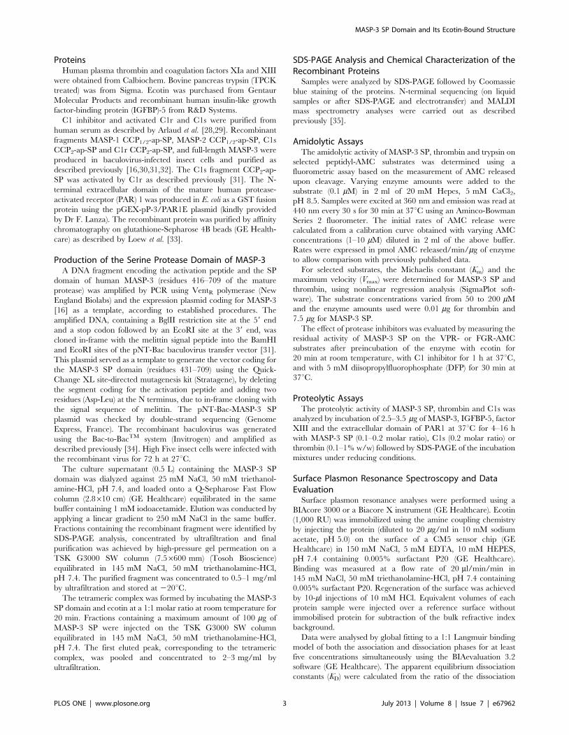
from 500 ml of culture supernatant was 700 mg. SDS-PAGE
analysis of the purified fragment yielded a band migrating at about
35 kDa under both non-reducing and reducing conditions,
corresponding to the expected mass (Fig. 2). In some preparations,
a doublet of lower mass (25 kDa) was detected, accounting for
about 5% of the purified material, and corresponding to a
degradation product. N-terminal sequence analysis yielded a
major sequence, Ile431-Ile-Gly-Gly-Arg-Asn-Ala-Glu…, corre-
sponding to that expected for the SP domain, indicating that the
melittin signal peptide had been correctly processed. A minor
sequence, Gly433-Gly-Arg-Asn-Ala-Glu-Pro-Gly…, accounting for
about 20% of the recombinant material, was also obtained,
corresponding to a fragment lacking the first two N-terminal
residues. Mass spectrometry analysis yielded a heterogeneous peak
centered on a mass value of 34,300627 Da. Given the predicted
mass of the polypeptide (30,551 Da), the deduced mass value for
the carbohydrate moieties (3,749 Da) is consistent with the
presence of three N-linked oligosaccharide chains comprising
two N-acetylglucosamine and five mannose residues (calculated
mass 1,218 Da). These data indicate that all predicted N-
glycosylation sites [14] are occupied by short high-mannose
oligosaccharides, in accordance with previous data on full-length
recombinant MASP-3 expressed in the same system [16].
Enzymatic Activity of the MASP-3 SP DomainThe catalytic activity of the recombinant SP domain of MASP-3
and its inhibition pattern were checked and further investigated.
Among the nine peptidyl-AMC substrates listed in Table 1, the
highest enzymatic activity was observed with substrates of tissue
plasminogen activator (FGR), kallikrein (PFR) and thrombin
(VPR). Significant cleavage was also observed for DPR, FSR,
GGR and KGR, which are substrates for thrombin/trypsin, factor
XIa, thrombin, and trypsin, respectively. We observed very low
cleavage of the thrombin substrate FVR-AMC and no cleavage of
APR-AMC, which mimics the cleavage site of PAR4, a protein
substrate of MASP-1 [44]. The values obtained for cleavage of
PFR, VPR and GGR and for the control enzyme thrombin are in
Figure 2. SDS-PAGE analysis of the recombinant MASP-3 SPdomain. The purified SP domain of MASP-3 was analyzed by SDS-PAGEunder non-reducing (NR) and reducing (R) conditions followed byCoomassie blue staining. The molecular masses of reduced and non-reduced standard proteins (expressed in kDa) are shown on the rightand left sides of the gel, respectively.doi:10.1371/journal.pone.0067962.g002
MASP-3 SP Domain and Its Ecotin-Bound Structure
PLOS ONE | www.plosone.org 4 July 2013 | Volume 8 | Issue 7 | e67962
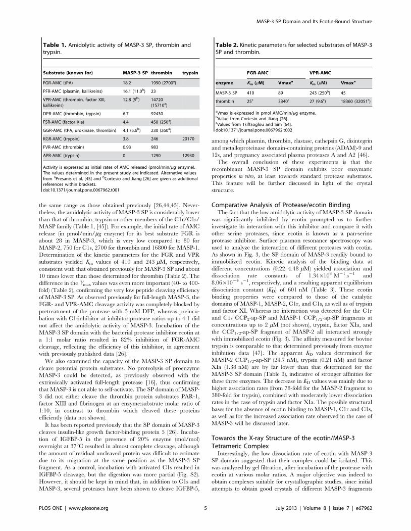
the same range as those obtained previously [26,44,45]. Never-
theless, the amidolytic activity of MASP-3 SP is considerably lower
than that of thrombin, trypsin or other members of the C1r/C1s/
MASP family (Table 1, [45]). For example, the initial rate of AMC
release (in pmol/min/mg enzyme) for its best substrate FGR is
about 28 in MASP-3, which is very low compared to 80 for
MASP-2, 750 for C1s, 2700 for thrombin and 16800 for MASP-1.
Determination of the kinetic parameters for the FGR and VPR
substrates yielded Km values of 410 and 243 mM, respectively,
consistent with that obtained previously for MASP-3 SP and about
10 times lower than those determined for thrombin (Table 2). The
difference in the Vmax values was even more important (40- to 400-
fold) (Table 2), confirming the very low peptide cleaving efficiency
of MASP-3 SP. As observed previously for full-length MASP-3, the
FGR- and VPR-AMC cleavage activity was completely blocked by
pretreatment of the protease with 5 mM DFP, whereas preincu-
bation with C1-inhibitor at inhibitor:protease ratios up to 4:1 did
not affect the amidolytic activity of MASP-3. Incubation of the
MASP-3 SP domain with the bacterial protease inhibitor ecotin at
a 1:1 molar ratio resulted in 82% inhibition of FGR-AMC
cleavage, reflecting the efficiency of this inhibitor, in agreement
with previously published data [26].
We also examined the capacity of the MASP-3 SP domain to
cleave potential protein substrates. No proteolysis of proenzyme
MASP-3 could be detected, as previously observed with the
extrinsically activated full-length protease [16], thus confirming
that MASP-3 is not able to self-activate. The SP domain of MASP-
3 did not either cleave the thrombin protein substrates PAR-1,
factor XIII and fibrinogen at an enzyme:substrate molar ratio of
1:10, in contrast to thrombin which cleaved these proteins
efficiently (data not shown).
It has been reported previously that the SP domain of MASP-3
cleaves insulin-like growth factor-binding protein 5 [26]. Incuba-
tion of IGFBP-5 in the presence of 20% enzyme (mol/mol)
overnight at 37uC resulted in almost complete cleavage, although
the amount of residual uncleaved protein was difficult to estimate
due to its migration at the same position as the MASP-3 SP
fragment. As a control, incubation with activated C1s resulted in
IGFBP-5 cleavage, but the digestion was more partial (Fig. S2).
However, it should be kept in mind that, in addition to C1s and
MASP-3, several proteases have been shown to cleave IGFBP-5,
among which plasmin, thrombin, elastase, cathepsin G, disintegrin
and metalloproteinase domain-containing proteins (ADAM)-9 and
12s, and pregnancy associated plasma proteases A and A2 [46].
The overall conclusion of these experiments is that the
recombinant MASP-3 SP domain exhibits poor enzymatic
properties in vitro, at least towards standard protease substrates.
This feature will be further discussed in light of the crystal
structure.
Comparative Analysis of Protease/ecotin BindingThe fact that the low amidolytic activity of MASP-3 SP domain
was significantly inhibited by ecotin prompted us to further
investigate its interaction with this inhibitor and compare it with
other serine proteases, since ecotin is known as a pan-serine
protease inhibitor. Surface plasmon resonance spectroscopy was
used to analyze the interaction of different proteases with ecotin.
As shown in Fig. 3, the SP domain of MASP-3 readily bound to
immobilized ecotin. Kinetic analysis of the binding data at
different concentrations (0.22–4.48 mM) yielded association and
dissociation rate constants of 1.346103 M21.s21 and
8.0661024 s21, respectively, and a resulting apparent equilibrium
dissociation constant (KD) of 601 nM (Table 3). These ecotin
binding properties were compared to those of the catalytic
domains of MASP-1, MASP-2, C1r, and C1s, as well as of trypsin
and factor XI. Whereas no interaction was detected for the C1r
and C1s CCP2-ap-SP and MASP-1 CCP1/2-ap-SP fragments at
concentrations up to 2 mM (not shown), trypsin, factor XIa, and
the CCP1/2-ap-SP fragment of MASP-2 all interacted strongly
with immobilized ecotin (Fig. 3). The affinity measured for bovine
trypsin is comparable to that determined previously from enzyme
inhibition data [47]. The apparent KD values determined for
MASP-2 CCP1/2-ap-SP (24.7 nM), trypsin (0.21 nM) and factor
XIa (1.38 nM) are by far lower than that determined for the
MASP-3 SP domain (Table 3), indicative of stronger affinities for
these three enzymes. The decrease in KD values was mainly due to
higher association rates (from 78-fold for the MASP-2 fragment to
380-fold for trypsin), combined with moderately lower dissociation
rates in the case of trypsin and factor XIa. The possible structural
bases for the absence of ecotin binding to MASP-1, C1r and C1s,
as well as for the increased association rate observed in the case of
MASP-3 will be discussed later.
Towards the X-ray Structure of the ecotin/MASP-3Tetrameric Complex
Interestingly, the low dissociation rate of ecotin with MASP-3
SP domain suggested that their complex could be isolated. This
was analyzed by gel filtration, after incubation of the protease with
ecotin at various molar ratios. A major objective was indeed to
obtain complexes suitable for crystallographic studies, since initial
attempts to obtain good crystals of different MASP-3 fragments
Table 1. Amidolytic activity of MASP-3 SP, thrombin andtrypsin.
Substrate (known for) MASP-3 SP thrombin trypsin
FGR-AMC (tPA) 18.2 1990 (2700a)
PFR-AMC (plasmin, kallikreins) 16.1 (11.0b) 23
VPR-AMC (thrombin, factor XIII,kallikreins)
12.8 (9b) 14720(15710a)
DPR-AMC (thrombin, trypsin) 6.7 92430
FSR-AMC (factor XIa) 4.4 450 (250a)
GGR-AMC (tPA, urokinase, thrombin) 4.1 (5.6b) 230 (260a)
KGR-AMC (trypsin) 3.8 246 20170
FVR-AMC (thrombin) 0.93 983
APR-AMC (trypsin) 0 1290 12930
Activity is expressed as initial rates of AMC released (pmol/min/mg enzyme).The values determined in the present study are indicated. Alternative valuesfrom aPresanis et al. [45] and bCortesio and Jiang [26] are given as additionalreferences within brackets.doi:10.1371/journal.pone.0067962.t001
Table 2. Kinetic parameters for selected substrates of MASP-3SP and thrombin.
FGR-AMC VPR-AMC
enzyme Km (mM) Vmaxa Km (mM) Vmaxa
MASP-3 SP 410 89 243 (250b) 45
thrombin 25c 3340c 27 (9.6c) 18360 (32051c)
aVmax is expressed in pmol AMC/min/mg enzyme.bValue from Cortesio and Jiang [26].cValues from Tsiftsoglou and Sim [64].doi:10.1371/journal.pone.0067962.t002
MASP-3 SP Domain and Its Ecotin-Bound Structure
PLOS ONE | www.plosone.org 5 July 2013 | Volume 8 | Issue 7 | e67962

had failed. Ecotin has been previously described as a molecular
tool to assist the crystallization of proteases [38], since the X-ray
structures of complexes between ecotin (or variants) and various
proteases have been reported, including trypsin [48], collagenase
[49], thrombin [50], granzyme B [51], coagulation factors Xa [52]
and XIa [38].
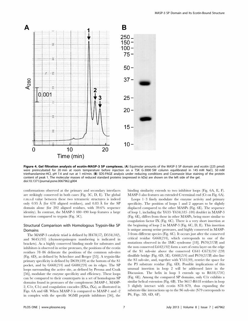
Three peaks were obtained by gel filtration when using
equimolar amounts of protease and ecotin, as shown in Fig. 4A.
The major peak 1 contained equal amounts of enzyme and
inhibitor, as judged by N-terminal sequencing and SDS-PAGE
analysis (Fig. 4B), and corresponded to the tetrameric complex
submitted to crystallization. The intensity of the second peak
increased when the inhibitor was in excess and was identified as a
trimeric complex between the ecotin dimer and the protease, as
previously observed for rat trypsin [53]. The third peak
corresponded to the uncomplexed protease and the ecotin dimer
(molecular mass 32 kDa) and its intensity increased when the
protease was in excess.
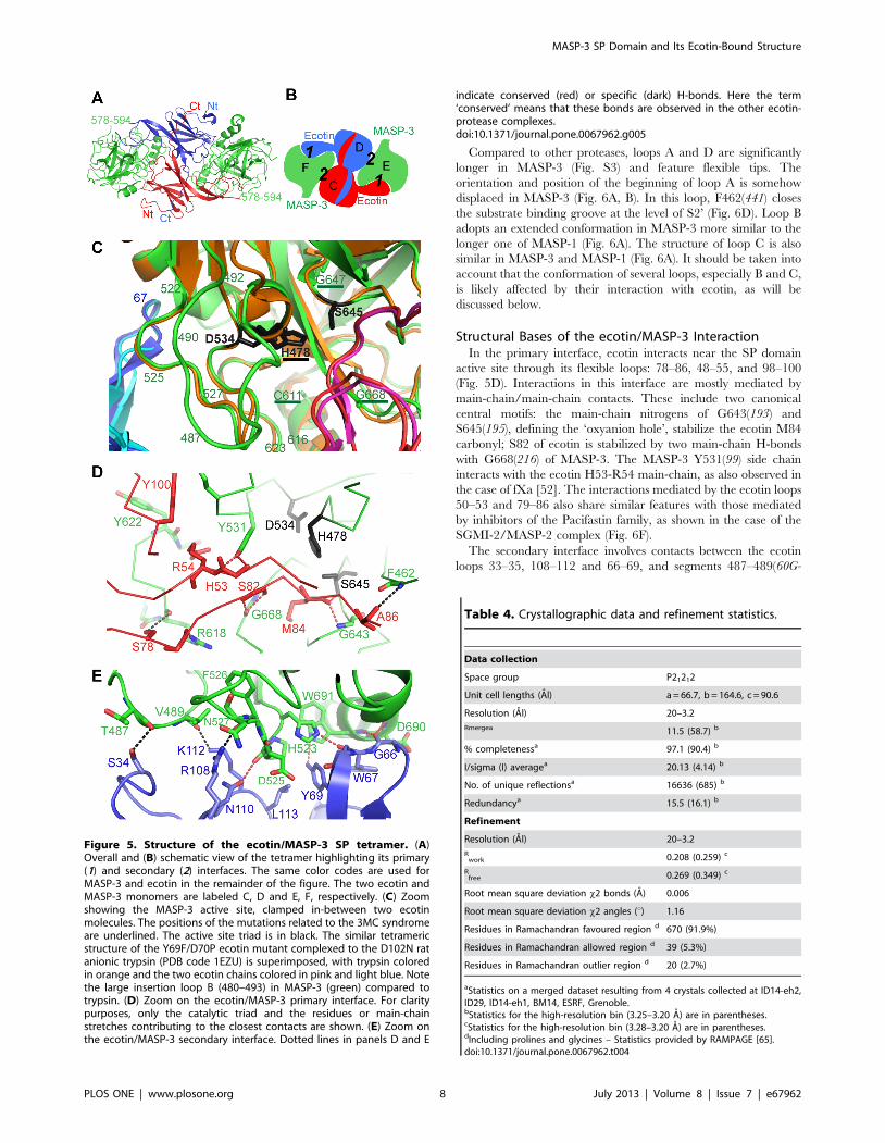
Crystallization of the ecotin/MASP-3 SP domain tetramer was
successful and reproducible. Four coherent datasets were merged
to yield a more complete and redundant dataset which was used to
solve the structure and refine it to 3.2 A resolution, with R and
Rfree values of 0.208 and 0.269 (Table 4). As observed previously
for other ecotin/protease complexes, the tetramer features a
central ecotin dimer, each ecotin molecule interacting with both
SP domains, hence defining two interfaces (Fig. 5A, B). The
primary interface (labeled 1 in Fig. 5B) is the one where ecotin
directly interacts with the SP domain catalytic triad and substrate-
binding site (Fig. 5C, D). Remarkably, the amino acids
corresponding to the mutations associated to the 3MC syndrome
all cluster in this area (Fig. 5C). The secondary interface (labeled 2in Fig. 5B) corresponds to the interaction with the other ecotin
molecule. Two SP surface loops (523–534 and 480–490) are
clamped in-between the two ecotin molecules, thus taking part in
both interfaces.
The tetramer is almost perfectly symmetrical, except for slight
differences at the edges of the interfaces and for some local
differences in the crystal contacts of the two copies of each protein.
Compared to other ecotin/protease complex structures, the most
similar tetramer is the one formed by the D102N rat anionic
trypsin and Y69F/D70P ecotin ([54], PDB code 1EZU). The
Figure 3. Comparative SPR analysis of the interaction of MASP-3 and other trypsin-like proteases with ecotin. Ecotin (1,000 RU) wasimmobilized on a CM5 sensor chip as described under Materials and Methods. Sixty microliters of varying concentrations of the MASP-3 SP domain(A), the MASP-2 CCP1/2-ap-SP fragment (B), trypsin (C), and factor XIa (D) were injected over immobilized ecotin in 145 mM NaCl, 50 mMtriethanolamine-HCl, pH 7.4 containing 0.005% surfactant P20 at a flow rate of 20 ml/min. The specific binding signals shown were obtained bysubtracting the background signal over a reference surface with no protein immobilized. Fits are shown as dotted lines and were obtained by globalfitting of the data using a 1:1 Langmuir binding model.doi:10.1371/journal.pone.0067962.g003
Table 3. Kinetic and dissociation constants for the interactionof selected proteases with immobilized ecotin.
Protease ka (M21 s21) kd (s21) KD (nM)
MASP-3 SP 1.346103 8.0661024 601
MASP-2 CCP1/2-ap-SP 1.046105 2.2861023 24.7
Trypsin 5.096105 1.0961024 0.21
Factor XIa 1.236105 1.6961024 1.38
doi:10.1371/journal.pone.0067962.t003
MASP-3 SP Domain and Its Ecotin-Bound Structure
PLOS ONE | www.plosone.org 6 July 2013 | Volume 8 | Issue 7 | e67962
conformations observed at the primary and secondary interfaces
are strikingly conserved in both cases (Fig. 5C, D, E). The global
r.m.s.d value between these two tetrameric structures is indeed
only 0.95 A (for 678 aligned residues), and 0.83 A for the SP
domain alone (for 202 aligned residues, with 39.6% sequence
identity). In contrast, the MASP-3 480–490 loop features a large
insertion compared to trypsin (Fig. 5C).
Structural Comparison with Homologous Trypsin-like SPDomains
The MASP-3 catalytic triad is defined by H478(57), D534(102),
and S645(195) (chymotrypsinogen numbering is indicated in
brackets). As a highly conserved binding mode for substrates and
inhibitors is observed in serine proteases, the positions of the ecotin
residues 78–86 delineate the positions of the common sub-sites
(Fig. 6D), as defined by Schechter and Berger [55]. A trypsin-like
primary specificity is defined by D639(189) at the bottom of the S1
pocket, and by G668(216) and G680(226) on its edges. The SP
loops surrounding the active site, as defined by Perona and Craik
[56], modulate the enzyme specificity and efficiency. These loops
can be compared to their counterparts in a set of homologous SP
domains found in proteases of the complement (MASP-1, MASP-
2, C1r, C1s) and coagulation cascades (fIXa, fXa), as illustrated in
Figs. 6A and 6B. When MASP-3 is compared to MASP-1 and -2
in complex with the specific SGMI peptide inhibitors [56], the
binding similarity extends to two inhibitor loops (Fig. 6A, E, F).
MASP-3 also features an extended C-terminal end (Ct on Fig. 6A).
Loops 1–3 finely modulate the enzyme activity and primary
specificity. The position of loops 1 and 2 appears to be slightly
displaced compared to the other MASPs (Fig. 6E). The sequence
of loop 1, including the Y633–Y634(185–186) doublet in MASP-3
(Fig. 6E), differs from those in other MASPs, being more similar to
coagulation factor IX (Fig. 6C). There is a very short insertion at
the beginning of loop 2 in MASP-3 (Fig. 6C, D, E). This insertion
is unique among serine proteases, and highly conserved in MASP-
3 from different species (Fig. 6C). It occurs just after the conserved
critical residue G668(216), which corresponds to one of the
mutations observed in the 3MC syndrome [18]. P670(217B) and
the non conserved L642(192) form a sort of extra layer on the edge
of the S1 sub-site above the conserved C641–C673(191–220)
disulfide bridge (Fig. 6D, 5E). G668(216) and P670(217B) also line
the S3 sub-site, and, together with Y531(99), restrict the space for
the P3 substrate residue (Fig. 6D). Possible implications of this
unusual insertion in loop 2 will be addressed later in the
Discussion. The helix in loop 3 extends up to R618(173C)
(Fig. 6E). Among the compared SP domains, only C1r exhibits a
similar helical extension (Fig. 6B). The S617-R618 residues in loop
3 slightly interact with ecotin S78–S79, thus expanding the
substrate-like interactions up to the S6 sub-site (S79 corresponds to
P6, Figs. 5D, 6D, 6F).
Figure 4. Gel filtration analysis of ecotin-MASP-3 SP complexes. (A) Equimolar amounts of the MASP-3 SP domain and ecotin (225 pmol)were preincubated for 20 min at room temperature before injection on a TSK G-3000 SW column equilibrated in 145 mM NaCl, 50 mMtriethanolamine-HCl, pH 7.4 and run at 1 ml/min. (B) SDS-PAGE analysis under reducing conditions and Coomassie blue staining of the proteincontent of peak 1. The molecular masses of reduced standard proteins (expressed in kDa) are shown on the left side of the gel.doi:10.1371/journal.pone.0067962.g004
MASP-3 SP Domain and Its Ecotin-Bound Structure
PLOS ONE | www.plosone.org 7 July 2013 | Volume 8 | Issue 7 | e67962
Compared to other proteases, loops A and D are significantly
longer in MASP-3 (Fig. S3) and feature flexible tips. The
orientation and position of the beginning of loop A is somehow
displaced in MASP-3 (Fig. 6A, B). In this loop, F462(441) closes
the substrate binding groove at the level of S2’ (Fig. 6D). Loop B
adopts an extended conformation in MASP-3 more similar to the
longer one of MASP-1 (Fig. 6A). The structure of loop C is also
similar in MASP-3 and MASP-1 (Fig. 6A). It should be taken into
account that the conformation of several loops, especially B and C,
is likely affected by their interaction with ecotin, as will be
discussed below.
Structural Bases of the ecotin/MASP-3 InteractionIn the primary interface, ecotin interacts near the SP domain
active site through its flexible loops: 78–86, 48–55, and 98–100
(Fig. 5D). Interactions in this interface are mostly mediated by
main-chain/main-chain contacts. These include two canonical
central motifs: the main-chain nitrogens of G643(193) and
S645(195), defining the ‘oxyanion hole’, stabilize the ecotin M84
carbonyl; S82 of ecotin is stabilized by two main-chain H-bonds
with G668(216) of MASP-3. The MASP-3 Y531(99) side chain
interacts with the ecotin H53-R54 main-chain, as also observed in
the case of fXa [52]. The interactions mediated by the ecotin loops
50–53 and 79–86 also share similar features with those mediated
by inhibitors of the Pacifastin family, as shown in the case of the
SGMI-2/MASP-2 complex (Fig. 6F).
The secondary interface involves contacts between the ecotin
loops 33–35, 108–112 and 66–69, and segments 487–489(60G-
Figure 5. Structure of the ecotin/MASP-3 SP tetramer. (A)Overall and (B) schematic view of the tetramer highlighting its primary(1) and secondary (2) interfaces. The same color codes are used forMASP-3 and ecotin in the remainder of the figure. The two ecotin andMASP-3 monomers are labeled C, D and E, F, respectively. (C) Zoomshowing the MASP-3 active site, clamped in-between two ecotinmolecules. The positions of the mutations related to the 3MC syndromeare underlined. The active site triad is in black. The similar tetramericstructure of the Y69F/D70P ecotin mutant complexed to the D102N ratanionic trypsin (PDB code 1EZU) is superimposed, with trypsin coloredin orange and the two ecotin chains colored in pink and light blue. Notethe large insertion loop B (480–493) in MASP-3 (green) compared totrypsin. (D) Zoom on the ecotin/MASP-3 primary interface. For claritypurposes, only the catalytic triad and the residues or main-chainstretches contributing to the closest contacts are shown. (E) Zoom onthe ecotin/MASP-3 secondary interface. Dotted lines in panels D and E
indicate conserved (red) or specific (dark) H-bonds. Here the term‘conserved’ means that these bonds are observed in the other ecotin-protease complexes.doi:10.1371/journal.pone.0067962.g005
Table 4. Crystallographic data and refinement statistics.
Data collection
Space group P21212
Unit cell lengths (Al) a = 66.7, b = 164.6, c = 90.6
Resolution (Al) 20–3.2
Rmergea 11.5 (58.7) b
% completenessa 97.1 (90.4) b
I/sigma (I) averagea 20.13 (4.14) b
No. of unique reflectionsa 16636 (685) b
Redundancya 15.5 (16.1) b
Refinement
Resolution (Al) 20–3.2
Rwork 0.208 (0.259) c
Rfree 0.269 (0.349) c
Root mean square deviation x2 bonds (A) 0.006
Root mean square deviation x2 angles (u) 1.16
Residues in Ramachandran favoured region d 670 (91.9%)
Residues in Ramachandran allowed region d 39 (5.3%)
Residues in Ramachandran outlier region d 20 (2.7%)
aStatistics on a merged dataset resulting from 4 crystals collected at ID14-eh2,ID29, ID14-eh1, BM14, ESRF, Grenoble.bStatistics for the high-resolution bin (3.25–3.20 A) are in parentheses.cStatistics for the high-resolution bin (3.28–3.20 A) are in parentheses.dIncluding prolines and glycines – Statistics provided by RAMPAGE [65].doi:10.1371/journal.pone.0067962.t004
MASP-3 SP Domain and Its Ecotin-Bound Structure
PLOS ONE | www.plosone.org 8 July 2013 | Volume 8 | Issue 7 | e67962
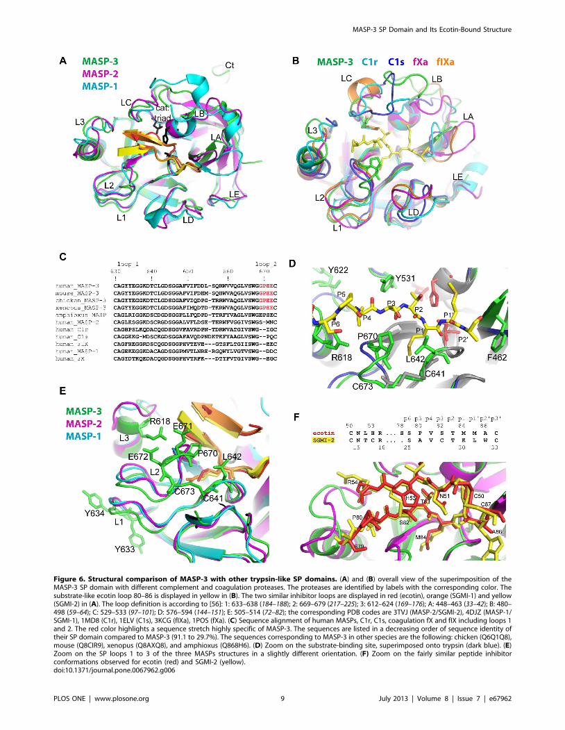
Figure 6. Structural comparison of MASP-3 with other trypsin-like SP domains. (A) and (B) overall view of the superimposition of theMASP-3 SP domain with different complement and coagulation proteases. The proteases are identified by labels with the corresponding color. Thesubstrate-like ecotin loop 80–86 is displayed in yellow in (B). The two similar inhibitor loops are displayed in red (ecotin), orange (SGMI-1) and yellow(SGMI-2) in (A). The loop definition is according to [56]: 1: 633–638 (184–188); 2: 669–679 (217–225); 3: 612–624 (169–176); A: 448–463 (33–42); B: 480–498 (59–64); C: 529–533 (97–101); D: 576–594 (144–151); E: 505–514 (72–82); the corresponding PDB codes are 3TVJ (MASP-2/SGMI-2), 4DJZ (MASP-1/SGMI-1), 1MD8 (C1r), 1ELV (C1s), 3KCG (fIXa), 1POS (fXa). (C) Sequence alignment of human MASPs, C1r, C1s, coagulation fX and fIX including loops 1and 2. The red color highlights a sequence stretch highly specific of MASP-3. The sequences are listed in a decreasing order of sequence identity oftheir SP domain compared to MASP-3 (91.1 to 29.7%). The sequences corresponding to MASP-3 in other species are the following: chicken (Q6Q1Q8),mouse (Q8CIR9), xenopus (Q8AXQ8), and amphioxus (Q868H6). (D) Zoom on the substrate-binding site, superimposed onto trypsin (dark blue). (E)Zoom on the SP loops 1 to 3 of the three MASPs structures in a slightly different orientation. (F) Zoom on the fairly similar peptide inhibitorconformations observed for ecotin (red) and SGMI-2 (yellow).doi:10.1371/journal.pone.0067962.g006
MASP-3 SP Domain and Its Ecotin-Bound Structure
PLOS ONE | www.plosone.org 9 July 2013 | Volume 8 | Issue 7 | e67962
60I), 523–526(91–94) and 690–691(236–237) of the protease
(Fig. 5E). A set of conserved interactions can be delineated in this
interface by comparison with other ecotin/protease complex
structures. These involve H523(91), P524(92), F526(94),
D690(236) and W691(237) on the protease side, and G66, W67,
Y69, R108, N110, L113 on the ecotin side (Fig. 5E). Of note, the
mutations Y69F/D70P shown to strongly increase the inhibitory
efficiency of ecotin towards trypsin [54] lie in this essential part of
the secondary interface. Again, very few protease side-chains
(W691(237) and H523(91)) mediate interactions in this area.
Finally, unique additional interactions are provided by the large
insertion loop in MASP-3, where T487(60G) and V489(60I)
interact with ecotin S34 and K112, respectively (Fig. 5E).
Following this analysis, we tried to identify the structural bases
accounting for the observed differences in ecotin binding by C1r,
C1s and the MASPs. The wide MASP-1 substrate-binding site
[23,57] is unlikely to prevent interaction with the flexible ecotin
molecule through the primary interface. Ecotin binding through
the secondary interface could however be affected by sequence
differences at the level of D525(95) and N527(97) of MASP-3
(Fig. 5E). Bulkier side chains are present at these positions in
MASP-1, C1r and C1s, contrary to MASP-2. Moreover, larger
insertions in loops B or C, which are clamped in-between two
ecotin subunits (Fig. 5C), would strongly restrict their possible
conformational space: this applies to the MASP-1 super-extended
B loop, and to the larger C1r and C1s C loops (Fig. 6A, B).
Conformational Flexibility in MASP-3 and its PotentialAllosteric Implications
Several surface loops are highly flexible in the MASP-3 SP
domain, especially the major insertion loops A and D. Flexibility of
some side-chains (mostly charged ones) are observed in loops C
and 2, and at the tip of loop 3. Even the N-terminal hydrophobic
dipeptide I431(16)-I432(17) is not as clearly defined as expected in
an active serine protease structure. It is stabilized by the activation
pocket, which lies in MASP-3 next to the disordered segment
578(145)-593(150). Very small patches of additional density
observed near L441(26) further suggest that the N-terminal
extremity may adopt alternative orientation(s) up to this level in
some molecules of the crystal. Similar features were indeed
observed in the structure of complement factor I [58], which,
although correctly processed at its activation cleavage site, is in a
zymogen-like conformation. No interpretable density was available
for the residues of the newly generated N-terminus in most
molecules of factor I up to the residue homologous to MASP-3
L441(26). As stated above, the slight N-terminal heterogeneity
observed in some batches of the recombinant MASP-3 SP domain
could also contribute to this partial local disorder.
Another indirect mark of flexibility resides in the probable
ecotin-constrained local conformation(s) of some surface loops.
Since the crystal structure of MASP-3 alone is not available, the
free and ecotin-bound conformations cannot be compared.
However, ecotin-induced conformational changes have been
observed previously in the case of thrombin and factor Xa [52],
and significant conformational changes were also observed in the
catalytic fragment of MASP-2 in complex with SGMI-2 [57].
Evidence of such a change may be inferred from the comparison
of the position and orientation of Tyr531(99) in MASP-3 and its
homologues. In MASP-1 and -2, the positions of the correspond-
ing Phe549 and Phe529 side chains, respectively, are significantly
displaced by their interaction with the peptide inhibitor (Fig. 7A).
In fXa, this residue is significantly displaced by its close interaction
with loop 50 of ecotin [52]. Its conformation in ecotin-bound
MASP-3 is far more similar to that in the ecotin-bound fXa (not
shown) or in the SGMI-bound MASP-1 and -2 than in the free
forms of MASP-1, MASP-2, C1r, C1s and fXa (Fig. 7B). As loops
B and C are clamped in-between two ecotin molecules (Fig. 5C),
they likely require an adaptive conformational change in MASP-3
to accommodate ecotin binding, which would be consistent with
the lower association rate observed (Table 3).
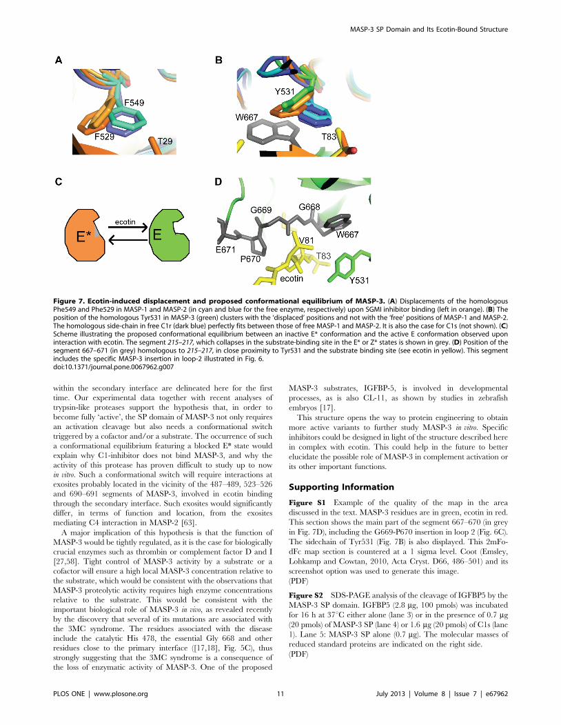
Towards a Plausible Conformational Switch in the MASP-3 SP Domain
Such a conformational flexibility could offer a clue to resolve the
following apparent MASP-3 paradox: on the one hand, this
protease clearly exhibits poor enzymatic properties in vitro; on the
other hand, its structure in complex with ecotin is quite similar to
that of ‘classical’ active proteases. In particular, this structure does
not explain why C1-inhibitor, the physiological inhibitor of all
other proteases of the C1r/C1s/MASP family, does not bind to
MASP-3. The concept of allostery was recently introduced as a
general property of the trypsin-like protease fold [27,59,60].
Besides the activation cleavage transforming a zymogen (Z) into an
enzyme (E), this concept adds a possible conformational equilib-
rium between an active E (or Z) conformation and an inactive one
(E* or Z*). Access to the binding site is blocked in E* or Z*, but a
conformational switch to the active form can be provided in situ by
an activator and/or a substrate. Our observations would thus be
consistent with the hypothesis that the MASP-3 SP domain is seen
in the E state in the crystal structure, as a result of its interaction
with ecotin, whereas it is mainly stabilized in the E* state when its
catalytic activity is assayed in vitro (Fig. 7C). Two major arguments
give credit to this hypothesis: (i) the poor activity of free thrombin
(in the E* state) has been correlated to its conformational
equilibrium [61,62] and MASP-3 also exhibits significant confor-
mational flexibility in its SP domain, even in the crystal structure;
(ii) the MASP-3 specific insertion in loop 2 occurs in the 667–
671(215–218) segment (in grey in Fig. 7D), homologous to the
segment collapsing into the substrate-binding site in the E* and Z*
structures, thus blocking its access [27,60]. This insertion could
thus promote the collapse of this fragment 667–671(215–218) into
the substrate-binding site in the absence of ecotin.
How could ecotin bind to MASP-3 in its initial E* state? A likely
hypothesis is that ecotin first binds through the secondary
interface, thereby triggering a conformational switch opening the
substrate binding site, which allows in turn the second ecotin
molecule to bind through the primary interface. The arguments
supporting this hypothesis are the following: (i) ecotin is unlikely to
bind first through the primary interface since the interactions
observed at this interface are quite poor, implying very few side-
chains; of note, the P1 residue in ecotin is Met84 instead of the
preferred lysine or arginine side-chains. The fact that ecotin does
not bind MASP-1 is likely due to steric conflicts at the secondary
interface and not at the primary interface, as discussed previously.
(ii) ecotin has been shown to trigger such a conformational switch
by binding to the exosite 2 of thrombin via its secondary interface
[50,56], and we have delineated a set of similar interactions in the
secondary interface of the ecotin/MASP-3 complex; (iii) the
reduced association rate constant observed in the case of MASP-3
(Table 3) would be fully consistent with the requirement of
conformational changes prior to binding through the primary
interface.
ConclusionThis study describes a further example of the wide inhibitory
spectrum of ecotin and illustrates how this inhibitor makes use of
two interfaces targeting essential main-chain functional elements
of trypsin-like proteases. In particular, conserved interactions
MASP-3 SP Domain and Its Ecotin-Bound Structure
PLOS ONE | www.plosone.org 10 July 2013 | Volume 8 | Issue 7 | e67962
within the secondary interface are delineated here for the first
time. Our experimental data together with recent analyses of
trypsin-like proteases support the hypothesis that, in order to
become fully ‘active’, the SP domain of MASP-3 not only requires
an activation cleavage but also needs a conformational switch
triggered by a cofactor and/or a substrate. The occurrence of such
a conformational equilibrium featuring a blocked E* state would
explain why C1-inhibitor does not bind MASP-3, and why the
activity of this protease has proven difficult to study up to now
in vitro. Such a conformational switch will require interactions at
exosites probably located in the vicinity of the 487–489, 523–526
and 690–691 segments of MASP-3, involved in ecotin binding
through the secondary interface. Such exosites would significantly
differ, in terms of function and location, from the exosites
mediating C4 interaction in MASP-2 [63].
A major implication of this hypothesis is that the function of
MASP-3 would be tightly regulated, as it is the case for biologically
crucial enzymes such as thrombin or complement factor D and I
[27,58]. Tight control of MASP-3 activity by a substrate or a
cofactor will ensure a high local MASP-3 concentration relative to
the substrate, which would be consistent with the observations that
MASP-3 proteolytic activity requires high enzyme concentrations
relative to the substrate. This would be consistent with the
important biological role of MASP-3 in vivo, as revealed recently
by the discovery that several of its mutations are associated with
the 3MC syndrome. The residues associated with the disease
include the catalytic His 478, the essential Gly 668 and other
residues close to the primary interface ([17,18], Fig. 5C), thus
strongly suggesting that the 3MC syndrome is a consequence of
the loss of enzymatic activity of MASP-3. One of the proposed
MASP-3 substrates, IGFBP-5, is involved in developmental
processes, as is also CL-11, as shown by studies in zebrafish
embryos [17].
This structure opens the way to protein engineering to obtain
more active variants to further study MASP-3 in vitro. Specific
inhibitors could be designed in light of the structure described here
in complex with ecotin. This could help in the future to better
elucidate the possible role of MASP-3 in complement activation or
its other important functions.
Supporting Information
Figure S1 Example of the quality of the map in the area
discussed in the text. MASP-3 residues are in green, ecotin in red.
This section shows the main part of the segment 667–670 (in grey
in Fig. 7D), including the G669-P670 insertion in loop 2 (Fig. 6C).
The sidechain of Tyr531 (Fig. 7B) is also displayed. This 2mFo-
dFc map section is countered at a 1 sigma level. Coot (Emsley,
Lohkamp and Cowtan, 2010, Acta Cryst. D66, 486–501) and its
screenshot option was used to generate this image.
(PDF)
Figure S2 SDS-PAGE analysis of the cleavage of IGFBP5 by the
MASP-3 SP domain. IGFBP5 (2.8 mg, 100 pmols) was incubated
for 16 h at 37uC either alone (lane 3) or in the presence of 0.7 mg
(20 pmols) of MASP-3 SP (lane 4) or 1.6 mg (20 pmols) of C1s (lane
1). Lane 5: MASP-3 SP alone (0.7 mg). The molecular masses of
reduced standard proteins are indicated on the right side.
(PDF)
Figure 7. Ecotin-induced displacement and proposed conformational equilibrium of MASP-3. (A) Displacements of the homologousPhe549 and Phe529 in MASP-1 and MASP-2 (in cyan and blue for the free enzyme, respectively) upon SGMI inhibitor binding (left in orange). (B) Theposition of the homologous Tyr531 in MASP-3 (green) clusters with the ‘displaced’ positions and not with the ‘free’ positions of MASP-1 and MASP-2.The homologous side-chain in free C1r (dark blue) perfectly fits between those of free MASP-1 and MASP-2. It is also the case for C1s (not shown). (C)Scheme illustrating the proposed conformational equilibrium between an inactive E* conformation and the active E conformation observed uponinteraction with ecotin. The segment 215–217, which collapses in the substrate-binding site in the E* or Z* states is shown in grey. (D) Position of thesegment 667–671 (in grey) homologous to 215–217, in close proximity to Tyr531 and the substrate binding site (see ecotin in yellow). This segmentincludes the specific MASP-3 insertion in loop-2 illustrated in Fig. 6.doi:10.1371/journal.pone.0067962.g007
MASP-3 SP Domain and Its Ecotin-Bound Structure
PLOS ONE | www.plosone.org 11 July 2013 | Volume 8 | Issue 7 | e67962
Figure S3 Multiple sequence alignments of a set of complement
and coagulation pro-teases serine protease domains. It includes the
complement C1r, C1s, MASP-1 and MASP-2 proteases, coagu-
lation factors 9, 10, 11 and thrombin; Rat trypsin as a reference;
MASP-3 from human and other species. The surface loops
defining the substrate specificity and catalytic efficiency are noted
on the top of the align-ment, following the nomenclature
introduced by Perona and Craik (1997, JBC, 272, 29987-90).
Yellow diamonds locate the position of the human MASP-3
glycosylation sites.
(PDF)
Acknowledgments
We thank Jean-Pierre Andrieu, Izabel Berard, Delphine Blot and Isabelle
Bally from the IBS platforms of the Partnership for Structural Biology in
Grenoble for assistance and access to the protein sequencing, mass
spectrometry, crystallization and BIAcore facilities, respectively. Access to
the ESRF beamlines ID14-eh1, ID14-eh2, ID29 and BM14 is greatly
acknowledged. We thank Sandor Cseh for construction of the pNT-Bac
vector containing the MASP-3 ap-SP segment, Ludovic Lenclume for
production of PAR1 and Evelyne Gout for baculovirus amplification and
insect cells infection.
Author Contributions
Conceived and designed the experiments: CG NT VR. Performed the
experiments: CG RKG ML LM NT. Analyzed the data: CG LS NT VR.
Contributed reagents/materials/analysis tools: FT. Wrote the paper: CG
GJA NT.
References
1. Dommett RM, Klein N, Turner MW (2006) Mannose-binding lectin in innateimmunity: past, present and future. Tissue Antigens 68: 193–209.
2. Hansen S, Selman L, Palaniyar N, Ziegler K, Brandt J, et al. (2010) Collectin 11
(CL-11, CL-K1) is a MASP-1/3-associated plasma collectin with microbial-binding activity. J Immunol 185: 6096–6104.
3. Keshi H, Sakamoto T, Kawai T, Ohtani K, Katoh T, et al. (2006) Identification
and characterization of a novel human collectin CL-K1. Microbiol Immunol 50:1001–1013.
4. Endo Y, Matsushita M, Fujita T (2011) The role of ficolins in the lectin pathway
of innate immunity. Int J Biochem Cell Biol 43: 705–712.
5. Sorensen R, Thiel S, Jensenius JC (2005) Mannan-binding-lectin-associatedserine proteases, characteristics and disease associations. Springer Semin
Immunopathol 27: 299–319.
6. Stover CM, Thiel S, Thelen M, Lynch NJ, Vorup-Jensen T, et al. (1999) Twoconstituents of the initiation complex of the mannan-binding lectin activation
pathway of complement are encoded by a single structural gene. J Immunol 162:
3481–3490.
7. Takahashi M, Endo Y, Fujita T, Matsushita M (1999) A truncated form of
mannose-binding lectin-associated serine protease (MASP)-2 expressed by
alternative polyadenylation is a component of the lectin complement pathway.Int Immunol 11: 859–863.
8. Degn SE, Hansen AG, Steffensen R, Jacobsen C, Jensenius JC, et al. (2009)
MAp44, a human protein associated with pattern recognition molecules of thecomplement system and regulating the lectin pathway of complement activation.
J Immunol 183: 7371–7378.
9. Skjoedt MO, Hummelshoj T, Palarasah Y, Honore C, Koch C, et al. (2010) Anovel mannose-binding lectin/ficolin-associated protein is highly expressed in
heart and skeletal muscle tissues and inhibits complement activation. J Biol
Chem 285: 8234–8243.
10. Degn SE, Jensen L, Hansen AG, Duman D, Tekin M, et al. (2012) Mannan-
Binding Lectin-Associated Serine Protease (MASP)-1 Is Crucial for Lectin
Pathway Activation in Human Serum, whereas neither MASP-1 nor MASP-3 IsRequired for Alternative Pathway Function. J Immunol 189: 3957–3969.
11. Heja D, Kocsis A, Dobo J, Szilagyi K, Szasz R, et al. (2012) Revised mechanism
of complement lectin-pathway activation revealing the role of serine proteaseMASP-1 as the exclusive activator of MASP-2. Proc Natl Acad Sci U S A 109:
10498–10503.
12. Gaboriaud C, Thielens NM, Gregory LA, Rossi V, Fontecilla-Camps JC, et al.(2004) Structure and activation of the C1 complex of complement: unraveling
the puzzle. Trends Immunol 25: 368–373.
13. Moller-Kristensen M, Thiel S, Sjoholm A, Matsushita M, Jensenius JC (2007)Cooperation between MASP-1 and MASP-2 in the generation of C3 convertase
through the MBL pathway. Int Immunol 19: 141–149.
14. Dahl MR, Thiel S, Matsushita M, Fujita T, Willis AC, et al. (2001) MASP-3 andits association with distinct complexes of the mannan-binding lectin complement
activation pathway. Immunity 15: 127–135.
15. Iwaki D, Kanno K, Takahashi M, Endo Y, Lynch NJ, et al. (2006) Small
mannose-binding lectin-associated protein plays a regulatory role in the lectincomplement pathway. J Immunol 177: 8626–8632.
16. Zundel S, Cseh S, Lacroix M, Dahl MR, Matsushita M, et al. (2004)
Characterization of recombinant mannan-binding lectin-associated serineprotease (MASP)-3 suggests an activation mechanism different from that of
MASP-1 and MASP-2. J Immunol 172: 4342–4350.
17. Rooryck C, Diaz-Font A, Osborn DP, Chabchoub E, Hernandez-Hernandez V,et al. (2011) Mutations in lectin complement pathway genes COLEC11 and
MASP1 cause 3MC syndrome. Nat Genet 43: 197–203.
18. Sirmaci A, Walsh T, Akay H, Spiliopoulos M, Sakalar YB, et al. (2010) MASP1mutations in patients with facial, umbilical, coccygeal, and auditory findings of
Carnevale, Malpuech, OSA, and Michels syndromes. Am J Hum Genet 87:
679–686.
19. Sekine H, Takahashi M, Iwaki D, Fujita T (2013) The Role of MASP-1/3 in
Complement Activation. Adv Exp Med Biol 734: 41–53.
20. Megyeri M, Harmat V, Major B, Vegh A, Balczer J, et al. (2013) Quantitative
Characterization of the Activation Steps of Mannan-binding Lectin (MBL)-
associated Serine Proteases (MASPs) Points to the Central Role of MASP-1 in
the Initiation of the Complement Lectin Pathway. J Biol Chem 288: 8922–8934.
21. Budayova-Spano M, Grabarse W, Thielens NM, Hillen H, Lacroix M, et al.
(2002) Monomeric structures of the zymogen and active catalytic domain of
complement protease c1r: further insights into the c1 activation mechanism.
Structure 10: 1509–1519.
22. Budayova-Spano M, Lacroix M, Thielens NM, Arlaud GJ, Fontecilla-Camps
JC, et al. (2002) The crystal structure of the zymogen catalytic domain of
complement protease C1r reveals that a disruptive mechanical stress is required
to trigger activation of the C1 complex. EMBO J 21: 231–239.
23. Dobo J, Harmat V, Beinrohr L, Sebestyen E, Zavodszky P, et al. (2009) MASP-
1, a promiscuous complement protease: structure of its catalytic region reveals
the basis of its broad specificity. J Immunol 183: 1207–1214.
24. Gaboriaud C, Rossi V, Bally I, Arlaud GJ, Fontecilla-Camps JC (2000) Crystal
structure of the catalytic domain of human complement c1s: a serine protease
with a handle. EMBO J 19: 1755–1765.
25. Harmat V, Gal P, Kardos J, Szilagyi K, Ambrus G, et al. (2004) The structure of
MBL-associated serine protease-2 reveals that identical substrate specificities of
C1s and MASP-2 are realized through different sets of enzyme-substrate
interactions. J Mol Biol 342: 1533–1546.
26. Cortesio CL, Jiang W (2006) Mannan-binding lectin-associated serine protease 3
cleaves synthetic peptides and insulin-like growth factor-binding protein 5. Arch
Biochem Biophys 449: 164–170.
27. Pozzi N, Vogt AD, Gohara DW, Di Cera E (2012) Conformational selection in
trypsin-like proteases. Curr Opin Struct Biol 22: 421–431.
28. Arlaud GJ, Reboul A, Sim RB, Colomb MG (1979) Interaction of C1-inhibitor
with the C1r and C1s subcomponents in human C1. Biochim Biophys Acta 576:
151–162.
29. Arlaud GJ, Sim RB, Duplaa AM, Colomb MG (1979) Differential elution of
Clq, Clr and Cls from human Cl bound to immune aggregates. Use in the rapid
purification of Cl subcomponents. Mol Immunol 16: 445–450.
30. Lacroix M, Ebel C, Kardos J, Dobo J, Gal P, et al. (2001) Assembly and
enzymatic properties of the catalytic domain of human complement protease
C1r. J Biol Chem 276: 36233–36240.
31. Rossi V, Bally I, Thielens NM, Esser AF, Arlaud GJ (1998) Baculovirus-
mediated expression of truncated modular fragments from the catalytic region of
human complement serine protease C1s. Evidence for the involvement of both
complement control protein modules in the recognition of the C4 protein
substrate. J Biol Chem 273: 1232–1239.
32. Rossi V, Cseh S, Bally I, Thielens NM, Jensenius JC, et al. (2001) Substrate
specificities of recombinant mannan-binding lectin-associated serine proteases-1
and -2. J Biol Chem 276: 40880–40887.
33. Loew D, Perrault C, Morales M, Moog S, Ravanat C, et al. (2000) Proteolysis of
the exodomain of recombinant protease-activated receptors: prediction of
receptor activation or inactivation by MALDI mass spectrometry. Biochemistry
39: 10812–10822.
34. Thielens NM, Cseh S, Thiel S, Vorup-Jensen T, Rossi V, et al. (2001)
Interaction properties of human mannan-binding lectin (MBL)-associated serine
proteases-1 and -2, MBL-associated protein 19, and MBL. J Immunol 166:
5068–5077.
35. Teillet F, Dublet B, Andrieu JP, Gaboriaud C, Arlaud GJ, et al. (2005) The two
major oligomeric forms of human mannan-binding lectin: chemical character-
ization, carbohydrate-binding properties, and interaction with MBL-associated
serine proteases. J Immunol 174: 2870–2877.
36. Kabsch W (2010) Xds. Acta Crystallogr D Biol Crystallogr 66: 125–132.
MASP-3 SP Domain and Its Ecotin-Bound Structure
PLOS ONE | www.plosone.org 12 July 2013 | Volume 8 | Issue 7 | e67962
37. McCoy AJ, Grosse-Kunstleve RW, Adams PD, Winn MD, Storoni LC, et al.
(2007) Phaser crystallographic software. J Appl Crystallogr 40: 658–674.38. Jin L, Pandey P, Babine RE, Gorga JC, Seidl KJ, et al. (2005) Crystal structures
of the FXIa catalytic domain in complex with ecotin mutants reveal substrate-
like interactions. J Biol Chem 280: 4704–4712.39. Jones TA, Zou JY, Cowan SW, Kjeldgaard M (1991) Improved methods for
building protein models in electron density maps and the location of errors inthese models. Acta Crystallogr A 47 (Pt 2): 110–119.
40. Emsley P, Cowtan K (2004) Coot: model-building tools for molecular graphics.
Acta Crystallogr D Biol Crystallogr 60: 2126–2132.41. Brunger AT, Adams PD, Clore GM, DeLano WL, Gros P, et al. (1998)
Crystallography & NMR system: A new software suite for macromolecularstructure determination. Acta Crystallogr D Biol Crystallogr 54: 905–921.
42. Bricogne GBE, Brandl M, Flensburg C, Keller P, Paciorek W, et al. (2009)BUSTER version 2.8, Global Phasing Ltd. Cambridge, United Kingdom.
43. Murshudov GN, Vagin AA, Dodson EJ (1997) Refinement of macromolecular
structures by the maximum-likelihood method. Acta Crystallogr D Biol Crystal-logr 53: 240–255.
44. Megyeri M, Mako V, Beinrohr L, Doleschall Z, Prohaszka Z, et al. (2009)Complement protease MASP-1 activates human endothelial cells: PAR4
activation is a link between complement and endothelial function. J Immunol
183: 3409–3416.45. Presanis JS, Hajela K, Ambrus G, Gal P, Sim RB (2004) Differential substrate
and inhibitor profiles for human MASP-1 and MASP-2. Mol Immunol 40: 921–929.
46. Beattie J, Allan GJ, Lochrie JD, Flint DJ (2006) Insulin-like growth factor-binding protein-5 (IGFBP-5): a critical member of the IGF axis. Biochem J 395:
1–19.
47. Yang SQ, Wang CI, Gillmor SA, Fletterick RJ, Craik CS (1998) Ecotin: a serineprotease inhibitor with two distinct and interacting binding sites. J Mol Biol 279:
945–957.48. McGrath ME, Erpel T, Bystroff C, Fletterick RJ (1994) Macromolecular
chelation as an improved mechanism of protease inhibition: structure of the
ecotin-trypsin complex. EMBO J 13: 1502–1507.49. Perona JJ, Tsu CA, Craik CS, Fletterick RJ (1997) Crystal structure of an ecotin-
collagenase complex suggests a model for recognition and cleavage of thecollagen triple helix. Biochemistry 36: 5381–5392.
50. Wang SX, Esmon CT, Fletterick RJ (2001) Crystal structure of thrombin-ecotinreveals conformational changes and extended interactions. Biochemistry 40:
10038–10046.
51. Waugh SM, Harris JL, Fletterick R, Craik CS (2000) The structure of the pro-
apoptotic protease granzyme B reveals the molecular determinants of itsspecificity. Nat Struct Biol 7: 762–765.
52. Wang SX, Hur E, Sousa CA, Brinen L, Slivka EJ, et al. (2003) The extended
interactions and Gla domain of blood coagulation factor Xa. Biochemistry 42:7959–7966.
53. Eggers CT, Wang SX, Fletterick RJ, Craik CS (2001) The role of ecotindimerization in protease inhibition. J Mol Biol 308: 975–991.
54. Gillmor SA, Takeuchi T, Yang SQ, Craik CS, Fletterick RJ (2000) Compromise
and accommodation in ecotin, a dimeric macromolecular inhibitor of serineproteases. J Mol Biol 299: 993–1003.
55. Schechter I, Berger A (1967) On the size of the active site in proteases. I. Papain.Biochem Biophys Res Commun 27: 157–162.
56. Perona JJ, Craik CS (1997) Evolutionary divergence of substrate specificitywithin the chymotrypsin-like serine protease fold. J Biol Chem 272: 29987–
29990.
57. Heja D, Harmat V, Fodor K, Wilmanns M, Dobo J, et al. (2012) Monospecificinhibitors show that both mannan-binding lectin-associated serine protease-1
(MASP-1) and -2 Are essential for lectin pathway activation and reveal structuralplasticity of MASP-2. J Biol Chem 287: 20290–20300.
58. Roversi P, Johnson S, Caesar JJ, McLean F, Leath KJ, et al. (2011) Structural
basis for complement factor I control and its disease-associated sequencepolymorphisms. Proc Natl Acad Sci U S A 108: 12839–12844.
59. Di Cera E (2009) Serine proteases. IUBMB Life 61: 510–515.60. Niu W, Chen Z, Gandhi PS, Vogt AD, Pozzi N, et al. (2011) Crystallographic
and kinetic evidence of allostery in a trypsin-like protease. Biochemistry 50:6301–6307.
61. Huntington JA (2009) Slow thrombin is zymogen-like. J Thromb Haemost 7
Suppl 1: 159–164.62. Lechtenberg BC, Johnson DJ, Freund SM, Huntington JA (2010) NMR
resonance assignments of thrombin reveal the conformational and dynamiceffects of ligation. Proc Natl Acad Sci U S A 107: 14087–14092.
63. Kidmose RT, Laursen NS, Dobo J, Kjaer TR, Sirotkina S, et al. (2012)
Structural basis for activation of the complement system by component C4cleavage. Proc Natl Acad Sci U S A 109: 15425–15430.
64. Tsiftsoglou SA, Sim RB (2004) Human complement factor I does not requirecofactors for cleavage of synthetic substrates. J Immunol 173: 367–375.
65. Lovell SC, Davis IW, Arendall WB 3rd, de Bakker PI, Word JM, et al. (2003)Structure validation by Calpha geometry: phi,psi and Cbeta deviation. Proteins
50: 437–450.
MASP-3 SP Domain and Its Ecotin-Bound Structure
PLOS ONE | www.plosone.org 13 July 2013 | Volume 8 | Issue 7 | e67962