Embed Size (px)
Citation preview

The SensesThe Senses
Slide 8.1Copyright © 2003 Pearson Education, Inc. publishing as Benjamin Cummings
Special senses Smell Taste Sight HearingHearing Equilibrium
Note: Touch is a “general” sense, not special sense

The Eye and VisionThe Eye and Vision
Slide 8.2Copyright © 2003 Pearson Education, Inc. publishing as Benjamin Cummings
70 percent of all sensory receptors are in the eyes
Each eye has over one million nerve fibers
Protection for the eye
Most of the eye is enclosed in a bony orbit
A cushion of fat surrounds most of the eye

Accessory Structures of the EyeAccessory Structures of the Eye
Slide 8.3aCopyright © 2003 Pearson Education, Inc. publishing as Benjamin Cummings
Figure 8.1b
Eyelids
Eyelashes

Slide 8.3b
Accessory Structures of the EyeAccessory Structures of the Eye
Copyright © 2003 Pearson Education, Inc. publishing as Benjamin Cummings
Meibomian glands – modified sebacious glands produce an oily secretion to lubricate the eye
Figure 8.1b

Accessory Structures of the EyeAccessory Structures of the Eye
Slide 8.3cCopyright © 2003 Pearson Education, Inc. publishing as Benjamin Cummings
Figure 8.1b
Ciliary glands –
modified sweat glands between the eyelashes

Slide 8.4a
Accessory Structures of the EyeAccessory Structures of the Eye
Copyright © 2003 Pearson Education, Inc. publishing as Benjamin Cummings
Conjunctiva Membrane that lines the eyelids
Connects to the surface of the eye
Secretes mucus to lubricate the eye

Accessory Structures of the EyeAccessory Structures of the Eye
Slide 8.4bCopyright © 2003 Pearson Education, Inc. publishing as Benjamin Cummings
Lacrimal apparatus
Lacrimal gland – produces lacrimal fluid
Lacrimal canals – drains lacrimal fluid from eyes
Figure 8.1a

Slide 8.4c
Accessory Structures of the EyeAccessory Structures of the Eye
Copyright © 2003 Pearson Education, Inc. publishing as Benjamin Cummings
Lacrimal sac – provides passage of lacrimal fluid towards nasal cavity
Figure 8.1a

Slide 8.4d
Accessory Structures of the EyeAccessory Structures of the Eye
Copyright © 2003 Pearson Education, Inc. publishing as Benjamin Cummings
Figure 8.1a
Nasolacrimal duct – empties lacrimal fluid into the nasal cavity

Function of the Lacrimal ApparatusFunction of the Lacrimal Apparatus
Slide 8.5Copyright © 2003 Pearson Education, Inc. publishing as Benjamin Cummings
Properties of lacrimal fluid
Dilute salt solution (tears)
Contains antibodies and lysozyme
Protects, moistens, and lubricates the eye
Empties into the nasal cavity
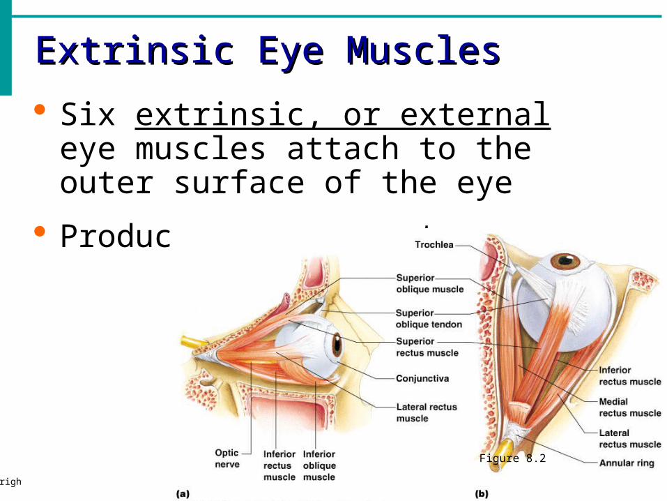
Extrinsic Eye MusclesExtrinsic Eye Muscles
Slide 8.6Copyright © 2003 Pearson Education, Inc. publishing as Benjamin Cummings
Six extrinsic, or external eye muscles attach to the outer surface of the eye
Produce eye movements
Figure 8.2

Structure of the EyeStructure of the Eye
Slide 8.7Copyright © 2003 Pearson Education, Inc. publishing as Benjamin Cummings
The wall is composed of three tunics
Fibrous tunic – outside layer
Choroid – middle layer
Sensory tunic – inside layer
Figure 8.3a

The Fibrous TunicThe Fibrous Tunic
Slide 8.8Copyright © 2003 Pearson Education, Inc. publishing as Benjamin Cummings
Sclera White connective tissue layer
Seen anteriorly as the “white of the eye”
Cornea Transparent, central anterior portion
Allows for light to pass through
Repairs itself easily
The only human tissue that can be transplanted without fear of rejection
sclera

Choroid LayerChoroid Layer
Slide 8.9Copyright © 2003 Pearson Education, Inc. publishing as Benjamin Cummings
Blood-rich nutritive tunic
Pigment prevents light from scattering
Modified anteriorly into two structures Ciliary body – smooth muscle
Iris
Pigmented layer that gives eye color
Pupil – rounded opening in the iris
iris
pupil


Sensory Tunic (Retina)Sensory Tunic (Retina)
Slide 8.10Copyright © 2003 Pearson Education, Inc. publishing as Benjamin Cummings
Contains receptor cells (photoreceptors) Rods
Cones
Signals pass from photoreceptors via a two-neuron chain Bipolar neurons
Ganglion cells
Signals leave the retina toward the brain through the optic nerve

Neurons of the RetinaNeurons of the Retina
Slide 8.11Copyright © 2003 Pearson Education, Inc. publishing as Benjamin Cummings
Figure 8.4

Neurons of the Retina and VisionNeurons of the Retina and Vision
Slide 8.12aCopyright © 2003 Pearson Education, Inc. publishing as Benjamin Cummings
More about photoreceptors…
Rods
Most are found towards the edges of the retina
Allow dim light vision and peripheral vision
Perception is all in gray tones

Neurons of the Retina and VisionNeurons of the Retina and Vision
Slide 8.12bCopyright © 2003 Pearson Education, Inc. publishing as Benjamin Cummings
Cones
Allow for detailed color vision
Densest in the center of the retina
Fovea centralis – area of the retina with only cones
No photoreceptor cells are at the optic disc, or blind spot
See book page 257 for blind spot test!


Cone SensitivityCone Sensitivity
Slide 8.13Copyright © 2003 Pearson Education, Inc. publishing as Benjamin Cummings
There are three types of cones
Different cones are sensitive to different wavelengths
Color blindness is the result of lack of one cone type
Figure 8.6

LensLens
Slide 8.14Copyright © 2003 Pearson Education, Inc. publishing as Benjamin Cummings
Biconvex crystal-like structure
Held in place by a suspensory ligament attached to the ciliary body
Figure 8.3a
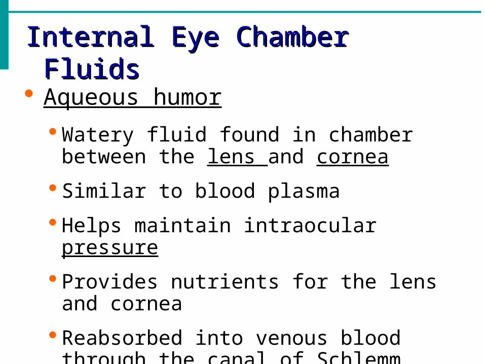
Internal Eye Chamber FluidsInternal Eye Chamber Fluids
Aqueous humor
Watery fluid found in chamber between the lens and cornea
Similar to blood plasma
Helps maintain intraocular pressure
Provides nutrients for the lens and cornea
Reabsorbed into venous blood through the canal of Schlemm

Internal Eye Chamber FluidsInternal Eye Chamber Fluids
Vitreous humor
Gel-like substance behind the lens
Keeps the eye from collapsing
Lasts a lifetime and is not replaced

vitreous humor
lens retina
extrinsic/external eye muscle

Images Formed on the RetinaImages Formed on the Retina
Figure 8.10 YouTube video link

Visual PathwayVisual Pathway
Slide 8.18a
Photoreceptors of the retina
Optic nerve
Optic nerve crosses at the optic chiasma
Figure 8.11

Visual PathwayVisual Pathway
Slide 8.18b
Optic tracts
Thalamus (axons form optic radiation)
Visual cortex of the occipital lobe
Figure 8.11

Lens AccommodationLens Accommodation
Slide 8.16
Light must be focused to a point on the retina for optimal vision
The eye is set for distance vision (over 20 ft away)
The lens must change shape to focus for closer objects
Figure 8.9

Poor Vision?Poor Vision?
Farsighted (Hyperopia)
Nearsighted (Myopia)

How corrective lenses work

AstigmatismAstigmatism
Lens is asymmetrical, leading to multiple focal points

CataractsCataracts
Lens has consistency of hardened jelly, but becomes increasingly hard and opaque with age. Could lead to blindness.

GlaucomaGlaucoma
Drainage of aqueous humor is blocked; pressure builds within eye and begins to compress retina and optic nerve. Could lead to blindness.
Eye ReflexesEye Reflexes Internal muscles are controlled by the
autonomic nervous system Bright light causes pupils to constrict
through action of radial and ciliary muscles
Viewing close objects causes accommodation
External muscles control eye movement to follow objects
Viewing close objects causes convergence (eyes moving medially)