Embed Size (px)
Citation preview
The second stage of labour has traditionally been regarded as the phase between full dilatation of the cervical os, and the birth of the baby. This is typically characterized by maternal restlessness, discomfort, desire for pain relief, a sense that the process is never-ending, and demands to attendants to end the whole process.
Appropriate midwifery care encompasses both knowledge of the usual physiological processes of this phase and of the mechanism of birth, and insight into the needs and choices of each individual labouring woman.
Chapter 28The transition and the second stage of labour:
physiology and the role of the midwife
The physiological changes are a continuation of the same forces which occurred in the earlier hours of labour, but activity is accelerated. This acceleration, however, does not occur abruptly.
Some women may experience an urge to push before the cervical os is fully dilated, and others may experience a quiet period before the onset of strong expulsive second stage contractions. This latter phenomenon has been termed the resting phase of the second stage of labour. The formal onset of the second stage of labour is traditionally confirmed with a vaginal examination to check for full dilatation of the cervical os. However, a finding of full cervical dilatation may occur some time after this stage has in fact been reached.
Contractions become stronger and longer but may be less frequent, allowing both mother and fetus regular recovery periods. The membranes often rupture spontaneously towards the end of the first stage or during transition to the second stage. The consequent drainage of liquor allows the hard, round fetal head to be directly applied to the vaginal tissues. This pressure aids distension. Fetal axis pressure increases flexion of the head, resulting in smaller presenting diameters, more rapid progress and less trauma to both mother and fetus. If the mother is upright during this time, these processes are optimized.
Uterine action
The contractions become expulsive as the fetus descends further into the vagina. Pressure from the presenting part stimulates nerve receptors in the pelvic floor. This phenomenon is termed the ‘Ferguson reflex’. As a consequence, the woman experiences the need to push. This reflex may initially be controlled to a limited extent but becomes increasingly compulsive, overwhelming and involuntary. The mother's response is to employ her secondary powers of expulsion by contracting her abdominal muscles and diaphragm.
As the fetal head descends, the soft tissues of the pelvis become displaced. Anteriorly, the bladder is pushed upwards into the abdomen where it is at less risk of injury during fetal descent.Posteriorly, the rectum becomes flattened into the sacral curve and the pressure of the advancing head expels any residual faecal matter. The fetal head becomes visible at the vulva, advancing with each contraction and receding between contractions until crowning takes place. The head is then born. The shoulders and body follow with the next contraction, accompanied by a gush of amniotic fluid and sometimes of blood. The second stage culminates in the birth of the baby.
Soft tissue displacement
Recognition of the commencement of the second stage of labourProgress from the first to the second stage is not always clinically apparent. Presumptive evidence1.Expulsive uterine contractionsSome women feel a strong desire to push before full dilatation occurs. Traditionally, it has been assumed that an early urge to push will lead to maternal exhaustion and/or cervical oedema or trauma. More recent research indicates that the early pushing urge may in fact be experienced by a significant minority of women, and that, in certain circumstances, early pushing may be physiological
It is not clear whether these findings are influenced by factors such as maternal or fetal position, or parity, and there is not enough evidence to date to determine the optimum response to the early pushing urge. The midwife needs to work with each individual woman in the context of each labour to determine the best approach in that specific case.
2. Rupture of the forewaters
Rupture of the forewaters may occur at any time during
labour.
3. Dilatation and gaping of the anus
Deep engagement of the presenting part may produce this
sign during the latter part of the first stage.
4. Anal cleft line
Some midwives have reported observing this line (also called
‘the purple line’) as a pigmented mark in the cleft of the
buttocks which creeps up the anal cleft as the labour
progresses (Hobbs 1998, Wickham 2007). The efficacy of this
observation remains to be tested formally.
5. Upper abdominal pressure and epidural analgesia
It has been observed that women who have an epidural in situ
often have a sense of discomfort under the ribs towards the
end of the first stage of labour. This seems to coincide with
full cervical dilatation. The efficacy of these observations in
predicting the onset of the anatomical second stage of labour
remains to be researched.

6. Show
This is the loss of bloodstained mucus which often
accompanies rapid dilatation of the cervical os
towards the end of the first stage of labour. It must be
distinguished from frank fresh blood loss caused by
partial separation of the placenta or a ruptured vasa
praevia.
7. Appearance of the presenting part
Excessive moulding may result in the formation of a large
caput succedaneum which can protrude through the cervix
prior to full dilatation of the os. Very occasionally, a baby
presenting by the vertex may be visible at the perineum at the
same time as remaining cervix. This is more common in
women of high parity. Similarly a breech presentation may be
visible when the cervical os is only 7–8 cm dilated.
Confirmatory evidence
a vaginal examination must be undertaken to confirm full
dilatation of the cervical os. This is both to ensure that a
woman is not pushing too early, and to provide a baseline for
timing the length of the second stage of labour.
However, some maternity settings and some individual
midwives do not insist on this unless there are observable
maternal and/or fetal signs that the labour is not progressing as
anticipated.
It was noted that vaginal assessment of cervical dilatation is
largely unevaluated. Despite this, regular vaginal
examinations are undertaken by most midwives and
obstetricians, and expected by many women. Whether the
midwife undertakes an examination or not, she should record
all the signs she observes and all the measurements she
takes, and she should advise and support the labouring
woman on the basis of accurate observation and assessment
of progress.
Phases and duration
A. The latent phase
In some women, full dilatation of the cervical os is recorded,
but the presenting part may not yet have reached the pelvic
outlet. She may not experience a strong expulsive urge until
the head has descended sufficiently to exert pressure on the
rectum and perineal tissues. More recent concerns over the
impact of epidural analgesia on spontaneous birth have led
to an increasing interest in the passive second stage of
labour (Simpson & James 2005, NICE 2007).
Due to the effect of epidural analgesia on the pelvic floor
muscles, there is little benefit in encouraging active
pushing until the head has descended and rotated as far
as possible if a woman has an effective epidural in situ.
Passive descent of the fetus can continue with good
midwifery support for the woman until the head is visible at
the vulva, or until the mother feels a spontaneous desire to
push.
Active phase
Once the fetal head is visible, the woman will usually experience
a compulsive urge to push.
Duration of the second stage
There is no good evidence about the absolute time limits of
physiological labour.Mothers and babies who labour for at least
twice as long as the traditional limits can do well. In the presence
of regular contractions, maternal and fetal well-being, and
progressive descent, considerable variation between women is to
be expected. While many maternity units do currently impose
limits on the second stage beyond which medical help should be
called, these are not based on good evidence.
Maternal response to transition and the second stagePushing
Traditionally, if the maternal urge to push occurs before
confirmation of full dilatation of the cervical os, or the
appearance of a visible vertex, the mother is encouraged to
avoid active pushing at this stage. This is done to conserve
maternal effort and allow the vaginal tissues to stretch
passively. Techniques include position change, often to the
left lateral, using controlled breathing, inhalation analgesia, or
even narcotic or epidural pain relief (Downe et al 2008).
Maternal response to transition and the second stagePushing
However, when mother and baby are well and labour has
progressed spontaneously, some midwives have adopted the
practice of supporting the urge to bear down without
confirming full dilatation of the cervical os, while paying close
attention to the maternal and fetal condition. As stated above,
the optimum response in this situation has not yet been
established.
There has been convincing evidence for over a decade
that managed active pushing in the second stage of labour
accompanied by breath holding (the Valsalva manoeuvre)
has adverse consequences (Aldrich et al 1995, Enkin et al
2000, p 290–1, Thomson 1993). Whenever active pushing
commences, the woman should be encouraged to follow
her own inclinations in relation to expulsive effort. Few
women need instruction on how to push unless they are
using epidural analgesia.
Spontaneous pushing efforts usually result in maximum
pressure being exerted at the height of a contraction. In
turn, this allows the vaginal muscles to become taut and
prevents bladder supports and the transverse cervical
ligaments from being pushed down in front of the baby's
head. It is believed that this may help to prevent prolapse
and urinary incontinence in later life, although this belief
has still not been formally tested (Benyon 1990).
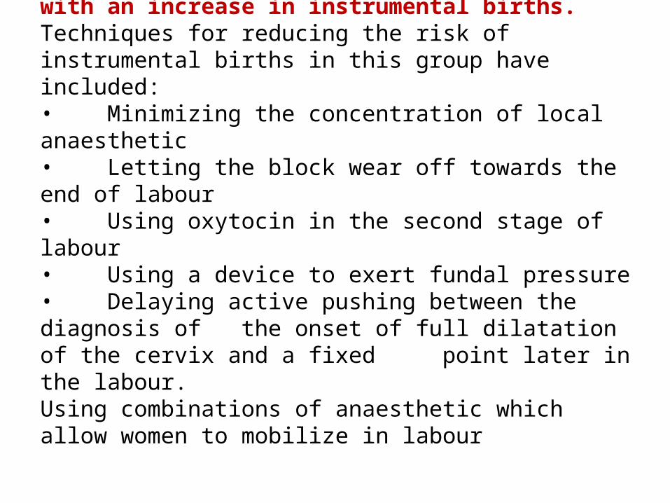
Epidural analgesia provides optimal pain relief for women, but its use is associated with an increase in instrumental births.Techniques for reducing the risk of instrumental births in this group have included:• Minimizing the concentration of local anaesthetic• Letting the block wear off towards the end of labour • Using oxytocin in the second stage of labour• Using a device to exert fundal pressure• Delaying active pushing between the diagnosis of
the onset of full dilatation of the cervix and a fixed point later in the labour.
Using combinations of anaesthetic which allow women to mobilize in labour
These techniques have had varying success rates.
Some mothers vocalize loudly as they push. This may aid in
coping with the contractions, so women should feel free to
express themselves in this way. Reassurance and praise will
help to boost confidence, enabling the mother to assert her
own control over events. The atmosphere should be calm
and the pace unhurried.
Position
If the mother lies flat on her back, resulting in hypotension.
This can lead to reduced placental perfusion and
diminished fetal oxygenation. The efficiency of uterine
contractions may also be reduced. The semi-recumbent
or supported sitting position, with the thighs abducted,
is the posture most commonly used in western
cultures. While this may afford the midwife good access
and a clear view of the perineum, the mother's weight is on
her sacrum, which directs the coccyx forwards and reduces
the pelvic outlet.
Left lateral position
This position was widely used in the UK in the last century,
although it is less common in current practice. The
perineum can be clearly viewed and uterine action is
effective, but an assistant may be required to support the
right thigh, which may not be ergonomic. It provides an
alternative for women who find it difficult to abduct their
hips. It may also aid fetal rotation, especially in the context
of epidural analgesia (Downe et al 2004).
Supported sitting position. Upright positions; squatting, kneeling, all fours, standing, using a birthing ball. (Figs. 28.1, 28.2)A review of studies examining upright versus recumbent positions during the second stage of labour. showed there were clear advantages for women in adopting an upright position (Gupta et al 2004). These included reduced duration of second stage labour, less assisted deliveries, less episiotomies, reduced severe pain in second stage labour, and fewer abnormal heart rate patterns. However, increased rates of second degree tears and of estimated blood loss >500 mL also occurred. The experimental group included women who used birthing chairs, a technique known to be associated with increased blood loss (Stewart & Spiby 1989
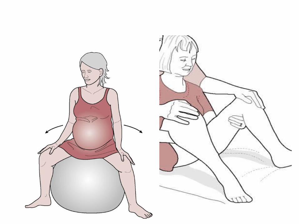
It is not clear if this risk accrues to all upright positions. The ‘upright position’ group in this review included women who were in supported sitting positions on a bed, and in the lateral position.
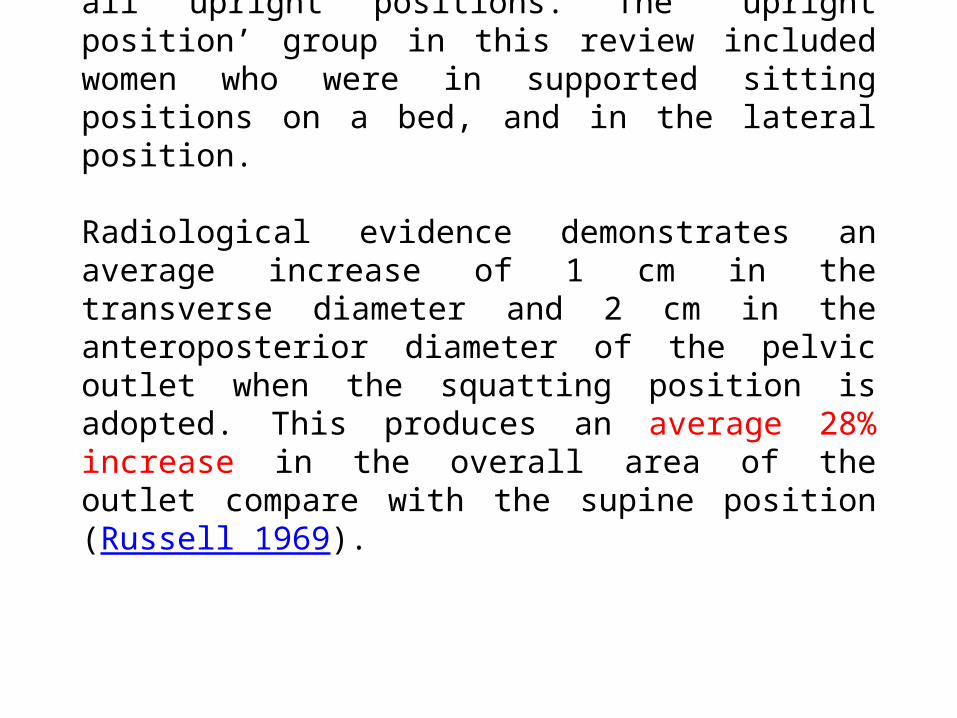
Radiological evidence demonstrates an average increase of 1 cm in the transverse diameter and 2 cm in the anteroposterior diameter of the pelvic outlet when the squatting position is adopted. This produces an average 28% increase in the overall area of the outlet compare with the supine position (Russell 1969).
Some women find the all fours position to be the optimum
approach for all or part of their labours, especially in the
case of an occipitoposterior position, due to relief of
backache. It can, however, be tiring to maintain for a long
period of time. A wide range of other standing and leaning
positions can be experimented with to help the mother cope
with her labour.
It is important not to insist on any position as the ‘right’ one.
Positive and dramatic effects on labour progress can be
achieved by encouraging the mother to change and adapt
her position in response to the way her body feels.
•The midwife's confidence: A full understanding of the
mechanism of labour should enable the midwife to adapt to
any position that the woman wishes to adopt.
Minimizing vaginal examinations in labour will also reduce
the risk of back injury to the midwife.
If the woman has an epidural in situ, it is essential that help
is called when the woman needs to be moved, and that
ergonomic lifting positions are used.
Minimizing unnecessary disturbance by other members of
staff is essential
•The midwife's confidence: A full understanding of the
mechanism of labour should enable the midwife to adapt to
any position that the woman wishes to adopt.
Minimizing vaginal examinations in labour will also reduce
the risk of back injury to the midwife.
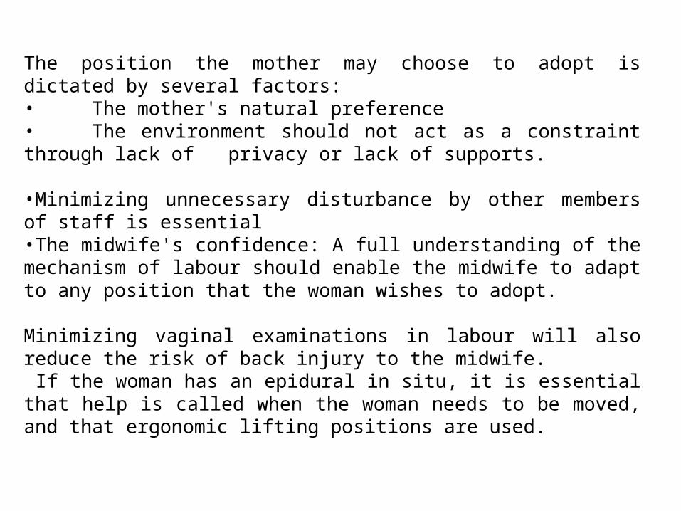
The position the mother may choose to adopt is dictated by several factors:• The mother's natural preference• The environment should not act as a constraint through lack of privacy or lack of supports.
•Minimizing unnecessary disturbance by other members of staff is essential•The midwife's confidence: A full understanding of the mechanism of labour should enable the midwife to adapt to any position that the woman wishes to adopt.
Minimizing vaginal examinations in labour will also reduce the risk of back injury to the midwife. If the woman has an epidural in situ, it is essential that help is called when the woman needs to be moved, and that ergonomic lifting positions are used.

The Process of Labor and BirthThe Process of Labor and BirthA number of forces affect the progress of labor and help to bring about childbirth. These critical factors are oftenreferred to as the “P’s” of labor:1.Powers (physiological forces)2. Passageway (maternal pelvis)3. Passenger (fetus and placenta)4. Passageway Passenger and their relationship(engagement, attitude, position)5. Psychosocial influences (previous experiences, emotional status)
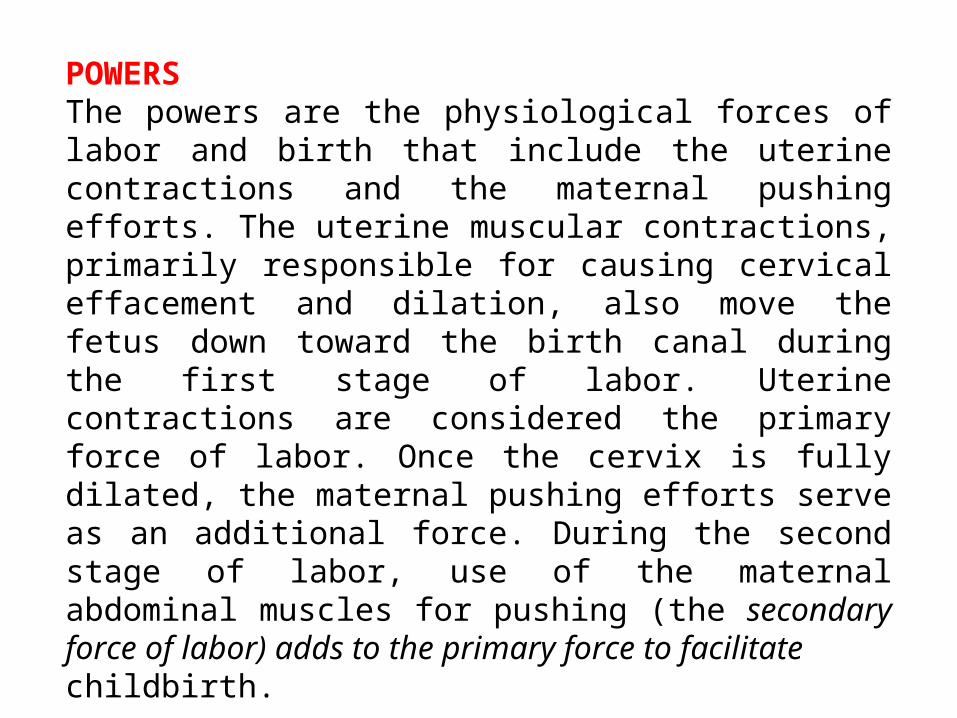
POWERSThe powers are the physiological forces of labor and birth that include the uterine contractions and the maternal pushing efforts. The uterine muscular contractions, primarily responsible for causing cervical effacement and dilation, also move the fetus down toward the birth canal during the first stage of labor. Uterine contractions are considered the primary force of labor. Once the cervix is fully dilated, the maternal pushing efforts serve as an additional force. During the second stage of labor, use of the maternal abdominal muscles for pushing (the secondary force of labor) adds to the primary force to facilitatechildbirth.
Characteristics of Uterine Contractions
Contractions are a rhythmic tightening of the uterus that
occurs intermittently. Over time, this action shortens the
individual uterine muscle fibers and aids in the process of
cervical effacement and dilation, birth, and postpartal
involution (the reduction in uterine size after birth.
Characteristics of Uterine Contractions
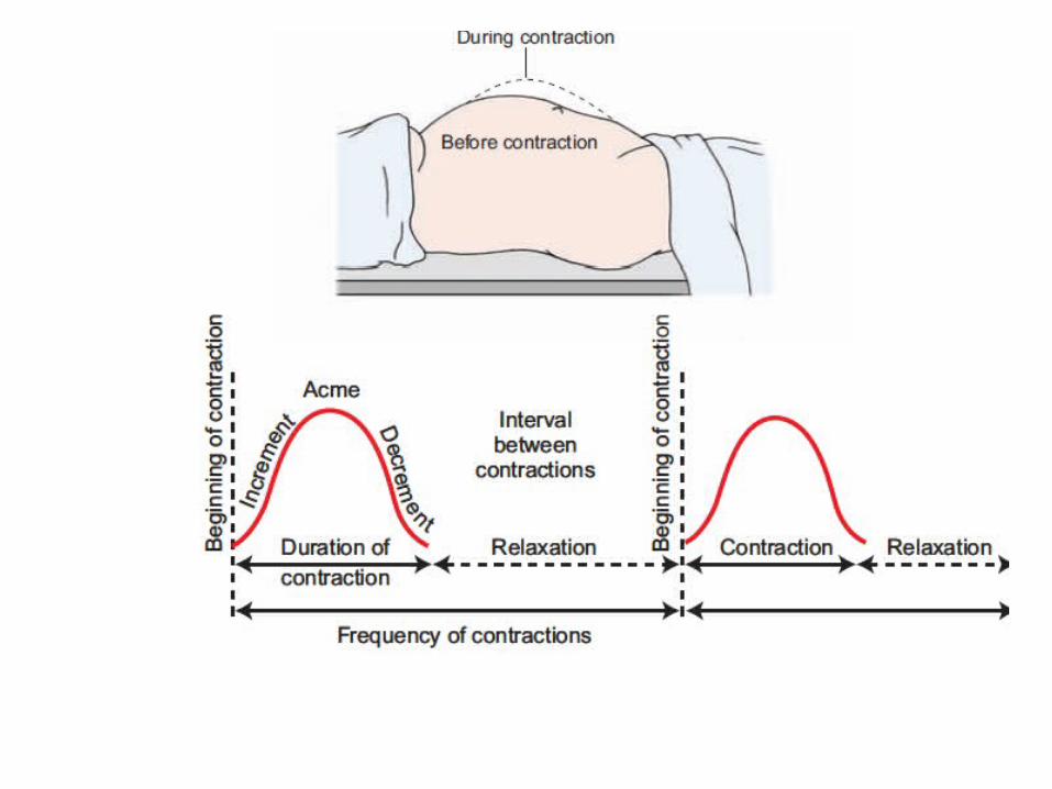
Each contraction consists of three distinct components: the
increment (building of the contraction), the acme (peak of
the contraction) and the decrement (decrease in the
contraction). Between contractions, the uterus normally
returns to a state of complete relaxation. This rest period
allows the uterine muscles to relax and provides the woman
with a short recovery period that helps her to avoid
exhaustion. In addition, uterine relaxation between
contractions is important for fetal oxygenation as it allows for
blood flow from the uterus to the placenta to be restored.
The lower uterine segment becomes thin-walled and passive. The boundary between the upper lower uterine segments becomes marked by a ridge on the inner uterine surface, known as the “physiological retraction ring.” With each contraction, the uterus elongates. Elongation causes a straightening of the fetal body so that the upper body is pressed against the fundus and the lower, presenting part is pushed toward the lower uterine segment and the cervix.
The pressure exerted by the fetus is called the fetal axis pressure. As the uterus elongates, the longitudinal muscle fibers are stretched upward over the presenting part. This force, along with the hydrostatic pressure of the fetal membranes, causes the cervix to dilate (open).
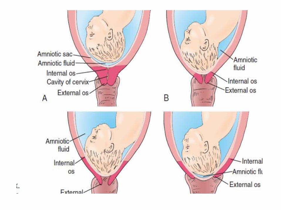
The coordinated efforts of the contractions help to bring
about effacement and dilatation of the cervix. Effacement is
the process of shortening and thinning of the cervix. As
contractions occur, the cervix becomes progressively shorter
until the cervical canal eventually disappears. The amount of
cervical effacement is usually expressed as a percentage
related to the length of the cervical canal, as compared to a
non effaced cervix.
For example, if a cervix has thinned to half the normal
length of a cervix it is considered to be 50% effaced. Dilation
is the opening and enlargement of the cervix that
progressively occurs throughout the first stage of labor.
Cervical dilation is expressed in centimeters and full dilation
is approximately 10 cm. With continued uterine contractions,
the cervix eventually opens large enough to allow the fetal
head to come through. At this point, the cervix is considered
fully dilated or completely dilated and measures 10 cm.
Maternal and fetal condition
If the mother has had analgesia, or if there is any concern
about the well-being of either the woman or her baby then
more frequent or continuous monitoring may limit the
choices available to her. However, there are often creative
solutions to these situations, and good midwifery care
involves finding these solutions where possible.
The mechanism of normal labour
Knowledge and recognition of the normal mechanism
enables the midwife to anticipate the next step in the
process of descent. Understanding and constant monitoring
of these movements can help to ensure that normal
progress is recognized, that the woman gives birth safely
and positively, or that early assistance can be sought should
any un resolvable problems occur. The fetal presentation,
position, and size relative to that of the woman will govern
the exact mechanism as the fetus responds to external
pressures.
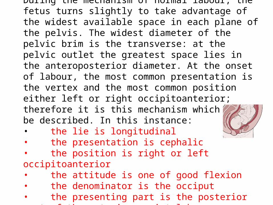
During the mechanism of normal labour, the fetus turns slightly to take advantage of the widest available space in each plane of the pelvis. The widest diameter of the pelvic brim is the transverse: at the pelvic outlet the greatest space lies in the anteroposterior diameter. At the onset of labour, the most common presentation is the vertex and the most common position either left or right occipitoanterior; therefore it is this mechanism which will be described. In this instance:• the lie is longitudinal• the presentation is cephalic• the position is right or left occipitoanterior• the attitude is one of good flexion• the denominator is the occiput• the presenting part is the posterior part of the anterior parietal bone.
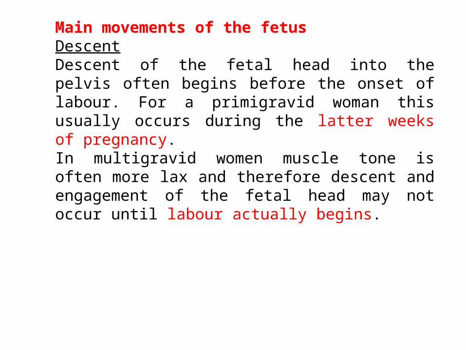
Main movements of the fetusDescentDescent of the fetal head into the pelvis often begins before the onset of labour. For a primigravid woman this usually occurs during the latter weeks of pregnancy. In multigravid women muscle tone is often more lax and therefore descent and engagement of the fetal head may not occur until labour actually begins.
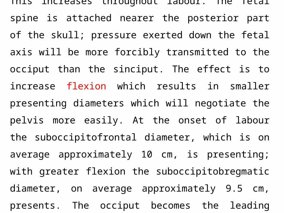
Flexion
This increases throughout labour. The fetal spine is attached
nearer the posterior part of the skull; pressure exerted down
the fetal axis will be more forcibly transmitted to the occiput
than the sinciput. The effect is to increase flexion which
results in smaller presenting diameters which will negotiate
the pelvis more easily. At the onset of labour the
suboccipitofrontal diameter, which is on average
approximately 10 cm, is presenting; with greater flexion the
suboccipitobregmatic diameter, on average approximately 9.5
cm, presents. The occiput becomes the leading part.
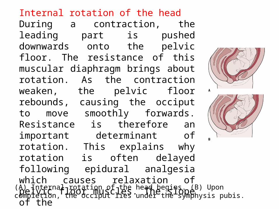
Internal rotation of the headDuring a contraction, the leading part is pushed downwards onto the pelvic floor. The resistance of this muscular diaphragm brings about rotation. As the contraction weaken, the pelvic floor rebounds, causing the occiput to move smoothly forwards. Resistance is therefore an important determinant of rotation. This explains why rotation is often delayed following epidural analgesia which causes relaxation of pelvic floor muscles. The slope of the
(A) Internal rotation of the head begins. (B) Upon completion, the occiput lies under the symphysis pubis.
pelvic floor determines the direction of rotation. In a well-flexed vertex presentation the occiput leads, and rotates anteriorly through ⅛ of a circle when it meets the pelvic floor. This causes a slight twist in the neck as the head is no longer in direct alignment with the shoulders. The anteroposterior diameter of the head now lies in the widest (anteroposterior) diameter of the pelvic outlet. The occiput slips beneath the sub-pubic arch and crowning occurs when the head no longer withdraws between contractions and the widest transverse diameter (biparietal) is born. If flexion is maintained, the sub-occipitobregmatic diameter, usually approximately 9.5 cm, distends the vaginal orifice.
Extension of the headOnce crowning has occurred the fetal head can extend, pivoting on the suboccipital region around the pubic bone. This releases the sinciput, face, and chin, which sweep the perineum, and then are born by a movement of extension (Fig. 28.4).RestitutionThe twist in the neck of the fetus which resulted from internal rotation is now corrected by a slight untwisting movement. The occiput moves ⅛ of a circle towards the side from which it started.
Internal rotation of the shoulders
The shoulders undergo a similar rotation to that of the head to
lie in the widest diameter of the pelvic outlet, namely
anteroposterior. The anterior shoulder is the first to reach the
levator ani muscle and it therefore rotates anteriorly to lie
under the symphysis pubis. This movement can be clearly
seen as the head turns at the same time (external rotation of
the head). It occurs in the same direction as restitution, and
the occiput of the fetal head now lies laterally.
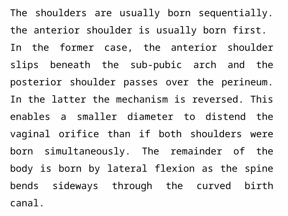
Lateral flexion
The shoulders are usually born sequentially. the anterior
shoulder is usually born first. In the former case, the anterior
shoulder slips beneath the sub-pubic arch and the posterior
shoulder passes over the perineum. In the latter the
mechanism is reversed. This enables a smaller diameter to
distend the vaginal orifice than if both shoulders were born
simultaneously. The remainder of the body is born by lateral
flexion as the spine bends sideways through the curved birth
canal.
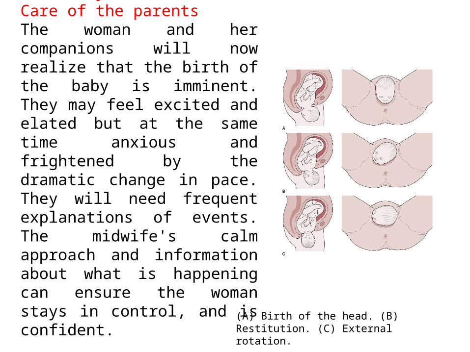
Midwifery careCare of the parentsThe woman and her companions will now realize that the birth of the baby is imminent. They may feel excited and elated but at the same time anxious and frightened by the dramatic change in pace. They will need frequent explanations of events. The midwife's calm approach and information about what is happening can ensure the woman stays in control, and is confident.
(A) Birth of the head. (B) Restitution. (C) External rotation.
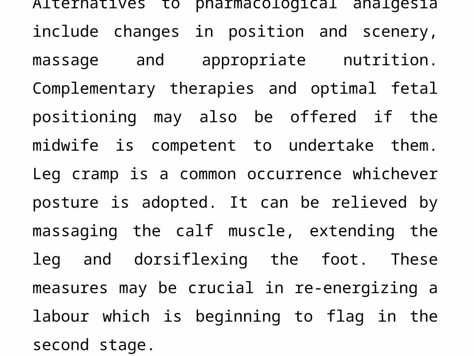
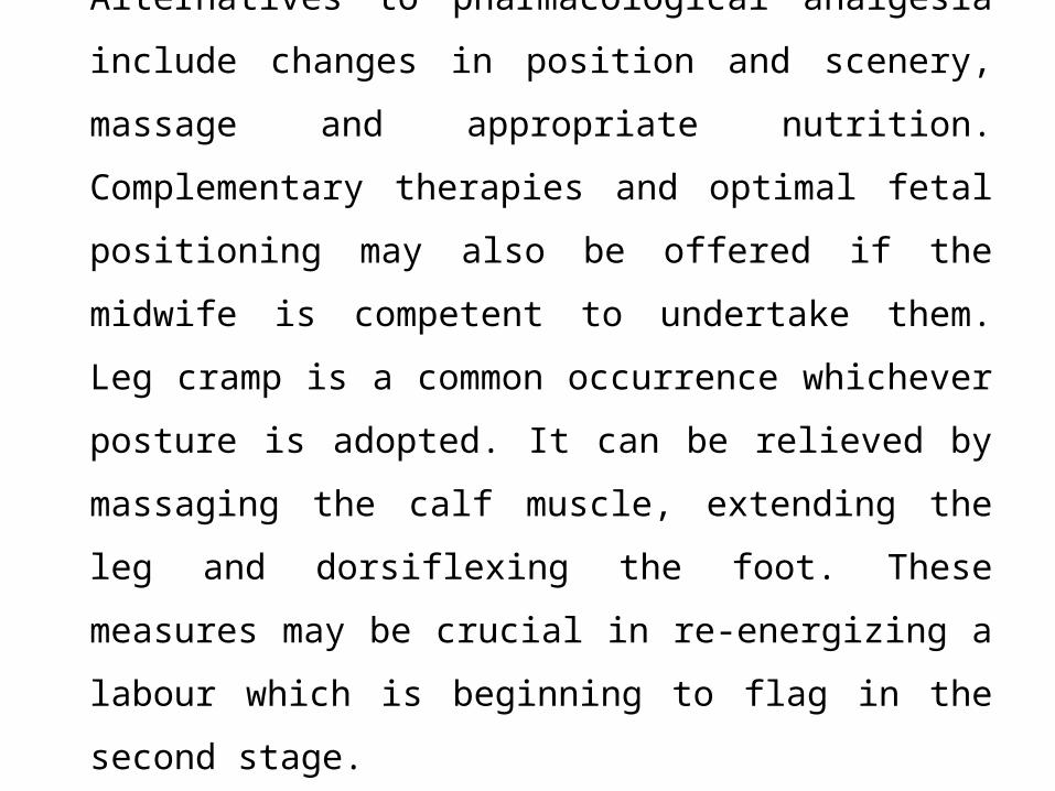
Alternatives to pharmacological analgesia include
changes in position and scenery, massage and
appropriate nutrition. Complementary therapies and
optimal fetal positioning may also be offered if the midwife
is competent to undertake them. Leg cramp is a common
occurrence whichever posture is adopted. It can be
relieved by massaging the calf muscle, extending the leg
and dorsiflexing the foot. These measures may be crucial
in re-energizing a labour which is beginning to flag in the
second stage.
Observations during the second stage of labourFour factors determine whether the second stage is continuing safely, and these must be carefully monitored:1 Uterine contractions2 Descent, rotation and flexion of the presenting part3 Fetal condition4 Maternal condition.Uterine contractionsThe strength, length and frequency of contractions should be assessed continuously by observation of maternal responses, and regularly by uterine palpation. They are usually stronger and longer than during the first stage of labour, with a longer resting phase. The posture and position adopted by the mother may influence the contractions.
Descent, rotation and flexionInitially, descent may occur slowly, especially in primigravid women, but it usually accelerates during the active phase. It may occur very rapidly in multigravid women. If there is a delay in descent on abdominal palpation, despite regular strong contractions and active maternal pushing, a vaginal examination may be performed with maternal permission. The purpose is to confirm whether or not internal rotation of the head has taken place, to assess the station of the presenting part, and to determine whether a caput succedaneum has formed. If the occiput has rotated anteriorly, the head is well flexed and caput succedaneum is not excessive it is likely that progress will continue. In the absence of good rotation and flexion, and/or a weakening of uterine contractions, change of position, nutrition and hydration, or use of optimal fetal positioning techniques may be helpful Consultation with a more experienced midwife may provide more suggestions to re-orientate the labour. However, if there is evidence that either fetal or maternal condition are compromised, an experienced obstetrician should be consulted.
Fetal condition
If the membranes are ruptured, the liquor amnii is observed
to ensure that it is clear. While thin old meconium staining is
not always regarded as a sign of fetal compromise, thick
fresh meconium is always ominous, and experienced
obstetric advice must be sought if this sign appears.
A well-grown healthy baby will not be compromised by this
transitory hypoxia. This will tend to produce early
decelerations of the fetal heart, with a swift return to the
normal baseline after a contraction, and good beat-to-beat
variation throughout. While early decelerations are always
deemed ‘suspicious’.
Suspicious/pathological changes in the fetal heartLate decelerations, a lack of return to the normal baseline, a rising baseline, or diminishing beat-to-beat variation are signs of concern. If these are heard for the first time in second stage, they may be due to cord or head compression, which may be helped by a change in position. However, if they persist, experienced obstetric aid must be sought. If the labour is taking place in a unit which is distant from an obstetric unit, an episiotomy may be considered if the birth is imminent, or midwives who are trained and experienced in ventouse birth may consider expediting the birth. Otherwise, with maternal consent, transfer to an obstetric unit should be expedited.
Maternal conditionThe midwife's observation includes an appraisal of the mother's ability to cope emotionally as well as an assessment of her physical well-being. Maternal comfortAs a result of her exertions the woman usually feels very hot and sticky and she will find it soothing to have her face and neck sponged with a cool flannel. Her mouth and lips may become very dry. Sips of iced water or other fluids are refreshing and a moisturizing cream can be applied to her lips. Her partner may help with these tasks as a positive contribution to ease her discomfort.
Preparation for the birthThere is usually little urgency if the woman is primigravid, but multigravid women may progress very rapidly.
The room should be warm with a spotlight available so that the perineum can be easily observed if necessary. A clean area should be prepared to receive the baby, and waterproof covers provided to protect the bed and floor. Sterile cord clamps, a clean apron, and sterile gloves are placed to hand. In some settings, sterile gowns are also used. An oxytocic agent may be prepared, either for the active management of the third stage if this is acceptable to the woman, or for use during an emergency.Neonatal resuscitation equipment must be thoroughly checked and readily accessible.
The birth of the babyDuring the birth, both mother and baby are particularly vulnerable to infection. While there is now evidence that strict antisepsis is unnecessary if the birth is straightforward (Keane & Thornton 1998),careful aseptic technique must be observed when preparing sterile equipment such as episiotomy scissors. Once she has scrubbed up the midwife prepares her equipment. This includes the following items:• warm swabbing solution or tap water• cotton wool and pads• sterile cord scissors and clamps• sterile episiotomy scissors.
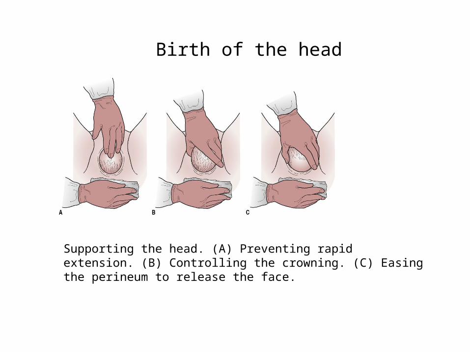
Supporting the head. (A) Preventing rapid extension. (B) Controlling the crowning. (C) Easing the perineum to release the face.
Birth of the head
If the cord is clamped, great care must be taken that
maternal tissues are not damaged. Holding a swab over
the cord as it is incised will reduce the risk of the
attendants being sprayed with blood during the
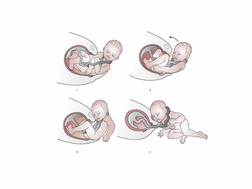
procedure. Once severed, the cord may be unwound
from around the neck.
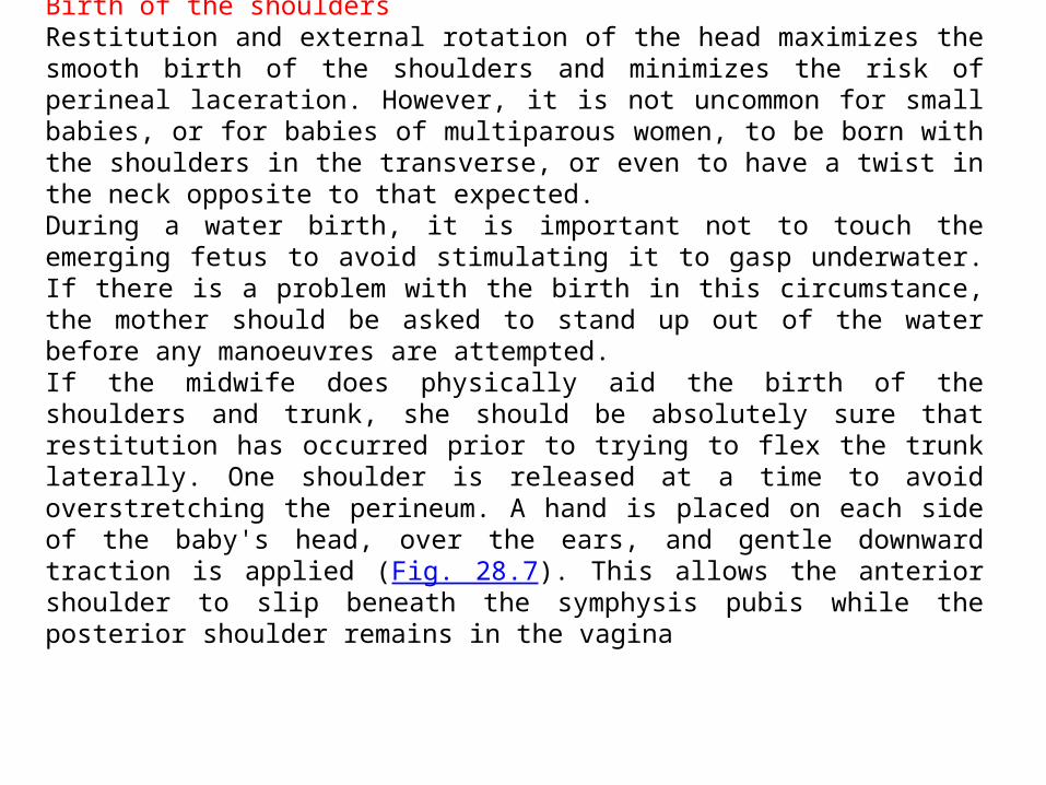
Birth of the shouldersRestitution and external rotation of the head maximizes the smooth birth of the shoulders and minimizes the risk of perineal laceration. However, it is not uncommon for small babies, or for babies of multiparous women, to be born with the shoulders in the transverse, or even to have a twist in the neck opposite to that expected. During a water birth, it is important not to touch the emerging fetus to avoid stimulating it to gasp underwater. If there is a problem with the birth in this circumstance, the mother should be asked to stand up out of the water before any manoeuvres are attempted.If the midwife does physically aid the birth of the shoulders and trunk, she should be absolutely sure that restitution has occurred prior to trying to flex the trunk laterally. One shoulder is released at a time to avoid overstretching the perineum. A hand is placed on each side of the baby's head, over the ears, and gentle downward traction is applied (Fig. 28.7). This allows the anterior shoulder to slip beneath the symphysis pubis while the posterior shoulder remains in the vagina
episiotomy may very occasionally be necessary. This is an incision through the perineal tissues which is designed to enlarge the vulval outlet during birth. As this is a surgical incision it cannot be undertaken unless the mother gives consent. She should be assured that its use is selective and discretional. The mother's wishes should be clearly documented and respected.The rationale for its use is the need to minimize the risk of severe trauma to the vagina and perineum, and to expedite the birth when there is evidence of fetal distress. However, the risks of its use should always be borne in mind. During a normal birth the indications are few and the midwife should adopt a restrictive policy.
Types of incision
There are two main directions of incision.
Mediolateral
This begins at the midpoint of the fourchette and is directed at a 45° angle
to the midline towards a point midway between the ischial tuberosity and
the anus. This line avoids the danger of damage to both the anal
sphincter and Bartholin's gland but it is more difficult to repair. This is
the incision largely used by midwives in the UK.
Median
This is a midline incision which follows the natural line of insertion of the
perineal muscles. It is associated with reduced blood loss but a higher
incidence of damage to the anal sphincter. It is the easier to repair and
results in less pain and dyspareunia. This incision is favoured in the USA.
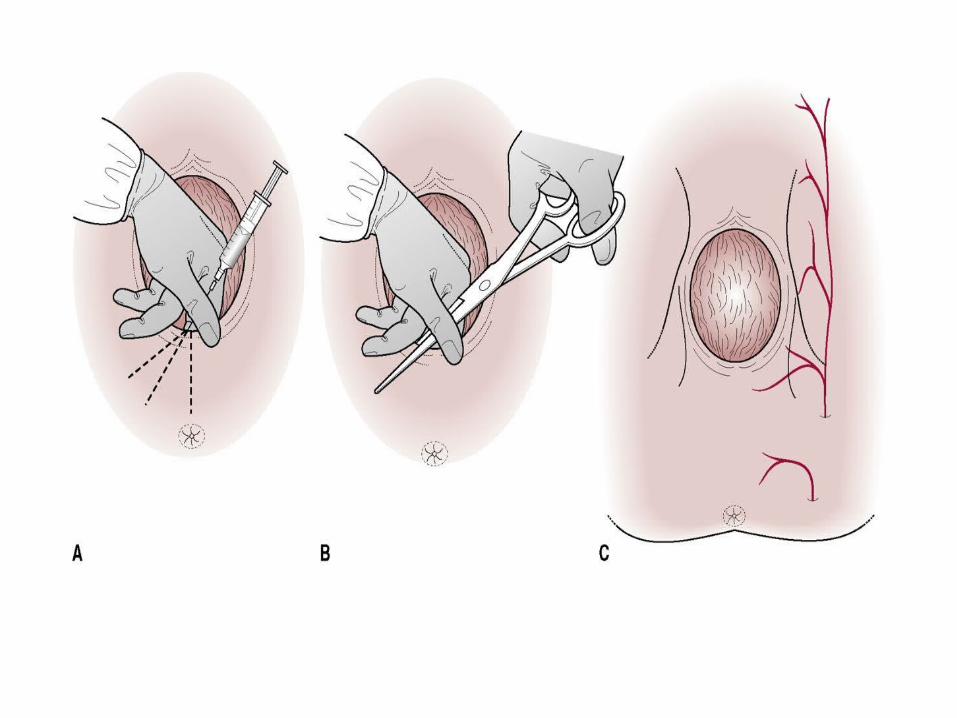
Infiltration of the perineum
The perineum should be adequately anaesthetized prior to the incision.
Lidocaine (formerly termed lignocaine) is commonly used, 0.5% 10 mL or
1% 5 mL. The advantage of the more concentrated solution is that a
smaller volume is needed. Lidocaine takes 3–4 min to take effect and, if
possible, two or three contractions should be allowed to occur between
infiltration and incision. The timing is not always easy to calculate but it is
better to infiltrate and not perform an episiotomy than to incise the
perineum without an effective local anaesthetic.
Method of infiltrationThe perineum is cleansed with antiseptic solution. Two fingers are inserted into the vagina along the line of the proposed incision in order to protect the fetal head. The needle is inserted beneath the skin for 4–5 cm following the same line.
The piston of the syringe should be withdrawn prior to injection to check whether the needle is in a blood vessel. If blood is aspirated, the needle should be repositioned and the procedure repeated until no blood is withdrawn.
Lidocaine is continuously injected as the needle is slowly withdrawn. Some practitioners inject the whole amount in one operation.
Anterior labial tearsIt is debatable whether or not these should be sutured. Much depends upon the control of bleeding as the labia are very vascular. A suture may be necessary to secure haemostasis.
Posterior perineal trauma Spontaneous tears are usually classified in degrees which are related to the anatomical structures.
• 1-degree tear involves the fourchette only
• 2-degree tear involves the fourchette and the superficial perineal
muscles, namely the bulbocavernosus and the transverse perineal
muscles and in some cases the pubococcygeus.
• 3-degree tear comprises a partial or complete disruption of the
anal sphincter muscles, which may involve either or both the external
and internal anal sphincter muscles
• 4-degree tear involves a disruption of the anal sphincter muscles
with a breach of the rectal mucosa.
Anterior labial tears
Third- and fourth-degree tears should be repaired by an
experienced obstetrician. A general anaesthetic or effective
epidural or spinal anaesthetic is necessary.
Prior to the commencement of repair, infiltration of the wound with
local anaesthetic will be required. Lidocaine 1% is used and time
must be allowed for it to take effect before repair begins. If an
epidural block is in progress, a ‘top up’ should be given.
The apex of the vaginal incision is identified and the posterior vaginal wall repaired from the apex downwards (Fig. 28.9). A continuous suture affords better haemostasis (Kettle & Johanson 1998). The thread should not be pulled too tightly as oedema will develop during the first 24–48 hrs. Care must be taken to identify other vaginal lacerations which need to be repaired.
Deeper interrupted sutures are then inserted to repair the perineal muscles. Good approximation of tissue is important. The subsequent strength of the pelvic floor will depend largely upon adequate repair of this layer.
If skin closure is carried out, a continuous subcuticular suture results in fewer short-term problems than interrupted transcutaneous suturing techniques.
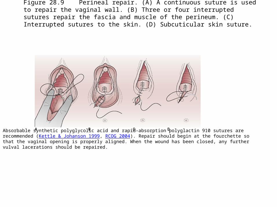
Figure 28.9 Perineal repair. (A) A continuous suture is used to repair the vaginal wall. (B) Three or four interrupted sutures repair the fascia and muscle of the perineum. (C) Interrupted sutures to the skin. (D) Subcuticular skin suture.
Absorbable synthetic polyglycolic acid and rapid-absorption polyglactin 910 sutures are recommended (Kettle & Johanson 1999, RCOG 2004). Repair should begin at the fourchette so that the vaginal opening is properly aligned. When the wound has been closed, any further vulval lacerations should be repaired.
The sutured areas should be inspected in order to confirm haemostasis before the vaginal pack is removed. A vaginal examination is made to ensure that the introitus has not been narrowed. Upon completion, and after warning the mother, a rectal examination is made to ensure that no sutures have penetrated the rectal mucosa. Any such sutures must be removed to prevent fistula formation.
The mother's legs are then gently and simultaneously removed from lithotomy support and she is made comfortable. The nature of the trauma and repair should be explained to her and information given on whether or not sutures will need to be removed.
Records
This should include details of any drugs administered, of the
duration and progress of labour, of the reason for
performing an episiotomy, and of perineal repair.
Details of the baby's condition including Apgar score are
also recorded.
The birth notification must be completed within 36 hrs of the
birth. This may be undertaken by anyone present at the
birth but is usually carried out by the midwife.
The notification is sent to the medical officer in the health
district in which the baby was born.
Conclusion
The processes of transition and of second stage labour are likely to
be very physically and emotionally intense, particularly for the
woman, but also for her partner and other birth companions.
The skill of the midwife is to support the woman effectively, to guide
her when her spirits or the labour are to enable her to accomplish
her birth safely.
Clear, comprehensive record keeping is essential. While much
practice in this area is not based on formal evidence, new
observations about normal birth are beginning to be recorded, and
these observations will form the basis for future research.