Embed Size (px)
Citation preview
489
Mesozoic Fishes 5 – Global Diversity and Evolution, G. Arratia, H.-P. Schultze & M. V. H. Wilson (eds.): pp. 489-497, 5 figs., 1 tab.© 2013 by Verlag Dr. Friedrich Pfeil, München, Germany – ISBN 978-3-89937-159-8
The second record of a mawsoniid coelacanth from the Lower Cretaceous Crato Formation,
Araripe Basin, northeastern Brazil, with comments on the development of coelacanths
Yoshitaka YABUMOTO and Paulo M. BRITO
Abstract
A well-preserved fossil coelacanth from the Lower Cretaceous Crato Formation in Araripe basin, northeast Bra-zil, is the second record of a coelacanth from this formation. The new specimen is slightly larger than the first one, with an estimated total length of about 100 mm. It is identified as Axelrodichthys araripensis MAISEY, 1986, based on the following features: the dorsal outline of the head is concave, the deepest portion of the lower jaw is located anteriorly, and the median extrascapular bone is present. The longer fin rays and the larger head as compared to those of adults are characteristics of juveniles of this species. The lung is already covered with thin, calcified plates at this stage. The toothed dermopalatine and ectopterygoid are described in this species for the first time. The scales have minute tubercles on the exposed area. The fact that the specimens of Axelrodichthys from the Crato Formation are juveniles as also are specimens of other species found in the Santana and the Crato formations led us to suggest paleoecological implications related to their reproductive biology.
Introduction
The Araripe basin in northeast Brazil is well known as a Lagerstätte for Early Cretaceous animals and plants, especially fossil fishes. Fossils are especially well preserved in two geological formations of this basin: The Albian Santana Formation with 26 nominal species of fossil fishes and the slightly older (Ap-tian) Crato Formation with 10 species (BRITO & YABUMOTO 2011). The species of the Crato Formation are: Lepidotes sp., Araripelepidotes cf. temnurus (AGASSIZ 1841), Obaichthidae gen. et sp. indet., Cratoamia gondwanica BRITO, YABUMOTO & GRANDE, 2008, Placidichthys bidorsalis BRITO, 2000, Vinctifer longirostris SANTOS, 1990, Cladocyclus gardneri AGASSIZ, 1841, Dastilbe crandalli JORDAN, 1910 (the most abundant fossil fish), Santanaichthys cf. diasii (SANTOS 1958) and Axelrodichthys sp. (see BRITO 2007). The last spe-cies, represented by a juvenile, was the first coelacanth reported from the Crato Formation (BRITO & MARTILL 1999). Among these taxa, Cratoamia gondwanica and Dastilbe crandalli have not been recorded from the Santana Formation. In the present study, we describe a second specimen of the coelacanth Axelrodichthys, also considered to be a juvenile, and compare it with the specimens from the Santana Formation. Both specimens of coel-acanth from the Crato Formation were collected in the commercial quarries for laminated limestones found near the town of Nova Olinda, State of Ceará. The age of the Crato Formation is considered as Aptian (see MARTILL & HEIMHOFER 2007). Four nominal species of coelacanths have been described from the Mesozoic deposits of Brazil: Maw-sonia gigas WOODWARD, 1907, from the Ilhas Group of Bahia, M. brasiliensis YABUMOTO, 2000, and Axelrodichthys araripensis MAISEY, 1986, from the Santana Formation of the Araripe basin, and Parnaibaia maranhaoensis YABUMOTO, 2008, from the Pastos Bons Formation of the Parnaíba Basin.

490
Material and methods
The new specimen here described is deposited in the Kitakyushu Museum of Natural History and Human His-tory, Kitakyushu, Japan under the number KMNH VP 100,262. It is an almost complete specimen, lacking only the caudal peduncle. The new specimen is slightly larger than the first one described from the same Formation and determined as Axelrodichthys sp. (see BRITO & MARTILL 1999); the latter specimen is catalogued under the number MPSC-287 (Museum of Santana do Cariri, Ceará, Brazil). Meristic characters and measurements were taken according to FOREY (1998: 298). Terminology of coelacanth bones follows FOREY (1998) and ARRATIA et al. (2000). To avoid possible confusion of names, the terminology for the caudal region proposed by UYENO (1991) will be presented in parentheses in the text.
Systematic paleontology
Order Coelacanthiformes HUXLEY, 1861 Suborder Latimerioidei SCHULTZE, 1993 Family Mawsoniidae SCHULTZE, 1993 Genus Axelrodichthys MAISEY, 1986
Axelrodichthys araripensis MAISEY, 1986Figs. 1-4
1999 Axelrodichthys sp.: BRITO & MARTILL, p. 311-314, fig. 1.
Description
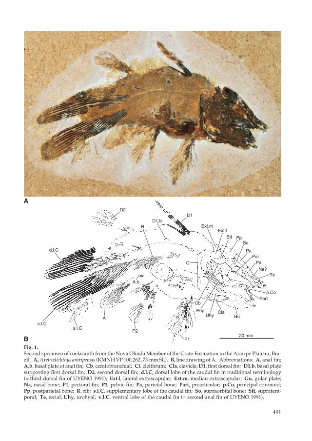
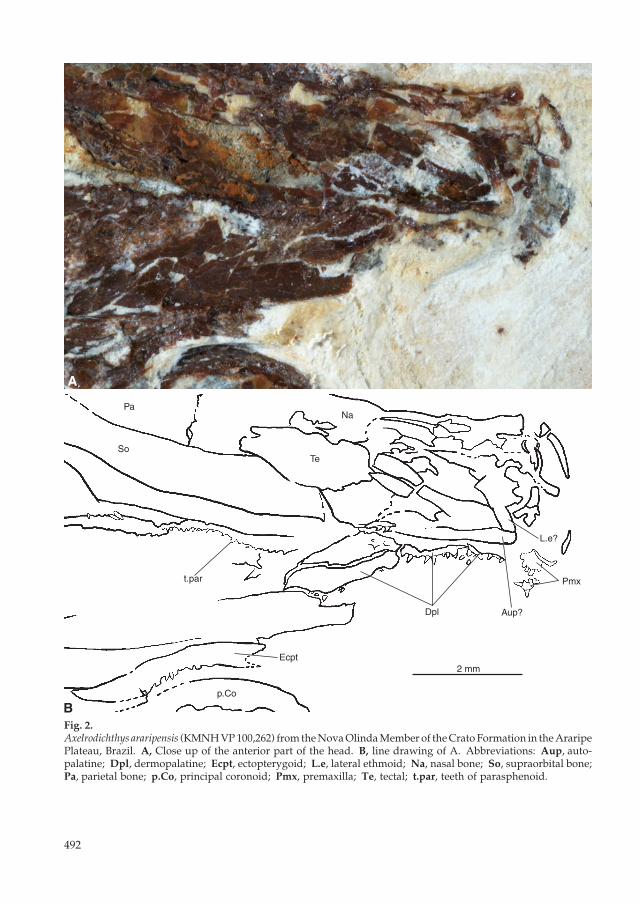
The new specimen (Fig. 1) is small, with an estimated total length of about 100 mm and an estimated length from the snout to the base of the caudal peduncle (standard length of Forey 1998: 298) of about 73 mm. Its head length is 25.6 mm. The body is bent in the middle; the body depth at the origin of the first dorsal fin is 21 mm. The parietonasal shield is about twice the length of the postparietal shield: the length of the parietonasal shield is 15.6 mm, whereas the estimated length of the postparietal shield is about 8 mm. Both parietals are narrow. Bones of the anterior part of the parietonasal shield are preserved, but the boundary of each bone is unclear. Supraorbitals are located alongside both parietal bones; the width of the supraorbitals is slightly nar-rower than that of the parietals (Fig. 1). The postparietal shield consists of a pair each of postparietals, supratemporals, and lateral extrascapu-lars, and a median extrascapular bone. The postparietal is largest and is slightly longer than the lateral extrascapular, which is almost the same size as the supratemporal. The median extrascapular (Fig. 1) is a small, square bone that is about one-third the size of the lateral extrascapular. The cheek bones are not well preserved. The postorbital, lachrymojugal and squamosal are missing. The preoperculum is preserved behind the quadrate. The surface of its ventral part has ridged ornamenta-tion. Both lower jaws are visible. Relatively large conical teeth (Fig. 2) present at the anterior end of the snout are probably premaxillary teeth. The lower jaw is deep, with the deepest point anterior to the midpoint of the lower jaw. The anterodorsal part of the angular is preserved, but the articular cannot be seen. The principal coronoid is a long, saddle-shaped bone, which articulates with the prearticular at its antero-ventral edge. Several conical teeth are preserved at the rostral or anterior end of the lower jaw (Fig. 3). The quadrate and pterygoid are well preserved. There is a distinct, wide strut along the posterior mar-gin of the pterygoid above the quadrate. The quadrate inclines caudally. The ventral end of the quadrate forms two condyles for the articulation of the lower jaw. The angle between the posterior margin of the quadrate and the ventral margin of the pterygoid is obtuse. Behind the premaxillary teeth, a flat bone, probably the autopalatine, is tightly articulated at its anterior end with a bone that is probably the lateral ethmoid (Fig. 2). The dermopalatines are located ventral to the autopalatine and have smaller teeth than the premaxilla. Two anterior dermopalatines can be seen. The ectopterygoid is located slightly apart from the posterior dermopalatine. The ectopterygoid teeth are almost the same size as those of the dermopalatines (Fig. 2). The metapterygoid is missing.
491
D1
Ext.lExt.m
Stt Pp
Pa
Na?
D2
Gu
Te
Pa
s.l.C
A
P2
P1
Cla
Cl
D1.bR
Cb
Uhy
So
Partp.Co
Pop
Par
A.b
d.l.C
v.l.C
20 mm
A
BFig. 1. Second specimen of coelacanth from the Nova Olinda Member of the Crato Formation in the Araripe Plateau, Bra-zil. A, Axelrodichthys araripensis (KMNH VP 100,262, 73 mm SL). B, line drawing of A. Abbreviations: A. anal fin; A.b, basal plate of anal fin; Cb, ceratobranchial; Cl, cleithrum; Cla, clavicle; D1, first dorsal fin; D1.b, basal plate supporting first dorsal fin; D2, second dorsal fin; d.l.C, dorsal lobe of the caudal fin in traditional terminology (= third dorsal fin of UYENO 1991); Ext.l, lateral extrascapular; Ext.m, median extrascapular; Gu, gular plate; Na, nasal bone; P1, pectoral fin; P2, pelvic fin; Pa, parietal bone; Part, prearticular; p.Co, principal coronoid; Pp, postparietal bone; R, rib; s.l.C, supplementary lobe of the caudal fin; So, supraorbital bone; Stt, supratem-poral; Te, tectal; Uhy, urohyal; v.l.C, ventral lobe of the caudal fin (= second anal fin of UYENO 1991).
492
Dpl
Ecpt
p.Co
Aup?
L.e?
Pmx
PaNa
t.par
TeSo
2 mm
B
A
Fig. 2.Axelrodichthys araripensis (KMNH VP 100,262) from the Nova Olinda Member of the Crato Formation in the Araripe Plateau, Brazil. A, Close up of the anterior part of the head. B, line drawing of A. Abbreviations: Aup, auto-palatine; Dpl, dermopalatine; Ecpt, ectopterygoid; L.e, lateral ethmoid; Na, nasal bone; So, supraorbital bone; Pa, parietal bone; p.Co, principal coronoid; Pmx, premaxilla; Te, tectal; t.par, teeth of parasphenoid.
493
The operculum is not preserved. Long ceratobranchials are preserved behind the preoperculum. A bone located ventral to the ceratobranchials is probably the urohyal, which broadens posteriorly (Fig. 1). This bone is not homologous to the tendon-bone urohyal found in teleosts (see ARRATIA & SCHULTZE 1990). The cleithrum is almost straight, but the ventral end bends slightly. The pectoral fin is about 20 mm long or about 27.4 % of the estimated standard length. The pelvic fin has 16 rays, each with a length of about 14 mm, or about 19.2 % of the estimated standard length. The pelvic and pectoral fin rays are segmented. The number of fin rays of the first dorsal fin is 10 and the length of the longest one is 19.4 mm, or about 26.6 % of the estimated standard length. About two-thirds of the distal region of first dorsal fin rays are segmented. The basal plate is not visible ventral to the dorsal fin rays, but a bone preserved between the first and the second dorsal fins is probably the basal plate. The second dorsal fin has 18 rays, which are 16.0 mm long, or about 21.9 % of the estimated standard length. The distal part of the fin rays is segmented. The basal plate of the second dorsal fin is not visible. The dorsal lobe of the caudal fin (= third dorsal fin of UYENO 1991) has 14 rays, and the fin rays are about 23 mm long, or about 31.5 % of the estimated standard length. There are 20 fin rays in the anal fin with a length of about 15 mm, or about 20.5 % of the estimated standard length. The anal fin rays are segmented almost entirely, except for a the short basal portion. The base of the anal fin consists of the slightly deep basal part and two cylindrical parts, which probably form the bifurcate anterior projections. The ventral lobe of the caudal fin (= second anal fin of UYENO 1991) has 14 fin rays that are almost of the same length as the fin rays of the dorsal lobe (Fig. 1). The supplementary lobe of the caudal fin is disarticulated, but a part of this structure is preserved under the ventral lobe. There are no ossified vertebral centra, indicating a persistent notochord. Most of the neural spines are not preserved, but their impressions are clearly seen. The anterior neural spines are ossified, extremely short, and gradually lengthen posteriad beginning at the mid point between the first and second dorsal fins. Neural spines articulating with ‘supraneural’ plus radial (ARRATIA et al. 2000) of the dorsal lobe of the caudal fin are long, and wide at the dorsal end. Most of the haemal spines are missing, but their impressions are visible. There are 14 haemal spines articulateds with 14 interhaemals plus ventral radials (ARRATIA et al. 2000) in the ventral lobe of the caudal fin. There are 27 very thin ribs, of which the first three are very short and the others gradually lengthen posteriad; 16 long ribs, which do not reach the ventral margin of the body, are located behind the stomach content.
Mm?
t.p
A
B 1 mm
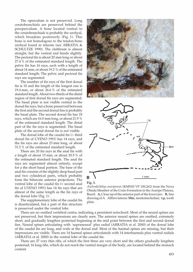
Fig. 3.Axelrodichthys araripensis (KMNH VP 100,262) from the Nova Olinda Member of the Crato Formation in the Araripe Plateau, Brazil. A, Close up of the anterior part of the lower jaw. B, line drawing of A. Abbreviations: Mm, mentomeckelian; t.p, tooth plate.
494
BA
C
1 mm1 mm 10 mm
10 mm10 mm
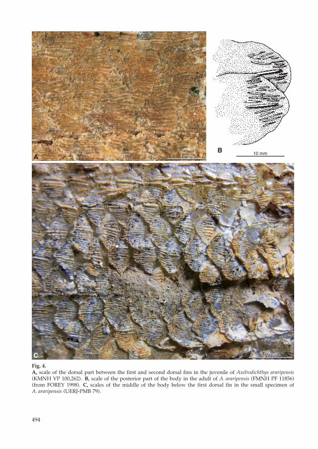
Fig. 4.A, scale of the dorsal part between the first and second dorsal fins in the juvenile of Axelrodichthys araripensis (KMNH VP 100,262). B, scale of the posterior part of the body in the adult of A. araripensis (FMNH PF 11856) (from FOREY 1998). C, scales of the middle of the body below the first dorsal fin in the small specimen of A. araripensis (UERJ-PMB 79).

495
Scales (Fig. 4A) are relatively large and exposed on the inner surface of the left side. Most of the scales show only embedded portions, but same scales show the exposed portions with minute tubercles. Many cylindrical fragments with several pores and a spiral-shaped lump are preserved in the abdo-men. These are probably stomach contents (Fig. 5). The calcified thin plates preserved above the stomach contents are probably a part of the calcified lung (Fig. 5). This organ, in adult specimens, consists of os-seous blades of variable thickness separated from each other, indicating that in the living individuals, these ossified plates were probably separated by connective tissue (BRITO et al. 2010).
Discussion
The new coelacanth specimen is almost complete, and is better preserved than the first smaller specimen described from the Crato Formation, although the supplementary or accessory lobe of the tail is partially disarticulated. It is a slightly larger specimen, with an estimated total length about 100 mm (the first one, with a total length of 70 mm, was identified as Axelrodichthys sp. by BRITO & MARTILL 1999). Based on the new specimen, we note two additional characteristics for the diagnosis of Axelrodicthys proposed by MAISEY (1986) and FOREY (1998). These are: (1) the concave dorsal margin of the parieto-nasal shield in lateral view and (2) the deepest point of the lower jaw located anteriorly, as the genus Axelrodicthys is clearly distinguished from its sister genus Mawsonia by these characteristics (MAISEY 1986: figs. 7, 18B, 26A; YABUMOTO 2002: fig. 3). We consider that both the new specimen and the first and smaller specimen represent juveniles of A. araripensis, due to the presence of the median extrascapular, the concave dorsal margins of the parietonasal shields, and the anterior position of the deepest point of the lower jaws (Fig. 1). Except for the two young individuals found in the Crato Formation, all other specimens of A. ararip-ensis occur in nodules from the Romualdo Member of the Santana Formation, which is slightly younger
Fig. 5.Stomach contents and calcified thin plates of the lung of Axelrodichthys araripensis (KMNH VP 100,262). Black arrows indicate the stomach contents. The white arrow indicates the calcified thin plates of the lung.
5 mm
496
than the Crato Formation. It is difficult to compare all the characters present in our specimen to adult specimens, since most of the larger specimens from the Santana Formation are incomplete, and especially because entire fin rays are not preserved. The ratio of the length of the parietonasal shield to that of the postparietal shield in the juvenile is almost the same as in adults, but the lower jaw is deeper and the head is larger than that of the adults (Table 1). The new juvenile specimen has conical teeth on the dermopalatine and ectoptery-goid (Fig. 2). These bones and teeth have
not previously been described in this species. The teeth at the anterior end of the lower jaws are needle-like in adults (FOREY 1998), but conical in the juvenile (Fig. 3). The quadrate is similar to that of adults, but it is inclined caudally, whereas it is almost vertical in adults. The angle between the posterior margin and the ventral margin of the pterygoid is obtuse, but it is acute in adults. The specimen described here has relatively long fin rays. The ratio of head length to estimated standard length is larger than that of adults. The long fin rays and the large head are interpreted here as juvenile characters of this species (Table 1). Surprisingly, the lung in this specimen (Fig. 5) has the calcified thin plates of some fossil coelacanths, rather than the phosphatic mass, probably formed by collagenic fibres during life, found in the 70 mm specimen and in others. The ornamentation of the scales of the juvenile is distinct from that of adults. The scales of the young specimen described here have minute tubercles on the exposed area (Fig. 4A). In contrast, in the adults of this species the scales are ornamented with short, irregularly spaced horizontal ridges (Fig. 4B) (see FOREY 1998: fig. 11.7). Some specimens of Axelrodichthys from the Santana Formation, considered as representing intermediate stages between juveniles and adults (cf. specimen UERJ-PMB 79) present a mixture of scale ornamentations, presenting both some scales with ornamentation as described here for the Crato specimen and other scales presenting the ornamentation as described by FOREY (1998) (Fig. 4C). Based on these observations, we suggest that the ornamentation of the scales changes from young to adult stages. Differences in the ornamentation in the scales, depending on the part of the body, were observed in other coelacanths such as Macropoma (see FOREY 1998); however, these differences are not ontogenetic. Ontogenetic changes in the ornamentation of scales are presently unknown in other fossil species and extant coelacanths and were not observed in the mawsoniid species Parnaibaia maranhaoensis where specimens of different sizes are known. Therefore, this pattern of ontogenetic variation should be sought in other species, especially in other mawsoniids. That the specimens of Axelrodichthys from the Crato Formation are juveniles, as well as the fact that juvenile specimens of other species also found in both the Santana and the Crato formations (e. g., Placidich-thys, Santanichthys, Cladocyclus) (LEAL & BRITO 2004; BRITO 2000, 2007) led us to suggest some paleoeco-logical implications relating to their reproductive biology. As BRITO (2007) mentioned, considering that the paleoenvironmental conditions of the Crato Formation are currently interpreted as lagoon-like with probable fluctuating salinities (MARTILL 1993), it is suggested that marine forms had entered the lagoon through one of the restricted links to the sea, perhaps for reproduction. The finding of young A. araripensis in the Crato Formation implies that this coelacanth gave birth in the lagoon and juveniles of this species remained there at least until they reached a total length of approximately 100 mm.
Acknowledgments
We are most grateful to Gloria ARRATIA for her encouragement and valuable assistance reviewing this manu-script. We also wish to express our sincere gratitude to David MARTILL for his comments and English editing of early drafts of the manuscript as well as the anonymous referees for their critical reading of this manuscript and their valuable comments. PMB’s research was supported by the CNPq and by a FAPERJ research grant.
Table 1.Some proportional measurements of Axelrodichthys araripensis. Abbreviations: CP, body depth at dorsal lobe of caudal fin origin (= 3rd dorsal of UYENO 1991); HD, depth of head; HL, head length; PD1, length from snout to 1st dorsal origin; PNL, length of parie-tonasal shield; PPL, length of postparietal shield; SL, estimated standard length; TD, body depth.
HL HD TD CP PD1 PNL/PPL/SL
MSPC-287 0.36 0.25 0.28 0.18 0.47 2.12KMNH VP 100,262 0.35 0.24 0.29 0.19 0.42 1.98AMNH 11756 0.29 0.20 0.29 0.25 0.37 1.99

497
References
AGASSIZ, L. (1841): On the fossil fishes found by Mr. Gardner in the province of Ceará, in north of Brazil. – Edinburgh New Philos. J. 30: 82-84.
ARRATIA, G. & SCHULTZE, H.-P. (1990): The urohyal: Development and homology within osteichthyans. – J. Morphol. 203: 247–282.
ARRATIA, G., SCHULTZE, H.-P. & CASCIOTTA, J. (2000): Vertebral column and associated elements in dipnoans and comparison with other fishes; development and homology. – J. Morphol. 250 (2): 101-172.
BRITO, P. M. (2000): A new halecomorph with two dorsal fins, Placidichthys bidorsalis n. g., n. sp. (Actinopterygii: Halecomorphi) from the Lower Cretaceous of the Araripe Basin, northeast Brazil. – C. R. Acad. Sci. Paris 331: 749-754.
– (2007): The Crato Formation fish fauna. – In: MARTILL, D. M., BECHLY, G. & LOVERIDGE, R. F. (eds.): The Crato Fossil Beds of Brazil: 429-443; Cambridge (Cambridge University Press)
BRITO, P. M. & MARTILL, D. M. (1999): Discovery of a juvenile coelacanth in the Lower Cretaceous, Crato Formation, Northeastern Brazil. – Cybium 23: 311-314.
BRITO, P. M., MEUNIER, F. J., CLÉMENT, G. & GEFFARD-KURIYAMA, D. (2010): The histological structure of the calcified lung of the fossil coelacanth Axelrodichthys araripensis (Actinistia: Mawsoniidae). – Palaeontol-ogy 53 (6): 1281-1290.
BRITO, P. M. & YABUMOTO, Y. (2011): An updated review of the fish faunas from the Crato and Santana for-mations in Brazil, a close relationship to Tethys fauna. – Bull. Kitakyushu Mus. Natur. Hist. Human. Hist., Ser. A 9: 107-136.
BRITO, P. M., YABUMOTO, Y. & GRANDE, L. (2008): New amiid fish (Halecomorphi; Amiiformes) from the Lower Cretaceous Crato Formation, Araripe Basin, Northeast Brazil. – J. Vert. Paleontol. 28: 1007-1014.
FOREY, P. L. (1998): History of the Coelacanth Fishes. – XIII + 419 pp.; London (Chapman and Hall).HUXLEY, T. H. (1861): Preliminary essay upon the systematic arrangement of the fishes of the Devonian epoc.
– Mem. Geol. Surv. U. K., London. Dec. 10: 1-49.JORDAN, D. S. (1910): Description of a collection of fishes from the bituminous shales of Riacho Doce, State of
Alagoas, Brazil. – Ann. Carnegie Mus. 7: 23-34.LEAL, M. E. C. & BRITO, P. M. (2004): The ichthyodectiform Cladocyclus gardneri (Actinopterygii: Teleostei)
from the Crato and Santana Formations, Lower Cretaceous of Araripe Basin, North-Eastern Brazil. – Ann. Paléontol. 90: 103-113.
MAISEY, J. G. (1986): Coelacanths from the Lower Cretaceous of Brazil. – Amer. Mus. Novitates 2866: 1-30.– (1991): Axelrodichthys Maisey, 1986. – In: MAISEY, J. G. (ed.): Santana Fossils: an Illustrated Atlas: 303-323;
Neptune City, New Jersey (T.F.H. Publications).MARTILL, D. M. (1993): Fossils of the Santana and Crato Formation, Brazil. – Palaeontological Association Field
Guides to Fossils 5: 159 pp.; London (The Palaeontological Association).MARTILL, D. M. & HEIMHOFER, U. (2007): Stratigraphy of the Crato Formation. – In: MARTILL, D. M.,
BECHLY, G. & LOVERIDGE, R. F. (eds.): The Crato Fossil Beds of Brazil: 25-43; Cambridge (Cambridge University Press).
SANTOS, R. da S. (1958): Leptolepis diasii, novo peixe fóssil da Serra do Araripe, Brasil. – Not. Prelim. Est., Div. Geol. Mineral., Dept, Nacl. Produção Mineral 108: 1-15.
– (1990): Vinctifer longirostris, do Cretáceo inferior da Formação Marizal, Estado da Bahia, Brasil. – An. Acad. Brasil. Ciênc. 62: 251-260.
SCHULTZE, H.-P. (1993): Osteichthyes: Sarcopterygii. – In: Benton, M. J. (ed.): The Fossil Record 2: 657-663; London (Chapman and Hall).
UYENO, T. (1991): Observations on locomotion and feeding of released coelacanths, Latimeria chalumnae. – En-vironm. Biol. Fish. 32: 267-273.
WOODWARD, A. S. (1907): On the Cretaceous formation of Bahia (Brazil), and on vertebrate fossils collected therein. II. The vertebrate fossils. – Quart. J. Geol. Soc. London 63: 131-139.
YABUMOTO, Y. (2000): A new coelacanth from the Early Cretaceous of Brazil (Sarcopterygii, Actinistia). – Pale-ontol. Res. 6: 343-350.
– (2002): A new coelacanth from the Early Cretaceous of Brazil (Sarcopterygii, Actinistia). – Paleontol. Res. 6: 343-350.
– (2008): A new Mesozoic coelacanth from Brazil (Sarcopterygii, Actinistia). – Paleontol. Res. 12: 329-343.
Authors’ addresses:Yoshitaka YABUMOTO, Department of Natural History, Kitakyushu Museum of Natural History and Human His-tory, 2-4-1 Higashida, Yahatahigashi-ku, Kitakyushu, Fukuoka, 805-0071, Japan; e-mail: [email protected] M. BRITO, Departamento de Zoologia, Universidade do Estado do Rio de Janeiro, rua São Francisco Xavier 524, Rio de Janeiro, 20559-900, Brazil; e-mail: [email protected]

498