Embed Size (px)
Citation preview
Mutation Research, 248 (1991) 371-374 © 1991 Elsevier Science Publishers B.V. 0027-5107/91/$03.50 ADONIS 0027510791001217
371
MUT 00095
The search for molecular mechanisms of non-genotoxic carcinogens
S. Green I CI Central Toxicology Laboratory, A lderley Park, Macclesfield, Cheshire SKIO 4TJ (U. K.)
(Accepted 25 October 1990)
Keywords: Non-genotoxic carcinogenesis; Molecular mechanisms
The mechanism(s) by which non-genotoxic carcinogens act are largely undetermined. This brief review discusses some of the many possible pathways that could be modulated by chemicals to result in tumour formation.
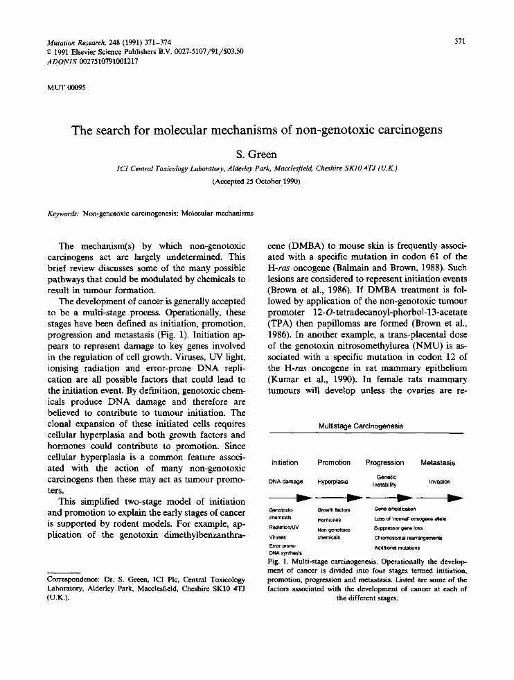
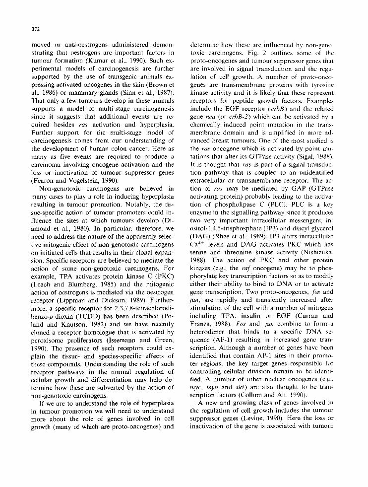
The development of cancer is generally accepted to be a multi-stage process. Operationally, these stages have been defined as initiation, promotion, progression and metastasis (Fig. 1). Initiation ap- pears to represent damage to key genes involved in the regulation of cell growth. Viruses, UV light, ionising radiation and error-prone DNA repli- cation are all possible factors that could lead to the initiation event. By definition, genotoxic chem- icals produce DNA damage and therefore are believed to contribute to tumour initiation. The clonal expansion of these initiated cells requires cellular hyperplasia and both growth factors and hormones could contribute to promotion. Since cellular hyperplasia is a common feature associ- ated with the action of many non-genotoxic carcinogens then these may act as tumour promo- ters.
This simplified two-stage model of initiation and promotion to explain the early stages of cancer is supported by rodent models. For example, ap- plication of the genotoxin dimethylbenzanthra-
Correspondence: Dr. S. Green, ICI Plc, Central Toxicology Laboratory, Alderley Park, Macclesfield, Cheshire SK10 4TJ (U.K.).
cene (DMBA) to mouse skin is frequently associ- ated with a specific mutation in codon 61 of the H-ras oncogene (Balmain and Brown, 1988). Such lesions are considered to represent initiation events (Brown et al., 1986). If DMBA treatment is fol- lowed by application of the non-genotoxic tumour promoter 12-O-tetradecanoyl-phorbol-13-acetate (TPA) then papillomas are formed (Brown et al., 1986). In another example, a trans-placental dose of the genotoxin nitrosomethylurea (NMU) is as- sociated with a specific mutation in codon 12 of the H-ras oncogene in rat mammary epithelium (Kumar et al., 1990). In female rats mammary tumours will develop unless the ovaries are re-
Multistage Carcinogenesis
Initiation Promotion Progression Metastasis
Genetic DNA damage Hyperplasia Instability Invasion
Genotoxi¢- Growth factors Gene am01ification
chemicals Hormones Loss of 'normal' oncogene allele
RadiatiorVUV Non-genotox ic- Suppressor gene loss
Viruses chemicals Chromosomal rearrangements
Error-pcone- Additional mutations DNA symhes~
Fig. 1. Multi-stage carcinogenesis. Operationally the develop- merit of cancer is divided into four stages termed initiation, promotion, progression and metastasis. Listed are some of the factors associated with the development of cancer at each of
the different stages.
372
moved or anti-oestrogens administered demon- strating that oestrogens are important factors in tumour formation (Kumar et al., 1990). Such ex- perimental models of carcinogenesis are further supported by the use of transgenic animals ex- pressing activated oncogenes in the skin (Brown et al., 1986) or mammary glands (Sinn et al., 1987). That only a few tumours develop in these animals supports a model of multi-stage carcinogenesis since it suggests that additional events are re- quired besides ras activation and hyperplasia. Further support for the multi-stage model of carcinogenesis comes from our understanding of the development of human colon cancer. Here as many as five events are required to produce a carcinoma involving oncogene activation and the loss or inactivation of tumour suppressor genes (Fearon and Vogelstein, 1990).
Non-genotoxic carcinogens are believed in many cases to play a role in inducing hyperplasia resulting in tumour promotion. Notably, the tis- sue-specific action of tumour promoters could in- fluence the sites at which tumours develop (Di- amond et al., 1980). In particular, therefore, we need to address the nature of the apparently selec- tive mitogenic effect of non-genotoxic carcinogens on initiated cells that results in their clonal expan- sion. Specific receptors are believed to mediate the action of some non-genotoxic carcinogens. For example, TPA activates protein kinase C (PKC) (Leach and Blumberg, 1985) and the mitogenic action of oestrogens is mediated via the oestrogen receptor (Lippman and Dickson, 1989). Further- more, a specific receptor for 2,3,7,8-tetrachlorodi- benzo-p-dioxin (TCDD) has been described (Po- land and Knutson, 1982) and we have recently cloned a receptor homologue that is activated by peroxisome proliferators (Issemann and Green, 1990). The presence of such receptors could ex- plain the tissue- and species-specific effects of these compounds. Understanding the role of such receptor pathways in the normal regulation of cellular growth and differentiation may help de- termine how these are subverted by the action of non-genotoxic carcinogens.
If we are to understand the role of hyperplasia in tumour promotion we will need to understand more about the role of genes involved in cell growth (many of which are proto-oncogenes) and
determine how these are influenced by non-geno- toxic carcinogens. Fig. 2 outlines some of the proto-oncogenes and turnout suppressor genes that are involved in signal transduction and the regu- lation of cell growth. A number of proto-onco- genes are transmembrane proteins with tyrosine kinase activity and it is likely that these represent receptors for peptide growth factors. Examples include the EGF receptor (erbB) and the related gene neu (or erbB-2) which can be activated by a chemically induced point mutation in the trans- membrane domain and is amplified in more ad- vanced breast tumours. One of the most studied is the ras oncogene which is activated by point mu- tations that alter its GTPase activity (Sigal, 1988). It is thought that ras is part of a signal transduc- tion pathway that is coupled to an unidentified extracellular or transmembrane receptor. The ac- tion of ras may be mediated by GAP (GTPase activating protein) probably leading to the activa- tion of phospholipase C (PLC). PLC is a key enzyme in the signalling pathway since it produces two very important intracellular messengers, in- ositol-l,4,5-trisphosphate (IP3) and diacyl glycerol (DAG) (Rhee et al., 1989). 1P3 alters intracellular Ca 2+ levels and DAG activates PKC which has serine and threonine kinase activity (Nishizuka, 1988). The action of PKC and other protein kinases (e.g., the raf oncogene) may be to phos- phorylate key transcription factors so as to modify either their ability to bind to DNA or to activate gene transcription. Two proto-oncogenes, los and jun, are rapidly and transiently increased after stimulation of the cell with a number of mitogens including TPA, insulin or EGF (Curran and Franza, 1988). Fos and jun combine to form a heterodimer that binds to a specific DNA se- quence (AP-1) resulting in increased gene tran- scription. Although a number of genes have been identified that contain AP-1 sites in their promo- ter regions, the key target genes responsible for controlling cellular division remain to be identi- fied. A number of other nuclear oncogenes (e.g., myc, myb and ski) are also thought to be tran- scription factors (Collum and Alt, 1990).
A new and growing class of genes involved in the regulation of cell growth includes the tumour suppressor genes (Levine, 1990). Here the loss or inactivation of the gene is associated with tumour
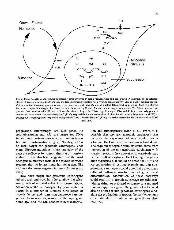
Growth Factors Hormones
"5 Autocrine
p- neu
erbB
ras PLC
PIP2 IP3+DAG
[c~ "+ ]
raf
TPA
PKC
fos myc "NN / M i t o g e n i c
jun myb ski ' ~ / Stimulus
Tag _ p53 - - E1B / T Z f - Suppression I erbA
J / Rb - - E1A
373
Fig. 2. Proto-oncogenes and tumour suppressor genes involved in signal transduction and cell growth. A selection of the different classes of gene are shown. ErbB and neu are transmembrane receptors with tyrosine kinase activity. Ras is a GTP-binding protein. Raf is a serine/threonine protein kinase. Fos, jun, myc, myb and ski are all nuclear DNA-binding proteins. ErbA is a thyroid hormone receptor homologue that does not bind hormone, p53 and Rb are tumour suppressor genes. The DNA turnout viral proteins that associate with Rb and p53 are also shown. Tag is the SV40 large T antigen. EIA and E1B are two early genes of adenovirus. Also shown are phospholipase C (PLC), responsible for the conversion of phosphatidyl inositol bisphosphate (PIP2) to inositol-l,4,5-trisphosphate (IP3) and diacyl glycerol (DAG). Protein kinase C (PKC) is a serine/threonine kinase activated by DAG
and TPA.
progression. Interestingly, two such genes, R b
(retinoblastoma) and p 5 3 , are targets for D N A tumour viral proteins associated with immortalisa- tion and transformation (Fig. 2). Notably, p 5 3 is an ideal target for genotoxic carcinogens since many different mutations in just one copy of the gene are sufficient for immortalisation or transfor- mation. It has also been suggested that the erbA
oncogene (a modified form of the thyroid hormone receptor that no longer binds hormone) acts, like p 5 3 , in a dominant negative fashion (Datum et al., 1989).
How then might non-genotoxic carcinogens subvert such pathways in order to allow the selec- tive growth of initiated cells? As discussed above, activation of the ras oncogene by point mutation occurs in a number of tumours. One action of growth factors and some non-genotoxic carcino- gens is to increase expression of the m y c gene. Since m y c and ras can cooperate in transforma-
tion and tumorigenesis (Sinn et al., 1987), it is possible that any non-genotoxic carcinogen that increases the expression of m y c would have a selective effect on cells that contain activated ras.
The required mitogenic stimulus could come from interaction of the non-genotoxic carcinogen with specific receptors (see above) or alternatively may be the result of a cytoxic effect leading to regener- ative hyperplasia. It should be noted that m y c and ras cooperation is just one example and that non- genotoxic carcinogens could potentially alter many different pathways involved in cell growth and differentiation. Modulation of these pathways could result in a growth advantage for cells con- taining either an activated oncogene or a mutated turnout suppressor gene. The growth of cells could also be altered if non-genotoxic carcinogens mod- ulate the production of growth factors (which may either stimulate or inhibit cell growth) or their receptors.
374
Non-genotoxic carcinogens could also alter cel- lular differentiation. For example the action of erbA is thought to block the differentiation of chicken erythroblasts thereby potentiating the ac- tion of erbB. Although the oncogenic form of erbA does not bind thyroid hormone, other mem- bers of this steroid/thyroid hormone receptor family can act in the presence of the appropriate ligand to promote differentiation.
Since normal cells surrounding those that con- tain transforming oncogenes are able to suppress the transformed phenotype it has been suggested that cell-cell communication via gap junctions is an important factor in suppressing tumorigenesis. Support for a role of gap junctions comes from studies showing that tumour promoters such as TPA and phenobarbitone reduce cell-cell com- munication (Yamasaki et al., 1988). Possibly other non-genotoxic carcinogens could also influence gap junctions.
Evidently there are many mechanisms by which non-genotoxic carcinogens could influence tumour development. If we are to dissect these mecha- nisms we need a good working model. The use of a receptor-mediated system may help determine the molecular mechanisms involved in the action of some non-genotoxic carcinogens and provide clues to which indicators could be used in the future to evaluate the carcinogenic potential of this important class of chemicals.
References
Balmain, A., and K. Brown (1988) Oncogene activation in chemical carcinogenesis. Adv. Cancer Res., 51,147-182.
Brown, K., M. Quintanilla, M. Ramsden, I.B. Kerr, S. Young and A. Balmain (1986) V-ras genes from Harvey and BALB murine sarcoma viruses can act as initiators of two-stage mouse skin carcinogenesis. Cell, 46, 447-456.
Collum, R.G., and F.W. Alt (1990) Are myc proteins transcrip- tion factors? Cancer Cells, 2, 69-75.
Curran, T., and B.R. Franza (1988) Fos and Jun: the AP.-1 connection, Cell, 55, 395-397.
Damm, K., C,C. Thompson and R.M. Evans (1989) Protein encoded by v-erbA functions as a thyroid-hormone recep- tor antagonist. Nature, 339, 593-597.
Diamond, L., T.G. O'Brien and W.M. Baird (1980) Tumour promoters and the mechanism of tumour promotion. Adv. Cancer Res., 32, 1-74.
Fearon, E.R., and B. Vogelstein (1990) A genetic model for colorectal tumorigenesis. Cell, 61,759-767.
Issemann, I., and S. Green, (1990) Activation of a member of the steroid hormone receptor superfamily by peroxisome proliferators. Nature, 347, 645-650.
Kumar, R., S. Sukumar and M. Barbacid (1990) Activation of ras oncogenes preceding the onset of neoplasia. Science, 248, 1101-1104.
Leach, K.L., and P.M. Blurnberg (1985) Modulation of protein kinase C activity and (3H) phorbol 12,13-dibutyrate bind- ing by various tumour promoters in mouse brain cytosol. Cancer Res., 45, 1958-1963.
Levine, A.J. (1990) Tumour suppressor genes. BioEssays, 12, 60-66.
Lippman, M.E., and R.B. Dickson (1989) Mechanisms of nor- mal and malignant breast epithelial growth regulation. J. Steroid Biochem., 34, 1-6.
Nishizuka, Y. (1988) The molecular heterogeneity of protein kinase C and its implications for cellular regulation. Na- ture, 334, 661-665.
Poland, A., and J.C, Knutson (1982) 2,3,7,8-Tetrachlorodi- benzo-p-dioxin and related halogenated aromatic hydro- carbons: examination of the mechanism of toxicity. Annu. Rev. Pharmacol. Toxicol., 22, 517-554.
Rhee, S.G., P.G. Suh, S.H. Ryu and S.Y. Lee (1989) Studies of inositol phospholipid-specific phospholipase C. Science. 244, 546-550.
Sigal, I.S. (1988) The ras oncogene: a structure and some function. Nature, 332, 485-486.
Sinn, E., W. Muller, P. Pattengale, I. Tepler, R. Wallace and Leder, P. (1987) Coexpression of MMTV/v-Ha-ras and MMTV/c-myc genes in transgenic mice: synergistic action of oncogenes in vivo. Cell, 49, 465-475.
Yamasaki, H., K. Enomoto, D.J. Fitzgerald, M. Mesnil, F. Katch and M. Hollstein (1988) Role of intercellular com- munication in the control of critical gene expression during multistage carcinogenesis. IARC Sci. Publ., 92, 57-75.


![Validation and development of an electrodeposition process ...970466/...Cr(III) is the most common. Cr(VI) compounds are genotoxic carcinogens [3]. CrO 3, which is a Cr(VI) compound,](https://img.pdfslide.us/doc/110x75/60e8111e4b8b9d41bb24c0dd/validation-and-development-of-an-electrodeposition-process-970466-criii.jpg)