Embed Size (px)
Citation preview

Developmental Biology 350 (2011) 255–266
Contents lists available at ScienceDirect
Developmental Biology
j ourna l homepage: www.e lsev ie r.com/deve lopmenta lb io logy
The Salvador/Warts/Hippo pathway controls regenerative tissue growth inDrosophila melanogaster
Felix A. Grusche a,b,c, Joffrey L. Degoutin a, Helena E. Richardson b,c,d, Kieran F. Harvey a,e,⁎a Cell Growth and Proliferation Laboratory, Peter MacCallum Cancer Centre, St Andrews Place, East Melbourne, Victoria 3002, Australiab Cell Cycle and Development Laboratory, Peter MacCallum Cancer Centre, St Andrews Place, East Melbourne, Victoria 3002, Australiac Department of Anatomy and Cell Biology, University of Melbourne, Parkville, Victoria 3010, Australiad Department of Biochemistry and Molecular Biology, University of Melbourne, Parkville, Victoria 3010, Australiae Department of Pathology, University of Melbourne, Parkville, Victoria 3010, Australia
⁎ Corresponding author. Cell Growth and ProliferationCancer Centre, St Andrews Place, East Melbourne, Victo9656 1411.
E-mail address: [email protected] (K.F. H
0012-1606/$ – see front matter © 2010 Elsevier Inc. Aldoi:10.1016/j.ydbio.2010.11.020
a b s t r a c t
a r t i c l e i n f oArticle history:Received for publication 20 July 2010Revised 12 November 2010Accepted 13 November 2010Available online 25 November 2010
Keywords:RegenerationHippo pathwayYorkieOrgan size control
During tissue regeneration, cell proliferation replaces missing structures to restore organ function.Regenerative potential differs greatly between organs and organisms; for example some amphibians canregrow entire limbs whereas mammals cannot. The process of regeneration relies on several signalingpathways that control developmental tissue growth, and implies the existence of organ size-controlcheckpoints that regulate both developmental, and regenerative, growth. Herewe explore the role of one suchcheckpoint, the Salvador–Warts–Hippo pathway, in tissue regeneration. The Salvador–Warts–Hippo pathwaylimits tissue growth by repressing the Yorkie transcriptional co-activator. Several proteins serve as upstreammodulators of this pathway including the atypical cadherins, Dachsous and Fat, whilst the atypical myosin,Dachs, functions downstream of Fat to activate Yorkie. Using Drosophila melanogaster imaginal discs we showthat Salvador–Warts–Hippo pathway activity is repressed in regenerating tissue and that Yorkie is rate-limiting for regeneration of the developing wing. We show that regeneration is compromised in dachsmutantwing discs, but that proteins in addition to Fat and Dachs are likely to modulate Yorkie activity in regeneratingcells. In conclusion our data reveal the importance of Yorkie hyperactivation for tissue regeneration andsuggest that multiple upstream inputs, including Fat–Dachsous signaling, sense tissue damage and regulateYorkie activity during regeneration of epithelial tissues.
Laboratory, Peter MacCallumria 3002, Australia. Fax: +61 3
arvey).
l rights reserved.
© 2010 Elsevier Inc. All rights reserved.
Introduction
Regeneration is a process whereby an organism can stimulatetissue growth to replace damaged body parts and re-establish tissuehomeostasis. The ability to regenerate varies greatly in the animalkingdom and also between different organs of the same species. Forexample planarians can regrow completely from a small bodyremnant, some crustaceans and amphibians can re-grow entirelimbs, zebrafish can regenerate hearts and fins, whereas mammalshave relatively poor regenerative capacity [reviewed in (Birnbaumand Sanchez Alvarado, 2008)]. Somemammalian organs, like the liver,can regrow whereas most other organs cannot. Drosophila melanoga-ster imaginal discs, the presumptive adult organs, were shown topossess regenerative capacity several decades ago (Hadorn, 1963).They can regenerate during a specific window of larval developmentin response to either surgically- or genetically-induced wounds(Hadorn, 1963; Smith-Bolton et al., 2009). Several growth-controlling
proteins and signaling pathways have been shown to regulate tissuegrowth not only during development, but also during regeneration;for example fibroblast growth factor in the zebrafish fin (Poss et al.,2000); the Wnt, transforming growth factor β and Hedgehogsignaling pathways in planarians (Forsthoefel and Newmark, 2009;Rink et al., 2009; Yazawa et al., 2009); and Wingless, Myc and the JunN-terminal kinase (JNK) pathway in D. melanogaster imaginal discs(Bosch et al., 2005; Mattila et al., 2005; Smith-Bolton et al., 2009).
The Salvador–Warts–Hippo (SWH) signaling pathway has recentlyemerged as a key modulator of tissue growth during development byregulating key proteins involved in cell proliferation, Cyclin E, andsurvival, DIAP1 (Tapon et al., 2002). Defective SWH pathway signalinghas been associated with a loss of tissue growth control in cancer(Harvey and Tapon, 2007). At the core of the SWH pathway are twoserine/threonine kinases, Warts (Wts) and Hippo (Hpo), and twoadaptor proteins, Salvador (Sav) and Mob as tumour suppressor(Mats) [reviewed in (Harvey and Tapon, 2007)]. These proteinsrestrict tissue growth by stimulating Wts to phosphorylate, andthereby repress, the Yorkie (Yki) transcriptional co-activator protein(Huang et al., 2005). The core SWH pathway proteins are regulated byinputs from several upstream regulatory branches, including the Fat–Dachsous (Ft–Ds) branch, the Kibra, Expanded, Merlin (KEM)

256 F.A. Grusche et al. / Developmental Biology 350 (2011) 255–266
complex, and the apicobasal polarity proteins, Lethal Giant Larvae(Lgl), Crumbs (Crb) and atypical Protein Kinase C (aPKC) [reviewed in(Grusche et al., 2010)]. The Ft–Ds branch of the SWH pathwaycontrols Yki activity by repressing activity of the atypical myosin,Dachs, which in turn is thought to inhibit Warts activity (Cho et al.,2006). Given that the SWH pathway is a key regulator of tissue growthin both insects and mammals during development, and of tissue sizehomeostasis in adult mice (Camargo et al., 2007; Dong et al., 2007),we postulated that the SWH pathway could also control regenerativetissue growth based on the following logic: SWH pathway activity isrepressed in mammalian cultured cells on the edge of physically-induced wounds in vitro (Zhao et al., 2007); SWH pathway activityregulates cell proliferation in a cell-density dependent manner(McPherson et al., 2004); the SWH pathway represses cell prolifer-ation, at least in part, by interactions between the Ft and Ds cadherinson neighbouring cells (Rogulja et al., 2008; Willecke et al., 2008), andtherefore we envisaged that these interactions might be abrogated inwounded tissue and this would cause pathway derepression.
To test this hypothesis, we induced tissue ablation in developing D.melanogaster imaginal discs and analysed SWH pathway activity. Wefound that Yki activity was elevated in tissue that was proliferatingpost wounding due to de-repression by Wts, and that Yki was rate-limiting for wing regeneration. In addition, the Ft–Ds upstreamsignaling branch of the SWH pathway was partially required forcontrolling this regenerative response.
Materials and methods
Immunohistochemistry
Imaginal discs were prepared as previously described (Gruscheet al., 2009; Harvey et al., 2003). The following antibodies were used:mouse anti-beta Galactosidase (1:200; Sigma #G-4664), rabbit anti-cleaved Caspase 3 (1:400; Cell Signaling Technologies #9661S),mouse anti-Cyclin E (1:50) (Richardson et al., 1995), rabbit anti-Yki(1:400; Oh and Irvine, 2008), rabbit anti-phospho-Histone H3 (1:400;Cell Signaling Technology), goat anti-mouse_555 and goat anti-rabbit_647 (1:600; Molecular Probes, Invitrogen). Images wererecorded on an Olympus FV-1000 confocal microscope and processedusing Adobe Photoshop CS2.
D. melanogaster strains
w1118flies were used as a wild-type control. Other stocks used
were: hydK3.5 (Lee et al., 2002), vps25PB2931 (Thompson et al., 2005),linesP14A (K. Harvey, unpublished) ept2 (Moberg et al., 2005), scrib1
(Bilder and Perrimon, 2000), diap1-lacZ (Hay et al., 1995), dachsGC13,dachs1 (Mao et al., 2006), ykiB5 (Huang et al., 2005), exMGH1 (Pellocket al., 2007) exe1 and ex697 (Boedigheimer and Laughon, 1993). TheUAS–Gal4 system (Brand and Perrimon, 1993) and Gal80ts (McGuireet al., 2003) were used to allow temporal expression of transgenes:rotund-Gal4, engrailed-Gal4, (Bloomington), puckered-Gal4 (Pastor-Pareja et al., 2004), UAS–GFP (Bloomington), UAS-myc–wts (Fengand Irvine, 2007), UAS-eiger, UAS-rpr (Smith-Bolton et al., 2009),UAS-Debcl (Colussi et al., 2000).
D. melanogaster genotypes by figure panels:
Figs. 1A–C, 2E, 7H, S1: y, w, hsFlp/+; ex697/+; FRT 82B, Ubi-GFP/FRT82B, hydK3.5
Fig. 2A: y, w, hsFlp/+; ex697/+; Ubi-GFP, FRT 80B/ept2, FRT80BFigs. 2B, 7F: y, w, hsFlp/+; ex697/+; FRT 82B, Ubi-GFP/FRT82B, scrib1
Fig. 2C: y, w, hsFlp/+; FRT 42D, Ubi-GFP/FRT42D, linesP14A ; thj5c8/+Fig. 2D: y, w, hsFlp/+; FRT 42D, Ubi-GFP/FRT42D, vps25PB2931; thj5c8/+Figs. 3A–D, 4A–C, S2: w; enGal4, UAS–GFP/ex697; tub-Gal80ts/UAS-DebclFig. 3E: w; UAS–GFP/+; rn-Gal4, tub-Gal80ts, UAS-eiger/+
Figs. 4D, 7J: w; ex697/+; rn-Gal4, tub-Gal80ts, UAS-eiger/+Figs. 4E–F, 5A: w;; rn-Gal4, tub-Gal80ts, UAS-eiger/+Fig. 5B: w; UAS-myc–wts/+; rn-Gal4, tub-Gal80ts, UAS-eiger/+Figs. 6A, C: w; enGal4, UAS–GFP/FRT42D; tub-Gal80ts/UAS-DebclFigs. 6B, D: w; enGal4, UAS–GFP/FRT42D, ykiB5; tub-G80ts/UAS-DebclFigs. 7A, B: wFigs. 7C, D: w; dachs1/GC13
Fig. 7G: y, w, hsFlp/+; dachsGC13, ex697, FRT40A/dachs1; FRT 82B,Ubi-GFP/FRT82B, scrib1
Fig. 7I: y, w, hsFlp/+; dachsGC13, ex697, FRT40A/dachs1; FRT 82B,Ubi-GFP/FRT82B, hydK3.5
Fig. 7K: w; dachsGC13, ex697, FRT40A/dachsGC13, FRT40A; rn-Gal4,tub-Gal80ts, UAS-eiger/+Fig. 8A: w; exe1, FRT40A/+; rn-Gal4, tub-Gal80ts, UAS-eiger/+Fig. 8B: w; ftfd, exe1, FRT40A/FRT40A, ftfd; rn-Gal4, tub-Gal80ts,UAS-eiger/+Fig. 8C: w; exe1, FRT40A/FRT40A, exMGH1; rn-Gal4, tub-Gal80ts,UAS-eiger/+Fig. S3: w; ex697/UAS–GFP; puc-G4/+.
Temporal expression of transgenes
To monitor phospho-Histone H3, cleaved Caspase 3, ex-lacZ andYki in en-Gal4; UAS-Debcl experiments, larvae were raised at thepermissive temperature (either 18 °C or 25 °C), then shifted to therestrictive temperature (29 °C) for 28 h on day 4 AEL (+/−12 h). Tomonitor ex-lacZ, Yki and GFP in rn-Gal4, UAS-eiger experiments, larvaewere raised at the permissive temperature (18 °C), then shifted to therestrictive temperature (27 °C) for 22 h on day 7 AEL (+/−12 h).
Wing size measurements
For yki heterozygosity experiments, eggs were collected on grapeplates for 6 h. Equal numbers of larvae (140) were transferred intofood vials. Vials were shifted from 25 °C to the restrictive temperatureof 29 °C at 96 h (+/−3 h) AEL for 21 h. For irradiation experiments,w1118 or dachsGC13/1 larvae were raised at RT and irradiatedwith 40 Gy γ-irradiation at day 7 (+/−12 h) AEL. Wings of 1- to 2-day-old female adults were mounted in Canada Balsam (Sigma,#C1795) and imaged on an Olympus BX-51 microscope. Wing sizewas quantitated using Adobe Photoshop CS3. The graphs displayaverage wing size, whilst error bars represent standard error of themean.
Analysis of Yorkie localization
High resolution cross-sections (x–z) were taken through en-Gal4;UAS-Debcl discs by confocal microscopy. Average staining intensity forYki in the nucleus of cells in both the anterior and posteriorcompartments was determined using Metamorph 7.6.3 software.Nuclei were determined by DAPI staining, anterior and posteriorcompartments were discerned by the presence or absence of GFP,respectively.
Immunoblotting
Between 18 and 22 imaginal discs were dissected from third instarlarvae of the appropriate genotypes, lysed in protein sample bufferand subjected to SDS-PAGE. Gels were transferred to PVDF andimmunoblotted with either rabbit anti-Yki (KH, unpublished), rabbitanti-Yki-phospho-S168 (Dong et al., 2007). Quantitation of immuno-blots was performed using Image J software.

Fig. 1. SWH pathway activity is regulated in a dynamic fashion around sites of genetically-induced tissue damage. hydK3.5 mutant clones, marked by absence of GFP (green), atdifferent stages of clone development. ex-lacZ (ex-Z), expression is shown in grey in single channels and in red in merged images. hyd clones are shown 32 h (A), 64 h (B) and 92 h(C) after induction. Scale bar=20 μm.
257F.A. Grusche et al. / Developmental Biology 350 (2011) 255–266
Statistical analysis
Statistical analysis in Fig. 6E was performed using Microsoft Excel2004 for Mac. The graph displays average wing size, whilst error barsrepresent standard error of the mean. Statistical significance wascalculated with a Student T test, two sample assuming unequalvariance, one-tailed.
Transplantation experiments
Wing discs of third instar larvae, 100–120 h AEL raised at 25 °Cwere dissected in Schneider's medium at room temperature,wounded with a sharpened Tungsten thread and either directlyfixed and stained for control experiments or transplanted into theabdomen of adult hosts using the method described by (Ursprung,1967). After 25 h of culture in vivo, discs were recovered, fixed andstained for further analysis.
Results
SWH pathway activity is repressed in cells that neighbour wounds
We hypothesized that if the SWH pathway is important forregeneration, it would be inactivated in regenerating cells, leading toactivation of Yki, a key growth-promoting component of the pathway.To test this hypothesis, we generated genetic wounds in developingimaginal discs ofD.melanogaster. Specifically, we employed the FLP/FRTtechnique to generate clones of mutant cells in a heterozygous tissue(Xu and Rubin, 1993). Clones of cells harbouring a range of mutationsrequired for cell viability often survive well for approximately two daysafter clone induction, possibly due to the perdurance of wild typeprotein, but disappear from the epithelium approximately three daysafter clone induction [for example see (Grusche et al., 2009)].
To visualise SWHpathway activity during this process we analysedthe expression of a well-characterized downstream target of the
258 F.A. Grusche et al. / Developmental Biology 350 (2011) 255–266
pathway, expanded (ex) using an enhancer trap in the ex locus (ex697),in tissuemosaic for amutation in the hyperplastic discs (hyd) gene. hydencodes for a HECT-domain E3 ubiquitin ligase (Lee et al., 2002), and
clones of hyd tissue die approximately 2–3 days post generation inlarval wing imaginal discs and are removed from the epithelium(Fig. 1). During the early and late stage of hyd mutant clonedevelopment, ex expression was observed at a basal level (Figs. 1Aand C). Strikingly, ex transcription was strongly upregulated in wildtype cells that surrounded mutant clones at the time when hyd cloneswere being lost from the tissue (64 h post clone induction),suggesting that the SWH pathway was specifically suppressed incells juxtaposing wound sites, to allow for activation of Yki (Fig. 1B).Furthermore, the observed increase in Yki activity corresponded withapoptosis of cells within the hyd clone, as determined by activatedCaspase 3 staining (Fig. S1).
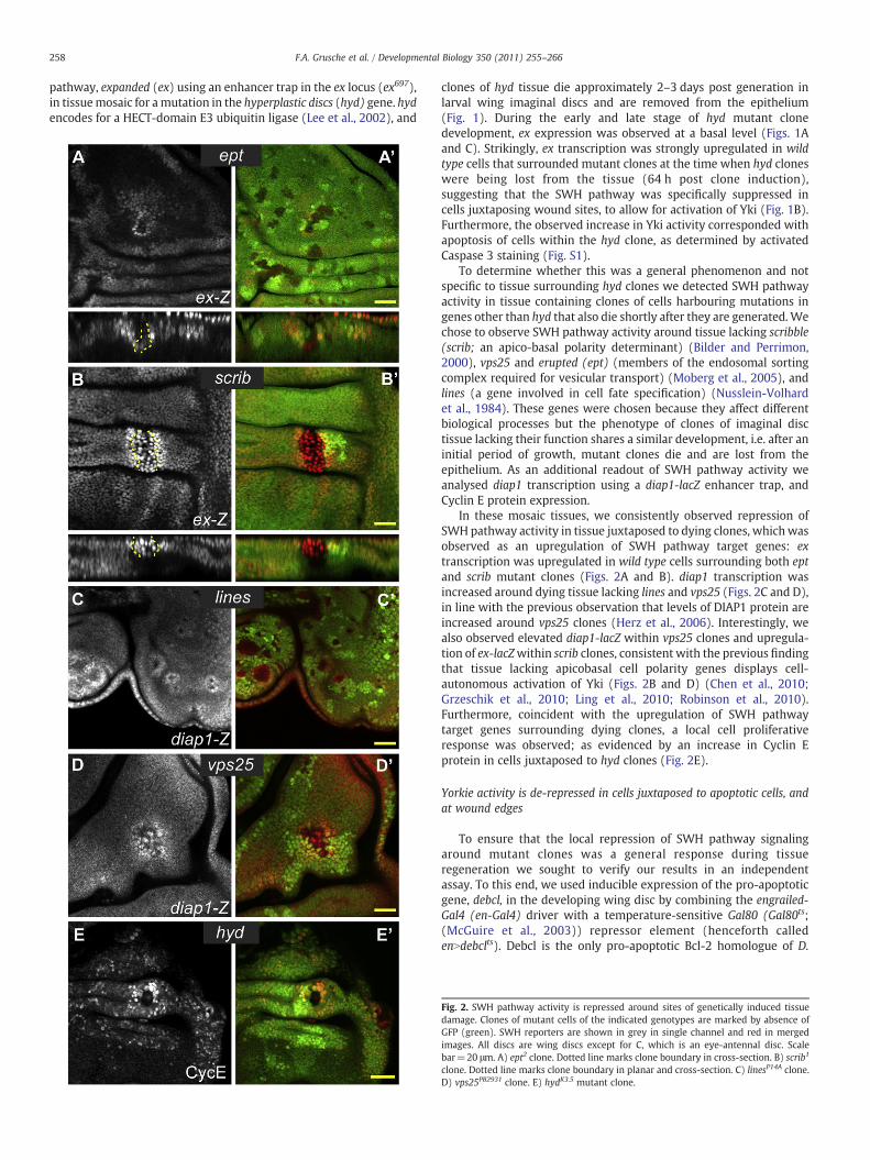
To determine whether this was a general phenomenon and notspecific to tissue surrounding hyd clones we detected SWH pathwayactivity in tissue containing clones of cells harbouring mutations ingenes other than hyd that also die shortly after they are generated.Wechose to observe SWH pathway activity around tissue lacking scribble(scrib; an apico-basal polarity determinant) (Bilder and Perrimon,2000), vps25 and erupted (ept) (members of the endosomal sortingcomplex required for vesicular transport) (Moberg et al., 2005), andlines (a gene involved in cell fate specification) (Nusslein-Volhardet al., 1984). These genes were chosen because they affect differentbiological processes but the phenotype of clones of imaginal disctissue lacking their function shares a similar development, i.e. after aninitial period of growth, mutant clones die and are lost from theepithelium. As an additional readout of SWH pathway activity weanalysed diap1 transcription using a diap1-lacZ enhancer trap, andCyclin E protein expression.
In these mosaic tissues, we consistently observed repression ofSWH pathway activity in tissue juxtaposed to dying clones, whichwasobserved as an upregulation of SWH pathway target genes: extranscription was upregulated in wild type cells surrounding both eptand scrib mutant clones (Figs. 2A and B). diap1 transcription wasincreased around dying tissue lacking lines and vps25 (Figs. 2C and D),in line with the previous observation that levels of DIAP1 protein areincreased around vps25 clones (Herz et al., 2006). Interestingly, wealso observed elevated diap1-lacZ within vps25 clones and upregula-tion of ex-lacZwithin scrib clones, consistent with the previous findingthat tissue lacking apicobasal cell polarity genes displays cell-autonomous activation of Yki (Figs. 2B and D) (Chen et al., 2010;Grzeschik et al., 2010; Ling et al., 2010; Robinson et al., 2010).Furthermore, coincident with the upregulation of SWH pathwaytarget genes surrounding dying clones, a local cell proliferativeresponse was observed; as evidenced by an increase in Cyclin Eprotein in cells juxtaposed to hyd clones (Fig. 2E).
Yorkie activity is de-repressed in cells juxtaposed to apoptotic cells, andat wound edges
To ensure that the local repression of SWH pathway signalingaround mutant clones was a general response during tissueregeneration we sought to verify our results in an independentassay. To this end, we used inducible expression of the pro-apoptoticgene, debcl, in the developing wing disc by combining the engrailed-Gal4 (en-Gal4) driver with a temperature-sensitive Gal80 (Gal80ts;(McGuire et al., 2003)) repressor element (henceforth calledenNdebclts). Debcl is the only pro-apoptotic Bcl-2 homologue of D.
Fig. 2. SWH pathway activity is repressed around sites of genetically induced tissuedamage. Clones of mutant cells of the indicated genotypes are marked by absence ofGFP (green). SWH reporters are shown in grey in single channel and red in mergedimages. All discs are wing discs except for C, which is an eye-antennal disc. Scalebar=20 μm. A) ept2 clone. Dotted line marks clone boundary in cross-section. B) scrib1
clone. Dotted line marks clone boundary in planar and cross-section. C) linesP14A clone.D) vps25PB2931 clone. E) hydK3.5 mutant clone.
259F.A. Grusche et al. / Developmental Biology 350 (2011) 255–266
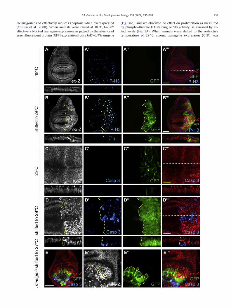
melanogaster and effectively induces apoptosis when overexpressed(Colussi et al., 2000). When animals were raised at 18 °C, Gal80ts
effectively blocked transgene expression, as judged by the absence ofgreen fluorescent protein (GFP) expression from a UAS–GFP transgene
(Fig. 3A″), and we observed no effect on proliferation as measuredby phospho-Histone H3 staining or Yki activity, as assessed by ex-lacZ levels (Fig. 3A). When animals were shifted to the restrictivetemperature of 29 °C, strong transgene expression (GFP) was
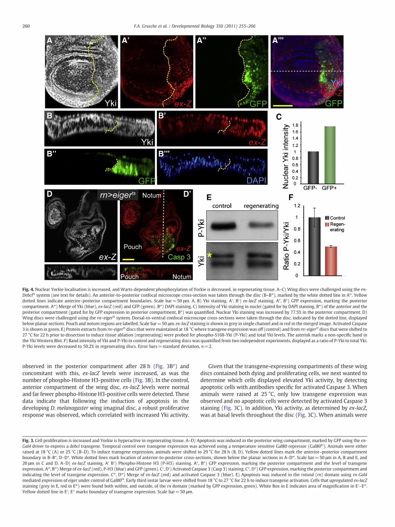
Fig. 4. Nuclear Yorkie localisation is increased, and Warts-dependent phosphorylation of Yorkie is decreased, in regenerating tissue. A–C) Wing discs were challenged using the en-Debclts system (see text for details). An anterior-to-posterior confocal microscope cross-section was taken through the disc (B–B‴), marked by the white dotted line in A‴. Yellowdotted lines indicate anterior–posterior compartment boundaries. Scale bar=50 μm. A, B) Yki staining. A′, B′) ex-lacZ staining. A″, B″) GFP expression, marking the posteriorcompartment. A‴) Merge of Yki (blue), ex-lacZ (red) and GFP (green). B‴) DAPI staining. C) Intensity of Yki staining in nuclei (gated for by DAPI staining, B‴) of the anterior and theposterior compartment (gated for by GFP expression in posterior compartment, B″) was quantified. Nuclear Yki staining was increased by 77.5% in the posterior compartment. D)Wing discs were challenged using the rnNeigerts system. Dorsal-to-ventral confocal microscope cross-sections were taken through the disc, indicated by the dotted line, displayedbelow planar sections. Pouch and notum regions are labelled. Scale bar=50 μm. ex-lacZ staining is shown in grey in single channel and in red in themerged image. Activated Caspase3 is shown in green. E) Protein extracts from rnNeigerts discs that weremaintained at 18 °Cwhere transgene expression was off (control) and from rnNeigerts discs that were shifted to27 °C for 22 h prior to dissection to induce tissue ablation (regenerating) were probed for phospho-S168-Yki (P-Yki) and total Yki levels. The asterisk marks a non-specific band inthe Yki Western Blot. F) Band intensity of Yki and P-Yki in control and regenerating discs was quantified from two independent experiments, displayed as a ratio of P-Yki to total Yki.P-Yki levels were decreased to 50.2% in regenerating discs. Error bars=standard deviation, n=2.
260 F.A. Grusche et al. / Developmental Biology 350 (2011) 255–266
observed in the posterior compartment after 28 h (Fig. 3B″) andconcomitant with this, ex-lacZ levels were increased, as was thenumber of phospho-Histone H3-positive cells (Fig. 3B). In the control,anterior compartment of the wing disc, ex-lacZ levels were normaland far fewer phospho-Histone H3-positive cells were detected. Thesedata indicate that following the induction of apoptosis in thedeveloping D. melanogaster wing imaginal disc, a robust proliferativeresponse was observed, which correlated with increased Yki activity.
Fig. 3. Cell proliferation is increased and Yorkie is hyperactive in regenerating tissue. A–D) AGal4 driver to express a debcl transgene. Temporal control over transgene expression was araised at 18 °C (A) or 25 °C (B–D). To induce transgene expression, animals were shifted tboundary in B–B″, D–D″. White dotted lines mark location of anterior-to-posterior cross-se20 μm in C and D. A–D) ex-lacZ staining. A′ B′) Phospho-Histone H3 (P-H3) staining. A″,expression. A‴, B‴)Merge of ex-lacZ (red), P-H3 (blue) and GFP (green). C′, D′) Activated Caspindicating the level of transgene expression. C‴, D‴) Merge of ex-lacZ (red) and activatedmediated expression of eiger under control of Gal80ts. Early third instar larvae were shifted frstaining (grey in E, red in E‴) were found both within, and outside, of the rn domain (markYellow dotted line in E′, E″ marks boundary of transgene expression. Scale bar=50 μm.
Given that the transgene-expressing compartments of these wingdiscs contained both dying and proliferating cells, we next wanted todetermine which cells displayed elevated Yki activity, by detectingapoptotic cells with antibodies specific for activated Caspase 3. Whenanimals were raised at 25 °C, only low transgene expression wasobserved and no apoptotic cells were detected by activated Caspase 3staining (Fig. 3C). In addition, Yki activity, as determined by ex-lacZ,was at basal levels throughout the disc (Fig. 3C). When animals were
poptosis was induced in the posterior wing compartment, marked by GFP using the en-chieved using a temperature sensitive Gal80 repressor (Gal80ts). Animals were eithero 29 °C for 28 h (B, D). Yellow dotted lines mark the anterior–posterior compartmentctions, shown below the planar sections in A–D‴. Scale bar=50 μm in A, B and E, andB″) GFP expression, marking the posterior compartment and the level of transgenease 3 (Casp 3) staining. C″, D″) GFP expression, marking the posterior compartment andCaspase 3 (blue). E) Apoptosis was induced in the rotund (rn) domain using rn-Gal4om 18 °C to 27 °C for 22 h to induce transgene activation. Cells that upregulated ex-lacZed by GFP expression, green). White Box in E indicates area of magnification in E′–E‴.
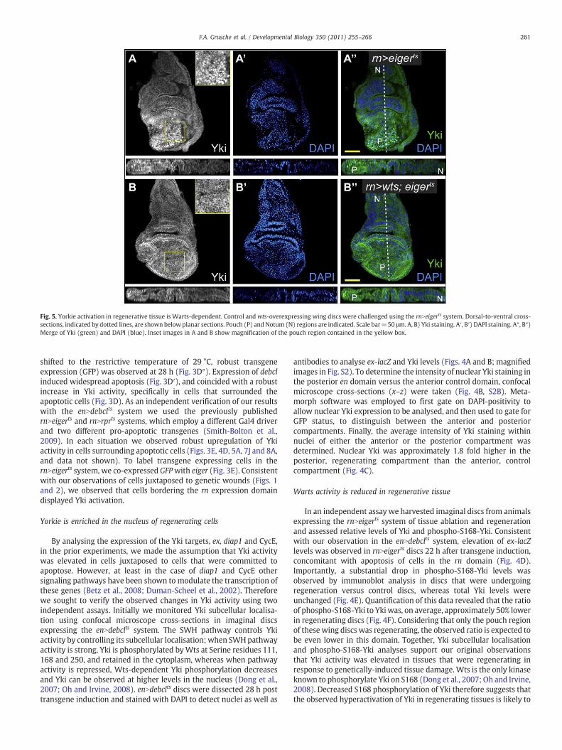
Fig. 5. Yorkie activation in regenerative tissue is Warts-dependent. Control andwts-overexpressing wing discs were challenged using the rnNeigerts system. Dorsal-to-ventral cross-sections, indicated by dotted lines, are shown below planar sections. Pouch (P) and Notum (N) regions are indicated. Scale bar=50 μm. A, B) Yki staining. A′, B′) DAPI staining. A″, B″)Merge of Yki (green) and DAPI (blue). Inset images in A and B show magnification of the pouch region contained in the yellow box.
261F.A. Grusche et al. / Developmental Biology 350 (2011) 255–266
shifted to the restrictive temperature of 29 °C, robust transgeneexpression (GFP) was observed at 28 h (Fig. 3D″). Expression of debclinduced widespread apoptosis (Fig. 3D′), and coincided with a robustincrease in Yki activity, specifically in cells that surrounded theapoptotic cells (Fig. 3D). As an independent verification of our resultswith the enNdebclts system we used the previously publishedrnNeigerts and rnNrprts systems, which employ a different Gal4 driverand two different pro-apoptotic transgenes (Smith-Bolton et al.,2009). In each situation we observed robust upregulation of Ykiactivity in cells surrounding apoptotic cells (Figs. 3E, 4D, 5A, 7J and 8A,and data not shown). To label transgene expressing cells in thernNeigerts system, we co-expressed GFPwith eiger (Fig. 3E). Consistentwith our observations of cells juxtaposed to genetic wounds (Figs. 1and 2), we observed that cells bordering the rn expression domaindisplayed Yki activation.
Yorkie is enriched in the nucleus of regenerating cells
By analysing the expression of the Yki targets, ex, diap1 and CycE,in the prior experiments, we made the assumption that Yki activitywas elevated in cells juxtaposed to cells that were committed toapoptose. However, at least in the case of diap1 and CycE othersignaling pathways have been shown to modulate the transcription ofthese genes (Betz et al., 2008; Duman-Scheel et al., 2002). Thereforewe sought to verify the observed changes in Yki activity using twoindependent assays. Initially we monitored Yki subcellular localisa-tion using confocal microscope cross-sections in imaginal discsexpressing the enNdebclts system. The SWH pathway controls Ykiactivity by controlling its subcellular localisation; when SWHpathwayactivity is strong, Yki is phosphorylated byWts at Serine residues 111,168 and 250, and retained in the cytoplasm, whereas when pathwayactivity is repressed, Wts-dependent Yki phosphorylation decreasesand Yki can be observed at higher levels in the nucleus (Dong et al.,2007; Oh and Irvine, 2008). enNdebclts discs were dissected 28 h posttransgene induction and stained with DAPI to detect nuclei as well as
antibodies to analyse ex-lacZ and Yki levels (Figs. 4A and B; magnifiedimages in Fig. S2). To determine the intensity of nuclear Yki staining inthe posterior en domain versus the anterior control domain, confocalmicroscope cross-sections (x–z) were taken (Fig. 4B, S2B). Meta-morph software was employed to first gate on DAPI-positivity toallow nuclear Yki expression to be analysed, and then used to gate forGFP status, to distinguish between the anterior and posteriorcompartments. Finally, the average intensity of Yki staining withinnuclei of either the anterior or the posterior compartment wasdetermined. Nuclear Yki was approximately 1.8 fold higher in theposterior, regenerating compartment than the anterior, controlcompartment (Fig. 4C).
Warts activity is reduced in regenerative tissue
In an independent assay we harvested imaginal discs from animalsexpressing the rnNeigerts system of tissue ablation and regenerationand assessed relative levels of Yki and phospho-S168-Yki. Consistentwith our observation in the enNdebclts system, elevation of ex-lacZlevels was observed in rnNeigerts discs 22 h after transgene induction,concomitant with apoptosis of cells in the rn domain (Fig. 4D).Importantly, a substantial drop in phospho-S168-Yki levels wasobserved by immunoblot analysis in discs that were undergoingregeneration versus control discs, whereas total Yki levels wereunchanged (Fig. 4E). Quantification of this data revealed that the ratioof phospho-S168-Yki to Yki was, on average, approximately 50% lowerin regenerating discs (Fig. 4F). Considering that only the pouch regionof these wing discs was regenerating, the observed ratio is expected tobe even lower in this domain. Together, Yki subcellular localisationand phospho-S168-Yki analyses support our original observationsthat Yki activity was elevated in tissues that were regenerating inresponse to genetically-induced tissue damage. Wts is the only kinaseknown to phosphorylate Yki on S168 (Dong et al., 2007; Oh and Irvine,2008). Decreased S168 phosphorylation of Yki therefore suggests thatthe observed hyperactivation of Yki in regenerating tissues is likely to

Fig. 6. Yorkie activity is rate-limiting for wing regeneration. Wing discs ofwild type andyki heterozygous animals were challenged using the en-Debclts system. A, B) wild type(A) and ykiB5/+ (B) wings at the permissive temperature. C, D)wild type (C) and ykiB5/+
(D) wings moved to the restrictive temperature for 21 h during larval development.E) Quantification of wing sizes. Error bars reflect standard error of themean (SEM).wildtype controls n=27, ykiB5/+ control wings n=28, wild type challenged wing n=20,ykiB5/+ challenged wing n=20. ykiB5/+ wings have a reduction of wing tissue size by20% after challenge. This reduction is statistically highly significant (pb0.001; one-tailed Student T test assuming unequal variance).
262 F.A. Grusche et al. / Developmental Biology 350 (2011) 255–266
be caused by repression of Wts kinase activity. To further investigatethe role of Wts in the activation of Yki during tissue regeneration, weassessed the ability of Wts overexpression to restrain Yki hyperacti-vation in regenerating tissue. We observed high levels of nuclear Ykiin rnNeigerts discs 22 h after transgene induction (Fig. 5A). Impor-tantly, co-expression of Wts consistently blocked nuclear Ykilocalisation (Fig. 5B). The decrease of Yki S168 phosphorylation inregenerative tissue and the inhibition of Yki nuclear localisation byWts overexpression therefore indicate that Yki hyperactivation inregenerating tissue is due to decreased repression by the core SWHpathway component Wts.
Finally, to determine whether Yki was also activated at surgicallyinduced wound edges in imaginal discs, we analysed Yki activity inwing imaginal discs that were wounded surgically and allowed toregenerate in the abdomens of host females as previously described(Ephrussi and Beadle, 1936; Hadorn, 1963). Strong induction of theJNK pathway reporter puckered (puc) has previously been describedaround wound edges in regenerating discs post surgical wounding(Bosch et al., 2005; Mattila et al., 2005). When discs were cultured invivo for 25 h post wounding, we observed strong induction of the JNKpathway reporter puc, at the wound edge using a puc-Gal4 incombination with UAS–GFP, which served as a positive control forregenerating cells (Fig. S3C′). Importantly, Yki activity, as measuredby ex-lacZ,was increased in cells in close proximity to thewound edge(Fig. S3C), compared with unwounded discs (Fig. S3A). In controlexperiments, discs were wounded, immediately fixed and stainedwithout in vivo culture. In this situation, puc activity was not observedat the wound edge (Fig. S3B′) and Yki activity was at baseline levelsthroughout the disc (Fig. S3B).
In summary, we observed robust activation of Yki in damagedimaginal disc tissues in different experimental scenarios: tissue
mosaic for genetically-induced dying clones, tissue induced to dieby induction of pro-apoptotic transgenes, and surgically-woundedtissue. In all cases, we observed consistent repression of SWHpathway activity, leading to increased Yki activity, in 2–5 rows ofcells bordering wound sites. The consistency of these results suggeststhat SWH pathway activity is repressed in proliferating cells thatjuxtapose wound sites.
Yorkie is rate-limiting for wing disc regeneration
To determine the functional significance of the SWH pathway inregenerative tissue growth, we assessed the regenerative capacity ofthe developing wing in both wild type and yki heterozygousbackgrounds. Yki is rate-limiting for tissue overgrowth caused byreduced SWH pathway activity owing to loss of genes such as ft(Bennett and Harvey, 2006; Silva et al., 2006; Willecke et al., 2006),and therefore we reasoned that if the SWH pathway indeed regulatesregenerative tissue growth, ykiwould be rate-limiting for this process.To test this hypothesis we used the enNdebclts system to transientlyinduce apoptosis of cells in the posterior compartment of thedeveloping wing discs of yki heterozygous and wild type animals,and then assessed regeneration by quantifying wing size of adultanimals. Importantly, in control experiments when debcl expressionwas not induced, yki heterozygosity was not rate-limiting for winggrowth (Figs. 6A, B and E). However, when tissue ablation wastransiently induced during wing development, we observed a 20%reduction in regenerative capacity of yki heterozygous wings,compared with wild type control wings (Figs. 6C–E), therebyproviding very strong genetic evidence that the SWH pathwaycontributes to regulation of regenerative tissue growth.
Upstream control of Yorkie activity in regenerative growth
Next we sought to investigate the mechanism by which SWHpathway activity is perturbed during regeneration to allowhyperactivation of Yki. Dying cells lose contact with their neigh-bours as a consequence of Caspase-mediated disassembly of cell–cell junctions (Kessler and Muller, 2009). Therefore cell–celladhesion proteins that mediate interactions between cells areobvious candidates for sensing loss of mutant or dying cells byremaining wild type tissue. In recent years, several reports haveshown that interactions between the atypical cadherins, Ft andDachsous (Ds), between neighbouring cells controls SWH pathwayactivity and tissue growth (Rogulja et al., 2008; Willecke et al.,2008). Moreover, Ds and Ft are required for oriented division of cellsthat occurs as a local response to apoptotic cells in wing imaginaldiscs (Li et al., 2009).
To determine whether Ft and Ds activity is abrogated inregenerating cells and thereby allows transient Yki activation weanalysed Yki induction in these cells in a dachs mutant background.Dachs is thought to induce Yki activity downstream of Ft by inhibitingthe Wts-mediated repression of Yki (Cho et al., 2006). In the absenceof dachs, the interaction between Ft and Ds does not influence Ykiactivity (Rogulja et al., 2008; Willecke et al., 2008). Therefore wereasoned that if Ft–Ds signaling is perturbed by tissue wounds, andthat this leads to induction of proliferation of wild type cellssurrounding the wound, then regenerative growth and Yki inductionshould be compromised in the absence of dachs. We thereforeattempted to assay the regenerative capacity of developing wings indachs mutant animals.
Initially, we wanted to assay the regenerative ability of dachsmutant animals using the rnNeigerts system (Smith-Bolton et al.,2009). Developmental timing is critical for this assay since youngeranimals have greater regenerative capacity (Smith-Bolton et al.,2009). Unfortunately, dachs mutant animals are developmentallydelayed by 1–2 days when raised at 18 °C (our own unpublished
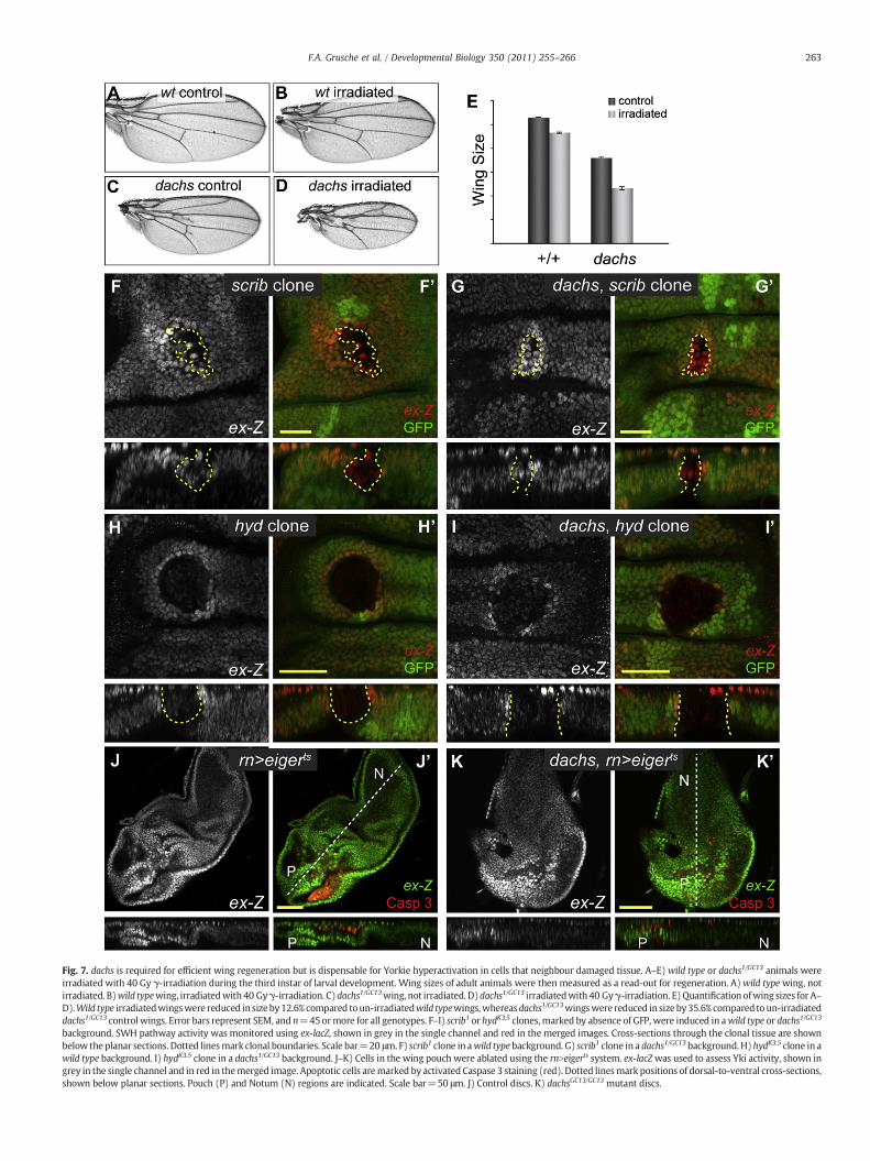
Fig. 7. dachs is required for efficient wing regeneration but is dispensable for Yorkie hyperactivation in cells that neighbour damaged tissue. A–E) wild type or dachs1/GC13 animals wereirradiated with 40 Gy γ-irradiation during the third instar of larval development. Wing sizes of adult animals were then measured as a read-out for regeneration. A)wild type wing, notirradiated. B)wild typewing, irradiatedwith40 Gyγ-irradiation. C)dachs1/GC13wing, not irradiated. D)dachs1/GC13 irradiatedwith 40 Gyγ-irradiation. E) Quantification ofwing sizes for A–D).Wild type irradiatedwingswere reduced in sizeby12.6% compared toun-irradiatedwild typewings,whereas dachs1/GC13wingswere reduced in size by35.6% compared to un-irradiateddachs1/GC13 control wings. Error bars represent SEM, and n=45ormore for all genotypes. F–I) scrib1 or hydK3.5 clones, marked by absence of GFP, were induced in awild type or dachs1/GC13
background. SWH pathway activity was monitored using ex-lacZ, shown in grey in the single channel and red in the merged images. Cross-sections through the clonal tissue are shownbelow the planar sections. Dotted linesmark clonal boundaries. Scale bar=20 μm. F) scrib1 clone in awild typebackground. G) scrib1 clone in a dachs1/GC13background. H)hydK3.5 clone in awild type background. I) hydK3.5 clone in a dachs1/GC13 background. J–K) Cells in the wing pouch were ablated using the rnNeigerts system. ex-lacZwas used to assess Yki activity, shown ingrey in the single channel and in red in themerged image. Apoptotic cells aremarked by activated Caspase 3 staining (red). Dotted linesmark positions of dorsal-to-ventral cross-sections,shown below planar sections. Pouch (P) and Notum (N) regions are indicated. Scale bar=50 μm. J) Control discs. K) dachsGC13/GC13 mutant discs.
263F.A. Grusche et al. / Developmental Biology 350 (2011) 255–266
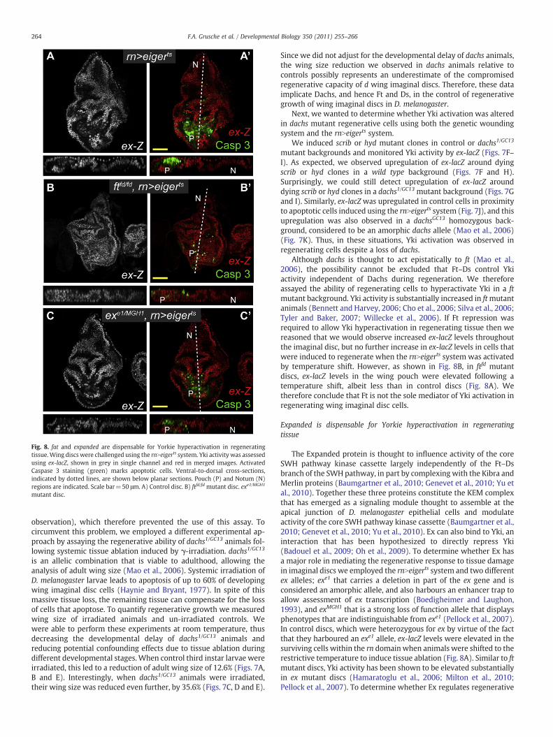
Fig. 8. fat and expanded are dispensable for Yorkie hyperactivation in regeneratingtissue.Wing discs were challenged using the rnNeigerts system. Yki activity was assessedusing ex-lacZ, shown in grey in single channel and red in merged images. ActivatedCaspase 3 staining (green) marks apoptotic cells. Ventral-to-dorsal cross-sections,indicated by dotted lines, are shown below planar sections. Pouch (P) and Notum (N)regions are indicated. Scale bar=50 μm. A) Control disc. B) ftfd/fd mutant disc. exe1/MGH1
mutant disc.
264 F.A. Grusche et al. / Developmental Biology 350 (2011) 255–266
observation), which therefore prevented the use of this assay. Tocircumvent this problem, we employed a different experimental ap-proach by assaying the regenerative ability of dachs1/GC13 animals fol-lowing systemic tissue ablation induced by γ-irradiation. dachs1/GC13
is an allelic combination that is viable to adulthood, allowing theanalysis of adult wing size (Mao et al., 2006). Systemic irradiation ofD. melanogaster larvae leads to apoptosis of up to 60% of developingwing imaginal disc cells (Haynie and Bryant, 1977). In spite of thismassive tissue loss, the remaining tissue can compensate for the lossof cells that apoptose. To quantify regenerative growth we measuredwing size of irradiated animals and un-irradiated controls. Wewere able to perform these experiments at room temperature, thusdecreasing the developmental delay of dachs1/GC13 animals andreducing potential confounding effects due to tissue ablation duringdifferent developmental stages. When control third instar larvae wereirradiated, this led to a reduction of adult wing size of 12.6% (Figs. 7A,B and E). Interestingly, when dachs1/GC13 animals were irradiated,their wing size was reduced even further, by 35.6% (Figs. 7C, D and E).
Since we did not adjust for the developmental delay of dachs animals,the wing size reduction we observed in dachs animals relative tocontrols possibly represents an underestimate of the compromisedregenerative capacity of d wing imaginal discs. Therefore, these dataimplicate Dachs, and hence Ft and Ds, in the control of regenerativegrowth of wing imaginal discs in D. melanogaster.
Next, we wanted to determine whether Yki activation was alteredin dachs mutant regenerative cells using both the genetic woundingsystem and the rnNeigerts system.
We induced scrib or hyd mutant clones in control or dachs1/GC13
mutant backgrounds and monitored Yki activity by ex-lacZ (Figs. 7F–I). As expected, we observed upregulation of ex-lacZ around dyingscrib or hyd clones in a wild type background (Figs. 7F and H).Surprisingly, we could still detect upregulation of ex-lacZ arounddying scrib or hyd clones in a dachs1/GC13 mutant background (Figs. 7Gand I). Similarly, ex-lacZ was upregulated in control cells in proximityto apoptotic cells induced using the rnNeigerts system (Fig. 7J), and thisupregulation was also observed in a dachsGC13 homozygous back-ground, considered to be an amorphic dachs allele (Mao et al., 2006)(Fig. 7K). Thus, in these situations, Yki activation was observed inregenerating cells despite a loss of dachs.
Although dachs is thought to act epistatically to ft (Mao et al.,2006), the possibility cannot be excluded that Ft–Ds control Ykiactivity independent of Dachs during regeneration. We thereforeassayed the ability of regenerating cells to hyperactivate Yki in a ftmutant background. Yki activity is substantially increased in ftmutantanimals (Bennett and Harvey, 2006; Cho et al., 2006; Silva et al., 2006;Tyler and Baker, 2007; Willecke et al., 2006). If Ft repression wasrequired to allow Yki hyperactivation in regenerating tissue then wereasoned that we would observe increased ex-lacZ levels throughoutthe imaginal disc, but no further increase in ex-lacZ levels in cells thatwere induced to regenerate when the rnNeigerts system was activatedby temperature shift. However, as shown in Fig. 8B, in ftfd mutantdiscs, ex-lacZ levels in the wing pouch were elevated following atemperature shift, albeit less than in control discs (Fig. 8A). Wetherefore conclude that Ft is not the sole mediator of Yki activation inregenerating wing imaginal disc cells.
Expanded is dispensable for Yorkie hyperactivation in regeneratingtissue
The Expanded protein is thought to influence activity of the coreSWH pathway kinase cassette largely independently of the Ft–Dsbranch of the SWH pathway, in part by complexing with the Kibra andMerlin proteins (Baumgartner et al., 2010; Genevet et al., 2010; Yu etal., 2010). Together these three proteins constitute the KEM complexthat has emerged as a signaling module thought to assemble at theapical junction of D. melanogaster epithelial cells and modulateactivity of the core SWH pathway kinase cassette (Baumgartner et al.,2010; Genevet et al., 2010; Yu et al., 2010). Ex can also bind to Yki, aninteraction that has been hypothesized to directly repress Yki(Badouel et al., 2009; Oh et al., 2009). To determine whether Ex hasa major role in mediating the regenerative response to tissue damagein imaginal discs we employed the rnNeigerts system and two differentex alleles; exe1 that carries a deletion in part of the ex gene and isconsidered an amorphic allele, and also harbours an enhancer trap toallow assessment of ex transcription (Boedigheimer and Laughon,1993), and exMGH1 that is a strong loss of function allele that displaysphenotypes that are indistinguishable from exe1 (Pellock et al., 2007).In control discs, which were heterozygous for ex by virtue of the factthat they harboured an exe1 allele, ex-lacZ levels were elevated in thesurviving cells within the rn domain when animals were shifted to therestrictive temperature to induce tissue ablation (Fig. 8A). Similar to ftmutant discs, Yki activity has been shown to be elevated substantiallyin ex mutant discs (Hamaratoglu et al., 2006; Milton et al., 2010;Pellock et al., 2007). To determine whether Ex regulates regenerative

265F.A. Grusche et al. / Developmental Biology 350 (2011) 255–266
tissue growth we assessed ex-lacZ levels in cells that were induced toregenerate when the rnNeigerts system was activated by temperatureshift. If Ex repression was required to allow Yki hyperactivation inregenerating tissue then Yki activity, as assessed by ex-lacZ, would beelevated throughout the imaginal disc, but not hyperactivated inregenerating cells in temperature-shifted rnNeigerts discs. However, asshown in Fig. 8C, in ex transheterozygous discs, ex-lacZ levels wereelevated to similar levels in regenerating cells in proximity toapoptotic cells compared with other regions of the disc, as wasobserved in control discs in Fig. 8A. This suggests that repression ofthe Ex protein is not required to hyperactivate Yki and driveproliferation of regenerating cells.
Discussion
We have shown that the SWH pathway regulates regenerativegrowth of D. melanogaster imaginal discs. Hyperactivation of Ykiactivity was observed in regenerating cells following tissue disruptionusing a range of genetic and surgical assays, and full Yki activity wasessential for efficient wing regeneration (Figs. 1–6). These findingsimply that the important role that the SWH pathway has inspecification of organ size during development involves an ability tocontrol proliferation in response to tissue damage. These resultsextend recent data describing a role for the SWH pathway inregeneration of the cricket leg (Bando et al., 2009). Interestingly,whilst this manuscript was under review, the SWH pathway wasfound to modulate regeneration of other epithelial tissues such as theadult D. melanogaster gut and the murine small intestine in responseto chemical and bacterial insults (Cai et al., 2010; Karpowicz et al.,2010; Shaw et al., 2010; Staley and Irvine, 2010). Coupled with thedata presented here, these findings suggest that the SWH pathway is aregulator of “epithelial fitness”, i.e. it is primed to sense damage inepithelial tissues, and to coordinate a robust repair mechanism inresponse to damage stimuli.
Other growth-regulatory proteins such asWgandMyc are importantfor D. melanogaster imaginal disc regeneration, as is the JNK pathway(Bosch et al., 2005; Mattila et al., 2005; Smith-Bolton et al., 2009).Interestingly however, regeneration of wing imaginal discs does notappear to be modulated by all growth pathways. For example, Smith-Bolton et al. (2009) provided evidence that the target of rapamycinpathway does not modulate regenerative growth of the wing imaginaldisc. This lends weight to the hypothesis that the SWH pathway has aspecific role in regulating regeneration, rather than regenerative tissuegrowth being controlled by all growth-regulatory pathways.
An important question arising from these studies is what are theupstream signaling mechanisms that, in response to tissue damage,permit Yki hyperactivation in regenerating cells? Given that weobserved a reduction in Wts-dependent phosphorylation of Yki inregenerating tissue, obvious candidate signaling inputs are the threemajor classes of upstream regulatory proteins of the SWH pathway; theFt–Ds branch, the KEM complex and the apicobasal polarity proteins,Lgl, Crb and aPKC (Grusche et al., 2010). The Ft and Ds cadherins limitcell proliferation byengaging in physical interactionswith each other onneighbouring cells and repressing the Yki activator, Dachs (Cho et al.,2006; Rogulja et al., 2008; Willecke et al., 2008). Ft and Ds are alsorequired for orientedmitoses of cells neighbouring apoptotic cells (Li etal., 2009). Given this,wehypothesized that in a damaged tissue, cell–cellcontactswould be broken, causing Ft–Ds signaling to be abrogated and aresultant elevation in Dachs, and hence Yki, activity. dachsmutant wingimaginal discs displayed impaired regenerative capacity following γ-irradiation-induced apoptosis, thus implicating the Ft–Ds signalingmodule in the control of Yki-dependent regeneration (Fig. 7). However,our genetic wounding and tissue ablation assays suggested that Ykihyperactivation in regenerating cells involves signaling inputs inaddition to the Ft–Ds branch of the SWH pathway (Fig. 7), since Yki
activation was still observed in regenerative tissue in the absence ofdachs (Fig. 7) or ft (Fig. 8B).
What are possible reasons for this apparent contradiction? Firstly,there could be subtle differences in Yki activation in wild type versusdachs cells that we failed to observe in our system, since lacZ enhancertrap lines offer poorly quantitative data, especially when comparingexpression levels between independent tissues. Secondly, there isevidence that Ds controls SWH pathway activity by functioning notonly as a ligand for Ft but also as a receptor (Willecke et al., 2008).Whether Ds signaling to downstream SWH pathway proteins requiresDachs, has not yet been determined. Thirdly, Ft and Ds signalingmightinfluence SWH pathway activity independent of Dachs in regenerat-ing cells. For example Ft has been proposed to control Yki activity byinfluencing the subcellular localization and levels of the Ex protein(Bennett and Harvey, 2006; Silva et al., 2006; Willecke et al., 2006).However we feel that this is unlikely given that Yki hyperactivation inregenerative tissue was maintained in ex and ft mutant backgrounds(Fig. 8C). A more likely scenario is that upstream SWH pathwayproteins in addition to Ft, Ds and Dachs participate in control of Ykiactivity in regenerative tissue growth. Candidates include the KEMcomplex components, Mer and Kibra, as well as the apicobasalpolarity proteins, Lethal Giant Larvae, Crumbs and atypical ProteinKinase C. Another possibility is that the altered physical status of cellsthat surround tissue wounds might regulate SWH pathway activityvia a mechanism such as tension, which has been hypothesized toinfluence the specification of Drosophilawing size (Aegerter-Wilmsenet al., 2007; Hufnagel et al., 2007). It is also plausible that signalingpathways that are known to control regeneration, such as the JNKpathway, regulate the SWH pathway during tissue regeneration.
Regenerating cellshavebeenpostulated to revert to amoreprimitivedifferentiation state [reviewed in (McClure and Schubiger, 2007)].Recently the murine Yki orthologue, YAP, was found to promote stemcell pluripotency along with other transcriptional regulatory proteinsincludingMyc (Lian et al., 2010). Significantly, Myc has previously beenshown to be induced in regeneratingD.melanogaster tissues, and to be apotent driver of the regenerative response (Smith-Bolton et al., 2009).Therefore a plausible hypothesis is that Yki hyperactivation promotesregeneration by altering the cell's transcriptional program, and thuscellular plasticity, in conjunction with Myc.
Regeneration has often been used as a paradigm to describe theidea of an organ size-checkpoint that limits the size of tissues duringboth development and adult homeostasis in metazoans. Our findingssuggest that the SWH pathway, which is a crucial regulator of organsize, also controls regeneration, providing evidence for a molecularlink between the two processes. In the future, it will be interesting todetermine whether the SWH pathway controls regeneration inanimals with robust regenerative capacity such as axolotls, hydraand planaria. It will also be important to determine whether the SWHpathway controls tissue regeneration in mammals; if this proves to bethe case, modulation of SWH pathway activity might be a powerfulapproach to modulate tissue regeneration post trauma or surgery.Finally, aberrant tissue regeneration upon chronic injury or inflam-mation has been proposed to contribute to tumour formation. Sinceincreasing evidence points to a role of aberrant SWH pathwaysignaling in cancer (Harvey and Tapon, 2007), our findings provide apotential molecular link between regeneration and tumorigenesis.
Supplementarymaterials related to this article can be found onlineat doi:10.1016/j.ydbio.2010.11.020.
Acknowledgments
We thank N. Tapon for comments and K. Irvine for thecommunication of results prior to publication. We thank I. Hariharan,B. Hay, K. Irvine, E. Martin-Blanco, K. Moberg, D. Pan, B. Thompson andL. Quinn for fly stocks and antibodies. We thankW. Gehring for adviceon tissue transplantation, J. Williamson for injection needle

266 F.A. Grusche et al. / Developmental Biology 350 (2011) 255–266
preparation, A. Moeller for advice with irradiation experiments and J.George for aiding statistical analysis. F.A.G. is supported by anInternational Postgraduate Research and Fee Remission Fellowshipfrom the University of Melbourne, H.E.R. is supported by a SeniorNHMRC Research Fellowship and K.F.H. is a Sylvia and Charles ViertelSenior Medical Research Fellow. This research was supported bygrants from the Australian NHMRC to K.F.H (#566700) and to H.E.R(#509020).
References
Aegerter-Wilmsen, T., et al., 2007. Model for the regulation of size in the wing imaginaldisc of Drosophila. Mech. Dev. 124, 318–326.
Badouel, C., et al., 2009. The FERM-domain protein Expanded regulates Hippo pathwayactivity via direct interactions with the transcriptional activator Yorkie. Dev. Cell16, 411–420.
Bando, T., et al., 2009. Regulation of leg size and shape by the Dachsous/Fat signallingpathway during regeneration. Development 136, 2235–2245.
Baumgartner, R., et al., 2010. The WW domain protein Kibra acts upstream of hippo inDrosophila. Dev. Cell 18, 309–316.
Bennett, F.C., Harvey, K.F., 2006. Fat cadherinmodulates organ size in Drosophila via theSalvador/Warts/Hippo signaling pathway. Curr. Biol. 16, 2101–2110.
Betz, A., et al., 2008. STAT92E is a positive regulator of Drosophila inhibitor of apoptosis1 (DIAP/1) and protects against radiation-induced apoptosis. Proc. Natl. Acad. Sci.U. S. A. 105, 13805–13810.
Bilder, D., Perrimon, N., 2000. Localization of apical epithelial determinants by thebasolateral PDZ protein Scribble. Nature 403, 676–680.
Birnbaum, K.D., Sanchez Alvarado, A., 2008. Slicing across kingdoms: regeneration inplants and animals. Cell 132, 697–710.
Boedigheimer, M., Laughon, A., 1993. Expanded: a gene involved in the control of cellproliferation in imaginal discs. Development 118, 1291–1301.
Bosch, M., et al., 2005. JNK signaling pathway required for wound healing inregenerating Drosophila wing imaginal discs. Dev. Biol. 280, 73–86.
Brand, A.H., Perrimon, N., 1993. Targeted gene expression as a means of altering cellfates and generating dominant phenotypes. Development 118, 401–415.
Cai, J., et al., 2010. The Hippo signaling pathway restricts the oncogenic potential of anintestinal regeneration program. Genes Dev. 24, 2383–2388.
Camargo, F.D., et al., 2007. YAP1 increases organ size and expands undifferentiatedprogenitor cells. Curr. Biol. 17, 2054–2060.
Chen, C.L., et al., 2010. The apical–basal cell polarity determinant crumbs regulatesHippo signaling in Drosophila. Proc. Natl. Acad. Sci. U. S. A. 107, 15810–15815.
Cho, E., et al., 2006. Delineation of a Fat tumor suppressor pathway. Nat. Genet. 38,1142–1150.
Colussi, P.A., et al., 2000. Debcl, a proapoptotic Bcl-2 homologue, is a component of theDrosophila melanogaster cell death machinery. J. Cell Biol. 148, 703–714.
Dong, J., et al., 2007. Elucidation of a universal size-control mechanism in Drosophilaand mammals. Cell 130, 1120–1133.
Duman-Scheel, M., et al., 2002. Hedgehog regulates cell growth and proliferation byinducing Cyclin D and Cyclin E. Nature 417, 299–304.
Ephrussi, B., Beadle, G.W., 1936. A technique of transplantation for Drosophila. Am. Soc.Nat. 70, 218–225.
Feng, Y., Irvine, K.D., 2007. Fat and expanded act in parallel to regulate growth throughwarts. Proc. Natl. Acad. Sci. U. S. A. 104, 20362–20367.
Forsthoefel, D.J., Newmark, P.A., 2009. Emerging patterns in planarian regeneration.Curr. Opin. Genet. Dev. 19, 412–420.
Genevet, A., et al., 2010. Kibra is a regulator of the Salvador/Warts/Hippo signalingnetwork. Dev. Cell 18, 300–308.
Grusche, F.A., et al., 2009. Sds22, a PP1phosphatase regulatory subunit, regulates epithelialcell polarity and shape [Sds22 in epithelial morphology]. BMC Dev. Biol. 9, 14.
Grusche, F.A., et al., 2010. Upstream regulation of the hippo size control pathway. Curr.Biol. 20, R574–R582.
Grzeschik, N.A., et al., 2010. Lgl, aPKC, and crumbs regulate the Salvador/Warts/Hippopathway through two distinct mechanisms. Curr. Biol. 20, 573–581.
Hadorn, E., 1963. Differenzierungsleistungen wiederholt fragmentierter Teilstückemännlicher Genitalscheiben von Drosophila melanogaster nach Kultur in vivo. Dev.Biol. 6, 617–629.
Hamaratoglu, F., et al., 2006. The tumour-suppressor genes NF2/Merlin and expandedact through Hippo signalling to regulate cell proliferation and apoptosis. Nat. CellBiol. 8, 27–36.
Harvey, K., Tapon, N., 2007. The Salvador–Warts–Hippo pathway — an emergingtumour-suppressor network. Nat. Rev. Cancer 7, 182–191.
Harvey, K.F., et al., 2003. The Drosophila Mst ortholog, hippo, restricts growth and cellproliferation and promotes apoptosis. Cell 114, 457–467.
Hay, B.A., et al., 1995. Drosophila homologs of baculovirus inhibitor of apoptosisproteins function to block cell death. Cell 83, 1253–1262.
Haynie, J.L., Bryant, P.J., 1977. The Effects of X-rays on the proliferation dynamics of cells inthe imaginalwing disc ofDrosophila melanogaster. Rouxs Arch. Dev. Biol. 183, 85–100.
Herz, H.M., et al., 2006. vps25 mosaics display non-autonomous cell survival andovergrowth, and autonomous apoptosis. Development 133, 1871–1880.
Huang, J., et al., 2005. The Hippo signaling pathway coordinately regulates cellproliferation and apoptosis by inactivating Yorkie, the Drosophila Homolog of YAP.Cell 122, 421–434.
Hufnagel, L., et al., 2007. On the mechanism of wing size determination in flydevelopment. Proc. Natl. Acad. Sci. U. S. A. 104, 3835–3840.
Karpowicz, P., et al., 2010. The Hippo tumor suppressor pathway regulates intestinalstem cell regeneration. Development 137, 4135–4145.
Kessler, T., Muller, H.A., 2009. Cleavage of Armadillo/beta-catenin by the caspase DrICEin Drosophila apoptotic epithelial cells. BMC Dev. Biol. 9, 15.
Lee, J.D., et al., 2002. The ubiquitin ligase Hyperplastic discs negatively regulateshedgehog and decapentaplegic expression by independent mechanisms. Develop-ment 129, 5697–5706.
Li, W., et al., 2009. Oriented cell division as a response to cell death and cell competition.Curr. Biol. 19, 1821–1826.
Lian, I., et al., 2010. The role of YAP transcription coactivator in regulating stem cell self-renewal and differentiation. Genes Dev. 24, 1106–1118.
Ling, C., et al., 2010. The apical transmembrane protein Crumbs functions as a tumorsuppressor that regulates Hippo signaling by binding to expanded. Proc. Natl. Acad.Sci. U. S. A. 107, 10532–10537.
Mao, Y., et al., 2006. Dachs: an unconventional myosin that functions downstream ofFat to regulate growth, affinity and gene expression in Drosophila. Development133, 2539–2551.
Mattila, J., et al., 2005. Role of Jun N-terminal Kinase (JNK) signaling in the woundhealing and regeneration of a Drosophila melanogaster wing imaginal disc. Int. J.Dev. Biol. 49, 391–399.
McClure, K.D., Schubiger, G., 2007. Transdetermination: Drosophila imaginal disc cellsexhibit stem cell-like potency. Int. J. Biochem. Cell Biol. 39, 1105–1118.
McGuire, S.E., et al., 2003. Spatiotemporal rescue of memory dysfunction in Drosophila.Science 302, 1765–1768.
McPherson, J.P., et al., 2004. Lats2/Kpm is required for embryonic development,proliferation control and genomic integrity. EMBO J. 23, 3677–3688.
Milton, C.C., et al., 2010. Differential requirement of Salvador–Warts–Hippo pathwaymembers for organ size control in Drosophila melanogaster. Development 137,735–743.
Moberg, K.H., et al., 2005. Mutations in erupted, the Drosophila ortholog of mammaliantumor susceptibility gene 101, elicit non-cell-autonomous overgrowth. Dev. Cell 9,699–710.
Nusslein-Volhard, C., et al., 1984. Mutations affecting the pattern of the larval cuticle inDrosophila melanogaster. I. Zygotic loci on the second chromosome. Rouxs Arch.Dev. Biol. 193, 267–282.
Oh, H., Irvine, K.D., 2008. In vivo regulation of Yorkie phosphorylation and localization.Development 135, 1081–1088.
Oh, H., et al., 2009. Phosphorylation-independent repression of Yorkie in Fat-Hipposignaling. Dev. Biol. 335, 188–197.
Pastor-Pareja, J.C., et al., 2004. Invasive cell behavior during Drosophila imaginal disceversion is mediated by the JNK signaling cascade. Dev. Cell 7, 387–399.
Pellock, B.J., et al., 2007. The Drosophila tumor suppressors expanded and merlindifferentially regulate cell cycle exit, apoptosis, and wingless signaling. Dev. Biol.304, 102–115.
Poss, K.D., et al., 2000. Roles for Fgf signaling during zebrafish fin regeneration. Dev.Biol. 222, 347–358.
Richardson, H., et al., 1995. Ectopic cyclin E expression induces premature entry into Sphase and disrupts pattern formation in the Drosophila eye imaginal disc.Development 121, 3371–3379.
Rink, J.C., et al., 2009. Planarian Hh signaling regulates regeneration polarity and linksHh pathway evolution to cilia. Science 326, 1406–1410.
Robinson, B.S., et al., 2010. Crumbs regulates Salvador/Warts/Hippo signaling inDrosophila via the FERM-domain protein expanded. Curr. Biol. 20, 582–590.
Rogulja, D., et al., 2008. Morphogen control of wing growth through the Fat signalingpathway. Dev. Cell 15, 309–321.
Shaw, R.L., et al., 2010. The Hippo pathway regulates intestinal stem cell proliferationduring Drosophila adult midgut regeneration. Development 137, 4147–4158.
Silva, E., et al., 2006. The tumor-suppressor gene fat controls tissue growth upstream ofexpanded in the hippo signaling pathway. Curr. Biol. 16, 2081–2089.
Smith-Bolton, R.K., et al., 2009. Regenerative growth in Drosophila imaginal discs isregulated by Wingless and Myc. Dev. Cell 16, 797–809.
Staley, B.K., Irvine, K.D., 2010. Warts and Yorkie mediate intestinal regeneration byinfluencing stem cell proliferation. Curr. Biol. 20, 1580–1587.
Tapon, N., et al., 2002. Salvador promotes both cell cycle exit and apoptosis inDrosophila and is mutated in human cancer cell lines. Cell 110, 467–478.
Thompson, B.J., et al., 2005. Tumor suppressor properties of the ESCRT-II complexcomponent Vps25 in Drosophila. Dev. Cell 9, 711–720.
Tyler, D.M., Baker, N.E., 2007. Expanded and fat regulate growth and differentiation inthe Drosophila eye through multiple signaling pathways. Dev. Biol. 305, 187–201.
Ursprung, H., 1967. In vivo culture of Drosophila imaginal discs. Meth. Dev. Biol.485–492.
Willecke, M., et al., 2006. The fat cadherin acts through the hippo tumor-suppressorpathway to regulate tissue size. Curr. Biol. 16, 2090–2100.
Willecke, M., et al., 2008. Boundaries of Dachsous cadherin activity modulate the Hipposignaling pathway to induce cell proliferation. Proc. Natl. Acad. Sci. U. S. A. 105,14897–14902.
Xu, T., Rubin, G.M., 1993. Analysis of genetic mosaics in developing and adultDrosophila tissues. Development (Cambridge, England) 117, 1223–1237.
Yazawa, S., et al., 2009. Planarian hedgehog/patched establishes anterior–posteriorpolarity by regulatingWnt signaling. Proc. Natl. Acad. Sci. U. S. A. 106, 22329–22334.
Yu, J., et al., 2010. Kibra functions as a tumor suppressor protein that regulates hipposignaling in conjunction with merlin and expanded. Dev. Cell 18, 288–299.
Zhao, B., et al., 2007. Inactivation of YAP oncoprotein by the Hippo pathway is involvedin cell contact inhibition and tissue growth control. Genes Dev. 21, 2747–2761.