Embed Size (px)
Citation preview
ORIGINAL RESEARCH ARTICLEpublished: 17 October 2014
doi: 10.3389/fmicb.2014.00548
The Salmonella effector protein SpvC, a phosphothreoninelyase is functional in plant cellsChristina Neumann1‡, Malou Fraiture2‡, Casandra Hernàndez-Reyes1, Fidele N. Akum1,
Isabelle Virlogeux-Payant3,4, Ying Chen2†, Stephanie Pateyron5, Jean Colcombet5, Karl-Heinz Kogel1,
Heribert Hirt5, Frédéric Brunner2 and Adam Schikora1*
1 Research Center for BioSystems, Land Use and Nutrition, Institute for Phytopathology and Applied Zoology, Justus-Liebig University Giessen, Giessen, Germany2 Department of Plant Biochemistry, Center for Plant Molecular Biology (ZMBP), Eberhard Karls University Tübingen, Tübingen, Germany3 Institut National de la Recherche Agronomique, UMR1282 Infectiologie et Santé Publique, Nouzilly, France4 Université François Rabelais de Tours, UMR1282 Infectiologie et Santé Publique, Tours, France5 Unité de Recherche en Génomique Végétale, Plant Genomics, Evry, France
Edited by:
Nicola Holden, The James HuttonInstitute, UK
Reviewed by:
Frederik Börnke, Leibniz-Institute forVegetable and Ornamental Crops(IGZ), GermanyJeri Barak, University ofWisconsin-Madison, USA
*Correspondence:
Adam Schikora, Research Center forBioSystems, Land Use andNutrition, Institute forPhytopathology and AppliedZoology, Justus-Liebig UniversityGiessen, Heinrich-Buff-Ring 26-32,Giessen 35392, Germanye-mail: [email protected]†Present address:
Ying Chen, Laboratory of ForestGenetics and Gene Engineering,Nanjing Forestry University, Nanjing,China
‡These authors have contributedequally to this work.
Salmonella is one of the most prominent causes of food poisoning and growingevidence indicates that contaminated fruits and vegetables are an increasing concernfor human health. Successful infection demands the suppression of the host immunesystem, which is often achieved via injection of bacterial effector proteins into hostcells. In this report we present the function of Salmonella effector protein in plantcell, supporting the new concept of trans-kingdom competence of this bacterium. Wescreened a range of Salmonella Typhimurium effector proteins for interference with plantimmunity. Among these, the phosphothreonine lyase SpvC attenuated the inductionof immunity-related genes when present in plant cells. Using in vitro and in vivosystems we show that this effector protein interacts with and dephosphorylates activatedArabidopsis Mitogen-activated Protein Kinase 6 (MPK6), thereby inhibiting defensesignaling. Moreover, the requirement of Salmonella SpvC was shown by the decreasedproliferation of the �spvC mutant in Arabidopsis plants. These results suggest thatsome Salmonella effector proteins could have a conserved function during proliferation indifferent hosts. The fact that Salmonella and other Enterobacteriaceae use plants as hostsstrongly suggests that plants represent a much larger reservoir for animal pathogens thanso far estimated.
Keywords: T3SS, trans-kingdom pathogenicity, Salmonella, plant infection
INTRODUCTIONVarious pathogenic bacteria such as Salmonella enterica,Pseudomonas aeruginosa, Staphylococcus aureus, Escherichia coliO157:H7, and Listeria monocytogenes are able to proliferate onboth animal and plant organisms (Prithiviraj et al., 2005; Mililloet al., 2008; Schikora et al., 2008, 2011; Haapalainen et al., 2009;Holden et al., 2009). Salmonella is a genus of Gram-negativeenteropathogenic bacteria that colonizes a wide range of hosts,including humans. These bacteria are the causal agents ofgastroenteritis and typhoid fever (Pang et al., 1995). The mostcommon mode of infection in humans is the ingestion of con-taminated food or water. Whereas 0.3% of fresh products werecontaminated with Salmonella bacteria in 2007 in the EuropeanUnion (Westrell et al., 2009), the proportion of raw-food relatedoutbreaks reached 25% in the USA in recent years (Rangel et al.,2005).
The study of the Salmonella infection mechanism was untilrecently mainly driven by its medical aspect; therefore the mouse
and human epithelial cell models are the best studied to date.Today, it is still poorly understood how these bacteria success-fully proliferate in such diversified hosts as animals or plants.However, important insights were obtained during last years.Stomata openings were identified as possible entry points of bac-teria into the inner layers of the mesophyll (Kroupitski et al.,2009). Interestingly, while some plant species (e.g., arugula)allow the Salmonella enterica subsp. enterica ser. Typhimurium(S. Typhimurium) strain SL1344 to internalize, some others (e.g.,parsley) seem to be capable of preventing internalization (Golberget al., 2011). In a previous report, we showed that in Arabidopsisthaliana, roots and especially root hair cells can be colonized bySalmonella (Schikora et al., 2008).
Studies of the infection mechanisms in animals revealedthat, besides remodeling the host cell architecture, Salmonellaactively suppresses the host immune system by injecting a cock-tail of effector proteins. These effectors are delivered by TypeIII Secretion Systems (T3SSs). S. Typhimurium possesses two
www.frontiersin.org October 2014 | Volume 5 | Article 548 | 1

Neumann et al. Activity of Salmonella SpvC in plant cells
distinct T3SSs, T3SS-1, and T3SS-2, encoded by two SalmonellaPathogenicity Islands, SPI-1 and SPI-2, respectively. To date,about 44 Salmonella effectors have been described and the func-tion of many of them is known [reviewed in Heffron et al. (2011)].In addition to SPIs, some Salmonella serovars carry plasmids witha common locus called salmonella plasmid virulence (spv) (Boydand Hartl, 1998). The spv operon encodes further effector pro-teins responsible for full virulence in humans and in the mousemodel (Montenegro et al., 1991; Fierer et al., 1992; Gulig andDoyle, 1993; Chu and Chiu, 2006).
Even though some Salmonella effectors have homologs inplant pathogenic bacteria, the role of Salmonella T3SS-dependenteffectors in the modulation of the plant immune system andtheir contribution to plant host susceptibility are less under-stood. Plants induce defense mechanisms after recognition ofpathogens. This recognition may occur at two levels: (i) at thecell surface, where Pattern Recognition Receptors (PRRs) recog-nize conserved microbial structures called Pathogen-AssociatedMolecular Patterns (PAMPs), and (ii) in the cytoplasm whereResistance (R) proteins recognize bacterial effectors injected intoplant cells. Both recognition events initiate immune responsesreferred to as Pattern-Triggered Immunity (PTI) [renamed fromPAMP-triggered immunity (Boller, 2012)] or Effector-TriggeredImmunity (ETI), respectively. An activation of MAPKs andenhanced expression of Pathogenesis Related (PR) genes are hall-marks of both: the PTI and the ETI responses. Both responseswere already observed after inoculation with Salmonella (Schikoraet al., 2008; Meng et al., 2013; Garcia et al., 2014). Recently, thesuppression of plant defense by Salmonella was reported in twodifferent systems. In contrast to living S. Typhimurium, treat-ment with dead or chloramphenicol-treated bacterial cells elicitedan oxidative burst and changes in apoplastic pH in tobacco(Shirron and Yaron, 2011). Similar responses were provokedby inoculation with the invA mutant, which has no functionalT3SS-1, showing that T3SS-deficient or dead bacteria inducedefense reactions while living wild-type bacteria actively sup-press their induction. We observed a very similar phenomenonin Arabidopsis plants (Schikora et al., 2011). Inoculation withwild-type S. Typhimurium strain 14028s provoked changes inexpression of 249 and 1318 genes at 2 and 24 h after infection,respectively (Schikora et al., 2011). However, inoculation withthe prgH mutant, which has no functional T3SS-1, changed theexpression of over 1600 genes at 24 h. Gene ontology (GO) termenrichment analysis of the 649 prgH-specific genes revealed anoverrepresentation of genes related to pathogen responses andubiquitin-mediated protein degradation (Schikora et al., 2011;Garcia et al., 2014). Interestingly, this set includes BAK1, BIK1,WRKY18, WRKY33, EIN3, PR4, FRK1, 4CL, Sec61, and PUB23,all of which are up-regulated upon inoculation with pathogen orPAMP treatment. The higher expression levels of these genes afterinoculation with the prgH mutant compared to the wild-typeimply that the mutant is lacking an effective suppression mech-anism to hinder plant defense. A powerful response to pathogenattack is the hypersensitive response (HR). This induced cell deathis often the reaction to bacterial proteins present in the host cyto-plasm (Jones and Dangl, 2006). In respect to Salmonella effectorproteins, SseF was the first effector reported to induce HR-like
symptoms in tobacco plants (Ustun et al., 2012). The fact thatSseF-induced HR-like symptoms can be suppressed by RNAi-mediated silencing of SGT1 (Suppressor of G2 allele of Skip1)indicates an R-protein-mediated response, identical to ETI.
In this report, we present two functional screens of Salmonellaeffector proteins and virulence factors in plants. Our screensresulted in the identification of the phosphothreonine lyase SpvC,which was able to suppress PTI. Using in vitro and in vivo sys-tems we showed that this effector protein actively interacts withand dephosphorylates activated Arabidopsis Mitogen-activatedProtein Kinase 6 (MPK6). MAPKs are important regulators of theimmune response in animals and plants and the dephosphory-lation of MPK6 hinders the induction of defense-related genesin Arabidopsis. Moreover, we showed that bacterial fitness onArabidopsis plants is compromised in mutants lacking the SpvCgene. These results strengthen the notion that some Salmonellaeffectors may be equally applied in plant and animal systems tosuppress the respective host immune systems.
MATERIALS AND METHODSPLANT GROWTH CONDITIONSArabidopsis thaliana Colombia-0 (N60000) plants were culti-vated on soil under stable climate conditions: 8 h light/16 h darkat 20◦C, 40–60% humidity, ∼120 μE m-2 s-1 light intensity.Leaves from 4-week old plants were used for protoplast prepa-ration and analysis of transient gene expression. AlternativelyArabidopsis seedlings were germinated on sterile half-strength MSagar medium and cultivated for 2 weeks in short-day condi-tions (at 21◦C, 60% humidity) in growing chambers. Nicotianabenthamiana plants were germinated and cultivated on soil, ina greenhouse under long-day conditions (16 h light at 22◦C,40–60% humidity) for 4 weeks.
CLONING OF SALMONELLA VIRULENCE FACTORS ANDSPI-DEPENDENT EFFECTOR PROTEINSFifty-four Salmonella virulence genes, which when mutatedcaused the attenuation of virulence in the mouse model,and genes coding 18 SPI-1- or SPI-2-encoded effectors fromSalmonella enterica subsp. enterica serovar Typhimurium strain14028s (S. Typhimurium) were cloned into the versatile Gateway(Invitrogen) vector system. All open reading frames (ORFs)were constructed in two versions: one including the native stopcodon: the STOP version and a second without the stop codon:the END version. Cloning was based on the ATOME cloningstrategy (http://urgv.evry.inra.fr/ATOMEdb). The consequentialentry clones were sequenced and those with correct ORFs wereused for further studies. For the screen in Arabidopsis protoplasts,the ORFs were further recombined into p2GW7 (VIB, Universityof Ghent). SpvC was additionally cloned into p2FGW7 (VIB,University of Ghent) for expression of the N-terminal GFP fusionprotein GFP-SpvC.
BACTERIAL MUTAGENESISThe SpvC mutant �spvC of the S. Typhimurium 14028strain was obtained using the λ-Red mutagenesis system asdescribed by Datsenko and Wanner (2000). The sequencesof the primers used were: 5′ATGCCCATAAATAGGCC
Frontiers in Microbiology | Plant-Microbe Interaction October 2014 | Volume 5 | Article 548 | 2
Neumann et al. Activity of Salmonella SpvC in plant cells
TAATCTAAATCTAAACATCCCTCCTTTGAATATGTGTAGGCTGGAGCTGCTTC3′ and 5′TTACTCTGTCATCAAACGATAAAACGGTTCCTCACGTAAAGCCTGTCTCTCATATGAATATCCTCCTTAG3′.
AGROBACTERIUM-MEDIATED TRANSFORMATIONThe Gateway compatible pGreen derivative vectors pJC005 andpJC001 for expression of 10xMyc- or 3xHA-tagged recom-binant proteins, respectively, carrying Salmonella ORFs, weretransformed into the Agrobacterium tumefaciens strain GV3101,pMP90. Transformed bacteria were cultivated until stationaryphase, washed in infiltration medium (10 mM MgCl2, 10 mMMES-KOH, pH 5, 4, 200 μM acetosyringone) and incubatedfor 2 h in the dark. OD600 of the infiltration solution wasthen adjusted to 0.3. Leaves of N. benthamiana were infiltratedone-sided.
PROTOPLAST TRANSFORMATIONThe preparation of Arabidopsis mesophyll protoplasts was per-formed according to the protocol from Yoo et al. (2007)with minor changes (Fraiture et al., 2014). Briefly, thin leafstripes were dipped into 1.5% cellulose “Onozuka” R10—0.4%macerozyme R10 solution (Yakult Pharmaceutical Industry),vacuum-infiltrated for 30 min and digested for 3 h at 20◦C inthe dark. After two subsequent washing steps with W5 bufferArabidopsis protoplasts were suspended to a concentration of2 × 105 cells/ml in MMG buffer and subjected to polyethyleneglycol-mediated transfection. 100 μg plasmid DNA/ml protoplastsuspension was used during transfection. Protoplasts sampleswere then incubated in W1 buffer at 20◦C in the dark for 12–16 hallowing plasmid gene expression.
LUCIFERASE REPORTER GENE ASSAYSLuciferase gene assays were conducted to screen for immunity-suppressing effects of effector proteins from Salmonella (Fraitureet al., 2014). For this, Arabidopsis protoplasts were co-transfectedwith pFRK1-Luciferase (pFRK1-Luc) and a candidate effector genein p2GW7 (or empty p2FGW7 serving as GFP control). Forthe assay, luciferin was added to 600 μl transfected protoplastsolution to a final concentration of 200 μM. Protoplasts weretransferred to an opaque 96-well plate (100 μl per well). Foreach sample, flg22 was added to 3 wells to a final concentra-tion of 500 nM. The remaining 3 replicates were left untreated.The luminescence reflecting the luciferase activity was mea-sured at different time-points using a Berthold Mithras LB 940luminometer.
RNA ISOLATION AND QUANTITATIVE REAL-TIME PCRTotal RNA from 400 μl protoplast solution was extracted withTRI reagent (Ambion) and treated with DNase I (Macherey-Nagel) following the suppliers’ protocols. Poly A-tailed RNA(1 μg) was converted to cDNA using the RevertAid reverse tran-scriptase (Fermentas) and oligo-dT primers. qRT-PCR reactionswere performed in triplicates with the Maxtra SYBR Green MasterMix (Fermentas) and run on a Biorad iCycler according to themanufacturer’s instructions. The primers used for the qRT-PCRare presented in Supplementary Table S1. Relative gene expres-sion was determined with a serial cDNA dilution standard curve.
The actin transcript was used as an internal control in all experi-ments. Data was processed with the iQ software (Biorad) (Zhenget al., 2014).
IMMUNOBLOT ANALYSISTo monitor the activation of MAPKs, Salmonella effector-genetransformed protoplasts were challenged with 500 nM flg22(Zheng et al., 2014). Pellets from 100 μl protoplast solution werecollected 0, 15, and 30 min after treatment and dissolved indenaturating protein loading buffer. Proteins were separated bySDS-PAGE, blotted onto nitrocellulose membranes (Hybond–ECL, Amersham) and stained with 0.1% Ponceau S to visualizeequal sample loading. The membranes were incubated with anti-phospho-p44/42 MAPK antibody (Cell Signaling Technology)diluted 1/1000 in 5% BSA TBS-T. The expression of GFP-tagged Salmonella virulence proteins and effectors was assessedin Arabidopsis protoplasts collected 24 h after transformationusing an anti-GFP antibody. The immunoblot was revealed inNBT/BCIP detection solution.
PROTEIN PURIFICATIONRecombinant GST-SpvC and 6xHis-SpvC proteins were producedin E. coli BL21 bacteria using the pDEST15 and pDEST17 vec-tors (Invitrogen). Protein expression was induced with 1 mMIPTG overnight at 30◦C. Cells were lysed and protein purifiedaccordingly to the manufacturers’ protocols (Macherey-Nagel forGTH-beads and Qiagen for Ni-beads purifications).
PULL-DOWN ASSAYFor the pull-down assay 50 μg of purified recombinant proteinswere incubated with 50 μg of total Arabidopsis protein extract inthe presence (or absence) of 80 μg BSA for 30 min in a final vol-ume of 200 μl at 21◦C together with the corresponding beads.Beads were washed 3 times and Ni- or GTH- binding complexesseparated by SDS-PAGE. Anti-MPK6, anti-MPK3, or anti-MPK4antibodies (Sigma-Aldrich) were used to visualize the bindingbetween SpvC and MAPKs.
BiFCBimolecular fluorescence complementation (BiFC) assay was per-formed using the full-length versions of MAPKs and SpvC cloneddown-stream of N-terminal or C-terminal part of gene codingfor the Yellow Fluorescent Protein (YFP) in both combinations,using pBIFC1-4 vectors. Arabidopsis epidermal cells were co-transformed with vectors carrying those constructs and vectorcarrying p35S-mCherry. Fluorescence was observed 24–48 h aftertransformation. Expression of mCherry was used as readout forsuccessful transformation. Reconstitution of functional YFP wasobserved with the 510–540 nm band pass filter on a Leica SP2confocal laser-scanning microscope.
IN VITRO DEPHOSPHORYLATION ASSAYThe phosphatase activity of SpvC on activated MAPKs wasassessed using 25 μg purified recombinant GST-SpvC or 6xHis-SpvC proteins and 50 μg of total protein extract from Arabidopsisseedlings treated or not (control) with 1 μM flg22 for 15 min.Recombinant effector proteins and Arabidopsis proteins were co-incubated for 30 min at 21◦C. Samples were precipitated using
www.frontiersin.org October 2014 | Volume 5 | Article 548 | 3
Neumann et al. Activity of Salmonella SpvC in plant cells
a chloroform/methanol procedure and separated by SDS-PAGE.The presence of the phosphorylated pTEpY epitope was probedwith anti-pERK1/2 antibody (see above).
PATHOGENICITY ASSAYTo assess the Salmonella proliferation rate in plants, soil-grown,4-week old Arabidopsis Col-0 plants were infiltrated with wild-type Salmonella enterica subsp. enterica ser. Typhimurium strain14028s or its isogenic mutant �spvC, using syringe infiltra-tion. Bacteria were grown in LB medium until early log phase,washed and re-suspended in 10 mM MgCl2. Infiltration solutionwas adjusted to OD600 = 0.01 (1.7 × 10⁶ bacteria/ml). Bacterialpopulation was monitored during 4 days post-infiltration asdescribed in Schikora et al. (2008).
INCOMPATIBLE INTERACTIONTo test the breach of non-host resistance, leaves from soil-grownArabidopsis plants were transformed with p35S-GFP-SpvC ormCherry via particle bombardment and inoculated with Blumeriagraminis f. sp. hordei (Bgh) conidia. After 48 h, leaves were stainedwith calcofluor to visualize fungal growth. The outcome of inter-action was counted on cells transformed either with mCherry(control) or plasmid carrying GFP-SpvC.
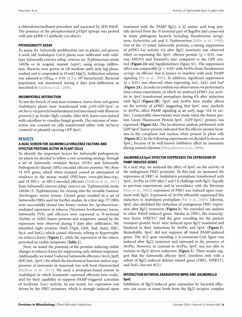
RESULTSA DUAL SCREEN FOR SALMONELLA VIRULENCE FACTORS ANDEFFECTOR PROTEINS ACTIVE IN PLANT CELLSTo identify the important factors for Salmonella pathogenicityon plants we decided to follow a two-screening-strategy througha set of Salmonella virulence factors (SVFs) and SalmonellaPathogenicity Islands (SPIs)-encoded effector proteins. We chose54 SVF genes, which when mutated caused an attenuation ofvirulence in the mouse model (PHI-base, www.phi-base.org),and 18 SPI-1- or SPI-2-encoded effectors (Heffron et al., 2011)from Salmonella enterica subsp. enterica ser. Typhimurium strain14028s (S. Typhimurium) for cloning into the versatile Gateway(Invitrogen) vector system. Cloned genes resulted in a set ofSalmonella ORFs used for further studies. In a first step, 37 ORFswere successfully cloned into binary vectors for Agrobacterium-mediated expression in tobacco (Nicotiana benthamiana) leaves.Salmonella SVFs and effectors were expressed as N-terminal10xMyc or 3xHA fusion proteins and symptoms caused by theexpression were observed during 5 days after infiltration. Weidentified eight proteins (SseF, OrgA, Orf4, SsaI, SsaQ, HilC,SicA, and SseG), which caused chlorosis, wilting or hypertrophyon tobacco leaves (Figure 1), while the expression of the others,provoked no visible symptoms (Table 1).
Next, we tested the potential of the proteins inducing visiblechanges in tobacco leaves for suppressing early defense responses.Additionally, we tested 5 selected Salmonella effectors (AvrA, SptP,SlrP, SseL, SpvC) for which the biochemical function and/or sup-pression of immunity in mammals has well been characterized(Heffron et al., 2011). We used a protoplast-based system inArabidopsis in which transiently expressed effectors were evalu-ated for their capability to suppress PAMP-triggered activationof luciferase (Luc) activity. In our screen Luc expression wasdriven by the FRK1 promoter, which is strongly induced upon
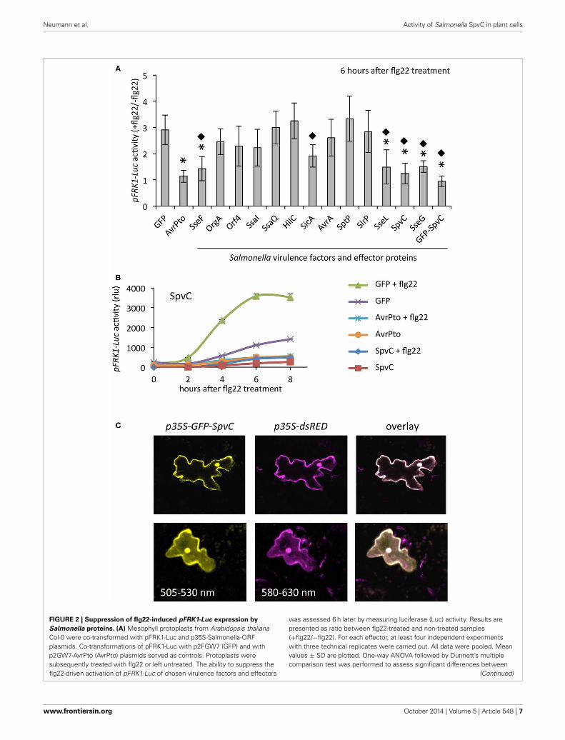
treatment with the PAMP flg22, a 22 amino acid long pep-tide derived from the N-terminal part of flagellin and conservedin many pathogenic bacteria including Pseudomonas aerugi-nosa, Escherichia coli and S. Typhimurium (Felix et al., 1999).Out of the 13 tested Salmonella proteins, a strong suppressionof pFRK1-Luc activity 6 h after flg22 treatment was observedwhen co-expressing the SpvC effector protein (p < 0.01, one-way ANOVA and Dunnett’s test) compared to the GFP con-trol (Figure 2A and Supplementary Figure S1). The suppressioneffect was comparable (p > 0.05) with AvrPto from Pseudomonassyringe, an effector that is known to interfere with early PAMPsignaling (He et al., 2006). In addition, significant suppression(p < 0.01) was observed when expressing SseL, SseG, and SseF(Figure 2A). In order to confirm our observations we performed atime-course experiment, in which we analyzed pFRK1-Luc activ-ity in SpvC-transformed protoplasts during 8 h after inductionwith flg22 (Figure 2B). SpvC and AvrPto have similar effectson the activity of pFRK1 suggesting that SpvC may, similarlyto AvrPto, affect PAMP signaling at an early stage (4 h or ear-lier). Comparable observations were made when the fusion pro-tein Green Fluorescent Protein-SpvC (GFP-SpvC) protein wasexpressed (Figure 2A). The localization analysis performed withGFP-SpvC fusion protein indicated that the effector protein local-izes to the cytoplasm and nucleus when present in plant cells(Figure 2C). In the following experiments we decided to focus onSpvC, because of its well-known inhibitory effect on immunityduring animal infection (Mazurkiewicz et al., 2008).
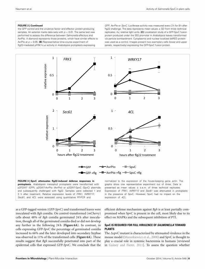
SALMONELLA SpvC EFFECTOR SUPPRESSES THE EXPRESSION OFPAMP-INDUCED GENESIn a next step, we analyzed the effect of SpvC on the activity ofthe endogenous FRK1 promoter. To this end, we measured theexpression of FRK1 in Arabidopsis protoplasts transformed withSpvC, AvrPto, or GFP after 1 and 3 h challenge with flg22. Equallyto previous experiments and in accordance with the literature(Asai et al., 2002), expression of FRK1 was induced upon treat-ment with flg22. Expression of AvrPto efficiently suppressed thisinduction in Arabidopsis protoplasts (He et al., 2006). Likewise,SpvC also abolished the induction of endogenous FRK1 expres-sion after flg22 treatment (Figure 3). We extended our analysisto other PAMP-induced genes. Similar to FRK1, the transcrip-tion factor WRKY17 and the gene encoding for the proteintransport protein Sec61 were induced upon flg22 treatment andhindered in their inductions by AvrPto and SpvC (Figure 3).Remarkably, SpvC did not suppress all tested PAMP-inducedgenes. The 4CL gene encoding a 4-coumarate-CoA ligase wasinduced after flg22 treatment and repressed in the presence ofAvrPto. However, in contrast to AvrPto, SpvC was not able torestrain its flg22-driven induction (Figure 3). These results sug-gest that the Salmonella effector SpvC interferes only with asubset of flg22-induced defense related genes (FRK1, WRKY17,and Sec61, but not 4CL).
INTERACTION BETWEEN ARABIDOPSIS MPK6 AND SALMONELLASpvCInhibition of flg22-induced gene expression by bacterial effec-tors can occur at many levels from the flg22 receptor complex
Frontiers in Microbiology | Plant-Microbe Interaction October 2014 | Volume 5 | Article 548 | 4
Neumann et al. Activity of Salmonella SpvC in plant cells
FIGURE 1 | Screen for Salmonella proteins that induce HR-like
symptoms in plants. SVF and effector genes were cloned into plantexpression vectors and expressed as HA- and Myc-tagged versions in N.benthamiana leaves via Agrobacterium-mediated transformation. Symptomswere observed 5 days after infiltration. Expression of eight proteins resulted
in macroscopic changes in leaf morphology when compared to thenon-transformed parts of the leaf (control), transformation withAgrobacterium GV3101 or infiltration with 10 mM MgCl2. The experimentwas repeated 4 times with both versions of bacterial fusion protein. Only theright side of the leaf was infiltrated.
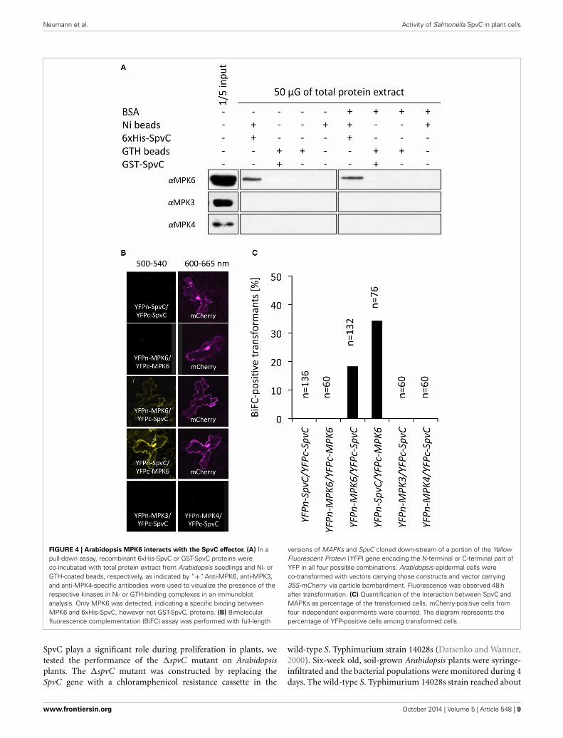
(FLS2-BAK1), through the MAPK cascade, down to transcrip-tional regulation of defense genes. MAPK cascades play a key rolein flg22 signal transduction and in pathogen defense. Among the20 Arabidopsis MAPKs, MPK3, MPK4, and MPK6 are stronglyactivated by flg22 (Asai et al., 2002; Pitzschke et al., 2009). Basedon the functional characteristics of SpvC during animal infec-tion as well as the function of other members of the OspF family[e.g., HopAI1 (Zhang et al., 2007)], we hypothesized that SpvCtargets plant MAPKs. To test our hypothesis, we analyzed pos-sible protein-protein interactions between SpvC and ArabidopsisMAPKs. Recombinant 6xHis-SpvC and GST-SpvC proteins wereexpressed and purified from E. coli BL21 cells. The recombi-nant proteins were subsequently co-incubated with total proteinextract from Arabidopsis seedlings and either Ni- or GTH-coatedbeads were used to precipitate the respective Ni- or GTH-bindingcomplexes. Pull-down samples were probed for the presence ofMAPKs in immunoblot assays. In the presence of His-tagged,but not GST-tagged SpvC, we detected the MPK6 in the pulled-down protein complex (Figure 4A), suggesting the interaction
between SpvC and MPK6. This interaction was observed evenin the presence of an excess of BSA. However, we did not detectMPK3 or MPK4, indicating a specific interaction between SpvCand MPK6.
The in vitro SpvC-MPK6 interaction was tested also inbimolecular fluorescent complementation (BiFC) assays. Full-length cDNAs of SpvC and the three MAPKs were cloneddownstream of sequences encoding either the N- or C-terminalpart of the Yellow Fluorescent Protein (YFP) and subsequentlytransiently expressed in Arabidopsis epidermal cells via parti-cle bombardment. Both tested combinations: (i) YFPn-MPK6with YFPc-SpvC and (ii) YFPn-SpvC with YFPc-MPK6, whenexpressed together, resulted in reconstitution of a functionalYFP protein (Figure 4B). We co-expressed the constructs withp35S-mCherry plasmid, allowing normalization of the interac-tion events (Figure 4C). Eighteen percent of all transformedcells showed visible interaction between SpvC and MPK6 whenYFPn-MPK6 was co-expressed with YFPc-SpvC, and 34% of allcells when YFPn-SpvC, and YFPc-MPK6 were used as interaction
www.frontiersin.org October 2014 | Volume 5 | Article 548 | 5
Neumann et al. Activity of Salmonella SpvC in plant cells
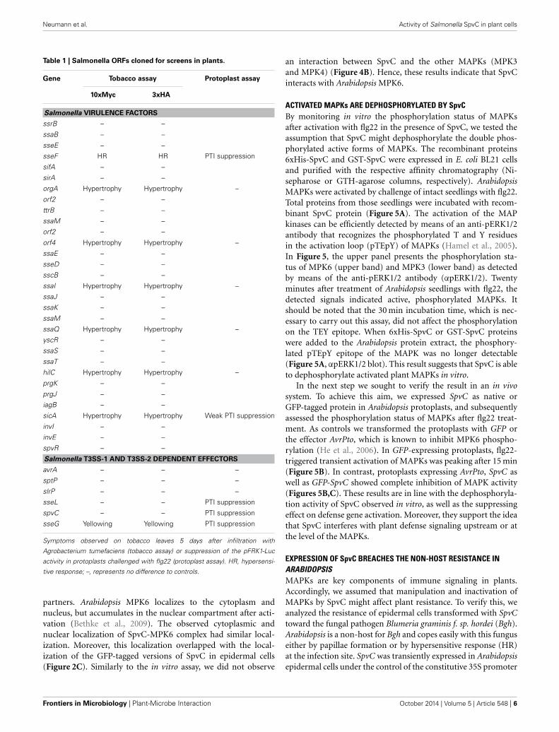
Table 1 | Salmonella ORFs cloned for screens in plants.
Gene Tobacco assay Protoplast assay
10xMyc 3xHA
Salmonella VIRULENCE FACTORS
ssrB – –
ssaB – –
sseE – –
sseF HR HR PTI suppression
sifA – –
sirA – –
orgA Hypertrophy Hypertrophy –
orf2 – –
ttrB – –
ssaM – –
orf2 – –
orf4 Hypertrophy Hypertrophy –
ssaE – –
sseD – –
sscB – –
ssaI Hypertrophy Hypertrophy –
ssaJ – –
ssaK – –
ssaM – –
ssaQ Hypertrophy Hypertrophy –
yscR – –
ssaS – –
ssaT – –
hilC Hypertrophy Hypertrophy –
prgK – –
prgJ – –
iagB – –
sicA Hypertrophy Hypertrophy Weak PTI suppression
invI – –
invE – –
spvR – –
Salmonella T3SS-1 AND T3SS-2 DEPENDENT EFFECTORS
avrA – – –
sptP – – –
slrP – – –
sseL – – PTI suppression
spvC – – PTI suppression
sseG Yellowing Yellowing PTI suppression
Symptoms observed on tobacco leaves 5 days after infiltration with
Agrobacterium tumefaciens (tobacco assay) or suppression of the pFRK1-Luc
activity in protoplasts challenged with flg22 (protoplast assay). HR, hypersensi-
tive response; –, represents no difference to controls.
partners. Arabidopsis MPK6 localizes to the cytoplasm andnucleus, but accumulates in the nuclear compartment after acti-vation (Bethke et al., 2009). The observed cytoplasmic andnuclear localization of SpvC-MPK6 complex had similar local-ization. Moreover, this localization overlapped with the local-ization of the GFP-tagged versions of SpvC in epidermal cells(Figure 2C). Similarly to the in vitro assay, we did not observe
an interaction between SpvC and the other MAPKs (MPK3and MPK4) (Figure 4B). Hence, these results indicate that SpvCinteracts with Arabidopsis MPK6.
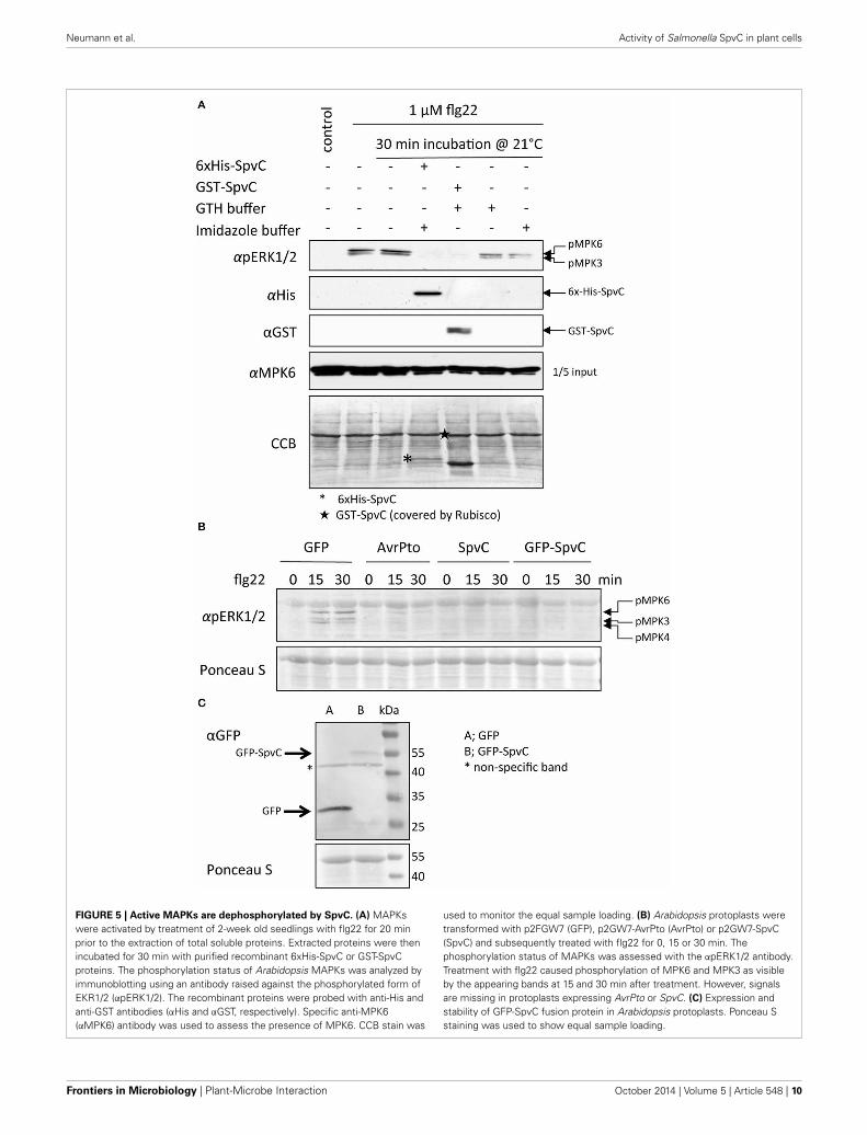
ACTIVATED MAPKs ARE DEPHOSPHORYLATED BY SpvCBy monitoring in vitro the phosphorylation status of MAPKsafter activation with flg22 in the presence of SpvC, we tested theassumption that SpvC might dephosphorylate the double phos-phorylated active forms of MAPKs. The recombinant proteins6xHis-SpvC and GST-SpvC were expressed in E. coli BL21 cellsand purified with the respective affinity chromatography (Ni-sepharose or GTH-agarose columns, respectively). ArabidopsisMAPKs were activated by challenge of intact seedlings with flg22.Total proteins from those seedlings were incubated with recom-binant SpvC protein (Figure 5A). The activation of the MAPkinases can be efficiently detected by means of an anti-pERK1/2antibody that recognizes the phosphorylated T and Y residuesin the activation loop (pTEpY) of MAPKs (Hamel et al., 2005).In Figure 5, the upper panel presents the phosphorylation sta-tus of MPK6 (upper band) and MPK3 (lower band) as detectedby means of the anti-pERK1/2 antibody (αpERK1/2). Twentyminutes after treatment of Arabidopsis seedlings with flg22, thedetected signals indicated active, phosphorylated MAPKs. Itshould be noted that the 30 min incubation time, which is nec-essary to carry out this assay, did not affect the phosphorylationon the TEY epitope. When 6xHis-SpvC or GST-SpvC proteinswere added to the Arabidopsis protein extract, the phosphory-lated pTEpY epitope of the MAPK was no longer detectable(Figure 5A, αpERK1/2 blot). This result suggests that SpvC is ableto dephosphorylate activated plant MAPKs in vitro.
In the next step we sought to verify the result in an in vivosystem. To achieve this aim, we expressed SpvC as native orGFP-tagged protein in Arabidopsis protoplasts, and subsequentlyassessed the phosphorylation status of MAPKs after flg22 treat-ment. As controls we transformed the protoplasts with GFP orthe effector AvrPto, which is known to inhibit MPK6 phospho-rylation (He et al., 2006). In GFP-expressing protoplasts, flg22-triggered transient activation of MAPKs was peaking after 15 min(Figure 5B). In contrast, protoplasts expressing AvrPto, SpvC aswell as GFP-SpvC showed complete inhibition of MAPK activity(Figures 5B,C). These results are in line with the dephosphoryla-tion activity of SpvC observed in vitro, as well as the suppressingeffect on defense gene activation. Moreover, they support the ideathat SpvC interferes with plant defense signaling upstream or atthe level of the MAPKs.
EXPRESSION OF SpvC BREACHES THE NON-HOST RESISTANCE INARABIDOPSISMAPKs are key components of immune signaling in plants.Accordingly, we assumed that manipulation and inactivation ofMAPKs by SpvC might affect plant resistance. To verify this, weanalyzed the resistance of epidermal cells transformed with SpvCtoward the fungal pathogen Blumeria graminis f. sp. hordei (Bgh).Arabidopsis is a non-host for Bgh and copes easily with this funguseither by papillae formation or by hypersensitive response (HR)at the infection site. SpvC was transiently expressed in Arabidopsisepidermal cells under the control of the constitutive 35S promoter
Frontiers in Microbiology | Plant-Microbe Interaction October 2014 | Volume 5 | Article 548 | 6
Neumann et al. Activity of Salmonella SpvC in plant cells
FIGURE 2 | Suppression of flg22-induced pFRK1-Luc expression by
Salmonella proteins. (A) Mesophyll protoplasts from Arabidopsis thalianaCol-0 were co-transformed with pFRK1-Luc and p35S-Salmonella-ORFplasmids. Co-transformations of pFRK1-Luc with p2FGW7 (GFP) and withp2GW7-AvrPto (AvrPto) plasmids served as controls. Protoplasts weresubsequently treated with flg22 or left untreated. The ability to suppress theflg22-driven activation of pFRK1-Luc of chosen virulence factors and effectors
was assessed 6 h later by measuring luciferase (Luc) activity. Results arepresented as ratio between flg22-treated and non-treated samples(+flg22/−flg22). For each effector, at least four independent experimentswith three technical replicates were carried out. All data were pooled. Meanvalues ± SD are plotted. One-way ANOVA followed by Dunnett’s multiplecomparison test was performed to assess significant differences between
(Continued)
www.frontiersin.org October 2014 | Volume 5 | Article 548 | 7
Neumann et al. Activity of Salmonella SpvC in plant cells
FIGURE 2 | Continued
the GFP control and the virulence factor- and effector- protein-producingsamples. An asterisk marks data sets with p < 0.01. The same test wasperformed to assess the difference between Salmonella effectors andAvrPto. A diamond represents those proteins, which have similar effects toAvrPto at p > 0.05. (B) Representative time-course experiment offlg22-mediated pFRK1-Luc activity in Arabidopsis protoplasts expressing
GFP, AvrPto or SpvC. Luciferase activity was measured every 2 h for 8 h afterflg22 challenge. The data represents mean values ± SD from three technicalreplicates. rlu; relative light units. (C) Localization study of a GFP-SpvC fusionprotein produced under the 35S promoter in Arabidopsis leaves transformedvia particle bombardment. Cytoplasmic and nuclear localized dsRED proteinwas used as a control. Images present two exemplary cells (lower and upperpanels, respectively) expressing the GFP-SpvC fusion protein.
FIGURE 3 | SpvC attenuates flg22-induced defense responses in
protoplasts. Arabidopsis mesophyll protoplasts were transformed withp2FGW7 (GFP), p2GW7-AvrPto (AvrPto) or p2GW7-SpvC (SpvC) plasmidsand subsequently challenged with flg22. Samples were collected 1 and3 h after treatment. Relative expression levels of FRK1, WRKY17,Sec61, and 4CL were assessed using quantitative RT-PCR and
normalized to the expression of the house-keeping gene actin. Thegraphs show one representative experiment out of three. Data ispresented as mean values ± s.e.m. of three technical replicates.Expression of FRK1, WRKY17, and Sec61 was attenuated in protoplastsin the presence of SpvC. However, SpvC had no impact on theexpression of 4CL.
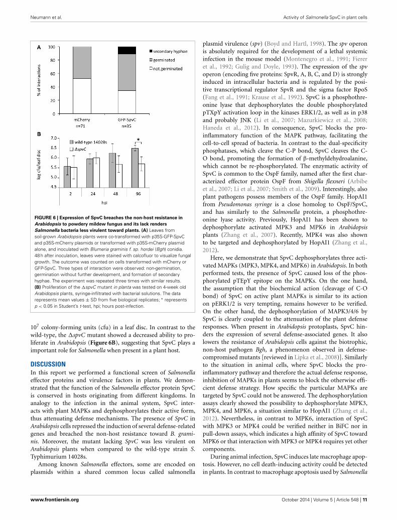
as a GFP-tagged version (GFP-SpvC) and transformed leaves wereinoculated with Bgh conidia. On control-transformed (mCherry)cells about 48% of Bgh conidia germinated 24 h after inocula-tion, though all of the germinated conidia died or did not developany further in the following 24 h (Figure 6A). In contrast, incells expressing GFP-SpvC the percentage of germinated conidiaincreased to 66% and the later developed into secondary hyphaewas observed in 11% of the transformed cells (Figure 6A). Theseresults suggest that Bgh successfully penetrated into part of theepidermal cells that expressed GFP-SpvC. We conclude that the
efficient defense mechanism against Bgh is at least partially com-promised when SpvC is present in the cell, most likely due to itseffect on MAPKs and the subsequent inhibition of PTI.
SpvC IS REQUIRED FOR FULL VIRULENCE OF SALMONELLA TOWARDPLANTSThe �spvC mutant is characterized by attenuated virulence in themouse model (Mazurkiewicz et al., 2008) and SpvC is thought toplay a crucial role in systemic bacteremia in humans [reviewedin Guiney and Fierer, 2011]. To assess the question whether
Frontiers in Microbiology | Plant-Microbe Interaction October 2014 | Volume 5 | Article 548 | 8
Neumann et al. Activity of Salmonella SpvC in plant cells
FIGURE 4 | Arabidopsis MPK6 interacts with the SpvC effector. (A) In apull-down assay, recombinant 6xHis-SpvC or GST-SpvC proteins wereco-incubated with total protein extract from Arabidopsis seedlings and Ni- orGTH-coated beads, respectively, as indicated by “+”. Anti-MPK6, anti-MPK3,and anti-MPK4-specific antibodies were used to visualize the presence of therespective kinases in Ni- or GTH-binding complexes in an immunoblotanalysis. Only MPK6 was detected, indicating a specific binding betweenMPK6 and 6xHis-SpvC, however not GST-SpvC, proteins. (B) Bimolecularfluorescence complementation (BiFC) assay was performed with full-length
versions of MAPKs and SpvC cloned down-stream of a portion of the YellowFluorescent Protein (YFP) gene encoding the N-terminal or C-terminal part ofYFP in all four possible combinations. Arabidopsis epidermal cells wereco-transformed with vectors carrying those constructs and vector carrying35S-mCherry via particle bombardment. Fluorescence was observed 48 hafter transformation. (C) Quantification of the interaction between SpvC andMAPKs as percentage of the transformed cells. mCherry-positive cells fromfour independent experiments were counted. The diagram represents thepercentage of YFP-positive cells among transformed cells.
SpvC plays a significant role during proliferation in plants, wetested the performance of the �spvC mutant on Arabidopsisplants. The �spvC mutant was constructed by replacing theSpvC gene with a chloramphenicol resistance cassette in the
wild-type S. Typhimurium strain 14028s (Datsenko and Wanner,2000). Six-week old, soil-grown Arabidopsis plants were syringe-infiltrated and the bacterial populations were monitored during 4days. The wild-type S. Typhimurium 14028s strain reached about
www.frontiersin.org October 2014 | Volume 5 | Article 548 | 9
Neumann et al. Activity of Salmonella SpvC in plant cells
FIGURE 5 | Active MAPKs are dephosphorylated by SpvC. (A) MAPKswere activated by treatment of 2-week old seedlings with flg22 for 20 minprior to the extraction of total soluble proteins. Extracted proteins were thenincubated for 30 min with purified recombinant 6xHis-SpvC or GST-SpvCproteins. The phosphorylation status of Arabidopsis MAPKs was analyzed byimmunoblotting using an antibody raised against the phosphorylated form ofEKR1/2 (αpERK1/2). The recombinant proteins were probed with anti-His andanti-GST antibodies (αHis and αGST, respectively). Specific anti-MPK6(αMPK6) antibody was used to assess the presence of MPK6. CCB stain was
used to monitor the equal sample loading. (B) Arabidopsis protoplasts weretransformed with p2FGW7 (GFP), p2GW7-AvrPto (AvrPto) or p2GW7-SpvC(SpvC) and subsequently treated with flg22 for 0, 15 or 30 min. Thephosphorylation status of MAPKs was assessed with the αpERK1/2 antibody.Treatment with flg22 caused phosphorylation of MPK6 and MPK3 as visibleby the appearing bands at 15 and 30 min after treatment. However, signalsare missing in protoplasts expressing AvrPto or SpvC. (C) Expression andstability of GFP-SpvC fusion protein in Arabidopsis protoplasts. Ponceau Sstaining was used to show equal sample loading.
Frontiers in Microbiology | Plant-Microbe Interaction October 2014 | Volume 5 | Article 548 | 10
Neumann et al. Activity of Salmonella SpvC in plant cells
FIGURE 6 | Expression of SpvC breaches the non-host resistance in
Arabidopsis to powdery mildew fungus and its lack renders
Salmonella bacteria less virulent toward plants. (A) Leaves fromsoil-grown Arabidopsis plants were co-transformed with p35S-GFP-SpvCand p35S-mCherry plasmids or transformed with p35S-mCherry plasmidalone, and inoculated with Blumeria graminis f. sp. hordei (Bgh) conidia.48 h after inoculation, leaves were stained with calcofluor to visualize fungalgrowth. The outcome was counted on cells transformed with mCherry orGFP-SpvC. Three types of interaction were observed: non-germination,germination without further development, and formation of secondaryhyphae. The experiment was repeated three times with similar results.(B) Proliferation of the �spvC mutant in planta was tested on 4-week oldArabidopsis plants, syringe-infiltrated with bacterial solutions. The datarepresents mean values ± SD from five biological replicates; ∗ representsp < 0.05 in Student’s t-test, hpi; hours post-infection.
107 colony-forming units (cfu) in a leaf disc. In contrast to thewild-type, the �spvC mutant showed a decreased ability to pro-liferate in Arabidopsis (Figure 6B), suggesting that SpvC plays aimportant role for Salmonella when present in a plant host.
DISCUSSIONIn this report we performed a functional screen of Salmonellaeffector proteins and virulence factors in plants. We demon-strated that the function of the Salmonella effector protein SpvCis conserved in hosts originating from different kingdoms. Inanalogy to the infection in the animal system, SpvC inter-acts with plant MAPKs and dephosphorylates their active form,thus attenuating defense mechanisms. The presence of SpvC inArabidopsis cells repressed the induction of several defense-relatedgenes and breached the non-host resistance toward B. grami-nis. Moreover, the mutant lacking SpvC was less virulent onArabidopsis plants when compared to the wild-type strain S.Typhimurium 14028s.
Among known Salmonella effectors, some are encoded onplasmids within a shared common locus called salmonella
plasmid virulence (spv) (Boyd and Hartl, 1998). The spv operonis absolutely required for the development of a lethal systemicinfection in the mouse model (Montenegro et al., 1991; Fiereret al., 1992; Gulig and Doyle, 1993). The expression of the spvoperon (encoding five proteins: SpvR, A, B, C, and D) is stronglyinduced in intracellular bacteria and is regulated by the posi-tive transcriptional regulator SpvR and the sigma factor RpoS(Fang et al., 1991; Krause et al., 1992). SpvC is a phosphothre-onine lyase that dephosphorylates the double phosphorylatedpTXpY activation loop in the kinases ERK1/2, as well as in p38and probably JNK (Li et al., 2007; Mazurkiewicz et al., 2008;Haneda et al., 2012). In consequence, SpvC blocks the pro-inflammatory function of the MAPK pathway, facilitating thecell-to-cell spread of bacteria. In contrast to the dual-specificityphosphatases, which cleave the C-P bond, SpvC cleaves the C-O bond, promoting the formation of β-methyldehydroalanine,which cannot be re-phosphorylated. The enzymatic activity ofSpvC is common to the OspF family, named after the first char-acterized effector protein OspF from Shigella flexneri (Arbibeet al., 2007; Li et al., 2007; Smith et al., 2009). Interestingly, alsoplant pathogens possess members of the OspF family. HopAI1from Pseudomonas syringe is a close homolog to OspF/SpvC,and has similarly to the Salmonella protein, a phosphothre-onine lyase activity. Previously, HopAI1 has been shown todephosphorylate activated MPK3 and MPK6 in Arabidopsisplants (Zhang et al., 2007). Recently, MPK4 was also shownto be targeted and dephosphorylated by HopAI1 (Zhang et al.,2012).
Here, we demonstrate that SpvC dephosphorylates three acti-vated MAPKs (MPK3, MPK4, and MPK6) in Arabidopsis. In bothperformed tests, the presence of SpvC caused loss of the phos-phorylated pTEpY epitope on the MAPKs. On the one hand,the assumption that the biochemical action (cleavage of C-Obond) of SpvC on active plant MAPKs is similar to its actionon pERK1/2 is very tempting, remains however to be verified.On the other hand, the dephosphorylation of MAPK3/4/6 bySpvC is clearly coupled to the attenuation of the plant defenseresponses. When present in Arabidopsis protoplasts, SpvC hin-ders the expression of several defense-associated genes. It alsolowers the resistance of Arabidopsis cells against the biotrophic,non-host pathogen Bgh, a phenomenon observed in defense-compromised mutants [reviewed in Lipka et al., 2008)]. Similarlyto the situation in animal cells, where SpvC blocks the pro-inflammatory pathway and therefore the actual defense response,inhibition of MAPKs in plants seems to block the otherwise effi-cient defense strategy. How specific the particular MAPKs aretargeted by SpvC could not be answered. The dephosphorylationassays clearly showed the possibility to dephosphorylate MPK3,MPK4, and MPK6, a situation similar to HopAI1 (Zhang et al.,2012). Nevertheless, in contrast to MPK6, interaction of SpvCwith MPK3 or MPK4 could be verified neither in BiFC nor inpull-down assays, which indicates a high affinity of SpvC towardMPK6 or that interaction with MPK3 or MPK4 requires yet othercomponents.
During animal infection, SpvC induces late macrophage apop-tosis. However, no cell death-inducing activity could be detectedin plants. In contrast to macrophage apoptosis used by Salmonella
www.frontiersin.org October 2014 | Volume 5 | Article 548 | 11
Neumann et al. Activity of Salmonella SpvC in plant cells
to facilitate the cell-to-cell spread in animal organism, cellulardeath in plants (hypersensitive response; HR) is very often adefense mechanism induced by recognition of pathogen effec-tor proteins by the plant intracellular R proteins. Despite the factthat SpvC is a T3SS-translocated effector in mammalian cells, thedescribed above screen in tobacco leaves suggests that SpvC doesnot induce the hallmark of effector-triggered immunity (ETI)in plants, the HR, implying that SpvC is not recognized by Rprotein(s). We also exclude the possibility that SpvC is recog-nized by surface located receptors by testing its PAMP activity.Growth inhibition and production of reactive oxygen species,both hallmarks of pattern-triggered immunity (PTI), were stud-ied in plants after contact with SpvC (Supplementary Figure S2).Our results suggest that SpvC is not toxic for plant cells whenexternally present and that plants do not recognize SpvC bypotential surface receptor(s).
As described above, the intracellular presence of SpvC atten-uated the activation of MPK3/4/6 and expression of severaldefense-related genes. Whether, besides inhibition of those twoaspects of plant defense, SpvC actively suppresses the HR responseremains to be verified in future experiments. Furthermore, thetranslocation of Salmonella effector proteins into plant cytoplasmwas not yet demonstrated. The function of SpvC requires itspresence in the host cytoplasm, therefore a direct evidence oftranslocation of this effector (or/and others) needs to be pro-vided in future work, as this would certainly help to understandhow these bacteria suppress plant immune responses. Interestingwas the observation that expression of other Salmonella effec-tors in planta induced visible changes. SseF and SseG, bothSPI-2 encoded effector proteins involved in the trafficking ofSalmonella Containing Vacuole (SCV) in animal cells, inducedHR-like (SseF) or yellowing (SseG) symptoms in tobacco leaves,when expressed via Agrobacterium-mediated transformation. Itconfirms the observation made by Ustun et al. (2012), whoshowed that SseF from S. enterica triggers HR-like symptoms intobacco plants when expressed transiently via Agrobacterium infil-tration or delivered via the T3SS from Xanthomonas campestris pv.vesicatoria. Moreover, the ability of SseF to trigger HR-like symp-toms was lost upon silencing of SGT1 (suppressor of G2 alleleof skp1), which is required for HR induction in tobacco. Theseresults indicate that Salmonella SseF is recognized in N. ben-thamiana via an R protein-mediated mechanism and triggersETI in consequence. Surprisingly, expression of SptP or SlrP,both postulated to be key effectors of Salmonella with the high-est number of predicted protein-protein interactions (Schlekeret al., 2012), induced no visible symptoms in tobacco leavesnor had an effect on the induction of pFRK1-Luc in Arabidopsisprotoplasts.
In summary, an increasing number of evidence indicates thatplants evolved diverse mechanisms to recognize Salmonella bacte-ria using surface receptors as well as intracellular R proteins. Ourstudy supports the view that Salmonella also evolved means tointerfere with plant immunity by efficiently employing its reper-toire of effector proteins to succumb plant immune responses.Consequently, Salmonella, and possibly other human pathogenicbacteria, seems to possess effective tools for suppression of theplant immune system.
ACKNOWLEDGMENTSThe work of Casandra Hernàndez-Reyes was supported byCONACYT fellowship from the Mexican Ministry for Science.Heribert Hirt was supported by a grant of the ERANET SystemsBiology project SHIPREC (Salmonella Host Interaction ProjectEuropean Consortium).
SUPPLEMENTARY MATERIALThe Supplementary Material for this article can be foundonline at: http://www.frontiersin.org/journal/10.3389/fmicb.2014.00548/abstract
REFERENCESArbibe, L., Kim, D. W., Batsche, E., Pedron, T., Mateescu, B., Muchardt, C., et al.
(2007). An injected bacterial effector targets chromatin access for transcrip-tion factor NF-kappaB to alter transcription of host genes involved in immuneresponses. Nat. Immunol. 8, 47–56. doi: 10.1038/ni1423
Asai, T., Tena, G., Plotnikova, J., Willmann, M. R., Chiu, W. L., Gomez-Gomez, L.,et al. (2002). MAP kinase signalling cascade in Arabidopsis innate immunity.Nature 415, 977–983. doi: 10.1038/415977a
Bethke, G., Unthan, T., Uhrig, J. F., Poschl, Y., Gust, A. A., Scheel, D., et al. (2009).Flg22 regulates the release of an ethylene response factor substrate from MAPkinase 6 in Arabidopsis thaliana via ethylene signaling. Proc. Natl. Acad. Sci. USA.106, 8067–8072. doi: 10.1073/pnas.0810206106
Boller, T. (2012). Experimental evidence of a role for RLKs in innate immunity.Signal. Commun. Plants 13, 67–77. doi: 10.1007/978-3-642-23044-8_4
Boyd, E. F., and Hartl, D. L. (1998). Salmonella virulence plasmid. Modularacquisition of the spv virulence region by an F-plasmid in Salmonella entericasubspecies I and insertion into the chromosome of subspecies II, IIIa, IV andVII isolates. Genetics 149, 1183–1190.
Chu, C., and Chiu, C. H. (2006). Evolution of the virulence plasmids of non-typhoid Salmonella and its association with antimicrobial resistance. MicrobesInfect. 8, 1931–1936. doi: 10.1016/j.micinf.2005.12.026
Datsenko, K. A., and Wanner, B. L. (2000). One-step inactivation of chromosomalgenes in Escherichia coli K-12 using PCR products. Proc. Natl. Acad. Sci. U.S.A.97, 6640–6645. doi: 10.1073/pnas.120163297
Fang, F. C., Krause, M., Roudier, C., Fierer, J., and Guiney, D. G. (1991). Growthregulation of a Salmonella plasmid gene essential for virulence. J. Bacteriol. 173,6783–6789.
Felix, G., Duran, J. D., Volko, S., and Boller, T. (1999). Plants have a sensitive per-ception system for the most conserved domain of bacterial flagellin. Plant J. 18,265–276. doi: 10.1046/j.1365-313X.1999.00265.x
Fierer, J., Krause, M., Tauxe, R., and Guiney, D. (1992). Salmonella typhimuriumbacteremia: association with the virulence plasmid. J. Infect. Dis. 166, 639–642.doi: 10.1093/infdis/166.3.639
Fraiture, M., Zheng, X., and Brunner, F. (2014). An Arabidopsis and tomatomesophyll protoplast system for fast identification of early MAMP-triggeredimmunity-suppressing effectors. Methods Mol. Biol. 1127, 213–230. doi:10.1007/978-1-62703-986-4_17
Garcia, A. V., Charrier, A., Schikora, A., Bigeard, J., Pateyron, S., De Tauzia-Moreau,M. L., et al. (2014). Salmonella enterica flagellin is recognized via FLS2 and acti-vates PAMP-triggered immunity in Arabidopsis thaliana. Mol. Plant 7, 657–674.doi: 10.1093/mp/sst145
Golberg, D., Kroupitski, Y., Belausov, E., Pinto, R., and Sela, S. (2011).Salmonella typhimurium internalization is variable in leafy vegetables andfresh herbs. Int. J. Food Microbiol. 145, 250–257. doi: 10.1016/j.ijfoodmicro.2010.12.031
Guiney, D. G., and Fierer, J. (2011). The role of the spv genes in Salmonellapathogenesis. Front. Microbiol. 2:129. doi: 10.3389/fmicb.2011.00129
Gulig, P. A., and Doyle, T. J. (1993). The Salmonella typhimurium virulence plas-mid increases the growth rate of salmonellae in mice. Infect. Immun. 61,504–511.
Haapalainen, M., Van Gestel, K., Pirhonen, M., and Taira, S. (2009). Soluble plantcell signals induce the expression of the type III secretion system of Pseudomonassyringae and upregulate the production of pilus protein HrpA. Mol. PlantMicrobe Interact. 22, 282–290. doi: 10.1094/MPMI-22-3-0282
Frontiers in Microbiology | Plant-Microbe Interaction October 2014 | Volume 5 | Article 548 | 12
Neumann et al. Activity of Salmonella SpvC in plant cells
Hamel, L. P., Miles, G. P., Samuel, M. A., Ellis, B. E., Seguin, A., and Beaudoin, N.(2005). Activation of stress-responsive mitogen-activated protein kinase path-ways in hybrid poplar (Populus trichocarpa x Populus deltoides). Tree Physiol.25, 277–288. doi: 10.1093/treephys/25.3.277
Haneda, T., Ishii, Y., Shimizu, H., Ohshima, K., Iida, N., Danbara, H., et al. (2012).Salmonella type III effector SpvC, a phosphothreonine lyase, contributes toreduction in inflammatory response during intestinal phase of infection. Cell.Microbiol. 14, 485–499. doi: 10.1111/j.1462-5822.2011.01733.x
He, P., Shan, L., Lin, N. C., Martin, G. B., Kemmerling, B., Nurnberger, T.,et al. (2006). Specific bacterial suppressors of MAMP signaling upstreamof MAPKKK in Arabidopsis innate immunity. Cell 125, 563–575. doi:10.1016/j.cell.2006.02.047
Heffron, F., Niemann, G., Yoon, H., Kidwai, A., Brown, R., McDemrott, J., et al.(2011). “Salmonella-secreted virulence factors,” in Salmonella from Genom toFunction, ed S. Porwollik (San Diego, CA: Caister Academic Press), 187–223.
Holden, N., Pritchard, L., and Toth, I. (2009). Colonization outwith the colon:plants as an alternative environmental reservoir for human pathogenicenterobacteria. FEMS Microbiol. Rev. 33, 689–703. doi: 10.1111/j.1574-6976.2008.00153.x
Jones, J. D., and Dangl, J. L. (2006). The plant immune system. Nature 444,323–329. doi: 10.1038/nature05286
Krause, M., Fang, F. C., and Guiney, D. G. (1992). Regulation of plasmid virulencegene expression in Salmonella dublin involves an unusual operon structure.J. Bacteriol. 174, 4482–4489.
Kroupitski, Y., Golberg, D., Belausov, E., Pinto, R., Swartzberg, D., Granot, D.,et al. (2009). Internalization of Salmonella enterica in leaves is induced by lightand involves chemotaxis and penetration through open stomata. Appl. Environ.Microbiol. 75, 6076–6086. doi: 10.1128/AEM.01084-09
Li, H., Xu, H., Zhou, Y., Zhang, J., Long, C., Li, S., et al. (2007). The phosphothreo-nine lyase activity of a bacterial type III effector family. Science 315, 1000–1003.doi: 10.1126/science.1138960
Lipka, U., Fuchs, R., and Lipka, V. (2008). Arabidopsis non-host resis-tance to powdery mildews. Curr. Opin. Plant Biol. 11, 404–411. doi:10.1016/j.pbi.2008.04.004
Mazurkiewicz, P., Thomas, J., Thompson, J. A., Liu, M., Arbibe, L., Sansonetti, P.,et al. (2008). SpvC is a Salmonella effector with phosphothreonine lyase activityon host mitogen-activated protein kinases. Mol. Microbiol. 67, 1371–1383. doi:10.1111/j.1365-2958.2008.06134.x
Meng, F., Altier, C., and Martin, G. B. (2013). Salmonella colonization activatesthe plant immune system and benefits from association with plant pathogenicbacteria. Environ. Microbiol. 15, 2418–2430. doi: 10.1111/1462-2920.12113
Milillo, S. R., Badamo, J. M., Boor, K. J., and Wiedmann, M. (2008). Growth andpersistence of Listeria monocytogenes isolates on the plant model Arabidopsisthaliana. Food Microbiol. 25, 698–704. doi: 10.1016/j.fm.2008.03.003
Montenegro, M. A., Morelli, G., and Helmuth, R. (1991). Heteroduplex analysisof Salmonella virulence plasmids and their prevalence in isolates of definedsources. Microb. Pathog. 11, 391–397. doi: 10.1016/0882-4010(91)90035-9
Pang, T., Bhutta, Z. A., Finlay, B. B., and Altwegg, M. (1995). Typhoid fever andother salmonellosis: a continuing challenge. Trends Microbiol. 3, 253–255. doi:10.1016/S0966-842X(00)88937-4
Pitzschke, A., Schikora, A., and Hirt, H. (2009). MAPK cascade signallingnetworks in plant defence. Curr. Opin. Plant Biol. 12, 421–426. doi:10.1016/j.pbi.2009.06.008
Prithiviraj, B., Bais, H. P., Jha, A. K., and Vivanco, J. M. (2005). Staphylococcusaureus pathogenicity on Arabidopsis thaliana is mediated either by a direct effectof salicylic acid on the pathogen or by SA-dependent, NPR1-independent hostresponses. Plant J. 42, 417–432. doi: 10.1111/j.1365-313X.2005.02385.x
Rangel, J. M., Sparling, P. H., Crowe, C., Griffin, P. M., and Swerdlow, D. L. (2005).Epidemiology of Escherichia coli O157:H7 outbreaks, United States, 1982-2002.Emerg. Infect. Dis. 11, 603–609. doi: 10.3201/eid1104.040739
Schikora, A., Carreri, A., Charpentier, E., and Hirt, H. (2008). The dark side ofthe salad: Salmonella typhimurium overcomes the innate immune response ofArabidopsis thaliana and shows an endopathogenic lifestyle. PLoS ONE 3:e2279.doi: 10.1371/journal.pone.0002279
Schikora, A., Virlogeux-Payant, I., Bueso, E., Garcia, A. V., Nilau, T., Charrier, A.,et al. (2011). Conservation of Salmonella infection mechanisms in plants andanimals. PLoS ONE 6:e24112. doi: 10.1371/journal.pone.0024112
Schleker, S., Garcia-Garcia, J., Klein-Seetharaman, J., and Oliva, B. (2012).Prediction and comparison of salmonellahuman and salmonellaarabidop-sis interactomes. Chem. Biodivers. 9, 991–1018. doi: 10.1002/cbdv.201100392
Shirron, N., and Yaron, S. (2011). Active suppression of early immune response intobacco by the human pathogen Salmonella typhimurium. PLoS ONE 6:e18855.doi: 10.1371/journal.pone.0018855
Smith, G. K., Ke, Z., Hengge, A. C., Xu, D., Xie, D., and Guo, H. (2009). Active-sitedynamics of SpvC virulence factor from Salmonella typhimurium and densityfunctional theory study of phosphothreonine lyase catalysis. J. Phys. Chem. B113, 15327–15333. doi: 10.1021/jp9052677
Ustun, S., Muller, P., Palmisano, R., Hensel, M., and Bornke, F. (2012). SseF, atype III effector protein from the mammalian pathogen Salmonella enterica,requires resistance-gene-mediated signalling to activate cell death in the modelplant Nicotiana benthamiana. New Phytol. 194, 1046–1060. doi: 10.1111/j.1469-8137.2012.04124.x
Westrell, T., Ciampa, N., Boelaert, F., Helwigh, B., Korsgaard, H., Chriel, M.,et al. (2009). Zoonotic infections in Europe in 2007: a summary of theEFSA-ECDC annual report. Euro Surveill. 14. Available online at: http://www.
eurosurveillance.org/ViewArticle.aspx?ArticleId=19100Yoo, S. D., Cho, Y. H., and Sheen, J. (2007). Arabidopsis mesophyll protoplasts:
a versatile cell system for transient gene expression analysis. Nat. Protoc. 2,1565–1572. doi: 10.1038/nprot.2007.199
Zhang, J., Shao, F., Li, Y., Cui, H., Chen, L., Li, H., et al. (2007). A Pseudomonassyringae effector inactivates MAPKs to suppress PAMP-induced immunity inplants. Cell Host Microbe 1, 175–185. doi: 10.1016/j.chom.2007.03.006
Zhang, Z., Wu, Y., Gao, M., Zhang, J., Kong, Q., Liu, Y., et al. (2012). Disruption ofPAMP-induced MAP kinase cascade by a Pseudomonas syringae effector acti-vates plant immunity mediated by the NB-LRR protein SUMM2. Cell HostMicrobe 11, 253–263. doi: 10.1016/j.chom.2012.01.015
Zheng, X., McLellan, H., Fraiture, M., Liu, X., Boevink, P. C., Gilroy, E. M., et al.(2014). Functionally redundant RXLR effectors from Phytophthora infestansact at different steps to suppress early flg22-triggered immunity. PLoS Pathog.10:e1004057. doi: 10.1371/journal.ppat.1004057
Conflict of Interest Statement: The authors declare that the research was con-ducted in the absence of any commercial or financial relationships that could beconstrued as a potential conflict of interest.
Received: 29 April 2014; accepted: 01 October 2014; published online: 17 October 2014.Citation: Neumann C, Fraiture M, Hernàndez-Reyes C, Akum FN, Virlogeux-PayantI, Chen Y, Pateyron S, Colcombet J, Kogel K-H, Hirt H, Brunner F and Schikora A(2014) The Salmonella effector protein SpvC, a phosphothreonine lyase is functionalin plant cells. Front. Microbiol. 5:548. doi: 10.3389/fmicb.2014.00548This article was submitted to Plant-Microbe Interaction, a section of the journalFrontiers in Microbiology.Copyright © 2014 Neumann, Fraiture, Hernàndez-Reyes, Akum, Virlogeux-Payant,Chen, Pateyron, Colcombet, Kogel, Hirt, Brunner and Schikora. This is an open-access article distributed under the terms of the Creative Commons Attribution License(CC BY). The use, distribution or reproduction in other forums is permitted, providedthe original author(s) or licensor are credited and that the original publication in thisjournal is cited, in accordance with accepted academic practice. No use, distribution orreproduction is permitted which does not comply with these terms.
www.frontiersin.org October 2014 | Volume 5 | Article 548 | 13