Embed Size (px)
Citation preview

The Saccharomyces cerevisiae Actin Patch Protein App1p Is aPhosphatidate Phosphatase Enzyme*�
Received for publication, September 21, 2012, and in revised form, October 11, 2012 Published, JBC Papers in Press, October 15, 2012, DOI 10.1074/jbc.M112.421776
Minjung Chae, Gil-Soo Han, and George M. Carman1
From the Department of Food Science, Rutgers Center for Lipid Research, and New Jersey Institute for Food, Nutrition,and Health, Rutgers University, New Brunswick, New Jersey 08901
Background: Phosphatidate phosphatase (PAP) plays diverse roles in lipid metabolism and cell signaling.Results: A novel yeast PAP is identified as the actin patch protein encoded by APP1.Conclusion: APP1 and other known genes (PAH1, DPP1, LPP1) are responsible for all detectable PAP activity in yeast.Significance: Identification of App1p as a PAP enzyme will facilitate the understanding of its cellular function.
Phosphatidate phosphatase (PAP) catalyzes the dephosphor-ylation of phosphatidate to yield diacylglycerol. In the yeast Sac-charomyces cerevisiae, PAP is encoded by PAH1, DPP1, andLPP1. The presence of PAP activity in the pah1� dpp1� lpp1�
triple mutant indicated another gene(s) encoding the enzyme.Wepurified PAP from the pah1�dpp1� lpp1� triplemutant bysalt extraction of mitochondria followed by chromatographywith DE52, Affi-Gel Blue, phenyl-Sepharose, MonoQ, andSuperdex 200. Liquid chromatography/tandemmass spectrom-etry analysis of a PAP-enriched sample revealed multiple puta-tive phosphatases. By analysis of PAPactivity inmutants lackingeach of the proteins, we found that APP1, a gene whose molec-ular function has been unknown, confers �30% PAP activity ofwild type cells. The overexpression ofAPP1 in the pah1�dpp1�
lpp1� mutant exhibited a 10-fold increase in PAP activity. ThePAP activity shown byApp1p heterologously expressed inEsch-erichia coli confirmed that APP1 is the structural gene for theenzyme. Introduction of the app1� mutation into the pah1�
dpp1� lpp1� triple mutant resulted in a complete loss of PAPactivity, indicating that distinct PAPenzymes inS. cerevisiae areencoded byAPP1,PAH1,DPP1, andLPP1. Lipid analysis of cellslacking the PAP genes, singly or in combination, showed thatPah1p is the onlyPAP involved in the synthesis of triacylglycerolas well as in the regulation of phospholipid synthesis. App1p,which shows interactions with endocytic proteins, may play arole in vesicular trafficking through its PAP activity.
PAP2 catalyzes the dephosphorylation of PA to produceDAGandPi (1). The productDAG is utilized for the synthesis ofthe neutral storage lipid TAG and for the synthesis of the phos-pholipids phosphatidylcholine and phosphatidylethanolaminevia the Kennedy pathway (1–6). The substrate PA is utilized forthe synthesis of phospholipids (e.g. phosphatidylinositol, phos-
phatidylglycerol, and cardiolipin) via the liponucleotide inter-mediate CDP-DAG (7–9). In addition to serving as intermedi-ates in lipid synthesis, PA and DAG are involved in lipidsignaling. For example, PA plays a role in cell growth activation,membrane proliferation, the transcription of lipid synthesisgenes, secretion, and vesicular transport, whereas DAG acti-vates protein kinase C (10–20). Thus, by the nature of its reac-tion, PAP activity may regulate the proportional synthesis ofphospholipids and TAG, the pathways by which phospholipidsare synthesized, and control the abundance of important sig-naling lipids. Indeed, biochemical and genetic studies usingyeast and mammalian experimental systems have shown thatPAP is an important regulator of lipid homeostasis and cellphysiology (21–25).Our laboratory has utilized the yeast Saccharomyces cerevi-
siae as a model eukaryote to study the enzymology and physio-logical roles of PAP enzymes (22, 26). In S. cerevisiae, PAP isknown to be encoded by three genes, namely PAH1 (27),DPP1(28), and LPP1 (29). Pah1p PAP is a Mg2�-dependent enzymewhose reaction is based on the DXDX(T/V) catalytic motifwithin the haloacid dehalogenase-like domain of the enzyme(27, 30). In contrast, the PAP activities ofDpp1p (28) andLpp1p(29) do not have a divalent cation requirement, and their reac-tions are based on a three-domain lipid phosphatase motifcomposed of the consensus sequences KX6RP, PSGH, andSRX5HX3D (31, 32). The Pah1p enzyme is specific for PA (27),whereas the Dpp1p and Lpp1p enzymes utilize PA and otherlipid phosphate molecules (e.g. DAG pyrophosphate andlysoPA) as substrates (28, 29, 33–36). The PAP activities inyeast also differ with respect to their cellular locations andmodes of regulation. Pah1p is found in the cytosol as a phos-phoprotein that is a consequence of multiple phosphoryla-tions (37–40). For catalysis, the phosphorylated enzymetranslocates to the nuclear/endoplasmic reticulum mem-brane through its dephosphorylation (38, 41–43). Dpp1p(28, 44, 45) and Lpp1p (29, 46) are integral membraneenzymes with six transmembrane-spanning regions that arelocalized to the vacuole and Golgi compartments, respec-tively, of the cell.Pah1p PAP plays an important role in lipid metabolism by
controlling the relative proportions of PA andDAG (27, 47). Animbalance of these intermediates due to a loss of PAP activity
* This work was supported, in whole or in part, by National Institutes of HealthGrant GM-28140 (to G. M. C.) from the United States Public Health Service.
� This article was selected as a Paper of the Week.1 To whom correspondence should be addressed: Dept. of Food Science, Rut-
gers University, 65 Dudley Rd., New Brunswick, NJ 08901. Tel.: 848-932-5407; E-mail: [email protected].
2 The abbreviations used are: PAP, phosphatidate phosphatase, PA, phos-phatidate; lysoPA, lysophosphatidic acid; DAG, diacylglycerol; TAG, triacyl-glycerol; SH3, Src homology 3; Ni2�-NTA, Ni2�-nitrilotriacetic acid.
THE JOURNAL OF BIOLOGICAL CHEMISTRY VOL. 287, NO. 48, pp. 40186 –40196, November 23, 2012© 2012 by The American Society for Biochemistry and Molecular Biology, Inc. Published in the U.S.A.
40186 JOURNAL OF BIOLOGICAL CHEMISTRY VOLUME 287 • NUMBER 48 • NOVEMBER 23, 2012
at Rutgers U
niversity, on Novem
ber 26, 2012w
ww
.jbc.orgD
ownloaded from
_profile.html http://www.jbc.org/content/suppl/2012/11/22/M112.421776.DCAuthorSupplemental Material can be found at:

results in cellular abnormalities that include a drastic reductionin TAG abundance and susceptibility to fatty acid-inducedlipotoxicity, themisregulation of phospholipid synthesis and anaberrant expansion of the nuclear/endoplasmic reticulummembrane, and defects in lipid droplet formation and vacuolehomeostasis (27, 30, 43, 47–49). The Dpp1p and Lpp1p are notinvolved in de novo lipid synthesis that occurs at the endoplas-mic reticulum (21, 28, 29). Instead, these enzymes are thoughtto have roles in lipid signaling by controlling the amounts of PA,DAG pyrophosphate, and lysoPA in the organelles where theyreside (21, 26).A significant amount ofMg2�-dependent PAP activity is still
present in the pah1� dpp1� lpp1� triple mutant (27). How-ever, unlike Pah1p PAP, this activity is sensitive to inhibition byN-ethylmaleimide (27). The enzyme activity is found in boththe cytosolic and the membrane fractions of the cell, and itsassociation with the membrane is peripheral in nature (27).Studies to gain insight into the physiological role of the PAPactivity have been hampered because the gene encoding theenzyme has yet to be identified. In this work, we purified PAPfrom the pah1� dpp1� lpp1� triple mutant and identifiedAPP1 as the gene encoding the enzyme. The lack of detectablePAP activity in the app1� pah1� dpp1� lpp1� quadruplemutant indicated that all PAP activity in yeast is encoded byAPP1, PAH1, DPP1, and LPP1. Moreover, this work identifiedthe molecular function of the actin patch protein App1p as aPAP enzyme.
EXPERIMENTAL PROCEDURES
Materials
All chemicals were reagent grade. Growth medium supplieswere purchased from Difco. Polyvinylidene difluoride mem-brane, the enhanced chemiluminescence Western blottingdetection kit, phenyl-Sepharose CL-4B, MonoQ, and Superdex200 columns were purchased from GE Healthcare. DE52(DEAE-cellulose) was purchased from Whatman. Affi-GelBlue, protein assay reagents, electrophoretic reagents, and pro-tein standards were purchased from Bio-Rad. Radiochemicals
were purchased from PerkinElmer Life Sciences. Bovineserum albumin, phenylmethylsulfonyl fluoride, benzamidine,aprotinin, leupeptin, pepstatin, isopropyl-�-D-thiogalactoside,sodium cholate, and Triton X-100 were purchased from Sigma.Lipids were obtained from Avanti Polar Lipids. Silica gel thin-layer chromatography plates were from EM Science. Restric-tion endonucleases, modifying enzymes, and recombinantVentR DNA polymerase were purchased from New EnglandBiolabs. Plasmid isolation and gel extraction kits and Ni2�-NTA-agarose resin were purchased from Qiagen. Invitrogenwas the source of theDNA size standards and the yeast deletionconsortium strain collection. The Yeast Maker yeast transfor-mation kit was purchased from Clontech. Stratagene suppliedthe QuikChange site-directed mutagenesis kit. Nourseothricin(LEXSY NTC) was purchased from Jena Bioscience. Mousemonoclonal anti-HA, and anti-His6 antibodies were fromRoche Applied Science. Anti-App1p antibodies were preparedin rabbits against the C-terminal portion (residues 490–502) ofthe protein at EZBiolab. Alkaline phosphatase-conjugated goatanti-rabbit IgG antibodies and alkaline phosphatase-conju-gated goat anti-mouse IgG antibodies were from Thermo Sci-entific and Pierce, respectively. Scintillation counting supplieswere purchased from National Diagnostics.
Strains and Growth Conditions
The strains used in this work are listed in Table 1. Yeast cellswere grown inYEPDmedium (1% yeast extract, 2%peptone, 2%glucose) or in synthetic complete medium containing 2% glu-cose at 30 °C as described previously (50, 51). For selection ofyeast cells bearing plasmids, the appropriate amino acids wereomitted from synthetic complete medium. Plasmid mainte-nance/amplifications (strain DH5�) and App1p expression(strain BL21(DE3)pLysS) were performed in Escherichia coli.The bacterial cells were grown in LB medium (1% Tryptone,0.5% yeast extract, 1% NaCl, pH 7.4) at 37 °C, and ampicillin(100 �g/ml) was added to select for the cells carrying plasmid.For growth on solid media, agar plates were prepared with sup-plementation of either 2% (yeast) or 1.5% (E. coli) agar. For
TABLE 1Strains and plasmids used in this work
Strain or plasmid Relevant characteristics Source or Ref.
E. coli strainsDH5� F� �80dlacZ�M15 � (lacZYA-argF)U169 deoR recA1 endA1 hsdR17 (rk� mk
�) phoA supE44 l�thi-1 gyrA96 relA1 Ref. 51BL21(DE3)pLysS F� ompT hsdSB (rB�mB
�) gal dcm (DE3) pLysS NovagenS. cerevisiae strainsBY4741 MATa his3�1 leu2�0 met15�0 ura3�0 Ref. 109BY4741-app1� app1�::kanMX4 derivative of BY4741 Deletion consortiumW303–1A MATa ade2–1 can1–100 his3–11,15 leu2–3,112 trp1–1 ura3–1 Ref. 110GHY63 app1�::natMX4 derivative of W303–1A This studyGHY57 pah1�::URA3 derivative of W303–1A Ref. 27GHY64 app1�::natMX4 pah1�::URA3 derivative of W303–1A This studyTBY1 dpp1�::TRP1/Kanr lpp1�::HIS3/Kanr derivative of W303–1A Ref. 29GHY65 app1�::natMX4 dpp1�::TRP1/Kanr lpp1�::HIS3/Kanr derivative of W303–1A This studyGHY58 pah1�::URA3 dpp1�::TRP1/Kanr lpp1�::HIS3/Kanr derivative of W303–1A Ref. 27GHY66 app1�::natMX4 pah1�::URA3 dpp1�::TRP1/Kanr lpp1�::HIS3/Kanr derivative of W303–1A This study
PlasmidsYEp351 Multicopy E. coli/yeast shuttle vector with LEU2 Ref. 91pMC102 APP1 gene inserted into YEp351 This studypMC102-D281E APP1 (D281E) derivative of pMC102 This studypMC103 APP1HA gene inserted into YEp351 This studypMC103-D281E APP1 (D281E) derivative of pMC103 This studypET-15b E. coli expression vector with N-terminal His6 tag fusion NovagenpMC101 APP1 coding sequence inserted into pET-15b This study
App1p Is a Phosphatidate Phosphatase
NOVEMBER 23, 2012 • VOLUME 287 • NUMBER 48 JOURNAL OF BIOLOGICAL CHEMISTRY 40187
at Rutgers U
niversity, on Novem
ber 26, 2012w
ww
.jbc.orgD
ownloaded from
App1p PAP purification in yeast, pah1� dpp1� lpp1� mutantcells were grown to late exponential phase in YEPDmedium at30 °C. For heterologous expression of His6-tagged App1p,E. coli BL21(DE3)pLysS cells bearing pMC101 were grown toA600 nm � 0.5 at 30 °C in 1 liter of LBmedium containing ampi-cillin (100�g/ml) and chloramphenicol (34�g/ml). The culturewas then incubated for 3 h with 1 mM isopropyl-B-D-thiogalac-toside to induce the expression of the PAP enzyme.
DNA Manipulations, Cloning of APP1, and Construction ofPlasmids
Standardmethods were used to isolate plasmid and genomicDNA and for manipulation of DNA using restriction enzymes,DNA ligase, and modifying enzymes (51). Transformations ofS. cerevisiae (52) and E. coli (51) with DNA/plasmids and PCRreactions (53) followed the standard protocols. The plasmidsused in this work are listed in Table 1. The S. cerevisiae APP1gene was cloned by PCR. A 3,024-bp DNA fragment that con-tains the entire coding sequence (1,764 bp) of APP1, the 5�-un-translated region (824 bp), and the 3�-untranslated region (436bp) was amplified fromW303-1A genomic DNA (forward, 5�-GATTATAAGCTTAACTGACACCCATATCGCTTGACCC-3�; reverse, 5�-GGTCAATATACGTGCATCTAGAAGGCCT-TTCCCGAC-3�). The APP1 DNA fragment was digested withHindIII/XbaI and inserted into plasmid YEp351 at the samerestriction enzyme sites. The multicopy plasmid containingAPP1 was named pMC102. The APP1 gene was used to con-structAPP1HA, inwhich the sequence for theHAepitope (YPY-DVPDYA) was located after the start codon. The 854-bp (for-ward, 5�-GATTATAAGCTTAACTGACACCCATATCGCT-TGACCC-3�; reverse, 5�-AGCGTAGTCTGGGACGTCGTA-TGGGTACATCTTTTTATTCCTTCTCCAAAGCAATTT-TTTCCCCC-3�) and 2,224-bp (forward, 5-TACCCATACGA-CGTCCCAGACTACGCTAATAGTCAAGGTTACGATGA-AAGCTCTTCCTCTACTGC-3�; reverse, 5�-GGTCAATAT-ACGTGCATCTAGAAGGCCTTTCCCGAC-3�) APP1 DNAfragments that contain theHA tag at the 3� and 5� ends, respec-tively, were amplified by PCR. These PCR products containing27-bp overlapping ends were combined by overlap extensionPCR. The combined 3,051-bp DNA was then amplified,digested with HindIII and XbaI, and inserted into YEp351 atHindIII/XbaI sites. The multicopy plasmid containing APP1HA
was named pMC103. Plasmids pMC102-D281E and pMC103-D281E were produced by PCR-mediated site-directedmutagenesis with the codon change of GAT to GAA. Thenucleotide change in the APP1 and APP1HA alleles was con-firmed by DNA sequencing. For expression of APP1 in E. coli,its coding sequence (except the first nucleotide) was amplifiedby PCR from genomic DNA template (forward, 5�-TGAATA-GTCAAGGTTACGATGAAAGCTCTTCC-3�; reverse, 5�-TAATCCTCGAGTTAGTTTGAATACTTCTCCCTAATTC-TGCG-3�). The 1,774-bp PCR product was digested with XhoI,and the vector pET-15b was digested with NdeI, filled withKlenow, and digested with XhoI. The blunt cohesive end PCRproducts were inserted into pET-15b at NdeI (Klenow)/XhoIsites. The plasmid bearing His6-tagged APP1 was namedpMC101.
Construction of the app1� Mutant and Its Derivatives
The app1� mutation in the wild type strainW303-1A and inthe dpp1� lpp1� mutant strain TBY1 was generated by one-step gene replacement (54). The strains were transformed withthe 1,219-bp disruption cassette (app1�::natMX4) that hadbeen amplified from pAG25 (EUROSCARF) (forward, 5�-AGT-TCCGTCAAAGGGGGAAAAAATTGCTTTGGAGAAGG-AATAAAAAGATGACATGGAGGCCCAGAATACCC-3�;reverse, 5�-TATACAATTTTTAAACTCCCTCCCGATGTA-TATAAATAACAGTGTATTTACAGTATAGCGACCAGC-ATTCAC-3�). Yeast transformants were selected on YEPDmedium containing 100 �g/ml nourseothricin, and the app1�mutation in the nourseothricin-resistant cells was confirmedby PCR amplification of the 1,217-bp fragment from genomicDNA (forward, 5�-AGGGGGAAAAAATTGCTTTGGA-3�;reverse, 5�-ATACAATTTTTAAACTCCCTCCCG-3�). Thepah1� mutation in the app1� mutant strain GHY63 and theapp1� dpp1� lpp1� mutant strain GHY65 was generated byone-step gene replacement as described previously (27).
Partial Purification of App1p PAP from S. cerevisiae
Thepah1�dpp1� lpp1�mutant strainGHY58 (27)was usedto purify PAP activity that is not encoded by the PAH1 (27),DPP1 (28), or LPP1 (29) gene. All steps were performed at 8 °C.Step 1: Preparation of Cell Extract—The pah1� dpp1� lpp1�
triplemutant cells were harvested in the late exponential phase,and cells were resuspended in buffer A (50 mM Tris-HCl (pH7.5), 0.3 M sucrose, 10mM2-mercaptoethanol, 1mMNa2EDTA,0.5 mM phenylmethanesulfonyl fluoride, 1 mM benzamide, 5�g/ml aprotinin, 5 �g/ml leupeptin, 5 �g/ml pepstatin). Cells(200 g, wet weight) were disrupted with glass beads (0.5-mmdiameter) using a Biospec Products BeadBeater as describedpreviously (55). The cell lysate was centrifuged at 1,500 � g for10 min to remove unbroken cells and glass beads. Protein con-centration was estimated by the method of Bradford (56) usingbovine serum albumin as the standard.Step 2: Preparation of Crude Mitochondria—Crude mito-
chondria were collected from the cell extract by centrifugationat 32,000 � g for 10 min (57).Step 3: Preparation of NaCl Extract—Mitochondria were
suspended in buffer A containing 1 M NaCl to a final proteinconcentration of 10 mg/ml. The suspension was centrifuged at32,000 � g for 10 min to remove salt-unextractable proteinsfrom mitochondrial membranes. The supernatant containingthe salt-extracted proteins was then dialyzed overnight againstbuffer B (50mMTris-HCl (pH 7.5), 1 mMMgCl2, 10mM 2-mer-captoethanol, 1 mM phenylmethylsulfonyl fluoride) containing0.5% sodium cholate. Sodium cholate was added to buffer B toprevent the precipitation of PAP that occurred after theremoval of salt from the enzyme preparation.Step 4:DE52Chromatography—ADE52 column (1.5� 7 cm)
was equilibrated with buffer B containing 0.5% sodium cholate.The enzyme preparation from the previous step was applied tothe column at a flow rate of 40 ml/h, and the column waswashed with 5 column volumes of buffer B plus 0.5% sodiumcholate followed by elution of the enzyme in 5-ml fractionswith
App1p Is a Phosphatidate Phosphatase
40188 JOURNAL OF BIOLOGICAL CHEMISTRY VOLUME 287 • NUMBER 48 • NOVEMBER 23, 2012
at Rutgers U
niversity, on Novem
ber 26, 2012w
ww
.jbc.orgD
ownloaded from

10 column volumes of a linear NaCl gradient (0–0.5 M) in thesame buffer. The peak of PAP activity eluted at 0.2 M NaCl.Step 5: Affi-Gel Blue Chromatography—AnAffi-Gel blue col-
umn (1.5 � 5 cm) was equilibrated with buffer B containing0.5% sodium cholate and 0.2 M NaCl. The DE52-purifiedenzymewas applied to the column at a flow rate of 60ml/h. Thecolumnwaswashedwith 5 columnvolumes of buffer B contain-ing 0.5% sodium cholate and 0.5 M NaCl. PAP activity was theneluted in 3-ml fractions with 12 column volumes of a lineargradient ofNaCl (0.5–3.0M) in buffer B containing 0.2% sodiumcholate. The peak of activity eluted at a NaCl concentration of1.3 M.Step 6: Phenyl-Sepharose Chromatography—A phenyl-Sep-
harose (0.8 � 2 cm) column was equilibrated with buffer Bcontaining 0.2% sodium cholate and 3 M NaCl. The Affi-GelBlue-purified enzyme was made to contain 3 M NaCl and thenapplied to the column under gravity flow. The column was firstwashed with 10 column volumes of the equilibration buffer andthen washed with 10 column volumes of the buffer withoutNaCl. PAP activity was then eluted in 1-ml fractions with 1%Triton X-100 in buffer B. Hereafter, the PAP activity was madesoluble with Triton X-100 instead of sodium cholate.Step 7: MonoQ Chromatography—AMonoQ column (0.5 �
5 cm) was equilibrated with buffer B containing 0.5% TritonX-100. The phenyl-Sepharose-purified enzyme was applied tothe column; the column was then washed with 5 column vol-umes of buffer B containing 0.5% Triton X-100 followed byelution of PAP in 1-ml fractions with 20 column volumes of alinear NaCl gradient (0–1 M) at a flow rate 24ml/h. The peak ofactivity was eluted at NaCl concentration of 0.5 M.Step 8: Superdex 200 Chromatography—A gel filtration
Superdex 200 column (1 � 30 cm) was equilibrated with bufferC containing 0.5 M NaCl and 0.5% Triton X-100. The MonoQ-purified enzyme was applied to the column at a flow rate of 40ml/h. The enzyme was then eluted in 0.5-ml fractions with 1column volume of the same buffer.
Purification of His6-tagged App1p PAP from E. coli
All stepswere performed at 8 °C.E. coli cells expressingHis6-tagged App1p were suspended in 20ml of 20 mMTris-HCl (pH8.0) buffer containing 0.5 M NaCl, 5 mM imidazole, and 1 mM
phenylmethylsulfonyl fluoride. Cells were disrupted by afreeze-thawing cycle and by two passes through a French pressat 20,000 p.s.i. Unbroken cells and cell debris were removed bycentrifugation at 12,000 g for 30 min at 4 °C. The supernatant(cell lysate) was gently mixed with 2 ml of 50% slurry of Ni2�-NTA-agarose for 2 h. TheNi2�-NTA-agarose/enzymemixturewas packed in a 10-ml column and washed with 20ml of 20mM
Tris-HCl (pH 8.0) buffer containing 0.5 MNaCl, 45mM imidaz-ole, 10% glycerol, and 7 mM 2-mercaptoethanol. His6-taggedApp1p was eluted from the column in 1-ml fractions with 20mM Tris-HCl (pH 8.0) buffer containing 0.5 M NaCl, 250 mM
imidazole, 10% glycerol, and 7mM 2-mercaptoethanol. Purifiedenzyme preparations were stored at �80 °C.
PAP Assay
PAP activity was measured for 20 min by following therelease of water-soluble [32P]Pi from chloroform-soluble
[32P]PA (5,000 cpm/nmol) at 30 °C as described previously (58).The [32P]PA substrate was synthesized enzymatically fromDAG and [�-32P]ATP with E. coli DAG kinase (58). The reac-tion mixture contained 50 mM Tris-HCl buffer (pH 7.5), 1 mM
MgCl2, 0.2 mM PA, 2 mM Triton X-100, 10 mM 2-mercaptoeth-anol, and enzyme protein in a total volume of 0.1 ml. Enzymeassays were conducted in triplicate, and the average standarddeviation of the assays was � 5%. All reactions were linear withtime and protein concentration. A unit of enzyme activity wasdefined as the amount of enzyme that catalyzed the formationof 1 nmol of product per minute.
SDS-PAGE and Western Blot Analysis
SDS-PAGE (59) using 10% slab gels and Western blotting(60, 61) using polyvinylidene difluoride membrane were per-formed as described previously. Proteins in polyacrylamide gelswere visualized by staining with Coomassie Blue R250. Theanti-HA, anti-His6, and anti-App1p antibodies were used atdilutions of 1:500, 1:5000, and 1:5000, respectively. Alkalinephosphatase-conjugated goat anti-mouse IgG antibodies andgoat anti-rabbit IgG antibodies were used at a dilution of1:5,000. Immune complexes were detected using the enhancedchemiluminescenceWestern blotting detection kit. Fluorimag-ing was used to acquire images fromWestern blots.
Identification of Proteins by Liquid Chromatography/TandemMass Spectrometry
An SDS-polyacrylamide gel slice containing proteins puri-fied through the Superdex 200 chromatography step was sub-jected to trypsin digestion (62) followed by liquid chromatog-raphy/tandem mass spectrometry using a Thermo FisherScientific LTQ Orbitrap Velos instrument (63). The spectrawere searched against the Swiss-Prot yeast database using theMASCOT (V.2.3) search engine (62). This work was performedat the Center for Advanced Proteomics Research facility of theUniversity of Medicine and Dentistry of New Jersey (Newark,NJ).
Labeling and Analysis of Lipids
Steady-state labeling of phospholipids and neutral lipidswith[32P]Pi and [2-14C]acetate, respectively, was performed asdescribed previously (27). Lipids were extracted from labeledcells by the method of Bligh and Dyer (64). Phospholipids wereanalyzed by two-dimensional thin-layer chromatography onsilica gel plates using chloroform/methanol/ammoniumhydroxide/water (45:25:2:3, v/v) as the solvent system fordimension one and chloroform/methanol/glacial acetic acid/water (32:4:5:1, v/v) as the solvent system for dimension two(65). Neutral lipids were analyzed by one-dimensional thin-layer chromatography on silica gel plates using the solvent sys-tem hexane/diethyl ether/glacial acetic acid (40:10:1, v/v) (66).The identity of the labeled lipids on TLC plates was confirmedby comparison with standards after exposure to iodine vapor.Radiolabeled lipids were visualized by phosphorimaging analy-sis. The relative quantities of labeled lipids were analyzed usingImageQuant software.
App1p Is a Phosphatidate Phosphatase
NOVEMBER 23, 2012 • VOLUME 287 • NUMBER 48 JOURNAL OF BIOLOGICAL CHEMISTRY 40189
at Rutgers U
niversity, on Novem
ber 26, 2012w
ww
.jbc.orgD
ownloaded from

Analyses of Data
Statistical analyses were performedwith SigmaPlot software.The p values 0.05 were taken as a significant difference.
RESULTS
PAP Activity in the pah1� dpp1� lpp1� Triple Mutant IsEncoded by the APP1 Gene—The pah1� dpp1� lpp1� triplemutant, which does not contain PAP encoded by known genes(i.e. PAH1, DPP1, and LPP1), was used to purify the unknownPAP enzyme. The PAP activity in the triple mutant was associ-ated with the cytosolic (0.7 � 0.3 nmol/min/mg), microsomal(1� 0.1 nmol/min/mg), andmitochondrial (5� 0.3 nmol/min/mg) fractions. The PAP specific activity was enriched 5-fold inthe mitochondrial fraction over the cell extract, and accord-ingly, this fractionwas used as the source of enzyme. PAP activ-ity was dissociated from mitochondrial membranes with 1 M
NaCl followed by dialysis for desalting and chromatographywith DE52, Affi-Gel Blue, phenyl-Sepharose, MonoQ, andSuperdex 200.We noted the precipitation of PAP activity uponthe removal of salt, and thus, sodium cholate or Triton X-100was included in the chromatography buffers to maintain itssolubility. The presence of salt also stabilized PAP activity. TheSuperdex 200 step afforded the greatest enrichment (4.5-fold)in specific activity (Table 2). The elution profiles of PAP activityand protein and an SDS-PAGE analysis indicated that theenzyme preparation was not pure (Fig. 1). Attempts to furtherpurify the enzyme were unsuccessful because the PAP activitywas labile. Overall, the enzyme was purified 2,143-fold over thecell extract to a final specific activity of 1,500 nmol/min/mgwith an activity yield of 5% (Table 2).The peak of PAP activity that emerged from the Superdex
200 column correlated with the enrichment of a minor proteinband that migrated just above the size of the 50-kDa molecularmass marker (Fig. 1, fraction 27, indicated by the asterisk). AnSDS-polyacrylamide gel slice containing this band was sub-jected to trypsin digestion followed by the analysis of peptidesby liquid chromatography-mass spectrometry. This analysisyielded a few proteins of unknown function that upon furtheranalysis (e.g. expression and purification in E. coli) showed noPAP activity. This indicated that the PAP enzyme in the prep-aration was very low in abundance. Accordingly, we used moresensitive liquid chromatography-tandemmass spectrometry todetect low abundance proteins. This analysis revealed that theenzyme preparation contained 112 proteins (or proteolyticfragments thereof). From the list, we focused on those proteins
with unknown molecular function that possessed a phospha-tase motif. We also considered proteins that are thought to beinvolved in phospholipid synthesis. Based on these criteria, weanalyzed cell extracts from 20 mutants from the yeast deletionstrain collection for a loss in PAP activity. Of the mutants, onlyapp1� exhibited a 30% decrease in PAP activity when com-pared with the wild type parental strain. These data indicatedthat the PAP activity might be directed by the APP1 gene.
The acronym APP1 stands for actin patch protein (67)because App1p is a component of cortical actin patches andinteracts with endocytic proteins (67–75) (Fig. 2). Pfam analysisindicated that App1p consisting of 587 amino acids in length(66.1 kDa) contains a region with weak sequence similarity to ahaloacid dehalogenase-like domain (76) (Fig. 2). Containedwithin this domain is a DXDX(T/V) (residues 281–285) cata-lytic motif that is present in the superfamily of Mg2�-depen-dent phosphatase enzymes (77) that include yeast Pah1p andmammalian lipin PAP enzymes (27, 78). App1p also containsseveral PXXPmotifs (Fig. 2) that are important for interactionswith proteins that possess SH3 domains (79, 80).To prove that APP1 encodes PAP, we used heterologous
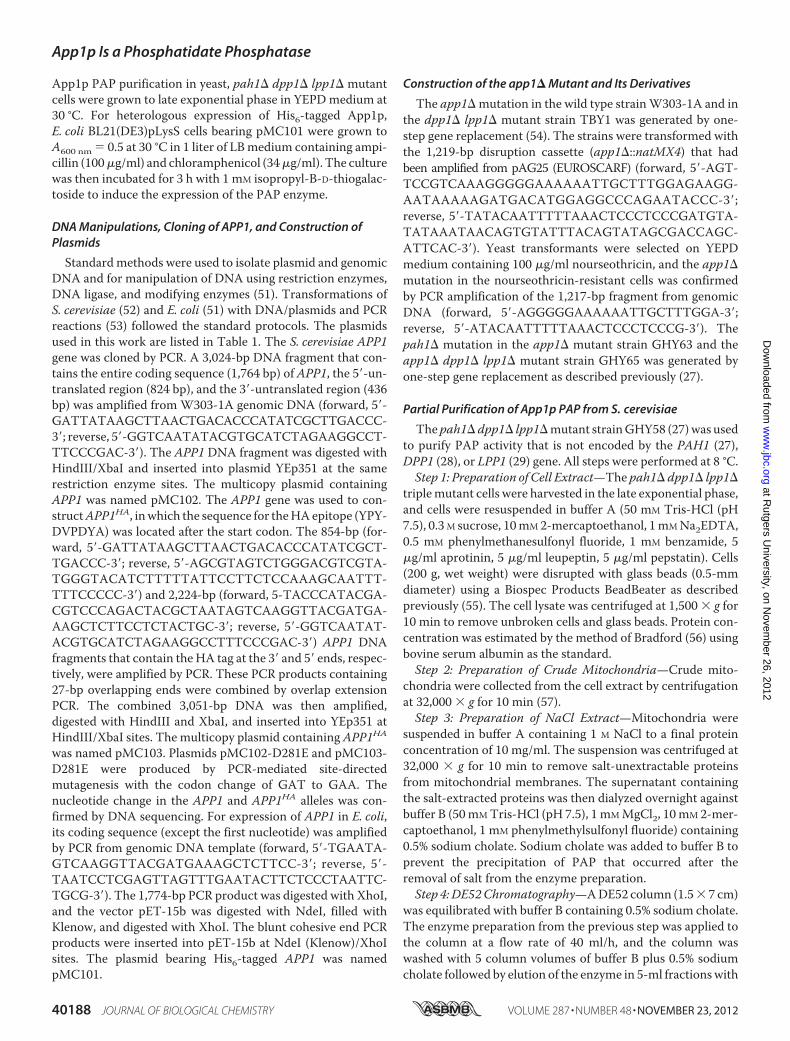
overexpression of APP1 in E. coli. The S. cerevisiae APP1 cod-ing sequence was amplified by PCR and inserted into plasmidpET-15b for the isopropyl-B-D-thiogalactoside-inducibleexpression of His6-tagged protein. The purified His6-taggedApp1pmigrateduponSDS-PAGEat the expected sizeof�68kDa(Fig. 3A). In addition, the purified protein was confirmed byimmunereactionwithantibodiesdirectedagainst theHis6 epitope
TABLE 2Partial purification of App1p PAP from S. cerevisiaePAPwas purified from pah1� dpp1� lpp1�mutant cells as described under “Exper-imental Procedures.” The data are based on starting with 200 g (wet weight) of cells.
Purification stepTotalunits Protein
Specificactivity Yield Purification
nmol/min mg units/mg % -foldCell extract 3,237 4,625 0.7 100 1Mitochondria 2,200 628 3.5 68 5NaCl extract 1,745 346 5 54 7DE52 1,053 65 16 33 23Affi-Gel Blue 770 20 39 24 56Phenyl-Sepharose 545 4 137 17 196MonoQ 300 0.9 330 9 471Superdex 200 150 0.1 1,500 5 2,143
FIGURE 1. Elution profiles of PAP activity and protein after chromatogra-phy with Superdex 200 and SDS-PAGE of the purified enzyme. TheMonoQ-purified PAP enzyme preparation was subjected to chromatographywith Superdex 200. Fractions were collected and assayed for PAP activity (●)and protein (E). Fractions 24 –30 were subjected to SDS-PAGE followed bystaining with Coomassie Blue (above plot). The positions of electrophoresismolecular mass standards are indicated in the figure. The band derived fromfraction 27 that was used for protein sequencing is indicated by the asterisk.mAU, milliabsorbance units.
App1p Is a Phosphatidate Phosphatase
40190 JOURNAL OF BIOLOGICAL CHEMISTRY VOLUME 287 • NUMBER 48 • NOVEMBER 23, 2012
at Rutgers U
niversity, on Novem
ber 26, 2012w
ww
.jbc.orgD
ownloaded from
and against a peptide sequence found at theC-terminal portion ofApp1p (Fig. 3A). The purified protein catalyzed theMg2�-depen-dentPAPreaction, and theadditionof10mMNa2EDTAabolishedPAP activity. Unlike the Mg2�-dependent Pah1p PAP, App1penzyme activity was sensitive to inhibition by N-ethylmaleimide(Fig. 3B).ThatApp1pexpressed inE. coliexhibitsPAPactivity alsoindicated that a posttranslationalmodification thatmight occur inyeast is not essential for enzyme activity.PAP Activity Is Affected by the app1� pah1� dpp1� lpp1�
Mutations and by the APP1(D281E) Mutation—We con-structed a variety of isogenic mutants where the app1� muta-
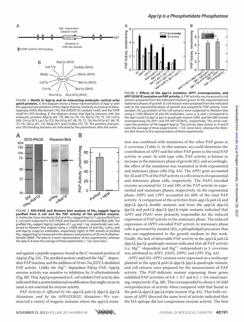
tion was combined with mutations of the other PAP genes inS. cerevisiae (Table 1). In this manner, we could determine thecontribution ofAPP1 and the other PAP genes to the total PAPactivity in yeast. In wild type cells, PAP activity is known toincrease in the stationary phase of growth (81), and accordingly,the effect of the mutations was examined in both exponentialand stationary phase cells (Fig. 4A). The APP1 gene accountedfor 32 and 37% of the PAP activity in cell extracts of exponentialand stationary phase cells, respectively. The PAH1-encodedenzyme accounted for 11 and 24% of the PAP activity in expo-nential and stationary phases, respectively. In the exponentialphase, DPP1 and LPP1 accounted for 60% of the total PAPactivity. A comparison of the activities from app1� pah1� anddpp1� lpp1� double mutants and from the app1� dpp1�lpp1� and pah1� dpp1� lpp1� triple mutants indicated thatAPP1 and PAH1 were primarily responsible for the inducedexpression of PAP activity in the stationary phase. The inducedexpression of DPP1-encoded PAP activity in stationary phasecells is governed by inositol (82), a phospholipid precursor thatwas not supplemented to the growth medium in this work.Finally, the lack of detectable PAP activity in the app1� pah1�dpp1� lpp1� quadruple mutant indicated that all PAP activity(i.e. Mg2�-dependent and Mg2�-independent) in S. cerevisiaewas attributed to APP1, PAH1, DPP1, and LPP1 (Fig. 4A).
APP1 andHA-APP1 versions were expressed on a multicopyplasmid in the app1� pah1� dpp1� lpp1� quadruple mutant,and cell extracts were prepared for the measurement of PAPactivity. The PAP-deficient mutant expressing these genesexhibited PAP activities of 6.8 � 0.7 and 6.5 � 0.6 nmol/min/mg, respectively (Fig. 4B). This corresponded to about a 10-foldoverproduction of activity when compared with that found inthe pah1� dpp1� lpp1� triple mutant (Fig. 4A). That both ver-sions of APP1 directed the same level of activity indicated thatthe HA epitope did not compromise enzyme activity. The first
FIGURE 2. Motifs in App1p and its interacting endocytic cortical actinpatch proteins. A, the diagram shows a linear representation of App1p withthe approximate positions of the region that has similarity to a haloacid deha-logenase (HAD)-like domain (76), the DXDX(T/V) catalytic motif, and the PXXPmotif for SH3 binding. B, the diagram shows that App1p interacts with theendocytic proteins Abp1p (69 –73), Bbc1p (70, 73), Bzz1p (70, 73, 74), Cof1p(69), Crn1p (67), Las17p (75), Rvs161p (67, 68, 70, 73, 74), Rvs167p (67, 68, 70,73, 74), Sla1p (67, 73), Sla2p (67), and Ysc84p (70, 73). The proteins that pos-sess SH3 binding domains are indicated by the parentheses after the name.
FIGURE 3. SDS-PAGE and Western blot analysis of His6-tagged App1ppurified from E. coli and the PAP activity of the purified enzyme.A, molecular mass standards (Std) and His6-tagged App1p (1 �g) purified fromE. coli were subjected to SDS-PAGE and stained with Coomassie Blue (left). Thepurified His6-tagged App1p (samples of 1 �g and 1 ng, respectively) was sub-jected to Western blot analysis using a 1:5000 dilution of anti-His6 (�His6) andanti-App1p (�App1p) antibodies, respectively (right). B, PAP activity of purifiedHis6-tagged App1p measured in the absence and presence of 20 mM N-ethylma-leimide (NEM). The data in A were representative of two experiments, whereasthe data in B were the average of three experiments � S.D. (error bars).
FIGURE 4. Effects of the app1� mutation, APP1 overexpression, andAPP1(D281E) mutation on PAP activity. A, PAP activity was measured in cellextracts prepared from the indicated mutants grown to the exponential andstationary phases of growth. B, cell extracts were prepared from the indicatedcells at the exponential phase of growth and assayed for PAP activity. Inset,samples (50 �g protein) of the cell extracts were subjected to Western blotusing a 1:500 dilution of anti-HA antibodies. Lanes a, b, and c correspond tothe app1� pah1� dpp1� lpp1� quadruple mutant (QM), and the QM mutantoverexpressing HA-APP1 and HA-APP1(D281E), respectively. The arrow indi-cates the position of HA-tagged App1p. The activity data shown in A and Bwere the average of three experiments � S.D. (error bars), whereas the West-ern blot shown in B is representative of three experiments.
App1p Is a Phosphatidate Phosphatase
NOVEMBER 23, 2012 • VOLUME 287 • NUMBER 48 JOURNAL OF BIOLOGICAL CHEMISTRY 40191
at Rutgers U
niversity, on Novem
ber 26, 2012w
ww
.jbc.orgD
ownloaded from

conserved aspartate residue in the DIDDT sequence in App1pwas mutated to glutamate by site-specific mutagenesis. Gluta-mate was chosen to replace aspartate to conserve the charge ofthe amino acid at this position, and thus, minimize a structuralchange in the enzyme. TheD281Emutation abolished the over-expressed PAP activity directed by the APP1 and HA-APP1genes (Fig. 4B). An immunoblot analysis using anti-HA anti-bodies showed that the D281E mutation in the HA-tagged ver-sion of App1p did not affect the expression of the enzyme (Fig.4B, inset). These data indicated that the DIDDT sequence inApp1p is responsible for its PAP catalytic function.Effects of the app1� pah1� dpp1� lpp1� Mutations on Lipid
Composition—The PAP deletion mutants were used to deter-mine the contribution ofAPP1, PAH1,DPP1, and LPP1 to lipidcomposition. In the first set of experiments, wild type andmutant cells were labeled to steady state with [2-14C]acetate toanalyze DAG, TAG, and total phospholipids. Cells were grownto the stationary phase (the growth phase when DAG and TAGare most affected (27)), and lipids were extracted and then ana-lyzed by one-dimensional TLC. When compared with thepah1� mutation, the app1� and dpp1� lpp1� mutations didnot have a significant effect on the relative amounts of DAG,TAG, and total phospholipids (Fig. 5). Moreover, alterations inlipid composition observed in the double, triple, and quadruplemutants were attributed to the pah1� mutation (Fig. 5). Theseresults indicate that of the genes encoding PAP in S. cerevisiae,PAH1 is responsible for the synthesis of TAG and the regula-tion of phospholipid synthesis. In the second set of experi-ments, the wild type and mutant cells were labeled to steadystatewith [32P]Pi to analyze the composition of individual phos-pholipids. The phospholipids were extracted from exponentialphase cells (the growth phase when phospholipid compositionis most affected (27)) and analyzed by two-dimensional TLC.The relative amounts of themajor phospholipids phosphatidyl-choline, phosphatidylethanolamine, phosphatidylinositol,phosphatidylserine, and the precursor PA were not affected bythe app1� and dpp1� lpp1� mutations (Fig. 6). The relativeamounts of these phospholipids, however, were affected by thepah1� mutation alone and in combination with the app1� anddpp1� lpp1� mutations (Fig. 6). As described previously (27),the pah1� mutation caused decreases in the amounts of phos-
phatidylcholine (37%) and phosphatidylserine (33%) andincreases in the amounts of phosphatidylethanolamine (108%),phosphatidylinositol (42%), and PA (86%). Interestingly, whencombined with the pah1� dpp1� lpp1� mutations, the app1�mutation reversed the effect of the pah1� mutation on thecomposition of phosphatidylcholine and phosphatidylethano-lamine. The reason for this change is unclear.The app1� Mutant Does Not Exhibit Obvious Phenotypes—
We noted that the app1� mutant in the BY4741 genetic back-ground exhibited slower growth on synthetic medium and wassensitive to elevated temperature (i.e. 37 °C). It is known thatthe BY4741 strain has more stringent growth requirementswhen compared with other strains used by the yeast researchcommunity (83). Accordingly, the growth requirements of theapp1� mutant were also examined in the W303-1A geneticbackground. This analysis showed that the app1�mutation didnot cause obvious growth defects. In addition, no striking phe-notypes (e.g. changes in cellular morphology, respiratory defi-ciency, salt sensitivity) were identified. In striking contrast, thepah1� mutant exhibits a slow growth phenotype, is tempera-ture-sensitive in several genetic backgrounds, and exhibitsdefects in cellularmorphology (27, 43, 48, 49, 84). None of thesephenotypes were complemented by the expression of APP1,substantiating that App1p and Pah1p PAP activities have dis-tinct functions in lipid metabolism and cell physiology.
DISCUSSION
PAP is generally recognized as an important enzyme ineukaryotic organisms because its substrate PA and productDAG play important roles in the synthesis of TAG and phos-pholipids and in other aspects of cell physiology (e.g. transcrip-tion, lipid signaling, and vesicular trafficking) (1–20). Twobasictypes of PAP activity are found in eukaryotic organisms: anactivity that is dependent on Mg2� and an activity that is notdependent on Mg2� or any other divalent cation (21, 22, 74).Mg2�-dependent PAP activity is governed by a DXDX(T/V)catalytic motif (77), whereas Mg2�-independent activity isdirected by a three-domain catalytic motif consisting of thesequences KX6RP, PSGH, and SRX5HX3D (31). In the yeastS. cerevisiae, the Mg2�-dependent PAP is encoded by PAH1
FIGURE 5. Effects of the PAP gene mutations on DAG, TAG, and total phos-pholipids. The indicated cells were grown to the stationary phase of growthin the presence of [2-14C]acetate (1 �Ci/ml). Lipids were extracted and sepa-rated by one-dimensional TLC, and the images were subjected to ImageQuantanalysis. The percentages shown for the individual lipids were normalized to thetotal 14C-labeled chloroform-soluble fraction, which also contained sterols, fattyacids, and unidentified neutral lipids. Each data point represents the average ofthree experiments � S.D. (error bars). PL, phospholipids.
FIGURE 6. Effects of the PAP gene mutations on phospholipid composi-tion. The indicated cells were grown to the exponential phase of growth inthe presence of [32P]Pi (10 �Ci/ml). Phospholipids were extracted and sepa-rated by two-dimensional TLC, and the images were subjected toImageQuant analysis. The percentages shown for the individual phospholip-ids were normalized to the total 32P-labeled chloroform-soluble fraction thatincluded sphingolipids and unidentified phospholipids. Each data point rep-resents the average of three experiments � S.D. (error bars). Abbreviations:PC, phosphatidylcholine; PE, phosphatidylethanolamine; PI, phosphatidyli-nositol; PS, phosphatidylserine.
App1p Is a Phosphatidate Phosphatase
40192 JOURNAL OF BIOLOGICAL CHEMISTRY VOLUME 287 • NUMBER 48 • NOVEMBER 23, 2012
at Rutgers U
niversity, on Novem
ber 26, 2012w
ww
.jbc.orgD
ownloaded from
(27), whereas the Mg2�-independent type of the enzyme isencoded byDPP1 (28) and LPP1 (29). The pah1� dpp1� lpp1�triple mutant lacking the known Mg2�-dependent and Mg2�-independent PAP enzymes still possesses theMg2�-dependentactivity that is sensitive to inhibition byN-ethylmaleimide (27),and the identity of the gene(s) encoding this activity was thefocus of this work.APP1 was identified through a reverse genetics approach
using protein sequence information derived from the PAPenzyme isolated from the pah1� dpp1� lpp1� triple mutant.Obtaining App1p sequence information was not straightfor-ward. The PAP activity was labile in the absence of high salt,which comprised the effectiveness of chromatography stepsthat required low salt for enzyme binding (e.g. ion exchangechromatography). Although the eight-step purification schemeresulted in a 2,143-fold enrichment in PAP specific activity, itdid not result in a homogeneous enzyme preparation thatcould facilitate unequivocal protein sequence determination.Although the sensitive liquid chromatography/tandem massspectrometry technology yielded many protein candidates, itallowed us to identify App1p that was present in very low abun-dance. In the end, the collective data (e.g. reduction of PAPactivity in the app1� mutant, the APP1-directed overexpres-sion of PAP activity in the app1� pah1� dpp1� lpp1� quadru-ple mutant, and the heterologous expression of App1p PAPactivity in E. coli) provided a conclusive level of evidence thatAPP1 is the structural gene encoding a PAP enzyme inS. cerevisiae.Efforts to identify APP1 by informatics and by genetic
approaches were unsuccessful. For example, a protein BLASTsearch using Pah1p as the query did not identify App1p or anyother homologs in the Saccharomyces genome database. ABLAST search against higher eukaryotic organism databasesidentified mammalian lipin proteins, but this was expectedbecause a BLAST search with mouse lipin-1 as the query iden-tifies S. cerevisiae Pah1p (78). Likewise, a BLAST search usingApp1p as the query does not identify Pah1p or the mammalianlipins. Instead, the BLAST search identifies App1p homologsonly found in fungi. A synthetic genetic array screen using thepah1�mutant in combinationwith the cho2� and opi3�muta-tions defective in the phosphatidylethanolamine methylationsteps of phosphatidylcholine synthesis via the CDP-DAG path-way (85, 86) has also been performed.3 The rationale for thisanalysis was that the loss of a gene encoding PAP in combina-tion with the loss of Pah1p causes lethality due to a lack of DAGproduction required for the synthesis of phosphatidylcholinevia the Kennedy pathway (85, 86). The genetic screen, however,did not lead to the identification of APP1,3 indicating that thecellular functions of App1p and Pah1p do not overlapwith eachother with respect to lipid synthesis. This assertion was furthersupported by the fact that APP1 did not complement pheno-types (e.g. temperature sensitivity) exhibited by the pah1�mutant and that the analysis of cells possessing the app1�mutation alone and in combination with mutations for otherknown PAP genes indicated that only Pah1p PAP was involvedin de novo lipid synthesis.
Although there is essentially no sequence homology betweenApp1p and Pah1p, both enzymes possess the canonicalDXDX(T/V) catalytic motif that is typical of Mg2�-dependentphosphatase enzymes (77). For Pah1p, its DIDGT catalyticsequence is contained within the haloacid dehalogenase-likedomain similar to that found in the mammalian lipin PAPenzymes (21, 23, 78). However, the haloacid dehalogenase-likedomain is not found in App1p, but instead, it contains a con-served domain found only in fungi that has overlapping regionsthat show weak sequence similarity to the haloacid dehaloge-nase-like domain found in Pah1p and lipin (76). It is within thisdomain that the App1p DIDDT sequence is found (Fig. 2), andindeed, theD281Emutation abolishedPAP activity, confirmingthis to be the catalytic sequence.PAP enzymes isolated from S. cerevisiae are known to have
molecular masses of 91 kDa (Mg2�-dependent) (87), 75 kDa(Mg2�-dependent) (88), 45 kDa (Mg2�-dependent) (89), and34 kDa (Mg2�-independent) (34). Protein sequence informa-tion has confirmed that the 91-kDa enzyme is a proteolysisproduct of Pah1p (27) and that the 34-kDa enzyme is Dpp1p(28). Lpp1p PAPwas identified based on its sequence homologywithDpp1p (29). The identity of the genes encoding the 75- and45-kDa forms of PAP has been an enigma. Although the basicenzymological properties of these enzymes are similar, the75-kDa enzyme is soluble, whereas the 45-kDa enzyme is asso-ciated with mitochondria (88, 89). Because of the differences insize and location, it has been assumed that the 75- and 45-kDaPAP enzymes are encoded by different genes. Based on theresult that no detectable PAP activity was present in the app1�pah1� dpp1� lpp1� quadruple mutant, we hypothesize thatthe 75-kDa PAP was the soluble form of App1p and that the45-kDa PAP was a proteolytic fragment of App1p bound tomitochondrialmembranes. Unfortunately, this hypothesis can-not be addressed because preparations of the 75- and 45-kDaPAP enzymes are no longer available. Obviously, the proteinused for sequence analysis in this study was a proteolytic frag-ment of App1p.The PAP enzymes in S. cerevisiae are found in different cel-
lular locations and play diverse roles in cell physiology (74).Pah1p is a cytosolic enzyme that associates with and functionsat the nuclear/endoplasmic reticulum membrane to regulatethe synthesis of TAG andmembrane phospholipids (21, 22, 27,42, 74). Dpp1p and Lpp1p, respectively, are thought to controlthe signaling functions of PA, DAGpyrophosphate, and lysoPAin vacuole and Golgi membranes (21, 22, 28, 29, 34, 44–46, 74,90). Like Pah1p,App1p is a cytosolic protein (46) that associateswith membranes, but the role this PAP plays in cell physiologyis not yet clear.The formation of endocytic vesicles in S. cerevisiae involves a
series of processes that include actin patch assembly, actinpolymerization, and changes in membrane structure and cur-vature (75, 92). These processes involve numerous endocyticproteins, and App1p is known to physically interact with manyof them (67–75) (Fig. 2). Although these interactions have yet tobe studied in detail, the presence of proline-rich regions (PXXPmotifs) in App1p suggests interactions with SH3 domains ofAbp1p, Ysc84p, Sla1p, Bbc1p, Bzz1p, and Rvs167p (79, 80).App1p interactions with proteins that do not possess SH33 C. R. McMaster, personal communication.
App1p Is a Phosphatidate Phosphatase
NOVEMBER 23, 2012 • VOLUME 287 • NUMBER 48 JOURNAL OF BIOLOGICAL CHEMISTRY 40193
at Rutgers U
niversity, on Novem
ber 26, 2012w
ww
.jbc.orgD
ownloaded from

domains (e.g. Las17p, Sla2p, Cof1p, Crn1p, and Rvs161) mustoccur through other mechanisms yet to be defined. Based onthese protein interaction data, App1p is postulated to play arole in endocytosis (67). However, until now the molecularfunction of App1p has been unknown. App1p PAP located atcortical actin patches (92) may regulate the local concentra-tions of PA andDAG.These lipids are known to facilitatemem-brane fission/fusion events in model systems (93–98), and theyare also known to interact with and regulate enzymes (e.g. phos-phatidylinositol 4-phosphate kinase, protein kinase C, proteinkinase D) that play important roles in vesicular trafficking (99–103). For example, in mammalian cells, the inhibition of PAPactivity by propranolol attenuates protein kinaseD recruitmentto Golgi membranes, blocking vesicle bud formation and pro-tein transport to the cell surface (103, 104). Unfortunately, pro-pranolol does not discriminate between Mg2�-dependent andMg2�-independent PAP activities (90, 105–107), and thus, theidentity of the type of PAP enzyme involved in this process isunknown.We speculate that in yeast, App1p PAP activity playsa role in vesicle formation through its recruitment from thecytosol to cortical actin patches via endocytic proteins. Theseproteinsmay tetherApp1pPAP to actin patches and/or serve toregulate the relative amounts of PA and DAG, which in turncontribute to the control of vesicle formation. Clearly, the workreported here provides an impetus to pursue these questions inmore detail. Although an App1p homolog does not exist inhigher eukaryotes, a functional homolog exists in the form ofthe lipin PAP enzymes that catalyze the same reaction (27, 106,108), and as indicated above, a PAP activity is implicated invesicular trafficking in mammalian cells.
Acknowledgments—Chris A. Marfo is acknowledged for the initialcharacterization of the anti-App1p antibodies. The mass spectrome-try data were obtained from an Orbitrap instrument funded in partby National Institutes of Health Grant NS046593, for the support ofthe University of Medicine and Dentistry of New Jersey Neuropro-teomics Core Facility.
REFERENCES1. Smith, S. W., Weiss, S. B., and Kennedy, E. P. (1957) The enzymatic
dephosphorylation of phosphatidic acids. J. Biol. Chem. 228, 915–9222. Kennedy, E. P., andWeiss, S. B. (1956)The function of cytidine coenzyme
in the biosynthesis of phospholipids. J. Biol. Chem. 222, 193–2143. Borkenhagen, L. F., and Kennedy, E. P. (1957) The enzymatic synthesis of
cytidine diphosphate choline. J. Biol. Chem. 227, 951–9624. Weiss, S. B., Smith, S. W., and Kennedy, E. P. (1958) The enzymatic
formation of lecithin from cytidine diphosphate choline and D-1,2-dig-lyceride. J. Biol. Chem. 231, 53–64
5. Kennedy, E. P. (1956) The synthesis of cytidine diphosphate choline,cytidine diphosphate ethanolamine, and related compounds. J. Biol.Chem. 222, 185–191
6. Weiss, S. B., Kennedy, E. P., and Kiyasu, J. Y. (1960) The enzymaticsynthesis of triglycerides. J. Biol. Chem. 235, 40–44
7. Paulus, H., and Kennedy, E. P. (1960) The enzymatic synthesis of inositolmonophosphatide. J. Biol. Chem. 235, 1303–1311
8. Kiyasu, J. Y., Pieringer, R. A., Paulus, H., and Kennedy, E. P. (1963) Thebiosynthesis of phosphatidylglycerol. J. Biol. Chem. 238, 2293–2298
9. Davidson, J. B., and Stanacev, N. Z. (1971) Biosynthesis of cardiolipin inmitochondria isolated from guinea pig liver.Biochem. Biophys. Res. Com-mun. 42, 1191–1199
10. Bishop, W. R., Ganong, B. R., and Bell, R. M. (1986) Attenuation of sn-
1,2-diacylglycerol second messengers by diacylglycerol kinase. J. Biol.Chem. 261, 6993–7000
11. Kearns, B. G., McGee, T. P., Mayinger, P., Gedvilaite, A., Phillips, S. E.,Kagiwada, S., and Bankaitis, V. A. (1997) Essential role for diacylglycerolin protein transport from the yeast Golgi complex.Nature 387, 101–105
12. Waggoner, D.W., Xu, J., Singh, I., Jasinska, R., Zhang,Q.X., andBrindley,D. N. (1999) Structural organization of mammalian lipid phosphatephosphatases: implications for signal transduction. Biochim. Biophys.Acta 1439, 299–316
13. Sciorra, V. A., andMorris, A. J. (2002) Roles for lipid phosphate phosphata-ses in regulation of cellular signaling. Biochim. Biophys. Acta 1582, 45–51
14. Testerink, C., and Munnik, T. (2005) Phosphatidic acid: a multifunc-tional stress signaling lipid in plants. Trends Plant Sci. 10, 368–375
15. Wang, X., Devaiah, S. P., Zhang,W., andWelti, R. (2006) Signaling func-tions of phosphatidic acid. Prog. Lipid Res. 45, 250–278
16. Brindley, D. N. (2004) Lipid phosphate phosphatases and related pro-teins: signaling functions in development, cell division, and cancer.J. Cell. Biochem. 92, 900–912
17. Howe, A.G., andMcMaster,C. R. (2006)Regulationof phosphatidylcholinehomeostasis by Sec14.Can. J. Physiol. Pharmacol. 84, 29–38
18. Foster, D. A. (2007) Regulation of mTOR by phosphatidic acid? CancerRes. 67, 1–4
19. Carman, G.M., and Henry, S. A. (2007) Phosphatidic acid plays a centralrole in the transcriptional regulation of glycerophospholipid synthesis inSaccharomyces cerevisiae. J. Biol. Chem. 282, 37293–37297
20. Carrasco, S., and Mérida, I. (2007) Diacylglycerol, when simplicity be-comes complex. Trends Biochem. Sci. 32, 27–36
21. Carman, G. M., and Han, G.-S. (2006) Roles of phosphatidate phospha-tase enzymes in lipid metabolism. Trends Biochem. Sci. 31, 694–699
22. Carman, G. M., and Han, G.-S. (2009) Phosphatidic acid phosphatase, akey enzyme in the regulation of lipid synthesis. J. Biol. Chem. 284,2593–2597
23. Csaki, L. S., and Reue, K. (2010) Lipins: multifunctional lipid metabolismproteins. Annu. Rev. Nutr. 30, 257–272
24. Reue, K., and Dwyer, J. R. (2009) Lipin proteins and metabolic homeo-stasis. J. Lipid Res. 50, (suppl.) S109–S114
25. Reue, K., and Brindley, D. N. (2008) Multiple roles for lipins/phos-phatidate phosphatase enzymes in lipid metabolism. J. Lipid Res. 49,2493–2503
26. Pascual, F., and Carman, G. M. (2012) Phosphatidate phosphatase, a keyregulator of lipid homeostasis. Biochim. Biophys. Acta, doi:10.1016/j.bbalip.2012.08.006
27. Han, G.-S., Wu, W.-I., and Carman, G. M. (2006) The Saccharomycescerevisiae lipin homolog is a Mg2�-dependent phosphatidate phospha-tase enzyme. J. Biol. Chem. 281, 9210–9218
28. Toke, D.A., Bennett,W. L., Dillon, D.A.,Wu,W.-I., Chen, X.,Ostrander,D. B., Oshiro, J., Cremesti, A., Voelker, D. R., Fischl, A. S., and Carman,G. M. (1998) Isolation and characterization of the Saccharomyces cerevi-siae DPP1 gene encoding for diacylglycerol pyrophosphate phosphatase.J. Biol. Chem. 273, 3278–3284
29. Toke, D. A., Bennett, W. L., Oshiro, J., Wu, W.-I., Voelker, D. R., andCarman, G.M. (1998) Isolation and characterization of the Saccharomy-ces cerevisiae LPP1 gene encoding a Mg2�-independent phosphatidatephosphatase. J. Biol. Chem. 273, 14331–14338
30. Han, G.-S., Siniossoglou, S., and Carman, G.M. (2007) The cellular func-tions of the yeast lipin homolog Pah1p are dependent on its phos-phatidate phosphatase activity. J. Biol. Chem. 282, 37026–37035
31. Stukey, J., and Carman, G. M. (1997) Identification of a novel phospha-tase sequence motif. Protein Sci. 6, 469–472
32. Toke, D. A.,McClintick,M. L., andCarman,G.M. (1999)Mutagenesis ofthe phosphatase sequence motif in diacylglycerol pyrophosphate phos-phatase from Saccharomyces cerevisiae. Biochemistry 38, 14606–14613
33. Dillon, D. A., Chen, X., Zeimetz, G. M., Wu, W.-I., Waggoner, D. W.,Dewald, J., Brindley, D.N., andCarman,G.M. (1997)MammalianMg2�-independent phosphatidate phosphatase (PAP2) displays diacylglycerolpyrophosphate phosphatase activity. J. Biol. Chem. 272, 10361–10366
34. Wu,W.-I., Liu, Y., Riedel, B.,Wissing, J. B., Fischl, A. S., and Carman, G.M.(1996) Purification and characterization of diacylglycerol pyrophosphate
App1p Is a Phosphatidate Phosphatase
40194 JOURNAL OF BIOLOGICAL CHEMISTRY VOLUME 287 • NUMBER 48 • NOVEMBER 23, 2012
at Rutgers U
niversity, on Novem
ber 26, 2012w
ww
.jbc.orgD
ownloaded from

phosphatase from Saccharomyces cerevisiae. J. Biol. Chem. 271, 1868–187635. Dillon, D. A., Wu, W.-I., Riedel, B., Wissing, J. B., Dowhan, W., and
Carman, G. M. (1996) The Escherichia coli pgpB gene encodes for adiacylglycerol pyrophosphate phosphatase activity. J. Biol. Chem. 271,30548–30553
36. Faulkner, A., Chen, X., Rush, J., Horazdovsky, B., Waechter, C. J., Car-man, G. M., and Sternweis, P. C. (1999) The LPP1 and DPP1 gene prod-ucts account formost of the isoprenoid phosphatase activities in Saccha-romyces cerevisiae. J. Biol. Chem. 274, 14831–14837
37. Choi, H.-S., Su, W.-M., Han, G.-S., Plote, D., Xu, Z., and Carman, G. M.(2012) Pho85p-Pho80p phosphorylation of yeast Pah1p phosphatidatephosphatase regulates its activity, location, abundance, and function inlipid metabolism. J. Biol. Chem. 287, 11290–11301
38. Choi, H.-S., Su, W.-M., Morgan, J. M., Han, G.-S., Xu, Z., Karanasios, E.,Siniossoglou, S., and Carman, G. M. (2011) Phosphorylation of phos-phatidate phosphatase regulates its membrane association and physio-logical functions in Saccharomyces cerevisiae: identification of Ser602,Thr723, and Ser744 as the sites phosphorylated by CDC28 (CDK1)-en-coded cyclin-dependent kinase. J. Biol. Chem. 286, 1486–1498
39. Su,W.-M.,Han,G.-S.,Casciano, J., andCarman,G.M. (2012)ProteinkinaseA-mediated phosphorylation of Pah1p phosphatidate phosphatase func-tions in conjunction with the Pho85p-Pho80p and Cdc28p-cyclin B kinasesto regulate lipid synthesis in yeast. J. Biol. Chem. 287, 33364–33376
40. Blom, N., Sicheritz-Pontén, T., Gupta, R., Gammeltoft, S., and Brunak, S.(2004) Prediction of post-translational glycosylation and phosphoryla-tion of proteins from the amino acid sequence. Proteomics 4, 1633–1649
41. Siniossoglou, S., Santos-Rosa, H., Rappsilber, J., Mann, M., and Hurt, E.(1998) A novel complex of membrane proteins required for formation ofa spherical nucleus. EMBO J. 17, 6449–6464
42. Karanasios, E., Han, G.-S., Xu, Z., Carman, G. M., and Siniossoglou, S.(2010) A phosphorylation-regulated amphipathic helix controls themembrane translocation and function of the yeast phosphatidate phos-phatase. Proc. Natl. Acad. Sci. U.S.A. 107, 17539–17544
43. Santos-Rosa, H., Leung, J., Grimsey, N., Peak-Chew, S., and Siniossoglou,S. (2005) The yeast lipin Smp2 couples phospholipid biosynthesis tonuclear membrane growth. EMBO J. 24, 1931–1941
44. Han, G.-S., Johnston, C. N., Chen, X., Athenstaedt, K., Daum, G., andCarman, G.M. (2001) Regulation of the Saccharomyces cerevisiae DPP1-encoded diacylglycerol pyrophosphate phosphatase by zinc. J. Biol.Chem. 276, 10126–10133
45. Han, G.-S., Johnston, C. N., and Carman, G. M. (2004) Vacuole membranetopography of the DPP1-encoded diacylglycerol pyrophosphate phospha-tase catalytic site from Saccharomyces cerevisiae. J. Biol. Chem. 279,5338–5345
46. Huh, W. K., Falvo, J. V., Gerke, L. C., Carroll, A. S., Howson, R. W.,Weissman, J. S., and O’Shea, E. K. (2003) Global analysis of protein local-ization in budding yeast. Nature 425, 686–691
47. Fakas, S., Qiu, Y., Dixon, J. L., Han, G.-S., Ruggles, K. V., Garbarino, J.,Sturley, S. L., and Carman, G. M. (2011) Phosphatidate phosphatase ac-tivity plays a key role in protection against fatty acid-induced toxicity inyeast. J. Biol. Chem. 286, 29074–29085
48. Adeyo, O., Horn, P. J., Lee, S., Binns, D. D., Chandrahas, A., Chapman,K. D., and Goodman, J. M. (2011) The yeast lipin orthologue Pah1p isimportant for biogenesis of lipid droplets. J. Cell Biol. 192, 1043–1055
49. Sasser, T., Qiu, Q. S., Karunakaran, S., Padolina, M., Reyes, A., Flood, B.,Smith, S., Gonzales, C., and Fratti, R. A. (2012) The yeast lipin 1 ortho-logue Pah1p regulates vacuole homeostasis andmembrane fusion. J. Biol.Chem. 287, 2221–2236
50. Rose,M.D.,Winston, F., andHeiter, P. (1990)Methods in Yeast Genetics:A Laboratory Course Manual, Cold Spring Harbor Laboratory Press,Cold Spring Harbor, NY
51. Sambrook, J., Fritsch, E. F., andManiatis, T. (1989)Molecular Cloning: ALaboratory Manual, 2nd Ed., Cold Spring Harbor Laboratory, ColdSpring Harbor, NY
52. Ito, H., Fukuda, Y., Murata, K., and Kimura, A. (1983) Transformation ofintact yeast cells treated with alkali cations. J. Bacteriol. 153, 163–168
53. Innis, M. A., and Gelfand, D. H. (1990) in PCR Protocols: A Guide toMethods and Applications (Innis, M. A., Gelfand, D. H., Sninsky, J. J., and
White, T. J., eds) pp. 3–12, Academic Press, Inc., San Diego54. Rothstein, R. (1991)Targeting,disruption, replacement, andallele rescue: Inte-
grativeDNA transformation in yeast.Methods Enzymol.194, 281–30155. Fischl, A. S., andCarman,G.M. (1983) Phosphatidylinositol biosynthesis
in Saccharomyces cerevisiae: purification and properties of microsome-associated phosphatidylinositol synthase. J. Bacteriol. 154, 304–311
56. Bradford,M.M. (1976)A rapid and sensitivemethod for the quantitationof microgram quantities of protein utilizing the principle of protein-dyebinding. Anal. Biochem. 72, 248–254
57. Belendiuk, G., Mangnall, D., Tung, B., Westley, J., and Getz, G. S. (1978)CTP-phosphatidic acid cytidyltransferase from Saccharomyces cerevi-siae: partial purification, characterization, and kinetic behavior. J. Biol.Chem. 253, 4555–4565
58. Carman, G. M., and Lin, Y.-P. (1991) Phosphatidate phosphatase fromyeast.Methods Enzymol. 197, 548–553
59. Laemmli, U. K. (1970) Cleavage of structural proteins during the assem-bly of the head of bacteriophage T4. Nature 227, 680–685
60. Burnette, W. (1981) Western blotting: Electrophoretic transfer of pro-teins from sodium dodecyl sulfate-polyacrylamide gels to unmodifiednitrocellulose and radiographic detection with antibody and radioiodi-nated protein A. Anal. Biochem. 112, 195–203
61. Haid, A., and Suissa, M. (1983) Immunochemical identification of mem-brane proteins after sodium dodecyl sulfate-polyacrylamide gel electro-phoresis.Methods Enzymol. 96, 192–205
62. Das, A., Li, H., Liu, T., and Bellofatto, V. (2006) Biochemical character-ization of Trypanosoma brucei RNA polymerase II.Mol. Biochem. Para-sitol. 150, 201–210
63. Jain, M. R., Li, Q., Liu, T., Rinaggio, J., Ketkar, A., Tournier, V., Madura,K., Elkabes, S., and Li, H. (2012) Proteomic identification of immunopro-teasome accumulation in formalin-fixed rodent spinal cords with exper-imental autoimmune encephalomyelitis. J. Proteome Res. 11, 1791–1803
64. Bligh, E. G., and Dyer, W. J. (1959) A rapid method of total lipid extrac-tion and purification. Can. J. Biochem. Physiol. 37, 911–917
65. Esko, J. D., and Raetz, C. R. (1980) Mutants of Chinese hamster ovarycells with altered membrane phospholipid composition: replacement ofphosphatidylinositol by phosphatidylglycerol in a myo-inositol aux-otroph. J. Biol. Chem. 255, 4474–4480
66. Henderson, R. J., and Tocher, D. R. (1992) in Lipid Analysis (Hamilton,R. J., and Hamilton, S., eds) pp. 65–111, IRL Press, New York
67. Drees, B. L., Sundin, B., Brazeau, E., Caviston, J. P., Chen, G. C., Guo,W.,Kozminski, K. G., Lau, M.W., Moskow, J. J., Tong, A., Schenkman, L. R.,McKenzie, A., 3rd, Brennwald, P., Longtine, M., Bi, E., Chan, C., Novick,P., Boone, C., Pringle, J. R., Davis, T. N., Fields, S., and Drubin, D. G.(2001) A protein interaction map for cell polarity development. J. CellBiol. 154, 549–571
68. Bon, E., Recordon-Navarro, P., Durrens, P., Iwase, M., Toh-E, A., andAigle, M. (2000) A network of proteins around Rvs167p and Rvs161p,two proteins related to the yeast actin cytoskeleton.Yeast 16, 1229–1241
69. Ho, Y., Gruhler, A., Heilbut, A., Bader, G. D., Moore, L., Adams, S. L.,Millar, A., Taylor, P., Bennett, K., Boutilier, K., Yang, L., Wolting, C.,Donaldson, I., Schandorff, S., Shewnarane, J.,Vo,M.,Taggart, J.,Goudreault,M., Muskat, B., Alfarano, C., Dewar, D., Lin, Z., Michalickova, K., Willems,A. R., Sassi, H., Nielsen, P. A., Rasmussen, K. J., Andersen, J. R., Johansen,L. E.,Hansen, L.H., Jespersen,H., Podtelejnikov,A.,Nielsen, E., Crawford, J.,Poulsen, V., Sørensen, B. D., Matthiesen, J., Hendrickson, R. C., Gleeson, F.,Pawson,T.,Moran,M. F.,Durocher,D.,Mann,M.,Hogue,C.W., Figeys,D.,and Tyers,M. (2002) Systematic identification of protein complexes in Sac-charomyces cerevisiae by mass spectrometry.Nature 415, 180–183
70. Tong, A. H., Drees, B., Nardelli, G., Bader, G. D., Brannetti, B., Castagnoli,L., Evangelista, M., Ferracuti, S., Nelson, B., Paoluzi, S., Quondam,M., Zuc-coni, A., Hogue, C. W., Fields, S., Boone, C., and Cesareni, G. (2002) Acombined experimental and computational strategy to define protein inter-action networks for peptide recognition modules. Science 295, 321–324
71. Fazi, B., Cope, M. J., Douangamath, A., Ferracuti, S., Schirwitz, K., Zuc-coni, A., Drubin, D. G., Wilmanns, M., Cesareni, G., and Castagnoli, L.(2002) Unusual binding properties of the SH3 domain of the yeast actin-binding protein Abp1: structural and functional analysis. J. Biol. Chem.277, 5290–5298
App1p Is a Phosphatidate Phosphatase
NOVEMBER 23, 2012 • VOLUME 287 • NUMBER 48 JOURNAL OF BIOLOGICAL CHEMISTRY 40195
at Rutgers U
niversity, on Novem
ber 26, 2012w
ww
.jbc.orgD
ownloaded from
72. Landgraf, C., Panni, S., Montecchi-Palazzi, L., Castagnoli, L., Schneider-Mergener, J., Volkmer-Engert, R., and Cesareni, G. (2004) Protein inter-action networks by proteome peptide scanning. PLoS. Biol. 2, E14
73. Tonikian, R., Xin, X., Toret, C. P., Gfeller, D., Landgraf, C., Panni, S.,Paoluzi, S., Castagnoli, L., Currell, B., Seshagiri, S., Yu, H., Winsor, B.,Vidal, M., Gerstein, M. B., Bader, G. D., Volkmer, R., Cesareni, G.,Drubin, D. G., Kim, P. M., Sidhu, S. S., and Boone, C. (2009) Bayesianmodeling of the yeast SH3 domain interactome predicts spatiotemporaldynamics of endocytosis proteins. PLoS. Biol. 7, e1000218
74. Yu, H., Braun, P., Yildirim,M.A., Lemmens, I., Venkatesan, K., Sahalie, J.,Hirozane-Kishikawa, T., Gebreab, F., Li, N., Simonis, N., Hao, T., Rual,J. F., Dricot, A., Vazquez, A., Murray, R. R., Simon, C., Tardivo, L., Tam,S., Svrzikapa, N., Fan, C., de Smet, A. S., Motyl, A., Hudson, M. E., Park,J., Xin, X., Cusick, M. E., Moore, T., Boone, C., Snyder, M., Roth, F. P.,Barabási, A. L., Tavernier, J., Hill, D. E., andVidal,M. (2008)High-qualitybinary protein interactionmap of the yeast interactome network. Science322, 104–110
75. Michelot, A., Costanzo,M., Sarkeshik, A., Boone, C., Yates, J. R., 3rd, andDrubin, D. G. (2010) Reconstitution and protein composition analysis ofendocytic actin patches. Curr. Biol. 20, 1890–1899
76. Punta,M., Coggill, P. C., Eberhardt, R. Y.,Mistry, J., Tate, J., Boursnell, C.,Pang, N., Forslund, K., Ceric, G., Clements, J., Heger, A., Holm, L., Sonn-hammer, E. L., Eddy, S. R., Bateman, A., and Finn, R. D. (2012) The Pfamprotein families database. Nucleic Acids Res. 40, D290-D301
77. Madera, M., Vogel, C., Kummerfeld, S. K., Chothia, C., and Gough, J.(2004) The SUPERFAMILY database in 2004: additions and improve-ments. Nucleic Acids Res. 32, D235-D239
78. Péterfy, M., Phan, J., Xu, P., and Reue, K. (2001) Lipodystrophy in the fldmouse results from mutation of a new gene encoding a nuclear protein,lipin. Nat. Genet. 27, 121–124
79. Feng, S., Chen, J. K., Yu, H., Simon, J. A., and Schreiber, S. L. (1994) Twobinding orientations for peptides to the Src SH3 domain: development ofa general model for SH3-ligand interactions. Science 266, 1241–1247
80. Lim, W. A., Richards, F. M., and Fox, R. O. (1994) Structural determi-nants of peptide-binding orientation and of sequence specificity in SH3domains. Nature 372, 375–379
81. Hosaka, K., and Yamashita, S. (1984) Regulatory role of phosphatidatephosphatase in triacylglycerol synthesis of Saccharomyces cerevisiae.Biochim. Biophys. Acta 796, 110–117
82. Oshiro, J., Rangaswamy, S., Chen, X., Han, G.-S., Quinn, J. E., and Car-man, G. M. (2000) Regulation of theDPP1-encoded diacylglycerol pyro-phosphate (DGPP) phosphatase by inositol and growth phase: inhibitionof DGPP phosphatase activity by CDP-diacylglycerol and activation ofphosphatidylserine synthase activity by DGPP. J. Biol. Chem. 275,40887–40896
83. Hanscho, M., Ruckerbauer, D. E., Chauhan, N., Hofbauer, H. F., Kra-hulec, S., Nidetzky, B., Kohlwein, S. D., Zanghellini, J., and Natter, K.(2012) Nutritional requirements of the BY series of Saccharomycescerevisiae strains for optimum growth. FEMS Yeast Res. 12, 796–808
84. Irie, K., Takase,M.,Araki,H., andOshima,Y. (1993)Agene,SMP2, involved inplasmid maintenance and respiration in Saccharomyces cerevisiae encodes ahighly charged protein.Mol.Gen.Genet.236, 283–288
85. Carman, G.M., andHan, G.-S. (2011) Regulation of phospholipid synthesis inthe yeast Saccharomyces cerevisiae.Ann. Rev. Biochem.80, 859–883
86. Henry, S. A., Kohlwein, S. D., and Carman, G.M. (2012)Metabolism andregulation of glycerolipids in the yeast Saccharomyces cerevisiae. Genet-ics 190, 317–349
87. Lin, Y.-P., and Carman, G.M. (1989) Purification and characterization ofphosphatidate phosphatase from Saccharomyces cerevisiae. J. Biol. Chem.264, 8641–8645
88. Hosaka, K., and Yamashita, S. (1984) Partial purification and propertiesof phosphatidate phosphatase in Saccharomyces cerevisiae.Biochim. Bio-phys. Acta 796, 102–109
89. Morlock, K. R., McLaughlin, J. J., Lin, Y.-P., and Carman, G. M. (1991)Phosphatidate phosphatase from Saccharomyces cerevisiae: isolation of45-kDa and 104-kDa forms of the enzyme that are differentially regulatedby inositol. J. Biol. Chem. 266, 3586–3593
90. Furneisen, J. M., and Carman, G. M. (2000) Enzymological properties ofthe LPP1-encoded lipid phosphatase from Saccharomyces cerevisiae.Biochim. Biophys. Acta 1484, 71–82
91. Hill, J. E., Myers, A. M., Koerner, T. J., and Tzagoloff, A. (1986) Yeast/E. colishuttle vectorswithmultiple unique restriction sites.Yeast2, 163–167
92. Weinberg, J., and Drubin, D. G. (2012) Clathrin-mediated endocytosis inbudding yeast. Trends Cell Biol. 22, 1–13
93. Liao, M. J., and Prestegard, J. H. (1979) Fusion of phosphatidic acid-phosphatidylcholine mixed lipid vesicles. Biochim. Biophys. Acta 550,157–173
94. Koter, M., de Kruijff, B., and van Deenen, L. L. (1978) Calcium-inducedaggregation and fusion of mixed phosphatidylcholine-phosphatidic acidvesicles as studied by 31P NMR. Biochim. Biophys. Acta 514, 255–263
95. Blackwood, R. A., Smolen, J. E., Transue, A., Hessler, R. J., Harsh, D. M.,Brower, R. C., and French, S. (1997) Phospholipase D activity facilitatesCa2�-induced aggregation and fusion of complex liposomes. Am. J.Physiol. 272, C1279-C1285
96. Weigert, R., Silletta,M.G., Spanò, S., Turacchio,G., Cericola, C., Colanzi,A., Senatore, S., Mancini, R., Polishchuk, E. V., Salmona, M., Facchiano,F., Burger, K.N.,Mironov,A., Luini, A., andCorda,D. (1999)CtBP/BARSinduces fission of Golgi membranes by acylating lysophosphatidic acid.Nature 402, 429–433
97. Goñi, F.M., and Alonso, A. (1999) Structure and functional properties ofdiacylglycerols in membranes. Prog. Lipid Res. 38, 1–48
98. Chernomordik, L., Kozlov, M. M., and Zimmerberg, J. (1995) Lipids inbiological membrane fusion. J. Membr. Biol. 146, 1–14
99. Roth,M.G. (2008)Molecularmechanisms of PLD function inmembranetraffic. Traffic. 9, 1233–1239
100. Morris, A. J. (2007) Regulation of phospholipase D activity, membranetargeting, and intracellular trafficking by phosphoinositides. Biochem.Soc. Symp. 247–257
101. Maissel, A., Marom, M., Shtutman, M., Shahaf, G., and Livneh, E. (2006)PKC� is localized in theGolgi, ER, and nuclear envelope and translocatesto the nuclear envelope upon PMAactivation and serum-starvation: C1bdomain and the pseudosubstrate containing fragment target PKC� tothe Golgi and the nuclear envelope. Cell. Signal. 18, 1127–1139
102. Lehel, C., Oláh, Z., Jakab, G., Szállási, Z., Petrovics, G., Harta, G., Blum-berg, P. M., and Anderson, W. B. (1995) Protein kinase C � subcellularlocalization domains and proteolytic degradation sites: a model for pro-tein kinase C conformational changes. J. Biol. Chem. 270, 19651–19658
103. Baron, C. L., and Malhotra, V. (2002) Role of diacylglycerol in PKD re-cruitment to the TGN and protein transport to the plasma membrane.Science 295, 325–328
104. Asp, L., Kartberg, F., Fernandez-Rodriguez, J., Smedh, M., Elsner, M.,Laporte, F., Bárcena, M., Jansen, K. A., Valentijn, J. A., Koster, A. J.,Bergeron, J. J., and Nilsson, T. (2009) Early stages of Golgi vesicle andtubule formation require diacylglycerol.Mol. Biol. Cell 20, 780–790
105. Jamal, Z., Martin, A., Gomez-Muñoz, A., and Brindley, D. N. (1991)Plasmamembrane fractions from rat liver contain a phosphatidate phos-phohydrolase distinct from that in the endoplasmic reticulum and cyto-sol. J. Biol. Chem. 266, 2988–2996
106. Han, G.-S., and Carman, G. M. (2010) Characterization of the humanLPIN1-encoded phosphatidate phosphatase isoforms. J. Biol. Chem. 285,14628–14638
107. Havriluk, T., Lozy, F., Siniossoglou, S., and Carman, G. M. (2008) Color-imetric determination of pure Mg2�-dependent phosphatidate phos-phatase activity. Anal. Biochem. 373, 392–394
108. Donkor, J., Sariahmetoglu, M., Dewald, J., Brindley, D. N., and Reue, K.(2007) Three mammalian lipins act as phosphatidate phosphatases withdistinct tissue expression patterns. J. Biol. Chem. 282, 3450–3457
109. Brachmann, C. B., Davies, A., Cost, G. J., Caputo, E., Li, J., Hieter, P., andBoeke, J. D. (1998) Designer deletion strains derived from Saccharomycescerevisiae S288C: a useful set of strains and plasmids for PCR-mediatedgene disruption and other applications. Yeast 14, 115–132
110. Thomas, B. J., and Rothstein, R. (1989) Elevated recombination rates intranscriptionally active DNA. Cell 56, 619–630
App1p Is a Phosphatidate Phosphatase
40196 JOURNAL OF BIOLOGICAL CHEMISTRY VOLUME 287 • NUMBER 48 • NOVEMBER 23, 2012
at Rutgers U
niversity, on Novem
ber 26, 2012w
ww
.jbc.orgD
ownloaded from
Papers of the Week
All Phosphatidate Phosphatase Genes in YeastIdentified�
� See referenced article, J. Biol. Chem. 2012, 287, 40186–40196
The Saccharomyces cerevisiae Actin Patch Protein App1p Is a Phosphatidate PhosphataseEnzyme
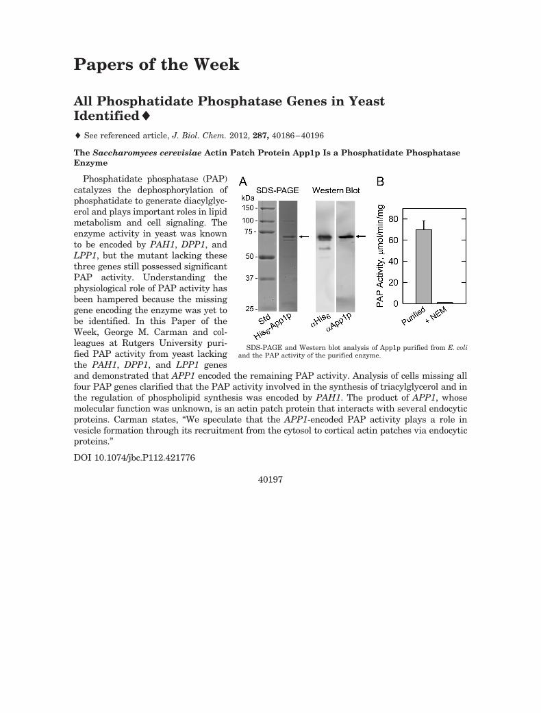
Phosphatidate phosphatase (PAP)catalyzes the dephosphorylation ofphosphatidate to generate diacylglyc-erol and plays important roles in lipidmetabolism and cell signaling. Theenzyme activity in yeast was knownto be encoded by PAH1, DPP1, andLPP1, but the mutant lacking thesethree genes still possessed significantPAP activity. Understanding thephysiological role of PAP activity hasbeen hampered because the missinggene encoding the enzyme was yet tobe identified. In this Paper of theWeek, George M. Carman and col-leagues at Rutgers University puri-fied PAP activity from yeast lackingthe PAH1, DPP1, and LPP1 genesand demonstrated that APP1 encoded the remaining PAP activity. Analysis of cells missing allfour PAP genes clarified that the PAP activity involved in the synthesis of triacylglycerol and inthe regulation of phospholipid synthesis was encoded by PAH1. The product of APP1, whosemolecular function was unknown, is an actin patch protein that interacts with several endocyticproteins. Carman states, “We speculate that the APP1-encoded PAP activity plays a role invesicle formation through its recruitment from the cytosol to cortical actin patches via endocyticproteins.”
DOI 10.1074/jbc.P112.421776
40197
SDS-PAGE and Western blot analysis of App1p purified from E. coliand the PAP activity of the purified enzyme.