Embed Size (px)
Citation preview
The role of water in the behavior of wood
Dominique Derome a,∗, Ahmad Rafsanjani a,b Stefan Hering c
Martin Dressler a,b Alessandra Patera a,b Christian Lanvermann d
Marjan Sedighi-Gilani a Falk K. Wittel c Peter Niemz d andJan Carmeliet a,e
aLaboratory for Building Science and Technology, Swiss Federal Laboratories forMaterials Science and Technology, EMPA, Dubendorf, Switzerland
bDepartment of Civil, Environmental and Geomatic Engineering, ETH Zurich, Zurich,Switzerland
cInstitute for Building Materials, ETH Zurich, Zurich, SwitzerlanddWood Physics Group, Institute for Building Materials, ETH Zurich, Zurich, Switzerland
eChair of Building Physics, ETH Zurich, Zurich, Switzerland
Abstract
Wood, due to its biological origin, has the capacity to interact with water. Sorption/desorptionof moisture is accompanied with swelling/shrinkage and softening/hardening of its stiff-ness. The correct prediction of the behavior of wood components undergoing environmentalloading requires that the moisture behavior and mechanical behavior of wood are consid-ered in a coupled manner. We propose a comprehensive framework using a fully coupledporomechanical approach, where its multiscale implementation provides the capacity totake into account, directly, the exact geometry of the wood cellular structure, using compu-tational homogenization. A hierarchical model is used to take into account the subcellularcomposite-like organization of the material. Such advanced modeling requires high reso-lution experimental data for the appropriate determination of inputs and for its validation.High-resolution x-ray tomography, digital image correlation, and neutron imaging are pre-sented as valuable methods to provide the required information.
Key words: Wood, coupling, water, mechanical behavior, poromechanics, multiscale,imaging methods
∗ Corresponding author. Uberlandstrasse 129, CH-8600 DubendorfEmail address: [email protected] (Dominique Derome).
Preprint submitted to Elsevier September 18, 2015
arX
iv:1
509.
0516
1v1
[co
nd-m
at.m
trl-
sci]
17
Sep
2015
1 Introduction
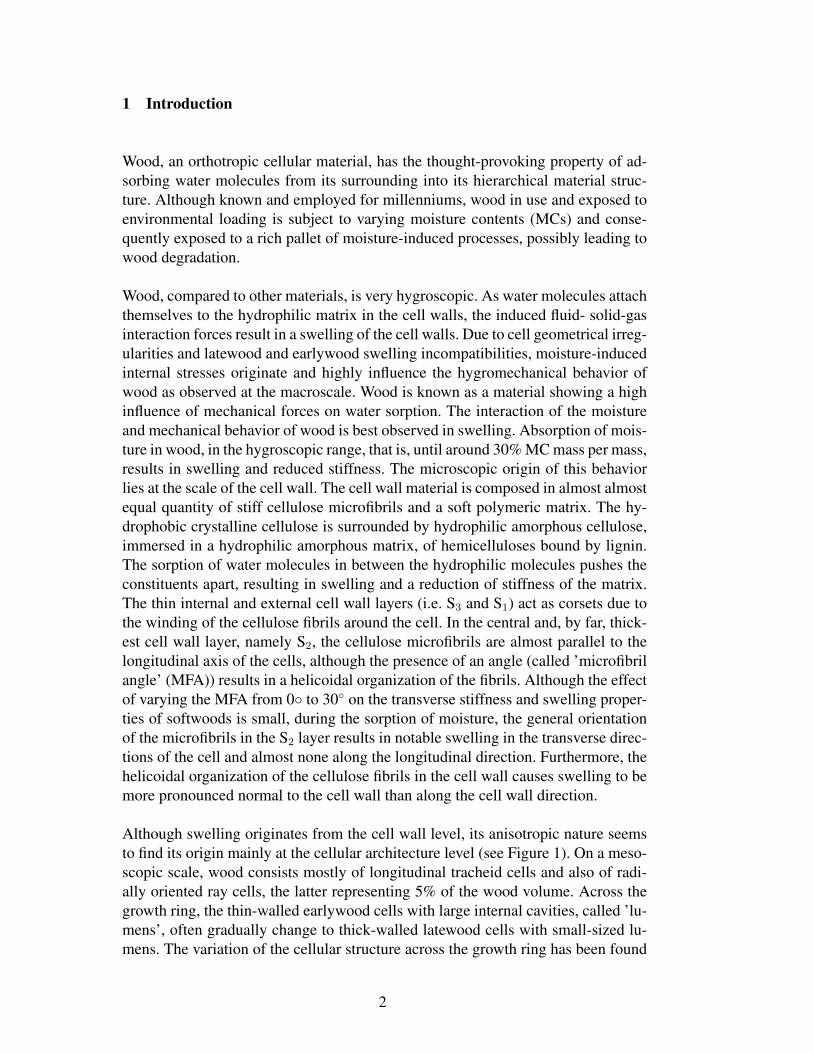
Wood, an orthotropic cellular material, has the thought-provoking property of ad-sorbing water molecules from its surrounding into its hierarchical material struc-ture. Although known and employed for millenniums, wood in use and exposed toenvironmental loading is subject to varying moisture contents (MCs) and conse-quently exposed to a rich pallet of moisture-induced processes, possibly leading towood degradation.
Wood, compared to other materials, is very hygroscopic. As water molecules attachthemselves to the hydrophilic matrix in the cell walls, the induced fluid- solid-gasinteraction forces result in a swelling of the cell walls. Due to cell geometrical irreg-ularities and latewood and earlywood swelling incompatibilities, moisture-inducedinternal stresses originate and highly influence the hygromechanical behavior ofwood as observed at the macroscale. Wood is known as a material showing a highinfluence of mechanical forces on water sorption. The interaction of the moistureand mechanical behavior of wood is best observed in swelling. Absorption of mois-ture in wood, in the hygroscopic range, that is, until around 30% MC mass per mass,results in swelling and reduced stiffness. The microscopic origin of this behaviorlies at the scale of the cell wall. The cell wall material is composed in almost almostequal quantity of stiff cellulose microfibrils and a soft polymeric matrix. The hy-drophobic crystalline cellulose is surrounded by hydrophilic amorphous cellulose,immersed in a hydrophilic amorphous matrix, of hemicelluloses bound by lignin.The sorption of water molecules in between the hydrophilic molecules pushes theconstituents apart, resulting in swelling and a reduction of stiffness of the matrix.The thin internal and external cell wall layers (i.e. S3 and S1) act as corsets due tothe winding of the cellulose fibrils around the cell. In the central and, by far, thick-est cell wall layer, namely S2, the cellulose microfibrils are almost parallel to thelongitudinal axis of the cells, although the presence of an angle (called ’microfibrilangle’ (MFA)) results in a helicoidal organization of the fibrils. Although the effectof varying the MFA from 0◦ to 30◦ on the transverse stiffness and swelling proper-ties of softwoods is small, during the sorption of moisture, the general orientationof the microfibrils in the S2 layer results in notable swelling in the transverse direc-tions of the cell and almost none along the longitudinal direction. Furthermore, thehelicoidal organization of the cellulose fibrils in the cell wall causes swelling to bemore pronounced normal to the cell wall than along the cell wall direction.
Although swelling originates from the cell wall level, its anisotropic nature seemsto find its origin mainly at the cellular architecture level (see Figure 1). On a meso-scopic scale, wood consists mostly of longitudinal tracheid cells and also of radi-ally oriented ray cells, the latter representing 5% of the wood volume. Across thegrowth ring, the thin-walled earlywood cells with large internal cavities, called ’lu-mens’, often gradually change to thick-walled latewood cells with small-sized lu-mens. The variation of the cellular structure across the growth ring has been found
2
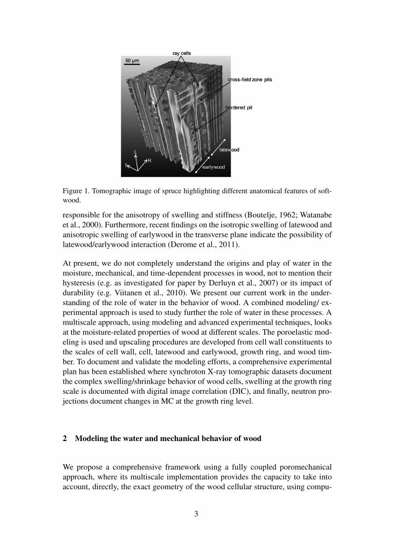
Figure 1. Tomographic image of spruce highlighting different anatomical features of soft-wood.
responsible for the anisotropy of swelling and stiffness (Boutelje, 1962; Watanabeet al., 2000). Furthermore, recent findings on the isotropic swelling of latewood andanisotropic swelling of earlywood in the transverse plane indicate the possibility oflatewood/earlywood interaction (Derome et al., 2011).
At present, we do not completely understand the origins and play of water in themoisture, mechanical, and time-dependent processes in wood, not to mention theirhysteresis (e.g. as investigated for paper by Derluyn et al., 2007) or its impact ofdurability (e.g. Viitanen et al., 2010). We present our current work in the under-standing of the role of water in the behavior of wood. A combined modeling/ ex-perimental approach is used to study further the role of water in these processes. Amultiscale approach, using modeling and advanced experimental techniques, looksat the moisture-related properties of wood at different scales. The poroelastic mod-eling is used and upscaling procedures are developed from cell wall constituents tothe scales of cell wall, cell, latewood and earlywood, growth ring, and wood tim-ber. To document and validate the modeling efforts, a comprehensive experimentalplan has been established where synchroton X-ray tomographic datasets documentthe complex swelling/shrinkage behavior of wood cells, swelling at the growth ringscale is documented with digital image correlation (DIC), and finally, neutron pro-jections document changes in MC at the growth ring level.
2 Modeling the water and mechanical behavior of wood
We propose a comprehensive framework using a fully coupled poromechanicalapproach, where its multiscale implementation provides the capacity to take intoaccount, directly, the exact geometry of the wood cellular structure, using compu-
3
tational homogenization. An hierarchical model is used to take into account thesubcellular composite-like organization of the material.
2.1 Poromechanical approach
A rigorous way of taking into account the interaction of fluids with the solid matrixin porous materials is based on the theory of poromechanics, introduced by Biot(1941). Within the context of thermodynamics of open porous continua, Coussy(2010) presented a general framework to formulate adequate constitutive equationsfor poroelastic behavior. The poromechanical approach starts from an energy view-point where stress/strain and chemical potential/MC are seen as pairs determiningthe energy content. With one equation expressing the energy content of the wholesystem, any change in stress field or chemical potential leads to a coupled responsein both the strain and the MC. Thus, the poromechanical approach is suited tocover coupled effects between the mechanical and moisture response as observedin wood. A full description of the model described below is found in Carmeliet etal. (2013).
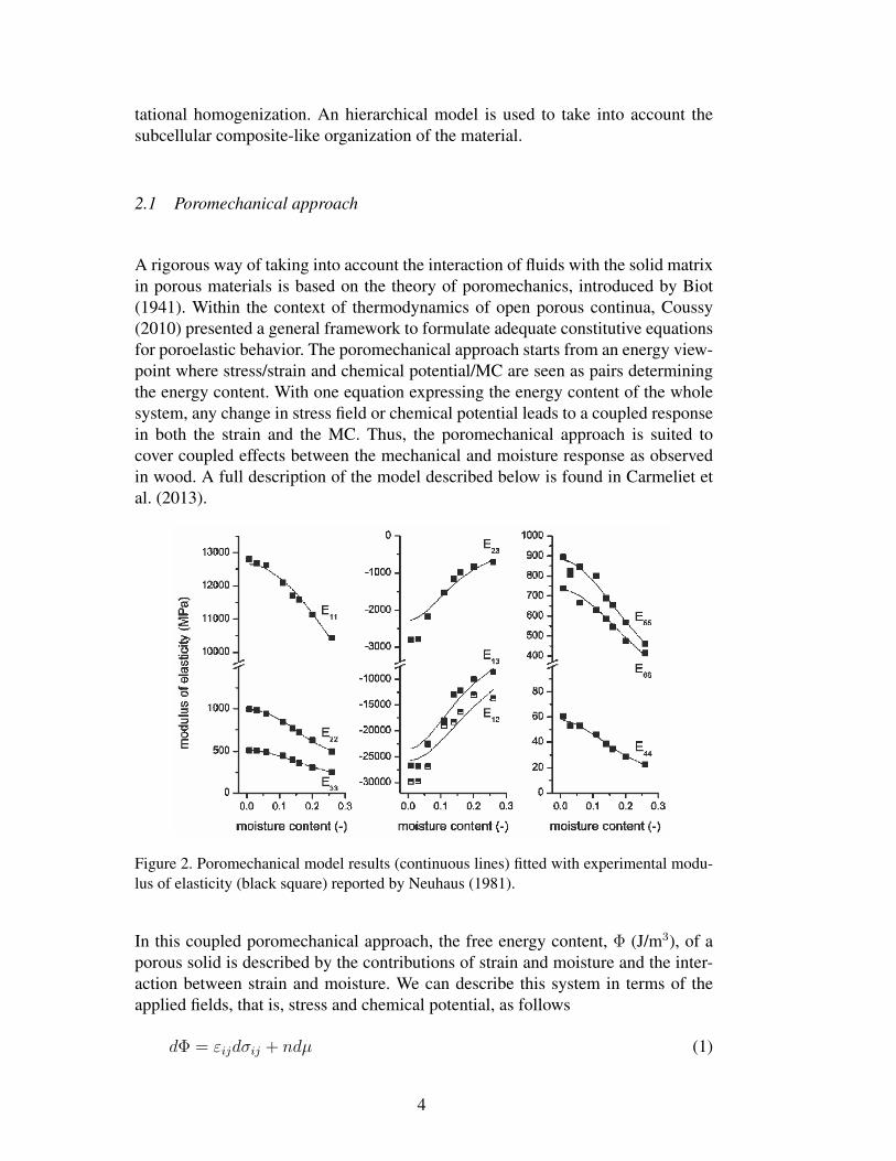
Figure 2. Poromechanical model results (continuous lines) fitted with experimental modu-lus of elasticity (black square) reported by Neuhaus (1981).
In this coupled poromechanical approach, the free energy content, Φ (J/m3), of aporous solid is described by the contributions of strain and moisture and the inter-action between strain and moisture. We can describe this system in terms of theapplied fields, that is, stress and chemical potential, as follows
dΦ = εijdσij + ndµ (1)
4
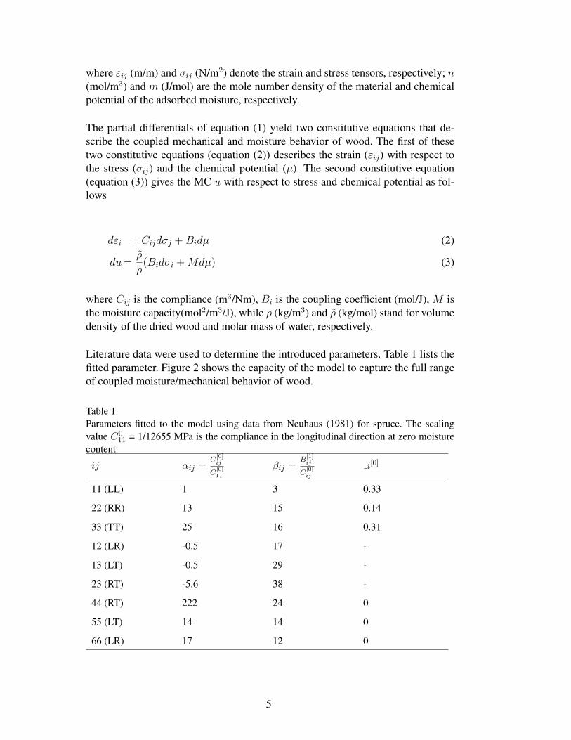
where εij (m/m) and σij (N/m2) denote the strain and stress tensors, respectively; n(mol/m3) and m (J/mol) are the mole number density of the material and chemicalpotential of the adsorbed moisture, respectively.
The partial differentials of equation (1) yield two constitutive equations that de-scribe the coupled mechanical and moisture behavior of wood. The first of thesetwo constitutive equations (equation (2)) describes the strain (εij) with respect tothe stress (σij) and the chemical potential (µ). The second constitutive equation(equation (3)) gives the MC u with respect to stress and chemical potential as fol-lows
dεi = Cijdσj +Bidµ (2)
du=ρ
ρ(Bidσi +Mdµ) (3)
where Cij is the compliance (m3/Nm), Bi is the coupling coefficient (mol/J), M isthe moisture capacity(mol2/m3/J), while ρ (kg/m3) and ρ (kg/mol) stand for volumedensity of the dried wood and molar mass of water, respectively.
Literature data were used to determine the introduced parameters. Table 1 lists thefitted parameter. Figure 2 shows the capacity of the model to capture the full rangeof coupled moisture/mechanical behavior of wood.
Table 1Parameters fitted to the model using data from Neuhaus (1981) for spruce. The scalingvalue C0
11 = 1/12655 MPa is the compliance in the longitudinal direction at zero moisturecontent
ij αij =C
[0]ij
C[0]11
βij =B
[1]ij
C[0]ij
i[0]
11 (LL) 1 3 0.33
22 (RR) 13 15 0.14
33 (TT) 25 16 0.31
12 (LR) -0.5 17 -
13 (LT) -0.5 29 -
23 (RT) -5.6 38 -
44 (RT) 222 24 0
55 (LT) 14 14 0
66 (LR) 17 12 0
5
2.2 Multiscale approach
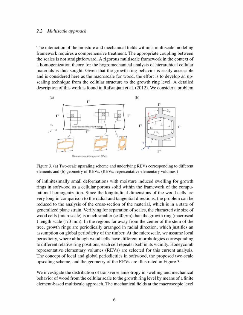
The interaction of the moisture and mechanical fields within a multiscale modelingframework requires a comprehensive treatment. The appropriate coupling betweenthe scales is not straightforward. A rigorous multiscale framework in the context ofa homogenization theory for the hygromechanical analysis of hierarchical cellularmaterials is thus sought. Given that the growth ring behavior is easily accessibleand is considered here as the macroscale for wood, the effort is to develop an up-scaling technique from the cellular structure to the growth ring level. A detaileddescription of this work is found in Rafsanjani et al. (2012). We consider a problem
Figure 3. (a) Two-scale upscaling scheme and underlying REVs corresponding to differentelements and (b) geometry of REVs. (REVs: representative elementary volumes.)
of infinitesimally small deformations with moisture induced swelling for growthrings in softwood as a cellular porous solid within the framework of the compu-tational homogenization. Since the longitudinal dimensions of the wood cells arevery long in comparison to the radial and tangential directions, the problem can bereduced to the analysis of the cross-section of the material, which is in a state ofgeneralized plane strain. Verifying for separation of scales, the characteristic size ofwood cells (microscale) is much smaller (≈40 µm) than the growth ring (macroscal) length scale (≈3 mm). In the regions far away from the center of the stem of thetree, growth rings are periodically arranged in radial direction, which justifies anassumption on global periodicity of the timber. At the microscale, we assume localperiodicity, where although wood cells have different morphologies correspondingto different relative ring positions, each cell repeats itself in its vicinity. Honeycombrepresentative elementary volumes (REVs) are selected for this current analysis.The concept of local and global periodicities in softwood, the proposed two-scaleupscaling scheme, and the geometry of the REVs are illustrated in Figure 3.
We investigate the distribution of transverse anisotropy in swelling and mechanicalbehavior of wood from the cellular scale to the growth ring level by means of a finiteelement-based multiscale approach. The mechanical fields at the macroscopic level
6
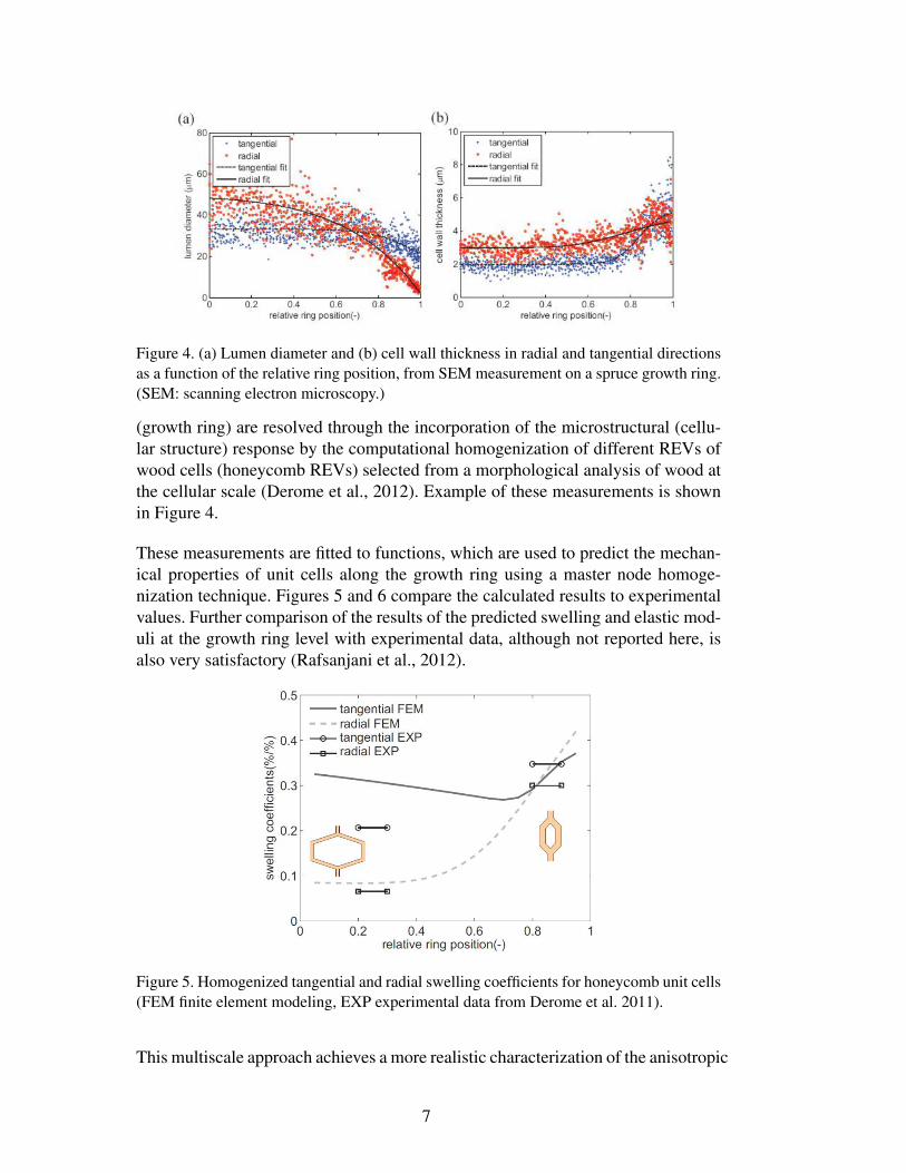
Figure 4. (a) Lumen diameter and (b) cell wall thickness in radial and tangential directionsas a function of the relative ring position, from SEM measurement on a spruce growth ring.(SEM: scanning electron microscopy.)
(growth ring) are resolved through the incorporation of the microstructural (cellu-lar structure) response by the computational homogenization of different REVs ofwood cells (honeycomb REVs) selected from a morphological analysis of wood atthe cellular scale (Derome et al., 2012). Example of these measurements is shownin Figure 4.
These measurements are fitted to functions, which are used to predict the mechan-ical properties of unit cells along the growth ring using a master node homoge-nization technique. Figures 5 and 6 compare the calculated results to experimentalvalues. Further comparison of the results of the predicted swelling and elastic mod-uli at the growth ring level with experimental data, although not reported here, isalso very satisfactory (Rafsanjani et al., 2012).
Figure 5. Homogenized tangential and radial swelling coefficients for honeycomb unit cells(FEM finite element modeling, EXP experimental data from Derome et al. 2011).
This multiscale approach achieves a more realistic characterization of the anisotropic
7
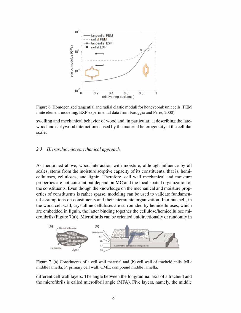
Figure 6. Homogenized tangential and radial elastic moduli for honeycomb unit cells (FEMfinite element modeling, EXP experimental data from Faruggia and Perre, 2000).
swelling and mechanical behavior of wood and, in particular, at describing the late-wood and earlywood interaction caused by the material heterogeneity at the cellularscale.
2.3 Hierarchic micromechanical approach
As mentioned above, wood interaction with moisture, although influence by allscales, stems from the moisture sorptive capacity of its constituents, that is, hemi-celluloses, celluloses, and lignin. Therefore, cell wall mechanical and moistureproperties are not constant but depend on MC and the local spatial organization ofthe constituents. Even though the knowledge on the mechanical and moisture prop-erties of constituents is rather sparse, modeling can be used to validate fundamen-tal assumptions on constituents and their hierarchic organization. In a nutshell, inthe wood cell wall, crystalline celluloses are surrounded by hemicelluloses, whichare embedded in lignin, the latter binding together the cellulose/hemicellulose mi-crofibrils (Figure 7(a)). Microfibrils can be oriented unidirectionally or randomly in
Figure 7. (a) Constituents of a cell wall material and (b) cell wall of tracheid cells. ML:middle lamella; P: primary cell wall; CML: compound middle lamella.
different cell wall layers. The angle between the longitudinal axis of a tracheid andthe microfibrils is called microfibril angle (MFA). Five layers, namely, the middle
8
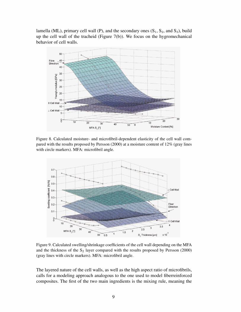
lamella (ML), primary cell wall (P), and the secondary ones (S1, S2, and S3), buildup the cell wall of the tracheid (Figure 7(b)). We focus on the hygromechanicalbehavior of cell walls.
Figure 8. Calculated moisture- and microfibril-dependent elasticity of the cell wall com-pared with the results proposed by Persson (2000) at a moisture content of 12% (gray lineswith circle markers). MFA: microfibril angle.
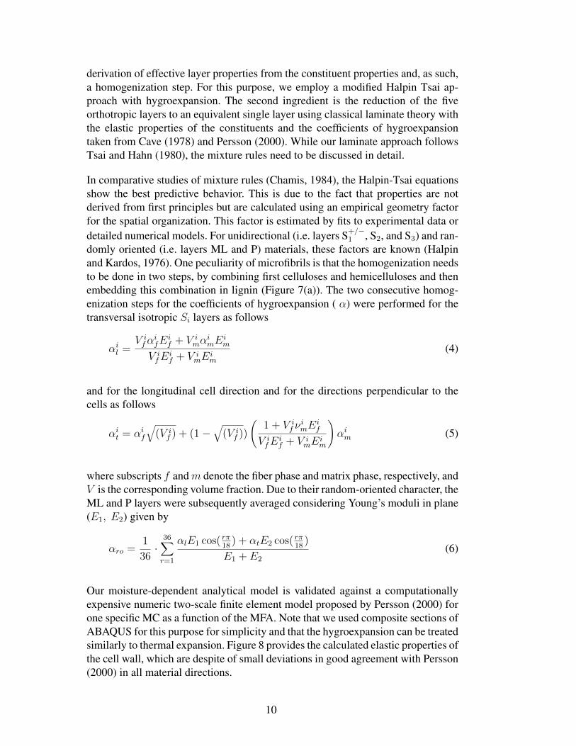
Figure 9. Calculated swelling/shrinkage coefficients of the cell wall depending on the MFAand the thickness of the S2 layer compared with the results proposed by Persson (2000)(gray lines with circle markers). MFA: microfibril angle.
The layered nature of the cell walls, as well as the high aspect ratio of microfibrils,calls for a modeling approach analogous to the one used to model fiberreinforcedcomposites. The first of the two main ingredients is the mixing rule, meaning the
9
derivation of effective layer properties from the constituent properties and, as such,a homogenization step. For this purpose, we employ a modified Halpin Tsai ap-proach with hygroexpansion. The second ingredient is the reduction of the fiveorthotropic layers to an equivalent single layer using classical laminate theory withthe elastic properties of the constituents and the coefficients of hygroexpansiontaken from Cave (1978) and Persson (2000). While our laminate approach followsTsai and Hahn (1980), the mixture rules need to be discussed in detail.
In comparative studies of mixture rules (Chamis, 1984), the Halpin-Tsai equationsshow the best predictive behavior. This is due to the fact that properties are notderived from first principles but are calculated using an empirical geometry factorfor the spatial organization. This factor is estimated by fits to experimental data ordetailed numerical models. For unidirectional (i.e. layers S+/−
1 , S2, and S3) and ran-domly oriented (i.e. layers ML and P) materials, these factors are known (Halpinand Kardos, 1976). One peculiarity of microfibrils is that the homogenization needsto be done in two steps, by combining first celluloses and hemicelluloses and thenembedding this combination in lignin (Figure 7(a)). The two consecutive homog-enization steps for the coefficients of hygroexpansion ( α) were performed for thetransversal isotropic Si layers as follows
αil =V ifα
ifE
if + V i
mαimE
im
V ifE
if + V i
mEim
(4)
and for the longitudinal cell direction and for the directions perpendicular to thecells as follows
αit = αif
√(V i
f ) + (1 −√
(V if ))
(1 + V i
f νimE
if
V ifE
if + V i
mEim
)αim (5)
where subscripts f andm denote the fiber phase and matrix phase, respectively, andV is the corresponding volume fraction. Due to their random-oriented character, theML and P layers were subsequently averaged considering Young’s moduli in plane(E1, E2) given by
αro =1
36·
36∑r=1
αlE1 cos( rπ18
) + αtE2 cos( rπ18
)
E1 + E2
(6)
Our moisture-dependent analytical model is validated against a computationallyexpensive numeric two-scale finite element model proposed by Persson (2000) forone specific MC as a function of the MFA. Note that we used composite sections ofABAQUS for this purpose for simplicity and that the hygroexpansion can be treatedsimilarly to thermal expansion. Figure 8 provides the calculated elastic properties ofthe cell wall, which are despite of small deviations in good agreement with Persson(2000) in all material directions.
10
To estimate the free swelling of a piece of cell wall, a block of two adjacent cellwalls is calculated. The swelling coefficients in the main directions of the cell wallare given in Figure 9. It is not surprising that the swelling in thickness directiondominates the one seen in the other orientations. Moreover, the dependency onMFA is more pronounced in the longitudinal direction and in the direction parallelto the cell wall. Compared to Persson (2000), all tendencies can be confirmed, eventhough the absolute values perpendicular to the cell wall differ. The latter can beexplained by different mixing rules used at the microfibril level. For consistency,we employ Halpin-Tsai as described above, while Persson (2000) uses a numerichomogenization scheme from a so-called base cell with adjusted geometry.
In addition to provide the cell wall properties, this approach could be further usedto study early/latewood for different cell wall and geometry properties in orderto identify the factors dominating the swelling anisotropy of wood seen at themacroscale.
3 Imaging methods
The models presented above require high-resolution experimental data for the ap-propriate determination of inputs and for their validation. High-resolution tomog-raphy, DIC, and neutron imaging are presented as valuable methods to provide therequired information.
3.1 Swelling of wood probed by phase-contrast X-ray synchroton tomography
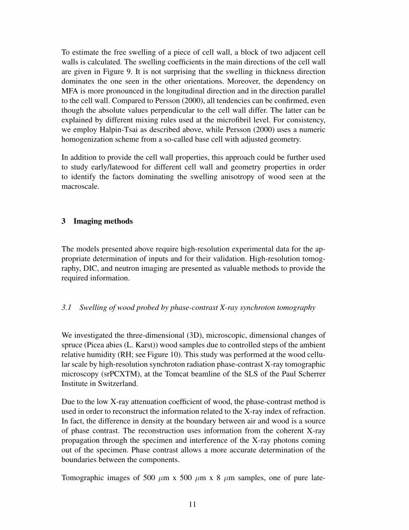
We investigated the three-dimensional (3D), microscopic, dimensional changes ofspruce (Picea abies (L. Karst)) wood samples due to controlled steps of the ambientrelative humidity (RH; see Figure 10). This study was performed at the wood cellu-lar scale by high-resolution synchroton radiation phase-contrast X-ray tomographicmicroscopy (srPCXTM), at the Tomcat beamline of the SLS of the Paul ScherrerInstitute in Switzerland.
Due to the low X-ray attenuation coefficient of wood, the phase-contrast method isused in order to reconstruct the information related to the X-ray index of refraction.In fact, the difference in density at the boundary between air and wood is a sourceof phase contrast. The reconstruction uses information from the coherent X-raypropagation through the specimen and interference of the X-ray photons comingout of the specimen. Phase contrast allows a more accurate determination of theboundaries between the components.
Tomographic images of 500 µm x 500 µm x 8 µm samples, one of pure late-
11
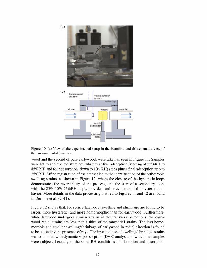
Figure 10. (a) View of the experimental setup in the beamline and (b) schematic view ofthe environmental chamber.
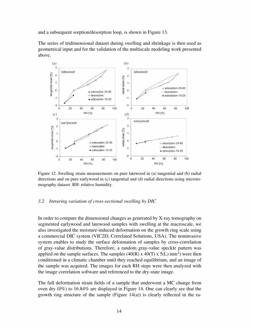
wood and the second of pure earlywood, were taken as seen in Figure 11. Sampleswere let to achieve moisture equilibrium at five adsorption (starting at 25%RH to85%RH) and four desorption (down to 10%RH) steps plus a final adsorption step to25%RH. Affine registration of the dataset led to the identification of the orthotropicswelling strains, as shown in Figure 12, where the closure of the hysteretic loopsdemonstrates the reversibility of the process, and the start of a secondary loop,with the 25%-10%-25%RH steps, provides further evidence of the hysteretic be-havior. More details in the data processing that led to Figures 11 and 12 are foundin Derome et al. (2011).
Figure 12 shows that, for spruce latewood, swelling and shrinkage are found to belarger, more hysteretic, and more homomorphic than for earlywood. Furthermore,while latewood undergoes similar strains in the transverse directions, the early-wood radial strains are less than a third of the tangential strains. The less homo-morphic and smaller swelling/shrinkage of earlywood in radial direction is foundto be caused by the presence of rays. The investigation of swelling/shrinkage strainswas combined with dynamic vapor sorption (DVS) analysis, in which the sampleswere subjected exactly to the same RH conditions in adsorption and desorption.
12
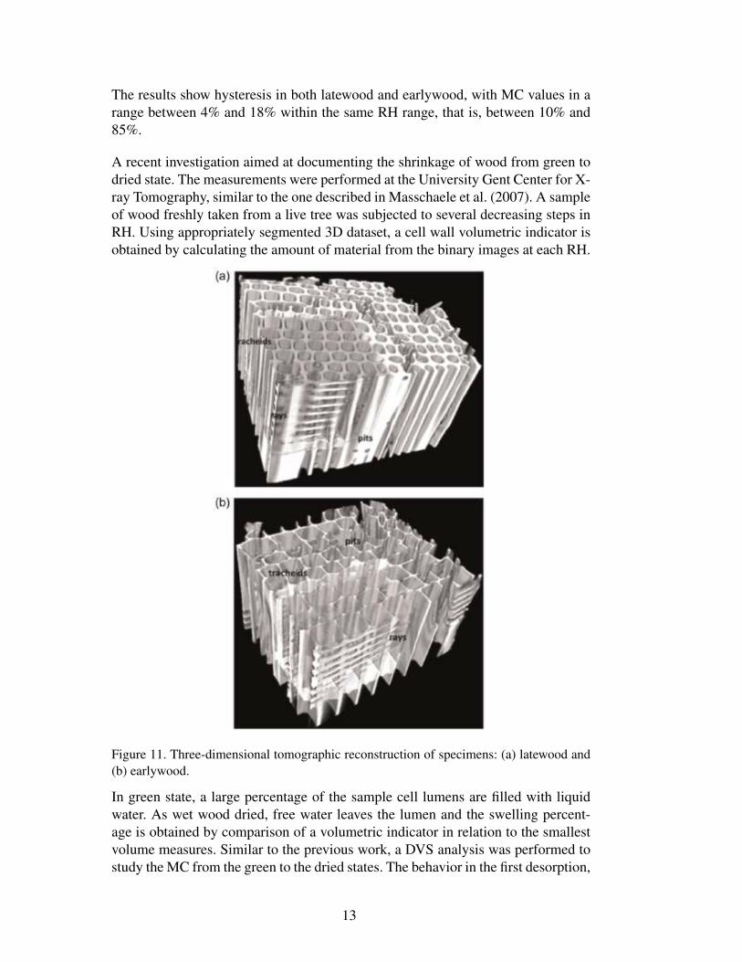
The results show hysteresis in both latewood and earlywood, with MC values in arange between 4% and 18% within the same RH range, that is, between 10% and85%.
A recent investigation aimed at documenting the shrinkage of wood from green todried state. The measurements were performed at the University Gent Center for X-ray Tomography, similar to the one described in Masschaele et al. (2007). A sampleof wood freshly taken from a live tree was subjected to several decreasing steps inRH. Using appropriately segmented 3D dataset, a cell wall volumetric indicator isobtained by calculating the amount of material from the binary images at each RH.
Figure 11. Three-dimensional tomographic reconstruction of specimens: (a) latewood and(b) earlywood.
In green state, a large percentage of the sample cell lumens are filled with liquidwater. As wet wood dried, free water leaves the lumen and the swelling percent-age is obtained by comparison of a volumetric indicator in relation to the smallestvolume measures. Similar to the previous work, a DVS analysis was performed tostudy the MC from the green to the dried states. The behavior in the first desorption,
13
and a subsequent sorption/desorption loop, is shown in Figure 13.
The series of tridimensional dataset during swelling and shrinkage is then used asgeometrical input and for the validation of the multiscale modeling work presentedabove.
Figure 12. Swelling strain measurements on pure latewood in (a) tangential and (b) radialdirections and on pure earlywood in (c) tangential and (d) radial directions using microto-mography dataset. RH: relative humidity.
3.2 Intraring variation of cross-sectional swelling by DIC
In order to compare the dimensional changes as generated by X-ray tomography onsegmented earlywood and latewood samples with swelling at the macroscale, wealso investigated the moisture-induced deformation on the growth ring scale usinga commercial DIC system (VIC2D; Correlated Solutions, USA). The noninvasivesystem enables to study the surface deformation of samples by cross-correlationof gray-value distributions. Therefore, a random gray-value speckle pattern wasapplied on the sample surfaces. The samples (40(R) x 40(T) x 5(L) mm3) were thenconditioned in a climatic chamber until they reached equilibrium, and an image ofthe sample was acquired. The images for each RH steps were then analyzed withthe image correlation software and referenced to the dry-state image.
The full deformation strain fields of a sample that underwent a MC change fromoven dry (0%) to 16.84% are displayed in Figure 14. One can clearly see that thegrowth ring structure of the sample (Figure 14(a)) is clearly reflected in the ra-
14
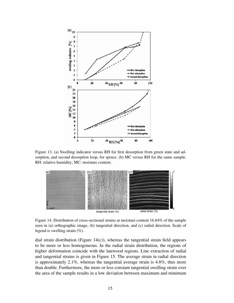
Figure 13. (a) Swelling indicator versus RH for first desorption from green state and ad-sorption, and second desorption loop, for spruce. (b) MC versus RH for the same sample.RH: relative humidity; MC: moisture content.
Figure 14. Distribution of cross-sectional strains at moisture content 16.84% of the sampleseen in (a) orthographic image, (b) tangential direction, and (c) radial direction. Scale oflegend is swelling strain (%).
dial strain distribution (Figure 14(c)), whereas the tangential strain field appearsto be more or less homogeneous. In the radial strain distribution, the regions ofhigher deformation coincide with the latewood regions. Line extraction of radialand tangential strains is given in Figure 15. The average strain in radial directionis approximately 2.1%, whereas the tangential average strain is 4.8%, thus morethan double. Furthermore, the more or less constant tangential swelling strain overthe area of the sample results in a low deviation between maximum and minimum
15
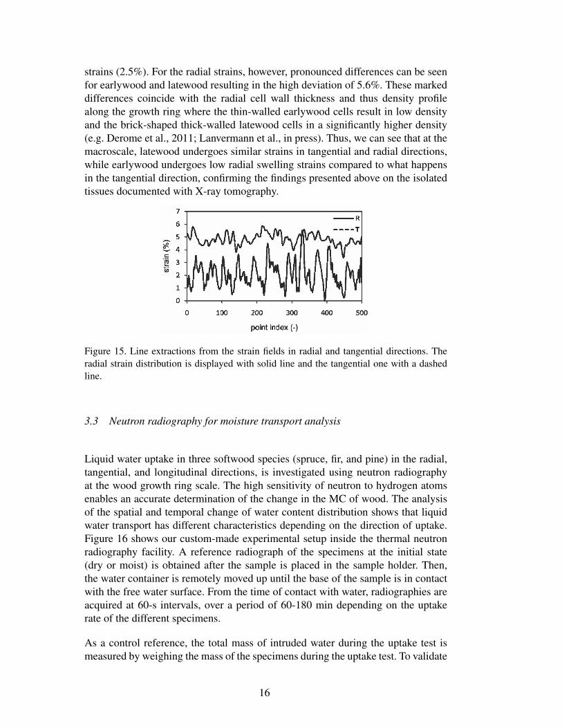
strains (2.5%). For the radial strains, however, pronounced differences can be seenfor earlywood and latewood resulting in the high deviation of 5.6%. These markeddifferences coincide with the radial cell wall thickness and thus density profilealong the growth ring where the thin-walled earlywood cells result in low densityand the brick-shaped thick-walled latewood cells in a significantly higher density(e.g. Derome et al., 2011; Lanvermann et al., in press). Thus, we can see that at themacroscale, latewood undergoes similar strains in tangential and radial directions,while earlywood undergoes low radial swelling strains compared to what happensin the tangential direction, confirming the findings presented above on the isolatedtissues documented with X-ray tomography.
Figure 15. Line extractions from the strain fields in radial and tangential directions. Theradial strain distribution is displayed with solid line and the tangential one with a dashedline.
3.3 Neutron radiography for moisture transport analysis
Liquid water uptake in three softwood species (spruce, fir, and pine) in the radial,tangential, and longitudinal directions, is investigated using neutron radiographyat the wood growth ring scale. The high sensitivity of neutron to hydrogen atomsenables an accurate determination of the change in the MC of wood. The analysisof the spatial and temporal change of water content distribution shows that liquidwater transport has different characteristics depending on the direction of uptake.Figure 16 shows our custom-made experimental setup inside the thermal neutronradiography facility. A reference radiograph of the specimens at the initial state(dry or moist) is obtained after the sample is placed in the sample holder. Then,the water container is remotely moved up until the base of the sample is in contactwith the free water surface. From the time of contact with water, radiographies areacquired at 60-s intervals, over a period of 60-180 min depending on the uptakerate of the different specimens.
As a control reference, the total mass of intruded water during the uptake test ismeasured by weighing the mass of the specimens during the uptake test. To validate
16
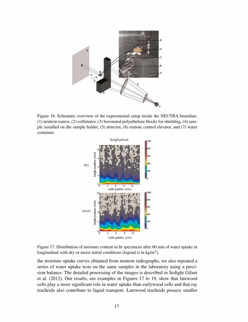
Figure 16. Schematic overview of the experimental setup inside the NEUTRA beamline:(1) neutron source, (2) collimator, (3) boronated polyethelene blocks for shielding, (4) sam-ple installed on the sample holder, (5) detector, (6) remote control elevator, and (7) watercontainer.
Figure 17. Distribution of moisture content in fir specimens after 60 min of water uptake inlongitudinal with dry or moist initial conditions (legend is in kg/m3).
the moisture uptake curves obtained from neutron radiographs, we also repeated aseries of water uptake tests on the same samples in the laboratory using a preci-sion balance. The detailed processing of the images is described in Sedighi Gilaniet al. (2012). Our results, see examples in Figures 17 to 19, show that latewoodcells play a more significant role in water uptake than earlywood cells and that raytracheids also contribute to liquid transport. Latewood tracheids possess smaller
17
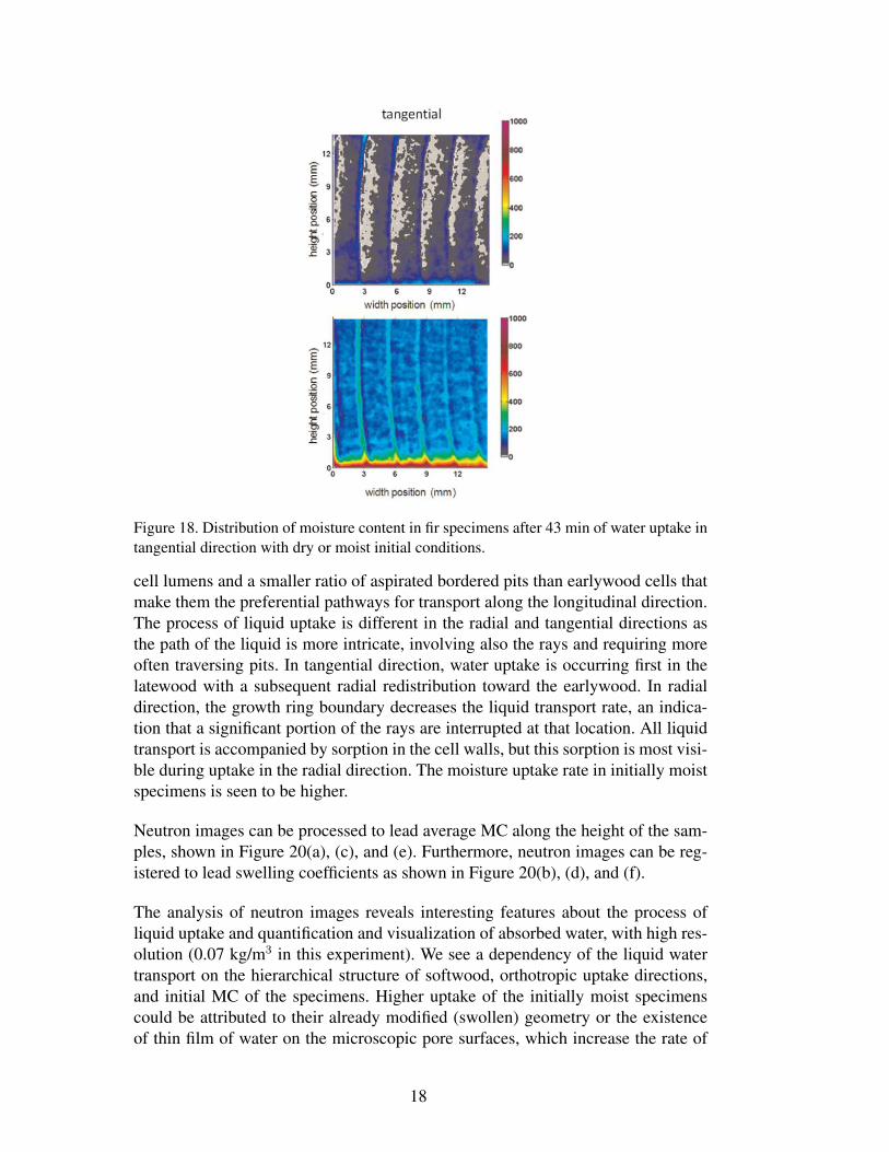
Figure 18. Distribution of moisture content in fir specimens after 43 min of water uptake intangential direction with dry or moist initial conditions.
cell lumens and a smaller ratio of aspirated bordered pits than earlywood cells thatmake them the preferential pathways for transport along the longitudinal direction.The process of liquid uptake is different in the radial and tangential directions asthe path of the liquid is more intricate, involving also the rays and requiring moreoften traversing pits. In tangential direction, water uptake is occurring first in thelatewood with a subsequent radial redistribution toward the earlywood. In radialdirection, the growth ring boundary decreases the liquid transport rate, an indica-tion that a significant portion of the rays are interrupted at that location. All liquidtransport is accompanied by sorption in the cell walls, but this sorption is most visi-ble during uptake in the radial direction. The moisture uptake rate in initially moistspecimens is seen to be higher.
Neutron images can be processed to lead average MC along the height of the sam-ples, shown in Figure 20(a), (c), and (e). Furthermore, neutron images can be reg-istered to lead swelling coefficients as shown in Figure 20(b), (d), and (f).
The analysis of neutron images reveals interesting features about the process ofliquid uptake and quantification and visualization of absorbed water, with high res-olution (0.07 kg/m3 in this experiment). We see a dependency of the liquid watertransport on the hierarchical structure of softwood, orthotropic uptake directions,and initial MC of the specimens. Higher uptake of the initially moist specimenscould be attributed to their already modified (swollen) geometry or the existenceof thin film of water on the microscopic pore surfaces, which increase the rate of
18
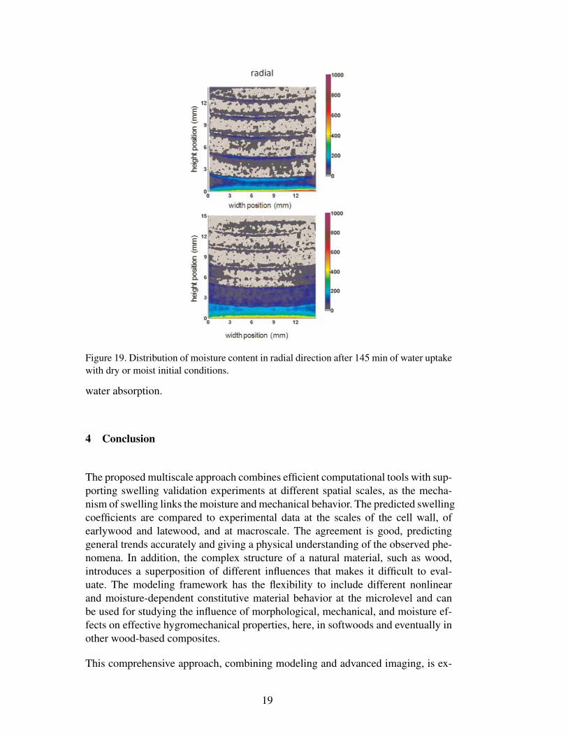
Figure 19. Distribution of moisture content in radial direction after 145 min of water uptakewith dry or moist initial conditions.
water absorption.
4 Conclusion
The proposed multiscale approach combines efficient computational tools with sup-porting swelling validation experiments at different spatial scales, as the mecha-nism of swelling links the moisture and mechanical behavior. The predicted swellingcoefficients are compared to experimental data at the scales of the cell wall, ofearlywood and latewood, and at macroscale. The agreement is good, predictinggeneral trends accurately and giving a physical understanding of the observed phe-nomena. In addition, the complex structure of a natural material, such as wood,introduces a superposition of different influences that makes it difficult to eval-uate. The modeling framework has the flexibility to include different nonlinearand moisture-dependent constitutive material behavior at the microlevel and canbe used for studying the influence of morphological, mechanical, and moisture ef-fects on effective hygromechanical properties, here, in softwoods and eventually inother wood-based composites.
This comprehensive approach, combining modeling and advanced imaging, is ex-
19
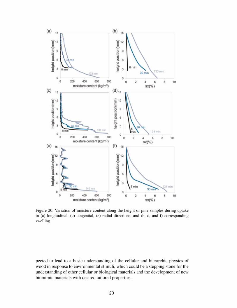
Figure 20. Variation of moisture content along the height of pine samples during uptakein (a) longitudinal, (c) tangential, (e) radial directions, and (b, d, and f) correspondingswelling.
pected to lead to a basic understanding of the cellular and hierarchic physics ofwood in response to environmental stimuli, which could be a stepping stone for theunderstanding of other cellular or biological materials and the development of newbiomimic materials with desired tailored properties.
20
Funding
This study was supported by the Swiss National Science Foundation SNF underSinergia grant no. 125184.
Acknowledgements
Phase-contrast synchrotron X-ray tomographic data were acquired at the Tomcatbeamline of SLS. Neutron images acquired at the Neutra beamline of SINQ. SLSand SINQ are at PSI, Villigen, Switzerland. X-ray tomography data for green woodexperiments were acquired at the University Ghent Center for X-ray Tomographyin Belgium.
References
Biot MA (1941) General theory of three-dimensional consolidation. Journalof Applied Physics 12: 155-164.
Boutelje JB (1962) The relationship of structure to transverse anisotropy inwood with reference to shrinkage and elasticity. Holzforschung 16(2): 33-46.
Carmeliet J, Derome D, Dressler M, et al. (2013) Nonlinear poro-elastic modelfor unsaturated porous solids; Olivier Coussy Memorial Issu. Journal of Ap-plied Mechanics 80(2): 020909.
Cave ID (1978) Modelling moisture-related mechanical properties of wood.Part I:properties of the wood constituents. Wood Science and Technology 12:75-86.
Chamis CC (1984) Simplified composite micromechanics equations for hy-gral, thermal, and mechanical properties. Sampe Quarterly: Society for theAdvancement of Material and Process Engineering 15(3): 14-23.
Coussy O (2010) Mechanics and Physics of Porous Solids. John Wiley andSons, West Sussex, United Kingdom.
Derluyn H, Janssen H, Diepens J, et al. (2007) Hygroscopic behavior of paperand books. Journal of Building Physics 31(9): 9-34.
Derome D, Griffa M, Koebel M, et al. (2011) Hysteretic swelling of wood atcellular scale probed by phase-contrast X-ray tomography. Journal of Struc-tural Biology 173: 180-190.
21
Derome D, Zillig W and Carmeliet J (2012) Variation of measured cross-sectional cell dimensions and calculated water vapor permeability across asingle growth ring of spruce wood. Wood Science and Technology 46(5): 827-840.
Farruggia F and Perre P (2000) Microscopic tensile tests in the transverseplane of earlywood and latewood parts of spruce. Wood Science and Technol-ogy 34(2): 65-82.
Halpin JC and Kardos JL (1976) The Halpin-Tsai equations: a review. Poly-mer Engineering and Science 16(5): 344-352.
Lanvermann C, Evans R, Schmitt U, et al. (in press) Distribution of structureand lignin within growth rings of Norway spruce. Wood Science and Technol-ogy.
Masschaele BC, Cnudde V, Dierick M, et al. (2007) UGCT: new X-ray radio-graphy and tomography facility. Nuclear Instruments and Methods in PhysicsResearch Section A: Accelerators Spectrometers Detectors and AssociatedEquipment 580: 266-269.
Neuhaus FH (1981) Elastizitaetszahlen von Fichtenholz in Abaengigkeit vonder Holzfeuchtigkeit. PhD Thesis, Institut fuer konstruktiven IngenieurbauRuhr-Universitaet Bochum, Germany.
Persson K (2000) Micromechanical Modelling of Wood and Fibre Properties.Doctoral Thesis, Lund University, Sweden.
Rafsanjani A, Derome D, Wittel F, et al. (2012) Computational up-scaling ofanisotropic swelling and mechanical behavior of hierarchical cellular materi-als. Composites Science and Technology 72: 744-751.
Sedighi-Gilani M, Griffa M, Mannes D, et al. (2012) Visualization and quan-tification of liquid water transport in softwood by means of neutron radiogra-phy. International Journal of Heat and Mass Transfer 55 (21-22): 6211-6221.
Tsai S and Hahn H (1980) Introduction to Composite Materials. Lancaster,PA: Technomic Publishing Company, Inc.
Viitanen H, Vinha J, Salmiren K, et al. (2010) Moisture and bio-deteriorationrisk of building materials and structures. Journal of Building Physics 33(3):201-224.
Watanabe U, Norimoto M and Morooka T (2000) Cell wall thickness and tan-gential Young’s modulus in coniferous early wood. Journal of Wood Science46(2): 109-114.
22