Embed Size (px)
Citation preview

Exp Brain ResDOI 10.1007/s00221-011-2579-1
123
REVIEW
The role of the occipital face area in the cortical face perception network
David Pitcher · Vincent Walsh · Bradley Duchaine
Received: 5 August 2009 / Accepted: 27 January 2011! Springer-Verlag 2011
Abstract Functional magnetic resonance imaging (fMRI)studies have identiWed spatially distinct face-selectiveregions in human cortex. These regions have been linkedtogether to form the components of a cortical network spe-cialized for face perception but the cognitive operationsperformed in each region are not well understood. In thispaper, we review the evidence concerning one of theseface-selective regions, the occipital face area (OFA), to bet-ter understand what cognitive operations it performs in theface perception network. Neuropsychological evidence andtranscranial magnetic stimulation (TMS) studies demon-strate the OFA is necessary for accurate face perception.fMRI and TMS studies investigating the functional role ofthe OFA suggest that it preferentially represents the parts ofa face, including the eyes, nose, and mouth and that it doesso at an early stage of visual perception. These studies areconsistent with the hypothesis that the OFA is the Wrst stagein a hierarchical face perception network in which the OFArepresents facial components prior to subsequent process-ing of increasingly complex facial features in higher face-selective cortical regions.
Keywords Face perception · Occipital face area (OFA) · Functional magnetic resonance imaging (fMRI) · Transcranial magnetic stimulation (TMS)
Introduction
The ubiquitous presence of faces makes them a uniquelysalient stimulus for studying the functions of human visualcortex. Functional magnetic resonance imaging (fMRI) stud-ies graphically illustrate this saliency by identifying multipleregions distributed across cortex that exhibit a stronger neuralresponse to faces than to other visual object categories (Puceet al. 1996; Kanwisher et al. 1997; McCarthy et al. 1997;Gauthier et al. 2000; Ishai et al. 2002). These regions havebeen linked together to form the components of a distributedcortical network specialized for face perception (Haxby et al.2000; Calder and Young 2005; Ishai 2008). While the cogni-tive operations performed in these regions are not yet fullyunderstood each of the regions has been shown to exhibitdiVerent functional properties (for reviews see Allison et al.2000; Kanwisher and Yovel 2006; Ishai 2008). In this paper,we focus on what is arguably the least understood face-selec-tive region, the occipital face area (OFA) (Puce et al. 1996;Gauthier et al. 2000) to clarify the functional role the OFAperforms within the cortical face perception network.
While it has not been as extensively studied as the spa-tially adjacent fusiform face area (FFA) (Kanwisher et al.1997; McCarthy et al. 1997) the OFA has been shown toperform face computations that functionally distinguish itfrom other face-selective cortical regions. SpeciWcally, theOFA preferentially represents the parts of the face, such asthe eyes, nose, and mouth (Pitcher et al. 2007; Liu et al.2010; Nichols et al. 2010). This representation of face partinformation is consistent with the OFA acting as the Wrststage in a distributed network for face perception in whichface computations of increasing complexity, such as iden-tity and facial expression discrimination, are performed athigher levels of cortex (Haxby et al. 2000). Experimentaltechniques with high temporal resolution have further
D. Pitcher (&) · V. WalshInstitute of Cognitive Neuroscience, University College London, Alexandra House, 17 Queen Square, London WC1N 3AR, UKe-mail: [email protected]
B. DuchaineDepartment of Psychology and Brain Sciences, Dartmouth College, Hanover, NH 03755, USA

Exp Brain Res
123
demonstrated that the OFA processes face informationapproximately 100 ms after stimulus onset, an early responseconsistent with the OFA acting as the Wrst face-selectivecortical region (Liu et al. 2002; Pitcher et al. 2007, 2008;Sadeh et al. 2010). Neuropsychological studies of patientswith acquired prosopagnosia (Rossion et al. 2003; Bouvierand Engel 2006) and transcranial magnetic stimulation(TMS) studies of healthy participants (Pitcher et al. 2007,2008, 2009) demonstrate that the OFA is functionally nec-essary for some face computations, and also suggest theexistence of cortical connections between early visual cor-tex and the FFA that bypass the OFA. This converging evi-dence from diVerent experimental techniques supports thehypothesis that the OFA is an essential component of thecortical face perception network and that it represents faceparts prior to subsequent processing of more complex facialaspects in higher face-selective cortical regions.
What is the OFA and where is it located?
The OFA is a functionally deWned face-selective regionlocated on the lateral surface of the occipital lobe either in,
or in the vicinity of, the inferior occipital gyrus (IOG).Early fMRI studies deWned the OFA using a contrast offaces greater than scrambled images and letter strings (Puceet al. 1996), or faces greater than letter strings only(Gauthier et al. 2000) but it is now more commonly deWnedusing a contrast of faces greater than a diverse non-face cat-egory such as objects (Yovel and Kanwisher 2005), or bothobjects and scenes (Large et al. 2008). The results from aconventional functional localizer in one participant using acontrast of faces greater than objects are shown in Fig. 1.We have included the OFA together with the FFA and aface-selective region in the posterior STS (pSTS) to illus-trate the location of these three core face-selective regionsin relation to each other.
The existence of a cortical region exhibiting a strongneural response to faces in the lateral occipital cortex wasdemonstrated in early positron emission tomography (PET)and fMRI studies of face and object perception (Sergentet al. 1992; Clark et al. 1996; Haxby et al. 1994; Malachet al. 1995; Puce et al. 1996; Kanwisher et al. 1997; Grill-Spector et al. 1999; Haxby et al. 1999; HoVman and Haxby2000) but it was Gauthier et al. (2000) who named thisregion the occipital face area. The OFA is larger and more
Fig. 1 The three core face-selective regions in the occipitotemporalcortex. From top to bottom: the right OFA, right FFA, and the face-selective region in the right posterior STS. The intersection of the gray
lines identiWes the region of interest (ROI) in each row. From left toright: coronal slice, horizontal slice, and sagittal slice. Face-selectiveROIs identiWed using a contrast of faces greater than objects

Exp Brain Res
123
frequently found in the right hemisphere (RH) than in theleft hemisphere (LH), a Wnding consistent with otherface-selective regions and with evidence from multipleexperimental techniques that demonstrate face perception ispreferentially lateralized in the RH (Young et al. 1985;Kanwisher et al. 1997; Barton et al. 2002; Pitcher et al.2007). As with other functionally deWned face-selectiveregions (including the FFA and pSTS), the OFA varies spa-tially between participants, with group peak Talairach coor-dinates placing the OFA in Brodmann area 18 or 19depending on the study. To illustrate the range of this vari-ability, the mean group peak Talairach coordinates for theright OFA from thirteen fMRI studies of face perceptionare shown in Table 1.
The lateral occipital lobe (the area of the brain in whichthe OFA is located) receives input from early visual cortexand is believed to represent increasingly complex objectshapes prior to further analysis in higher cortical regions(Grill-Spector et al. 1998; Lerner et al. 2001; Kourtzi et al.2003; Rotshtein et al. 2005). This hypothesis is supportedby the presence of additional functionally deWned category-selective regions for motion (Watson et al. 1993) objects(Malach et al. 1995) and bodies (Downing et al. 2001) thatare also found in lateral occipital cortex. The intermediateposition of the OFA in a cortical hierarchy between earlyvisual cortex and the FFA was cleverly demonstrated in anfMRI study that compared the neural response across theseareas to faces presented in the ipsilateral and contralateralvisual Weld (Hemond et al. 2007). In this study, the OFAresponded to faces presented in the ipsilateral visual Weldslightly less than two-thirds as strongly as to faces pre-sented in the contralateral visual Weld. This diVered from
the neural response seen in primary visual cortex thatresponded only to faces shown in the contralateral Weld andnot at all to faces in the ipsilateral Weld. By contrast, theFFA response to faces in the contralateral and ipsilateralvisual Welds was almost identical. This pattern of results isconsistent with the hypothesis that the OFA is positionedbetween early visual cortex and the FFA in the visual corti-cal hierarchy.
Is the OFA essential for face perception?
Neuropsychological studies of patients with category-selec-tive visual agnosias oVer the unique opportunity to investi-gate which cortical regions are essential for accurateperception of the impaired category. However, suchpatients are exceptionally rare and to date there are noreported cases of prosopagnosic patients with discretelesions that exclusively encompass the right IOG. How-ever, there is neuropsychological evidence from patientswith more diVuse lesions to larger areas of cortex (includ-ing to the right IOG) that suggest the right OFA is a neces-sary component of the face perception network.
Bouvier and Engel (2006) conducted a meta-analysis of57 patients with either achromatopsia or prosopagnosiaresulting from cortical damage. The analysis includeddetails of behavioral testing for all patients and high-resolu-tion structural MRI scans of damaged brain areas in morethan half of the reported cases. The analysis revealed thatthe majority of prosopagnosic patients with structural MRIscans exhibited lesions in the vicinity of the right OFA. Bycomparison, fewer prosopagnosic patients exhibited damage
Table 1 Table showing the variability of the group mean Talairach coordinates for the peak voxel in the right OFA from thirteen fMRI studies offace perception
Study Group mean Talairach co-ordinates
fMRI contrast N with right OFA
Fox et al. (2009) 38, ¡78, ¡12 Faces > Objects 13/15
Gauthier et al. (2000) 31, ¡75, 0 Faces > Letter strings 19/20
Kovács et al. (2008) 47, ¡71, ¡7 Faces > Fourier noise images 15/16
Large et al. (2008) 36, ¡75, ¡13 Faces > Places, objects, and scrambled pictures Not reported
Liu et al. (2010) 46, ¡78, ¡7 Faces > Objects Not reported
Nichols et al. (2010) 40, ¡71, ¡9 Faces > Houses 17/17
Pitcher et al. (2009) 45, ¡78, ¡6 Faces > Objects 15/15
Puce et al. (1996) 36, ¡66, ¡17 Faces > Letter strings Not reported
Puce et al. (1996) 38, ¡62, ¡18 Faces > Textures Not reported
Ramon et al. (2010a, b) 31, ¡85, ¡7 Faces > Cars and scrambled faces 13/13
Rhodes et al. (2009) 40, ¡78, ¡6 Faces > Objects 11/16
Rossion et al. (2003) 38, ¡80, ¡7 Faces > Objects 9/11
Rotshtein et al. (2005) 43, ¡61, ¡20 Faces > Houses and scrambled faces 7/8
Schiltz and Rossion (2006) 39, ¡77, ¡11 Faces > Objects and scrambled faces 11/12

Exp Brain Res
123
to the region usually encompassing the right FFA and veryfew patients exhibited damage to the right posterior STS.However, it is important to note that this type of analysiscan be misleading. Averaging lesions together in an overlapanalysis can highlight the borders between the location oftwo critical lesions rather than a single lesion hotspot, so itis important to consider this analysis with regard for evi-dence from single case studies of acquired prosopagnosia.The authors also noted the slices chosen for lesion illustra-tion in the reported patients often omitted the ventral sur-face of the brain where the FFA is located which may havebiased the results of the meta-analysis. However, despitethese concerns this study still suggests the right OFA is oneof the face-selective regions necessary for accurate faceperception.
Single case studies of acquired prosopagnosic patientshave provided more detailed examples of the functionalimportance of the OFA. Rossion et al. (2003) reported thecase of patient P.S., a right-handed woman with a lesionextending from the posterior part of the right inferior occip-ital gyrus into the right posterior fusiform gyrus (seeFig. 2). This lesion leaves P.S. without a right OFA,although intriguingly she still has a right FFA. The study ofP.S. has informed a number of issues, but because sheexhibits additional lesions to the left fusiform gyrus (shehas no left FFA) and right anterior middle temporal gyrus,the relation between her behavioral deWcits and her rightIOG lesion is unclear. In particular, it should be noted thatcortical damage restricted to the right anterior temporal
lobe has been shown to cause severe face perception impair-ments (Evans et al. 1995; Barton 2008). Barton (2008) alsosuggested that patients with bilateral lesions to face-selectiveregions exhibit a more severe form of prosopagnosia thanpatients with unilateral lesions. It is therefore important tointerpret the face discrimination impairments in P.S. withrespect to all her lesions and not solely with regard to thedamage to her right inferior occipital gyrus.
P.S. has great diYculty with face recognition in dailylife, and testing has shown she is impaired with matchingunfamiliar faces seen from diVerent viewing angles, facialgender discrimination, and facial expression matching(Rossion et al. 2003). P.S. also shows reduced holistic pro-cessing as measured by the face composite eVect (Ramonet al. 2010a) and the part-whole eVect (Ramon and Rossion2010). By contrast, P.S. is unimpaired with basic-level andwithin-class object discrimination and recognition tasks(Rossion et al. 2003; Busigny et al. 2010). Despite herextensive cortical damage, a standard fMRI face localizerdemonstrated that P.S. exhibited a normal right FFA com-pared with aged-matched controls (Rossion et al. 2003).Neural activity in her right FFA can still be modulatedby emotionally expressive faces despite her somewhatimpaired behavioral performance on a facial expressiondiscrimination task (Peelen et al. 2009). These results dem-onstrate that face information can still be processed in theright FFA despite the absence of the right OFA suggestingthe presence of alternate cortical routes between earlyvisual cortex and the fusiform gyrus.
Fig. 2 Diagram illustrating the cortical damage in P.S., a neuro-psychological patient with severe acquired prosopagnosia (Rossion et al. 2003). The dam-aged regions are highlighted with blue arrows: lesion 1 right inferior occipital gyrus, lesion 2 left fusiform gyrus, lesion 3 right anterior middle temporal gyrus. Despite the extensive cortical damage P.S. still has a right FFA (circled in red). Right FFA deW-ned using a contrast of faces greater than tools (peak voxel Talairach coordinates 34, ¡52, ¡20, signiWcant P < 0.05, Bonferroni corrected)

Exp Brain Res
123
Our studies that have used TMS to selectively disruptface discrimination also suggest the right OFA is an essen-tial component of the face perception network. TMS avoidssome of the potential diYculties of patient studies that canlimit their interpretation, such as individual diVerences inpre-morbid ability (Farah 2004) and compensatory corticalplasticity following the lesion (Robertson and Murre 1999).In our most recent study, repetitive TMS delivered over theright OFA selectively impaired a face discrimination taskbut had no eVect on sensitive object and body discrimina-tion tasks (Pitcher et al. 2009). This result demonstratesthat TMS possesses the necessary spatial speciWcity toselectively impair face discrimination when delivered overthe right OFA. It is important to note, however, that TMSdelivered over the right OFA does not impair all face per-ception tasks but only tasks dependent on particular aspectsof face perception. For example, TMS to the right OFAimpaired the discrimination of face parts but not the spac-ing between these parts on a facial identity task (Pitcheret al. 2007). This study also reported that while there wereno signiWcant eVects when TMS was delivered over the LHthere was a trend in the data that suggested face parts mightalso be represented in left OFA. In a second study, TMSdelivered over the right OFA impaired a discrimination taskin which facial expressions were matched across diVerentfacial identities but had no eVect on a matched control taskin which facial identities were matched across diVerentfacial expressions (Pitcher et al. 2008). These selectivelyinduced TMS impairments demonstrate that the right OFAis crucial for only some aspects of face perception and pro-vide further evidence for cortical routes between earlyvisual cortex and face-selective regions in the fusiformgyrus that bypass the OFA.
Does the OFA preferentially represent the parts of a face?
In their seminal cognitive model of face perception Bruceand Young (1986) proposed the Wrst stage of face process-ing involved the structural encoding of view-centered facialdescriptions. In this model, the structural encoding stagepreceded all subsequent face processing operations such asthose tailored for identity and expression discrimination.This hypothesis, that diVerent aspects of face perception areperformed in diVerent components of a distributed and hier-archical network, was based on behavioral and neuropsy-chological studies and was later adapted to account for theemerging neuroimaging evidence (Haxby et al. 2000).Early fMRI studies suggested the IOG would be the mostlikely cortical locus of this initial structural encoding stagebased on its location in extrastriate cortex (Haxby et al.1999; HoVman and Haxby 2000).
More recent studies have further characterized how thisstructural encoding stage may operate by demonstratingthat the OFA preferentially represents the physical structureand component parts of a face (Rotshtein et al. 2005;Nichols et al. 2010). One recent fMRI study employed, a2-by-2 blocked design with an orthogonal manipulation inwhich face parts (eyes, nose, and mouth) were present orabsent and Wrst order face relational conWgurations (thelocation of these parts in a face) were normal or scrambled(Liu et al. 2010). The results demonstrated that the magni-tude of the BOLD response in the OFA was larger forblocks that included the face parts than for blocks withoutface parts. Notably, the blocks in which the parts were in anormal or a scrambled conWguration produced an equivalentBOLD response in the OFA.
This preferential representation of face parts in the OFAbut not the spacing between these parts is consistent withour TMS study (Pitcher et al. 2007). In the Wrst experiment,TMS was delivered over the right OFA at a frequency of10 Hz for 500 ms while participants performed delayedmatch to sample face and house discrimination tasks. Boththe faces and houses varied either in the parts (the eyes andthe mouth for the faces, the windows and door for thehouses) or the second order spacing between these parts.TMS delivered over the right OFA selectively impaired thediscrimination of the face part stimuli but had no eVect onthe face spacing or the house part and spacing stimuli. Thesimilar conclusions reported in these two studies usingdiVerent experimental methods provide complimentary evi-dence that the OFA preferentially represents the parts of aface, not the spacing between these parts.
However, it is important to note that two other fMRIadaptation studies suggest that the OFA is sensitive to thesecond order spacing between the component parts of aface. Rotshtein et al. (2007) reported a study in which par-ticipants were presented with face stimuli that varied theface parts (eyes, nose, and mouth) or the spacing betweenthe parts. The results of a group average whole brain con-trast (this study did not use functional localizers) revealedthat the right lateral occipital sulcus (MNI co-ordinates¡39, ¡90, 0) showed increased neural sensitivity (it wasreleased from adaptation) when face parts diVered acrosstrials while neural sensitivity in the inferior occipital gyrus(MNI co-ordinates ¡33, ¡87, ¡18) increased when thespacing between face parts diVered across trials. Theauthors concluded that face parts and the spacing betweenthese parts were preferentially represented in two diVerentregions of lateral occipital cortex. A more recent studydirectly tested what role the OFA may perform in repre-senting the spacing between face parts (Rhodes et al. 2009).In this study, the spacing of the parts was manipulated butthe face parts themselves were not, so while it does notaddress whether the OFA preferentially represents face
Exp Brain Res
123
parts the results contradict the conclusion that the OFAdoes not represent the spacing between face parts reachedby Liu et al. (2010) and Pitcher et al. (2007).
DiVerences in experimental design between these studiesmay account for the discrepancy concerning whether theOFA represents the spacing of face parts. Liu et al. (2010)functionally localized the OFA using independent data sowere able to measure the response to the manipulated facepart and face spacing stimuli in face-selective voxels only.By contrast, Rotshtein et al. (2007) used a group averagewhole brain contrast and it is possible that the region thatresponded to face spacing changes contained voxels thatwere not face-selective and thus not in the OFA (note thatthis study reported that a nearby region in the lateral occipi-tal sulcus was sensitive to face part changes). Note also thatin the Liu study, the Wrst order face spacing relations werescrambled while the Rotshtein study and the Rhodes studymanipulated the second order relations suggesting that theOFA may compute only second order relations. However,our TMS study (Pitcher et al. 2007) also manipulated thesecond order relations and TMS delivered over the rightOFA had no eVect on the discrimination of the face spacingstimuli. Both Rotshtein et al. (2007) and Rhodes et al.(2009) used an fMRI adaptation design, which is thought tobe a comparatively sensitive measure of neural activity(Grill-Spector et al. 2006). By contrast, TMS studies dem-onstrate which stimulus aspects are causally necessary foraccurate discrimination and therefore this study providesconvincing evidence that the representation of face parts inthe OFA is essential for accurate face discrimination. Bycontrast, this result also suggests that the representation ofthe spacing between face parts in the OFA does not directlycontribute to face discrimination. It is also possible that thespacing between face parts is represented in additionalregions of visual cortex as well as in the OFA. These addi-tional representations could have compensated for the TMSdisruption of the OFA and contributed to the unimpairedperformance on the face spacing task in our study.
There is evidence that supports an alternative accountof the neural representation of face parts. In a PET study,Rossion et al. (2000) reported that attending to changes inface parts produced greater neural activity in the left FFAthan attending to whole face changes. The opposite patternwas demonstrated in the right FFA, which showed greateractivity when participants attended to changes in wholefaces than to changes in face parts. This study did notexamine the role of the OFA in discriminating face parts soit does not directly contradict our hypothesis but the prefer-ential representation of face parts in face-selective regionsin the LH may warrant further study (but see Schiltz andRossion 2006; Pitcher et al. 2007).
More recently, Rossion (2008) proposed a cortical faceperception model that is seemingly inconsistent with our
hypothesis that the OFA preferentially represents face partsprior to subsequent processing in the FFA. This model pro-posed that the FFA is the Wrst face-selective cortical regionand that the FFA followed by the OFA holistically repre-sents the percept of a whole face. The holistic face repre-sentation in the OFA, where neurons are believed to havesmaller receptive Welds, then reWnes the initially coarserholistic face representation in the FFA to facilitate identiW-cation via re-entrant processing between the two face-selec-tive regions. Evidence in support of this model came fromtwo neuropsychological patients (P.S. and D.F.) with faceperception deWcits both of whom exhibited a normal rightFFA (compared with aged-matched controls) in the absenceof a right OFA (Dricot et al. 2008; Steeves et al. 2009).This was interpreted as evidence that FFA activation is notdependent on prior activation in the OFA but that computa-tions in the OFA are essential for subsequently reWning theinitial face representation in the FFA.
While the above case studies provide valuable informa-tion neither of the cited patients exhibit lesions exclusive tothe right inferior occipital gyrus. This limits the scope ofthe conclusions one is able to draw regarding the functionalrole of the right OFA from studies of these patients. PatientP.S. exhibits lesions to her right inferior occipital gyrus, leftfusiform gyrus, and right anterior middle temporal gyrus(see Fig. 2). As stated earlier, cortical damage restricted tothe right anterior temporal lobe can result in severe faceperception deWcits (Evans et al. 1995; Barton 2008), soaccounting for the impairments observed in P.S. withoutconsidering the disruptive eVect of the additional lesions ispotentially problematic. For example, it seems plausiblethat accurate face discrimination is as dependent on corticalconnections between the FFA and the anterior temporallobe (Kriegeskorte et al. 2007) as on cortical connectionsbetween the OFA and the FFA. Patient D.F has extensivebilateral damage to her lateral occipital lobes resulting insevere visual form agnosia (Milner et al. 1991; Steeveset al. 2009). This restricts the conclusions that can be maderegarding face-selective regions in this patient as any per-formance deWcits may result from her extensive wider per-ceptual impairments that are not limited to face perception.
Rossion (2008) also cited an fMRI study of healthy par-ticipants in support of his model. Schiltz and Rossion(2006) conducted an fMRI adaptation study of the facecomposite eVect, a behavioral face illusion in which the tophalves of faces are perceived as diVerent faces when theyare presented with the bottom half of a diVerent face(Young et al. 1987). This eVect is interpreted as evidencefor the holistic processing of faces as the whole of the face,rather than just the top half, is necessary for accurate dis-crimination. Results demonstrated that both the FFA andthe OFA were more sensitive to stimulus changes when thetop halves of faces were aligned with the bottom halves of

Exp Brain Res
123
diVerent faces than with the original bottom halves of thesame face. This demonstration that the OFA is engaged inholistic representations of the whole face is inconsistentwith our hypothesis that the OFA preferentially representsface parts. More recently, the same authors reported a fol-low-up fMRI study (Schiltz et al. 2010) repeating the samebasic design but switching to an event-related trial-by-trialadaptation design in place of the blocked adaptation designused in their earlier study. Changing the experimentaldesign changed the pattern of the results: in this new studyonly the FFA was sensitive to the face composite eVect butthe OFA was not. This is seemingly inconsistent withRossion’s hypothesis as this proposes that the OFA con-structs a holistic representation of the whole face that isreWned via re-entrant processing with the FFA. We agreewith Rossion’s (2008) conclusion that face perception is, inpart, dependent on the operation of bilateral connectionsbetween the OFA and the FFA (although such connectionshave yet to be demonstrated). However, we contend thehypothesis that the OFA represents face parts prior tosubsequent processing in the FFA during the initial feed-forward sweep of visual perception provides a morecompelling interpretation of the existing evidence.
Is the OFA the Wrst stage of a cortical network?
Models of the cortical components of the face perceptionnetwork propose that the OFA computes an early structuraldescription of a face (Haxby et al. 2000; Calder and Young2005) while higher-level face-selective regions such as theFFA (HoVman and Haxby et al. 2000; Grill-Spector et al.2004) and the anterior temporal lobe (Kriegeskorte et al.2007) compute the invariant aspects of a face such as facialidentity. This hypothesis is consistent with hierarchicalmodels which propose that complex visual objects are rec-ognized via a series of stages in which features of increas-ing complexity are extracted and analyzed at progressivelyhigher levels of the visual processing stream (Grill-Spectoret al. 1998; Lerner et al. 2001; Ullman et al. 2002; Grill-Spector and Malach 2004; Fairhall and Ishai 2007).
Understanding how the hierarchical connections in thecortical face network operate will beneWt from establishingwhen each face-selective region actively performs its func-tional role. Precise temporal information informs funda-mental questions such as whether the extended facenetwork operates in a predominantly feed-forward sweep orwhether functionally diVerent face-selective regions oper-ate in parallel. In addition, demonstrating that particularface-selective areas are functionally active at multiple timeswill address whether the predicted feedback mechanismsare operating in the network (Haxby et al. 2000; Calder andYoung 2005; Fairhall and Ishai 2007). As the OFA is the
proposed Wrst stage of the cortical network then any face-speciWc neural activity should be observed in the OFA priorto activity in all subsequent stages of the network. There issome evidence from experimental techniques with a hightemporal resolution that this may be the case.
Electroencephalography (EEG) studies of face percep-tion oVer a well-established and reliable method for estab-lishing the timing of face-speciWc events (Rossion andJacques 2008). The N170 is an event-related potential(ERP) component that peaks approximately 170 ms afterstimulus onset that is stronger for faces than for other cate-gories of visual object (Bentin et al. 1996). Establishingwhich cortical areas generate the N170 is problematicowing to source localization issues (Slotnick 2004). Studiesthat have attempted to localize the N170 to face-selectiveregions suggest the N170 records neural activity arisingfrom the FFA (Horovitz et al. 2004), pSTS (Henson et al.2003), or both the FFA and pSTS (Sadeh et al. 2010) butnot the OFA.
The P1 is an earlier ERP component that peaks approxi-mately 100 ms after stimulus onset, is sometimes face-selective, and is typically recorded from electrodes over themedial occipital pole (Eimer 1998) (see Fig. 3). The earlierlatency of the P1, which precedes the N170, suggests that itmay, in part, be recording neural activity generated by theOFA. Although the P1 is often larger for faces than forobjects (Eimer 1998; Itier and Taylor 2004; Herrmann et al.2005; Thierry et al. 2007), this is not always the case. Mod-ulation of the P1 also occurs in response to a variety of non-face stimuli, for example the orientation of letter strings
Fig. 3 The P1 and N170 ERP components. The graph shows the grandaverage ERP responses from ten subjects to faces (in red) and to chairs(in blue). Note that the peaks at 100 ms and at 170 ms are larger for fac-es. Three channels have been averaged in each subject (P8, PO8, andP10)

Exp Brain Res
123
(Rosazza et al. 2009), and checkerboard patterns (Martinezet al. 2001). This inconsistency has led some authors toquestion whether the P1 actually constitutes a face-selec-tive ERP component or whether it responds to more low-level visual characteristics (for a recent review see Rossionand Jacques 2008). However, given that some studies haveshown the P1 can be modulated by face stimuli it remainspossible that face-selective neurons in the OFA contributeto the P1 along with a variety of other non-face-selectivesources. A recent study that looked for correlations betweenface-selective cortical regions and simultaneously mea-sured face-selective ERP components demonstrated that theOFA was correlated with ERPs peaking 110 ms after stim-ulus onset (Sadeh et al. 2010).
Face-speciWc activity occurring as early as 100 ms hasalso been demonstrated in magnetoencephalography (MEG)studies. MEG oVers a similarly precise temporal resolutionto EEG but beneWts from increased spatial resolution.MEG studies of face perception have identiWed two earlyface-selective components, the M100, and the M170 (Liuet al. 2002; Itier et al. 2006). The M100 occurs bilaterallyapproximately 100 ms after stimulus onset and exhibits alarger response amplitude to faces containing scrambledface parts (eyes, nose, and mouth) than to faces with theparts masked out but in a normal conWguration (Liu et al.2002). The M100 occurs within the same temporal windowas the P1 (approximately, 80–140 ms after stimulus onset),and both are recorded from a similar scalp location. Giventhat the OFA and the M100 each exhibit a preference forface parts (Liu et al. 2002, 2010; Pitcher et al. 2007), it ispossible that both may be recording the same underlyingneural activity. However, at this stage it is diYcult todraw strong conclusions as the later face-selective MEGcomponent, the M170, also demonstrates sensitivity to facecomponent parts (Harris and Aguirre 2008).
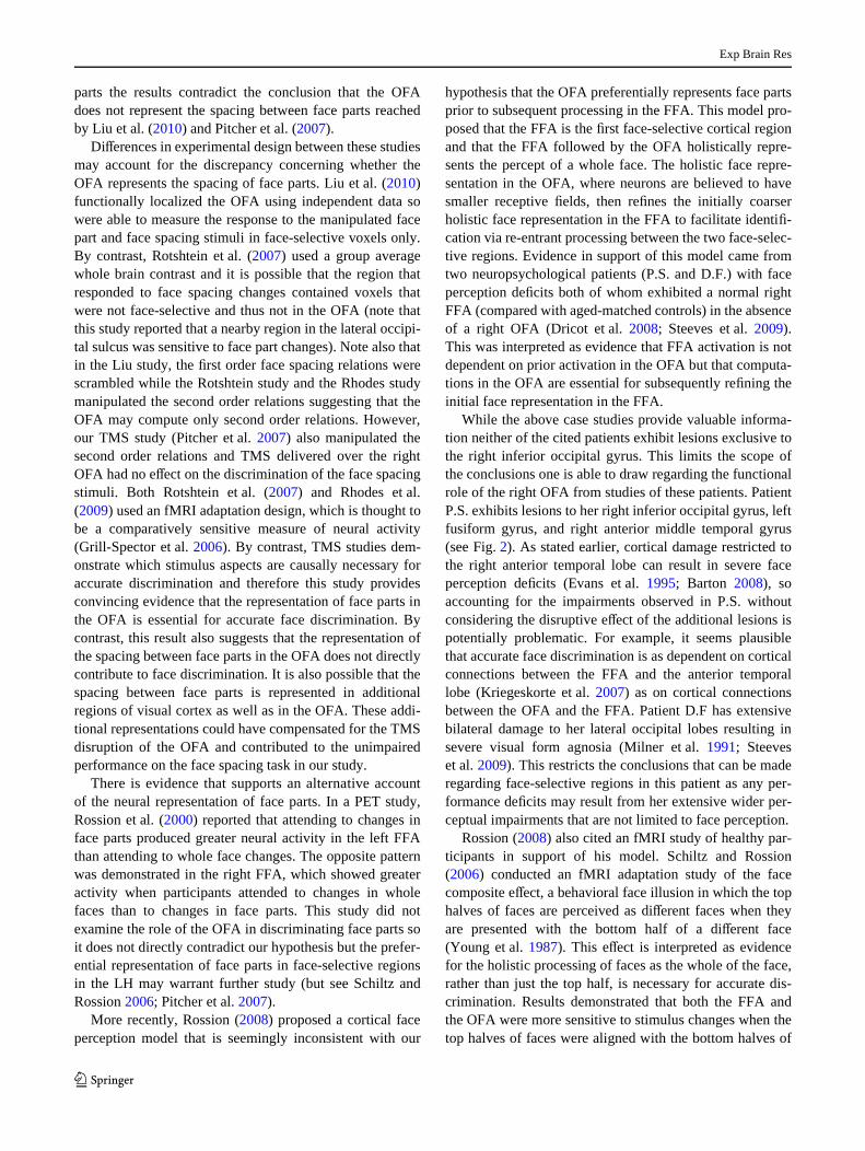
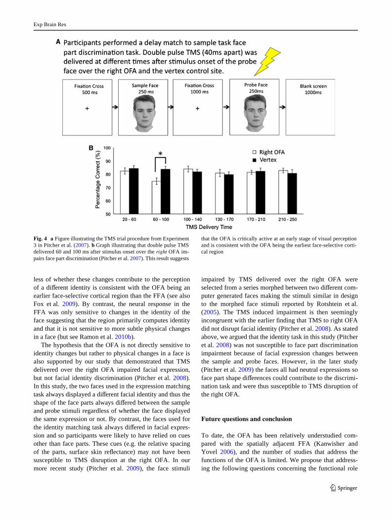
TMS studies can also address when a particular corticalregion is engaged in a speciWc cognitive task with a highdegree of temporal resolution. This is achieved by deliver-ing TMS pulses over a targeted cortical region at diVerenttimes from stimulus onset. Plotting the disruptive eVect ofthe TMS demonstrates when the targeted region was cru-cially engaged in the task and suggests when the region iscritically active. In our Wrst TMS study, we delivered twopulses of TMS separated by 40 ms at diVerent times fromstimulus onset during diVerent time windows while partici-pants performed a face part discrimination task (Pitcheret al. 2007). TMS impaired discrimination when delivered60 and 100 ms after stimulus onset but had no eVect whendelivered during all other time windows up to 250 ms (seeFig. 4). This 60–100 ms TMS induced impairment at therOFA was replicated in a second study in which partici-pants had to discriminate facial expressions (Pitcher et al.2008). The temporal proximity of these TMS induced
impairments to the P1/M100 components reported in elec-trophysiological studies (Eimer 1998; Liu et al. 2002; Itierand Taylor 2004; Thierry et al. 2007) further suggests thatthe OFA and the P1/M100 components may reXect thesame underlying neural activity. If so, then the Wrst wave offace-speciWc activity in the OFA peaks approximately100 ms after stimulus onset. OFA neural activity at 100 mswould then precede the timing of the intracranial ERPresponses to faces recorded in the right fusiform gyrus ofneuropsychological patients that has been shown peak attimes varying from 110 to 700 ms (Allison et al. 1999;McCarthy et al. 1999; Puce et al. 1999; Barbeau et al.2008). Face-speciWc neural activity in the OFA precedingface-speciWc neural activity in the fusiform gyrus (the corti-cal locus of the FFA) is consistent with the OFA being theearliest face-selective cortical region as originally proposedby Haxby et al. (2000) (but see Rossion 2008).
Does the OFA contribute to facial identity discrimination?
Evidence that the OFA is involved in facial identity compu-tations comes from fMRI studies (HoVman and Haxby2000; Yovel and Kanwisher 2004) but the exact nature ofhow the OFA contributes to identiWcation is not fullyunderstood. While FFA activation has been shown to corre-late with facial identity discrimination (Grill-Spector et al.2004), the role of the OFA for facial identiWcation isbelieved to involve the structural description of a face priorto further analysis in the FFA (Haxby et al. 1999; 2000;HoVman and Haxby 2000). Rotshtein et al. (2005) reportedan fMRI adaptation study that elegantly demonstrates howthis process may function. Face stimuli were drawn from aseries morphed at diVerent gradations between images oftwo famous people (for example, Margaret Thatcher andMarilyn Monroe). In the scanner, participants were pre-sented with two successive faces that were either same ordiVerent, faces in the diVerent trials varied by 30% alongthe physical morphing dimension. In half of the diVerenttrials, the two faces were both perceived as the same iden-tity (for example, both Marilyn or both Margaret), while inthe other half of the diVerent trials the two faces were per-ceived to be diVerent identities (e.g., Wrst Marilyn thenMargaret or vice versa). Results showed that the OFAexhibited increased neural sensitivity (it was released fromadaptation) during the diVerent trials regardless of whetherthe faces were perceived as a diVerent identity. By contrast,the FFA showed increased neural sensitivity (it wasreleased from adaptation) only during the diVerent trialsthat presented a diVerent identity and not during diVerenttrials that presented the same identity. This evidence thatthe OFA is sensitive to physical changes in a face regard-
Exp Brain Res
123
less of whether these changes contribute to the perceptionof a diVerent identity is consistent with the OFA being anearlier face-selective cortical region than the FFA (see alsoFox et al. 2009). By contrast, the neural response in theFFA was only sensitive to changes in the identity of theface suggesting that the region primarily computes identityand that it is not sensitive to more subtle physical changesin a face (but see Ramon et al. 2010b).
The hypothesis that the OFA is not directly sensitive toidentity changes but rather to physical changes in a face isalso supported by our study that demonstrated that TMSdelivered over the right OFA impaired facial expression,but not facial identity discrimination (Pitcher et al. 2008).In this study, the two faces used in the expression matchingtask always displayed a diVerent facial identity and thus theshape of the face parts always diVered between the sampleand probe stimuli regardless of whether the face displayedthe same expression or not. By contrast, the faces used forthe identity matching task always diVered in facial expres-sion and so participants were likely to have relied on cuesother than face parts. These cues (e.g. the relative spacingof the parts, surface skin reXectance) may not have beensusceptible to TMS disruption at the right OFA. In ourmore recent study (Pitcher et al. 2009), the face stimuli
impaired by TMS delivered over the right OFA wereselected from a series morphed between two diVerent com-puter generated faces making the stimuli similar in designto the morphed face stimuli reported by Rotshtein et al.(2005). The TMS induced impairment is then seeminglyincongruent with the earlier Wnding that TMS to right OFAdid not disrupt facial identity (Pitcher et al. 2008). As statedabove, we argued that the identity task in this study (Pitcheret al. 2008) was not susceptible to face part discriminationimpairment because of facial expression changes betweenthe sample and probe faces. However, in the later study(Pitcher et al. 2009) the faces all had neutral expressions soface part shape diVerences could contribute to the discrimi-nation task and were thus susceptible to TMS disruption ofthe right OFA.
Future questions and conclusion
To date, the OFA has been relatively understudied com-pared with the spatially adjacent FFA (Kanwisher andYovel 2006), and the number of studies that address thefunctions of the OFA is limited. We propose that address-ing the following questions concerning the functional role
Fig. 4 a Figure illustrating the TMS trial procedure from Experiment3 in Pitcher et al. (2007). b Graph illustrating that double pulse TMSdelivered 60 and 100 ms after stimulus onset over the right OFA im-pairs face part discrimination (Pitcher et al. 2007). This result suggests
that the OFA is critically active at an early stage of visual perceptionand is consistent with the OFA being the earliest face-selective corti-cal region

Exp Brain Res
123
of the OFA will inform our understanding of the humanface perception cortical network.
1. How face-selective is the OFA?—Face-selective corti-cal regions show a greater neural response to faces thanto any other category of visual stimuli, but whetherthese regions only represent faces is disputed (Haxbyet al. 2001; Spiridon and Kanwisher 2002). The face-selectivity of the right OFA was recently demonstratedby our study that reported that TMS delivered over theright OFA impaired face discrimination but not objectand body discrimination (Pitcher et al. 2009). How-ever, more recent evidence has suggested that the OFAmay also represent the orientation of two-dimensionalshapes (Silvanto et al. 2010) and that the IOG may con-tain overlapping neuronal populations that respond toboth faces and limbs (Weiner and Grill-Spector 2010).
2. Are there feedback connections to the OFA?—Themammalian visual system contains an extensive net-work of feedback connections from higher corticalareas to lower cortical areas and feedback connectionsare predicted in cortical face perception networks(Haxby et al. 2000; Fairhall and Ishai 2007). The OFAshould be the subject of feedback signals from higherface-selective cortical regions but such connectionshave not yet been demonstrated.
3. Is there a non-human primate homolog of theOFA?—The most compelling evidence that face per-ception is performed in a network of face-selectivecortical regions comes from recent studies that havecombined fMRI and microstimulation in macaques(Tsao et al. 2006; Moeller et al. 2008). Evidence froma recent paper that aimed to establish homologiesbetween face-selective cortical regions across speciessuggests that the posterior lateral face patch (PL), themost posterior of six patches reported in the macaque,is the most likely candidate for the primate homologof the OFA (Tsao et al. 2008). However, the functionsof these regions in humans and macaques will need tobe better understood before exact homologies can bemade.
4. Is the OFA a face detector?—Gauthier et al. (2000)hypothesized that a possible functional role for theOFA might involve face detection. Evidence thatthe OFA represents faces and the location of faces inthe visual Weld is consistent with this hypothesis(Kovács et al. 2008; Nestor et al. 2008; Schwarzloseet al. 2008). However, more recent evidence that theOFA is sensitive to changes in face stimuli evenwhen the subject is behaviorally unaware of thechange runs contrary to the hypothesis that the OFAoperates as face detector (Large et al. 2008; Foxet al. 2009).
If the OFA is the Wrst stage of a distributed cortical networkspecialized for face perception (Haxby et al. 2000; Calderand Young 2005; Ishai 2008), then a better understandingof the functional role the OFA performs will be essential inestablishing how this network operates. In this paper, wehave presented evidence consistent with the hypothesis thatthe OFA represents the parts of a face and that it does soprior to more detailed analyses performed at higher face-selective cortical regions. Our conclusions are, in part,based on adding temporal information to the existing corti-cal model of face perception proposed by Haxby et al.(2000), and we contend that experimental techniques withhigh temporal resolution add essential information in thestudy of cortical networks. To further illustrate this point,we have included a model adapted from Haxby et al. (2000)that includes information concerning when some of theface-selective cortical regions are functionally active (seeFig. 5).
Acknowledgments We thank Galit Yovel and Danny Dilks for theirtypically perceptive comments and Marius Peelen and Boaz Sadeh forsupplying Wgures. This work was supported by BBSRC grant BB/F022875/1 to BD and VW.
Fig. 5 A cortical model of the face-processing network (adapted fromHaxby et al. 2000) with temporal information added from intracranialERP and TMS studies (1 Pitcher et al. 2007; 2 Pitcher et al. 2008; 3Allison et al. 1999; 4 McCarthy et al. 1999; 5 Puce et al. 1999; 6 Bar-beau et al. 2008). The model and the connections between the func-tional areas are hypothetical

Exp Brain Res
123
References
Allison T, Puce A, Spencer DD, McCarthy G (1999) Electrophysiolog-ical studies of human face perception. I: potentials generated inoccipito-temporal cortex by face and non-face stimuli. Cereb Cor-tex 9:415–430
Allison T, Puce A, McCarthy G (2000) Social perception from visualcues: role of the STS region. Trends Cogn Sci 4:267–278
Barbeau EJ, Taylor MJ, Regis J, Marquis P, Chauvel P, Liegeois-Chauvel C (2008) Spatio temporal dynamics of face recognition.Cereb Cortex 18:997–1009
Barton JJS (2008) Structure and function in acquired prosospagnosia:lessons from a series of ten patients with brain damage.J Neuropsychol 2:197–225
Barton JJS, Press DZ, Keenan JP, O’Connor M (2002) Lesions of thefusiform face area impair perception of facial conWguration inprosopagnosia. Neurology 58:71–78
Bentin S, Allison T, Puce A, Perez E, McCarthy G (1996) Electrophys-iological studies of face perception in humans. J Cogn Neurosci8:551–565
Bouvier SE, Engel SA (2006) Behavioral deWcits and cortical damageloci in cerebral achromatopsia. Cereb Cortex 16:183–191
Bruce V, Young A (1986) Understanding face recognition. Br JPsychol 77:305–327
Busigny T, Graf M, Mayer E, Rossion B (2010) Acquired prosopagno-sia as a face-speciWc disorder: ruling out the general visualsimilarity account. Neuropsychologia 48:2051–2067
Calder AJ, Young AW (2005) Understanding the recognition of facialidentity and facial expression. Nat Rev Neurosci 6:641–651
Clark VP, Keil K, Maisog JM, Courtney S, Ungerleider LG, Haxby JV(1996) Functional magnetic resonance imaging of human visualcortex during face matching: a comparison with positron emissiontomography. Neuroimage 4:1–15
Downing P, Jiang Y, Shuman M, Kanwisher N (2001) A cortical areaselective for visual processing of the human body. Science293:2470–2473
Dricot L, Sorger B, Schiltz C, Goebel R, Rossion B (2008) The rolesof “face” and “non-face” areas during individual face perception:evidence by fMRI adaptation in a brain-damaged prosopagnosicpatient. NeuroImage 40:318–332
Eimer M (1998) Does the face-speciWc N170 component reXect theactivity of a specialized eye processor? Neuroreport 9:2945–2948
Evans J, Heggs A, Antoun N, Hodges J (1995) Progressive prosopag-nosia associated with selective right temporal lobe atrophy: a newsyndrome? Brain 118:1–13
Fairhall SL, Ishai A (2007) EVective connectivity within the distrib-uted cortical network for face perception. Cereb Cortex 17:2400–2406
Farah MJ (2004) Visual agnosia: disorders of object recognition andwhat they tell us about normal vision. MIT Press, Cambridge
Fox CJ, Moon S-Y, Iaria G, Barton JJS (2009) The correlates of sub-jective perception of identity and expression in the face network:an fMRI adaptation study. Neuroimage 44:569–580
Gauthier I, Tarr MJ, Moylan J, Skudlarski P, Gore JC, Anderson AW(2000) The fusiform “face area” is part of a network that pro-cesses faces at the individual level. J Cogn Neurosci 12:495–504
Grill-Spector K, Malach R (2004) The human visual cortex. Annu RevNeurosci 27:649–677
Grill-Spector K, Kushnir T, Hendler T, Edelman S, Itzchak Y, MalachR (1998) A sequence of object-processing stages revealed byfMRI in the human occipital lobe. Hum Brain Mapp 6:316–328
Grill-Spector K, Kushnir T, Edelman S, Avidan G, Itzchak Y, MalachR (1999) DiVerential processing of objects under various viewingconditions in the human lateral occipital complex. Neuron24:187–203
Grill-Spector K, Knouf N, Kanwisher N (2004) The fusiform face areasubserves face perception, not generic within-category identiWca-tion. Nat Neurosci 7:555–562
Grill-Spector K, Henson R, Martin A (2006) Repetition and the brain:neural models of stimulus speciWc eVects. Trends Cogn Sci10:14–23
Harris A, Aguirre GK (2008) The representation of parts and wholes inface-selective cortex. J Cogn Neurosci 20:863–878
Haxby JV, Horwitz B, Ungerleider LG, Maisog JM, Pietrini P, GradyCL (1994) The functional organization of human extrastriate cor-tex: a pet-rcbf study of selective attention to faces and locations.J Neurosci 14:6336–6353
Haxby JV, Ungerleider LG, Clark VP, Schouten JL, HoVman EA,Martin A (1999) The eVect of face inversion on activity in humanneural systems for face and object perception. Neuron 22:189–199
Haxby JV, HoVman EA, Gobbini MI (2000) The distributed humanneural system for face perception. Trends Cogn Sci 4:223–233
Haxby JV, Gobbini MI, Furey ML, Ishai A, Schouten JL, Pietrini P(2001) Distributed and overlapping representations of faces andobjects in ventral temporal cortex. Science 293:2425–2430
Hemond CC, Kanwisher N, Op de Beeck HP (2007) A preference forcontralateral stimuli in human object- and face-selective cortex.PLoS ONE 2(6):e574
Henson RN, Goshen-Gottstein Y, Ganel T, Otten LJ, Quayle A, RuggMD (2003) Electrophysiological and haemodynamic correlates offace perception, recognition and priming. Cereb Cortex 13:793–805
Herrmann MJ, Ehlis A-C, Ellgring H, Fallgatter AJ (2005) Early stages(P100) of face perception in humans as measured with event-related potentials (ERPs). J Neural Transm 112:1073–1081
HoVman EA, Haxby JV (2000) Distinct representations of eye gazeand identity in the distributed human neural system for face per-ception. Nat Neurosci 3:80–84
Horovitz SG, Rossion B, Skudlarski P, Gore JC (2004) Parametric de-sign and correlational analyses help integrating fMRI and electro-physiological data during face processing. Neuroimage 22:1587–1595
Ishai A (2008) Let’s face it: it’s a cortical network. Neuroimage40(2):415–419
Ishai A, Haxby JV, Ungerleider LG (2002) Visual imagery of famousfaces: eVects of memory and attention revealed by fMRI. Neuro-Image 17:1729–1741
Itier RJ, Taylor MJ (2004) N170 or N1? Spatiotemporal diVerences be-tween object and face processing using ERPs. Cereb Cortex14:132–142
Itier RJ, Herdman AT, George N, Cheyne D, Taylor MJ (2006) Inver-sion and contrast-reversal eVects on face processing assessed byMEG. Brain Res 1115:108–120
Kanwisher N, Yovel G (2006) The fusiform face area: a cortical regionspecialized for the perception of faces. Philos Trans R Soc LondB Biol Sci 361:2109–2128
Kanwisher N, McDermott J, Chun MM (1997) The fusiform face area:a module in human extrastriate cortex specialized for face percep-tion. J Neurosci 17:4302–4311
Kourtzi Z, Tolias AS, Altmann CF, Augath M, Logothetis NK (2003)Integration of local features into global shapes: monkey andhuman fMRI studies. Neuron 37:333–346
Kovács G, Cziraki C, Vidnyánszky Z, Schweinberger SR, GreenleeMW (2008) Position-speciWc and position-invariant face after-eVects reXect the adaptation of diVerent cortical areas. Neuroimage43:156–164
Kriegeskorte N, Formisano E, Sorger B, Goebel R (2007) Individualfaces elicit distinct response patterns in human anterior temporalcortex. PNAS 104:20600–20605

Exp Brain Res
123
Large ME, Cavina-Pratesi C, Vilis T, Culham JC (2008) The neuralcorrelates of change detection in the face perception network.Neuropsychologia 46(8):2169–2176
Lerner Y, Hendler T, Ben-Nashat D, Harel M, Malach R (2001) A hier-archical axis of object processing stages in the human visual cor-tex. Cereb Cortex 11:287–297
Liu J, Harris A, Kanwisher N (2002) Stages of processing in face per-ception: an MEG study. Nat Neurosci 5:910–916
Liu J, Harris A, Kanwisher N (2010) Perception of face parts and faceconWgurations: an fMRI study. J Cogn Neurosci 22:203–211
Malach R, Reppas J, Benson R, Kwong K, Jiang H, Kennedy W, Led-den P, Brady T, Rosen B, Tootell R (1995) Object-related activityrevealed by functional magnetic resonance imaging in humanoccipital cortex. PNAS 92:8135–8139
Martinez A, Di Russo F, Anllo-Vento L, Sereno MI, Buxton RB, Hill-yard SA (2001) Putting spatial attention on the map: timing andlocalization of stimulus selection processes in striate and extras-triate visual areas. Vis Res 41:1437–1457
McCarthy G, Puce A, Gore J, Allison T (1997) Face-speciWc process-ing in the fusiform gyrus. J Cogn Neurosci 9:605–610
McCarthy G, Puce A, Belger A, Allison T (1999) Electrophysiologicalstudies of human face perception. II: response properties of face-speciWc potentials generated in occipitotemporal cortex. CerebCortex 9:431–444
Milner AD, Perrett DI, Johnston RS, Benson PJ, Jordan TR, HeeleyDW, Bettucci D, Mortara F, Mutani R, Terassi E, Davidson DL(1991) Perception and action in “visual form agnosia”. Brain114:405–428
Moeller S, Freiwald WA, Tsao DY (2008) Patches with links: a uniWedsystem for processing faces in the macaque temporal lobe.Science 320:1355–1359
Nestor A, Vettel JM, Tarr MJ (2008) Task-speciWc codes for face rec-ognition: how they shape the neural representation of features fordetection and individuation. Plos ONE 3:e3978
Nichols DF, Betts LR, Wilson HR (2010) Decoding of faces and facecomponents in face-sensitive human visual cortex. Front Psychol1:28. doi:10.3389/fpsyg.2010.00028
Peelen MV, Lucas N, Mayer E, Vuilleumier P (2009) Emotional atten-tion in acquired prosopagnosia. Soc Cogn AVect Neurosci 4:268–277
Pitcher D, Walsh V, Yovel G, Duchaine B (2007) TMS evidence forthe involvement of the right occipital face area in early face pro-cessing. Curr Biol 17(18):1568–1573
Pitcher D, Garrido L, Walsh V, Duchaine B (2008) TMS disrupts theperception and embodiment of facial expressions. J Neurosci28(36):8929–8933
Pitcher D, Charles L, Devlin JT, Walsh V, Duchaine B (2009) Tripledissociation of faces, bodies, and objects in extrastriate cortex.Curr Biol 19(4):319–324
Puce A, Allison T, Asgari M, Gore JC, McCarthy G (1996) DiVerentialsensitivity of human visual cortex to faces, letterstrings, and tex-tures: a functional magnetic resonance imaging study. J Neurosci16:5205–5215
Puce A, Allison T, McCarthy G (1999) Electrophysiological studies ofhuman face perception. III: eVects of top-down processing onface-speciWc potentials. Cereb Cortex 9:445–458
Ramon M, Rossion B (2010) Impaired processing of relative distancesbetween features and of the eye region in acquired prosopagno-sia—two sides of the same holistic coin? Cortex 46:374–389
Ramon M, Busigny T, Rossion B (2010a) Impaired holistic processingof unfamiliar individual faces in acquired prosopagnosia. Neuro-psychologia 48:933–944
Ramon M, Dricot L, Rossion B (2010b) Personally familiar faces areperceived categorically in face-selective regions other than theFFA. Eur J Neurosci 32:1587–1598
Rhodes G, Michie PT, Hughes ME, Byatt G (2009) FFA and OFAshow sensitivity to spatial relations in faces. Eur J Neurosci30:721–733
Robertson I, Murre JMJ (1999) Rehabilitation of brain damage: brainplasticity and principles of guided recovery. Psychol Bull125:544–575
Rosazza C, Cai Q, Minati l, Paulignan Y, Nazir T (2009) Earlyinvolvement of dorsal and ventral pathways in visual word recog-nition: an ERP study. Brain Res 1272:32–44
Rossion B (2008) Constraining the cortical face network by neuroim-aging studies of acquired prosopagnosia. Neuroimage 40:423–426
Rossion B, Jacques C (2008) Does physical interstimulus varianceaccount for early electrophysiological face sensitive responses inthe human brain? Ten lessons on the N170. Neuroimage 39:1959–1979
Rossion B, de Gelder B, Dricot L, Zoontjes R, De Volder A, BodartJ-M, Crommelinck M (2000) Hemispheric asymmetries forwhole-based and part-based face processing in the human fusi-form gyrus. J Cogn Neurosci 12:793–802
Rossion B, Caldara R, Seghier M, Schuller AM, Lazeyras F, Mayer E(2003) A network of occipito-temporal face-sensitive areasbesides the right middle fusiform gyrus is necessary for normalface processing. Brain 126:2381–2395
Rotshtein P, Henson RN, Treves A, Driver J, Dolan RJ (2005) Morp-hing Marilyn into Maggie dissociates physical and identity facerepresentations in the brain. Nat Neurosci 8:107–113
Rotshtein P, Geng JJ, Driver J, Dolan RJ (2007) Role of features andsecond-order spatial relations in face discrimination, face recog-nition, and individual face skills: behavioral and functional mag-netic resonance imaging data. J Cogn Neurosci 19:1435–1452
Sadeh B, Podlipsky I, Zadanov A, Yovel G (2010) Face-selectivefMRI and event-related potential responses are highly correlated:evidence from simultaneous ERP-fMRI investigation. Hum BrainMapp 31:1490–1501
Schiltz C, Rossion B (2006) Faces are represented holistically in thehuman occipito-temporal cortex. Neuroimage 32:1385–1394
Schiltz C, Dricot L, Goebel R, Rossion B (2010) Holistic perception ofindividual faces in the right middle fusiform gyrus as evidencedby the composite face illusion. J Vis 10:1–16
Schwarzlose RF, Swisher JD, Dang S, Kanwisher N (2008) The distri-bution of category and location information across object-selec-tive regions in human visual cortex. PNAS 105:4447–4452
Sergent J, Ohta S, MacDonald B (1992) Functional neuroanatomy offace and object processing. Brain 115:15–36
Silvanto J, Schwarzkopf DS, Gilaie-Dotan S, Rees G (2010) DiVeringcausal roles for lateral occipital cortex and occipital face area ininvariant shape recognition. Eur J Neurosci 32:165–171
Slotnick SD (2004) Source localization of ERP generators. In: HandyTC (ed) Event-related potentials: a methods handbook. MITPress, Cambridge, pp 149–166
Spiridon M, Kanwisher N (2002) How distributed is visual categoryinformation in human occipital-temporal cortex? An fMRI study.Neuron 35:1157–1165
Steeves JKE, Dricot L, Goltz HC, Sorger B, Peters J, Milner AD, Goo-dale MA, Goebel R, Rossion B (2009) Abnormal face identitycoding in the middle fusiform gyrus of two brain-damaged pros-opagnosic patients. Neuropsychologia 47:2584–2592
Thierry G, Martin CD, Downing PE, Pegna AJ (2007) Controlling forinterstimulus perceptual variance abolishes N170 face selectivity.Nat Neurosci 10:505–511
Tsao DY, Freiwald WA, Tootell RBH, Livingstone M (2006) A corti-cal region consisting entirely of face cells. Science 311:670–674
Tsao DY, Moeller S, Freiwald WA (2008) Comparing face patch sys-tems in macaques and humans. PNAS 105:19514–19519

Exp Brain Res
123
Ullman S, Vidal-Naquet M, Sali E (2002) Visual features of interme-diate complexity and their use in classiWcation. Nat Neurosci5:682–687
Watson JD, Myers R, Frackowiak RS, Hajnal JV, Woods RP, Mazzi-otta J, Zeki S (1993) Area V5 of the human brain: evidence froma combined study using positron emission tomography and mag-netic resonance imaging. Cereb Cortex 3:79–94
Weiner KS, Grill-Spector K (2010) Sparsely-distributed organizationof face and limb activations in human ventral temporal cortex.NeuroImage 52:1559–1573
Young AW, Hay DC, McWeeny KH, Ellis AW, Barry C (1985) Famil-iarity decisions for faces presented to the left and right cerebralhemispheres. Brain Cogn 4:439–450
Young AW, Hellawell D, Hay DC (1987) ConWgurational informationin face perception. Perception 16:747–759
Yovel G, Kanwisher N (2004) Face perception: domain speciWc, notprocess speciWc. Neuron 44:889–898
Yovel G, Kanwisher N (2005) The neural basis of the behavioral face-inversion eVect. Curr Biol 15:2256–2262