Embed Size (px)
Citation preview
THE ROLE OF THE HIPPOCAMPUS AND MATRIX METALOPROTEINASES ON
HABITUATION OF THE HEAD-SHAKE RESPONSE TASK/CLASSICAL
CONDITIONING PARADIGM
by
ROBERTA V WIEDIGER
A dissertation submitted in partial fulfillment of the requirements for the degree of
DOCTOR OF PHILOSOPHY
WASHINGTON STATE UNIVERSITY
Department of Psychology
December 2008
To the Faculty of Washington State University: The members of the Committee appointed to examine the dissertation of
ROBERTA V. WIEDIGER find if satisfactory and recommend that it be accepted.
____________________________________ Chair
____________________________________
____________________________________
____________________________________
ii
ACKNOWLEDGMENTS
First and foremost, I would like to thank the incredible mentoring of Dr. Jay Wright. He not only inspired me to search for answers by doing research, but his outstanding guidance and patience encouraged me to look for answers in places that I would otherwise not thought possible. Thank you for being understanding of my needs and not giving up on me, especially during my post-partum depression. A very special thanks to Dr. Fran McSweeney, who believed in me by opening the doors to her lab, which made all of this possible. Thanks to Ruth Day and AnnMarie Gooch, whose love and support made WSU feel like home. Thanks to my parents, who taught me that anything is possible with hard work, patient, and perseverance. Their love and support kept me going during many hard times. Thanks to my dad for teaching me the priorities in life, and thanks to my mom for supporting me during my post-partum depression. And last but not least, to my wonderful, caring, loving, amazing, very patient husband, whose support and dedication made life in graduate school a dream come true. I not only fell in love, but I was also given the most beautiful and wonderful gift in the world, Melanie, my amazing daughter. You will always be in my heart. I am where I am in my life because I was fortunate to have crossed paths with all of you. Thank you for believing in me even when I had doubts.
iii
THE ROLE OF THE HIPPOCAMPUS AND MATRIX METALOPROTEINASES
ON HABITUATION OF THE HEAD-SHAKE RESPONSE TASK/CLASSICAL CONDITIONING PARADIGM
Abstract
By Roberta V. Wiediger, Ph.D. Washington State University
December 2008 Chair: John W. Wright These experiments were designed to find evidence regarding the relationship
between the hippocampus and matrix metalloproteinases (MMP) during the head-shake
response (HSR) task/classical conditioning paradigm. Habituation is the simplest form of
learning, where an organism’s response is decreased due to a repeated presentation of a
harmless stimulus. The HSR task has been very predictable in showing habituation to
repeated air stimulation to a rat’s ear and the spontaneous recovery of the habituated
response twenty-four hours later. Since the hippocampus and MMPs are implicated in
learning and memory, these experiments investigated their role during a HSR task when a
classical conditioning paradigm was added. Therefore these studies were designed to
investigate the following: 1) Can a tone serve as the conditioned stimulus (CS) during
the HSR task? 2) Is the dorsal hippocampus important during the HSR/Classical
conditioning paradigm? 3) Are hippocampal MMPs important during the CS-US
iv
(unconditioned stimulus) association? 4) Is hippocampal MMP-3 important during the
CS-US association? Findings revealed: 1) the tone presented 1-s prior to the US became
a CS. 2) Dorsal hippocampectomized animals were not able to make the CS-US
association. 3) Injections of FN-439, a general MMP inhibitor, into the dorsal
hippocampus interfered with the CS-US association. 4) Dorsal hippocampus injections
of MMP-3 inhibitor also interfered with the CS-US association. Therefore, animals
which were not able to form the CS-US association showed similar rates of responding
twenty-four hours later. Based on the collective findings of these experiments it is
evident that during a HSR/classical conditioning paradigm, the hippocampus plays an
important role in consolidating and storing the CS-US association. More specifically
dorsal hippocampus MMP-3 was found to be particularly important in the formation of
the CS-US association.
v
TABLE OF CONTENTS
ACKNOWLEDGEMENTS................................................................................... iii ABSTRACT........................................................................................................... iv LIST OF FIGURES ............................................................................................. viii DEDICATION....................................................................................................... ix CHAPTER ONE ......................................................................................................1
A. General introduction ....................................................................................2
1. Definition of learning...............................................................................2
B. Types of learning .........................................................................................2 1. Associative learning.................................................................................2 2. Non-associative learning..........................................................................3 C. Physiological basis of learning ....................................................................5
1. Extracellular matrix .................................................................................6 2. Long-term potentiation ............................................................................7 3. Importance of calcium ions during learning ............................................8
D. Physiological model of learning ..................................................................9 1. Molecular view ........................................................................................9 E. Proposed experiments ................................................................................14 F. References..................................................................................................16
CHAPTER TWO ...................................................................................................21 Abstract ......................................................................................................23
1. Introduction..........................................................................................24
vi
2. Materials and methods .........................................................................25
3. Results..................................................................................................29
4. Discussion............................................................................................32
References..................................................................................................37 Figure captions...........................................................................................43
CHAPTER THREE ...............................................................................................46 Abstract ......................................................................................................48
1. Introduction..........................................................................................49
2. Results..................................................................................................50
3. Discussion............................................................................................53
4. Material and methods...........................................................................55
References..................................................................................................60 Figure captions...........................................................................................63
CHAPTER FOUR..................................................................................................65
General discussion .....................................................................................66 Summary of findings..................................................................................67
vii
LIST OF FIGURES
1. Learning model ..........................................................................................10
2. No tone, tone and random tone comparisons between session I
and session II..............................................................................................44
3. Reconstruction of aspiration lesion............................................................44
4. No tone hippocampectomy, tone hippocampectomy, no tone neocortex control,
tone neocortex control comparisons between session I and session II ......45
5. High FN439, low FN439 and aCSF comparisons between session I
and session II..............................................................................................64
6. MMP-3i and aCSF comparisons between session I and session II............64
viii

Dedication
To my husband and my father, who inspired me to reach high and never give up.
Thank you!
ix

CHAPTER ONE
GENERAL INTRODUCTION
1
A. General Introduction 1. Definition of Learning
Learning has been defined as “a relatively long-term change in behavior that results from
experience” (Kosslyn & Rosenberg, 2004). “A process by which experience produces a
relatively enduring change in the organism’s behavior or capabilities” (Passer & Smith, 2004).
“A relatively permanent change in an organism’s behavior due to experience” (Myers, 2004). It
is known that the hippocampus plays a role in learning and memory and therefore dorsal
hippocampectomy was presently employed in an effort to determine its importance in
spontaneous recovery and habituation of the head-shake response (HSR). The protocols used
included the use of the HSR habituation task, the introduction of a tone (conditioned stimulus –
CS) prior to each introduction of the air stimulus (unconditioned stimulus – US),
hippocampectomized rats and the infusion of matrix-metalloproteinase inhibitors (MMPIs)
injected in the dorsal hippocampus. These paradigms permitted the examination of the role of
the dorsal hippocampus during a HSR task combined with a CS-US association, and the role of
MMPs during this task.
B. Types of learning
1. Associative Learning
Experimental studies concerning learning are usually divided into the categories of
associative and non-associative learning (Eisenstein, Eisenstein, & Bonheim, 1991). However,
the majority of introductory psychology textbooks only describe associative learning (Myers,
2004; Weiten, 2004). Associative learning relates to the occurrence of certain events together in
time and space (Wieten, 2004). It includes classical and operant conditioning. The former is a
subtype of associative learning characterized by the association of an originally neutral stimulus
2
with an unconditioned stimulus causing a conditioned response to be emitted that was previously
unconditioned. An example of classical conditioning is the use of an aversive stimulus to
encourage people to stop smoking. Specifically, giving a smoker a drug (UCS) that causes
nausea (UCR), and later introducing cigarette smoking behavior (CS) right before the UCS will
decrease cigarette smoking behavior by its association with (CR). Classical conditioning, also
referred to as Pavlovian conditioning, alters the way in which animals process events and may
produce a variety of consequences (Holland, 1997). Classical conditioning changes the way the
animal attends to CSs including the production of various types of CRs, the acquisition of
reinforcing power, and the direction of attention toward or away from those stimuli, in addition it
also may involve distinct, relatively independent neural circuitry (Holland, 1997).
On the other hand, operant conditioning is a subtype of learning that associates the
behavior with the consequence of that behavior making it more or less likely to reoccur in the
future. An example of operant conditioning is receiving a ticket for speeding and also having
your automobile insurance rate increase. Presumably this will cause the driver to avoid speeding
in the future.
2. Non-associative learning
Non-associative learning includes habituation and sensitization (Eisenstein et al., 1991).
A definition of non-associative learning could not be found in the literature. Most behavioral
scientists use the term non-associative learning as an antonym to associative learning. This
nomenclature has been adopted without providing operational definitions. Habituation is defined
as “a decrease in responsiveness to repeated presentations of a stimulus” (Groves & Thompson,
1970), and is considered the simplest form of learning (Harris, 1943; Thorpe, 1966). According
to Staddon (2001), habituation is the waning of a reflexive response to repeated stimulation.
3
Consisted with this definition Zaccardi, and colleagues (2001) have proposed that habituation is
the simplest form of learning seen in all animals consisting of a decrease in responsiveness due
to a repeatedly applied innocuous stimulus.
Thompson and Spencer (1966) listed nine parametric characteristics proper to
habituation. Recently, McSweeney and Murphy (2000) added five more characteristics to this
list ranging from, spontaneous recovery, “the recovery of a habituated response to a stimulus
when that stimulus is not present for a period of time,” to stimulus intensity, “habituation
sometimes is more pronounced and faster for less intense than for more intense stimuli.” Two
out of these fourteen characteristics are concerned with the phenomenon called sensitization.
According to Eisenstein et al. (1991), sensitization has been employed with several different
procedural variations and meanings. Groves et al. (1970) have described sensitization as an
increase in responsiveness due to early stimulus presentation. Sensitization has also been
defined as “an increase in responsiveness due to the presentation of a stimulus from different
modality” (Swithers & Hall, 1994). Some researchers have named this phenomenon
dishabituation (Marcus, Nolen, Rankin, & Carew, 1998). As a result, sensitization has been
employed with several different procedural variations and meanings (Eisenstein et al., 1991), but
the most commonly used is that of an initial increase in responsiveness due to the repeated
presentation of a stimulus (Groves et al., 1970).
McSweeney, Hinson & Cannon (1996) proposed a sensitization-habituation hypothesis.
According to this hypothesis early-session increases in responding are primarily due to
sensitization, while late-session decreases are primarily produced by habituation to the
repeatedly presented reinforcer. This approach is important because it compares the phenomena
of sensitization and habituation with empirical characteristics of within-session changes in
4
operant responding, thus suggesting that the two types are produced by similar variables.
Therefore, both phenomena can occur within a wide variety of species, performing a wide
variety of responses (McSweeney et al., 1996).
According to Harris (1943) the most ubiquitous phenomenon in animal behavior is that of
response decrement as the result of repeated stimulation. This phenomenon has been reported in
studies ranging from ameba’s locomotion, to humans’ reflex mechanism. Marcus et al. (1998),
have reported habituation of the siphon withdrawal reflex in the marine mollusk Aplysia.
Murphy et al., (2005), have noted habituation in rats during the HSR task. Eisenstein and
colleagues, (1990), studied habituation and sensitization of the palmar galvanic skin response
(GSR) to shock in college males. Ornitz and Guthrie (1989) showed habituation and
sensitization of the acoustical startle response in adult humans. The behaviors described above
demonstrate that the animal does not necessarily need to learn to respond to certain stimuli.
Inborn reflexive responses are extremely important for survival, therefore requiring little or no
time to develop associations.
C. Physiological basis of learning
As mentioned above an organism is constantly exposed to a variety of information that
must be learned and stored in memory so that it can be retrieved at a later time. However, little
is known about the neural mechanisms underlying learning and memory. Nevertheless, many
studies have shown that the hippocampus is involved in learning and memory (Collingridge,
Isaac, & Wang, 2004; Lee, Everitt, & Thomas, 2004; Wright, et al., 2004b). One fascinating and
exceedingly important aspect of the brain is its capacity to efficiently pass information from one
neuron to another and to modify these circuits with experience. This property is called “synaptic
plasticity”, the ability of the brain to change, reconfigure and in this way consolidate vast
5
amounts of behavioral information (Collingridge et al., 2004). The neural plasticity underlying
memory appears to undergo a continuous pattern of maintenance and reconsolidation made
possible by extracellular matrix (ECM) molecules (reviewed in Wright & Harding, 2004a, and
described below).
1. Extracellular Matrix
Within the Central Nervous System is an extracellular matrix composed of a network of
proteins involved in cellular development, migration, connectivity and reorganization (Novak &
Kaye, 2000). Many ECM molecules are responsible for maintaining and changing the synaptic
configuration presumed to be indispensable for the processes of neural plasticity, memory and
learning. Recently, researches have begun to identify those molecules responsible for
reconfiguring and storing memory. Much attention has been given to the matrix
metalloproteinases (MMPs), enzymes critical to the maintenance and reconstruction of the ECM
(Wright et al., 2004b). The MMPs are responsible for ECM degradation, and the mediation of
new synaptic reconfiguration. Therefore, MMPs are extremely important in learning, as they are
markers of neural plasticity. Several studies have measured changes in MMP expression in the
hippocampus after an animal has successfully performed a task, implying that memory
consolidation is taking place (Meighan, et al., 2006; and Meighan et al., 2007).
The hippocampus is a region of the limbic cortex located in the temporal lobe, linked to
learning and memory (Collingridge et al., 2004; Kandel, Schwartz & Jessel, 2000; Kandel,
2001). The hippocampal formation is composed of the hippocampus proper, dentate gyrus and
the subiculum (Carlson, 2001). The major neocortical input of the hippocampal formations is
through the entorhinal cortex, a region of the limbic system. Neurons in the entorhinal cortex
send information to the granule cells, found in the dentate gyrus, through axons that form the
6
perforant path. The hippocampus is also composed of subfields (CA1, CA2 and CA3). The
CA1 field posses many pyramidal cells, large neurons with pyramid shaped soma, which provide
primary output for the hippocampus. They send axons to neurons in the subicular complex,
which project out of the hippocampal formation to the entorhinal cortex and to the basal
forebrain. The CA3 pyramidal cells branch in two directions. One pathway goes to the CA1
field and the other goes to the fornix and structures in the basal forebrain, including the septum
and the mammillary bodies (Carlson, 2001). The CA2 field connects CA3 and CA1 fields thus
completing the neural circuit.
There are three major pathways in the hippocampus: 1) The perforant pathway,
projecting from the entorhinal cortex to the granule cells of the dentate gyrus. 2) The mossy
fiber pathway, containing the axons of granule cells that project to the pyramidal cells in the
CA3 field of the hippocampus. 3) The Schaffer collateral pathway, which consists of the
excitatory collaterals of the pyramidal cells in the CA3 region and ends on the pyramidal cells in
the CA1 region (Carlson, 2001).
2. Long-term potentiation
According to Collingridge et al. (2004), one form of synaptic plasticity, known as long-
term potentiation (LTP), has consolidated its status as a synaptic model for investigating the
molecular basis of memory. LTP has been identified as the biological substrate for at least some
types of memories (Lynch, 2004). LTP has been most frequently studied in the hippocampus,
although it is also seen in other CNS structures, such as the amygdala and cerebellum.
Long-term potentiation has two phases, an early and a late phase. The early phase (E-
LTP) lasts for a few hours and does not require protein synthesis, while the late phase (L-LTP)
can last for at least twenty-four hours and requires protein and RNA synthesis (Kandel et al.,
7
2000; Kandel, 2001). This requirement for new mRNA and protein synthesis implies that
transcriptional regulation represents the principal control point for the consolidation of synaptic
plasticity, and translation of new synthesized mRNAs playing a more secondary role (Kelleher,
Govindarajan & Tonegawa, 2004). To inhibit L-LTP expression, treatment must occur before
protein synthesis has taken place, and before repeated tetanization has occurred. The application
of a protein synthesis inhibitor, e.g. anisomycin, after the induction of L-LTP is ineffective.
However, the ability of protein synthesis inhibitors to block the enhancement of protein
synthesis, without depleting the ones necessary for basal neuronal and synaptic function, seems
to be of brief duration (Kelleher et al., 2004). This suggests that anisomycin interferes only with
the induction of L-LTP not with its maintenance.
ECM molecules are regulated via expression of MMPs that function to degrade existing
matrix, but it is offset by tissue inhibitors of MMPs (TIMPs) that serve to preserve the matrix
(Wright et al., 2002). Therefore, the ECM is well suited to mediate the synaptic plasticity
requirements assumed to occur during LTP and underlie learning and memory. Degradation of
ECM by MMPs is controlled by three mechanisms: 1) Regulation of gene transcription, which
occurs via stimulation of growth factors, oncofene products, phorbol esters, cell-to-cell and cell-
to-ECM interactions. 2) Regulation of pro-enzyme activation and 3) through the presence of
TIMPs.
3. Importance of calcium ions during learning
Calcium plays a very important role in the learning process, as a primary component of
the signaling process, beginning with the action potential. The flow of calcium ions through the
ionotropic receptors into the neuron depolarizes the plasma membrane (Kandel et al., 2000).
When the cell reaches threshold, action potentials are generated moving down the axon and
8
reaching the terminal buttons. The terminal buttons release neurotransmitters, e.g. glutamate, to
the next neuron. Therefore, a chemical signal is given to the next neuron which in turn initiates
an action potential. Calcium ions are extremely important to the induction of action potentials
since they contribute to membrane depolarization resulting in LTP, and new spine formation.
Structural changes in the synapses can be due to cell-to-cell rearrangements and extracellular
matrix-to-cell interactions (Dityatev, Dityateva, Sytnyk, Delling, Toni, Nikonenko, Muller, &
Schachner, 2004). New neuronal spines are formed during memory consolidation. Experiments
have demonstrated the role of neurogenesis in long-term memory in rats. Snyder and colleagues
(2005), have shown that newly formed dentate granule neurons may be required in order to form
long-term spatial memories in rats. Therefore, dynamic changes in structural characteristics of
the synapses are thought to underlie synaptic plasticity, thus permitting memory consolidation.
Recently it had been reported that rats allowed to explore a compartmentalized environment for
30 min showed 30% more synaptic spines with dense phosphorylated cofilin immunoreactivity
in hippocampal field CA1 than rats pretreated with an NMDA receptor antagonist (Fedulov, Rex,
Simmons, Palmer, Gall, & Lynch, 2007). The authors interpreted these results to suggest that
cellular events associated with LTP such as synaptic spine changes, occur during learning and
form the basis of a new memory. If these results are confirmed it will be the first time LTP has
been directly linked to a change in synaptic spine induced by experience in a novel environment.
D. Physiological model of learning
1. A molecular view
The model below illustrates how learning may take place.
9
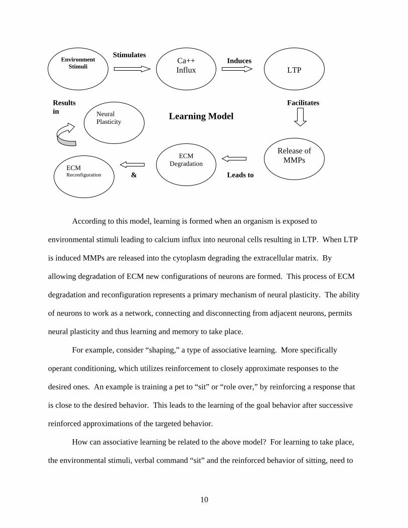
According to this model, learning is formed when an organism is exposed to
environmental stimuli leading to calcium influx into neuronal cells resulting in LTP. When LTP
is induced MMPs are released into the cytoplasm degrading the extracellular matrix. By
allowing degradation of ECM new configurations of neurons are formed. This process of ECM
degradation and reconfiguration represents a primary mechanism of neural plasticity. The ability
of neurons to work as a network, connecting and disconnecting from adjacent neurons, permits
neural plasticity and thus learning and memory to take place.
For example, consider “shaping,” a type of associative learning. More specifically
operant conditioning, which utilizes reinforcement to closely approximate responses to the
desired ones. An example is training a pet to “sit” or “role over,” by reinforcing a response that
is close to the desired behavior. This leads to the learning of the goal behavior after successive
reinforced approximations of the targeted behavior.
How can associative learning be related to the above model? For learning to take place,
the environmental stimuli, verbal command “sit” and the reinforced behavior of sitting, need to
Environment Stimuli
Ca++ Influx
LTP
ECM Reconfiguration
ECM Degradation
Stimulates Induces
Release of MMPs
& Leads to
Learning Model Results Facilitates in Neural
Plasticity
10
be presented contiguously. When the organism is presented this new information calcium influx
into the cell occurs. With a sufficient increase in action potential firing rate LTP is induced. In
turn, LTP stimulates MMPs to be released leading to ECM degradation and reconfiguration,
allowing the neuronal network to reconfigure. Therefore, when the animal learns the meaning of
the word “sit” it responds accordingly, and an increase in hippocampal MMP expression occurs
accompanied by the formation of new synaptic spines. In this way, new synapses are formed due
to the degradation of ECM.
According to Thompson (1990), memory traces are formed in the hippocampus and in the
cerebellum during classical conditioning of discrete behavioural responses. Learning and
retrieval both activate hippocampal circuits and activate molecular pathways (Power, Berlay,
McGaugh, & Steward, 2006). The hippocampus is thought to participate in the processing of
contextual and temporal information (Hoehler & Thompson, 1980; Lee & Kim, 2004). The
hippocampus also appears to be involved in certain types of conditioned fear memory, i.e. fear to
a contextual cue, but not to a tone cue (Kim & Jung, 2006). Although the hippocampus plays a
role in certain aspects of conditioning, it is not necessary for learning and memory of the basic
conditioned responses. The cerebellum and its associated brain-stem circuitry, on the other hand,
do appear to be essential for learning and memory of the conditioned response (Thompson,
1990). According to Lee and Kim (2004), the cerebellum is essential to establish the CS-US
association for conditioned eyeblink responses, and the hippocampus on the other hand, is not
critical for eyeblink responses but affects eyeblink CRs perhaps by processing contextual
information during conditioning. Lesion of the dorsal part of the hippocampus decreases
performances in associative spatial tasks like the radial maze or the Morris water maze (Stupien,
Florian, & Roullet, 2003).
11
However, differences in neural substrates have recently been noted between associative
and non-associative learning. For example, habituation of the HSR in rats occurs with repeated
puffs of air to the ear. Habituation occurs rapidly and is evidenced as a decreasing number of
head-shakes during the session. Nonetheless, twenty-four hours later the rat will exhibit
spontaneous recovery, presenting nearly the same within-session change in response to the air
puff as it did twenty-four hours earlier. If learning represents a permanent change in the
organism’s behavior, then spontaneous recovery should not occur. Looking at the model,
perhaps the ECM was not reconfigured, thus no neural plasticity took place.
Many studies on cellular mechanisms of gill-withdraw reflex in Aplysia, a large marine
snail, have shown several forms of learning including, habituation, sensitization, and classical
conditioning (Carew, Castellucci, & Kandel, 1971; Carew, Walters, & Kandel, 1981). A recent
study done by Hawkins, Clark and Kandel (2006), found that gill withdrawal in Aplysia can also
undergo operant conditioning. They showed that the mechanism of operant conditioning of gill
withdrawal might be an elaboration of mechanisms that contribute to non-associative effects of
the siphon shock. However, the associative and non-associative effects had different time
courses, suggesting that they may involve different neural mechanisms of learning (Hawkins et
al., 2006).
Wright et al., (2004b) have shown that hippocampectomized rats revealed habituation of
the HSR task, demonstrating the ability to develop non-associative learning even though no
hippocampus was present. Anisomycin, a protein inhibitor, when infused in the CA1 region of
the hippocampus have been found to have no effect on long-term memory formation of spatial
habituation (Vianna, Alonso, Viola, Quevedo, Paris, Furman, Stein, Medina, & Izquierdo, 2000).
This could indicate that the hippocampus does not play a role in reflexive, non-associative
12
learning. Perhaps this is a way organisms have to preserve a behavior that is critically important
for survival. According to Vienna et al., (2000), different neural mechanisms are involved in
memory formation of hippocampal-dependent associative and non-associative memories.
Since habituation of HSR was not affected by hippocampectomy, then where does that
non-associative learning take place? Given that the HSR is a reflex, like eye blinking and
galvanic skin response, and is important for survival but is not plastic, then mediation could
occur in subcortical areas of the brain. Brain stem structures are likely candidates to hold such
important processes as non-associative learning. Swithers-Mulvey and Hall (1993), have argued
that oral habituation, the opening or closing of jaws and/or movement of tongue, is neurally
represented in the brainstem, since habituation continues to be expressed in rat pups after
decerebration. According to Kandel et al., (2000), the midbrain controls many sensory and
motor functions, including eye movements and the coordination of visual and auditory reflexes,
and is important for this type of learning. An animal in its natural habitat habituates to a startle
response due to the harmless repeated noise produced by the wind passing through the tree
branches. However, the next day that similar noise could be caused by something else, perhaps,
a predator. Thus, the startle response is important for survival. As a result the system does not
modify this behavior via synaptic plasticity, because in a different situation that startle reflex
could indeed be useful. Perhaps that is the reason that such important non-associative learning
does not take place in the hippocampus, but elsewhere, (perhaps in the brainstem), sending the
information to the neocortex to be processed. And that is why repeated presentation of a puff of
air to the rat’s ear will decrease the HSR behavior, conserving energy, however twenty-four
hours later, the same situation is presented and the rat responds as if it had little prior experience
with the stimulus.
13
However, a recent head-shake experiment in rats, has shown the latter to be inaccurate
(Wright et al., in preparation). Sensitization-habituation hypothesis is easily seen in this type of
experiment. The rat’s response at the beginning of the session is higher, due to sensitization,
than at the end of the session. Habituation takes place soon after the session begins with the
animal reducing the number of head-shake response. However, in this experiment the rats were
subjected to five sessions of twenty-four trials each. The more sessions the animals were
exposed to, the less likely spontaneous recovery was to occur. When the inter-session-interval
(ISI) was five minutes the animals revealed very little spontaneous recovery. Even when the ISI
was twenty-four hours, spontaneous recovery was diminished. In other words, the more
exposure to the stimulus the less likely the animal was to present spontaneous recovery on the
next session. Some “savings” of habituation was transferred from one session to the next.
According to Skinner (1950) the only way to achieve full extinction in the presence of the
stimulation of starting an experiment is to start the experiment repeatedly. Meaning, the only
way to extinguish spontaneous recovery, in a new session, is to restart the experiment many
times. This would consequently end the novelty of the initialization of the session. Perhaps this
was the reason for these results.
E. Proposed experiments
Consequently, the question of whether the hippocampus and MMPs are important for the
CS-US association to occur during the HSR task was asked. Specifically, chapter two looked at
habituation of the HSR, and the influence of a tone (CS) preceding the air-puff (US). Dorsal
hippocampectomy was performed on a subset of animals in order to determine whether this
structure was necessary for this association to occur. Chapter three consisted of two
experiments: The first examined whether a general MMPi inhibitor infused into the dorsal
14
hippocampus had an effect on spontaneous recovery of the HSR when the CS preceded the US.
The second experiment investigated the role of a more specific MMPi inhibitor, MMP-3i
delivered into the dorsal hippocampus and its affect on spontaneous recovery of the HSR when a
CS precedes a US.
15
F. References
Carlson, N. R., (2001). Physiology of Behavior (7th ed.). Needham Heights, MA: Allyn &
Bacon.
Carew, T. J., Castellucci, V. F., & Kandel, E. R., (1971). An analysis of dishabituation and
sensitization of the gill withdrawal reflex in Aplysia. Int. J. Neurosc. 2, 79-98.
Carew, T. J., Walters, E. T., & Kandel, E. R., (1981). Associative learning in Aplysia: cellular
correlates supporting a conditioned fear hypothesis. Science. 211, (4481), 501-504.
Collingridge, G. L., Isaac J. T., & Wang Y. T., (2004). Receptor trafficking and synaptic
plasticity. Nature Reviews Neuroscience, 5, 952-962.
Dityatev, A., Dityateva, G., Sytnyk, V., Delling, M., Toni, N., Nikonenko, I., Muller, D., &
Schachner, M., (2004).
Polysialylated Neural Cell Adhesion Molecule Promotes
Remodeling and Formation of Hippocampal Synapses. Journal of Neuroscience, 24 (42),
9372-9382.
Eisenstein, E. M., Eisenstein, D., & Bonheim, P., (1991). Initial habituation or sensitization of
the GSR depends on magnitude of first response. Physiology and Behavior, 49 (1), 211-5.
Eisenstein, E. M., Eisenstein, D., Bonheim, P., & Welch, E. A., (1990). Habituation of the
galvanic skin response in adult males as a function of age. Physiology and Behavior, 48
(1), 169-73.
Fedulov, V., Rex, C. S., Simmons, D. A., Palmer, L., Gall, C. M., & Lynch, G., (2007).
Evidence that long-term potentiation occurs within individual hippocampal synapses
during learning. J Neuroscience 27(30): 8031-9.
Groves, P. M., & Thompson, R. F., (1970). Habituation: a dual-process theory. Psychological
Review, 77 (5), 419-50.
16
Harris, J. D., (1943). Habituatory response decrement in the intact organism. Psychological
Bulletin, 40, 385-422.
Hawkins, R. D., Clark, G. A., & Kandel, E. R., (2006). Operant conditioning of gill withdrawal
in Aplysia. The Journal of Neuroscience. 26 (9), 2443–2448
Hoehler, F. K., & Thompson, R. F., (1980). Effect of the interstimulus (CS-UCS) interval on
hippocampal unit activity during classical conditioning of the nictitating membrane
response of the rabbit (Oryctolagus cuniculus). J Comp Physiol Psychol. 94 (2), 201-15.
Holland, P. C., (1997). Brain mechanisms for changes in processing of conditioned stimuli in
Pavlovian conditioning: implications for behavior theory. Anim. Learn. Behav. 25, 373 –
399.
Kandel, E. R., (2001). The molecular biology of memory storage: a dialog between genes and
synapses. Bioscience Reports, 21 (5), 565-611.
Kandel, E. R., Schwartz, J. H., & Jessel, T. M., (2000). Principles of neural science (4th ed.).
New York, NY: McGraw-Hill.
Kelleher, R. J., Govindarajan, A., & Tonegawa, S., (2004). Translational regulatory mechanisms
in persistent forms of synaptic plasticity. Neuron, 44, 59–73.
Kim, J. J., & Jung, M. W., (2006). Neural circuits and mechanisms involved in Pavlovian fear
conditioning: A critical review. Neurosci. And Biobeh. Reviews. 30, 188 – 202.
Kosslyn, S. M., & Rosenberg, R. S., (2004). Psychology, the brain, the person the world (2nd
ed.). Boston, MA: Allyn & Bacon.
Lee, T., & Kim, J. J., (2004). Differential effects of cerebellar, amygdalar and hippocampal
lesions on classical eyeblink conditioning in rats. J. Neurosci. 24, 3242 – 3250.
17
Lee, J. L. C., Everitt, B. J., & Thomas, K. L. (2004). Independent cellular processes for
hippocampal memory consolidation and reconsolidation. Science, 304, 839-843.
Lynch, M. A., (2004). Long-term potentiation and memory. Physiological Reviews, 84, 87-136.
Marcus, E. A., Nolen, T. G., Rankin, C. H., & Carew, T. J., (1998). Behavioral dissociation of
dishabituation, sensitization, and inhibition in Aplysia. Science, 241 (4862), 210–213.
Meighan, S. E., Meighan, P. C., Choudhury, P., Davis, C. J., Olson, M. L., Zornes, P. A.,
Wright, J. W., & Harding, J. W., (2006). Effects of extracellular matrix-degrading
proteases matrix metalloproteinases 3 and 9 on spatial learning and synaptic plasticity.
Journal of Neurochemistry, 96, 1227–1241.
Meighan PC, Meighan SE, Davis CJ, Wright JW, Harding JW. (2007). Effects of matrix
metalloproteinase inhibition on short- and longterm plasticity of schaffer collateral/CA1
synapses. Journal of Neurochemistry, 102, 2085–2096.
McSweeney, F. K., Hinson, J. M., & Cannon, C. B., (1996). Sensitization-habituation may occur
during operant conditioning. Psychological Bulletin, 120, 256-271.
McSweeney, F. K., & Murphy, E.S., (2000). Criticisms of the satiety hypothesis as an
explanation for within-session decreases in responding. Journal of the Experimental
Analysis of Behavior, 74 (3), 347–361.
Murphy, E. S., Harding, J. W., Muhunthan, K., Holtfreter, K., & Wright, J. W. (2005). Role of
mitogen-activated protein kinases during recovery from head-shake response habituation
in rats. Brain Research, 1050, 170-179.
Myers, D. G., (2004). Psychology (7th ed.). New York, NY: Worth.
Novak, U., & Kaye, A. H., (2000). Extracellular matrix and the brain: components and function.
Journal of Clinical Neurosciensce, 7 (4), 280-290.
18
Ornitz, E. M., & Guthrie, D., (1989). Long-term habituation and sensitization of the acoustic
startle response in the normal adult human. Psychophysiology, 26 (2), 166-73.
Passer, M. W., & Smith, R. E., (2004). Psychology, the science of mind and behavior (3rd ed.).
New York, NY: McGraw-Hill.
Power, A. E., Berlau, D. J., McGaugh, J. L., & Steward, O. (2006) Anisomycin infused into the
hippocampus fails to block “reconsolidation” but impairs extinction: the role of re-
exposure duration. Learning and Memory. 13, 27 – 34.
Skinner, B. F., (1950). Are theories of learning necessary? Psychological Review, 57 (4), 193-
216.
Snyder, J. S., Hong, N. S., McDonald, R. J., & Wojtowicz, J. M., (2005). A role for adult
neurogenesis in spatial long-term memory. Neuroscience, 130 (4), 843-52.
Staddon, J. E. R., (2001). The new behaviorism. Philadelphia, PA: Taylor & Francis.
Stupien, G., Florian, C., & Roullet, P. (2003). Involvement of the hippocampal CA3-region in
acquisition and in memory consolidation of spatial but not in object information in mice.
Neurobio. of learning and memory. 80, 32 – 41.
Swithers, S. E., & Hall, W. G., (1994). Does oral experience terminate ingestion? Appetite, 23
(2), 113-38.
Swithers-Mulvey, S. E., & Hall, W. G. (1993). Integration of oral habituation and gastric signals
in decerebrate rat pups. Am. J. Physiol. Regulatory Integrative Comp. Physiol. 265, 216
– 219.
Thompson, R. F., (1990). Neural mechanism of classical conditioning in mammals. Philos.
Trans. R. Soc. London Ser. B. 329, 161 – 170.
19
Thompson, R. F, & Spencer, W. A., (1966). Habituation: a model phenomenon for the study of
neuronal substrates of behavior. Psychological Review, 73 (1), 16-43.
Thorpe, W. H., (1966). Learning and instinct in animals. Cambridge, MA: Harvard University
Press.
Vianna, M. R. M., Alonso, M., Viola, H., Quevedo, J., Paris, F., Furman, M., Stein, M. L.,
Medina, J. H., & Izquierdo, I. (2000). Role of hippocampal signaling pathways in long-
term memory formation of a nonassociative learning task in the rat. Learning and
memory. 7, 333 – 340.
Weiten, W., (2004). Psychology, themes and variations (6th ed.). Belmont, CA: Wadsworth &
Thomson.
Wright, J. W., & Harding, J. W., (2004a). The brain angiotensin system and extracellular matrix
molecules in neural plasticity, learning, and memory. Progress in Neurobiology, 72,
263-293.
Wright, J.W., Reichert, J. R., Davis, C. J, Harding, J. W., (2002). Neural plasticity and the brain
rennin-angiotensis system. Neuroscience and Biobehavioral Reviews. 26, 529-552.
Wright, J. W., Murphy, E. S., Elijah, I. E., Holtfreter, K. L., Davis, C. J., Olson, M. L.,
Muhunthan, K., & Harding, J. W., (2004b). Influence of hippocampectomy on
habituation, exploratory behavior, and spatial memory in rats. Brain Research, 1023, 1-
14.
Zaccardi, M. L., Traina, G., Cataldo, E., Brunelli, M., (2001). Nonassociative learning in the
leech Hirudo medicinalis. Behavioral Brain Research, 126 (1-2), 81-92.
20
CHAPTER TWO INFLUENCE OF DORSAL HIPPOCAMPUS LESIONS ON SPONTANEOUS
RECOVERY FOLLOWING HABITUATION OF A HEAD-SHAKE/CLASSICAL
CONDITIONING RESPONSE
21
Editor-in-Chief: Dr. Paul Gold Neurobiology of Learning and Memory
Influence of dorsal hippocampus lesions on spontaneous recovery
following habituation of a head-shake/classical conditioning response
Roberta V. Wiedigera and John W. Wrightb*
aDepartment of Psychology, Lincoln Land Community College
Springfield, IL 62794
bDepartments of Psychology, Veterinary and
Comparative Anatomy, Pharmacology and Physiology,
Washington State University, Pullman, WA 99164-4820 USA
Running head: Habituation and hippocampectomy
*Corresponding author: Roberta V. Wiediger
Lincoln Land Community College
5250 Shepherd Road
Springfield, IL 62794 USA
Tel: 1 217 786 2394
Fax: 1 217 786 2470
E-mail address: [email protected]
22
Abstract
The present investigation combined a classical conditioning paradigm with a head-shake
response (HSR) habituation task in intact and bilateral dorsal hippocampus lesioned rats. HSRs
were elicited by applying a mild air stimulus to the ear. In the first experiment animals tested for
HSR habituation revealed rapid habituation and an 85% level of spontaneous recovery following
a 24-h inter-session-interval. The addition of a tone immediately prior to the air stimulus
produced a similar pattern of habituation during the first session; however the level of
spontaneous recovery was significantly reduced (44%) during the second session. Random
presentation of the tone resulted in a spontaneous recovery level equivalent with the no tone
group (87%). In a second experiment dorsal hippocampus lesioned rats placed on the tone/HSR
paradigm responded at nearly the same rate during Sessions I and II, as did dorsal hippocampus
lesioned rats that did not experience the tone. In contrast, neocortex lesioned rats placed on the
tone/HSR paradigm displayed a significantly reduced level of spontaneous recovery ( 55.4%);
while neocortex lesioned-no tone animals revealed a spontaneous recovery level of 83.8%.
These results demonstrate the importance of the dorsal hippocampus in the formation of
conditioned associations that can impact spontaneous recovery without changing the initial
pattern of habituation. It appears that the dorsal hippocampus influences habituation by
conserving responses and reducing spontaneous recovery during a second habituation session
only if a temporally contingent signaling cue is present.
Keywords: Habituation; Spontaneous recovery; Head-shake response; Dorsal hippocampus
lesions; Classical conditioning; Learning
23
1. Introduction
Habituation is considered a form of nonassociative learning and is characterized by a
decrease in the strength of a response to a repeatedly presented stimulus (Harris, 1943;
McSweeney & Murphy, 2008; Thompson & Spencer, 1966; Staddon, Chelaru, & Higa, 2002;
Thorpe, 1966). This decrement in response strength cannot be attributed to sensory adaptation or
motor fatigue, but is thought to involve neural plasticity within the central nervous system
(Carew & Kandel, 1973). The hippocampus has been implicated in the control of inhibitory
processes, particularly habituation (Douglas 1967; Jarrard & Bunnell, 1968; Kimble, 1968;
Leaton, 1965, 1981; Oswald, Yee, Rawlins, Bannerman, Good, & Honey, 2002; Pribram, 1967;
Roberts, Dember, & Brodwick, 1962). For example, patients with hippocampal damage show
disrupted cortical response to novel stimuli (Yamaguchi & Knight 1991) and altered habituation
(Yamaguchi, Hale, D’Esposito, & Knight, 2004). Several researchers have shown that the
hippocampus also underlies successful associative learning, including classical and operant
conditioning (reviewed in Suzuki, 2007; Thompson, 2005; Vianna, Alonso, Viola, Quevedo,
Paris, Furman, Stein, Medina, & Izquierdo, 2000).
Our laboratory recently reported that hippocampectomized rats revealed severe impairment
in spatial memory and habituation to objects in an open field; however they retained normal
habituation to the head-shake response (HSR) task (Wright, Murphy, Elijah, Holtfreter, Davis,
Olson, Muhunthan, & Harding, 2004). The HSR consists of a rapid rotation of the head about
the anterior to posterior axis due to a mild air stimulus applied to the ear (Askew, Leibrecht, &
Ratner, 1969). This response follows a decreasing negatively accelerated function of stimulus
frequency, such that the higher the rate of stimulus presentation the faster the rate of habituation.
Following habituation the HSR spontaneously recovers as a function of the inter-session-interval
24
(ISI), reaching approximately 85-90% of its original response strength following 24-h of rest
(Murphy, Harding, Muhunthan, Holtfreter, & Wright, 2005). From these studies we concluded
that the hippocampus is important regarding the interpretation and consolidation of spatial
information required in open field, radial and Morris water maze tasks, but appears to be much
less important to habitation of reflex behaviors such as the HSR, and perhaps startle response and
lick suppression.
The present study further explored the role of the hippocampus in habituation by combining
classical conditioning and HSR protocols. This was accomplished by presenting a short duration
tone (CS) prior to the air stimulus (US) on each habituation trial. We predicted that the
introduction of the tone would significantly reduce spontaneous recovery following a 24-h ISI
when the hippocampus was intact. In contrast, dorsal hippocampus lesioned animals were
expected to loose the ability to make use of the tone as an anticipatory associative learning signal
resulting in maximum spontaneous recovery. This latter prediction assumed that a hippocampus-
dependent association between the CS and US would be formed in intact animals; while
hippocampus lesioned animals would be unable to form this association. We focused on the
dorsal hippocampus (and the CA1 field) given recent indications that it is important in the
temporal ordering of objects (Hoge & Kesner, 2007), spatial working memory (Dillon, Qu,
Marcus, & Dodart, 2008), contextual fear conditioning (Chang, Chen, & Liang, 2008) and
conditioned place preference (Meyers, Zavala, & Neusewander, 2003) in laboratory animals.
Damage to the CA1 field also results in severe antegrade amnesia in human patients (Rempel-
Clower, Zola, Squire, & Amaral, 1996).
2. Materials and methods
25
The protocols used in this investigation were approved by the Washington State University
Institutional Animal Care and Use Committee and conformed to the guidelines for the care and
use of laboratory animals as required by the American Psychological Association and the
National Institutes of Health (NIH Publication #80-23).
2.1. Subjects
The first experiment utilized twenty-four male Sprague-Dawley rats, ranging in age from 3-4
months (350-450 g, breeding stock derived from Taconic, Germantown, NY) placed on a 12-h
light/dark cycle initiated at 06:00-h, and were housed separately with food and water ad libitum.
The second experiment used thirty-two Sprague-Dawley rats of the same gender and age as
above, under the same housing conditions except that food was removed the night prior to
surgery.
2.2. HSR habituation
The apparatus was patterned after that used by Leibrecht and Askew (1969) and consisted of
an elevated platform measuring 7.5 x 16 cm. This platform was positioned on a 0.9 m tall
wooden column that allowed the subject freedom of movement during testing. To discourage
escape attempts the surface of the platform was covered with 1 cm square wire mesh cloth
surrounded by plywood sloping down and outward thus forming a collar 17 x 24 cm at its
extreme. The base of the wooden column pivoted 360° thus permitting the experimenter to
compensate for movements by the subject and maintain a face-to-face orientation. The test room
was painted black with ambient light set at the minimum level (5.7 Fc) necessary to score
responses in order to reduce spatial cues. Head-shake responses were elicited by moving a hand
held tube (orifice diameter = 0.5 mm) that provided a continuous stream of air across the center
of the subject’s left ear at an approximate rate of 3 cycles per s. The air tube was held 1-1.5 cm
26
from the animal’s ear during the entire 15-s trial. The intensity of the air stream was set at 5-6
cm displacement of a 40 cm column of 95% ethanol in a 0.5 mm inside diameter U-shaped
manometer. The standard habituation session consisted of a 5-min adaptation period on the test
stand, followed by 24, 15-s stimulus presentations separated by a fixed 15-s inter-trial-interval
(ITI). An IBM compatible computer signaled the intervals to the experimenter by displaying the
15-s trial in green numbers, and the 15-s ITI in red numbers. The experimenter recorded the
number of HSRs following each trial. A tone (1-s duration @ 74 dB) was generated from
speakers attached to the IBM compatible computer.
The rats were randomly assigned to one of three groups (n=8 each) and were tested over two
habituation sessions separated by a 24-h ISI. Members of Group 1 served as a “no tone group”,
and received the standard HSR protocol. Members of Group 2, “tone group”, received the same
protocol as Group 1, however a tone (1-s in duration) was presented 2-s prior to the presentation
of the air stimulus on each trial. Group 3, “random tone group”, received the same protocol as
Group 2; but the tone was randomly presented with an equal probability of occurring during
either the 15-s HSR trial or during the 15-s ITI, i.e. “random contingency”. In this way the
presentation of the CS was non-contingent with the presentation of the US and the occurrence of
the CS provided no information about the subsequent occurrence of the US. Thus, this
procedure eliminated the CS-US contingency (Rescorla, 1967).
2.3. Surgery
Each member of the four groups (n=8 each) of the second experiment was anesthetized using
Ketamine hydrochloride and Xylazine (100 and 2 mg/ml/kg, i.m., respectively; Phoenix
Scientific, St. Joseph, MO, and Mobay, Shawnee, KS). Members of two of these groups
received bilateral aspiration lesions of the dorsal hippocampus according to procedures described
27
by Isaacson and Woodruff (1976). Each rat was placed in a stereotaxic frame equipped with a
nose clamp rather than ear bars (Model 900, David Kopf Instruments, Tujunga, CA). A previous
investigation (Kramer & Wright, 1971) determined that insertion of ear bars resulted in hyper-
responsiveness during subsequent HSR habituation trials. A midline incision was made thus
exposing the dorsal cranium. Bone was removed between bregma and lambda, and the dura was
retracted permitting tissue aspiration using a 21 g x 4 cm length blunt tip stainless steel
hypodermic needle attached to a 1 ml syringe barrel inserted into Tygon tubing (ID= 7 mm,
Norton, Akron, Ohio) under vacuum. Gelfoam (Upjohn, Kalamazoo, MI) soaked in sterile 0.15
M NaCl with vitamin K (20 mg/ml) added was used to pack the lesion sites. The incision was
closed and the animal was placed onto a pre-warmed gel pack (Model 390P, BrainTree
Scientific, BrainTree, MA) until fully conscious at which time it was transferred to its home
cage. Members of the two remaining groups had the neocortex dorsal to the hippocampus
removed by aspiration. Each animal received Buprenorphine hydrochloride analgesia (0.25
mg/kg, i.m.; Rechitt Benckiser Healthcare, Hull, UK) immediately following surgery. All rats
were permitted 6 days of recovery prior to testing.
Members of Group 4, no tone hippocampectomized, were tested over two habituation
sessions with an ISI of 24-h as described in the first experiment. Members of Group 5, tone
hippocampectomized, were treated as described for Group 4 but with the contingent tone as
described in the first experiment. Members of Group 6, no tone neocortex lesioned, experienced
the same protocol as Group 4. Members of Group 7, tone neocortex lesioned, were treated the
same as group 5.
2.4. Histology
28
Once behavioral testing was completed each member of Groups 4-7 was deeply anesthetized
with Equithesin (pentobarbital: 100 mg/kg, i.p., Jensen-Salsbury Labs, Kansas City, MO) and
transcardally perfused (0.15 M NaCl followed by 10% formalin). The brains were immediately
removed and stored in a 30% sucrose in 10% formalin solution at 4° C for at least 10 days. Each
brain was then placed on the stage of a freezing microtome (Spencer Lens, Buffalo, NY) and
sectioned in the coronal plane at 40 µm. The sections were mounted on gelatin-coated slides and
stained with Cresyl violet. Once the slides were cover-slipped they were viewed using an
overhead slide projector (Model X-1000, Ken-A-Vision, Raytown, MO) thus permitting
localization of the aspiration lesions in each hemisphere according to the rat brain atlas of
Paxinos and Watson (1986).
2.5. Data analyses
The twenty-four trials were grouped into 8 blocks of 3 trials each, and the mean number of
HSRs of each trial block was calculated for each animal. The data from Groups 1-3 were
analyzed using a 3 (groups) X 8 (trial blocks) analysis of variance (ANOVA), with repeated
measures on the second factor, for each of the two sessions. A one-way ANOVA was used to
compare the groups on the first trial block of Session II. Significant effects were further
analyzed using Newman-Keuls post-hoc tests with a level of significance set at p< .05. In
addition, t-tests for repeated measures were used to analyze the first trial blocks of Sessions I and
II for each group. Alpha levels were adjusted according to the Bonferonni technique. The data
from Groups 4-7 were analyzed using a 4 (groups) x 8 (trial blocks) ANOVA, with repeated
measures on the second factor. Additional analyses were as described above.
3. Results
3.1. HSR habituation and spontaneous recovery
29
Figure 1 presents the results of HSR habituation comparing the no tone, the tone, and the
random tone groups of the first experiment. These groups indicated similar patterns of
habituation over trials during Session I. The no tone group revealed a mean (SEM) response
level of 6.5 (.5) HSRs on the first trial block of Session I, while 24-h later mean responding
decreased to 5.5 (.6) HSRs during the first trial block of Session II, representing a spontaneous
recovery level of 84.6%. The tone and random tone groups revealed mean response levels of 6.6
(.8) and 6.9 (.5) HSRs, respectively, on the first trial block of Session I, decreasing to 2.9 (.5)
and 6.0 (.5) HSRs, respectively, during the first trial block of Session II, representing
spontaneous recovery levels of 43.9 and 86.9%, respectively. Thus, the levels of spontaneous
recovery were equivalent for the no tone and random tone groups, but greatly reduced for the
contingent tone group.
The statistical analyses to support the above observations concerning Session I indicated a
significant effect for trial blocks (F7,147 = 133.52; p<.0001), revealing an expected within-session
decrease in responding during the session (i.e. habituation). No groups x trials interaction was
found (F14,147 = 1.10; p>.10), and no groups effect (F2,21 = .16; p>.10), suggesting that the
overall levels of HSRs during the session were similar across groups. Analyses of Session II
data revealed a significant effect for trial blocks (F7,147 = 97.51; p<.0001), suggesting an
expected within-session decrease in responding over sessions. A significant interaction was
found (F14,147 = 9.00; p<.0001), but no groups effect (F2,21 = .56; p>.10). The significant
interaction indicated that the within-session changes in responding varied across the groups. A
one-way ANOVA comparing these three groups on the first trial block of Session II was
significant (F2,21 = 12.16; p<.0001). Post-hoc tests revealed that the contingent tone group’s
level of spontaneous recovery was less than the other groups; while the other two groups did not
30
differ. Finally, t-tests for repeated measures indicated that the number of HSRs emitted during
the first trail block of Session II was significantly less than that of Session I for the contingent
tone group (t7 = 6.09; p<.001). The other groups did not differ on this measure.
3.2. Histological results
Figure 2 presents hippocampus lesions in the animals of Groups 4 and 5 extended from -2.3
to -6.3 mm posterior to bregma and impacted the CA1 field and dentate gyrus (Figure 2A). The
dorsal hippocampus was spared at its anterior extreme (-1.8 to -2.3 mm) and at medial and lateral
extremes from -2.6 to -6.3 posterior to bregma. There was evidence of slight damage to
laterodorsal thalamic nuclei in 5 rats (2 in the no tone hippocampus lesioned group and 3 in the
tone hippocampus lesioned group). In addition to the dorsal hippocampal damage the corpus
callosum, subcortical white matter, and neocortex overlying the hippocampus were extensively
damaged in all rats. The lesions in all animals approximated bilateral symmetry. Damage to
members of the neocortex lesioned groups (Groups 6 and 7) extended from -2.3 to -6.3 mm
posterior to bregma (Figure 2B) and impacted the corpus callosum at its dorsal extreme.
3.3. Influence of dorsal hippocampus lesions on HSRs and spontaneous recovery
Figure 3 presents the behavioral results comparing dorsal hippocampus and neocortex
lesioned groups. The no tone hippocampus lesioned group indicted a mean of 8.3 HSRs during
the first trial block of Session I, decreasing to 7.1 HSRs on the first trial block of Session II,
representing a spontaneous recovery level of 85.7%. The tone hippocampus lesioned group
revealed mean responding of 9.1 (.9) HSRs during the first trial block of Session I; 24-h later
responding decreased to 7.9 (.9) HSRs on the initial trial block of Session II. This represented a
spontaneous recovery level of 86.8%. The no tone neocortex lesioned group evidenced 8.17
(0.4) and 6.85 (0.3) HSRs on the first trial blocks of Sessions I and II, respectively, representing
31
a spontaneous recovery of 83.8%. In contrast, the tone neocortex lesioned group displayed a
mean of 8.3 (.8) HSRs during the first trial block of Session I, decreasing to 4.6 (.9) HSRs on the
initial trial block of Session II, representing a spontaneous recovery level of 55.4%. Thus,
members of the tone neocortex lesioned group utilized the tone as a cue to the onset of the air
stimulus and reduced responding at the initiation of Session II. On the other hand, members of
the tone hippocampus lesioned group failed to make use of the signaling value of the tone.
These results suggest that an intact dorsal hippocampus is necessary in order for the tone to be
utilized to reduce responding during Session II.
The statistical analysis to support these conclusions concerning Session I indicated a
significant effect of trial blocks (F7,196 = 240.53; p<.001), suggesting a within-session decrease in
responding during session I. No groups x trials interaction (F21,196 = 1.50; p>.10), or groups
effect (F3,28 = .67; p>.10), were found. Statistical analysis of Session II suggested a significant
effect of trial blocks (F7,196 = 179.34; p<.001), indicating a within-session decrease in responding
during the session. A significant interaction of groups x trials was also found (F21,196 = 3.29;
p<.001), but no groups effect (F3,28 = 2.67; p>.05). The significant interaction suggested that the
within-session changes in responding varied across the groups. A one-way ANOVA comparing
the groups on the first trial block was significant (F3,28 = 9.47; p<.001). Finally, t-tests for
repeated measures indicted that the number of HSRs emitted by the tone neocortex lesioned
group during the first trial block of Session II was less than that measured in Session I (t7 = 6.85;
p<.001). The other groups did not differ on this measure.
4. Discussion
These patterns of HSR habituation and spontaneous recovery are consistent with previous
reports in that a decreasing negatively accelerated level of responding was present accompanied
32
by substantial spontaneous recovery following a 24-h ISI (Murphey, Harding, Muhunthan,
Holtfreter, & Wright, 2005; Wright, Meighan, Murphy, Holtfreter, Davis, Olson, Benoist,
Muhunthan, & Harding, 2006). The hippocampus has been implicated in the control of
inhibitory processes including habituation (Douglas, 1967; Jarrard & Bunnell, 1968; Kimble,
1968; Leaton, 1965; Primbram, 1967; Roberts, Dember, & Brodwick, 1962), fear extinction and
spontaneous recovery (Ji & Maren, 2007), and in the formation and utilization of spatial memory
(Bures, Fenton, Kaminsky, & Zinyuk, 1997; McNaughton, Barnes, Meltzer, & Sutherland, 1989;
Morris, Garrud, Rawlins, & O’Keefe, 1982; Nadel, 1991; O’Keefe & Nadel 1978; Wishaw,
1987). Thus, hippocampal damage has been shown to result in an impaired ability to solve tasks
that rely on spatial search strategies in several mammalian species including rat (Goodrich-
Hunsaker, Hunsaker, & Kesner, 2008; Jarrard, 1993; Morris, Garrud, Rawlins, & O’Keefe, 1982;
Olton, Walker, & Gage, 1978; Sutherland & McDonald, 1990), mouse (Dillon, Qu, Marcus, &
Dodart, 2008) and human (Cummings, Tomiyasu, Read, & Benson, 1984; Volpe & Hirst, 1983;
Zola-Morgan, Squire, & Amaral, 1986).
Investigations concerned with the influence of brain lesions on HSR determined that frontal
cortex aspiration lesions failed to alter the habituation of this response (Kramer & Wright, 1971);
as did bilateral ibotenic-acid-induced lesions of the nucleus basalis magnocellularis (Dokla,
Parker, & Thal, 1990). Recently, suprachiasmatic nucleus lesions were reported to reduce
spontaneous recovery of the HSR following a 24-h ISI, suggesting that “clock genes” located in
this nucleus may be involved in resetting responsiveness (Holtfreter, Murphy, Harding, &
Wright, 2008). Most relevant to the present findings, reasonably large bilateral hippocampus
lesions failed to alter the pattern of HSR habituation and spontaneous recovery (Wright, Murphy,
Elijah, Holtfreter, Davis, Olson, Muhunthan, & Harding, 2004). However, the relative
33
importance of the dorsal hippocampus with regard to signaling cues during habituation had not
been previously investigated.
The present study combined a classical conditioning paradigm with HSR habituation in order
to examine the influence of the dorsal hippocampus on spontaneous recovery. We predicted that
the presentation of a short duration signaling cue prior to the air stimulus to the ear would permit
an association to form and be stored in the dorsal hippocampus. The consolidation of this
association was expected to reduce the strength of responding, and thus spontaneous recovery,
during a second HSR session. We further hypothesized that bilateral dorsal hippocampus lesions
would prevent such an association from forming, and maximum spontaneous recovery would
occur during the second session despite the contingent presentation of the tone. In other words,
the signaling value of the tone would be lost.
The results of the first experiment demonstrated a decreasing negatively accelerated response
pattern over trials asymptoting by about the fifth trial block (Figure 1). Following a 24-h ISI the
first trial block of Session II compared with that of Session I, indicated a spontaneous recovery
level of 85% by the HSR group. The addition of a randomly applied tone failed to change this
pattern. A contingent tone presented prior to the onset of the air stimulus on each trial of Session
I did not alter the pattern of habituation, however, following a 24-h ISI the response strength was
reduced resulting in a 44% level of spontaneous recovery (Figure 1). Thus, the addition of a
signaling tone that alerted the animal to the impending air stimulus significantly reduced the
number of HSRs during the first trial block of Session II. This suggests that the CS-US
association formed during Session I influenced spontaneous recovery of the HSR such that 24-h
later these animals responded less when compared with the no tone HSR rats. In contrast, non-
contingent presentation of the tone did not affect spontaneous recovery. These results establish
34
that a CS-US association can be formed during HSR habituation and that this paradigm
significantly influenced the subsequent level of spontaneous recovery.
The no tone and tone dorsal hippocampus lesioned groups of the second experiment revealed
robust spontaneous recovery following a 24-h ISI (85.7 and 86.8% respectively, Figure 3). The
tone neocortex lesioned group showed a similar level of spontaneous recovery as the tone group
of the first experiment (55 and 44%, respectively); although the neocortex lesioned animals
evidenced some heightened responsiveness. The no tone neocortex lesioned group indicated
significant spontaneous recovery during Session II (83.8%), equivalent with the no tone and tone
dorsal hippocampus lesioned groups. Thus, when the tone was presented prior to the onset of the
air stimulus to dorsal hippocampus damaged animals maximum spontaneous recovery was
present following the 24-h ISI. In contrast, when the hippocampus was intact the presentation of
the tone prior to the onset of the air stimulus resulted in a significant decrease in spontaneous
recovery. These results suggest that the formation of the association during Session I was
hippocampal-dependent and illustrate the importance of monitoring and controlling potential
environmental cues that could be associated with the eliciting stimulus during habituation trials.
These findings further suggest that the addition of a contingent cue to a habituation paradigm
involving reflex-like responses engages the dorsal hippocampus.
In summary, this investigation is the first to examine the role of the dorsal hippocampus in
the association formed between a signaling cue and the stimulus to induce HSR habituation. The
results indicate an important role for the dorsal hippocampus when this combined paradigm
includes a contingently placed tone. Under this condition intact animals conserved responses,
however dorsal hippocampus damaged animals were unable to make use of this cue forcing a
less efficient response pattern with more HSRs emitted than necessary to cope with the
35
repeatedly applied air stimulus. Taken together these findings suggest a modified hypothesis
concerning the role of the hippocampus in habituatory processes. Specifically, the hippocampus
may be essential to the associations formed during classical conditioning (Suzuki, 2007;
Thompson, 2005; Vianna, Alonso, Viola, Quevedo, Paris, Furman, Stein, Medina, & Izquierdo,
2000) and during the interpretation of spatial information as required in open-field, eight arm
radial maze, and Morris water maze tasks. However, the hippocampus may not be as important
regarding the habituation of reflex-like behaviors such as lick suppression, startle response, and
HSR unless contingent cues are present. Thus, the neural mechanism(s) underlying the
habituation that occurs with reflex-like behaviors, and the habituation that occurs in the presence
of a signaling cue, appear to be mediated by different anatomical/neurochemical systems.
Acknowledgements
The authors thank Dr. Robert A. Rescorla for valuable advice concerning the random
contingency placement of the tone during the training of the “random tone group”, Dr. Eric S.
Murphy for helpful comments during the writing of this manuscript, and Mr. Mathew D.
Wiediger for assisting with computer programs. This study was supported by a Laing
Endowment for Alzheimer’s disease grant to J.W.W., and funds provided by Washington State
University.
36
References
Askew, H. R., Leibrecht, B. C., & Ratner, S. C. (1969). Effects of stimulus duration and
repeated sessions on habituation of the head-shake response in the rat. Journal of
Comparative & Physiological Psychology, 67, 497–503.
Bures, J., Fenton, A. D., Kaminsky, Y., & Zinyuk, L. (1997). Place cells and place navigation.
Proceedings National Academy of Sciences USA, 94, 343–350.
Carew, T. J., & Kandel, E. R. (1973). Acquisition and retention of long-term habituation in
Aplysia. Science, 182, 1158-1160.
Chang, S. D., Chen, D. Y., & Liang, K. C. (2008). Infusion of lidocaine into the dorsal
hippocampus before or after the sock training phase impaired conditioned freezing in a
two-phase training task of contextual fear conditioning. Neurobiology of Learning and
Memory, 89, 95-105.
Cummings, J. L., Tomiyasu, U., Read, S., & Benson, D. F. (1984). Amnesia with hippocampal
lesions after cardiopulmonary arrest. Neurology, 34, 679– 681.
Dillon, G. M., Qu, X., Marcus, J. N., & Dodart, J. C. (2008). Excitotixic lesions restricted to the
dorsal CA1 field of the hippocampus impair spatial memory and extinction learning in
C57BL/6 mice. Neurobiology of Learning and Memory, 90, 426-433.
Dokla, C. P., Parker, S. C., & Thal, L. J. (1990). Habituation and retention of the head-shake
response: lack of impairment by nucleus basalis magnocellularis lesions. Pharmacology
Biochemical & Behavior, 35, 151– 155.
Douglas, R. J. (1967). The hippocampus and behavior. Psychological Bulletin, 67, 416 – 422.
37
Goodrich-Hunsaker, N. J., Hunsaker, M. R., & Kesner, R. P. (2008). The interactions and
dissociations of the dorsal hippocampus subregions: how the dentate gyrus, CA3, and
CA1 process spatial information. Behavioral Neuroscience, 122(1), 16-26.
Harris, J. D. (1943). Habituatory response decrement in the intact organism. Psychological
Bulletin, 40, 385-422.
Hoge, J. & Kesner, R. P. (2007). Role of CA3 and CA1 subregions of the dorsal hippocampus
on temporal processing of objects. Neurobiology of Learning and Memory, 88, 225-231.
Holtfreter, K.L., Murphy, E.S., Harding, J.W., & Wright, J.W. (2008). Effects of
suprachiasmatic nucleus lesions on habituation of the head-shake response.
Neuroscience Letters 439, 203-207.
Isaacson, R. L., & Woodruff, M. L. (1976). Spontaneous alternation and passive avoidance in
rats after hippocampal lesions. In: B. L. Hart (Ed.), Experimental Psychobiology, A
Laboratory Manual (pp. 103-109). New York, NY: Freeman.
Jarrard, L. E. (1993). On the role of the hippocampus in learning and memory in the rat.
Behavioral & Neural Biology, 60, 9– 26.
Jarrard, L. E., & Bunnell, B. N. (1968). Open-field behavior of hippocampal lesioned rats and
hamster. Journal of Comparative & Physiological Psychology, 66, 500-502.
Ji, J., & Maren, S. (2007). Hippocampal involvement in contextual modulation of fear
extinction. Hippocampus, 17, 749-758.
Kimble, D. P. (1968). Hippocampus and internal inhibition. Psychological Bulletin, 70, 285 –
295.
Kramer, T. J., & Wright, J. W. (1971). Effects of bilateral frontal lesions on habituation of the
head-sake response in the rat. Physiology & Behavior, 7, 211 – 214.
38
Leaton, R. N. (1965). Exploratory behavior in rats with hippocampal lesions. Journal of
Comparative & Physiological Psychology, 59, 325–330.
Leaton, R. N. (1981). Habituation of startle response, lick suppression, and exploratory behavior
in rats with hippocampal lesions. Journal of Comparative & Physiological Psychology,
95, 813– 826.
Leibrecht, B. C., & Askew, H. R. (1969). Habituation of the head-shake response in the rat:
recovery, transfer, and changes in topography. Journal of Comparative & Physiological
Psychology, 69, 699 – 708.
McNaughton, B. L., Barnes, C. A., Meltzer, J., & Sutherland, R. J. (1989). Hippocampal granule
cells are necessary for normal spatial learning but not for spatially- selective pyramidal
cell discharge. Experimental Brain Research, 76, 485–496.
McSweeney, F.K., and Murphy, E.S. (2008). Sensitization and habituation regulate reinforce
effectiveness. Neurobiology of Learning and Memory, Aug 18. [Epub ahead of print].
Meyers, R.A., Zavala, A.R. & Neusewander, J.L. (2003). Dorsal, but not ventral, hippocampal
lesions disrupt cocaine place conditioning. Learning & Memory, 14, 2127-2131.
Morris R.G.M., Garrud P., Rawlins J.N.P., & O’Keefe J. (1982). Place navigation impaired in
rats with hippocampal lesions. Nature, 297, 681– 683.
Murphy, E. S., Harding, J. W., Muhunthan, K., Holtfreter, K., & Wright, J. W. (2005). Role of
mitogen-activated protein kinases during recovery from head-shake response habituation
in rats. Brain Research, 1050, 170-179.
Nadel L. (1991). The hippocampus and space revisited. Hippocampus, 1, 221– 229.
O’Keefe, J., & Nadel, L. (1978). The Hippocampus as a Cognitive Map (pp. 265-290). Oxford,
UK: Oxford University Press.
39
Olton, D. S., Walker, J. A., & Gage, F. H. (1978). Hippocampal connections and spatial
discrimination. Brain Research, 139, 295–308.
Oswald, C.J.P., Yee, B.K., Rawlins, M.N.P., Bannerman, D.B., Good, M. & Honey, R.C.
(2002). The influence of selective lesions to components of the hippocampal system on
the orientating response, habituation and latent inhibition. European Journal of
Neuroscience, 15, 1983-1990.
Paxinos, G., & Watson, C. (1986). The Rat Brain in Stereotaxic Coordinates (2nd ed.). New
York: Academic Press.
Pribram, K. H. (1967). The limbic systems efferent control of neural inhibition and behavior.
Progress in Brain Research, 27, 318 – 336.
Rempel-Clower, N. L., Zola, S. M., Squire, L. R. & Amaral, D. G. (1996). Three cases of
enduring memory impairment after bilateral damage limited to the hippocampal
formation. Journal of Neuroscience, 16(16), 5233-5255.
Rescorla, R. A. (1967). Pavlovian conditioning and its proper control procedures. Psychological
Review, 74, 71-80.
Roberts, W. W., Dember, W. N., & Brodwick, M. (1962). Alternation and exploration in rats
with hippocampal lesions. Journal of Comparative & Physiological Psychology, 55, 695–
700.
Staddon, J.E.R., Chelaru, I.M., & Higa, J.J. (2002). Habituation, memory and the brain: The
dynamics of interval timing. Behavioural Processes 57, 71-88.
Sutherland, R. J., & McDonald, R. J. (1990). Hippocampus, amygdala and memory deficits in
rats. Behavioural Brain Research, 37, 57– 79.
40
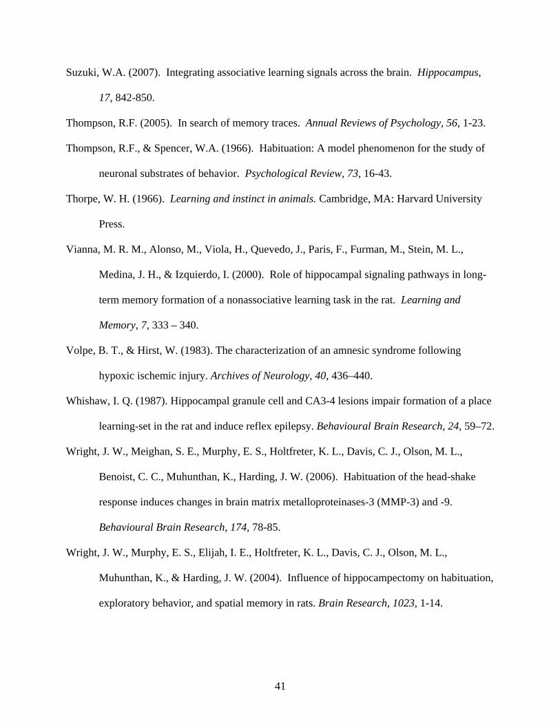
Suzuki, W.A. (2007). Integrating associative learning signals across the brain. Hippocampus,
17, 842-850.
Thompson, R.F. (2005). In search of memory traces. Annual Reviews of Psychology, 56, 1-23.
Thompson, R.F., & Spencer, W.A. (1966). Habituation: A model phenomenon for the study of
neuronal substrates of behavior. Psychological Review, 73, 16-43.
Thorpe, W. H. (1966). Learning and instinct in animals. Cambridge, MA: Harvard University
Press.
Vianna, M. R. M., Alonso, M., Viola, H., Quevedo, J., Paris, F., Furman, M., Stein, M. L.,
Medina, J. H., & Izquierdo, I. (2000). Role of hippocampal signaling pathways in long-
term memory formation of a nonassociative learning task in the rat. Learning and
Memory, 7, 333 – 340.
Volpe, B. T., & Hirst, W. (1983). The characterization of an amnesic syndrome following
hypoxic ischemic injury. Archives of Neurology, 40, 436–440.
Whishaw, I. Q. (1987). Hippocampal granule cell and CA3-4 lesions impair formation of a place
learning-set in the rat and induce reflex epilepsy. Behavioural Brain Research, 24, 59–72.
Wright, J. W., Meighan, S. E., Murphy, E. S., Holtfreter, K. L., Davis, C. J., Olson, M. L.,
Benoist, C. C., Muhunthan, K., Harding, J. W. (2006). Habituation of the head-shake
response induces changes in brain matrix metalloproteinases-3 (MMP-3) and -9.
Behavioural Brain Research, 174, 78-85.
Wright, J. W., Murphy, E. S., Elijah, I. E., Holtfreter, K. L., Davis, C. J., Olson, M. L.,
Muhunthan, K., & Harding, J. W. (2004). Influence of hippocampectomy on habituation,
exploratory behavior, and spatial memory in rats. Brain Research, 1023, 1-14.
41
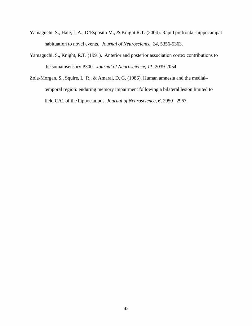
Yamaguchi, S., Hale, L.A., D’Esposito M., & Knight R.T. (2004). Rapid prefrontal-hippocampal
habituation to novel events. Journal of Neuroscience, 24, 5356-5363.
Yamaguchi, S., Knight, R.T. (1991). Anterior and posterior association cortex contributions to
the somatosensory P300. Journal of Neuroscience, 11, 2039-2054.
Zola-Morgan, S., Squire, L. R., & Amaral, D. G. (1986). Human amnesia and the medial–
temporal region: enduring memory impairment following a bilateral lesion limited to
field CA1 of the hippocampus, Journal of Neuroscience, 6, 2950– 2967.
42
Figure Captions
Figure 1. Mean (± SEM) group changes in number of head-shake responses per 3-trial blocks
during Sessions I and II of habituation trials separated by a 24-h ISI. There were no differences
among the groups concerning the pattern of habituation during Session I. There were differences
among the groups during the first trial block of Session II with the contingent tone group
revealing a significantly reduced level of spontaneous recovery as compared with the other two
groups which did not differ. *p<.001.
Figure 2. Schematic reconstruction of the smallest (solid black region) and largest (solid black +
hatched regions) aspiration lesions within the dorsal hippocampus (A) and neocortex (B) for
members of the groups utilized in Experiment 2. Coordinates for these coronal sections are
indicated in mm posterior to bregma with reference to the stereotaxic brain atlas of Paxinos and
Watson (1986).
Figure 3. Mean (± SEM) number of head-shake responses per 3-trial blocks for dorsal
hippocampectomized (HippX) and neocortex (NeoX) lesioned rats during Sessions I and II of
habituation trials separated by a 24-h ISI. These groups did not differ regarding pattern of
habituation during Session I; however, members of the tone neocortex lesioned group revealed
significantly attenuated levels of spontaneous recovery during the first trial block of Session II.
*p<.001.
43
3-Trial Blocks
0 2 4 6 8 10 12 14 16
Num
ber o
f Hea
d-S
hake
Res
pons
es
0
2
4
6
8 No Tone Session I No Tone Session II Tone Session I Tone Session II Random Tone Session I Random Tone Session II
*
Figure 1
Figure 2
lowing a habituation/classical conditioning head-shake task
44
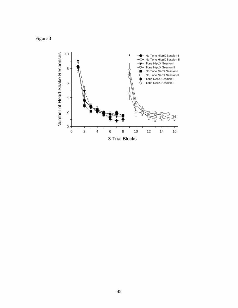
Figure 3
3-Trial Blocks0 2 4 6 8 10 12 14 16
Num
ber o
f Hea
d-Sh
ake
Res
pons
es
0
2
4
6
8
10 No Tone HippX Session I No Tone HippX Session II Tone HippX Session I Tone HippX Session II No Tone NeoX Session I No Tone NeoX Session II Tone NeoX Session I Tone NeoX Session II
*
45
CHAPTER 03
THE ROLE OF MATRIX METALLOPROTEINASES ON HABITUATION DURING A
HEAD SHAKE RESPONSE TASK
46
Editor-in-Chief: Dr. F. E. Bloom Brain Research
The role of matrix metalloproteinases on habituation during a head shake response task
Roberta V. Wiedigera* and John W. Wrightb,c,d
aDepartment of Psychology, Lincoln Land Community College,
Springfield, IL 62794
bDepartment of Psychology, Washington State University, Pullman, WA 99164-4820
cDepartment of Veterinary and Comparative Anatomy, Pharmacology and Physiology,
Washington State University, Pullman, WA 99164-6520
dProgram in Neuroscience, Washington State University, Pullman, WA 99164-6520
Running head: Head-shake response habituation
*Corresponding author: Roberta V. Wiediger
Lincoln Land Community College
5250 Shepherd Road
Springfield, IL 62794 USA
Tel: 1 217 786 2394
Fax: 1 217 786 2470
E-mail address: [email protected]
47
Abstract
Matrix Metalloproteinases (MMPs) have been implicated as markers of neural plasticity.
The present investigation employed a head-shake response (HSR)/classical conditioning
paradigm as the habituated response in rats. A tone served as the conditioned stimulus (CS) and
an stream of air to the ear as the unconditioned stimulus (US). The bilateral injection of a
general MMP inhibitor, FN-439, into the dorsal hippocampus resulted in maximal spontaneous
recovery during the second habituation session conducted 24-h following the first session. The
control group was injected with artificial cerebrospinal fluid and showed a significant attenuation
of spontaneous recovery during the second session. Members of a separate group of rats were
injected with a specific MMP-3 inhibitor into the dorsal hippocampus. The results were similar
to those animals treated with FN-439. These findings indicate that during this HSR/classical
conditioning task when animals were given an MMP inhibitor no association between the CS-US
was formed in the dorsal hippocampus, therefore maximum spontaneous recovery was present
24h later. In contrast, when the dorsal hippocampus was functioning this association was formed
and spontaneous recovery was reduced thus conserving the number of responses emitted. This
experiment suggests that activated MMP-3 is an important enzyme in the formation and
consolidation of this association in the hippocampus.
Key words: Habituation; Spontaneous recovery; Head-shake response; Classical conditioning;
Matrix metalloproteinases; FN-439; MMP-3 inhibitor; Hippocampus; Learning
48
1. Introduction Habituation is characterized by a decrease in the strength of a response to a repeatedly presented
stimulus (Harris, 1943; Thompson and Spencer, 1966; Staddon, et al., 2002; Thorpe, 1966) that
is not related to sensory adaptation or motor fatigue (McSweeney et al., 2005). This
phenomenon is a well documented form of learning (Mackintosh, 1987) that occurs across many
species for a number of response systems ranging from the gill-withdrawal reflex in Aplysia
(Castellucci and Kandel, 1974), tap withdrawal or chemotaxic response in the nematode
Caenorhabditis elegans (Bernhard and van der Kooy, 200; Rose and Rankin, 2001), to acoustic
startle response in rats and mice (Masini et al., 2008; Plappert and Pilz, 2005; Stevenson and
Gratton, 2004), and feeding in humans (Ernst and Epstein, 2002). The present investigation
focused on the head-shake response (HSR) which consists of a rapid rotation of the head about
the anterior to posterior axis due to a mild air stimulus applied to the ear (Askew et al., 1969).
This response follows a decreasing negatively accelerating function of stimulus frequency, such
that the higher the rate of stimulus presentation the faster the rate of habituation (Murphy et al.,
2005). Following habituation the HSR spontaneously recovers as a function of the inter-session-
interval (ISI), reaching approximately 85-90% of its original response strength following 24-h of
rest.
The hippocampus has been linked to learning and memory storage which appears to be
dependent upon synaptic plasticity (Collingridge et al., 2004). Thus, memory consolidation
requires synaptic reconfiguration which in turn is dependent upon changes in extracellular matrix
(ECM) molecules mediated by matrix metalloproteinases (MMPs; Wright et al., 2002). ECM
molecules are composed of proteins involved in cellular development, migration, connectivity
and reorganization (Novak and Kaye, 2000). The temporary dissociation of ECM molecules by
49
MMPs permits the reconfiguration of the brain’s synaptic architecture thought to be critical to
the processes of neural plasticity, learning and memory (Dityatev and Schachner, 2003). We
have recently shown that the hippocampus is involved in the HSR task when a tone (1-s in
duration) is presented 2-s prior to the air stimulus (Wiediger and Wright, submitted). Dorsal
hippocampectomized rats exposed to the tone revealed high levels of spontaneous recovery after
a 24-h ISI; however animals with an intact hippocampus revealed attenuated levels of
spontaneous recovery, suggesting that the hippocampus is important during the formation of this
CS-US pairing and promotes conservation of responding.
The present study examined the role of MMPs in the dorsal hippocampus during
habituation utilizing this combined classical conditioning/HSR habituation protocol. We
reasoned that if dorsal hippocampal MMPs are critical to reconfiguration of ECM molecules
during learning, then animals treated with an MMP inhibitor (MMPi) should loose the ability to
make use of the tone as an anticipatory associative cue. This would result in maximum
spontaneous recovery during a second habituation session following a 24-h ISI. In contrast,
animals injected with vehicle should make use of this cue and attenuation in spontaneous
recovery would be anticipated during the second session. We initially tested this hypothesis with
a general MMPi, FN-439, and then with a specific MMP-3i. MMP-3 has been shown to be
important to both spatial and associative learning (Wright et a., 2006; Olson et al., 2008; Brown
et al., 2007; Meighan et al., 2006; Wright et al., 2004).
2. Results
2.1 Hippocampal infusion of FN-439
Figure 1 presents the results of HSR habituation comparing the high and low dose FN-
439 groups, and the aCSF group. Similar patterns of habituation over trials were seen among
50
these groups during Session I. The group that received the high dose of FN-439 revealed a mean
(SEM) response level of 7.6 (.5) HSRs on the first trial block of Session I. The low dose FN-439
and aCSF groups indicated mean response levels of 6.2 (.9) and 6.5 (.5) HSRs, respectively.
During the first trial block of Session II the high and low dose FN-439 groups and the aCSF
group indicated mean HSR levels of 6.1 (.4), 2.4 (.8) and 2.9 (.4), respectively, representing
spontaneous recovery levels of 81.1, 39.2 and 44.5%, respectively. Thus, the levels of
spontaneous recovery were attenuated and equivalent for the low dose FN-439 and aCSF groups
as compared with the high dose FN-439 group.
The statistical analyses to support the above observations concerning Session I indicated
a significant effect for trial blocks, F(7,112)= 144.32, p<.0001, confirming an expected within-
session decrease in responding during the session. No group or interaction of groups x trials was
found, F(2,16)= 2.56, p>.10 and F(14,112)=1.34, p>.10, respectively, suggesting that the overall
levels of HSRs during the session were similar across groups. Statistical analyses of Session II
data revealed a significant effect for trial blocks, F(7,112)=75.44, p<.0001, suggesting an
expected within-session decrease in responding over trials. Significant group and interaction
effects were also found, F(2,16)=5.32 and F(14,112)=12.33, p<.0001, respectively. The
significant interaction suggested that the within-session changes in responding varied across the
groups. The significant group effect suggested that the groups differed in responding during
session II. A one-way ANOVA comparing these three groups on the first trial block of Session
II was significant, F(2,18)=16.937, p<.0001. Post-hoc tests indicated that the low dose FN-439
and the aCSF groups were each different from the high dose FN-439 group, but the two former
groups did not differ. Repeated measures t-test confirmed that the low dose FN-439 and aCSF
groups revealed significant decreases in HSRs comparing the first trial blocks of Session I and II,
51
t(7)=4.33 and 9.00 respectively, p<.01. The high dose FN-439 group did not differ, t(7)=1.69,
p>.05.
2.2 Hippocampal infusion of MMP-3 inhibitor
Figure 2 presents the results of HSR habituation comparing the MMP-3i treated group
and the aCSF control group. Similar patterns of habituation over trials were seen during Session
I. The group that received the MMP-3i revealed mean responding of 6.8 (.3) HSRs during the
first trial block of Session I. The aCSF group displayed mean responding of 6.9 (.3) HSRs.
During the first trial block of Session II the MMP-3i and the aCSF groups indicated mean HSR
levels of 5.3 (.2) and 2.8 (.2), respectively, representing spontaneous recovery levels of 77.9 and
40.6%, respectively Thus, the level of spontaneous recovery was low for the aCSF group, but
remained high for the MMP-3i group.
The statistical analysis to support the above observations concerning Session I indicated a
significant effect of trial blocks, F(7,98)=175.84, p<.001, suggesting a within-session decrease in
responding during session I. No interaction of groups x trials, F(7,98)=.41, p>.10, and no groups
effect, F(1,14)=.09, p>.10, were found. Statistical analysis of Session II indicated a significant
effect of trial blocks, F(7,98)=134.73, p<.001, indicating a within-session decrease in
responding. A significant interaction of groups x trials was found, F(7,98)=23.89, p<.0001, but
no groups effect, F(1,14)=1.13, p>.05. The significant interaction suggested that the within-
session changes in responding varied across the groups. An independent t-test indicated that the
aCSF group’s level of spontaneous recovery during the first trial block of Session II was
significantly less than that of the MMP-3i group, t(14)=6.609, p<.0001. Repeated measures t-
test confirmed that the aCSF group emitted fewer HSRs during the first trial block of Session II
52
as compared with Session I, t(7)=8.99, p<.001; while the MMP-3i group did not differ,
t(7)=2.08, p>.05.
3. Discussion
MMPs have been shown to be critical to the maintenance and reconstruction of the ECM
(Wright et al., 2004), suggesting that MMPs are important in learning given that they are markers
of neural plasticity. Currently over 25 MMPs have been identified. Of these MMP-9 and MMP-
3 have been shown to be particularly instrumental regarding synaptic plasticity and memory
formation (Olson et al., 2008). Several studies have measured changes in MMP expression in
the hippocampus after the animal has successfully solved the Morris water maze task, HSR task,
passive avoidance and conditioned place preference implying that memory consolidation is
taking place (Wright et al., 2006; Olson et al., 2008; Brown et al., 2007; Meighan et al., 2006;
Wright et al., 2004). Thus, MMP activity appears to produce transient alterations in the ECM
that may be prerequisite to hippocampal-dependent learning.
Meighan et al. (2006) employed the general MMP inhibitor FN-439 infused
intracerebroventricularly (icv) to disrupt the acquisition of the Morris water maze task of spatial
memory. FN-439 has also been shown to inhibit the induction and stability of long-term
potentiation (LTP) in hippocampal slices (Meighan et al., 2006; Meighan et al., 2007). Recently,
Olson et al. (2008), measured high levels of MMP-3 at 1 and 4 hours after the acquisition of a
passive avoidance conditioning task. These investigators also showed that the icv infusion of an
MMP-3 inhibitor during the acquisition of a passive avoidance conditioning task resulted in
dose-dependent learning deficits. Of particular relevance to the present investigation, Wright et
al. (2006) measured elevations in MMP-3 protein expression in the hippocampus, pre-frontal
cortex and piriform cortex at the 2-h ISI during a HSR task.
53
The present study combined a classical conditioning paradigm with HSR habituation in
order to examine the influence of MMPs on spontaneous recovery. The application of a
signaling cue, a tone prior to the air stimulus to the ear, permitted an association to form,
presumably stored in the dorsal hippocampus, that reduced the strength of responding and thus
spontaneous recovery during a second HSR session. We hypothesized that the injection of an
MMP inhibitor into the dorsal hippocampus would prevent such an association from forming and
maximum spontaneous recovery would occur during the second session despite the contingent
presentation of the tone. In other words, the signaling value of the tone would be lost due to the
inhibition of MMPs acting on ECM molecules with a resulting disruption of synaptogenesis.
The results concerned with FN-439 infusion demonstrated a predictable decreasing
negatively accelerated response pattern over trials asymptoting by about the fifth trial block
(Figure 1). Following a 24-h ISI the first trial block of Session II compared with that of Session
I indicated a spontaneous recovery level of 81.1% by the high dose FN-439 group. However the
low dose FN-439 group and the control group showed a spontaneous recovery of 39.2 and
44.5%, respectively. Thus, the high dose of FN-439 interfered with the CS-US association and
prevented memory consolidation. On the other hand, the group that received the low dose of
FN-439 and the control group formed the association resulting in attenuated spontaneous
recovery during the second session. These results suggest that dorsal hippocampus MMPs play a
role in the formation of such an association.
In the second experiment a predictable decreasing negatively accelerated response pattern
was also present over trials (Figure 2). Following a 24-h ISI the first trial block of Session II
compared with that of Session I indicated a spontaneous recovery level of 77.9% by the MMP-3i
group. The control group showed a level of spontaneous recovery of 40.6%. When MMP-3 was
54
inhibited no association was formed and a high level of spontaneous recovery was measured
during session II. In contrast, animals that received aCSF made use of the tone as a signaling cue
and an attenuated level of spontaneous recovery occurred during the second session. Thus, the
targeted MMP-3 appears to be an important enzyme involved in the formation of this CS-US
association.
In summary, this investigation is the first to examine the impact of MMP inhibitors
delivered to the dorsal hippocampus on the association formed between a signaling cue and HSR
habituation. The results indicate an important role for MMP-3 in the dorsal hippocampus when a
HSR habituation paradigm is combined with a contingently placed tone. Under control
conditions the animal conserved responses during the initial trial blocks of the second session,
thus reducing spontaneous recovery. However, when an MMP inhibitor was injected bilaterally
into the dorsal hippocampus the animals failed to make use of this cue thus forcing a less
efficient response pattern with more HSRs emitted than necessary to cope with the repeatedly
applied air stimulus. Taken together these findings suggest that MMP-3 may be essential to the
forming of the CS-US association during this HSR task.
4. Materials and methods
The protocols used in these studies were approved by the Washington State University
Institutional Animal Care and Use Committee and conformed to the guidelines for the care and
use of laboratory animals as required by the National Institutes of Health.
4.1 Animals
Male Sprague-Dawley rats (300-350 g, breeding stock derived from Taconic,
Germantown, NY) were adapted to a 12-h light/dark cycle initiated at 0600 h in an American
Association for the Accreditation of Laboratory Animal Care-approved vivarium at a
55
temperature of 21 ± 1° C. The animals were housed in pairs and provided water and food (Harlan
Teklad F6 Rodent Diet, Madison, WI) ad libitum, except the night prior to surgery when food
was removed.
4.2. Head-shake habituation
Independent groups of rats (8 per group) were tested over the course of 24 trials during
the first habituation session, followed by an additional 24 trials during a second session separated
by a 24-h ISI. The apparatus was patterned after that used by Askew et al. (1969) and consisted
of an elevated platform measuring 7.5 x 16 cm. This surface was positioned on a 0.9 m tall
wooden column that allowed the subject freedom of movement during testing. To discourage
escape attempts the surface of the platform was covered with 1 cm square wire mesh cloth
surrounded by plywood sloping down and outward, forming a collar 17 x 24 cm at its extreme.
The base of the wooden column pivoted 360˚, thus permitting the experimenter to compensate
for movements by the animal and maintain a face-to-face orientation. HSRs were elicited by a
hand held tube (orifice diameter = 0.5 mm) that provided a continuous stream of air that was
oscillated across the center of the subject’s left ear at an approximate rate of 3 cycles/s. The air
tube was held 1-1.5 cm from the animal’s ear. The intensity of the air stream was set at 5-6 cm
displacement of a 40 cm column of 95% ethanol in a 0.5 mm inside diameter U-shaped
manometer (Askew et al., 1969). The test room was painted black with ambient light set at the
minimum level necessary to score responses in order to reduce spatial cues.
4.3. Procedure
The habituation session consisted of a 5-min undisturbed adaptation period on the test stand,
followed by 24, 15-s stimulus presentations. Each stimulus presentation was separated by a fixed
15-s inter-trial interval (ITI). An IBM-compatible computer signaled the intervals to the
56
experimenter by displaying the 15-s trial in green numbers, and the 15-s ITI in red numbers. A
tone (1-s in duration) was presented 2-s prior to the air stimulus on each trial. From where the
animal was positioned in the room, it received a buzzer sound of 74 dB from speakers of an IBM
compatible computer. The experimenter recorded the number of HSRs following each trial. At
the end of Session I animals were taken to an adjacent room to receive the designated injections
within 5 min post-testing and again at 1-h post-testing. This injections schedule was chosen to
ensure that the inhibitor was present at 1-2 h post-conditioning, when elevations in active MMPs
have been measured (Olson et al., 2008).
4.4. Surgery
All rats were anesthetized using ketamine hydrochloride and xylazine (100 and 2
mg/ml/kg, i.m., respectively; Phoenix Scientific, St. Joseph, MO, and Mobay, Shawnee, KS).
Each rat received bilateral guide cannulas targeting the dorsal hippocampi (flat skull coordinates
relative to bregma: posterior: -4.0 mm, lateral: +-2.5 mm from midline). The guide was
constructed from PE-60 tubing (Clay Adams, Parsippany, NJ) with a heat bulge that rested on
top of the cranium thus serving as a stop to further penetration. The total length of the guide was
2.5 cm with a distance from the beveled tip to the heat bulge of 2.5 mm. All infusions were
made using a 30-gauge stainless-steel tubing injector with a beveled end that protruded 2.0 mm
beyond the tip of the guide cannula. The injector was attached to a 10 ul Hamilton syringe by
PE-20 tubing.
4.5. Hippocampal infusion of FN-439
The first experiment examined whether the general MMP inhibitor, FN-439 (4-Abz-Gly-
Pro-D-Leu-D-Ala-OH, mw=490.6; MMP inhibitor 1 #444250, Calbiochem, San Diego, CA),
interfered with the CS-US association formed during session I, by looking at the level of
57
spontaneous recovery during session II. This was accomplished by injecting a low dose (25 µg
in 2.5 µl aCSF) or a high dose (50 µg in 2.5 µl aCSF) of FN-439 bilaterally at 5 min and 1 h
post-testing after the termination of Session I. Members of the control group received aCSF (2.5
µl) delivered to each side at the same times post-testing.
4.6. Hippocampal infusion of MMP-3 inhibitor
The second experiment examined whether a specific MMP-3 inhibitor (Ac-Arg-Cys-Gly-
Val-Pro-Asp-NH2, mw=686.8; #444218, Calbiochem, San Diego, CA) interfered with the CS-
US association formed during session I, by looking at the level of spontaneous recovery during
session II. This was accomplished by injecting MMP-3i (50 µg in 2.5 µl aCSF) to each side at 5
min and 1 h post-testing after the termination of Session I. Members of the control group
received aCSF (2.5 µl) delivered to each side following the same procedure. Each group
consisted of 8 rats.
4.7. Histology
Once behavioral testing was completed each animal was deeply anesthetized with
Equithesin (pentobarbital: 100 mg/kg, i.p., Jensen-Salsbury Labs, Kansas City, MO) and
transcardally perfused (0.15 M NaCl followed by 10% formalin). The brains were immediately
removed and stored in a 30% sucrose in 10% formalin solution at 4° C for at least 30 days. Each
brain was then placed on the stage of a freezing microtome (Spencer Lens, Buffalo, NY) and
sectioned in the coronal plane at 40 µm. The sections were mounted on gelatin-coated slides and
stained with Cresyl violet. Once the slides had been cover-slipped they were viewed using an
overhead slide projector (Model X-1000, Ken-A-Vision, Raytown, MO) thus permitting
localization of the aspiration lesions in each hemisphere according to the rat brain atlas of
Paxinos and Watson (1986).
58
4.8 Statistical analyses
The twenty-four trials of each session were grouped into 8 blocks of 3 trials each, and the
mean of each trial block was calculated for each animal. These data were analyzed using a 3
(groups) x 8 (trial blocks) analysis of variance (ANOVA) with repeated measures on the second
factor, for each of the two sessions. A one-way ANOVA was used to compare the groups on the
first trial block of Session II. Significant effects were further analyzed using Newman-Keuls
post-hoc tests with a level of significance set at p< .05. In addition, t-tests for repeated measures
were used to analyze the first trial blocks of Sessions I and II for each group. Alpha levels were
adjusted according to the Bonferonni technique. The data of the second experiment were
analyzed using a 2 (groups) x 8 (trial blocks) ANOVA with repeated measures on the second
factor. Post-hoc tests were as described above.
Acknowledgements
The authors thank Mr. Mathew D. Wiediger for assisting with computer programs. This
study was supported by a Laing Endowment for Alzheimer’s disease grant to J.W.W., and funds
provided by Washington State University.
59
References
Askew, H.R., Leibrecht, B.C., Ratner, S.C., 1969. Effects of stimulus duration and repeated sessions on habituation of the head-shake response in the rat. J. Comp. Physiol. Psychol. 67, 497–503.
Bernhard, N., van der Kooy, D., 2000. A behavioral and genetic dissection of two forms of
olfactory plasticity in Caenorhabditis elegans: adaptation and habituation. Learn. Mem. 7, 199–212.
Brown, T.E., Forquer, M.R., Cocking, D.L., Jansen, H.T., Harding, J.W., Sorg, B.A., 2007. Role
of matrix metalloproteinases in the acquisition and reconsolidation of cocaine-induced conditioned place preference. Learn. Mem. 14, 214–223.
Castellucci, V.F., Kandel, E.R., 1974. A quantal analysis of the synaptic depression underlying habituation of the gill-withdrawal reflex in Aplysia. Proc. Natl. Acad. Sci. U. S. A. 77, 7492–7496. Collingridge, G.L., Isaac, J.T., Wang, Y.T., 2004. Receptor trafficking and synaptic plasticity.
Nat. Rev. Neurosci. 5, 952-962. Dityatev, A., Schachner, M., 2003. Extracellular matrix molecules and synaptic plasticity. Nat.
Rev. Neurosci. 4, 456-468. Ernst, M.M., Epstein, L.H., 2002. Habituation of responding for food in humans. Appetite 38,
224–234. Harris, J.D., 1943. Habituatory response decrement in the intact organism. Psychol. Bull. 40,
385-422. Mackintosh, N.J., 1987. Neurobiology, psychology, and habituation. Behav. Res. Ther. 25, 81-
97. Masini, C.V., Day, H.E.W., Campeau, S., 2008. Long-term habituation to repeated loud noise is
impaired by relatively short inter-stressor intervals in rats. Behav. Neurosci. 122, 210-223.
McSweeney, F.K., Kowal, B.P., Murphy, E.S., Wiediger, R.V., 2005. Stimulus change
dishabituates operant responding supported by water reinforcers. Behav. Proces. 70, 235-246.
Meighan, S.E., Meighan, P.C., Choudhury, P., Davis, C.J., Olson, M.L., Zornes, P.A., Wright,
J.W., Harding, J.W., 2006. Effects of extracellular matrix-degrading proteases matrix metalloproteinases 3 and 9 on spatial learning and synaptic plasticity. J. Neurochem. 96, 1227 – 1241.
60
Meighan, P.C., Meighan, S.E., Davis, C.J., Wright, J.W., Harding, J.W., 2007. Effects of matrix metalloproteinase inhibition on short- and longterm plasticity of schaffer collateral/CA1 synapses. J. Neurochem. 102, 2085–2096.
Murphy, E.S., Harding, J.W., Muhunthan, K., Holtfreter, K., Wright, J.W., 2005. Role of
mitogen-activated protein kinases during recovery from head-shake response habituation in rats. Brain Research. 1050, 170-179.
Novak, U., Kaye, A.H., 2000. Extracellular matrix and the brain: components and function. J.
Clin. Neurosci. 7, 280-290. Olson, M.L, Meighan, P.C, Brown, T.E, Asay, A.L, Benoist, C.C, Harding, J.W, Wright, J.W.,
2008. Hippocampal MMP-3 elevation is associated with passive avoidance conditioning. Regul. Pept. 146, 19–25.
Paxinos, G., Watson, C., 1986. The Rat Brain in Stereotaxic Coordinates (2nd ed.). New York:
Academic Press. Plappert, C.F., Pilz, P.K., 2005. Long-term habituation of the startle response in mice evoked by
acoustic and tactile stimuli. Behav. Brain Res. 162, 307–310. Rose, J.K., Rankin, C.H., 2001. Analysis of habituation in Caenorhabditis elegans. Lear. Mem. 8,
63-69. Staddon, J.E.R., Chelaru, I.M., Higa, J.J., 2002. Habituation, memory and the brain: The
dynamics of interval timing. Behav. Proces. 57, 71-88. Stevenson, C.W., Gratton, A., 2004. Basolateral amygdala dopamine receptor antagonism
modulates initial reactivity to but not habituation of the acoustic startle response. Behav. Brain Res. 153, 383–387.
Thompson, R.F., Spencer, W.A., 1966. Habituation: A model phenomenon for the study of
neuronal substrates of behavior. Psychol. Rev. 73, 16-43. Thorpe, W.H., 1966. Learning and instinct in animals. Cambridge, MA: Harvard University
Press. Wright, J.W., Reichert, J. R., Davis, C. J, Harding, J. W., 2002. Neural plasticity and the brain
rennin-angiotensis system. Neurosci. Biobeh. Rev. 26, 529-552. Wright, J.W., Meighan, S.E., Murphy, E.S., Holtfreter, K.L., Davis, C.J., Olson, M.L., Benoist,
C.C., Muhunthan, K., Harding, J.W., 2006. Habituation of the head-shake response induces changes in brain matrix metalloproteinases-3 (MMP-3) and -9. Behav. Brain Res. 174, 78-85.
61

Wright, J.W., Murphy, E.S., Elijah, I.E., Holtfreter, K.L., Davis, C.J., Olson, M.L., Muhunthan, K., & Harding, J.W., 2004. Influence of hippocampectomy on habituation, exploratory behavior, and spatial memory in rats. Brain Res. 1023, 1-14.
62
Figure Captions: Figure 1. Mean (± SEM) group changes in number of head-shake responses per 3-trial blocks
during Sessions I and II of habituation trials separated by a 24-h ISI. There were no differences
among the groups concerning the pattern of habituation during Session I. There were differences
among the groups during the first trial block of Session II with the high FN439 group revealing a
significantly high level of spontaneous recovery as compared with the other two groups which
did not differ. *p<.001.
Figure 2. Mean (± SEM) number of head-shake responses per 3-trial blocks for the MMP-3i
and aCSF injected rats during Sessions I and II of habituation trials separated by a 24-h ISI.
These two groups did not differ regarding pattern of habituation during Session I; however,
members of the aCSF group revealed significantly attenuated levels of spontaneous recovery
during the first trial block of Session II. *p<.001.
63
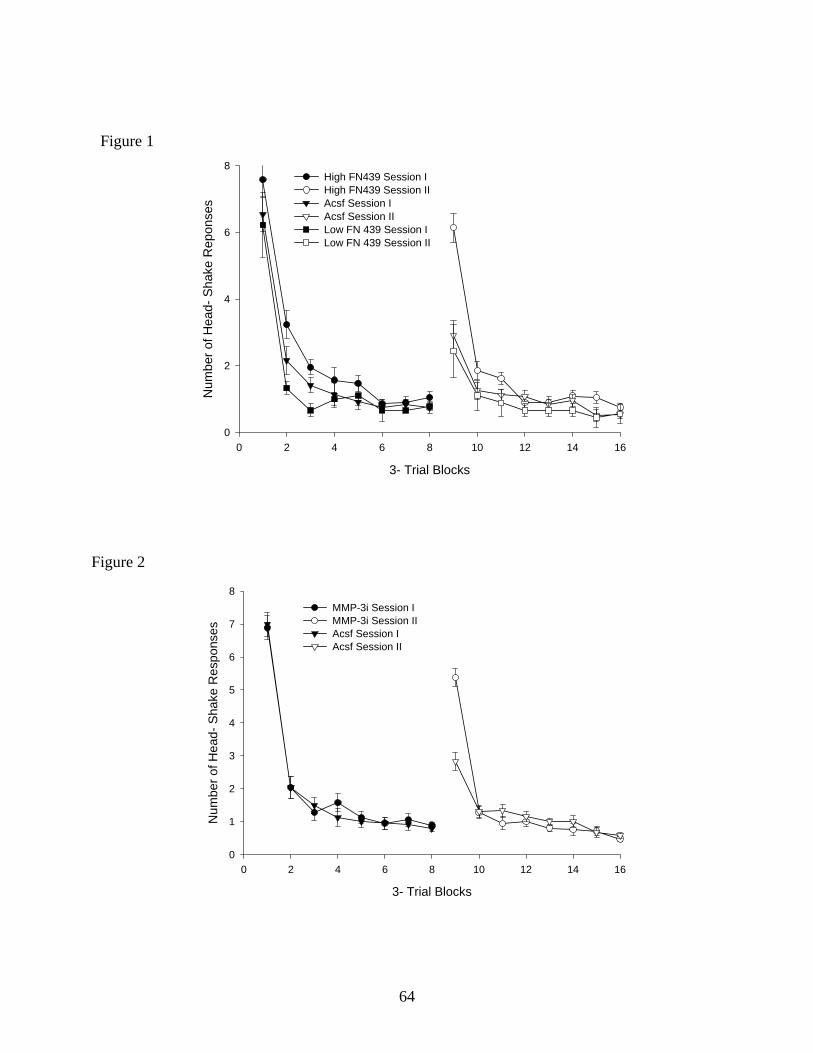
3- Trial Blocks
0 2 4 6 8 10 12 14 16
Num
ber o
f Hea
d- S
hake
Rep
onse
s
0
2
4
6
8High FN439 Session IHigh FN439 Session II Acsf Session I Acsf Session IILow FN 439 Session I Low FN 439 Session II
Figure 1
Figure 2
3- Trial Blocks
0 2 4 6 8 10 12 14 16
Num
ber o
f Hea
d- S
hake
Res
pons
es
0
1
2
3
4
5
6
7
8MMP-3i Session I MMP-3i Session II Acsf Session I Acsf Session II
64
CHAPTER FOUR
GENERAL DISCUSSION
65
General Discussion These experiments were designed to provide a better understanding concerning the role
of the hippocampus and the matrix metalloproteinases (MMPs) during the head-shake response
task combined with a classical conditioning paradigm.
Patterns of the HSR habituation and spontaneous recovery showed a reliable decreasing
negatively accelerated response pattern that was followed by a substantial spontaneous recovery
following a 24-h ISI (Askew et al., 1969; Leibrecht et al., 1969; Murphy et al., 2005; Wright et
al., 2006; McSweeney & Murphy, 2000). The hippocampus has been implicated in the control
of inhibitory processes including habituation (Douglas, 1967; Jarrard & Bunnell, 1968; Kimble,
1968; Leaton, 1965; Primbram, 1967; Roberts et al., 1962), fear extinction and spontaneous
recovery (Ji & Maren, 2007), and in the formation and utilization of spatial memory (Bures et al.,
1997; McNaughton et al., 1989; Morris et al., 1982; Nadel, 1991; O’Keefe and Nadel 1978;
Wishaw, 1987).
The hippocampus is also linked to learning and memory storage because of its substantial
synaptic plasticity (Collingridge et al., 2004). ECM molecules mediate changes in the brain’s
synaptic architecture thought to be critical to the processes of neural plasticity, learning and
memory (Dityatev and Schachner, 2003). Wright and colleagues (2007) reported a compromised
ability to reconfigure ECM molecules by inhibiting MMP activity in the dorsal hippocampus
during spatial memory acquisition. Meighan et al. (2006) used the general MMP inhibitor FN-
439 to interfere with the late phase of long-term potentiation (LTP), and when FN-439 was
infused intracerebroventricularly (icv) it disrupted the acquisition of the Morris water maze task
of spatial memory. FN-439 has also been shown to disrupt the induction and stability of long-
term potentiation (LTP) in hippocampal slices (Meighan et al., 2006 and Meighan et al., 2007).
66
Recently, Olson et al, (2008) measured high levels of MMP-3 at 1 and 4 hours after the
acquisition of a passive avoidance conditioning task. They also showed that the icv infusion of
an MMP-3 inhibitor during the acquisition of a passive avoidance conditioning task resulted in
dose-dependent learning deficits. Therefore the overall objective of this dissertation was to
answer the following questions: 1) Can a CS-US association be formed during a HSR task, and
if so, what is its effect on spontaneous recovery? 2) Is the dorsal hippocampus important during
the CS-US association during a HSR task, and if so, what is its effect on spontaneous recovery?
3) What is the role of MMPs on the CS-US association during a HSR task? Will the inhibition
of MMPs interfere with the acquisition of the CS-US association? Will the inhibition of MMP-3
interfere with the acquisition of the CS-US association?
Summary of Findings
An association between the tone (CS) and the air stimulus to the ear (US) was formed
during the HSR task. Thus, animals that received the CS-US pairing showed reduced
spontaneous recovery after a 24-h ISI; while the control group showed high spontaneous
recovery. This was interpreted to suggest that the animals were able to recognize the association
of the two stimuli and therefore little HSR occurred. The association between the CS-US was
confirmed when a random tone control group presented a pattern of responding very similar to
the no tone control group. In other words, when the tone was noncontingent, or when no
presentation of the tone was made, spontaneous recovery of the HSR 24-h later was high. These
findings suggest that when the CS-US association is acquired animals conserve energy by
reducing responding during a second HSR session. To further understand this phenomenon the
dorsal hippocampus was bilaterally removed. Under this condition the animals were unable to
learn the CS-US association, therefore 24-h later they showed the same pattern of responding as
67
the no tone control group described above. These results strongly suggest that when the general
MMP inhibitor, FN439, or the specific MMP-3 inhibitor was infused into the dorsal
hippocampus the animals were not able to acquire the CS-US association. Thus, their pattern of
responding 24-h later during the second session was very similar to that displayed by members
of the no tone control group which never experienced the CS-US pairing.
In conclusion, the dorsal hippocampus appears to play a significant role in mediating
learning when a classical conditioning paradigm is combined with the HSR task. Removing the
hippocampus, or inhibiting MMPs in this structure, interfered with the learning of the CS-US
association. These results highlight the importance of the dorsal hippocampus with respect to the
pairing of a signaling cue with the habituation of a reflex-like behavior such as HSR. Once this
association was in place the animal’s subsequent behavior was changed such that fewer
responses were emitted. This represents a much more rapid adaptation to the repeated
presentation of an innocuous stimulus than when the association is unavailable to the animal.
These results illustrate the usefulness of combining associative and nonassociative learning
paradigms in an effort to understand the parameters under which habituation and spontaneous
recovery can be modified, and further point out the importance of MMPs to this phenomenon.
68
References
Askew, H. R., Leibrecht, B. C., & Ratner, S. C. (1969). Effects of stimulus duration and
repeated sessions on habituation of the head-shake response in the rat. Journal of
Comparative & Physiological Psychology, 67, 497–503.
Bures, J., Fenton, A. D., Kaminsky, Y., & Zinyuk, L. (1997). Place cells and place navigation.
Proceedings National Academy of Sciences USA, 94, 343–350.
Collingridge, G. L., Isaac J. T., & Wang Y. T., (2004). Receptor trafficking and synaptic
plasticity. Nature Reviews Neuroscience, 5, 952-962.
Dityatev, A., and Schachner, M., (2003). Extracellular matrix molecules and synaptic plasticity.
Nature Reviews Neuroscience. Vol.4, no.6, pp.456-468.
Douglas, R. J. (1967). The hippocampus and behavior. Psychological Bulletin, 67, 416 – 422.
Jarrard, L. E., & Bunnell, B. N. (1968). Open-field behavior of hippocampal lesioned rats and
hamster. Journal of Comparative & Physiological Psychology, 66, 500-502.
Ji, J., & Maren, S. (2007). Hippocampal involvement in contextual modulation of fear
extinction. Hippocampus, 17, 749-758.
Kimble, D. P. (1968). Hippocampus and internal inhibition. Psychological Bulletin, 70, 285 –
295.
Leaton, R. N. (1965). Exploratory behavior in rats with hippocampal lesions. Journal of
Comparative & Physiological Psychology, 59, 325–330.
Leibrecht, B. C., & Askew, H. R. (1969). Habituation of the head-shake response in the rat:
recovery, transfer, and changes in topography. Journal of Comparative & Physiological
Psychology, 69, 699 – 708.
69
McNaughton, B. L., Barnes, C. A., Meltzer, J., & Sutherland, R., J. (1989). Hippocampal
granule cells are necessary for normal spatial learning but not for spatially-selective
pyramidal cell discharge. Experimental Brain Research, 73 (3), 1432-1106
McSweeney, F. K., & Murphy, E.S., (2000). Criticisms of the satiety hypothesis as an
explanation for within-session decreases in responding. Journal of the Experimental
Analysis of Behavior, 74 (3), 347–361.
Meighan, S. E., Meighan, P. C., Choudhury, P., Davis, C. J., Olson, M. L., Zornes, P. A.,
Wright, J. W., & Harding, J. W. (2006). Effects of extracellular matrix-degrading
proteases matrix metalloproteinases 3 and 9 on spatial learning and synaptic plasticity.
Journal of Neurochemistry, 96, 1227 – 1241.
Meighan PC, Meighan SE, Davis CJ, Wright JW, & Harding JW. (2007). Effects of matrix
metalloproteinase inhibition on short- and longterm plasticity of schaffer collateral/CA1
synapses. Journal of Neurochemistry, 102, 2085–2096.
Morris R.G.M., Garrud P., Rawlins J.N.P., & O’Keefe J. (1982). Place navigation impaired in
rats with hippocampal lesions. Nature, 297, 681– 683.
Murphy, E. S., Harding, J. W., Muhunthan, K., Holtfreter, K., & Wright, J. W. (2005). Role of
mitogen-activated protein kinases during recovery from head-shake response habituation
in rats. Brain Research, 1050, 170-179.
Nadel L. (1991). The hippocampus and space revisited. Hippocampus, 1, 221– 229.
O’Keefe, J., & Nadel, L. (1978). The Hippocampus as a Cognitive Map (pp. 265-290). Oxford,
UK: Oxford University Press.
70
Olson ML, Meighan PC, Brown TE, Asay AL, Benoist CC, Harding JW, Wright JW. 2008.
Hippocampal MMP-3 elevation is associated with passive avoidance conditioning.
Regulatory Peptides, 146, 19–25.
Pribram, K. H. (1967). The limbic systems efferent control of neural inhibition and behavior.
Progress in Brain Research, 27, 318 – 336.
Roberts, W. W., Dember, W. N., & Brodwick, M. (1962). Alternation and exploration in rats
with hippocampal lesions. Journal of Comparative & Physiological Psychology, 55, 695–
700.
Whishaw, I. Q. (1987). Hippocampal granule cell and CA3-4 lesions impair formation of a place
learning-set in the rat and induce reflex epilepsy. Behavioural Brain Research, 24, 59–72.
Wright, J.W., Reichert, J. R., Davis, C. J, Harding, J. W. (2002). Neural plasticity and the brain
rennin-angiotensis system. Neuroscience and Biobehavioral Reviews. 26, 529-552.
Wright, J. W., Brown, T. E., Harding, J. W. (2007). Inhibition of hippocampal matrix
metalloproteinase-3 and -9 disrupts spatial memory. Neural Plasticity. 1-8.
Wright, J. W., Meighan, S. E., Murphy, E. S., Holtfreter, K. L., Davis, C. J., Olson, M. L.,
Benoist, C. C., Muhunthan, K., Harding, J. W. (2006). Habituation of the head-shake
response induces changes in brain matrix metalloproteinases-3 (MMP-3) and -9.
Behavioural Brain Research, 174, 78-85.
71