Embed Size (px)
Citation preview
International Journal of
Molecular Sciences
Review
The Role of Small Noncoding RNA in DNADouble-Strand Break Repair
Iwona Rzeszutek 1,* and Gabriela Betlej 2
1 Institute of Biology and Biotechnology, Department of Biotechnology, University of Rzeszow, Pigonia 1,35-310 Rzeszow, Poland
2 Institute of Physical Culture Studies, College of Medical Sciences, University of Rzeszow,35-310 Rzeszow, Poland; [email protected]
* Correspondence: [email protected]; Tel.: +48-17-851-86-20; Fax: +48-17-851-87-64
Received: 7 October 2020; Accepted: 26 October 2020; Published: 28 October 2020�����������������
Abstract: DNA damage is a common phenomenon promoted through a variety of exogenous andendogenous factors. The DNA damage response (DDR) pathway involves a wide range of proteins,and as was indicated, small noncoding RNAs (sncRNAs). These are double-strand break-inducedRNAs (diRNAs) and DNA damage response small RNA (DDRNA). Moreover, RNA binding proteins(RBPs) and RNA modifications have also been identified to modulate diRNA and DDRNA functionin the DDR process. Several theories have been formulated regarding the synthesis and functionof these sncRNAs during DNA repair; nevertheless, these pathways’ molecular details remainunclear. Here, we review the current knowledge regarding the mechanisms of diRNA and DDRNAbiosynthesis and discuss the role of sncRNAs in maintaining genome stability.
Keywords: noncoding RNA; double-strand breaks (DSBs), DNA repair; DNA damage response (DDR)
1. Introduction
During life, all cells in our body are continuously challenged by factors that induce damages inour DNA. Such factors are commonly present in our environment and can be both exogenousand endogenous to the cell [1–3]. Exogenous sources of DNA damage include: an ultravioletlight (UV), ionizing radiation (IR) and X-ray [4], while endogenous DNA damage often arisesas a product or by-product of cells’ own metabolism [5]. Additionally, DNA damage can alsooccur during the replication or transcription process. Interestingly, these processes can also occurconcomitantly, creating opportunities for either cooperation or conflict [6,7]. The resulting DNAlesions (nucleotide adducts, inter-strand cross-links or single-/double-strand breaks (SSBs/DSBs))created by these factors are very dangerous and if left unrepaired or repaired incorrectly can lead to:(a) mutations, (b) chromosomal rearrangements, (c) aberrant DNA repair gene expression profiles,all of each contribute to cancer [4].
To counteract these lesions and preserve genome stability, cells evolved a molecular system thatdetects damaged DNA, signals their presence, promotes their repair as well as initiates signalingpathways that impact a wide range of cellular processes. These actions are collectively known asthe DNA-damage response (DDR) [3,4,8]. In fact, cells have developed a number of pathways,each of which recognizes and repairs specific types of damage that occur to DNA. Typically, DSBs arerepaired by two main mechanisms: homologous recombination (HR) or an error-prone mechanismknown as classical non-homologous end joining (C-NHEJ) [9,10]. In addition, alternative error-pronemechanisms, namely alternative end joining (alt-EJ) and single-strand annealing (SSA) are used [11].Large nucleotide adducts are repaired by nucleotide excision repair (NER), while individual or smallbase lesions are repaired by base-excision repair (BER) [10].
Int. J. Mol. Sci. 2020, 21, 8039; doi:10.3390/ijms21218039 www.mdpi.com/journal/ijms
Int. J. Mol. Sci. 2020, 21, 8039 2 of 13
Previously it has been thought that DDR involves only enzymatic reactions carried out by proteinsfacilitating signaling as well as the repair process. Interestingly, a number of reports have implicatedthe function of noncoding RNA (ncRNA) in DDR [12–14], which we would like to discuss here inmore detail.
2. An Overview of DNA Double-Strand Break Repair Mechanisms
Nuclear DNA, the storage of life information, is undoubtedly the most precious componentof a cell, therefore needs to be protected from any lesions, especially from DSBs, which are one ofthe most harmful forms of DNA damage. Once a DSB is present in the genome, the cell initiatesthe DDR process of trying to repair the lesion [3,15]. However, if this is inauspicious the cell entersapoptosis [15]. As mentioned previously, these lesions can be repaired either by C-NHEJ, which isa fast, nonspecific pathway, or by HR, a slower, but higher fidelity process [15]. HR uses the sisterchromatid as a template during G2 and S-phase for repair (the exception being HR repairing DSB inthe rDNA [16]), while the C-NHEJ pathway is predominantly used by the cell during the G1 phase,although it can occur throughout most of the cell cycle [15].
Activation of the cellular response in both pathways starts with the recruitment of proteins that areable to recognize the damaged DNA [17–22]. After the recognition of DNA breaks by the sensor proteins,three key phosphatidylinositol 3-kinase like proteins (PIKs): Ataxia Telangiectasia Mutated (ATM),DNA-dependent protein kinase catalytic subunit (DNA-PKcs), and Ataxia Telangiectasia and RAD3related-protein (ATR) are activated [3]. Once activated, they phosphorylate many downstream targets,including histone variant H2A.X on Ser139 [23]. In case H2A.X gets phosphorylated, it accumulatesaround DSB sites (up to 1 Mb away from the break site) [24,25]. This modification, known as γH2A.X,provides a scaffold for the amplification of DDR signaling as well as DNA repair [24]. The recruitmentof a Mediator of DNA damage checkpoint protein 1 (MDC1) to γH2AX facilitates the activationof the E3-ubiquitin ligases RNF8 and RNF168. This initiates a modification cascade resulting inpolyubiquitination of the H1-linker histone and H2A [26–28]. Ubiquitination signals the recruitmentP53-binding protein 1 (53BP1) and breast cancer type 1 protein (BRCA1), which then, by the opposingactivities, govern the repair pathway choice. Binding of 53BP1 to the break stimulates the C-NHEJ,whereas BRCA1 promotes the HR pathway [29].
C-NHEJ occurs via sensing and binding of the Ku heterodimer composed of Ku70-Ku80 subunitsto the DSB [30]. Once the Ku heterodimer is bound to the DSB ends, it recruits other downstream factors,such as DNA-PKcs [31,32], X-ray cross-complementing protein 4 (XRCC4) [30,33,34], DNA LigaseIV [31,33], XRCC4-like factor (XLF) [35], Artemis [36] and Aprataxin-and-PNK-like factor (APLF) [37–39]that process and ligate the ends to repair the break. If not repaired by C-NHEJ, DSB is fixed by HR whichis initiated by the nucleolytic degradation of the 5′ terminated strands in a process termed end resection.End resection occurs via a two-step mechanism involving partially redundant nucleases Sae2/CtIP,Mre11-Rad50-Xrs2/Nbs1 (MRX/N), Exo1, and/or Dna2. Generated in this process, the 3′ single-strandoverhangs get coated in a RAD51 protein filament to catalyze the homologous pairing and exchange ofDNA strands [9].
Interestingly, recent studies indicated a new component in the DDR cascade: the noncoding RNAas well as RNA binding proteins which will be discussed in the next sections.
3. Noncoding RNA in Double-Strand Break Repair
The term noncoding RNA is commonly used in the case of RNA that does not encode a protein,however, it does not mean that such RNA does not contain any information or possess any function.They are mainly processed from double-stranded RNA (dsRNA) precursors by Dicer or Dicer-likeproteins. Next, associated with Argonaute (AGO) or Piwi proteins (a central factor in the RNA-inducedsilencing complex (RISC)), they regulate the gene expression at the post-transcriptional level or targetDNA for excision [40,41]. It turns out that many species evolved processes in which noncoding RNAis involved. For instance, these small, 20–30 nucleotides (nt) ncRNAs are able to silence gene expression
Int. J. Mol. Sci. 2020, 21, 8039 3 of 13
through the interaction with mRNA: microRNA (miRNA) and small interfering RNA (siRNA).Similarly, Piwi-interacting RNA (piRNA) and siRNA are involved in epigenetic regulation [42,43].Moreover, it has been shown that ciliates, a class of unicellular eukaryotes, possess small ncRNAs thatare able to target DNA for elimination/excision [41,44–47]. Recent data also indicated the involvementof small noncoding RNAs in the DDR process [12,13], which we would like to describe in more detailin the following sections.
3.1. De Novo RNA Synthesis at DSBs
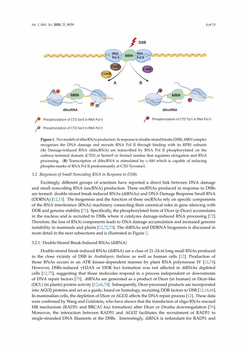
It is widely known that DDR induces signaling pathways that remodel chromatin in the proximityof DSBs [48,49] causing an inhibitory effect on transcription at nearby promoters in order to avoidcollisions or interference of the transcriptional and repair machinery [50–54]. The DSBs’ inhibitoryeffect of transcription by RNA polymerase II (RNA Pol II) depends on the ATM and/or DNA-PKcs,the chromatin remodeler BRG1, Poly (ADP-ribosyl) polymerase 1 (PARP1), E subunit of the negativeelongation factor (NELF-E) as well as on the distance to the DSBs (as far as 1Mb away from thebreak) [50,51,55–58]. Despite the well-established transcriptional inhibition described above, the firstinsights regarding the production of small RNAs in the vicinity of DSBs come from the studies performedon filamentous fungus Neurospora crassa [59]. A growing body of evidence shows that transcriptionoccurs at the DSBs (in the absence of promoters) and that the DSBs repair process involves not onlyDDR proteins, but also RNA that are produced in the proximity of DSBs. Interestingly, studies haveshown that de novo transcription occurs at the open DNA ends in vitro and in vivo in yeast andmammalian cancer cells [12,13,60–64]. Specifically, RNA Pol II accumulates at DSB and initiatestranscription from the broken ends to produce non-polyadenylated damage-induced long noncodingRNAs (dilncRNAs) (Figure 1) [63]. Importantly, the dependency of dilncRNA synthesis on RNAPol II was supported by the following observations: increased occupancy of RNAPII around thebreak sites, mapping analyses of increased transcripts to sequences around the breaks, as well asdetection of dilncRNA in native RNA pull-down after the induction of sequence-specific DSB [56,65].Additionally, DSB-dependent transcription, like promoter associated-transcription, is sensitive tothe inhibition of transcription factor IIH (TFIIH) as well as cyclin-dependent kinase 9 (CDK9) byTriptolide and 5,6-Dichloro-1-beta-Ribo-furanosyl Benzimidazole (DRB), respectively, leading to thephosphorylation of the RNA Pol II carboxy-terminal domain (CTD) residue Ser2/5 (Figure 1A) [63,65,66].Despite the well-established affinity of RNA Pol II to the free DNA [56,67], regulated recruitment ofRNA Pol II to the DSB has been recently described [65]. Specifically, c-Abl (nuclear tyrosine kinasewith multiple functions in the DDR [68]) stimulate the phospho-marks of RNA Pol II predominantlyat CTD Tyr1, at DSBs, which in turn is necessary for dilncRNA transcription (Figure 1B) [65].Furthermore, the transcription initiation of dilncRNA at the broken DNA is regulated by theMRE11-RAD50-NBS1 (MRN) complex which directly binds to the RNA Pol II (RPB1 subunit) [63].However, the details regarding the mechanism of transcription initiation at the broken end remainfully uncovered.
Additionally, this lncRNA produced by the transcription from broken ends has also been shown tostimulate the recruitment of factors involved in DSB repair through HR [61]. In the S/G2 cell-cycle phase,when CtIP is phosphorylated, dilncRNA binds to the resected DNA ends forming a DNA:RNA hybridrecognized by BRCA1. This event subsequently stimulates the recruitment of other HR factors,namely breast cancer type 2 (BRCA2), RAD51 [69] and RAD52 [70]. Moreover, it has been shownthat BRCA2 controls DNA:RNA hybrid levels at DSBs by interacting with and mediating RNase H2recruitment to DSBs [69]. Together, these data show that dilncRNAs play a role in the signalling andDSBs repair.
Int. J. Mol. Sci. 2020, 21, 8039 4 of 13
Figure 1. Two models of dilncRNAs production. In response to double-strand breaks (DSB), MRN complexrecognizes the DNA damage and recruits RNA Pol II through binding with its RPB1 subunit.(A) Damage-induced RNA (dilncRNA) are transcribed by RNA Pol II phosphorylated on thecarboxy-terminal domain (CTD) at Serine5 or Serine2 residue that regulates elongation and RNAprocessing. (B) Transcription of dilncRNA is stimulated by c-Abl which is capable of inducingphospho-marks of RNA Pol II predominantly at CTD Tyrosine1.
3.2. Biogenesis of Small Noncoding RNA in Response to DSBs
Excitingly, different groups of scientists have reported a direct link between DNA damageand small noncoding RNA (sncRNA) production. These sncRNAs produced in response to DSBsare termed: double-strand break-induced RNAs (diRNAs) and DNA Damage Response Small RNA(DDRNA) [12,13]. The biogenesis and the function of these sncRNAs rely on specific componentsof the RNA interference (RNAi) machinery connecting their canonical roles in gene silencing withDDR and genome stability [71]. Specifically, the phosphorylated form of Dicer (p-Dicer) accumulatesin the nucleus and is recruited to DSBs where it catalyzes damage-induced RNA processing [72].Therefore, the loss of RNAi components leads to DNA damage accumulation and increased genomeinstability in mammals and plants [12,72,73]. The diRNAs and DDRNA biogenesis is discussed inmore detail in the next subsections and is illustrated in Figure 2.
3.2.1. Double-Strand Break-Induced RNAs (diRNA)
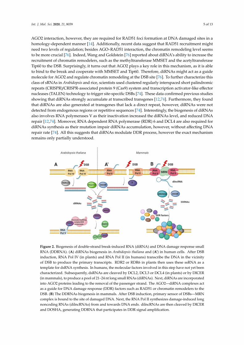
Double-strand break-induced RNAs (diRNA) are a class of 21–24 nt long small RNAs producedin the close vicinity of DSB in Arabidopsis thaliana as well as human cells [12]. Production ofthose RNAs occurs in an ATR kinase-dependent manner by plant RNA polymerase IV [12,74].However, DSBs-induced γH2AX or DDR foci formation was not affected in diRNAs depletedcells [12,75], suggesting that those molecules respond in a process independent or downstreamof DNA repair factors [75]. diRNAs are generated as a product of Dicer (in human) or Dicer-like(DCL) (in plants) protein activity [12,66,74]. Subsequently, Dicer-processed products are incorporatedinto AGO2 proteins and act as a guide, based on homology, recruiting DDR factors to DSB [12,14,66].In mammalian cells, the depletion of Dicer or AGO2 affects the DNA repair process [12]. These datawere confirmed by Wang and Goldstein, who have shown that the transfection of oligo-RNAs rescuedHR mechanism (RAD51 and BRCA1 foci formation) after Dicer or Drosha downregulation [76].Moreover, the interaction between RAD51 and AGO2 facilitates the recruitment of RAD51 tosingle-stranded DNA filaments at the DSBs. Interestingly, diRNA is redundant for RAD51 and
Int. J. Mol. Sci. 2020, 21, 8039 5 of 13
AGO2 interaction, however, they are required for RAD51 foci formation at DNA damaged sites in ahomology-dependent manner [14]. Additionally, recent data suggest that RAD51 recruitment mightneed two levels of regulation; besides AGO–RAD51 interaction, the chromatin remodeling level seemsto be more crucial [76]. Indeed, Wang and Goldstein [76] reported about diRNA’s ability to increase therecruitment of chromatin remodelers, such as the methyltransferase MMSET and the acetyltransferaseTip60 to the DSB. Surprisingly, it turns out that AGO2 plays a key role in this mechanism, as it is ableto bind to the break and cooperate with MMSET and Tip60. Therefore, diRNAs might act as a guidemolecule for AGO2 and regulate chromatin remodeling at the DSB site [76]. To further characterize thisclass of sRNAs in Arabidopsis and rice, scientists used clustered regularly interspaced short palindromicrepeats (CRISPR)/CRISPR-associated protein 9 (Cas9) system and transcription activator-like effectornucleases (TALEN) technology to trigger site-specific DSBs [74]. These data confirmed previous studiesshowing that diRNAs strongly accumulate at transcribed transgenes [12,74]. Furthermore, they foundthat diRNAs are also generated at transgenes that lack a direct repeat, however, diRNAs were notdetected from endogenous regions or repetitive sequences [74]. Interestingly, the biogenesis of diRNAsalso involves RNA polymerases V as their inactivation increased the diRNAs level, and reduced DNArepair [12,74]. Moreover, RNA dependent RNA polymerase (RDR) 6 and DCL4 are also required fordiRNAs synthesis as their mutation impair diRNAs accumulation, however, without affecting DNArepair rate [74]. All this suggests that diRNAs modulate DDR process, however the exact mechanismremains only partially understood.
Figure 2. Biogenesis of double-strand break-induced RNA (diRNA) and DNA damage response smallRNA (DDRNA). (A) diRNAs biogenesis in Arabidopsis thaliana and (A’) in human cells. After DSBinduction, RNA Pol IV (in plants) and RNA Pol II (in humans) transcribe the DNA in the vicinityof DSB to produce the primary transcripts. RDR2 or RDR6 in plants then uses these ssRNA as atemplate for dsRNA synthesis. In humans, the molecular factors involved in this step have not yet beencharacterized. Subsequently, dsRNAs are cleaved by DCL2, DCL3 or DCL4 (in plants) or by DICER(in mammals), to produce a pool of 21–24 nt long small RNAs (diRNAs). Next, diRNAs are incorporatedinto AGO2 proteins leading to the removal of the passenger strand. The AGO2—diRNA complexes actas a guide for DNA damage response (DDR) factors such as RAD51 or chromatin remodelers to theDSB. (B) The DDRNAs biogenesis in mammals. After DSB induction, primary sensor of DSBs—MRNcomplex is bound to the site of damaged DNA. Next, the RNA Pol II synthesizes damage-induced longnoncoding RNAs (dilncRNAs) from and towards DNA ends. dilncRNAs are then cleaved by DICERand DOSHA, generating DDRNA that participates in DDR signal amplification.
Int. J. Mol. Sci. 2020, 21, 8039 6 of 13
3.2.2. DNA Damage Response Small RNA (DDRNA)
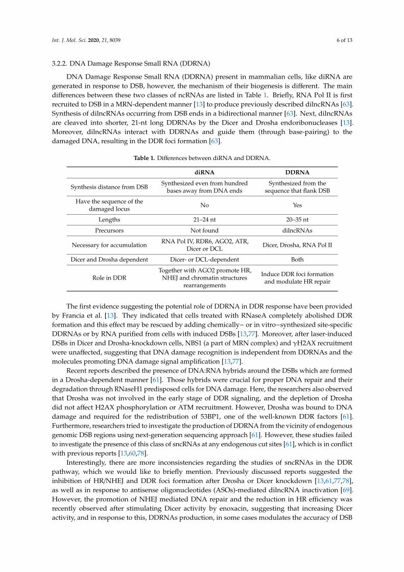
DNA Damage Response Small RNA (DDRNA) present in mammalian cells, like diRNA aregenerated in response to DSB, however, the mechanism of their biogenesis is different. The maindifferences between these two classes of ncRNAs are listed in Table 1. Briefly, RNA Pol II is firstrecruited to DSB in a MRN-dependent manner [13] to produce previously described dilncRNAs [63].Synthesis of dilncRNAs occurring from DSB ends in a bidirectional manner [63]. Next, dilncRNAsare cleaved into shorter, 21-nt long DDRNAs by the Dicer and Drosha endoribonucleases [13].Moreover, dilncRNAs interact with DDRNAs and guide them (through base-pairing) to thedamaged DNA, resulting in the DDR foci formation [63].
Table 1. Differences between diRNA and DDRNA.
diRNA DDRNA
Synthesis distance from DSB Synthesized even from hundredbases away from DNA ends
Synthesized from thesequence that flank DSB
Have the sequence of thedamaged locus No Yes
Lengths 21–24 nt 20–35 nt
Precursors Not found dilncRNAs
Necessary for accumulation RNA Pol IV, RDR6, AGO2, ATR,Dicer or DCL Dicer, Drosha, RNA Pol II
Dicer and Drosha dependent Dicer- or DCL-dependent Both
Role in DDRTogether with AGO2 promote HR,
NHEJ and chromatin structuresrearrangements
Induce DDR foci formationand modulate HR repair
The first evidence suggesting the potential role of DDRNA in DDR response have been providedby Francia et al. [13]. They indicated that cells treated with RNaseA completely abolished DDRformation and this effect may be rescued by adding chemically− or in vitro−synthesized site-specificDDRNAs or by RNA purified from cells with induced DSBs [13,77]. Moreover, after laser-inducedDSBs in Dicer and Drosha-knockdown cells, NBS1 (a part of MRN complex) and γH2AX recruitmentwere unaffected, suggesting that DNA damage recognition is independent from DDRNAs and themolecules promoting DNA damage signal amplification [13,77].
Recent reports described the presence of DNA:RNA hybrids around the DSBs which are formedin a Drosha-dependent manner [61]. Those hybrids were crucial for proper DNA repair and theirdegradation through RNaseH1 predisposed cells for DNA damage. Here, the researchers also observedthat Drosha was not involved in the early stage of DDR signaling, and the depletion of Droshadid not affect H2AX phosphorylation or ATM recruitment. However, Drosha was bound to DNAdamage and required for the redistribution of 53BP1, one of the well-known DDR factors [61].Furthermore, researchers tried to investigate the production of DDRNA from the vicinity of endogenousgenomic DSB regions using next-generation sequencing approach [61]. However, these studies failedto investigate the presence of this class of sncRNAs at any endogenous cut sites [61], which is in conflictwith previous reports [13,60,78].
Interestingly, there are more inconsistencies regarding the studies of sncRNAs in the DDRpathway, which we would like to briefly mention. Previously discussed reports suggested theinhibition of HR/NHEJ and DDR foci formation after Drosha or Dicer knockdown [13,61,77,78],as well as in response to antisense oligonucleotides (ASOs)-mediated dilncRNA inactivation [69].However, the promotion of NHEJ mediated DNA repair and the reduction in HR efficiency wasrecently observed after stimulating Dicer activity by enoxacin, suggesting that increasing Diceractivity, and in response to this, DDRNAs production, in some cases modulates the accuracy of DSB
Int. J. Mol. Sci. 2020, 21, 8039 7 of 13
repair [79]. Additionally, researchers also elucidated the role of DDRNAs in the stimulation of 53BP1foci formation [63,78,79], one of the key factors involved in choosing the DBS repair pathway bypromoting NHEJ and inhibiting HR [80]. The role of 53BP1 in the regulation of DNA repair and thegenerally prevailing view of competition between NHEJ and HR support the results presented byGioia et al. [79]. Several research groups abreast about the competition of those mechanisms [81,82],however, recent data supports entwined relationship between HR and NHEJ in repairing DSBs ratherthan exclusive competition [83]. Taken together, the discussed results demonstrate that DDRNApromotes DSB repair and DDR foci formation, however, it seems circumspect to investigate not onlythe molecular details of this regulation, but also whether DDRNA, in some part, can participate in theselection of the DNA repair pathway.
3.2.3. Telomeric DNA Damage Response Small RNAs (tDDRNAs)
Interestingly, the production of DDRNs and their longer precursors have also been shown attelomeres. These sncRNAs are transcribed from both strands of deprotected telomeres, hence are calledtelomeric DDRNAs (tDDRNAs) and telomeric dilncRNAs (tdilncRNAs) [78,84,85]. Similarly, as inDDRNAs biogenesis, silencing of Dicer and Drosha fully abolished tDDRNA production whileDrosha-knockdown lead to the accumulation of tdilncRNA [78], suggesting that both the proteins arecrucial for tDDRNAs biosynthesis.
Moreover, 53BP1 foci formation in response to DDRNA induction was described in telomeresas well. Despite unaltered total 53BP1 protein concentration, cells treated with IR were sensitive toRNaseA and displayed decreased DDR foci formation. Interestingly, the rescue of foci was possible onlyby DDRNAs with telomeric sequences. Moreover, cells depleted for Dicer or Drosha were ineffectivein 53BP1 foci formation, confirming the role of these two proteins in tDDRNAs generation and DDRresponse [78]. Additionally, in fibroblasts isolated from patients with Hutchinson–Gilford progeriasyndrome (HGPS), characterized by telomeres dysfunction and premature aging, levels of tdilncRNAsand tDDRNAs increased. Further study on a mouse model of HGPS indicated that the concentrationof markers of DDR activation such as 53BP1 and ATM were higher compared to wild-type cells.Additionally, using telomeric sequence-specific ASOs, which block both—tDDRNAs and tdilncRNAs,inhibited DDR in telomeres providing further evidence for the regulation of 53BP1 foci formation viaDDRNAs [84].
3.3. RNA Modification Involved in DNA Damage Response
Another piece of evidence suggesting the involvement of RNA in the DNA repair process hasbeen shown by Xiang et al. [86]. The authors detected the presence of N6-methyladenosine (m6A)at DNA damage sites in response to ultraviolet (UV) laser irradiation within a short period (2 min).RNA m6A is known to be regulated by the methyltransferase METTL3 (methyltransferase-like 3) [87]and the demethylase FTO (fat mass and obesity-associated protein) [88]. The absence of METTL3catalytic activity leads to delays in the repair of UV-induced cyclobutane pyrimidine dimers (CPD).METTL3 absence also affects the localization of DNA polymerase κ (Pol κ) at DNA damaged sites.Taken together, authors demonstrated the importance of m6A modification of RNA in the UV-responsiveDNA damage response [86].
A few years later, Svobodova Kovarikova et al. [89] confirmed the observations made by Xiang et al.,showing the accumulation of 6mA RNA modification at the DNA lesions explaining that this effectcould be a consequence of the coregulatory function of METTL-like enzymes or diffusion of m6A RNAto UVA-damaged chromatin [86]. They further observed that m6A RNAs being present in the vicinity ofDSBs likely concerns noncoding RNAs, rather than mRNA or rRNA [89]. Additionally, they indicatedthat inhibition of Suv4-20h1/h2 methyltransferases, responsible for H4K20me2/me3 which is recognizedby the 53BP1 protein (NHEJ component) did not affect m6A RNAs at the DNA lesions. These datasupport the results obtained by Xiang et al. showing that m6A RNA is likely playing a role in the NERmechanism [86,89].
Int. J. Mol. Sci. 2020, 21, 8039 8 of 13
Moreover, recent studies performed by Zhang et al. [90] have shown that METTL3 isphosphorylated by ATM in response to DSBs and subsequently recruited to the DNA lesions [90].This led to the m6A modification on the DNA damage-associated RNA, recognized and protected by theRNA m6A reader protein: YTH domain-containing protein 1 (YTHDC1). Consequently, the modifiedRNA forms DNA:RNA hybrids at DSBs which stimulate proteins involved in DDR, such as BRCA1and RAD51.
4. Conclusions and Outlook
For a long time, RNA was considered only as a DNA working photocopy, produced in the processof transcription to generate functional proteins. However, the discovery of noncoding RNAs initiateda series of studies to explore their potential role in various biological processes. In this review, we havediscussed the potential function of small noncoding RNAs associated with DSBs in the maintenance ofgenome stability. We have mainly focused on the mechanisms of diRNAs’ and DDRNAs’ biosynthesisand their role in the DDR pathway. We have also mentioned the process of de novo RNA synthesis atthe DSBs. Based on these studies, it is becoming clear that RNA may be employed by the DDR process,as many of DDR factors are required to process RNA in response to DSBs. For instance, DNA-PKcs,RAD52, BRCA1, 53BP1 interact with RNA as well as DNA:RNA hybrids, which are key structuresrequired for DDR progression [70,91,92]. An attractive hypothesis that supports the function of RNAin this phenomenon is that the dilncRNA forms DNA:RNA hybrids that serve a platform to stimulaterepair agents [69,70]. Besides, many RNAi components are ostensibly involved in DDR. Although it isagreed that Dicer, Drosha and AGO2 have a role in the DNA repair process, their mechanism remainsintangible [12–14,61,76,77]. Moreover, RNA modification has been identified as an important biologicalprocess involved in DNA damage response [86,89,90].
While we have a basic understanding of how sncRNAs participate in the maintenance ofgenome stability, numerous outstanding questions remain open. For instance, dilncRNAs’ associationto the transcriptionally active chromatin, the timing of dilncRNAs’ biogenesis, or dilncRNAs’regulation of DDR response remains elusive. It would also be interesting to investigate the moleculardetails of the regulation of the DDR process by sncRNAs as well as to discern whether DDRNA,in some part, can participate in the selection of the DNA repair pathway. Additionally, 6mA RNAmodification’s involvement in the DDR process that has been recently identified require furtherstudies. For instance, it would be interesting to investigate the possible role of other modifications,such as N1-methyladenosine (m1A) and N5-methylcytosine (m5C), in the DNA repair process.Further investigations of sncRNA, RNA binding proteins (RBPs) as well as RNA modifications afterDSBs will provide a deeper understanding of mechanisms that govern the maintenance of genomestability. This, in addition, can further help the research community to develop novel RNA therapeuticstrategies in the treatment of diseases linked to DNA damage or genetic disorders.
Author Contributions: Conceptualization: I.R.; writing—original draft preparation: I.R., G.B.; writing—reviewand editing: I.R.; figures preparation: G.B.; supervision: I.R. Both authors have read and agreed to the publishedversion of the manuscript.
Funding: This research received no external funding.
Acknowledgments: We would like to thank reviewers for their valuable comments.
Conflicts of Interest: The authors declare no conflict of interest.
References
1. Hoeijmakers, J.H.J. DNA damage, aging, and cancer. N. Engl. J. Med. 2009, 361, 1475–1485. [CrossRef] [PubMed]2. Mehta, A.; Haber, J.E. Sources of DNA double-strand breaks and models of recombinational DNA repair.
Cold Spring Harb. Perspect. Biol. 2014, 6. [CrossRef] [PubMed]3. Ciccia, A.; Elledge, S.J. The ScienceDirect—Molecular Cell: The DNA Damage Response: Making It Safe to
Play with Knives. Mol. Cell 2010, 40, 179–204. [CrossRef] [PubMed]
Int. J. Mol. Sci. 2020, 21, 8039 9 of 13
4. Jackson, S.P.; Bartek, J. The DNA-damage response in human biology and disease. Nature 2010, 461, 1071–1078.[CrossRef] [PubMed]
5. Valko, M.; Rhodes, C.J.; Moncol, J.; Izakovic, M.; Mazur, M. Free radicals, metals and antioxidants in oxidativestress-induced cancer. Chem. Biol. Interact. 2006, 160, 1–40. [CrossRef] [PubMed]
6. Helmrich, A.; Ballarino, M.; Nudler, E.; Tora, L. Transcription-replication encounters, consequences andgenomic instability. Nat. Struct. Mol. Biol. 2013, 20, 412–418. [CrossRef] [PubMed]
7. Jinks-Robertson, S.; Bhagwat, A.S. Transcription-Associated Mutagenesis. Annu. Rev. Genet. 2014, 48, 341–359.[CrossRef] [PubMed]
8. Harper, J.W.; Elledge, S.J. The DNA Damage Response: Ten Years After. Mol. Cell 2007, 28, 739–745.[CrossRef] [PubMed]
9. Brandsma, I.; Gent, D.C. Pathway choice in DNA double strand break repair: Observations of a balancing act.Genome Integr. 2012, 3. [CrossRef] [PubMed]
10. Rastogi, R.P.; Kumar, A.; Tyagi, M.B.; Sinha, R.P. Molecular Mechanisms of Ultraviolet Radiation-InducedDNA Damage and Repair. J. Nucl. Acids 2010, 2010, 592980. [CrossRef] [PubMed]
11. Sallmyr, A.; Tomkinson, A.E. Repair of DNA double-strand breaks by mammalian alternative end-joiningpathways. J. Biol. Chem. 2018, 293, 10536–10549. [CrossRef]
12. Wei, W.; Ba, Z.; Gao, M.; Wu, Y.; Ma, Y.; Amiard, S.; White, C.I.; Danielsen, J.M.R.; Yang, Y.G.; Qi, Y. A role forsmall RNAs in DNA double-strand break repair. Cell 2012, 149, 101–112. [CrossRef] [PubMed]
13. Francia, S.; Michelini, F.; Saxena, A.; Tang, D.; De Hoon, M.; Anelli, V.; Mione, M.; Carninci, P.;D’adda Di Fagagna, F. Site-specific DICER and DROSHA RNA products control the DNA-damage response.Nature 2012, 488, 231–235. [CrossRef] [PubMed]
14. Gao, M.; Wei, W.; Li, M.M.; Wu, Y.S.; Ba, Z.; Jin, K.X.; Li, M.M.; Liao, Y.Q.; Adhikari, S.; Chong, Z.; et al.Ago2 facilitates Rad51 recruitment and DNA double-strand break repair by homologous recombination.Cell Res. 2014, 24, 532–541. [CrossRef] [PubMed]
15. Ceccaldi, R.; Rondinelli, B.; D’Andrea, A.D. Repair Pathway Choices and Consequences at the Double-StrandBreak. Trends Cell Biol. 2016, 26, 52–64. [CrossRef] [PubMed]
16. Van Sluis, M.; McStay, B. A localized nucleolar DNA damage response facilitates recruitment of thehomology-directed repair machinery independent of cell cycle stage. Genes Dev. 2015, 29, 1151–1163.[CrossRef] [PubMed]
17. Lee, J.-H. ATM Activation by DNA Double-Strand Breaks Through the Mre11-Rad50-Nbs1 Complex. Science2005, 308, 551–554. [CrossRef]
18. Mimori, T.; Hardin, J.A. Mechanism of interaction between Ku protein and DNA. J. Biol. Chem. 1986, 261,10375–10379.
19. Benjamin, R.C.; Gill, D.M. Poly(ADP-ribose) synthesis in vitro programmed by damaged DNA. A comparisonof DNA molecules containing different types of strand breaks. J. Biol. Chem. 1980, 255, 10502–10508.
20. Ohgushi, H.; Yoshihara, K.; Kamiya, T. Bovine thymus poly(adenosine diphosphate ribose) polymerase.Physical properties and binding to DNA. J. Biol. Chem. 1980, 255, 6205–6211.
21. Wold, M.S. Replication protein A: A heterotrimeric, single-stranded DNA-binding protein required foreukaryotic DNA metabolism. Annu. Rev. Biochem. 1997, 66, 61–92. [CrossRef] [PubMed]
22. De Murcia, G.; de Murcia, J.M. Poly(ADP-ribose) polymerase: A molecular nick-sensor. Trends Biochem. Sci.1994, 19, 172–173. [CrossRef]
23. Burma, S.; Chen, B.P.; Murphy, M.; Kurimasa, A.; Chen, D.J. ATM Phosphorylates Histone H2AX in Responseto DNA Double-strand Breaks. J. Biol. Chem. 2001, 276, 42462–42467. [CrossRef]
24. Rogakou, E.P.; Boon, C.; Redon, C.; Bonner, W.M. Megabase chromatin domains involved in DNAdouble-strand breaks in vivo. J. Cell Biol. 1999, 146, 905–916. [CrossRef]
25. Iacovoni, J.S.; Caron, P.; Lassadi, I.; Nicolas, E.; Massip, L.; Trouche, D.; Legube, G. High-resolution profilingof γh2AX around DNA double strand breaks in the mammalian genome. EMBO J. 2010, 29, 1446–1457.[CrossRef] [PubMed]
26. Mattiroli, F.; Vissers, J.H.A.; Van Dijk, W.J.; Ikpa, P.; Citterio, E.; Vermeulen, W.; Marteijn, J.A.; Sixma, T.K.RNF168 ubiquitinates K13-15 on H2A/H2AX to drive DNA damage signaling. Cell 2012, 150, 1182–1195.[CrossRef] [PubMed]
Int. J. Mol. Sci. 2020, 21, 8039 10 of 13
27. Thorslund, T.; Ripplinger, A.; Hoffmann, S.; Wild, T.; Uckelmann, M.; Villumsen, B.; Narita, T.; Sixma, T.K.;Choudhary, C.; Bekker-Jensen, S.; et al. Histone H1 couples initiation and amplification of ubiquitin signallingafter DNA damage. Nature 2015, 527, 389–393. [CrossRef]
28. Schwertman, P.; Bekker-Jensen, S.; Mailand, N. Regulation of DNA double-strand break repair by ubiquitinand ubiquitin-like modifiers. Nat. Rev. Mol. Cell Biol. 2016, 17, 379–394. [CrossRef]
29. Escribano-Díaz, C.; Orthwein, A.; Fradet-Turcotte, A.; Xing, M.; Young, J.T.F.; Tkác, J.; Cook, M.A.;Rosebrock, A.P.; Munro, M.; Canny, M.D.; et al. A Cell Cycle-Dependent Regulatory Circuit Composed of53BP1-RIF1 and BRCA1-CtIP Controls DNA Repair Pathway Choice. Mol. Cell 2013, 49, 872–883. [CrossRef]
30. Mari, P.O.; Florea, B.I.; Persengiev, S.P.; Verkaik, N.S.; Brüggenwirth, H.T.; Modesti, M.; Giglia-Mari, G.;Bezstarosti, K.; Demmers, J.A.A.; Luider, T.M.; et al. Dynamic assembly of end-joining complexes requiresinteraction between Ku70/80 and XRCC4. Proc. Natl. Acad. Sci. USA 2006, 103, 18597–18602. [CrossRef]
31. Hsu, H.L.; Yannone, S.M.; Chen, D.J. Defining interactions between DNA-PK and ligase IV/XRCC4.DNA Repair 2002, 1, 225–235. [CrossRef]
32. Uematsu, N.; Weterings, E.; Yano, K.I.; Morotomi-Yano, K.; Jakob, B.; Taucher-Scholz, G.; Mari, P.O.;Van Gent, D.C.; Chen, B.P.C.; Chen, D.J. Autophosphorylation of DNA-PKCS regulates its dynamics at DNAdouble-strand breaks. J. Cell Biol. 2007, 177, 219–229. [CrossRef] [PubMed]
33. Costantini, S.; Woodbine, L.; Andreoli, L.; Jeggo, P.A.; Vindigni, A. Interaction of the Ku heterodimer withthe DNA ligase IV/Xrcc4 complex and its regulation by DNA-PK. DNA Repair 2007, 6, 712–722. [CrossRef]
34. Nick McElhinny, S.A.; Snowden, C.M.; McCarville, J.; Ramsden, D.A. Ku Recruits the XRCC4-Ligase IVComplex to DNA Ends. Mol. Cell. Biol. 2000, 20, 2996–3003. [CrossRef] [PubMed]
35. Yano, K.I.; Morotomi-Yano, K.; Wang, S.Y.; Uematsu, N.; Lee, K.J.; Asaithamby, A.; Weterings, E.; Chen, D.J.Ku recruits XLF to DNA double-strand breaks. EMBO Rep. 2008, 9, 91–96. [CrossRef] [PubMed]
36. Drouet, J.; Frit, P.; Delteil, C.; De Villartay, J.P.; Salles, B.; Calsou, P. Interplay between Ku, artemis, and theDNA-dependent protein kinase catalytic subunit at DNA ends. J. Biol. Chem. 2006, 281, 27784–27793.[CrossRef] [PubMed]
37. Kanno, S.I.; Kuzuoka, H.; Sasao, S.; Hong, Z.; Lan, L.; Nakajima, S.; Yasui, A. A novel human AP endonucleasewith conserved zinc-finger-like motifs involved in DNA strand break responses. EMBO J. 2007, 26, 2094–2103.[CrossRef]
38. Macrae, C.J.; McCulloch, R.D.; Ylanko, J.; Durocher, D.; Koch, C.A. APLF (C2orf13) facilitates nonhomologousend-joining and undergoes ATM-dependent hyperphosphorylation following ionizing radiation. DNA Repair2008, 7, 292–302. [CrossRef]
39. Grundy, G.J.; Rulten, S.L.; Zeng, Z.; Arribas-Bosacoma, R.; Iles, N.; Manley, K.; Oliver, A.; Caldecott, K.W.APLF promotes the assembly and activity of non-homologous end joining protein complexes. EMBO J.2013, 32, 112–125. [CrossRef]
40. Bartel, D.P. MicroRNAs: Genomics, Biogenesis, Mechanism, and Function. Cell 2004, 116, 281–297. [CrossRef]41. Sandoval, P.; Swart, E.; Arambasic, M.; Nowacki, M. Functional Diversification of Dicer-like Proteins and
Small RNAs Required for Genome Sculpting. Dev. Cell 2014, 28, 174–188. [CrossRef] [PubMed]42. Ha, M.; Kim, V.N. Regulation of microRNA biogenesis. Nat. Rev. Mol. Cell Biol. 2014, 15, 509–524. [CrossRef]43. Czech, B.; Hannon, G.J. One Loop to Rule Them All: The Ping-Pong Cycle and piRNA-Guided Silencing.
Trends Biochem. Sci. 2016. [CrossRef] [PubMed]44. Lepère, G.; Nowacki, M.; Serrano, V.; Gout, J.F.; Guglielmi, G.; Duharcourt, S.; Meyer, E.
Silencing-associated and meiosis-specific small RNA pathways in Paramecium tetraurelia. Nucleic Acids Res.2009, 37, 903–915. [CrossRef]
45. Zahler, A.M.; Neeb, Z.T.; Lin, A.; Katzman, S. Mating of the stichotrichous Ciliate Oxytricha trifallaxinduces production of a class of 27 nt small RNAs derived from the parental macronucleus. PLoS ONE2012, 7, e42371. [CrossRef]
46. Fang, W.; Wang, X.; Bracht, J.R.; Nowacki, M.; Landweber, L.F. Piwi-interacting RNAs protect DNA againstloss during oxytricha genome rearrangement. Cell 2012, 151, 1243–1255. [CrossRef]
47. Rzeszutek, I.; Maurer-Alcalá, X.X.; Nowacki, M. Programmed genome rearrangements in ciliates. Cell. Mol.Life Sci. 2020. [CrossRef]
48. Morrison, A.J.; Highland, J.; Krogan, N.J.; Arbel-Eden, A.; Greenblatt, J.F.; Haber, J.E.; Shen, X. INO80 andγ-H2AX interaction links ATP-dependent chromatin remodeling to DNA damage repair. Cell 2004, 119,767–775. [CrossRef]
Int. J. Mol. Sci. 2020, 21, 8039 11 of 13
49. Berkovich, E.; Monnat, R.J.; Kastan, M.B. Roles of ATM and NBS1 in chromatin structure modulation andDNA double-strand break repair. Nat. Cell Biol. 2007, 9, 683–690. [CrossRef] [PubMed]
50. Shanbhag, N.M.; Rafalska-Metcalf, I.U.; Balane-Bolivar, C.; Janicki, S.M.; Greenberg, R.A. ATM-Dependentchromatin changes silence transcription in cis to dna double-strand breaks. Cell 2010, 141, 970–981.[CrossRef] [PubMed]
51. Pankotai, T.; Bonhomme, C.; Chen, D.; Soutoglou, E. DNAPKcs-dependent arrest of RNA polymerase IItranscription in the presence of DNA breaks. Nat. Struct. Mol. Biol. 2012, 19, 276–282. [CrossRef] [PubMed]
52. Vítor, A.C.; Sridhara, S.C.; Sabino, J.C.; Afonso, A.I.; Grosso, A.R.; Martin, R.M.; De Almeida, S.F.Single-molecule imaging of transcription at damaged chromatin. Sci. Adv. 2019, 5, 1–12. [CrossRef] [PubMed]
53. Morales, J.C.; Richard, P.; Patidar, P.L.; Motea, E.A.; Dang, T.T.; Manley, J.L.; Boothman, D.A. XRN2 LinksTranscription Termination to DNA Damage and Replication Stress. PLoS Genet. 2016, 12, 1–22.[CrossRef] [PubMed]
54. Kruhlak, M.; Crouch, E.E.; Orlov, M.; Montão, C.; Gorski, S.A.; Nussenzweig, A.; Misteli, T.; Phair, R.D.;Casellas, R. The ATM repair pathway inhibits RNA polymerase I transcription in response to chromosomebreaks. Nature 2007, 447, 730–734. [CrossRef]
55. Kakarougkas, A.; Ismail, A.; Chambers, A.L.; Riballo, E.; Herbert, A.D.; Künzel, J.; Löbrich, M.; Jeggo, P.A.;Downs, J.A. Requirement for PBAF in Transcriptional Repression and Repair at DNA Breaks in ActivelyTranscribed Regions of Chromatin. Mol. Cell 2014, 55, 723–732. [CrossRef]
56. Iannelli, F.; Galbiati, A.; Capozzo, I.; Nguyen, Q.; Magnuson, B.; Michelini, F.; D’Alessandro, G.;Cabrini, M.; Roncador, M.; Francia, S.; et al. A damaged genome’s transcriptional landscape throughmultilayered expression profiling around in situ-mapped DNA double-strand breaks. Nat. Commun.2017, 8, 15656. [CrossRef]
57. Awwad, S.W.; Abu-Zhayia, E.R.; Guttmann-Raviv, N.; Ayoub, N. NELF-E is recruited to DNA double-strandbreak sites to promote transcriptional repression and repair. EMBO Rep. 2017, 18, 745–764. [CrossRef]
58. Nechaev, S.; Adelman, K. Transcription Initiation Into Productive Elongation. Biochim. Biophys. Acta 2012, 1809,34–45. [CrossRef]
59. Lee, H.C.; Chang, S.S.; Choudhary, S.; Aalto, A.P.; Maiti, M.; Bamford, D.H.; Liu, Y. qiRNA is a new type ofsmall interfering RNA induced by DNA damage. Nature 2009, 459, 274–277. [CrossRef]
60. Michalik, K.M.; Böttcher, R.; Förstemann, K. A small RNA response at DNA ends in Drosophila.Nucleic Acids Res. 2012, 40, 9596–9603. [CrossRef]
61. Lu, W.T.; Hawley, B.R.; Skalka, G.L.; Baldock, R.A.; Smith, E.M.; Bader, A.S.; Malewicz, M.; Watts, F.Z.;Wilczynska, A.; Bushell, M. Drosha drives the formation of DNA:RNA hybrids around DNA break sites tofacilitate DNA repair. Nat. Commun. 2018, 9, 532. [CrossRef] [PubMed]
62. Ohle, C.; Tesorero, R.; Schermann, G.; Dobrev, N.; Sinning, I.; Fischer, T. Transient RNA-DNA Hybrids AreRequired for Efficient Double-Strand Break Repair. Cell 2016, 167, 1001–1013.e7. [CrossRef]
63. Michelini, F.; Pitchiaya, S.; Vitelli, V.; Sharma, S.; Gioia, U.; Pessina, F.; Cabrini, M.; Wang, Y.; Capozzo, I.;Iannelli, F.; et al. Damage-induced lncRNAs control the DNA damage response through interaction withDDRNAs at individual double-strand breaks. Nat. Cell Biol. 2017, 19, 1400–1411. [CrossRef] [PubMed]
64. Pessina, F.; Giavazzi, F.; Yin, Y.; Gioia, U.; Vitelli, V.; Galbiati, A.; Barozzi, S.; Garre, M.; Oldani, A.;Flaus, A.; et al. Functional transcription promoters at DNA double-strand breaks mediate RNA-driven phaseseparation of damage-response factors. Nat. Cell Biol. 2019, 21, 1286–1299. [CrossRef]
65. Burger, K.; Schlackow, M.; Gullerova, M. Tyrosine kinase c-Abl couples RNA polymerase II transcription toDNA double-strand breaks. Nucleic Acids Res. 2019, 47, 3467–3484. [CrossRef] [PubMed]
66. Bonath, F.; Domingo-Prim, J.; Tarbier, M.; Friedländer, M.R.; Visa, N. Next-generation sequencing reveals twopopulations of damage-induced small RNAs at endogenous DNA double-strand breaks. Nucleic Acids Res.2018, 46, 11869–11882. [CrossRef] [PubMed]
67. Berretta, J.; Morillon, A. Pervasive transcription constitutes a new level of eukaryotic genome regulation.EMBO Rep. 2009, 10, 973–982. [CrossRef] [PubMed]
68. Meltser, V.; Ben-Yehoyada, M.; Shaul, Y. C-Abl tyrosine kinase in the DNA damage response: Cell death andmore. Cell Death Differ. 2011, 18, 2–4. [CrossRef]
69. D’Alessandro, G.; Whelan, D.R.; Howard, S.M.; Vitelli, V.; Renaudin, X.; Adamowicz, M.; Iannelli, F.;Jones-Weinert, C.W.; Lee, M.Y.; Matti, V.; et al. BRCA2 controls DNA:RNA hybrid level at DSBs by mediatingRNase H2 recruitment. Nat. Commun. 2018, 9. [CrossRef]
Int. J. Mol. Sci. 2020, 21, 8039 12 of 13
70. Yasuhara, T.; Kato, R.; Hagiwara, Y.; Shiotani, B.; Yamauchi, M.; Nakada, S.; Shibata, A.; Miyagawa, K.Human Rad52 Promotes XPG-Mediated R-loop Processing to Initiate Transcription-Associated HomologousRecombination Repair. Cell 2018, 175, 558–570. [CrossRef]
71. Burger, K.; Gullerova, M. Swiss army knives: Non-canonical functions of nuclear Drosha and Dicer. Nat. Rev.Mol. Cell Biol. 2015, 16, 417–430. [CrossRef]
72. Burger, K.; Schlackow, M.; Potts, M.; Hester, S.; Mohammed, S.; Gullerova, M. Nuclear phosphorylatedDicer processes doublestranded RNA in response to DNA damage. J. Cell Biol. 2017, 216, 2373–2389.[CrossRef] [PubMed]
73. Modzelewski, A.J.; Hilz, S.; Crate, E.A.; Schweidenback, C.T.H.; Fogarty, E.A.; Grenier, J.K.; Freire, R.;Cohen, P.E.; Grimson, A. Dgcr8 and Dicer are essential for sex chromosome integrity during meiosis in males.J. Cell Sci. 2015, 128, 2314–2327. [CrossRef] [PubMed]
74. Miki, D.; Zhu, P.; Zhang, W.; Mao, Y.; Feng, Z.; Huang, H.; Zhang, H.; Li, Y.; Liu, R.; Zhang, H.; et al.Efficient generation of diRNAs requires components in the posttranscriptional gene silencing pathway.Sci. Rep. 2017, 7, 1–11. [CrossRef] [PubMed]
75. Qi, Y.; Zhang, Y.; Baller, J.A.; Voytas, D.F. Histone H2AX and the small RNA pathway modulate bothnon-homologous end-joining and homologous recombination in plants. Mutat. Res. Fundam. Mol.Mech. Mutagen. 2016, 783, 9–14. [CrossRef]
76. Wang, Q.; Goldstein, M. Small RNAs recruit chromatin-modifying enzymes MMSET and Tip60 to reconfiguredamaged DNA upon double-strand break and facilitate repair. Cancer Res. 2016, 76, 1904–1915. [CrossRef]
77. Francia, S.; Cabrini, M.; Matti, V.; Oldani, A. Research Article Dicer, Drosha and DNA damage responseRNAs are necessary for the secondary recruitment of DNA damage response factors. J. Cell Sci. 2016, 129,1468–1476. [CrossRef] [PubMed]
78. Rossiello, F.; Aguado, J.; Sepe, S.; Iannelli, F.; Nguyen, Q.; Pitchiaya, S.; Carninci, P.; Di Fagagna, F.D.A.DNA damage response inhibition at dysfunctional telomeres by modulation of telomeric DNA damageresponse RNAs. Nat. Commun. 2017, 8. [CrossRef]
79. Gioia, U.; Francia, S.; Cabrini, M.; Brambillasca, S.; Michelini, F.; Jones-weinert, C.W. Pharmacological boostof DNA damage response and repair by enhanced biogenesis of DNA damage response RNAs. Sci. Rep.2019, 1–15. [CrossRef]
80. Guo, X.; Bai, Y.; Zhao, M.; Zhou, M.; Shen, Q.; Yun, C.; Zhang, H.; Zhu, W.; Wang, J. Acetylation of 53BP1dictates the DNA double strand break repair pathway. Nucleic Acids Res. 2018, 46, 689–703. [CrossRef]
81. Allen, C.; Halbrook, J.; Nickoloff, J.A. Interactive Competition Between Homologous Recombination andNon-Homologous End Joining. Mol. Cancer Res. 2003, 1, 913–920. [PubMed]
82. Shrivastav, M.; De Haro, L.P.; Nickoloff, J.A. Regulation of DNA double-strand break repair pathway choice.Cell Res. 2008, 18, 134–147. [CrossRef] [PubMed]
83. Ingram, S.P.; Warmenho, J.W.; Henthorn, N.T.; Smith, E.A.K.; Chadwick, A.L.; Burnet, N.G.; Mackay, R.I.;Kirkby, N.F.; Kirkby, K.J. Mechanistic modelling supports entwined rather than exclusively competitiveDNA double-strand break repair pathway. Sci. Rep. 2019. [CrossRef]
84. Aguado, J.; Sola-Carvajal, A.; Cancila, V.; Revêchon, G.; Ong, P.F.; Jones-Weinert, C.W.; Wallén Arzt, E.;Lattanzi, G.; Dreesen, O.; Tripodo, C.; et al. Inhibition of DNA damage response at telomeres improvesthe detrimental phenotypes of Hutchinson–Gilford Progeria Syndrome. Nat. Commun. 2019, 10, 1–11.[CrossRef] [PubMed]
85. Nguyen, Q.; Aguado, J.; Iannelli, F.; Suzuki, A.M.; Rossiello, F.; D’Adda Di Fagagna, F.; Carninci, P.Target-enrichment sequencing for detailed characterization of small RNAs. Nat. Protoc. 2018, 13,768–786. [CrossRef]
86. Xiang, Y.; Laurent, B.; Hsu, C.H.; Nachtergaele, S.; Lu, Z.; Sheng, W.; Xu, C.; Chen, H.; Ouyang, J.; Wang, S.;et al. RNA m6 A methylation regulates the ultraviolet-induced DNA damage response. Nature 2017, 543,573–576. [CrossRef]
87. Liu, J.; Yue, Y.; Han, D.; Wang, X.; Fu, Y.; Zhang, L.; Jia, G.; Yu, M.; Lu, Z.; Deng, X.; et al. A METTL3-METTL14complex mediates mammalian nuclear RNA N6-adenosine methylation. Nat. Chem. Biol. 2014, 10, 93–95.[CrossRef] [PubMed]
88. Jia, G.; Fu, Y.; Zhao, X.; Dai, Q.; Zheng, G.; Yang, Y.; Yi, C.; Lindahl, T.; Pan, T.; Yang, Y.G.; et al.N6-Methyladenosine in nuclear RNA is a major substrate of the obesity-associated FTO. Nat. Chem. Biol.2011, 7, 885–887. [CrossRef]
Int. J. Mol. Sci. 2020, 21, 8039 13 of 13
89. Kovarikova, A.S.; Stixova, L.; Kovarik, A.; Komurkova, D.; Legartova, S.; Fagherazzi, P.; Bártová, E.N6-Adenosine Methylation in RNA and a Reduced m3G/TMG Level in Non-Coding RNAs Appear atMicroirradiation-Induced DNA Lesions. Cells 2020, 9, 12–14.
90. Zhang, C.; Chen, L.; Peng, D.; Ren, J.; Wang, W.; Zhang, C.; Chen, L.; Peng, D.; Jiang, A.; He, Y.; et al.Article METTL3 and N6-Methyladenosine Promote Homologous Recombination-Mediated Repair of DSBsby Modulating DNA-RNA Hybrid Accumulation ll Article METTL3 and N6-Methyladenosine PromoteHomologous Recombination-Mediated Repair of DSBs by Modulating DNA-RN. Mol. Cell 2020, 79,425–442. [CrossRef]
91. Castello, A.; Horos, R.; Strein, C.; Fisher, B.; Eichelbaum, K.; Steinmetz, L.M.; Krijgsveld, J.; Hentze, M.W.Comprehensive Identification of RNA-Binding Proteins by RNA Interactome Capture. Methods Mol. Biol.2016, 1358, 131–139. [CrossRef]
92. Pryde, F.; Khalili, S.; Robertson, K.; Selfridge, J.; Ritchie, A.M.; Melton, D.W.; Jullien, D.; Adachi, Y.53BP1 exchanges slowly at the sites of DNA damage and appears to require RNA for its association withchromatin. J. Cell Sci. 2005, 118, 2043–2055. [CrossRef]
Publisher’s Note: MDPI stays neutral with regard to jurisdictional claims in published maps and institutionalaffiliations.
© 2020 by the authors. Licensee MDPI, Basel, Switzerland. This article is an open accessarticle distributed under the terms and conditions of the Creative Commons Attribution(CC BY) license (http://creativecommons.org/licenses/by/4.0/).