Embed Size (px)
Citation preview
THE ROLE OF SIGMA-1 RECEPTOR IN MODULATING
ENDOPLASMIC RETICULUM STRESS: PUTATIVE RELEVANCE
TO ALZHEIMER DISEASE
Louis-Alexandre Tassé
A thesis submitted in partial fulfillment of the requirements for the
Master’s degree in Neuroscience
Department of Cellular and Molecular Medicine
Neuroscience Program
Faculty of Medicine, University of Ottawa
© Louis-Alexandre Tasse, Ottawa, Canada, 2018
ii
ABSTRACT
Alzheimer’s Disease and other neurodegenerative diseases have been linked to dysfunction
in proteostasis in the endoplasmic reticulum (ER). The ER provides an exclusive
environment for protein synthesis and folding, which is vital to the cellular function. Under
normal conditions, the synthesis and degradation of proteins remain in balance. During
aging or during pathological states, disturbances of ER occur and consequently the failure
of protein homeostasis. The cells rely on a system, the unfolded protein response (UPR),
which regulates the homeostasis by three ER sensors: PERK, ATF6, and IRE-1.
Perturbations of ER function result in UPR. In physiological condition, the cell may
overcome the insult and regain homeostasis. However, prolonged or chronic UPR activates
apoptotic pathways and may cause cell death. The sigma-1 receptor (Sig-1R) is a 25 kD
polypeptide and a chaperone protein concentrated at the mitochondria-associated ER
membrane domain (MAM). The Sig-1R plays significant roles governing calcium
signalling, mitochondrial function, oxidative stress, protein chaperoning and ER stress.
Results of this investigation demonstrate that immortalized mouse embryonic fibroblasts
(MEFs) derived from Sig-1R-/—(KO) mice have higher baseline activation in all three
branches of the UPR in the absence of ER stress compared to MEFs derived from Wild-
type mice. Despite this increase in baseline activation, the PERK and ATF6 pathways have
a significantly blunted response to acute stress. Rescue experiments by expressing the Sig-
1R in KO MEFs did not recover the WT MEFs phenotype. Primary Sig-1R KO MEFs did
not show baseline ER stress, but did show inhibited recovery following treatment with the
acute ER stressor DTT. Overall, our data suggests that Sig-1R is important for the
reestablishment of proteostasis following acute stress.
iii
TABLE OF CONTENTS
ABSTRACT....................................................................................................................................ii
TABLE OF CONTENTS .......................................................................................................... iii
LIST OF FIGURES .................................................................................................................... vi
LIST OF ABREVIATIONS ..................................................................................................... vii
ACKNOWLEDGEMENTS ........................................................................................................ ix
1. INTRODUCTION .................................................................................................................... 1
1.1 Alzheimer's Disease .......................................................................................................... 1
1.1.1 Background ............................................................................................................................. 1
1.1.2 Hallmarks ................................................................................................................................ 3
1.1.3 Risk factors ............................................................................................................................. 4
1.1.4 Amyloid cascade hypothesis ............................................................................................. 5
1.1.5 Amyloid-beta peptide .......................................................................................................... 6
1.1.6 Clinical trials .......................................................................................................................... 6
1.2.1 Sigma-1 Receptor background ......................................................................................... 7
1.2.2 Sigma-1 receptor and Alzheimer's disease.................................................................. 10
1.2.3 Sigma-1 receptor knockout .............................................................................................. 11
1.3 Endoplasmic reticulum stress ..................................................................................... 12
1.3.1 Endoplasmic reticulum control of protein ................................................................... 12
1.3.2 The Unfolded Protein Response ..................................................................................... 13
1.3.3 Alzheimer’s Disease is an Endoplasmic Reticulum Stress disease ..................... 16
iv
1.4 Focus of this study ......................................................................................................... 17
2. HYPOTHESIS ....................................................................................................................... 18
3. OBJECTIVES ........................................................................................................................ 18
4. MATERIALS AND METHODS......................................................................................... 18
4.1 MEF isolation and cell culture ............................................................................. 18
4.2 Semi-quantitative RT-PCR................................................................................... 19
4.3 Western blot ............................................................................................................. 20
4.4 Antibodies ................................................................................................................. 20
4.5 Viability assay .......................................................................................................... 21
4.6 Generation of stable Sig-1R–eYFP-expressing Sig- 1R-/- MEFs ................ 21
4.7 Live cell Imaging ........................................................................................................ 22
4.8 Generation of stable Sig-1R Knock down MEFs ................................................. 22
4.9 Analysis and statistics ................................................................................................ 23
5. RESULTS ........................................................................................................................... 23
5.1 Immortalized Sig-1R KO MEFs are baseline stressed ................................... 23
5.2 Induced, acute ER stress elicits a robust response in Sig-1R wild-type and
an attenuated response in knock-out ............................................................................... 23
5.3 Recovery from an acute ER stress is similar in WT and KO ....................... 24
5.4 Primary KO MEFs and Primary WT MEFs are not baseline stressed ..... 25
v
5.5 Primary Sig-1R WT and KO MEFs show a robust difference in the
recovery from an acute ER stressor DTT ...................................................................... 25
6. DISCUSSION .................................................................................................................... 26
6.1 Baseline ER stress in Immortalized KO not rescued by WT Sig-1R .......... 26
6.2 Response to ER stress is attenuated in the immortalized KO. ..................... 27
6.3 Primary Knockout and knockdown better model........................................... 27
6.4 Cell homeostasis following acute DTT stress or a response to oxidative
stress? 28
6.5 Exogenous Aβ peptide induces autophagic and apoptotic response ........... 29
6.6 Future Directions .................................................................................................... 30
7 CONCLUSION .................................................................................................................. 31
8 FIGURE ............................................................................................................................. 33
9 APPENDIX ........................................................................................................................ 41
10 REFERENCE ............................................................................................................... 47
vi
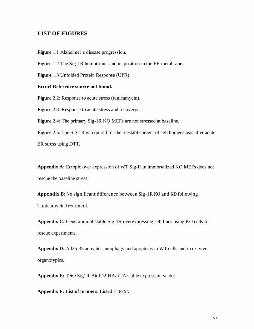
LIST OF FIGURES
Figure 1.1 Alzheimer’s disease progression.
Figure 1.2 The Sig-1R homotrimer and its position in the ER membrane.
Figure 1.3 Unfolded Protein Response (UPR).
Error! Reference source not found.
Figure 2.2: Response to acute stress (tunicamycin).
Figure 2.3: Response to acute stress and recovery.
Figure 2.4: The primary Sig-1R KO MEFs are not stressed at baseline.
Figure 2.5. The Sig-1R is required for the reestablishment of cell homeostasis after acute
ER stress using DTT.
Appendix A: Ectopic over expression of WT Sig-R in immortalized KO MEFs does not
rescue the baseline stress.
Appendix B: No significant difference between Sig-1R KO and KD following
Tunicamycin treatment.
Appendix C: Generation of stable Sig-1R overexpressing cell lines using KO cells for
rescue experiments.
Appendix D: Aβ25-35 activates autophagy and apoptosis in WT cells and in ex-vivo
organotypics.
Appendix E: TetO Sig1R-BioID2-HA/rtTA stable expression vector.
Appendix F: List of primers. Listed 3’ to 5’.
vii
LIST OF ABBREVIATIONS
AD: Alzheimer’s Disease
Aβ: Amyloid-Beta
AβOs: Amyloid-Beta Oligomers (AβOs
APP: Amyloid Precursor Protein
ApoE: apolipoprotein E
ATF4: Activating transcription factor 4
ATF6: Activating Transcription Factor 6
Bcl2: Apoptosis regulator Bcl-2
BiP: Binding immunoglobulin Protein
Ca: Calcium
CHOP: C/EBP homologous protein
DTT: Dithiothreitol
eIF2 : Eukaryotic translation initiation
factor 2A
EOAD: Early-Onset Alzheimer’s
Disease
ER: Endoplasmic Reticulum
ERAD: Endoplasmic Reticulum
Associated Degradation
ERSE: Endoplasmic Reticulum stress
element
G1 phase: Gap 1 phase
GADD34: DNA damage—inducible 34
Herp: Homocysteine-induced
endoplasmic reticulum protein
IKK: IκB kinase
IP3R: Inositol 1, 4, 5-trisphosphate
Receptor
IRE1: Inositol-requiring enzyme 1
JNK: c-Jun N-terminal kinase
KO: Knock Out
LOAD: Late-Onset Alzheimer’s Disease
MAM: Mitochondrial—endoplasmic
reticulum Associated Membrane
mRNA: messenger Ribonucleic Acid
MWM: Moris Water Maze
NADPH: Nicotinamide Adenine
Dinucleotide Phosphate
NF-κB: Nuclear Factor kappa B
NF—: Nuclear transcription Factor
Gama
NFTs: NeuroFibrillary Tangles
Nrf2: Nuclear factor-erythroid 2-related
Factor 2
viii
Orai1: Calcium release-activated
calcium channel protein 1
ORF: Open reading Frame
PM: Plasma Membranes
PERK: Protein kinase RNA-like
Endoplasmic Reticulum Kinase
PP1C: Protein Phosphatase 1C
PS1: Presenilin 1
PS2: Presenilin 2
PTZ: Pentazocine
Rac1: Ras-related C3 botulinum toxin
substrate 1
RIDD: Regulated IRE1-dependent
mRNA-decay
ROS: Reactive Oxygen Species
Sig-1R: Sigma-1 receptor
SKF: 10,047: (2S,6S,11S)-1,2,3,4,5,6-
Hexahydro-6,11-dimethyl-3- (2-
propenyl)-2.6-methano-3-benzazocin-8-
ol hydrochloride
S1P: Site 1 Protease
S2P : Site 2 Protease
SOCE: Store-Operated Ca2+ Entry
STIM1: stromal interaction molecule 1
Tau: Tubulin-Associated Unit
TNF-α: Tumour Necrosis Factor alpha
TRAF2: TNF receptor-associated
factor 2
UPR: Unfolded Protein Response
UPRE: Unfolded Protein Response
Elements
WT: Wild Type
XBP1: X-box protein

ix
ACKNOWLEDGEMENTS
I would like to thank Dr. Richard Bergeron for the opportunity to complete my M.Sc. in
Neuroscience degree in his laboratory. This opportunity has allowed me to improve my
expertise and to be exposed to the fascinating field of neuroscience. I also had the
opportunity to participate in other projects, which have led to the recent publication of a
research article, in which I am third author. I would like to thank Dr. Prakash Chudalayandi
and for his guidance and support throughout my project. I am grateful for all the support
he provided me over my journey in the lab. I am also thankful for the help I received from
Nina Ahlskog, Madelyn Abraham, Kayla Ferguson, and Thinh Nguyen. I would like to
extend my gratitude to all former and present members of Dr. Bergeron’s laboratory.
I would also like to thank Prakash Chudalayandi for his help with presentations and my
TAC member: Laura Trinkle-Mulcahy and Jonhy Ngsee for their retroaction on my project.
Finally, I would like to sincerely thank my family and friends for the support and
motivation they provided me over the years. To my parents, my wife and my two boys:
Mathis and Anthony, I am truly grateful for your continuous love and encouragement.
1
1. INTRODUCTION
1.1 Alzheimer’s Disease
1.1.1 Background
Alzheimer’s disease (AD), a neurodegenerative disorder, is most common cause of
dementia witnessed among aged people. AD represents 2/3 of all types of dementia. The
major characteristic of AD is the decline in cognitive functions and consequently a
dysfunction in social, behavioural and other daily activities. AD was first described in 1906
by the German psychiatrist Alois Alzheimer (Alzheimer et al. 1995; Maurer et al. 1997;
Möller & Graeber 1998; Reitz et al. 2011; Scheltens et al. 2016). The socio-economic
impact of AD amount over the billions, with considerable suffering experienced for the
patients and entourage. With over 35 million people suffering from dementia worldwide,
this growing social and economic burden on society is in part due to the ever-increasing
aging of the worldwide population (Mota et al. 2014; Prince et al. 2013; Wimo et al. 2013).
Despite decades of intense fundamental and clinical research, there are still no cures for
AD and only limited therapeutic interventions are available to treat the symptoms.
AD is primarily characterized by synaptic dysfunction. This induces dysfunction of
neuronal networks is manifested as episodic memory loss in early stages of the disorder
which slowly transgress into dementia as the disorder progress (Jacobsen et al. 2006;
Ondrejcak et al. 2010; Roy et al. 2016; Terry et al. 1991). The severity of the dementia is
highly correlated with neuronal loss that succeeds to the loss of synapses in the brain
(DeKosky & Scheff 1990; Shankar & Walsh 2009). The progressive loss of synapses and
neurones begins in the hippocampus, a brain region essential in learning and memory
2
process, and vicinal region of the medial temporal lobes (Braak & Braak 1991; Braak &
Braak 1997; Henderson 2014; Isik 2010; Vorhees & Williams 2014). Over the progression
of the disease, synapses and neurons loss increasingly affect the association areas of the
cerebral cortex, resulting in the progressive loss of short-term/working memory (also
referred as visuospatial memory), cognitive flexibility, and other cognitive abilities
(Carlesimo & Oscar-Berman 1992; Elcombe et al. 2014; Mucke & Selkoe 2012; Twamley
et al. 2006).
There are two forms of AD, that could be determined based on the age of the onset
of symptoms. The first form, a rare familial type (early onset; EOAD) is caused by
autosomal dominant mutations, with an early onset that occurs before the age of 60 years.
The second form and the most common one is the sporadic form (late onset; LOAD), with
an onset occurring after 60 years of age (Brouwers et al. 2008; Kukull & Bowen 2002;
Kukull et al. 2002; Reitz & Mayeux 2014). The EOAD is associated with mutations in the
amyloid precursor protein (APP), presenilin 1 (PS1), and presenilin 2 (PS2) genes, all of
which are linked to excessive production, accumulation, or deposition of amyloid beta (Aβ)
peptides in the brain (Cummings 2004; Goate 2006; Lannfelt et al. 2014; Selkoe 1989)
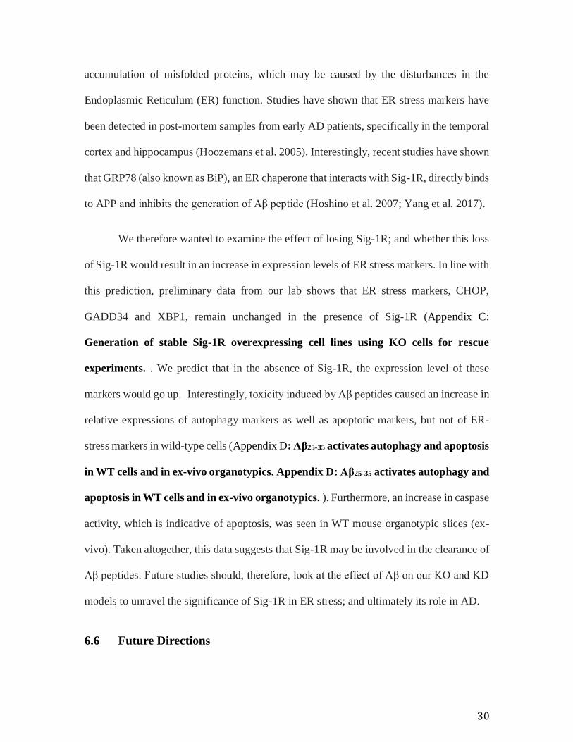
(Figure 1.1 Alzheimer’s disease progression. ). However, despite the great amount of
research on the elucidation of the complex molecular mechanisms of LOAD, the
environmental and genetic components are not yet, fully understood (Balin & Hudson
2014; Zou et al. 2014).
3
1.1.2 Hallmarks
The pathophysiology of AD develops as a consequence of neurofibrillary tangle formation.
This consists of hyperphosphorylated microtubule associated tau protein and senile plaques
of amyloid-β (Aβ) peptide in specific brain regions that result in synaptic loss and neuronal
death. The molecular hallmarks that characterized AD include neurofibrillary tangles
(NFTs) and neuritic plaques (Ballenger 2006; Blennow et al. 2006; Dennis J Selkoe 2011a).
These NFTs consist of intracellular filaments, primarily. Composed of
hyperphosphorylated tubulin-associated units (tau), a protein involved in the stabilization
of microtubules, which are able to self-assembly (Binder et al. 2005; Claeysen et al. 2012;
Mucke & Selkoe 2012). These NFTs are basically located within cell bodies of
pathological neurons of the cerebral cortex and brain stem (Braak & Braak 1991;
Henderson 2014). As for the neuritic plaques, they are mainly constitute of Aβ, a peptide
derived from the cleavage of APP (Mucke & Selkoe 2012; D. Puzzo et al. 2015; Zetterberg
& Mattsson 2014). This peptide adopts a β-pleated sheet configuration which has the
potential of self-aggregated to form soluble oligomers as well as insoluble amyloid beta
sheets. This is these insoluble sheets that accumulate in the extracellular space between
neurons, build up and associate with astrocytes, microglia, and dystrophic neurites (Selkoe
1989; Dennis J Selkoe 2011b; Toyn & Ahlijanian 2014). The neuritic plaques are primarily
formed in the basal isocortex (layer I) and gradually spread through the entire isocortex
structure as the disease progresses. In the late stages of AD, neuritic plaques are found
throughout the brain, with larger density in the isocortex (Braak & Braak 1991).
However, more recently, there is increasing evidence that soluble Aβ oligomers
(AβOs) are the molecules involve in the severity of the cytotoxicity and neuronal viability
4
as well as for the negative effects on synaptic function (Goate 2006; Lambert et al. 1998;
Lannfelt et al. 2014; Mucke & Selkoe 2012; Oda et al. 1994).
1.1.3 Risk factors
Sporadic AD (LOAD), the most common form, represents more than 90% of AD case, is
caused by an amalgam of environmental and genetic risk factors, for which very little are
currently known. The complexity of the disease is further enhanced by a suggested sex
differences in pathophysiology, which reinforcing its heterogeneity. Among the
environmental risk factors, studies have identified as potential, the levels of physical
activity, obesity, smoking, and alcohol consumption (Graves et al. 1991; Rovio et al. 2005)
However, the predominant risk factor for AD is aging (Blennow et al. 2006; Ferreira et al.
2015; Jagust 2013; Prince et al. 2013).
As regards to genetic factors, the predominant mutation occurs in apolipoprotein E
(ApoE) gene on chromosome 19, and the inheritance ApoE alleles ε4, which are associated
with higher Aβ load in the brain (Andreasson et al. 2014; Selkoe 1994a; Strittmatter et al.
1993). This polymorphism correlate with an increased risk of sporadic AD that
interestingly, prevail more in women than in men (Bretsky et al. n.d.; Farrer et al. 1997;
Mielke et al. 2014; Reitz & Mayeux 2014). Finally, hormonal changes produced by
menopause impact neuronal processes involved in cognition and has been suggested to be
a risk factor in AD (Pike 2017; Vest & Pike 2013).
5
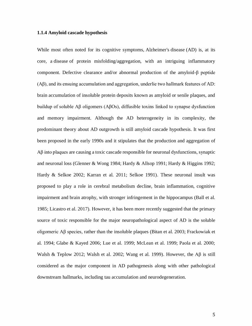
1.1.4 Amyloid cascade hypothesis
While most often noted for its cognitive symptoms, Alzheimer's disease (AD) is, at its
core, a disease of protein misfolding/aggregation, with an intriguing inflammatory
component. Defective clearance and/or abnormal production of the amyloid-β peptide
(Aβ), and its ensuing accumulation and aggregation, underlie two hallmark features of AD:
brain accumulation of insoluble protein deposits known as amyloid or senile plaques, and
buildup of soluble Aβ oligomers (AβOs), diffusible toxins linked to synapse dysfunction
and memory impairment. Although the AD heterogeneity in its complexity, the
predominant theory about AD outgrowth is still amyloid cascade hypothesis. It was first
been proposed in the early 1990s and it stipulates that the production and aggregation of
Aβ into plaques are causing a toxic cascade responsible for neuronal dysfunctions, synaptic
and neuronal loss (Glenner & Wong 1984; Hardy & Allsop 1991; Hardy & Higgins 1992;
Hardy & Selkoe 2002; Karran et al. 2011; Selkoe 1991). These neuronal insult was
proposed to play a role in cerebral metabolism decline, brain inflammation, cognitive
impairment and brain atrophy, with stronger infringement in the hippocampus (Ball et al.
1985; Licastro et al. 2017). However, it has been more recently suggested that the primary
source of toxic responsible for the major neuropathological aspect of AD is the soluble
oligomeric Aβ species, rather than the insoluble plaques (Bitan et al. 2003; Frackowiak et
al. 1994; Glabe & Kayed 2006; Lue et al. 1999; McLean et al. 1999; Paola et al. 2000;
Walsh & Teplow 2012; Walsh et al. 2002; Wang et al. 1999). However, the Aβ is still
considered as the major component in AD pathogenesis along with other pathological
downstream hallmarks, including tau accumulation and neurodegeneration.
6
1.1.5 Amyloid-beta peptide
Interestingly, APP, a highly conserved type-1 transmembrane glycoprotein located on
chromosome 21 in humans, has been suggested to be essential for normal brain
development as well as brain plasticity in adults; (Selkoe 1994b; Shariati & De Strooper
2013). However, the Aβ peptide is produced following a sequential proteolytic cleavage of
APP (Hamley 2012; Kopan & Ilagan 2004). The proteolytic cleavage is initiated by the
catalytic activity of either α- or β-secretases. The α-secretases are responsible for the
cleavage of the soluble extracellular domains (αAPP) and an 83 amino acid carboxy-
terminal fragments (C83) at one site. As for the β-secretase, it is responsible for the
production of βAPP and C99 peptides on another cleavage site (Cummings 2004; Daniela
Puzzo et al. 2015; D. J. Selkoe 2011). The maturation of the toxic peptides is then insured
by the γ-secretase, a large multiprotein complex constitute, among others, of the
subcomponents: PS1 and PS2. The subsequent proteolytic cleavage of βAPP by the γ-
secretase then gives rise to a 40 or 42 amino acid Aβ peptide (Aβ1-40 or Aβ1-42) (Kopan &
Ilagan 2004; Selkoe 1994a). Finally, because of the exposure of two hydrophobic alanine
and isoleucine residues, the Aβ1-42 peptide are able to self-aggregate into toxic AβOs (Mucke
& Selkoe 2012; Toyn & Ahlijanian 2014; Weiner et al. 2013).
1.1.6 Clinical trials
As mentioned previously, the soluble and insoluble Aβ peptides are considered as the
primary component of the AD pathogenicity in which they initiate and supply the cascade
involves neuronal and cognitive dysfunction (Gharibyan et al. 2007; Hamley 2012; Lee et
al. 2012; Pepys 2006; Williams & Serpell 2011). However, some clinicopathological
7
studies found unclear, the relationship between cognitive dysfunctions and Aβ load
(Castellani & Smith 2011; Castellani et al. 2009; Gustafson et al. 2006; Näslund et al. n.d.;
Vos et al. 2013). Moreover, results from therapeutic approaches targeting Aβ production
or clearance has not been promising (Mangialasche et al. 2010). Several Aβ-targeting drug
candidates such as secretase inhibitors and anti-Aβ antibodies have failed to improve the
patient's prognostic (Barten et al. 2006; Doody et al. 2013; Doody et al. 2014; Forman et
al. 2012; Hardy et al. 2014; Martenyi et al. 2012; Salloway et al. 2014). These result may
suggests that they have another component that drives the pathogenicity of AD. However
it is also possible that clinical trials failed because the drugs were given too late; if dementia
is due to loss of cortical neurons, then drugs are unlikely to replace these lost neurons.
1.2 Sigma-1 Receptor
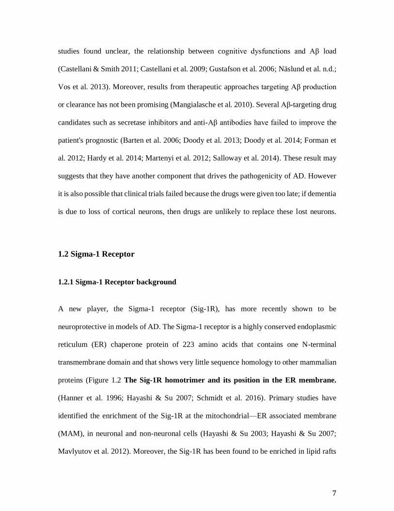
1.2.1 Sigma-1 Receptor background
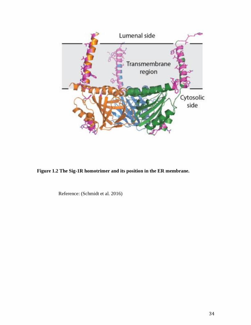
A new player, the Sigma-1 receptor (Sig-1R), has more recently shown to be
neuroprotective in models of AD. The Sigma-1 receptor is a highly conserved endoplasmic
reticulum (ER) chaperone protein of 223 amino acids that contains one N-terminal
transmembrane domain and that shows very little sequence homology to other mammalian
proteins (Figure 1.2 The Sig-1R homotrimer and its position in the ER membrane.
(Hanner et al. 1996; Hayashi & Su 2007; Schmidt et al. 2016). Primary studies have
identified the enrichment of the Sig-1R at the mitochondrial—ER associated membrane
(MAM), in neuronal and non-neuronal cells (Hayashi & Su 2003; Hayashi & Su 2007;
Mavlyutov et al. 2012). Moreover, the Sig-1R has been found to be enriched in lipid rafts
8
where several cellular functions are regulated and possibly at the nucleus (Hayashi & Su
2003; Natsvlishvili et al. 2015; Srivats et al. 2016; Wu & Bowen 2008). In physiological
conditions, the Sig-1R interacts with the ubiquitous binding immunoglobulin protein (BiP)
ER chaperone (Hayashi & Su 2007). Following ER stress or ligand activation, as per an
unfolded protein response (UPR) sensor, the Sig-1R dissociates from BiP and modulates
ER and plasma membranes (PM) ionic channel receptors, more specifically potassium and
calcium channels (Gao et al. 2012; Hayashi & Su 2007; Hayashi et al. 2012; Johannessen
et al. 2009; Mavlyutov et al. 2010). The Sig-1R has been shown to be transcriptionally up
regulated by the UPR, more precisely through the protein kinase RNA-like endoplasmic
reticulum kinase (PERK) pathway, directly through activating transcription factor 4
(ATF4) transcriptional activation. Interestingly, the ATF4 transcription expression level
has also shown to be upregulated following Sig-1R activation (Mitsuda et al. 2011; Omi et
al. 2014).
One of the roles of the Sig-1R is to regulate the Ca2+ signalling at two distinct sites.
At the cell surface, it has been shown to negatively regulate the store-operated Ca2+ entry
(SOCE) by preventing stromal interaction molecule 1 (Stim1)- Calcium release-activated
calcium channel protein 1 (Orai1) interaction (Srivats et al. 2016). In addition to SOCE
modulations, at the MAM it also plays a regulatory role by promoting the calcium efflux
to mitochondria through dissociation of inositol 1, 4, 5-trisphosphate receptor (IP3R) from
Ankyrin and by stabilizing it in its active conformation (Wu & Bowen 2008). This same
complex, in conjunction with Apoptosis regulator Bcl-2 (Bcl-2) and Ras-related C3
botulinum toxin substrate 1 (Rac1), has also been suggested to be involved in reactive

9
oxygen species (ROS) signalling through nicotinamide adenine dinucleotide phosphate
(NADPH)-oxidase activation (Natsvlishvili et al. 2015). A more recent study has shown
that the Sig-1R directly interacts with Inositol-requiring enzyme 1 (IRE1) to stabilize its
active form at the ER membrane. This complex enriched at the MAM, was also suggested
to monitor the ROS level generated by the mitochondria (Mori et al. 2013; Omi et al. 2014)
This is further strengthened by other studies that suggested a role for Sig-1R in suppression
or reduction of the accumulation/production of reactive oxygen species (ROS)
neighbouring mitochondria since Sig-1R knock-down shows an increased accumulation of
ROS. This accumulation is suggested to be attributed to the nuclear factor-erythroid 2-
related factor 2 (Nrf2), nuclear factor kappa B (NF-κB) and tumour necrosis factor alpha
(TNF-α) signalling (Allahtavakoli & Jarrott 2011; Hayashi et al. 2011; Meunier & Hayashi
2010; Mori et al. 2012; Tsai et al. 2012; Tsai et al. 2015; Zhao et al. 2014)
The activation of Sig1-R by Pentazocine (PTZ) and (2S,6S,11S)-1,2,3,4,5,6-
Hexahydro-6,11-dimethyl-3-(2-propenyl)-2.6-methano-3-benzazocin-8-ol hydrochloride
(SKF 10,047; SKF), two selective Sig-1R agonists, alleviated the ER stress response
related UPR markers and reduced the cell lethality in a primary neuronal retinal cell lines
prone to oxidative stress. This suggests a neuroprotective role for the Sig-1R (Ha et al.
2011; Omi et al. 2014). To further support this effect, overexpression of the Sig-1R
decreased cell apoptotic index whereas, an increase in apoptosis is observed in Sig-1R
knock out (KO) cells following ER stress activation (Ha et al. 2014; Hayashi & Su 2007).
It is therefore a potential therapeutic target to overcome ER stress-related disease (Ono et
al. 2013).
10
1.2.2 Sigma-1 receptor and Alzheimer's disease
Sig-1R had first been found to have a decreased ligand binding and lower density in ex
vivo experiments and in patients suffering from AD (Jansen et al. 1993; Mishina et al.
2008). Afterward, a polymorphism found in the 5'-upstream region of Sig-1R genes
(SIGMAR1), have shown to reduce its transcriptional activity, thereby reduces Sig-1R
expression and have been associated with an increased risk of developing AD (Huang et
al. 2011; Miyatake et al. 2004; Fehér et al. 2012; Jin et al. 2015).
Moreover, the Sig-1R role in AD has been fortified by studies which show that Sig-
1R agonists attenuate memory deficits in various rodent models of amnesia (Earley et al.
1991; Matsuno et al. 1994; Matsuno et al. 1997; Maurice & Privat 1997; Maurice et al.
1994; Senda et al. 1998; Zou et al. 2000). The agonized benefit of Sig-1R as anti-amnesic
agents is furfur more fortified by studies in males that shown neuroprotective and anti-
amnesic effect in AD animal models (Ishikawa & Hashimoto 2010; Maurice & Su 2009;
Maurice et al. 1998; Maurice et al. 2006; Meunier et al. 2009; Nguyen et al. 2015; Urani
et al. 2002).
Activation of the Sig-1R with SKF has also shown to ameliorate memory deficits
in an AD mouse model, and reduce oxidative stress markers induced by Aβ in-vitro
(Maurice et al. 2016; Nguyen et al. 2015).
Due to its neuroprotective effects in models of AD combined with the fact it is down
regulated in AD pathology, it is clear that the Sig-1R’s role in AD is worth investigating
further.
11
1.2.3 Sigma-1 receptor knockout
The absence of homology of Sig-1R with any other mammalian protein suggests a
fundamental function for Sig-1Rs. The cloning of the Sig-1R was therefore a big step in
the study of its structure and function but also the elaboration of Sig-1R KO mice (Langa
et al. 2003). Interestingly, Sig-1R KO mice were shown to have a lower axon density as
well as impaired hippocampus neurogenesis (Sha et al. 2013; Sha et al. 2015; Tsai et al.
2015). Our laboratory has revealed through electrophysiology, mild synaptic plasticity
deficits in Sig-1R KO compared to the wild type (WT) male mice, without change in other
aspects of basic cellular physiology. (Snyder et al. 2016).
Surprisingly, the KO mice developed normally. However it did show subtle, sex-
specific, behavioural phenotypes in pain, depression, anxiety, and cognition paradigms.
Behavioural studies using chemically induced and neuropathic pain models reveal an
attenuated response to pain in Sig-1R KO mice from both sexes as compared to WT
(Cendán et al. 2005; Entrena et al. 2009; Nieto et al. 2014; Puente et al. 2009). The
depressive-like phenotype seems to predominate in male as several behavioural studies
shown depressive-like phenotypes in two to eight-month-old male Sig-1R KO, but not
female (Chevallier et al. 2011; Sabino et al. 2009; Sha et al. 2015; Zhang et al. 2017).
Moreover, anxiety behaviours were detected only in Sig-1R KO males using behavioural
tasks (Chevallier et al. 2011). However, only the Sig-1R KO female revealed learning and
memory deficits in the behavioural tasks, suggesting that in the AD paradigm, the female
could be hardly impacted (Chevallier et al. 2011).
12
1.3 Endoplasmic reticulum stress
1.3.1 Endoplasmic reticulum control of protein
Proteostasis is of prime importance for cell health and regulation. The ER, along with Golgi
apparatus, is the major organelles responsible for the proteins folding, post-translational
modification and, export and secretion (Ron & Walter 2007; Walter & Ron 2012). The
proper folding of protein is ensured by a large variety of chaperones that also modulate
degradation (Vembar & Brodsky 2008). To regulate proteostasis, the ER use highly
conserved mechanism use for quality control or engage following ER stress activation.
Among those mechanisms, there is the ER associated degradation (ERAD), responsible for
the degradation of misfolded protein, and the UPR, trigger following the accumulation of
unfolded protein (Hetz & Mollereau 2014). If the UPR fail to restore proper protein load
and folding among the ER, it is known to trigger signalling cascades that induced apoptosis
(Walter & Ron 2012). Defects in UPR are known to be involved in neurodegenerative
disorder such as Alzheimer’s disease, amyotrophic lateral sclerosis, Huntington’s disease,
frontotemporal lobar degeneration, Parkinson’s disease but also other dysfunction like
psychiatric disorder, diabetes and cancer (Gold et al. 2013; Hayashi et al. 2009; Kakiuchi
et al. 2004; Kawada et al. 2014; Nevell et al. 2014; Prell et al. 2014; Tsai et al. 2014; Wang
& Kaufman 2014). It is therefore important to get a good understanding of these
mechanisms to be able to overcome these diseases.
13
1.3.2 The Unfolded Protein Response
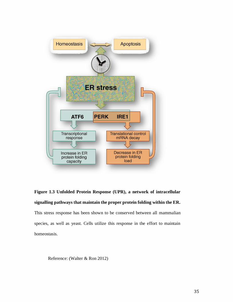
The UPR is mediated by three major ER receptors: Activating Transcription Factor 6
(ATF6), PERK and IRE1. The activation of these receptor result in a global decrease in
gene transcription and, an up regulation of chaperones and ER stress effectors (Ron &
Walter 2007). (Figure 1.3 Unfolded Protein Response (UPR)
The most conserved branch of the UPR is mediated by IRE1 receptors which
remains very similar to its yeast homologue (Walter & Ron 2012). This receptor is an ER
membrane resident protein consisting of, a sensor luminal domain, a single transmembrane
domain and a cytoplasmic domain responsible for a double functionality
kinase/endonuclease (Tirasophon et al. 1998). Following UPR activation, IRE1 dissociate
from BiP, self-phosphorylate and dimerize into its active form (Carrara et al. 2015; Pincus
et al. 2010). Following activation, IRE1 exerts its function through three different
pathways; X-box protein 1 (XBP1), TNF receptor-associated factor 2 (TRAF2) and
regulated IRE1-dependent mRNA-decay (RIDD) (Hollien & Weissman 2006; Imagawa et
al. 2008; Zeng et al. 2015). The XBP1 pathway is activated following an uncanonical exon
excision from XBP1 mRNA allowing the transcription of XBP1p, a transcription factor
that binds to ER stress element (ERSE), sequence CCAAT-N9-CCACG, and unfolded
protein response elements (UPRE), sequence TGACGTGG/A (Keisuke Yamamoto,
Hiderou Yoshida, Koichi Kokame 2004; Yoshida et al. 2001) This transcription factor
promote transcription of UPR target gene, cell survival, ER expansion, through lipid
synthesis, ER associated degradation (ERAD) and active secretion (Reimold et al. 2001;
Zhang et al. 2015; Yoshida et al. 1998; Samali et al. 2010).
14
The second signalling pathway consists in the recruitment of TRAF2 which
activated stress kinase such as c-Jun N-terminal kinase (JNK) and IκB kinase (IKK),
leading to induction of pro-inflammatory and inflammatory cytokines(Zhang et al. 2015).
The last function of IRE1, RIDD, mainly occurs following IRE1 hyper-activation (Zhang
et al. 2015). This function is exerted through the endonuclease activity of IRE1, on
unspecific mRNA and resulted in a global decrease of the transcript (Zhang et al. 2015).
PERK, the second branch of the UPR is observed only in higher eukaryotic cells
(Li et al. 2010; Walter & Ron 2012). Like the IRE1 sensor, PERK is an ER membrane
resident protein that consists of a single transmembrane domain with an N-terminal luminal
sensing domain but with a C-terminal domain that only exert kinase activity (Walter & Ron
2012). Following UPR activation, BiP dissociate from PERK allowing it to dimerize and
self-phosphorylate in its active form (Carrara et al. 2015; Walter & Ron 2012). The active
PERK complexes phosphorylate the omnipresent initiation transnational factor elF2a
(Scheuner et al. 2001). This phosphorylation results in eukaryotic translation initiation
factor 2A (eIF2a) inactivation which inhibits global protein translation to ultimately reduce
transitory protein amount in the ER and induce the cell cycle arrest at G1 phase (Bhakta-
Guha & Efferth 2015; Wek & Cavener 2007). Following the inactivation of elF2a, some
small open reading frame (ORF) located in the generally 5’ untranslated region of specific
mRNA, are preferentially translated (Andreev et al. 2015). Among these, the activating
transcription factor 4 (ATF4), induces the transcription of two key genes in the UPR:
C/EBP homologous protein (CHOP), a transcription factor and growth arrest and DNA
damage—inducible 34 (GADD34) a regulatory subunit (Wek & Cavener 2007). Although
the signalling pathway for PERK-mediated UPR has a protective effect by promoting
15
chaperone synthesis, oxidative stress response, autophagy and amino acid metabolism,
CHOP is controlling genes involved in apoptosis (Hetz & Mollereau 2014). This dualism
is mediated in a time-dependent manner through the ratio of phospho-elf2a/elf2a that is
among others, regulated by GADD34, a regulatory subunit of the PP1C phosphatase.
GADD34/ PP1C phosphatase inhibits the PERK signalling through elF2a
dephosphorization by feedback inhibition (Harding et al. 2009; Marciniak et al. 2004;
Tsaytler et al. 2011).
The third UPR pathway, namely the ATF6 pathway, is also observed only in higher
eukaryotes (Walter & Ron 2012). ATF6 is a transcription factor that is synthesized as an
ER plasma membrane receptor (Haze et al. 1999; Schindler & Schekman 2009). Like IRE1
and PERK, ATF6 consists of a luminal sensing domain that is stabilized by BiP, but in
addition includes a Golgi localization sequence (Shen et al. 2002; Shen et al. 2005) This
domain is linked by a short transmembrane domain to a cytoplasmic portion that contains
a basic leucine zipper and a transcriptional activation domain (Hetz et al. 2011). Following
stress activation, BiP disassociates from ATF6 allowing ATF6 to be translocated to the
Golgi apparatus by vesicular transport (Haze et al. 1999). It is then cleaved by the protease
site 1/2 (S1P/S2P) at both ends of the transmembrane domain to release the N-terminal and
C-terminal portions (Ye et al. 2000). The C-terminal portion, containing the transcription
factor component, is then routed to the nucleus where it targets ERSE I, UPRE, ERSE II
and XBP1 promoter elements (Keisuke Yamamoto, Hiderou Yoshida, Koichi Kokame
2004; Yoshida et al. 2001) ERSE is mainly responsible for activating the transcription of
chaperones and can be fully activated by ATF6 in a nuclear transcription factors Gama
(NF-Y) dependent manner whereas UPRE depend solely on XBP1 and is more specifically
16
responsible for the transcription of the ERAD component (Keisuke Yamamoto, Hiderou
Yoshida, Koichi Kokame 2004). The ERSE II (ATTGG-N-CCACG), as to it, seem to be
more specifically responsible for the transcription of Homocysteine-induced endoplasmic
reticulum protein (Herp), an ER membrane protein. This protein is responsible for protein
trafficking to proteasome through ubiquitin modulations of the ER-resident calcium release
channels, aiming to maintain calcium homoeostasis, preserving mitochondria functions
and suppressing caspase-3 activity (Belal et al. 2012; Keisuke Yamamoto, Hiderou
Yoshida, Koichi Kokame 2004; Kim et al. 2008; Kokame et al. 2001; Slodzinski et al.
2009; Chan et al. 2004).
1.3.3 Alzheimer’s Disease is an Endoplasmic Reticulum Stress disease
AD is a progressive neurodegenerative disease involving loss of synaptic function followed
by neuronal loss by apoptosis. The salient features involve accumulation of aggregates of
misfolded proteins occurring in conjunction with abnormalities in the redox regulation, Ca
homeostasis. Accumulation of the Aβ peptide along with hyper phosphorylated Tau is
seen in post-mortem samples of most AD patients. Accumulating evidence suggests that
the improper regulation of proteostasis and ER stress is a major pathological hallmark for
AD based on studies of several post-mortem brain tissue samples (Cornejo & Hetz 2013;
Plácido et al. 2014; Kennedy et al. 2014). The AD brains show evidence for the activated
UPR elements, like the phospho eIF2a has been shown in the neurons but not in the glial
cells (Salminen et al. 2009). A direct correlation has been observed in the AD pathogenesis
and activation of the IRE1 pathway. Further, mutations in the XBP promoter reported to
be a risk factor for AD. Genetic ablation of the IRE1 or the deletion of the RNase domain
17
of IRE1 led to reduced amyloid accumulation indicating the activation of IRE1 pathways
to be a cause for the progression of AD (Duran-Aniotz et al. 2014). The mutation in
PS1linked to familial AD has been reported to down regulate the UPR response and makes
cells susceptible to ER stress (Imaizumi et al. 2001; Katayama et al. 1999).
The literature suggests that ER stress is a pathological hallmark for AD (Cornejo &
Hetz 2013; Kennedy et al. 2014; Plácido et al. 2014). When taken together with the reports
of the Sig-1R as a protein chaperone (Hayashi & Su 2007) and its role in the regulation of
ER stress (Hayashi & Su 2005; Mori et al. 2013) it makes investigation of cell biology and
biochemistry of Sig-1R with respect to ER stress and cell homeostasis crucial for
understanding the etiology of AD in the mouse model.
1.4 Focus of this study
Together, these studies suggest that loss of Sig-1R function may predispose an individual
to AD and potentially contribute, at least partially, to the progression of AD. Therefore, to
fully elucidate how loss of the Sig-1R contribute to AD and cognitive decline, apart from
using animal models, it is also necessary to investigate the cell biology of Sig-1R when
cells are stressed.
The overall mechanism by which Sig-1R modulates ER stress and how it exerts its
protective effect remains unclear. The dissection of the UPR pathway with respect to Sig-
1R will therefore allow a better understanding of the mechanism underlying
neuroprotective effects of the Sig-1R.
18
2. HYPOTHESIS
Deletion of Sigma-1-receptor compromises the ER stress response, specifically the UPR
pathways PERK, IRE1, and ATF6..
3. OBJECTIVES
1. Determine if immortalized Sig-1R-/- MEFs show baseline activation of all three
branches of the UPR.
2. Characterize the UPR response during different types of ER stress for
immortalized and primary Sig-1R+/+ and immortalized and primary Sig-1R-/-
.
a. Acute Stress – Tunicamycin
b. Acute stress and recovery time - DTT
4. MATERIALS AND METHODS
4.1 MEF isolation and cell culture
Mouse embryonic fibroblasts (MEF) were isolated from WT or Sig-1R-/- C57/BL6 mouse
embryos (E14.5) using standard procedures (Xu 2005; Wong et al. 2016). In brief, E14.5
foetuses were isolated from pregnant dames and organs removed. The tissue was
trypsinized with 0.25% trypsin-EDTA (Gibco/Invitrogen, Burlington, ON Canada),
19
homogenized, and passed through a 70 μm cell strainer. The single cell suspension was
cultured in standard MEF media consisting of DMEM (Wisent Inc., Saint-Jean-Baptiste,
QC, Canada), 10% fetal bovine serum, 100 U/ml penicillin/streptomycin, and 2 mM L-
glutamine (all from Gibco/Invitrogen). MEFs were grown in a humidified 37°C, 5% CO2
incubator, passaged every 3–4 days using trypsinization (0.05% trypsin; Gibco/Invitrogen)
and plated as a monolayer at a density of ~2 × 10⁶ cells/ml.
4.2 Semi-quantitative RT-PCR
MEFs derived from WT and Sig-1R-/- mice were grown to 70–90% confluence, on 35 mm
dish was treated with 3 μM Tun for 8, 12, 16, or 24 h. Total RNA was isolated using 1 ml
TRIzol (Invitrogen) according to the manufacturer's instructions, and quantified using a
NanoDrop 2000 spectrophotometer (Thermo Scientific, Burlington, ON, Canada). Reverse
transcription was performed on 1.25 μg RNA per reaction using SuperScript III reverse
transcriptase (Invitrogen) according to the manufacturer's instructions. The housekeeping
gene, glyceraldehyde 3-phosphate dehydrogenase (GAPDH) was used as a control. The
following PCR conditions were used: an initial denaturation step at 95°C for 3 min,
followed by denaturation at 94°C for 30 s, annealing at Tm (indicated in Table 1) for 30 s,
extension at 72°C for 1 min repeated the indicated number of cycles (Table 1), and a final
extension at 72°C for 5 min. The product was separated on 3% agarose gels and visualized
using AlphaImager Mini (ProteinSimple, San Jose, CA, USA). Each data point
corresponds to six separately isolated biological samples with one technical replicate.
20
4.3 Western blot
MEFs were plated onto 10 cm (78.54 cm2) dishes and treated with 3 μM Tun for 4, 8, 16,
or 24 h. At collection, cells were gently washed twice in ice-cold PBS (10 mM Na2HPO4,
1.76 mM KH2PO4, 137 mM NaCl, and 2.68 mM KCl; pH 7.2) and lysed on ice with 700 μl
RIPA buffer (150 mM NaCl, 50 mM Tris, 0.5% sodium deoxycholate, 0.1% NP-40, 5 mM
sodium pyrophosphate, 2 mM β-glycerophosphate, 1×EDTA-free protease inhibitor
(Fisher Scientific), pH 7.5). MEFs were then scraped off, sonicated for 15 s at 20% effect
on a FB120 sonicator (Fisher Scientific), and cleared by centrifugation. Total protein (1.5
mg per lane) was resolved on Tris-glycine SDS-PAGE and transferred onto PVDF
membrane. After incubation with primary and secondary antibodies (see below),
membranes were developed using Luminata Crescendo (Millipore, Darmstadt, Germany)
and visualized using LI-COR Odyssey Fc (LI-COR, Lincoln, Nebraska USA). Band
intensities were normalized to total protein as determined by Fast Green stain (125 μM Fast
Green FCF (Sigma-Aldrich, Oakville, ON, Canada), 6.7% acetic acid, 30% methanol;
[37]). Each n corresponds to a single biological replicate containing 2–4 technical
replicates.
4.4 Antibodies
The following antibodies and dilutions were used: rabbit anti-Sig-1R (1:2000; Atlas
Antibodies, Bromma, Sweden), rabbit and mice anti-ATF6 (1:1000; Abcam, Cambridge,
MA, USA and Novus Biologicals, Oakville ON, Canada, respectively), rabbit anti-
GADD34 (1:200; MyBioSource, San Diego, CA, USA), mouse anti-CHOP (1:100;
Developmental Studies Hybridoma Bank, University of Iowa, Iowa City, IA, USA), and
21
mouse-Caspase 3 (1;400; Abcam, Cambridge, MA, USA). HRP-conjugated anti-mouse
and anti-rabbit secondary antibodies were all purchased from Jackson ImmunoResearch
(West Grove, PA, USA).
4.5 Viability assay
MEFs were plated at the density of 2 × 10⁴ in 100 μl per well onto black 96-well clear
bottom plates. After allowing 24 h for attachment, the cells were treated with 3 μM Tun as
previously described. At the end of the treatment, the media was carefully aspirated from
the wells, the cells washed in warm PBS, and incubated with 100 μl Calcein AM (3 μM;
Invitrogen) in DPBS (PBS with 90 μM CaCl2 and 50 μM MgCl2; Wisent Inc.) for 30 min.
Fluorescent Calcein AM hydrolyzed by live cells was measured at 535 nm (485 nm ex).
4.6 Generation of stable Sig-1R–eYFP-expressing Sig- 1R-/- MEFs
A Geneticin/G418 resistant Sig-1R-eYFP construct driven by a CAG promoter was
linearized with VspI before transfection into Sig-1R-/- MEFs using TransIT 2020 according
to the manufacturer’s instructions (Mirus Bio). After 48 h, the cells were split, diluted and
selected on medium containing 1 mg/ml G418 (Sigma Aldrich). Media was changed every
third day for 2 weeks until single colonies were visible. Single colonies were picked and
expanded before being screened for the presence of Sig-1R-YFP using epifluorescence
imaging on Zeiss Observer.A1 inverted microscope (Zeiss Instruments, Oberkirchen,
Germany) after each passage. Colonies that showed less than 100% expression of Sig-1R-
YFP was discarded.
22
4.7 Live cell Imaging
Stable Sig-1R-YFP expressing MEFs was plated on to 35 mm imaging dishes (ibidi GmbH,
Martinsreid, Germany) and live-cell imaged using Airyscan confocal imaging at 488, 515,
and 559 nm on an inverted Zeiss LSM 880 63× (NA 1.4) oil immersion objective (Zeiss).
The ER was stained with ER-Tracker Red (Life Technologies/Thermo Fisher Scientific)
and the plasma membrane was stained with CellMask Deep Red plasma membrane stain
(Life Technologies/Thermo Fisher Scientific) as a morphological marker 20 min prior to
imaging. Cells were imaged in Phenol Red free MEM (Wisent) containing 10% FBS on a
pre-warmed 37 °C stage with 5% CO2. For co-localization analysis, cells were imaged at a
resolution of 1024×1024 pixels, dwell time of 4 μs/pixel, and Z-stacks were obtained with
0.75–1 μm step size (6–10 sections per stack). Surface-surface co-localization analysis was
performed using IMARIS Imaging Software (Bitplane, Concord, MA, USA).
4.8 Generation of stable Sig-1R Knock down MEFs
WT Sig-1R MEFs were transiently transfected with Puromycin resistant shRNAs
TRCN0000194512 (D10) targeting sequence (5’-TTCACCAGAGATTACTACAGG-3’),
TRCN0000194052 (D11) targeting sequence (5’-TAATATCTGCATGGTATACGC-3’),
and TRCN0000193914 (E1) targeting sequence (5’-ATAATAGTCAGAATCAGGGTG-
3’), all purchased from GE Dharmacon. Cells were split and replated 48 h post transfection
ensuring a threefold dilution of cells and selected on puromycin (3 μg/ml) selective
medium. The resultant colonies were picked and expanded in puromycin-containing media
before knockdown efficiency were determined via Western blotting. Cells stably
transfected with D10 shRNA showed the largest knockdown of the 3 shRNAs tested.
23
4.9 Analysis and statistics
Densiometric analysis of RT-PCR results were performed using ImageJ, while Western
blots were analyzed using Image Studio (LI-COR). OriginPro 8.5 (OriginLab Corporation,
Northampton, MA, USA) was used to plot graphs. Data are expressed as mean ± SEM.
Statistical significance was determined with one-way ANOVA and Dunnett's T3 post-hoc
tests with a critical value of 0.05 using IBM SPSS Statistics 23 software (IBM, Armonk,
NY, USA).
5. RESULTS
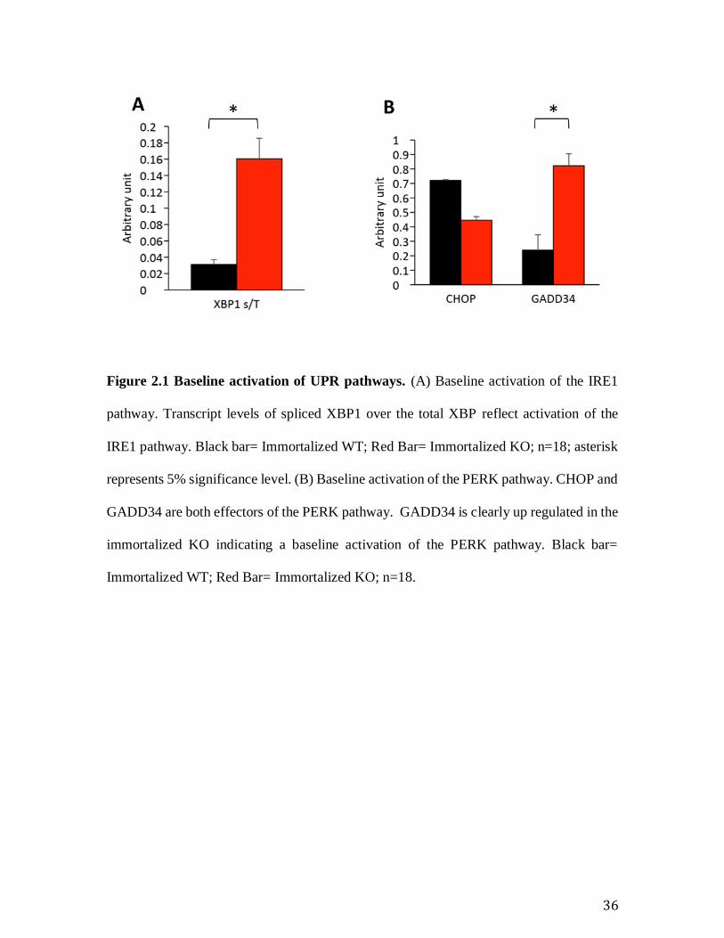
5.1 Immortalized Sig-1R KO MEFs is baseline stressed
RT-PCR analysis showed baseline activation in the effectors of the IRE1 and PERK
pathways. The increased ratio of spliced XBP XBPsp over total XBP XBPtot showed that
the IRE pathway is activated at baseline in the immortalized KO MEFs (Error! Reference
source not found.). The increased transcript levels of GADD34, indicates activation of the
PERK pathway (Fig. 1B). Interestingly, however, the transcript level of CHOP, another
component of the PERK pathway is not up regulated in immortalized KO cells. ATF6
pathway is not activated at baseline, however, as ATF6 transcripts level remain unchanged
(Error! Reference source not found.).
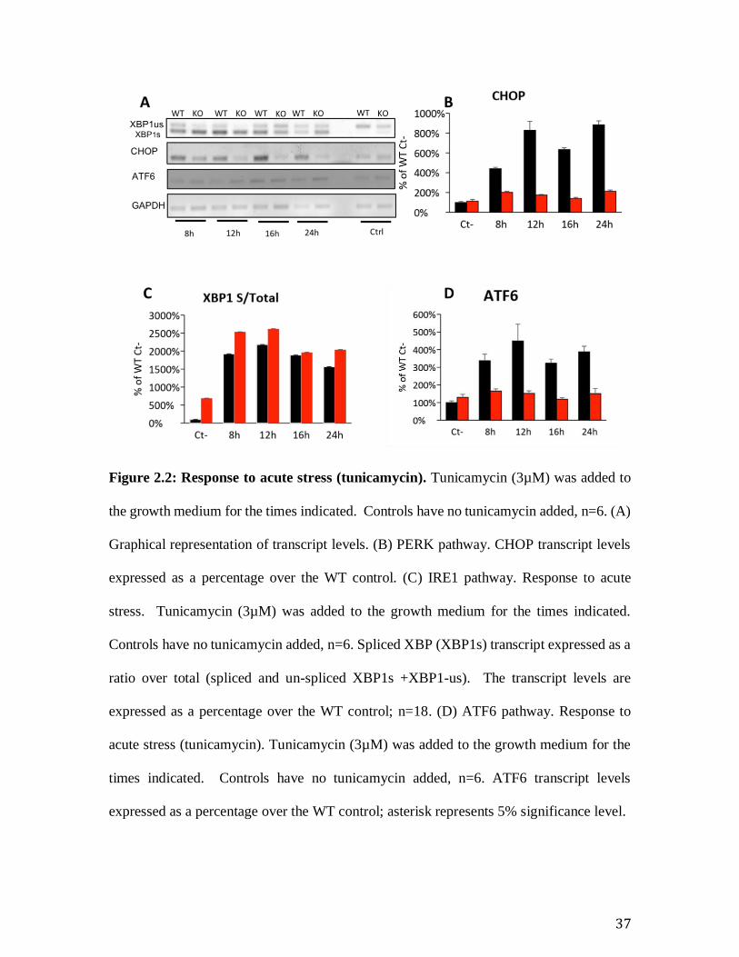
5.2 Induced, acute ER stress elicits a robust response in Sig-1R wild-type
and an attenuated response in knockout
24
Tunicamycin induces the Unfolded Protein Response in cells. We therefore wanted to
examine the effect of this acute stress on our models. In order to do so, we added
tunicamycin (3µM) to cells and examined the response at various time points ranging from
8 hours to 24 hours. Following acute stress, all the effectors of the UPR pathway show a
robust up regulation in the WT; however, the response is attenuated in the KO as reflected
in the modest up regulation especially of the PERK, ATF6, and IRE1 (Figure 2.2: Response
to acute stress (tunicamycin). ). The observation that the immortalized KO showed a
baseline stress and the attenuated response to a acute stress induced by tunicamycin suggest
an adaptation to ER stress in the immortalized KO MEF line.
5.3 Recovery from an acute ER stress is similar in WT and KO
Seeing the differences in response to acute stress between immortalized WT and KO cells,
we wanted to determine whether acute stress would lead to the same observation. We
therefore used DTT for this experiment. DTT induces an acute stress in cells by inhibiting
the formation of sulphydryl bridges in proteins and induces an unfolded protein response
within minutes. Cells were stressed with 5µM DTT for 30 minutes. Cells were then
washed off and allowed recover in regular media (with FBS and Pen/Strep). Response to
stress was monitored for 16 hours. Cells were collected at indicated time points. Controls
were not treated with DTT. The immortalized WT and KO both respond to the addition of
DTT almost identically; although two of the three pathways show a modest attenuation of
the response in Sig1R KO MEFs as compared with WT (CHOP and ATF6; Fig3C and 3D).
However, there is no change in the recovery kinetics from the acute stress in the KO when
25
compared to the WT (Figure 2.3: Response to acute stress and recovery. . This further
suggests that there is an adaptation to ER stress in our current model, immortalized cells.
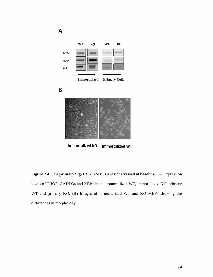
5.4 Primary KO MEFs and Primary WT MEFs are not baseline stressed
We then asked the question: is the primary KO MEFs stressed at baseline as well. The
primary KO MEFs are not stressed at baseline when compared with the immortalized KO
MEFs (Fig 4B). This finding further argues for an adaptation to stress in the immortalized
KO cells. Furthermore, the morphology of immortalized KO MEFs is very different from
the immortalized WT (Fig 4A), which indicates that the mutations acquired during
immortalization are different in the WT and KO cell lines. Differences in immortalization
adaptation between the WT and KO MEFs may be the cause of the morphological
differences we observe (Figure 2.4: The primary Sig-1R KO MEFs are not stressed at
baseline. .
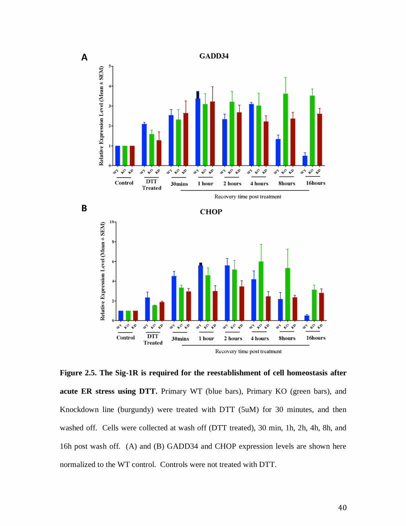
5.5 Primary Sig-1R WT and KO MEFs show a robust difference in the
recovery from an acute ER stressor DTT
Preliminary experiments showed that the primary KOs respond to the acute stress
tunicamycin identically to the primary WT. However, the primary KO displays a robust
phenotype during the recovery from an acute stress induced by DTT (Figure 2.5. The Sig-
1R is required for the reestablishment of cell homeostasis after acute ER stress using
DTT. ). WT cells were efficient at recovering from acute stress. The level of stress,
indicated by the expression level of CHOP and GADD34, remained high in primary KO
cells after 16 hours of recovery. This data leads us to the question of specificity. We wanted
26
to determine whether the response seen in Primary KO cells was caused by the absence of
the Sig-1R. We therefore generated a knockdown (KD) cell line using Sig-1R shRNA.
Recovery from the acute stress induced by DTT observed in the primary KO is
phenocopied by the Sig-1R knockdown (Fig 5). Both Sig-1R KD and primary KO cells
show a persistence of the stress phenotype for 16 hours following DTT wash off. The KD
cells show the same phenotype as KO primary cells in the expression levels of effectors of
the PERK pathway (CHOP and GADD34), (Figure 2.5. The Sig-1R is required for the
reestablishment of cell homeostasis after acute ER stress using DTT. ). Taken together,
our data from primary cells suggests that Sig-1R is required for the re-establishment of cell
homeostasis following acute ER stress.
6. DISCUSSION
6.1 Baseline ER stress in Immortalized KO not rescued by WT Sig-1R
RT-PCR analysis showed baseline activation in the effectors of the IRE1 and PERK
pathways. The increased ratio of spliced XBP1 XBP1sp over total XBP1 XBP1t showed
that the IRE pathway is activated at baseline in the immortalized KO MEFs (Error!
Reference source not found.). The increased transcript levels of GADD34, indicates
activation of the PERK pathway (Error! Reference source not found.). Interestingly,
however, the transcript level of CHOP, another component of the PERK pathway is not up
regulated in immortalized KO cells. This discrepancy might suggest a crosstalk between
the PERK pathway and another pathway, which might regulate either GADD34 directly or
might feed into CHOP (Takayanagi et al. 2013; Yao et al. 2013; Li et al. 2014; Yeganeh et
27
al. 2015; Chikka et al. 2013; Song et al. 2009). ATF6 pathway is not activated at baseline,
however, as ATF6 transcripts level remain unchanged (Figure 2.2: Response to acute
stress (tunicamycin). ).
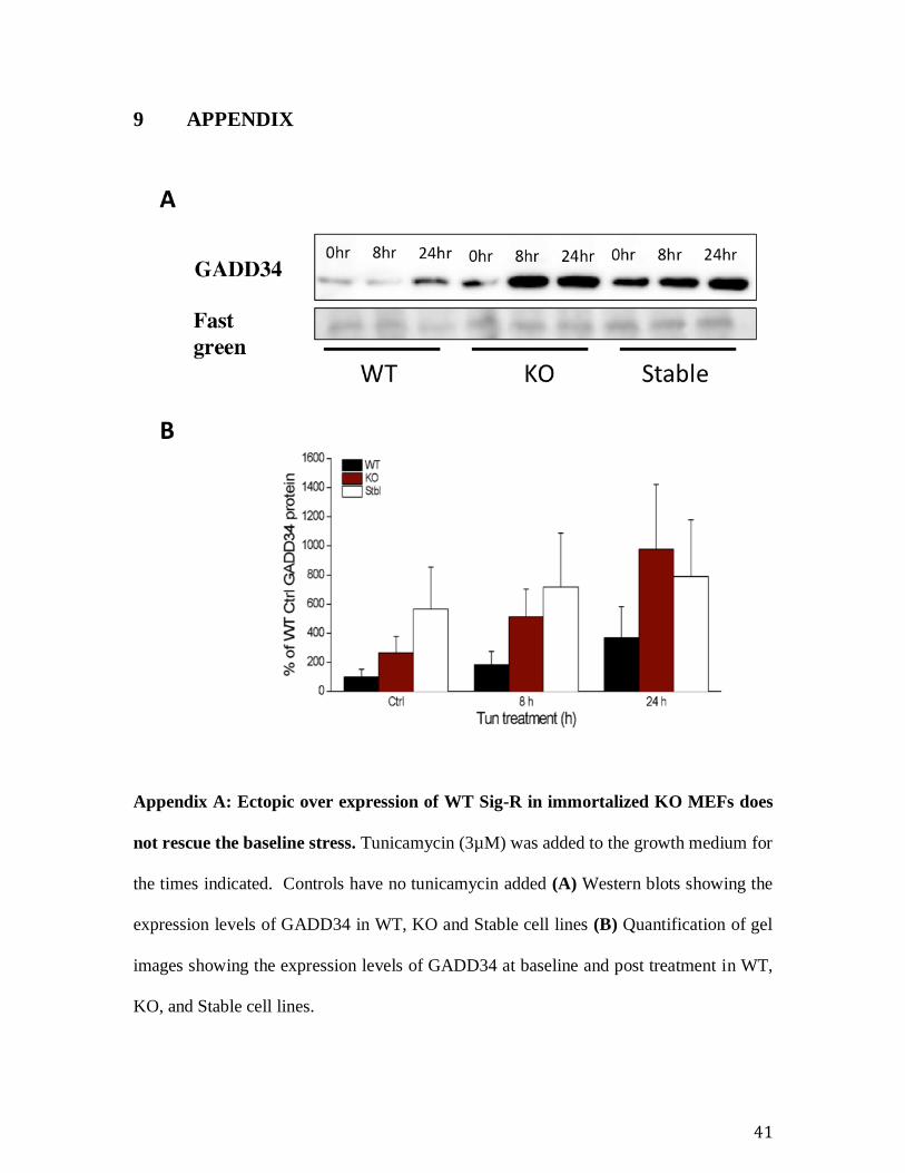
Further we have tried to rescue the baseline stress observed in the immortalized KO
by stably over-expressing WT Sig1R and found that the baseline stress as shown by the
up-regulation of the UPR elements remained intact (Appendix C: Generation of stable Sig-
1R overexpressing cell lines using KO cells for rescue experiments. . This observation
strongly suggests an adaptation to ER stress.
6.2 Response to ER stress is attenuated in the immortalized KO.
Tunicamycin induces the Unfolded Protein Response in cells. We therefore wanted to
examine the effect of this acute stress on our models. In order to do so, we added
tunicamycin (3µM) to cells and examined the response at various time points ranging from
8 hours to 24 hours. Following acute stress, all the effectors of the UPR pathway show a
robust up regulation in the WT; however, the response is attenuated in the KO as reflected
in the modest up regulation especially of the PERK, ATF6, and IRE1 (Figure 2.2: Response
to acute stress (tunicamycin). . The observation that the immortalized KO showed a
baseline stress and the attenuated response to a acute stress induced by tunicamycin suggest
an adaptation to ER stress in the immortalized KO MEF line.
6.3 Primary Knockout and knockdown better model
Primary MEFs isolated from the KO mice strain did not show any baseline ER stress as
against the immortalized KO MEFs. The process of spontaneous immortalization of Sig-
28
1R KO MEFs probably induced other mutations and thus the immortalized WT and KO
are not isogenic. The attenuated response to ER stress and the inability of WT Sig-1R to
rescue the baseline ER stress in immortalized KO, indicates an adaptation to ER stress in
the immortalized KO MEFs. The immortalized cell lines of UPR mutants are known to
show an attenuated response to the ER stressors and this is not uncommon in the field,
although not evident from the literature (vom Brocke et al. 2006). Regardless, it is well
established that immortalization process induces mutagenesis (vom Brocke et al. 2006;
Busuttil et al. 2003; Fridman & Tainsky 2008) and thus the likelihood that the immortalized
WT and KO are isogenic is remote.
However, the knockdown cells showed no baseline stress levels. Furthermore, the
KD cells phenocopy the primary knockout MEFs in the recovery from DTT acute stress
(Figure 2.5. The Sig-1R is required for the reestablishment of cell homeostasis after
acute ER stress using DTT. . This suggests that the KD cells are a better model to study
the effect of the Sig-1R in ER stress.
6.4 Cell homeostasis following acute DTT stress or a response to oxidative
stress?
The primary knockout and knockdown MEFs both have a compromised recovery from
DTT induced acute stress, which suggests that the Sig-1R is responsible returning the cell
to homeostasis following an acute stress. This might also suggest that Sig-1R plays a role
in responding to oxidative stress. Prolonged stress induced by reducing conditions has
been reported to generate free radicals, which initiates an oxidative stress response (Maity
29
et al. 2016). Although, here we have induced ER stress by DTT (5mM) just for 30 minutes,
it is possible that the Sig-1R knockout strains are susceptible to oxidative stress and are
responding to oxidative stress effects. Sig-1R knockout strains have been reported to
display higher levels of oxidative stress compared to the WT (Pal et al. 2012), and that
activation of Sig-1R protects retinal cells from oxidative stress damage (Ha et al. 2011;
Wang et al. 2016; Zhao et al. 2014). The Sig-1R KO and KD strains were not significantly
different in their responses when compared to the WT MEFs when treated with either
tunicamycin or thapsigargin (Appendix A: Ectopic over expression of WT Sig-R in
immortalized KO MEFs does not rescue the baseline stress.). However, the Sig-1R KO
and KD MEFs both displayed a robust phenotype when acutely stressed with DTT (Figure
2.5. The Sig-1R is required for the reestablishment of cell homeostasis after acute ER
stress using DTT. . While the WT MEFs showed recovery of ER stress markers CHOP
and GADD34 to baseline levels, the Sig-1R KO and KD MEFs remained elevated in these
transcripts; suggesting that the Sig-1R may be important for returning the cell to
homeostasis following acute ER stress. It will be important to show if Sig-1R plays a role
in responding to oxidative stress and if the ER stress response observed is downstream of
oxidative stress.
6.5 Exogenous Aβ peptide induces autophagic and apoptotic response
Alzheimer’s disease (AD) is a neurodegenerative disease that impairs memory and
cognitive function. The clinical manifestation of AD, and many other neurodegenerative
diseases, is initiated by alterations in protein functionalities of distinctive neuronal
populations. This disruption of functionality has been shown to be correlated to the
30
accumulation of misfolded proteins, which may be caused by the disturbances in the
Endoplasmic Reticulum (ER) function. Studies have shown that ER stress markers have
been detected in post-mortem samples from early AD patients, specifically in the temporal
cortex and hippocampus (Hoozemans et al. 2005). Interestingly, recent studies have shown
that GRP78 (also known as BiP), an ER chaperone that interacts with Sig-1R, directly binds
to APP and inhibits the generation of Aβ peptide (Hoshino et al. 2007; Yang et al. 2017).
We therefore wanted to examine the effect of losing Sig-1R; and whether this loss
of Sig-1R would result in an increase in expression levels of ER stress markers. In line with
this prediction, preliminary data from our lab shows that ER stress markers, CHOP,
GADD34 and XBP1, remain unchanged in the presence of Sig-1R (Appendix C:
Generation of stable Sig-1R overexpressing cell lines using KO cells for rescue
experiments. . We predict that in the absence of Sig-1R, the expression level of these
markers would go up. Interestingly, toxicity induced by Aβ peptides caused an increase in
relative expressions of autophagy markers as well as apoptotic markers, but not of ER-
stress markers in wild-type cells (Appendix D: Aβ25-35 activates autophagy and apoptosis
in WT cells and in ex-vivo organotypics. Appendix D: Aβ25-35 activates autophagy and
apoptosis in WT cells and in ex-vivo organotypics. ). Furthermore, an increase in caspase
activity, which is indicative of apoptosis, was seen in WT mouse organotypic slices (ex-
vivo). Taken altogether, this data suggests that Sig-1R may be involved in the clearance of
Aβ peptides. Future studies should, therefore, look at the effect of Aβ on our KO and KD
models to unravel the significance of Sig-1R in ER stress; and ultimately its role in AD.
6.6 Future Directions
31
While the results obtained from this study indicate that the Sig-1R may play a role in the
ER stress response and recovery in immortalized and primary MEF cell lines, the molecular
mechanism underlying its effect it is still unclear. Is the Sig-1R interacting with the three
branches of the UPR at their initiation (PERK, IRE1, and ATF6), or is there a process
upstream of ER stress that the Sig-1R is a part of, such as the oxidative stress response? To
elucidate the proteins and pathway(s) for which the Sig-1R is a direct interactor we propose
to use a BioID assay to map the Sig-1R interactome at baseline and under cellular stress.
The BioID assay was first developed by the Roux lab (Roux et al. 2012) to
successfully identify the proteins that interact with and are proximal to the lamin-A protein,
a well-characterizes structural component of the nuclear envelope. The BioID assay relies
on the expression of a biotin ligase enzyme fused to a protein of interest. Upon expression
of the fusion protein, the biotin ligase enzyme will biotinylate proteins that interact with,
or are proximal to, the protein of interest. Biotinylated proteins can then be pulled down
through affinity purification and subsequently identified via mass spectrometry. Roux lab
followed up their work with an improved BioID tag that was much smaller in size (Kim et
al., 2016). We used this tag and generated a tetracycline inducible Sig-1R-BioID
(Appendix D: Aβ25-35 activates autophagy and apoptosis in WT cells and in ex-vivo
organotypics. using the vector deposited in addgene (addgene #24418) by the Hsaio lab
(Hsiao et al. 2011). A pulse-chase experiment will be conducted to determine the optimal
induction time for the Sig-1R-BioID to reach endogenous Sig-1R protein levels. Optimal
induction time along with biotin supplementation to the culture media results in the Sig-
1R-BioID construct being expressed and able to biotinylate any proteins that interact with
or are proximal to the Sig-1R-BioID. Knowing the Sig-1R interactome under baseline and
32
under cellular stress is a crucial step towards understanding its role in cellular function, the
molecular mechanism underlying its ability to modulate ER stress response and recovery,
and finally enables the scientific community to make more informed decisions when using
the Sig-1R as a pharmacological target for treating AD.
7 CONCLUSION
Taken together, the data shows that immortalized Sig-1R KO MEFs are baseline stressed
whereas primary Sig-1R KO MEFs are not. I showed that using immortalized KO MEFs
may not be a reliable model especially when examining ER stress pathways. Using primary
WT and Sig-1R KO MEFs, however, I have shown that there is a hindered recovery from
the acute ER stressor DTT in the KO MEFs; suggesting a role for the Sig-1R in returning
the cell to homeostasis following ER stress. Future studies will investigate the effect of a
Sig-1R KO on Aβ toxicity and focus on elucidating the molecular pathway for which the
Sig-1R is a part of using Bio-ID.
33
8 FIGURE
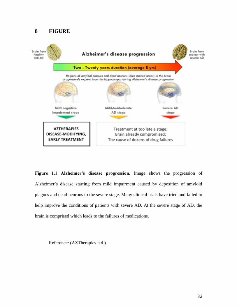
Figure 1.1 Alzheimer’s disease progression. Image shows the progression of
Alzheimer’s disease starting from mild impairment caused by deposition of amyloid
plagues and dead neurons to the severe stage. Many clinical trials have tried and failed to
help improve the conditions of patients with severe AD. At the severe stage of AD, the
brain is comprised which leads to the failures of medications.
Reference: (AZTherapies n.d.)
34
Figure 1.2 The Sig-1R homotrimer and its position in the ER membrane.
Reference: (Schmidt et al. 2016)
35
Figure 1.3 Unfolded Protein Response (UPR), a network of intracellular
signalling pathways that maintain the proper protein folding within the ER.
This stress response has been shown to be conserved between all mammalian
species, as well as yeast. Cells utilize this response in the effort to maintain
homeostasis.
Reference: (Walter & Ron 2012)
36
Figure 2.1 Baseline activation of UPR pathways. (A) Baseline activation of the IRE1
pathway. Transcript levels of spliced XBP1 over the total XBP reflect activation of the
IRE1 pathway. Black bar= Immortalized WT; Red Bar= Immortalized KO; n=18; asterisk
represents 5% significance level. (B) Baseline activation of the PERK pathway. CHOP and
GADD34 are both effectors of the PERK pathway. GADD34 is clearly up regulated in the
immortalized KO indicating a baseline activation of the PERK pathway. Black bar=
Immortalized WT; Red Bar= Immortalized KO; n=18.
37
Figure 2.2: Response to acute stress (tunicamycin). Tunicamycin (3µM) was added to
the growth medium for the times indicated. Controls have no tunicamycin added, n=6. (A)
Graphical representation of transcript levels. (B) PERK pathway. CHOP transcript levels
expressed as a percentage over the WT control. (C) IRE1 pathway. Response to acute
stress. Tunicamycin (3µM) was added to the growth medium for the times indicated.
Controls have no tunicamycin added, n=6. Spliced XBP (XBP1s) transcript expressed as a
ratio over total (spliced and un-spliced XBP1s +XBP1-us). The transcript levels are
expressed as a percentage over the WT control; n=18. (D) ATF6 pathway. Response to
acute stress (tunicamycin). Tunicamycin (3µM) was added to the growth medium for the
times indicated. Controls have no tunicamycin added, n=6. ATF6 transcript levels
expressed as a percentage over the WT control; asterisk represents 5% significance level.
38
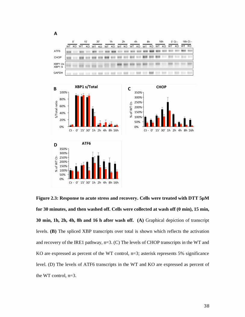
Figure 2.3: Response to acute stress and recovery. Cells were treated with DTT 5µM
for 30 minutes, and then washed off. Cells were collected at wash off (0 min), 15 min,
30 min, 1h, 2h, 4h, 8h and 16 h after wash off. (A) Graphical depiction of transcript
levels. (B) The spliced XBP transcripts over total is shown which reflects the activation
and recovery of the IRE1 pathway, n=3. (C) The levels of CHOP transcripts in the WT and
KO are expressed as percent of the WT control, n=3; asterisk represents 5% significance
level. (D) The levels of ATF6 transcripts in the WT and KO are expressed as percent of
the WT control, n=3.
39
Figure 2.4: The primary Sig-1R KO MEFs are not stressed at baseline. (A) Expression
levels of CHOP, GADD34 and XBP1 in the immortalized WT, immortalized KO, primary
WT and primary KO. (B) Images of immortalized WT and KO MEFs showing the
differences in morphology.
40
Figure 2.5. The Sig-1R is required for the reestablishment of cell homeostasis after
acute ER stress using DTT. Primary WT (blue bars), Primary KO (green bars), and
Knockdown line (burgundy) were treated with DTT (5uM) for 30 minutes, and then
washed off. Cells were collected at wash off (DTT treated), 30 min, 1h, 2h, 4h, 8h, and
16h post wash off. (A) and (B) GADD34 and CHOP expression levels are shown here
normalized to the WT control. Controls were not treated with DTT.
41
9 APPENDIX
Appendix A: Ectopic over expression of WT Sig-R in immortalized KO MEFs does
not rescue the baseline stress. Tunicamycin (3µM) was added to the growth medium for
the times indicated. Controls have no tunicamycin added (A) Western blots showing the
expression levels of GADD34 in WT, KO and Stable cell lines (B) Quantification of gel
images showing the expression levels of GADD34 at baseline and post treatment in WT,
KO, and Stable cell lines.
42
0
0.2
0.4
0.6
0.8
1
Ct- 8h 12h 16h
s/T
ota
l rat
ioXBP1 s/T
WT
#9
KO
D10
A
0%
50%
100%
150%
200%
250%
300%
350%
400%
Ct- 8h 12h 16h
% o
f W
T c
t-
CHOP
WT
#9
KO
D10
B
0%
50%
100%
150%
200%
250%
300%
Ct- 8h 12h 16h
% o
f W
T c
t-
ATF6WT
#9
KO
D10
C
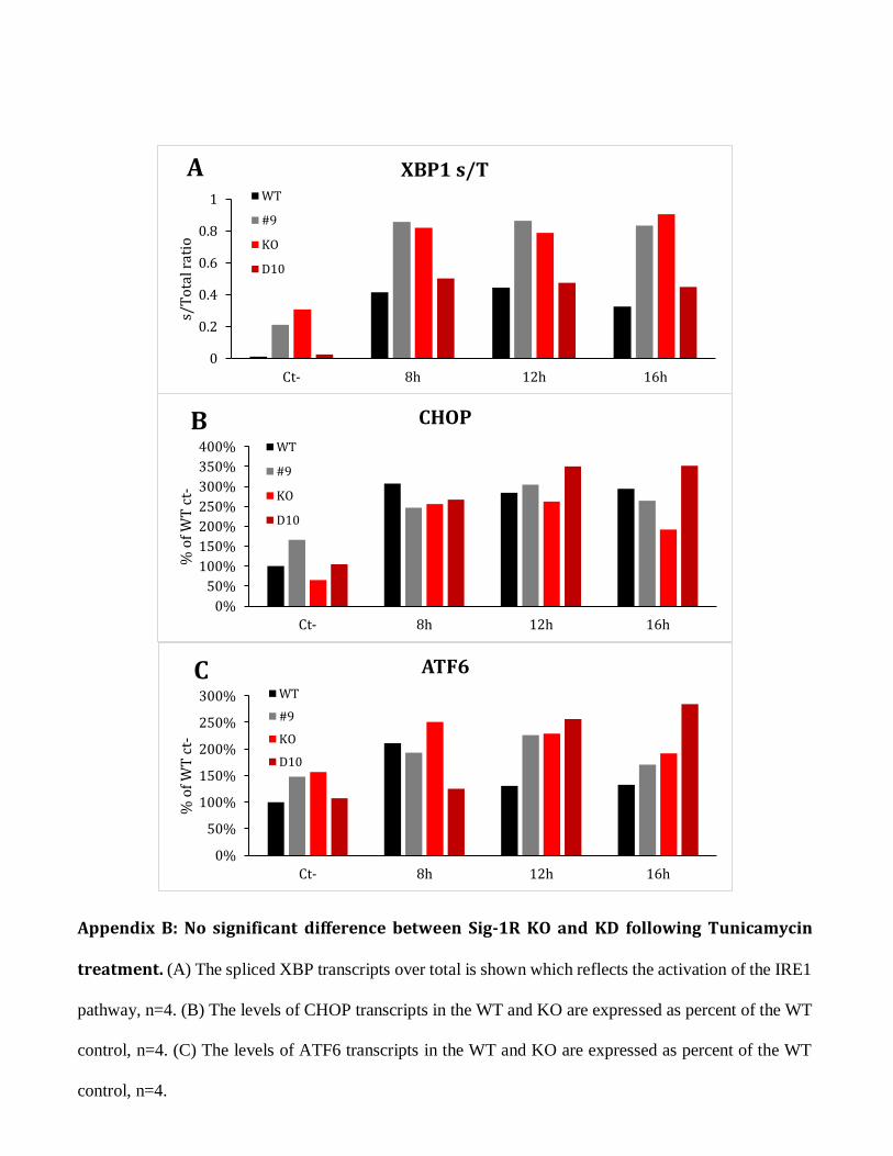
Appendix B: No significant difference between Sig-1R KO and KD following Tunicamycin
treatment. (A) The spliced XBP transcripts over total is shown which reflects the activation of the IRE1
pathway, n=4. (B) The levels of CHOP transcripts in the WT and KO are expressed as percent of the WT
control, n=4. (C) The levels of ATF6 transcripts in the WT and KO are expressed as percent of the WT
control, n=4.
43
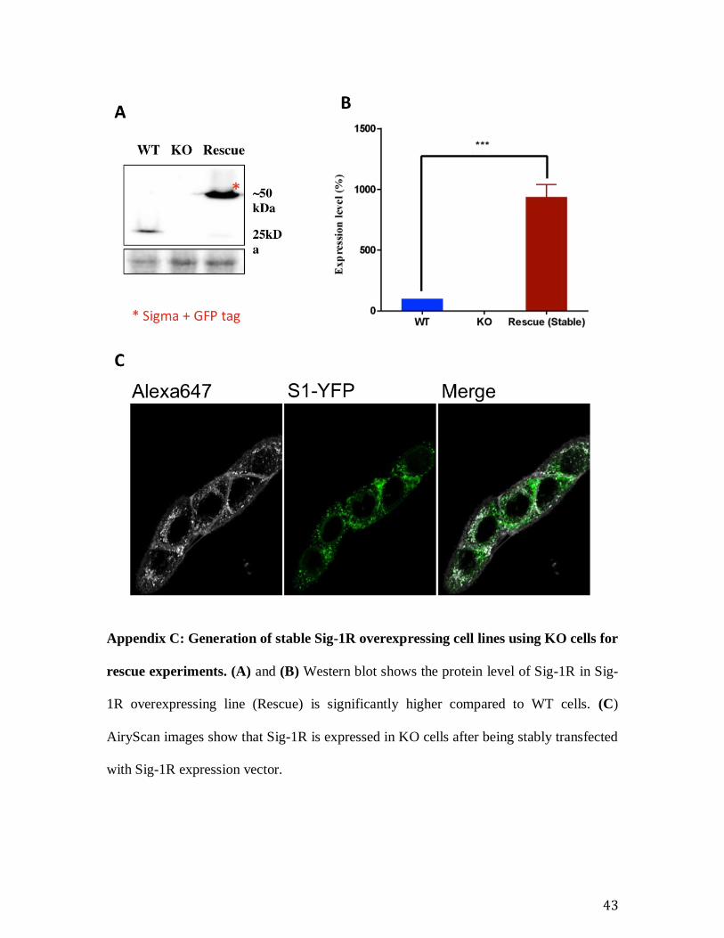
Appendix C: Generation of stable Sig-1R overexpressing cell lines using KO cells for
rescue experiments. (A) and (B) Western blot shows the protein level of Sig-1R in Sig-
1R overexpressing line (Rescue) is significantly higher compared to WT cells. (C)
AiryScan images show that Sig-1R is expressed in KO cells after being stably transfected
with Sig-1R expression vector.
44
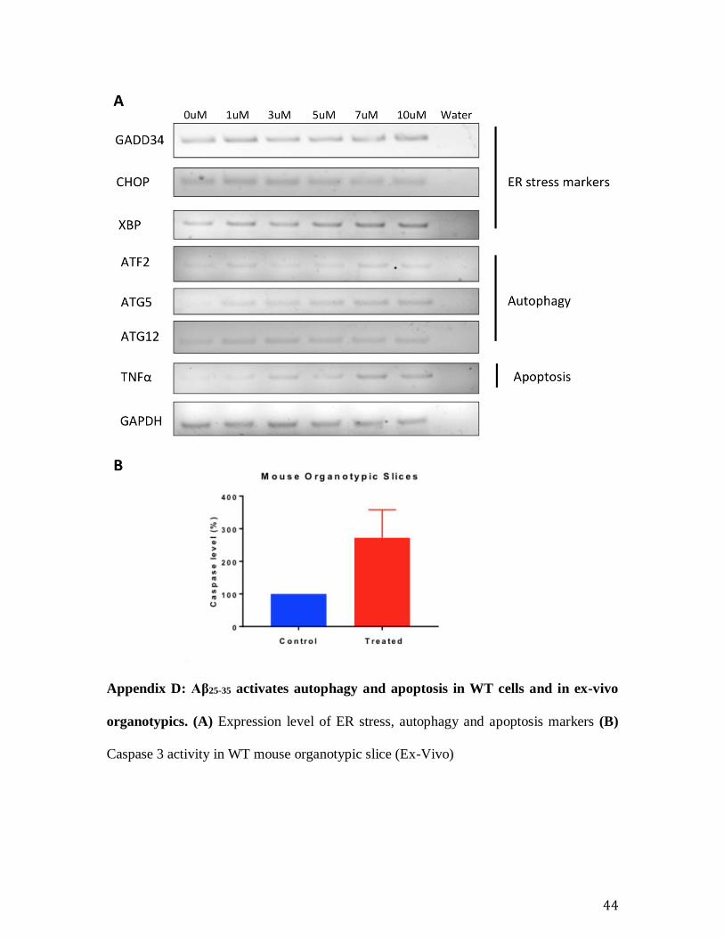
Appendix D: Aβ25-35 activates autophagy and apoptosis in WT cells and in ex-vivo
organotypics. (A) Expression level of ER stress, autophagy and apoptosis markers (B)
Caspase 3 activity in WT mouse organotypic slice (Ex-Vivo)
45
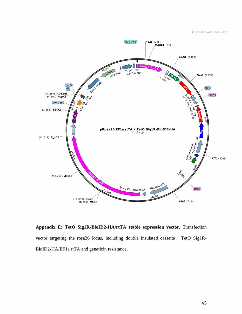
Appendix E: TetO Sig1R-BioID2-HA/rtTA stable expression vector. Transfection
vector targeting the rosa26 locus, including double insulated cassette : TetO Sig1R-
BioID2-HA/EF1a rtTA and geneticin resistance.
46
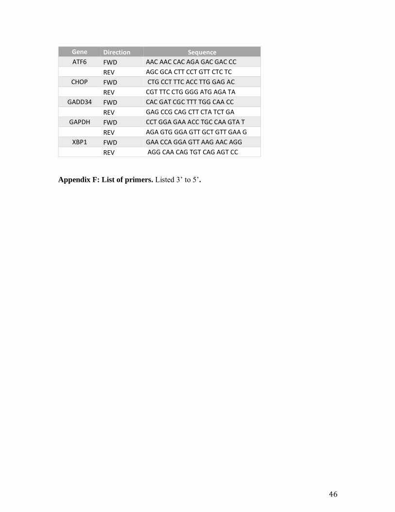
Gene Direction Sequence
ATF6 FWD AAC AAC CAC AGA GAC GAC CC REV AGC GCA CTT CCT GTT CTC TC
CHOP FWD CTG CCT TTC ACC TTG GAG AC REV CGT TTC CTG GGG ATG AGA TA
GADD34 FWD CAC GAT CGC TTT TGG CAA CC REV GAG CCG CAG CTT CTA TCT GA
GAPDH FWD CCT GGA GAA ACC TGC CAA GTA T REV AGA GTG GGA GTT GCT GTT GAA G
XBP1 FWD GAA CCA GGA GTT AAG AAC AGG REV AGG CAA CAG TGT CAG AGT CC
Appendix F: List of primers. Listed 3’ to 5’.
47
10 REFERENCE
Allahtavakoli, M. & Jarrott, B., 2011. Sigma-1 receptor ligand PRE-084 reduced infarct
volume, neurological deficits, pro-inflammatory cytokines and enhanced anti-
inflammatory cytokines after embolic stroke in rats. Brain Research Bulletin, 85(3–
4), pp.219–224. Available at: http://dx.doi.org/10.1016/j.brainresbull.2011.03.019.
Alzheimer, A. et al., 1995. An english translation of alzheimer’s 1907 paper, "Uber
eine eigenartige erkankung der hirnrinde? Clinical Anatomy, 8(6), pp.429–431.
Available at: http://www.ncbi.nlm.nih.gov/pubmed/8713166 [Accessed August 13,
2018].
Andreasson, U. et al., 2014. CSF biomarkers for Alzheimer’s pathology and the effect
size of APOE ɛ4. Molecular Psychiatry, 19(2), pp.148–149. Available at:
http://www.ncbi.nlm.nih.gov/pubmed/23419830 [Accessed August 13, 2018].
Andreev, D.E. et al., 2015. Translation of 5’ leaders is pervasive in genes resistant to
eIF2 repression. eLife, 2015(4), pp.1–21.
AZTherapies, I., Overview – AZTherapies, Inc. Available at:
https://aztherapies.com/company-info/overview/ [Accessed August 16, 2018].
Balin, B.J. & Hudson, A.P., 2014. Etiology and pathogenesis of late-onset Alzheimer’s
disease. Current allergy and asthma reports, 14(3), p.417. Available at:
http://link.springer.com/10.1007/s11882-013-0417-1 [Accessed August 13, 2018].
Ball, M.J. et al., 1985. A new definition of Alzheimer’s disease: a hippocampal dementia.
Lancet (London, England), 1(8419), pp.14–6. Available at:
http://www.ncbi.nlm.nih.gov/pubmed/2856948 [Accessed August 13, 2018].

48
Ballenger, J.F., 2006. Progress in the history of Alzheimer’s disease: the importance of
context. Journal of Alzheimer’s disease : JAD, 9(3 Suppl), pp.5–13. Available at:
http://www.ncbi.nlm.nih.gov/pubmed/17004361 [Accessed August 13, 2018].
Barten, D.M. et al., 2006. Gamma-secretase inhibitors for Alzheimer’s disease: balancing
efficacy and toxicity. Drugs in R&D, 7(2), pp.87–97. Available at:
http://www.ncbi.nlm.nih.gov/pubmed/16542055 [Accessed August 13, 2018].
Belal, C. et al., 2012. The homocysteine-inducible endoplasmic reticulum (ER) stress
protein herp counteracts mutant α-synuclein-induced ER stress via the homeostatic
regulation of ER-resident calcium release channel proteins. Human Molecular
Genetics, 21(5), pp.963–977.
Bhakta-Guha, D. & Efferth, T., 2015. Hormesis: Decoding two sides of the same coin.
Pharmaceuticals, 8(4), pp.865–883.
Binder, L.I. et al., 2005. Tau, tangles, and Alzheimer’s disease. Biochimica et Biophysica
Acta (BBA) - Molecular Basis of Disease, 1739(2–3), pp.216–223. Available at:
http://www.ncbi.nlm.nih.gov/pubmed/15615640 [Accessed August 13, 2018].
Bitan, G. et al., 2003. Amyloid -protein (A ) assembly: A 40 and A 42 oligomerize
through distinct pathways. Proceedings of the National Academy of Sciences,
100(1), pp.330–335. Available at: http://www.ncbi.nlm.nih.gov/pubmed/12506200
[Accessed August 13, 2018].
Blennow, K., de Leon, M.J. & Zetterberg, H., 2006. Alzheimer’s disease. The Lancet,
368(9533), pp.387–403. Available at:
http://www.ncbi.nlm.nih.gov/pubmed/16876668 [Accessed August 13, 2018].
Braak, H. & Braak, E., 1991. Neuropathological stageing of Alzheimer-related changes.
49
Acta Neuropathologica, 82(4), pp.239–259. Available at:
http://link.springer.com/10.1007/BF00308809 [Accessed August 13, 2018].
Braak, H. & Braak, E., 1997. Staging of Alzheimer-related cortical destruction.
International psychogeriatrics, 9 Suppl 1, pp.257-61; discussion 269-72. Available
at: http://www.ncbi.nlm.nih.gov/pubmed/9447446 [Accessed August 13, 2018].
Bretsky, P.M. et al., Evidence for an interaction between apolipoprotein E genotype,
gender, and Alzheimer disease. Alzheimer disease and associated disorders, 13(4),
pp.216–21. Available at: http://www.ncbi.nlm.nih.gov/pubmed/10609670 [Accessed
August 13, 2018].
vom Brocke, J. et al., 2006. MEF immortalization to investigate the ins and outs of
mutagenesis. Carcinogenesis, 27(11), pp.2141–7. Available at:
https://academic.oup.com/carcin/article-lookup/doi/10.1093/carcin/bgl101
[Accessed August 14, 2018].
Brouwers, N., Sleegers, K. & Van Broeckhoven, C., 2008. Molecular genetics of
Alzheimer’s disease: An update. Annals of Medicine, 40(8), pp.562–583. Available
at: http://www.ncbi.nlm.nih.gov/pubmed/18608129 [Accessed August 13, 2018].
Busuttil, R.A. et al., 2003. Oxygen accelerates the accumulation of mutations during the
senescence and immortalization of murine cells in culture. Aging Cell, 2(6), pp.287–
294. Available at: http://doi.wiley.com/10.1046/j.1474-9728.2003.00066.x.
Carlesimo, G.A. & Oscar-Berman, M., 1992. Memory deficits in Alzheimer’s patients: A
comprehensive review. Neuropsychology Review, 3(2), pp.119–169. Available at:
http://link.springer.com/10.1007/BF01108841 [Accessed August 13, 2018].
Carrara, M. et al., 2015. Noncanonical binding of BiP ATPase domain to Ire1 and Perk is
50
dissociated by unfolded protein CH1 to initiate ER stress signaling. eLife, 2015(4),
pp.1–16.
Castellani, R.J. et al., 2009. Reexamining Alzheimer’s Disease: Evidence for a Protective
Role for Amyloid-β Protein Precursor and Amyloid-β G. Bissette, ed. Journal of
Alzheimer’s Disease, 18(2), pp.447–452. Available at:
http://www.ncbi.nlm.nih.gov/pubmed/19584435 [Accessed August 13, 2018].
Castellani, R.J. & Smith, M.A., 2011. Compounding artefacts with uncertainty, and an
amyloid cascade hypothesis that is ‘too big to fail.’ The Journal of Pathology,
224(2), pp.147–152. Available at: http://www.ncbi.nlm.nih.gov/pubmed/21557219
[Accessed August 13, 2018].
Cendán, C.M. et al., 2005. Formalin-induced pain is reduced in σ1 receptor knockout
mice. European Journal of Pharmacology, 511(1), pp.73–74. Available at:
https://www.sciencedirect.com/science/article/pii/S0014299905001391 [Accessed
August 14, 2018].
Chan, S.L. et al., 2004. Herp stabilizes neuronal Ca2+ homeostasis and mitochondrial
function during endoplasmic reticulum stress. Journal of Biological Chemistry,
279(27), pp.28733–28743.
Chevallier, N., Keller, E. & Maurice, T., 2011. Behavioural phenotyping of knockout
mice for the sigma-1 (σ 1 ) chaperone protein revealed gender-related anxiety,
depressive-like and memory alterations. Journal of Psychopharmacology, 25(7),
pp.960–975. Available at: http://www.ncbi.nlm.nih.gov/pubmed/21555330
[Accessed August 14, 2018].
Chikka, M.R. et al., 2013. C/EBP homologous protein (CHOP) contributes to suppression
51
of metabolic genes during endoplasmic reticulum stress in the liver. The Journal of
biological chemistry, 288(6), pp.4405–15. Available at:
http://www.ncbi.nlm.nih.gov/pubmed/23281479 [Accessed August 15, 2018].
Claeysen, S. et al., 2012. Alzheimer culprits: Cellular crossroads and interplay. Cellular
Signalling, 24(9), pp.1831–1840. Available at:
http://linkinghub.elsevier.com/retrieve/pii/S0898656812001453 [Accessed August
13, 2018].
Cornejo, V.H. & Hetz, C., 2013. The unfolded protein response in Alzheimer’s disease.
Seminars in Immunopathology, 35(3), pp.277–292. Available at:
http://link.springer.com/10.1007/s00281-013-0373-9 [Accessed August 13, 2018].
Cummings, J.L., 2004. Treatment of Alzheimer’s disease: current and future therapeutic
approaches. Reviews in neurological diseases, 1(2), pp.60–9. Available at:
http://www.ncbi.nlm.nih.gov/pubmed/16400259 [Accessed August 13, 2018].
DeKosky, S.T. & Scheff, S.W., 1990. Synapse loss in frontal cortex biopsies in
Alzheimer’s disease: Correlation with cognitive severity. Annals of Neurology,
27(5), pp.457–464. Available at: http://doi.wiley.com/10.1002/ana.410270502
[Accessed August 13, 2018].
Doody, R.S. et al., 2013. A Phase 3 Trial of Semagacestat for Treatment of Alzheimer’s
Disease. New England Journal of Medicine, 369(4), pp.341–350. Available at:
http://www.nejm.org/doi/10.1056/NEJMoa1210951 [Accessed August 13, 2018].
Doody, R.S. et al., 2014. Phase 3 Trials of Solanezumab for Mild-to-Moderate
Alzheimer’s Disease. New England Journal of Medicine, 370(4), pp.311–321.
Available at: http://www.nejm.org/doi/10.1056/NEJMoa1312889 [Accessed August
52
13, 2018].
Duran-Aniotz, C. et al., 2014. Aggregate-Depleted Brain Fails to Induce Aβ Deposition
in a Mouse Model of Alzheimer’s Disease J. Bartz, ed. PLoS ONE, 9(2), p.e89014.
Available at: http://dx.plos.org/10.1371/journal.pone.0089014 [Accessed August 13,
2018].
Earley, B. et al., 1991. Evidence for an anti-amnesic effect of JO 1784 in the rat: a potent
and selective ligand for the sigma receptor. Brain research, 546(2), pp.282–6.
Available at: http://www.ncbi.nlm.nih.gov/pubmed/1676927 [Accessed August 14,
2018].
Elcombe, E.L. et al., 2014. Clinical and Cognitive Correlates of Structural Hippocampal
Change in “At-Risk” Older Adults. Journal of Geriatric Psychiatry and Neurology,
27(2), pp.67–76. Available at: http://www.ncbi.nlm.nih.gov/pubmed/24196661
[Accessed August 13, 2018].
Entrena, J.M. et al., 2009. Sigma-1 receptors are essential for capsaicin-induced
mechanical hypersensitivity: Studies with selective sigma-1 ligands and sigma-1
knockout mice. Pain, 143(3), pp.252–261. Available at:
http://www.ncbi.nlm.nih.gov/pubmed/19375855 [Accessed August 14, 2018].
Farrer, L.A. et al., 1997. Effects of age, sex, and ethnicity on the association between
apolipoprotein E genotype and Alzheimer disease. A meta-analysis. APOE and
Alzheimer Disease Meta Analysis Consortium. JAMA, 278(16), pp.1349–56.
Available at: http://www.ncbi.nlm.nih.gov/pubmed/9343467 [Accessed August 13,
2018].
Fehér, Á. et al., 2012. Association between a variant of the sigma-1 receptor gene and
53
Alzheimer’s disease. Neuroscience Letters, 517(2), pp.136–139. Available at:
http://www.ncbi.nlm.nih.gov/pubmed/22561649 [Accessed August 14, 2018].
Ferreira, S.T. et al., 2015. Soluble amyloid-Î2 oligomers as synaptotoxins leading to
cognitive impairment in Alzheimerâ€TMs disease. Frontiers in Cellular
Neuroscience, 9, p.191. Available at:
http://www.ncbi.nlm.nih.gov/pubmed/26074767 [Accessed August 13, 2018].
Forman, M. et al., 2012. The novel BACE inhibitor MK-8931 dramatically lowers
cerebrospinal fluid Aβ peptides in healthy subjects following single- and multiple-
dose administration. Alzheimer’s & Dementia, 8(4), p.P704. Available at:
https://linkinghub.elsevier.com/retrieve/pii/S1552526012020353 [Accessed August
13, 2018].
Frackowiak, J., Zoltowska, A. & Wisniewski, H.M., 1994. Non-fibrillar beta-amyloid
protein is associated with smooth muscle cells of vessel walls in Alzheimer disease.
Journal of neuropathology and experimental neurology, 53(6), pp.637–45.
Available at: http://www.ncbi.nlm.nih.gov/pubmed/7964904 [Accessed August 13,
2018].
Fridman, A.L. & Tainsky, M.A., 2008. Critical pathways in cellular senescence and
immortalization revealed by gene expression profiling. Oncogene, 27(46), pp.5975–
5987. Available at: http://www.ncbi.nlm.nih.gov/pubmed/18711403 [Accessed
August 14, 2018].
Gao, X.F. et al., 2012. Sigma-1 Receptor Agonists Directly Inhibit NaV1.2/1.4 Channels.
PLoS ONE, 7(11).
Gharibyan, A.L. et al., 2007. Lysozyme Amyloid Oligomers and Fibrils Induce Cellular
54
Death via Different Apoptotic/Necrotic Pathways. Journal of Molecular Biology,
365(5), pp.1337–1349. Available at:
http://www.ncbi.nlm.nih.gov/pubmed/17134716 [Accessed August 13, 2018].
Glabe, C.G. & Kayed, R., 2006. Common structure and toxic function of amyloid
oligomers implies a common mechanism of pathogenesis. Neurology, 66(Issue 1,
Supplement 1), pp.S74–S78. Available at:
http://www.ncbi.nlm.nih.gov/pubmed/16432151 [Accessed August 13, 2018].
Glenner, G.G. & Wong, C.W., 1984. Alzheimer’s disease: initial report of the
purification and characterization of a novel cerebrovascular amyloid protein.
Biochemical and biophysical research communications, 120(3), pp.885–90.
Available at: http://www.ncbi.nlm.nih.gov/pubmed/6375662 [Accessed August 13,
2018].
Goate, A., 2006. Segregation of a missense mutation in the amyloid beta-protein
precursor gene with familial Alzheimer’s disease. Journal of Alzheimer’s disease :
JAD, 9(3 Suppl), pp.341–7. Available at:
http://www.ncbi.nlm.nih.gov/pubmed/16914872 [Accessed August 13, 2018].
Gold, P.W., Licinio, J. & Pavlatou, M.G., 2013. Pathological parainflammation and
endoplasmic reticulum stress in depression: potential translational targets through
the CNS insulin, klotho and PPAR-γ systems. Molecular psychiatry, 18(2), pp.154–
65. Available at: http://www.ncbi.nlm.nih.gov/pubmed/23183489.
Graves, A.B. et al., 1991. Occupational exposures to solvents and lead as risk factors for
Alzheimer’s disease: a collaborative re-analysis of case-control studies. EURODEM
Risk Factors Research Group. International journal of epidemiology, 20 Suppl 2,
55
pp.S58-61. Available at: http://www.ncbi.nlm.nih.gov/pubmed/1833355 [Accessed
August 13, 2018].
Gustafson, D.R. et al., 2006. Cerebrospinal fluid -amyloid 1-42 concentration may
predict cognitive decline in older women. Journal of Neurology, Neurosurgery &
Psychiatry, 78(5), pp.461–464. Available at:
http://www.ncbi.nlm.nih.gov/pubmed/17098843 [Accessed August 13, 2018].
Ha, Y. et al., 2011. Sigma receptor 1 modulates endoplasmic reticulum stress in retinal
neurons. Investigative ophthalmology & visual science, 52(1), pp.527–540.
Ha, Y. et al., 2014. Sigma receptor 1 modulates ER stress and Bcl2 in murine retina. Cell
and Tissue Research, 356(1), pp.15–27.
Hamley, I.W., 2012. The Amyloid Beta Peptide: A Chemist’s Perspective. Role in
Alzheimer’s and Fibrillization. Chemical Reviews, 112(10), pp.5147–5192.
Available at: http://www.ncbi.nlm.nih.gov/pubmed/22813427 [Accessed August 13,
2018].
Hanner, M. et al., 1996. Purification, molecular cloning, and expression of the
mammalian sigma1-binding site. Proceedings of the National Academy of Sciences
of the United States of America, 93(15), pp.8072–8077.
Harding, H.P. et al., 2009. Ppp1r15 gene knockout reveals an essential role for translation
initiation factor 2 alpha (eIF2alpha) dephosphorylation in mammalian development.
Proceedings of the National Academy of Sciences of the United States of America,
106(6), pp.1832–7. Available at:
http://www.ncbi.nlm.nih.gov/pubmed/19181853%5Cnhttp://www.pubmedcentral.ni
h.gov/articlerender.fcgi?artid=PMC2644123.
56
Hardy, J. et al., 2014. Pathways to Alzheimer’s disease. Journal of internal medicine,
275(3), pp.296–303. Available at: http://www.ncbi.nlm.nih.gov/pubmed/24749173
[Accessed August 13, 2018].
Hardy, J. & Allsop, D., 1991. Amyloid deposition as the central event in the aetiology of
Alzheimer’s disease. Trends in pharmacological sciences, 12(10), pp.383–8.
Available at: http://www.ncbi.nlm.nih.gov/pubmed/1763432 [Accessed August 13,
2018].
Hardy, J. & Selkoe, D.J., 2002. The Amyloid Hypothesis of Alzheimer’s Disease:
Progress and Problems on the Road to Therapeutics. Science, 297(5580), pp.353–
356. Available at: http://www.ncbi.nlm.nih.gov/pubmed/12130773 [Accessed
August 13, 2018].
Hardy, J.A. & Higgins, G.A., 1992. Alzheimer’s disease: the amyloid cascade hypothesis.
Science (New York, N.Y.), 256(5054), pp.184–5. Available at:
http://www.ncbi.nlm.nih.gov/pubmed/1566067 [Accessed August 13, 2018].
Hayashi, A. et al., 2009. Aberrant endoplasmic reticulum stress response in
lymphoblastoid cells from patients with bipolar disorder. The international journal
of neuropsychopharmacology / official scientific journal of the Collegium
Internationale Neuropsychopharmacologicum (CINP), 12(1), pp.33–43. Available
at: http://www.ncbi.nlm.nih.gov/pubmed/18771604.
Hayashi, T. et al., 2012. Dynamic Interaction between Sigma-1 Receptor and Kv1 . 2
Shapes Neuronal and Behavioral Responses to Cocaine.
Hayashi, T. et al., 2011. Targeting ligand-operated chaperone sigma-1 receptors in the
treatment of neuropsychiatric disorders. Expert opinion on therapeutic targets,
57
15(5), pp.557–77. Available at: http://www.scopus.com/inward/record.url?eid=2-
s2.0-79954498271&partnerID=tZOtx3y1.
Hayashi, T. & Su, T.-, 2005. The Sigma Receptor: Evolution of the Concept in
Neuropsychopharmacology. Current Neuropharmacology.
Hayashi, T. & Su, T.P., 2007. Sigma-1 Receptor Chaperones at the ER- Mitochondrion
Interface Regulate Ca2+ Signaling and Cell Survival. Cell, 131(3), pp.596–610.
Hayashi, T. & Su, T.P., 2003. Sigma-1 receptors (sigma(1) binding sites) form raft-like
microdomains and target lipid droplets on the endoplasmic reticulum: roles in
endoplasmic reticulum lipid compartmentalization and export. J Pharmacol Exp
Ther, 306(2), pp.718–725. Available at:
http://www.ncbi.nlm.nih.gov/pubmed/12730355%5Cnhttp://jpet.aspetjournals.org/c
ontent/306/2/718.full.pdf.
Haze, K. et al., 1999. Mammalian Transcription Factor ATF6 Is Synthesized as a
Transmembrane Protein and Activated by Proteolysis in Response to Endoplasmic
Reticulum Stress. Molecular Biology of the Cell, 10(11), pp.3787–3799. Available
at: http://www.molbiolcell.org/content/10/11/3787.long.
Henderson, V.W., 2014. Alzheimer’s disease: Review of hormone therapy trials and
implications for treatment and prevention after menopause. The Journal of Steroid
Biochemistry and Molecular Biology, 142, pp.99–106. Available at:
http://www.ncbi.nlm.nih.gov/pubmed/23727128 [Accessed August 13, 2018].
Hetz, C. et al., 2011. The Unfolded Protein Response: Integrating Stress Signals Through
the Stress Sensor IRE1 . Physiological Reviews, 91(4), pp.1219–1243.
Hetz, C. & Mollereau, B., 2014. Disturbance of endoplasmic reticulum proteostasis in
58
neurodegenerative diseases. Nature reviews neuroscience, 15(4), pp.233–249.
Available at:
http://eutils.ncbi.nlm.nih.gov/entrez/eutils/elink.fcgi?dbfrom=pubmed&id=246
19348&retmode=ref&cmd=prlinks.
Hollien, J. & Weissman, J.S., 2006. Decay of Endoplasmic Reticulum-Localized mRNAs
During the Unfolded Protein Response. , (July), pp.104–107.
Hoozemans, J.J.M. et al., 2005. The unfolded protein response is activated in
Alzheimer’s disease. Acta Neuropathologica, 110(2), pp.165–172. Available at:
http://link.springer.com/10.1007/s00401-005-1038-0 [Accessed August 14, 2018].
Hoshino, T. et al., 2007. Involvement of prostaglandin E2 in production of amyloid-beta
peptides both in vitro and in vivo. The Journal of biological chemistry, 282(45),
pp.32676–88. Available at: http://www.ncbi.nlm.nih.gov/pubmed/17767011
[Accessed August 14, 2018].
Hsiao, E.C. et al., 2011. Constitutive Gs activation using a single-construct tetracycline-
inducible expression system in embryonic stem cells and mice. Stem cell research &
therapy, 2(2), p.11. Available at: http://stemcellres.com/content/2/2/11.
Huang, Y. et al., 2011. Genetic polymorphisms in sigma-1 receptor and apolipoprotein E
interact to influence the severity of Alzheimer’s disease. Current Alzheimer
research, 8(7), pp.765–70. Available at:
http://www.ncbi.nlm.nih.gov/pubmed/21605063 [Accessed August 14, 2018].
Imagawa, Y. et al., 2008. RNase domains determine the functional difference between
IRE1α and IRE1β. FEBS Letters, 582(5), pp.656–660.
Imaizumi, K. et al., 2001. The unfolded protein response and Alzheimer’s disease.
59
Biochimica et biophysica acta, 1536(2–3), pp.85–96. Available at:
http://www.ncbi.nlm.nih.gov/pubmed/11406343 [Accessed August 13, 2018].
Ishikawa, M. & Hashimoto, K., 2010. The role of sigma-1 receptors in the
pathophysiology of neuropsychiatric diseases. Journal of Receptor, Ligand and
Channel Research, 3, pp.25–36.
Isik, A.T., 2010. Late onset Alzheimer’s disease in older people. Clinical interventions in
aging, 5, pp.307–11. Available at: http://www.ncbi.nlm.nih.gov/pubmed/21103401
[Accessed August 13, 2018].
Jacobsen, J.S. et al., 2006. Early-onset behavioral and synaptic deficits in a mouse model
of Alzheimer’s disease. Proceedings of the National Academy of Sciences of the
United States of America, 103(13), pp.5161–6. Available at:
http://www.pnas.org/cgi/doi/10.1073/pnas.0600948103 [Accessed August 13, 2018].
Jagust, W., 2013. Vulnerable neural systems and the borderland of brain aging and
neurodegeneration. Neuron, 77(2), pp.219–34. Available at:
http://www.ncbi.nlm.nih.gov/pubmed/23352159 [Accessed August 13, 2018].
Jansen, K.L. et al., 1993. Loss of sigma binding sites in the CA1 area of the anterior
hippocampus in Alzheimer’s disease correlates with CA1 pyramidal cell loss. Brain
research, 623(2), pp.299–302. Available at:
http://www.ncbi.nlm.nih.gov/pubmed/8221112 [Accessed August 13, 2018].
Jin, J.-L. et al., 2015. Roles of sigma-1 receptors in Alzheimer’s disease. International
journal of clinical and experimental medicine, 8(4), pp.4808–20. Available at:
http://www.ncbi.nlm.nih.gov/pubmed/26131055 [Accessed August 14, 2018].
Johannessen, M. et al., 2009. Voltage-gated sodium channel modulation by sigma-
60
receptors in cardiac myocytes and heterologous systems. American journal of
physiology. Cell physiology, 296(5), pp.C1049–C1057.
Kakiuchi, C. et al., 2004. Association of the XBP1-116C/G polymorphism with
schizophrenia in the Japanese population. Psychiatry and clinical neurosciences,
58(4), pp.438–440.
Karran, E., Mercken, M. & Strooper, B. De, 2011. The amyloid cascade hypothesis for
Alzheimer’s disease: an appraisal for the development of therapeutics. Nature
Reviews Drug Discovery, 10(9), pp.698–712. Available at:
http://www.ncbi.nlm.nih.gov/pubmed/21852788 [Accessed August 13, 2018].
Katayama, T. et al., 1999. Presenilin-1 mutations downregulate the signalling pathway of
the unfolded-protein response. Nature cell biology, 1(8), pp.479–85. Available at:
http://www.nature.com/articles/ncb1299_479 [Accessed August 13, 2018].
Kawada, K. et al., 2014. Aberrant neuronal differentiation and inhibition of dendrite
outgrowth resulting from endoplasmic reticulum stress. Journal of Neuroscience
Research, 92(9), pp.1122–1133.
Keisuke Yamamoto, Hiderou Yoshida, Koichi Kokame, R.J.K. and K.M., 2004.
Differential Contribution of ATF6 and XBP1to the Activation os Endoplasmic
Reticulum Stress-Responcive cis-acting Elements ERSE, UPRE and ERSE-II. , 606,
pp.601–606.
Kennedy, B.K. et al., 2014. Geroscience: Linking Aging to Chronic Disease. Cell,
159(4), pp.709–713. Available at:
https://www.sciencedirect.com/science/article/pii/S009286741401366X [Accessed
August 13, 2018].
61
Kim, T.Y. et al., 2008. Herp enhances ER-associated protein degradation by recruiting
ubiquilins. Biochemical and Biophysical Research Communications, 369(2),
pp.741–746.
Kokame, K., Kato, H. & Miyata, T., 2001. Identification of ERSE-II, a New cis-Acting
Element Responsible for the ATF6-dependent Mammalian Unfolded Protein
Response. Journal of Biological Chemistry, 276(12), pp.9199–9205.
Kopan, R. & Ilagan, M.X.G., 2004. γ-Secretase: proteasome of the membrane? Nature
Reviews Molecular Cell Biology, 5(6), pp.499–504. Available at:
http://www.ncbi.nlm.nih.gov/pubmed/15173829 [Accessed August 13, 2018].
Kukull, W.A. et al., 2002. Dementia and Alzheimer disease incidence: a prospective
cohort study. Archives of neurology, 59(11), pp.1737–46. Available at:
http://www.ncbi.nlm.nih.gov/pubmed/12433261 [Accessed August 13, 2018].
Kukull, W.A. & Bowen, J.D., 2002. Dementia epidemiology. The Medical clinics of
North America, 86(3), pp.573–90. Available at:
http://www.ncbi.nlm.nih.gov/pubmed/12168560 [Accessed August 13, 2018].
Lambert, M.P. et al., 1998. Diffusible, nonfibrillar ligands derived from Abeta1-42 are
potent central nervous system neurotoxins. Proceedings of the National Academy of
Sciences of the United States of America, 95(11), pp.6448–53. Available at:
http://www.ncbi.nlm.nih.gov/pubmed/9600986 [Accessed August 13, 2018].
Langa, F. et al., 2003. Generation and phenotypic analysis of sigma receptor type I
(sigma 1) knockout mice. Eur J Neurosci, 18(8), pp.2188–2196.
Lannfelt, L. et al., 2014. Perspectives on future Alzheimer therapies: amyloid-β
protofibrils - a new target for immunotherapy with BAN2401 in Alzheimer’s
62
disease. Alzheimer’s Research & Therapy, 6(2), p.16. Available at:
http://www.ncbi.nlm.nih.gov/pubmed/25031633 [Accessed August 13, 2018].
Lee, C.-C., Sun, Y. & Huang, H.W., 2012. How Type II Diabetes-Related Islet Amyloid
Polypeptide Damages Lipid Bilayers. Biophysical Journal, 102(5), pp.1059–1068.
Available at: http://www.ncbi.nlm.nih.gov/pubmed/22404928 [Accessed August 13,
2018].
Li, H. et al., 2010. Mammalian endoplasmic reticulum stress sensor IRE1 signals by
dynamic clustering. Proc Natl Acad Sci U S A, 107(37), pp.16113–16118. Available
at: http://www.ncbi.nlm.nih.gov/pubmed/20798350.
Li, Y. et al., 2014. New insights into the roles of CHOP-induced apoptosis in ER stress.
Acta Biochimica et Biophysica Sinica, 46(8), pp.629–640. Available at:
https://academic.oup.com/abbs/article-lookup/doi/10.1093/abbs/gmu048 [Accessed
August 15, 2018].
Licastro, F. et al., 2017. Gene Signature in Alzheimer ’ s Disease and Environmental
Factors: The Virus Chronicle. In Handbook of infection and Alzheimer’s disease. pp.
5, 371 – 379.
Lue, L.-F. et al., 1999. Soluble Amyloid β Peptide Concentration as a Predictor of
Synaptic Change in Alzheimer’s Disease. The American Journal of Pathology,
155(3), pp.853–862. Available at:
https://www.sciencedirect.com/science/article/pii/S000294401065184X [Accessed
August 13, 2018].
Maity, S. et al., 2016. Oxidative Homeostasis Regulates the Response to Reductive
Endoplasmic Reticulum Stress through Translation Control. Cell Reports, 16(3),
63
pp.851–865. Available at:
http://linkinghub.elsevier.com/retrieve/pii/S2211124716307653 [Accessed August
14, 2018].
Mangialasche, F. et al., 2010. Alzheimer’s disease: clinical trials and drug development.
The Lancet Neurology, 9(7), pp.702–716. Available at:
http://www.ncbi.nlm.nih.gov/pubmed/20610346 [Accessed August 13, 2018].
Marciniak, S.J. et al., 2004. CHOP induces death by promoting protein synthesis and
oxidation in the stressed endoplasmic reticulum. Genes and Development, 18(24),
pp.3066–3077.
Martenyi, F. et al., 2012. BACE inhibitor LY2886721 safety and central and peripheral
PK and PD in healthy subjects (HSs). Alzheimer’s & Dementia, 8(4), pp.P583–P584.
Available at: https://linkinghub.elsevier.com/retrieve/pii/S1552526012017207
[Accessed August 13, 2018].
Matsuno, K. et al., 1994. Ameliorating effects of sigma receptor ligands on the
impairment of passive avoidance tasks in mice: involvement in the central
acetylcholinergic system. European journal of pharmacology, 261(1–2), pp.43–51.
Available at: http://www.ncbi.nlm.nih.gov/pubmed/8001652 [Accessed August 14,
2018].
Matsuno, K. et al., 1997. SA4503, a novel cognitive enhancer, with σ1 receptor agonistic
properties. Behavioural Brain Research, 83(1–2), pp.221–224. Available at:
https://www.sciencedirect.com/science/article/pii/S0166432897860743 [Accessed
August 14, 2018].
Maurer, K., Volk, S. & Gerbaldo, H., 1997. Auguste D and Alzheimer ’ s disease. The
64
lancet, 349, pp.1906–1909.
Maurice, T. et al., 2016. Learning performances and vulnerability to amyloid toxicity in
the butyrylcholinesterase knockout mouse. Behavioural Brain Research, 296,
pp.351–360. Available at: http://www.ncbi.nlm.nih.gov/pubmed/26306824
[Accessed August 14, 2018].
Maurice, T. et al., 1994. PRE-084, a sigma selective PCP derivative, attenuates MK-801-
induced impairment of learning in mice. Pharmacology, biochemistry, and behavior,
49(4), pp.859–69. Available at: http://www.ncbi.nlm.nih.gov/pubmed/7886099
[Accessed August 14, 2018].
Maurice, T., Grégoire, C. & Espallergues, J., 2006. Neuro(active)steroids actions at the
neuromodulatory sigma1 (σ1) receptor: Biochemical and physiological evidences,
consequences in neuroprotection. Pharmacology Biochemistry and Behavior, 84(4),
pp.581–597. Available at:
https://www.sciencedirect.com/science/article/pii/S009130570600219X [Accessed
August 14, 2018].
Maurice, T. & Privat, A., 1997. SA4503, a novel cognitive enhancer with sigma1
receptor agonist properties, facilitates NMDA receptor-dependent learning in mice.
European journal of pharmacology, 328(1), pp.9–18. Available at:
http://www.ncbi.nlm.nih.gov/pubmed/9203561 [Accessed August 14, 2018].
Maurice, T. & Su, T.P., 2009. The pharmacology of sigma-1 receptors. Pharmacology
and Therapeutics, 124(2), pp.195–206. Available at:
http://dx.doi.org/10.1016/j.pharmthera.2009.07.001.
Maurice, T., Su, T.P. & Privat, A., 1998. Sigma1 (sigma 1) receptor agonists and
65
neurosteroids attenuate B25-35-amyloid peptide-induced amnesia in mice through a
common mechanism. Neuroscience, 83(2), pp.413–28. Available at:
http://www.ncbi.nlm.nih.gov/pubmed/9460750 [Accessed August 14, 2018].
Mavlyutov, T. a et al., 2010. THE SIGMA-1 RECEPTOR IS ENRICHED IN
POSTSYNAPTIC SITES OF C-TERMINALS IN MOUSE MOTONEURONS. AN
ANATOMICAL AND BEHAVIORAL STUDY. , 167(2), pp.247–255.
Mavlyutov, T.A. et al., 2012. Development of the sigma-1 receptor in C-terminals of
motoneurons and colocalization with the N,N’-dimethyltryptamine forming enzyme,
indole-N-methyl transferase. Neuroscience, 206, pp.60–68.
McLean, C.A. et al., 1999. Soluble pool of Abeta amyloid as a determinant of severity of
neurodegeneration in Alzheimer’s disease. Annals of neurology, 46(6), pp.860–6.
Available at: http://www.ncbi.nlm.nih.gov/pubmed/10589538 [Accessed August 13,
2018].
Meunier, J. & Hayashi, T., 2010. Sigma-1 receptors regulate Bcl-2 expression by reactive
oxygen species-dependent transcriptional regulation of nuclear factor κB. Journal of
Pharmacology and Experimental, 332(2), pp.388–397. Available at:
http://jpet.aspetjournals.org/content/332/2/388.short.
Meunier, J., Ieni, J. & Maurice, T., 2009. The anti-amnesic and neuroprotective effects of
donepezil against amyloid β25-35 peptide-induced toxicity in mice involve an
interaction with the σ1 receptor. British Journal of Pharmacology, 149(8), pp.998–
1012. Available at: http://www.ncbi.nlm.nih.gov/pubmed/17057756 [Accessed
August 14, 2018].
Mielke, M.M. et al., 2014. Cerebrospinal fluid sphingolipids, β-amyloid, and tau in adults
66
at risk for Alzheimer’s disease. Neurobiology of Aging, 35(11), pp.2486–2494.
Available at: http://www.ncbi.nlm.nih.gov/pubmed/24952994 [Accessed August 13,
2018].
Mishina, M. et al., 2008. Low density of sigma1 receptors in early Alzheimer’s disease.
Annals of Nuclear Medicine, 22(3), pp.151–156. Available at:
http://www.ncbi.nlm.nih.gov/pubmed/18498028 [Accessed August 13, 2018].
Mitsuda, T. et al., 2011. Sigma-1Rs are upregulated via PERK/eIF2a/ATF4 pathway and
execute protective function in ER stress. Biochemical and Biophysical Research
Communications, 415(3), pp.519–525.
Miyatake, R. et al., 2004. Functional polymorphisms in the sigma1 receptor gene
associated with alcoholism. Biological psychiatry, 55(1), pp.85–90. Available at:
http://www.ncbi.nlm.nih.gov/pubmed/14706429 [Accessed August 14, 2018].
Möller, H.J. & Graeber, M.B., 1998. The case described by Alois Alzheimer in 1911.
Historical and conceptual perspectives based on the clinical record and
neurohistological sections. European archives of psychiatry and clinical
neuroscience, 248(3), pp.111–22. Available at:
http://www.ncbi.nlm.nih.gov/pubmed/9728729 [Accessed August 11, 2018].
Mori, T. et al., 2013. Sigma-1 Receptor Chaperone at the ER-Mitochondrion Interface
Mediates the Mitochondrion-ER-Nucleus Signaling for Cellular Survival. PLoS
ONE, 8(10).
Mori, T., Hayashi, T. & Su, T.-P., 2012. Compromising σ-1 receptors at the endoplasmic
reticulum render cytotoxicity to physiologically relevant concentrations of dopamine
in a nuclear factor-κB/Bcl-2-dependent mechanism: potential relevance to
67
Parkinson’s disease. The Journal of pharmacology and experimental therapeutics,
341(3), pp.663–71. Available at:
http://www.pubmedcentral.nih.gov/articlerender.fcgi?artid=3362887&tool=pmcentr
ez&rendertype=abstract.
Mota, S.I., Ferreira, I.L. & Rego, A.C., 2014. Dysfunctional synapse in Alzheimer’s
disease – A focus on NMDA receptors. Neuropharmacology, 76, pp.16–26.
Available at: http://www.ncbi.nlm.nih.gov/pubmed/23973316 [Accessed August 13,
2018].
Mucke, L. & Selkoe, D.J., 2012. Neurotoxicity of amyloid β-protein: synaptic and
network dysfunction. Cold Spring Harbor perspectives in medicine, 2(7),
p.a006338. Available at:
http://perspectivesinmedicine.cshlp.org/lookup/doi/10.1101/cshperspect.a006338
[Accessed August 13, 2018].
Näslund, J. et al., Correlation between elevated levels of amyloid beta-peptide in the
brain and cognitive decline. JAMA, 283(12), pp.1571–7. Available at:
http://www.ncbi.nlm.nih.gov/pubmed/10735393 [Accessed August 13, 2018].
Natsvlishvili, N. et al., 2015. Sigma-1 receptor directly interacts with Rac1-GTPase in the
brain mitochondria. BMC Biochem, 16(1), p.11. Available at:
http://www.ncbi.nlm.nih.gov/pubmed/25924612.
Nevell, L. et al., 2014. Elevated systemic expression of ER stress related genes is
associated with stress-related mental disorders in the Detroit Neighborhood Health
Study. Psychoneuroendocrinology, 43(December 2015), pp.62–70.
Nguyen, L. et al., 2015. Role of sigma-1 receptors in neurodegenerative diseases. Journal
68
of Pharmacological Sciences, 127(1), pp.17–29. Available at:
http://dx.doi.org/10.1016/j.jphs.2014.12.005.
Nieto, F.R. et al., 2014. Genetic inactivation and pharmacological blockade of sigma-1
receptors prevent paclitaxel-induced sensory-nerve mitochondrial abnormalities and
neuropathic pain in mice. Molecular pain, 10, p.11. Available at:
http://www.ncbi.nlm.nih.gov/pubmed/24517272 [Accessed August 14, 2018].
Oda, T. et al., 1994. Purification and Characterization of Brain Clusterin. Biochemical
and Biophysical Research Communications, 204(3), pp.1131–1136. Available at:
http://www.ncbi.nlm.nih.gov/pubmed/7980587 [Accessed August 13, 2018].
Omi, T. et al., 2014. Fluvoxamine alleviates ER stress via induction of Sigma-1 receptor.
Cell death & disease, 5(7), p.e1332. Available at:
http://www.ncbi.nlm.nih.gov/pubmed/25032855.
Ondrejcak, T. et al., 2010. Alzheimer’s Disease Amyloid β-Protein and Synaptic
Function. NeuroMolecular Medicine, 12(1), pp.13–26. Available at:
http://link.springer.com/10.1007/s12017-009-8091-0 [Accessed August 13, 2018].
Ono, Y. et al., 2013. A sigma-1 receptor antagonist (NE-100) prevents tunicamycin-
induced cell death via GRP78 induction in hippocampal cells. Biochemical and
Biophysical Research Communications, 434(4), pp.904–909.
Pal, A. et al., 2012. The sigma-1 receptor protects against cellular oxidative stress and
activates antioxidant response elements. European Journal of Pharmacology,
682(1–3), pp.12–20. Available at: http://www.ncbi.nlm.nih.gov/pubmed/22381068
[Accessed August 14, 2018].
Paola, D. et al., 2000. Oxidative Stress Induces Increase in Intracellular Amyloid β-
69
Protein Production and Selective Activation of βI and βII PKCs in NT2 Cells.
Biochemical and Biophysical Research Communications, 268(2), pp.642–646.
Available at:
https://www.sciencedirect.com/science/article/pii/S0006291X00921648 [Accessed
August 13, 2018].
Pepys, M.B., 2006. Amyloidosis. Annual Review of Medicine, 57(1), pp.223–241.
Available at:
http://www.annualreviews.org/doi/10.1146/annurev.med.57.121304.131243
[Accessed August 13, 2018].
Pike, C.J., 2017. Sex and the development of Alzheimer’s disease. Journal of
Neuroscience Research, 95(1–2), pp.671–680. Available at:
http://www.ncbi.nlm.nih.gov/pubmed/27870425 [Accessed August 13, 2018].
Pincus, D. et al., 2010. BiP binding to the ER-stress sensor Ire1 tunes the homeostatic
behavior of the unfolded protein response. PLoS Biology, 8(7).
Plácido, A.I. et al., 2014. The role of endoplasmic reticulum in amyloid precursor protein
processing and trafficking: Implications for Alzheimer’s disease. Biochimica et
Biophysica Acta (BBA) - Molecular Basis of Disease, 1842(9), pp.1444–1453.
Available at: https://www.sciencedirect.com/science/article/pii/S0925443914001318
[Accessed August 13, 2018].
Prell, T. et al., 2014. Endoplasmic reticulum stress is accompanied by activation of NF-
κB in amyotrophic lateral sclerosis. Journal of Neuroimmunology, 270(1–2), pp.29–
36.
Prince, M. et al., 2013. The global prevalence of dementia: A systematic review and
70
metaanalysis. Alzheimer’s & Dementia, 9(1), p.63–75.e2. Available at:
https://www.sciencedirect.com/science/article/pii/S1552526012025319 [Accessed
August 13, 2018].
Puente, B. de la et al., 2009. Sigma-1 receptors regulate activity-induced spinal
sensitization and neuropathic pain after peripheral nerve injury. Pain, 145(3),
pp.294–303. Available at: http://www.ncbi.nlm.nih.gov/pubmed/19505761
[Accessed August 14, 2018].
Puzzo, D. et al., 2015. Rodent models for Alzheimer’s disease drug discovery. Expert
Opinion on Drug Discovery, 10(7), pp.703–711. Available at:
http://www.ncbi.nlm.nih.gov/pubmed/25927677 [Accessed August 13, 2018].
Puzzo, D. et al., 2015. The keystone of Alzheimer pathogenesis might be sought in Aβ
physiology. Neuroscience, 307, pp.26–36. Available at:
https://www.sciencedirect.com/science/article/abs/pii/S030645221500771X
[Accessed August 13, 2018].
Reimold, a M. et al., 2001. Plasma cell differentiation requires the transcription factor
XBP-1. Nature, 412(6844), pp.300–307.
Reitz, C. et al., 2011. Genetics and genomics of late-onset Alzheimer’s disease and its
endophenotypes. International journal of Alzheimer’s disease, 2011, p.284728.
Available at: http://www.ncbi.nlm.nih.gov/pubmed/21660206 [Accessed August 11,
2018].
Reitz, C. & Mayeux, R., 2014. Alzheimer disease: Epidemiology, diagnostic criteria, risk
factors and biomarkers. Biochemical Pharmacology, 88(4), pp.640–651. Available
at: http://www.ncbi.nlm.nih.gov/pubmed/24398425 [Accessed August 13, 2018].
71
Ron, D. & Walter, P., 2007. Signal integration in the endoplasmic reticulum unfolded
protein response. Nature reviews. Molecular cell biology, 8(7), pp.519–529.
Roux, K.J. et al., 2012. A promiscuous biotin ligase fusion protein identifies proximal
and interacting proteins in mammalian cells. Journal of Cell Biology, 196(6),
pp.801–810.
Rovio, S. et al., 2005. Leisure-time physical activity at midlife and the risk of dementia
and Alzheimer’s disease. The Lancet Neurology, 4(11), pp.705–711. Available at:
http://www.ncbi.nlm.nih.gov/pubmed/16239176 [Accessed August 13, 2018].
Roy, D.S. et al., 2016. Memory retrieval by activating engram cells in mouse models of
early Alzheimer’s disease. Nature, 531(7595), pp.508–512. Available at:
http://www.nature.com/articles/nature17172 [Accessed August 13, 2018].
Sabino, V. et al., 2009. Sigma-1 receptor knockout mice display a depressive-like
phenotype. Behavioural Brain Research, 198(2), pp.472–476. Available at:
http://www.ncbi.nlm.nih.gov/pubmed/19100292 [Accessed August 14, 2018].
Salloway, S. et al., 2014. Two Phase 3 Trials of Bapineuzumab in Mild-to-Moderate
Alzheimer’s Disease. New England Journal of Medicine, 370(4), pp.322–333.
Available at: http://www.nejm.org/doi/10.1056/NEJMoa1304839 [Accessed August
13, 2018].
Salminen, A. et al., 2009. Inflammation in Alzheimer’s disease: Amyloid-β oligomers
trigger innate immunity defence via pattern recognition receptors. Progress in
Neurobiology, 87(3), pp.181–194. Available at:
https://www.sciencedirect.com/science/article/pii/S0301008209000185 [Accessed
August 13, 2018].
72
Samali, a et al., 2010. Methods for monitoring endoplasmic reticulum stress and the
unfolded protein response. International journal of cell biology, 2010, p.830307.
Available at: http://www.ncbi.nlm.nih.gov/pubmed/20169136.
Scheltens, P. et al., 2016. Alzheimer’s disease. The Lancet, 388(10043), pp.505–517.
Available at: http://www.ncbi.nlm.nih.gov/pubmed/26921134 [Accessed August 13,
2018].
Scheuner, D. et al., 2001. Translational control is required for the unfolded protein
response and in vivo glucose homeostasis. Molecular cell, 7(6), pp.1165–76.
Available at: http://www.ncbi.nlm.nih.gov/pubmed/11430820.
Schindler, A.J. & Schekman, R., 2009. In vitro reconstitution of ER-stress induced ATF6
transport in COPII vesicles. Proceedings of the National Academy of Sciences of the
United States of America, 106(42), pp.17775–17780.
Schmidt, H.R. et al., 2016. Crystal structure of the human σ1 receptor. Nature,
532(7600), pp.527–530. Available at:
http://www.nature.com/doifinder/10.1038/nature17391.
Selkoe, D.J., 2011. Alzheimer’s Disease. Cold Spring Harbor Perspectives in Biology,
3(7), pp.a004457–a004457. Available at:
http://www.ncbi.nlm.nih.gov/pubmed/21576255 [Accessed August 13, 2018].
Selkoe, D.J., 1994a. Alzheimer’s disease beyond 1994: the path to therapeutics.
Neurobiology of aging, 15 Suppl 2, pp.S131-3. Available at:
http://www.ncbi.nlm.nih.gov/pubmed/7700437 [Accessed August 13, 2018].
Selkoe, D.J., 1991. In the beginning ... Nature, 354(6353), pp.432–433. Available at:
http://www.nature.com/articles/354432a0 [Accessed August 13, 2018].
73
Selkoe, D.J., 1994b. Normal and Abnormal Biology of the beta-Amyloid Precursor
Protein. Annual Review of Neuroscience, 17(1), pp.489–517. Available at:
http://www.ncbi.nlm.nih.gov/pubmed/8210185 [Accessed August 13, 2018].
Selkoe, D.J., 2011a. Resolving controversies on the path to Alzheimer’s therapeutics.
Nature Medicine, 17(9), pp.1060–1065. Available at:
http://www.nature.com/articles/nm.2460 [Accessed August 13, 2018].
Selkoe, D.J., 2011b. Resolving controversies on the path to Alzheimer’s therapeutics.
Nature Medicine, 17(9), pp.1060–1065. Available at:
http://www.ncbi.nlm.nih.gov/pubmed/21900936 [Accessed August 13, 2018].
Selkoe, D.J., 1989. The deposition of amyloid proteins in the aging mammalian brain:
implications for Alzheimer’s disease. Annals of medicine, 21(2), pp.73–6. Available
at: http://www.ncbi.nlm.nih.gov/pubmed/2504258 [Accessed August 13, 2018].
Senda, T. et al., 1998. Ameliorative effect of SA4503, a novel cognitive enhancer, on the
basal forebrain lesion-induced impairment of the spatial learning performance in
rats. Pharmacology, biochemistry, and behavior, 59(1), pp.129–34. Available at:
http://www.ncbi.nlm.nih.gov/pubmed/9443547 [Accessed August 14, 2018].
Sha, S. et al., 2015. Sex-related neurogenesis decrease in hippocampal dentate gyrus with
depressive-like behaviors in sigma-1 receptor knockout mice. European
Neuropsychopharmacology, 25(8), pp.1275–1286. Available at:
http://www.ncbi.nlm.nih.gov/pubmed/25983018 [Accessed August 14, 2018].
Sha, S. et al., 2013. Sigma-1 Receptor Knockout Impairs Neurogenesis in Dentate Gyrus
of Adult Hippocampus Via Down-Regulation of NMDA Receptors. CNS
Neuroscience & Therapeutics, 19(9), pp.705–713. Available at:
74
http://www.ncbi.nlm.nih.gov/pubmed/23745740 [Accessed August 14, 2018].
Shankar, G.M. & Walsh, D.M., 2009. Alzheimer’s disease: synaptic dysfunction and Aβ.
Molecular Neurodegeneration, 4(1), p.48. Available at:
http://molecularneurodegeneration.biomedcentral.com/articles/10.1186/1750-1326-
4-48 [Accessed August 13, 2018].
Shariati, S.A.M. & De Strooper, B., 2013. Redundancy and divergence in the amyloid
precursor protein family. FEBS Letters, 587(13), pp.2036–2045. Available at:
http://www.ncbi.nlm.nih.gov/pubmed/23707420 [Accessed August 13, 2018].
Shen, J. et al., 2002. ER stress regulation of ATF6 localization by dissociation of
BiP/GRP78 binding and unmasking of golgi localization signals. Developmental
Cell, 3(1), pp.99–111.
Shen, J. et al., 2005. Stable Binding of ATF6 to BiP in the Endoplasmic Reticulum Stress
Response Stable Binding of ATF6 to BiP in the Endoplasmic Reticulum Stress
Response. Molecular and cellular biology, 25(3), pp.921–932.
Slodzinski, H. et al., 2009. Homocysteine-induced endoplasmic reticulum protein (Herp)
is up-regulated in parkinsonian substantia nigra and present in the core of Lewy
bodies. , 28(5), pp.333–343.
Snyder, M.A. et al., 2016. Sigma receptor type 1 knockout mice show a mild deficit in
plasticity but no significant change in synaptic transmission in the CA1 region of the
hippocampus. Journal of Neurochemistry, 138(5), pp.700–709. Available at:
http://www.ncbi.nlm.nih.gov/pubmed/27260635 [Accessed August 14, 2018].
Song, H. et al., 2009. Characterization of Myogenic Differentiation under Endoplasmic
Reticulum Stress and Taurine Treatment. In Springer, New York, NY, pp. 253–261.
75
Available at: http://link.springer.com/10.1007/978-0-387-75681-3_26 [Accessed
August 15, 2018].
Srivats, S. et al., 2016. Sigma1 receptors inhibit store-operated Ca2+entry by attenuating
coupling of STIM1 to Orai1. Journal of Cell Biology, 213(1), pp.65–79.
Strittmatter, W.J. et al., 1993. Apolipoprotein E: high-avidity binding to beta-amyloid
and increased frequency of type 4 allele in late-onset familial Alzheimer disease.
Proceedings of the National Academy of Sciences of the United States of America,
90(5), pp.1977–81. Available at: http://www.ncbi.nlm.nih.gov/pubmed/8446617
[Accessed August 13, 2018].
Takayanagi, S. et al., 2013. Gene regulatory network of unfolded protein response genes
in endoplasmic reticulum stress. Cell stress & chaperones, 18(1), pp.11–23.
Available at: http://www.ncbi.nlm.nih.gov/pubmed/22802018 [Accessed August 15,
2018].
Terry, R.D. et al., 1991. Physical basis of cognitive alterations in alzheimer’s disease:
Synapse loss is the major correlate of cognitive impairment. Annals of Neurology,
30(4), pp.572–580. Available at: http://doi.wiley.com/10.1002/ana.410300410
[Accessed August 13, 2018].
Tirasophon, W., Welihinda, A.A. & Kaufman, R.J., 1998. A stress response pathway
from the endoplasmic reticulum to the nucleus requires a novel bifunctional protein
kinase/endoribonuclease (Ire1p) in mammalian cells. Genes and Development,
12(12), pp.1812–1824.
Toyn, J.H. & Ahlijanian, M.K., 2014. Interpreting Alzheimer’s disease clinical trials in
light of the effects on amyloid-β. Alzheimer’s Research & Therapy, 6(2), p.14.
76
Available at: http://www.ncbi.nlm.nih.gov/pubmed/25031632 [Accessed August 13,
2018].
Tsai, S.-Y.A. et al., 2014. Sigma-1 receptor chaperones in neurodegenerative and
psychiatric disorders. Expert Opin. Ther. Targets, 18(12), pp.1461–1476.
Tsai, S.Y. et al., 2015. Sigma-1 receptor regulates Tau phosphorylation and axon
extension by shaping p35 turnover via myristic acid. Proc Natl Acad Sci U S A,
112(21), pp.6742–6747. Available at:
http://www.ncbi.nlm.nih.gov/pubmed/25964330.
Tsai, S.Y., Rothman, R.K. & Su, T.P., 2012. Insights into the sigma-1 receptor
chaperone’s cellular functions: A microarray report. Synapse, 66(1), pp.42–51.
Tsaytler, P. et al., 2011. Selective inhibition of a regulatory subunit of protein
phosphatase 1 restores proteostasis. Science (New York, N.Y.), 332(6025), pp.91–94.
Twamley, E.W., ROPACKI, S.A.L. & BONDI, M.W., 2006. Neuropsychological and
neuroimaging changes in preclinical Alzheimer’s disease. Journal of the
International Neuropsychological Society, 12(05), pp.707–35. Available at:
http://www.ncbi.nlm.nih.gov/pubmed/16961952 [Accessed August 13, 2018].
Urani, A. et al., 2002. Enhanced antidepressant effect of sigma(1) (sigma(1)) receptor
agonists in beta(25-35)-amyloid peptide-treated mice. Behavioural brain research,
134(1–2), pp.239–47. Available at: http://www.ncbi.nlm.nih.gov/pubmed/12191810
[Accessed August 14, 2018].
Vembar, S.S. & Brodsky, J.L., 2008. One step at a time: endoplasmic reticulum-
associated degradation. Nature reviews. Molecular cell biology, 9(12), pp.944–57.
Available at:
77
http://www.ncbi.nlm.nih.gov/pubmed/19002207%5Cnhttp://www.pubmedcentral.ni
h.gov/articlerender.fcgi?artid=PMC2654601.
Vest, R.S. & Pike, C.J., 2013. Gender, sex steroid hormones, and Alzheimer’s disease.
Hormones and behavior, 63(2), pp.301–7. Available at:
http://linkinghub.elsevier.com/retrieve/pii/S0018506X12001134 [Accessed August
13, 2018].
Vorhees, C. V. & Williams, M.T., 2014. Assessing Spatial Learning and Memory in
Rodents. ILAR Journal, 55(2), pp.310–332. Available at:
https://academic.oup.com/ilarjournal/article-lookup/doi/10.1093/ilar/ilu013
[Accessed August 13, 2018].
Vos, S.J. et al., 2013. Preclinical Alzheimer’s disease and its outcome: a longitudinal
cohort study. The Lancet Neurology, 12(10), pp.957–965. Available at:
http://www.ncbi.nlm.nih.gov/pubmed/24012374 [Accessed August 13, 2018].
Walsh, D.M. et al., 2002. Naturally secreted oligomers of amyloid β protein potently
inhibit hippocampal long-term potentiation in vivo. Nature, 416(6880), pp.535–539.
Available at: http://www.ncbi.nlm.nih.gov/pubmed/11932745 [Accessed August 13,
2018].
Walsh, D.M. & Teplow, D.B., 2012. Alzheimer’s Disease and the Amyloid β-Protein. In
Progress in molecular biology and translational science. pp. 101–124. Available at:
http://www.ncbi.nlm.nih.gov/pubmed/22482449 [Accessed August 13, 2018].
Walter, P. & Ron, D., 2012. The Unfolded Protein Response: From stress pathway to
homeostatic regulation. Science, 334(2011), pp.1081–1086.
Wang, J. et al., 2016. Activation of the molecular chaperone, sigma 1 receptor, preserves
78
cone function in a murine model of inherited retinal degeneration. Proceedings of
the National Academy of Sciences, p.201521749. Available at:
http://www.pnas.org/lookup/doi/10.1073/pnas.1521749113.
Wang, J. et al., 1999. The Levels of Soluble versus Insoluble Brain Aβ Distinguish
Alzheimer’s Disease from Normal and Pathologic Aging. Experimental Neurology,
158(2), pp.328–337. Available at: http://www.ncbi.nlm.nih.gov/pubmed/10415140
[Accessed August 13, 2018].
Wang, M. & Kaufman, R.J., 2014. The impact of the endoplasmic reticulum protein-
folding environment on cancer development. Nature reviews. Cancer, 14(9),
pp.581–97. Available at: http://www.ncbi.nlm.nih.gov/pubmed/25145482.
Weiner, M.W. et al., 2013. The Alzheimer’s Disease Neuroimaging Initiative: a review of
papers published since its inception. Alzheimer’s & dementia : the journal of the
Alzheimer’s Association, 9(5), pp.e111-94. Available at:
http://www.ncbi.nlm.nih.gov/pubmed/23932184 [Accessed August 13, 2018].
Wek, R.C. & Cavener, D.R., 2007. Translational control and the unfolded protein
response. Antioxidants & Redox Signaling, 9(12), pp.2357–2371. Available at:
http://www.ncbi.nlm.nih.gov/pubmed/17760508.
Williams, T.L. & Serpell, L.C., 2011. Membrane and surface interactions of Alzheimer’s
Aβ peptide - insights into the mechanism of cytotoxicity. FEBS Journal, 278(20),
pp.3905–3917. Available at: http://www.ncbi.nlm.nih.gov/pubmed/21722314
[Accessed August 13, 2018].
Wimo, A. et al., 2013. The worldwide economic impact of dementia 2010. Alzheimer’s &
Dementia, 9(1), p.1–11.e3. Available at:
79
https://www.sciencedirect.com/science/article/pii/S1552526012025307 [Accessed
August 13, 2018].
Wong, A.Y. et al., 2016. Aberrant Subcellular Dynamics of Sigma-1 Receptor Mutants
Underlying Neuromuscular Diseases. Molecular Pharmacology. Available at:
http://molpharm.aspetjournals.org/cgi/doi/10.1124/mol.116.104018.
Wu, Z. & Bowen, W.D., 2008. Role of sigma-1 receptor C-terminal segment in inositol
1,4,5-trisphosphate receptor activation: Constitutive enhancement of calcium
signaling in MCF-7 tumor cells. Journal of Biological Chemistry, 283(42),
pp.28198–28215.
Xu, J., 2005. Preparation, Culture, and Immortalization of Mouse Embryonic Fibroblasts.
Current Protocols in Molecular Biology, Chapter 28, pp.1–8. Available at:
http://www.ncbi.nlm.nih.gov/pubmed/18265366.
Yang, M. et al., 2017. Apolipoprotein A-II accelerates reactive AA amyloidosis. Amyloid,
24(sup1), pp.147–148. Available at:
https://www.tandfonline.com/doi/full/10.1080/13506129.2017.1295949 [Accessed
August 15, 2018].
Yao, S. et al., 2013. Activating Transcription Factor 6 Mediates Oxidized LDL-Induced
Cholesterol Accumulation and Apoptosis in Macrophages by Up-Regulating CHOP
Expression. Journal of Atherosclerosis and Thrombosis, 20(1), pp.94–107.
Available at:
http://jlc.jst.go.jp/DN/JST.JSTAGE/jat/13425?lang=en&from=CrossRef&type=abst
ract [Accessed August 15, 2018].
Ye, J. et al., 2000. ER stress induces cleavage of membrane-bound ATF6 by the same
80
proteases that process SREBPs. Molecular Cell, 6(6), pp.1355–1364.
Yeganeh, B. et al., 2015. Induction of Autophagy: Role of Endoplasmic Reticulum Stress
and Unfolded Protein Response. Autophagy: Cancer, Other Pathologies,
Inflammation, Immunity, Infection, and Aging, pp.91–101. Available at:
https://www.sciencedirect.com/science/article/pii/B9780128010433000054
[Accessed August 15, 2018].
Yoshida, H. et al., 1998. Identification of the cis -Acting Endoplasmic Reticulum Stress
Response Element Responsible for Transcriptional Induction of Mammalian
Glucose-regulated Proteins. the Journal of Biological Chemistry, 273(50),
pp.33741–33749.
Yoshida, H. et al., 2001. XBP1 mRNA is induced by ATF6 and spliced by IRE1 in
response to ER stress to produce a highly active transcription factor. Cell, 107(7),
pp.881–891.
Zeng, T. et al., 2015. IRE1α-TRAF2-ASK1 complex-mediated endoplasmic reticulum
stress and mitochondrial dysfunction contribute to CXC195-induced apoptosis in
human bladder carcinoma T24 cells. Biochemical and Biophysical Research
Communications, 460(3), pp.530–536. Available at:
http://dx.doi.org/10.1016/j.bbrc.2015.03.064.
Zetterberg, H. & Mattsson, N., 2014. Understanding the cause of sporadic Alzheimer’s
disease. Expert Review of Neurotherapeutics, 14(6), pp.621–630. Available at:
http://www.ncbi.nlm.nih.gov/pubmed/24852227 [Accessed August 13, 2018].
Zhang, B. et al., 2017. Sigma-1 receptor deficiency reduces GABAergic inhibition in the
basolateral amygdala leading to LTD impairment and depressive-like behaviors.
81
Neuropharmacology, 116, pp.387–398. Available at:
http://www.ncbi.nlm.nih.gov/pubmed/28108357 [Accessed August 14, 2018].
Zhang, S.X. et al., 2015. The unfolded protein response in retinal vascular diseases:
Implications and therapeutic potential beyond protein folding. Progress in Retinal
and Eye Research, 45(December), pp.111–131. Available at:
http://dx.doi.org/10.1016/j.preteyeres.2014.12.001.
Zhao, J. et al., 2014. Sigma receptor ligand, (+)-pentazocine, suppresses inflammatory
responses of retinal microglia. Investigative Ophthalmology and Visual Science,
55(6), pp.3375–3384.
Zou, L.B. et al., 2000. Effects of sigma(1) receptor agonist SA4503 and neuroactive
steroids on performance in a radial arm maze task in rats. Neuropharmacology,
39(9), pp.1617–27. Available at: http://www.ncbi.nlm.nih.gov/pubmed/10854906
[Accessed August 14, 2018].
Zou, Z. et al., 2014. Clinical Genetics of Alzheimer’s Disease. BioMed Research
International, 2014, pp.1–10. Available at:
http://www.ncbi.nlm.nih.gov/pubmed/24955352 [Accessed August 13, 2018].


![Endoplasmic reticulum[1]](https://img.pdfslide.us/doc/110x75/58ed5fc71a28aba1678b4611/endoplasmic-reticulum1.jpg)