Embed Size (px)
Citation preview

REV.CHIM.(Bucharest)♦ 69♦ No. 6 ♦ 2018 http://www.revistadechimie.ro 1509
The Role of Several Angiogenesis Peptides Markersin the Management of Hypertensive Pregnant Women
ELENA MIHALCEANU1, DANIELA CRISTINA DIMITRIU2*, IOAN TUDOR LAZAR1, BRINDUSA ALINA PETRE3,GABRIEL CONSTANTINESCU4*, DRAGOS NEMESCU1, IOANA SCRIPCARIU1
1 Grigore T. Popa University of Medicine and Pharmacy, Department of Obstetrics and Gynecology, 16 Universitatii Str., 700115,Iasi, Romania 2 Grigore T. Popa University of Medicine and Pharmacy, Department of Biochemistry, 16 Universitatii Str., 700115, Iasi, Romania3 Alexandru Ioan Cuza University of Iasi, Department of Chemistry, 11 Carol I Blvd., 700506, Iasi, Romania4 Carol Davila University of Medicine, Clinical Emergency Hospital of Bucharest, 37 Dionisie Lupu, Str., 020021, Bucharest,Romania
The objectives of the research herein are to detect if the modifications of sFlt-1 and PlGF can be correlatedwith the clinical and biochemical status of the hypertensive pregnant woman and if the sFlt-1/PlGF ratio isa good predictor for preeclampsia in case of the investigated patients. In the study herein, 100 pregnantwomen were evaluated; they were distributed in the following groups: The group of pregnant womendiagnosed with HTN at the time of hospital admission – including 50 pregnant women, of which 16 pregnantwomen presented medium and severe forms of preeclampsia and 34 pregnant women with pregnancy-induced hypertension and 50 pregnant women with normal pregnancy evolution as control group. We haveperformed hematological and biochemical tests, using serum samples from all analyzed patients andmeasured the serum concentrations of sFlt-1 and PlGF angiogenic factors. We found a significant correlationbetween the value of the arterial blood pressure, the proteinuria, the serum creatinine and the AST, on onehand, and the sFlt-1/PlGF ratio, on the other hand. The ROC curve has emphasized the fact that the proteinuria(AUC = 0.849), the AST (AUC = 0.664), the value of the arterial blood pressure SBP/DBP (AUC = 0.683/0.631), the serum creatinine (AUC = 0.674) and the sFlt-1/PlGF ratio, at a cut off value = 200, are effectivepreeclampsia predictors. Similarly, statistically significant increase (p = 0.001) of the sFlt-1 serumconcentration in case of the pregnant women from the PE group (14365±6464 pg/mL) and from the PIHgroup (9892±8443 pg/mL), compared to the control group (3278±1444 pg/mL) and, respectively, asignificant decrease (p = 0.003) of the PlGF pro-angiogenic marker for the group of pregnant womensuffering from preeclampsia (119.30±56.63 pg/mL) and from PIH (129.12±21.41 pg/mL), compared tothe control group (327.57±59.62 pg/mL). The results obtained within this research show that sFlt-1/PlGFratio represents an effective preeclampsia predictor, confirming the fact that the study of the angiogenicfactors may help to analyze the stratification of the risk degree in HTN pregnant women.
Keywords: soluble fms-like tyrosine kinase-1(sFlt-1), pro-angiogenic placental growth (PlGF), preeclampsia,gestational hypertension
Preeclampsia, a systemic vascular disease, may affectvarious organs, leading to severe complications of the fetusand of the pregnant mother. Even if precise causes thatdetermine preeclampsia are not yet established, severalstudies have shown, that placental dysfunctions play animportant role in the emergence of this disease, beingcorrelated to the subsequent maternal endothelialdisfunctions [1, 2]. The anatomical presentation of placentain case of pregnant women suffering from severepreeclampsia has frequently shown the presence ofinfarcts, fibrinoid necrosis of the blood vessels walls,thrombosis and signs of chronic inflammation [1, 3]. Incase of preeclampsia, the cytotrophoblast invasion isincomplete, as the cells are present only in the superficiallayers of the uterus endometrium [1, 4]. Therefore, thevascular resistance will significantly grow, because thespiral arteries of the endometrium cannot be properlymodeled [5]. The anti-angiogenic factors, such as sFlt-1(soluble fms-like tyrosine kinase-1 receptor), sEng (solubleendoglin) play an important role during the first part of thepregnancy, being related to the physiological vascular neo-formation; also, they are important during the second partof the pregnancy, contributing to the endothelial function
* email: [email protected]; [email protected]
and to the physiological vascular remodeling. The solubleFlt-1 is an anti-angiogenic circulating protein related to thereceptor of the PlGF (placental growth factor) and VEGFfields (Vascular Endothelial Growth Factor), thus preventingthe interaction with the endothelin receptors, causingendothelial dysfunctions. The endoglin is a surface co-receptor protein of TGF (transforming growth factor) β1and β3 [6]. Its soluble form, the sEng factor, is a new anti-angiogenic that acts in synergy with sFlt-1. During normalpregnancy, a pro-angiogenic status appears, with reducedsFlt-1 levels and increased PlGF levels, until the end of thesecond trimester; towards the end of pregnancy, theselevels reach the normal value. In case of pregnant womensuffering from preeclampsia, we may notice abnormalitiesrelated to the angiogenic profile, with early changes of theanti-angiogenic status predominance, leading toendothelial dysfunctions. Thus, the PlGF and VEGF levelsare lower than the normal value, while the sFlt-1 and sEnglevels are high. The sF1t-1 released in a large amount incirculation from the placental level will destroy thehomeostasis of the maternal endothelium. The result isthe emergence of hypertension, proteinuria and other

http://www.revistadechimie.ro REV.CHIM.(Bucharest)♦ 69♦ No. 6 ♦ 20181510
systemic manifestations of preeclampsia and metabolicsindrom [7]. In addition, the sFlt-1 factor is an antagonistof VEGF on the endothelial cells in renal, cerebral andhepatic vessels. Several researchers confirmed the factthat the increased circulating level of the sFlt-1 factorpredicts preeclampsia outbreak, being correlated with aserious level of the disease manifestation [6, 8, 9].Nevertheless, not all pregnant women suffering frompreeclampsia have presented modified levels of sFlt-1(thesoluble fms-like tyrosine kinase-1 receptor) and of PlGF. Itis not clear yet if patients diagnosed with preeclampsia,presenting low sFlt-1 levels, suffer from an alternative non-angiogenic form of the disease. Alternatively, pregnantwomen suffering from old vascular diseases can developsigns and symptoms of the disease, even if the sFlt-1 valuesare low [6, 8, 9]. The rennin-angiotensin-aldosteronesystem (RAAS) is also affected in case of preeclampsia.Unlike normal pregnant women, whose vascular responseis reduced in the presence of vasoactive peptides, such asangiotensin II and epinephrine, in case of pregnant womenwho develop preeclampsia, the vascular response is muchtoo strong [10, 11]. There are studies that have identifiedagonist antibodies on the angiotensin II receptors. This typeof antibodies was subsequently injected to pregnant femalemice; they have developed hypertension, proteinuria andincreased sFlt-1 and sEng levels [12-14].
Experimental partStudy groups in this study, we have evaluated a number
of 100 pregnant women at the Cuza Voda Obstetrics andGynecology Hospital, Iaºi, during the period January 2013- May 2014. For this research, the approval of the EthicsCommittee within the hospital was obtained from allsubjects. The processing of the biological evidencecomplies with the norms and legal procedures in force[15-17]. The patients were distributed in the followinggroups: The group represented by pregnant womendiagnosed with hypertensive (HTN) – including 50 pregnantwomen who were diagnosed with HTN at hospitaladmission; this group included 16 pregnant womensuffering from medium and severe forms of preeclampsia(PE group), aged between 19 and 41 years old, (table 1)and 34 pregnant women diagnosed with non-complicatedPIH (PIH group), aged between 22 and 44 years old (table1). The Control group – including a number of 50 normalpregnant women, aged between 16 and 41 years old (tabel1).
Baseline characteristics: The age of the pregnantwomen varied from 16 to 44 years old, registering asignificantly higher average value (p = 0.001) in case ofpregnant women diagnosed with PIH (31.35 y vs. 28.88 yin the group with patients diagnosed with PE and 26.50 in
the Control Group). The height (p < 0.008) and the weight(p = 0.001) at the beginning of the research weresignificantly higher in case of patients suffering from PIH.The gestational age at moment of diagnosis variedbetween 22 and 42 weeks, being significantly lower incase of patients suffering from PE (32.13 week) and PIH(33.74 week), compared to the Control Group (37.48 week)(p = 0.001). We notice that the blood pressure values variedin the PE group from 100/55 to 180/120 mmHg, the groupaverage being the highest 153.44/92.5 mmHg, comparedto the group of patients suffering from PIH (158.65/88.50mmHg) or to normal pregnant women (120.40/72.56mmHg) (p = 0.001). Gestity and parity did not registersignificant discrepancies between the analyzed groups (p> 0.05).
Laborator y evaluations: For the hematologicinvestigations, we have used whole blood collected onK3EDTA anticoagulant, as well as an automatedhematology analyzer (Celltac MEK- 6318 K). For thebiochemical investigations, we have resorted to serumobtained after the blood was collected in clot activator testtubes and centrifuged at 4000 rpm, during 10 min. In orderto determine the biochemical parameters, we used aClinical Chemistry automated analyzer, RX Imola type, withcalibrators and control serums. For the immunologicalinvestigations, the serum was frozen at - 80ºC. The testswere performed for the C-reactive protein, through thechemiluminescence technique, using an IMMULITE 1000automated device; for the sFlt-1 and PlGF markers, wehave resorted to the electroluminescence technique, usinga COBAS E 411 automated device. The proteinuria wasdetermined through the analysis of the urine samplescollected at hospital admission, using the VITROS 950 drychemistry automated analyzer, the samples being dilutedaccordingly.
Statistical methodsThe data were uploaded and processed with the help of
the SPSS 17 statistic functions. The ANOVA tests have beenapplied-using descriptive indicators of the monitoredparameters with confidence intervals of 95%; also the Ftest (ANOVA) was applied -a quantitative test, whose aimis to analyze the significant discrepancy between threeaverages, as well as the χ2 test -a qualitative test, throughwhich we have compared 2 or more frequency distributionscoming from the same population.
Results and discussionsClinical and laboratory markers
The C-reactive protein (CRP) was calculated in case of56,3% of patients suffering from preeclampsia and of 73.5%of patients suffering from PIH; it has registered low values,
Tabel 1BASELINE
CHARACTERISTICS OF THESTUDY POPULATION
REV.CHIM.(Bucharest)♦ 69♦ No. 6 ♦ 2018 http://www.revistadechimie.ro 1511
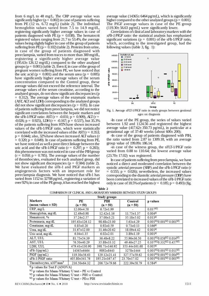
from 6 mg/L to 48 mg/L. The CRP average value wassignificantly higher (p = 0.002) in case of patients sufferingfrom PE (12 vs. 6.72 mg/L) (table 2). The individualhemoglobin values varied from 7.5 to 14.9 mg/dL,registering significantly higher average values in case ofpatients diagnosed with PE (p = 0.038). The hematocritregistered values ranging from 21.7 to 53.4%, the averagevalues being slightly higher in case of the group of patientssuffering from PE (p = 0.102) (table 2). Proteins from urine,in case of the group of patients diagnosed withpreeclampsia, varied from traces to more than 300 mg/dL,registering a significantly higher average value(195.63±126.12 mg/dL) compared to the other analyzedgroups (p = 0.001) (table 2). Even if, in case of the group ofpregnant women suffering from PE, we have noticed thatthe uric acid (p = 0.001) and the serum urea (p = 0.005)have significantly higher average values of the serumconcentration compared to the Control group, theseaverage values did not exceed the reference interval. Theaverage values of the serum creatinine, according to theanalyzed groups, do not show significant discrepancies (p= 0.312). The average values of the enzymatic markers(AST, ALT and LDH) corresponding to the analyzed groupsdid not show significant discrepancies (p > 0.05). In caseof patients suffering from preeclampsia, we did not noticesignificant correlations between the hepatic markers andthe sFlt-1/PlGF ratio: AST (r = -0.031; p = 0.909), ALT (r =-0.026; p = 0.925), LDH (r = -0.167; p = 0.537), but 35.3%of the patients suffering from HTN have shown increasedvalues of the sFlt-1/PlGF ratio, which were statisticallycorrelated with the increased values of the AST (r = 0.353;p = 0.044); also, 32% have shown increased ALT values (r= 0.320; p = 0.049). In case of patients suffering from PE,we have noticed as well a poor direct linkage between theuric acid and the sFlt-1/PlGF ratio (r = 0.297; p = 0.283);this phenomenon was not noticed in case of the PIH group(r = 0.050; p = 0.785). The average values of the numberof thrombocytes, evaluated for each analyzed group, didnot show significant discrepancies (p = 0.584) (table 2).We have evaluated the sFlt-1 and PlGF markers asangiogenesis factors with an important role forpreeclampsia diagnosis. We have noticed that sFlt-1 hasvaried from 1152 to 32708 pg/mL, registering a variation ofover 92%; in case of the PE group, it has reached the highest
average value (14365±6464 pg/mL), which is significantlyhigher compared to the other analyzed groups (p = 0.001).The PlGF average values in case of the PE group(119.30±56.63 pg/mL) were significantly lower.
Correlations of clinical and laboratory markers with thesFlt-1/PlGF ratio: the statistical analysis has emphasizedsignificant variations (p = 0.001) of the sFlt-1/PlGF ratio,which, according to the investigated group, had thefollowing values (table 3, fig. 1):
Table 2COMPARISON OF CLINICAL AND LABORATORY MARKERS BETWEEN STUDY GROUPS
Fig. 1. Average sFLT-1/PIGF ratio in study groups between gestionalages on diagnosis
-In case of the PE group, the series of values variedbetween 1.92 and 1124.56 and registered the highestaverage value (417.62±350.73 pg/mL), in particular at agestational age of 37-40 weeks (about 600±200);
-In case of the group of patients diagnosed with PIH,the ratio varied from 2.87 to 1309.18, with an averagegroup value of 199.09±186.14;
-in case of the witness group, the sFLT-1/PlGF ratiovaried from 0.88 to 110.64; the lowest average value(23.70±17.02) was registered.
In case of patients suffering from preeclampsia, we havenoticed a direct and moderated correlation between thesystolic arterial pressure (SBP) and the sFlt-1/PlGF ratio (r= 0.555; p = 0.026); nevertheless, the increased valuescorresponding to the diastolic arterial pressure (DBP) havebeen correlated to increased values of the sFlt-1/PlGF ratioonly in case of 18.5% of patients (r = 0.185; p = 0.493) (fig.

http://www.revistadechimie.ro REV.CHIM.(Bucharest)♦ 69♦ No. 6 ♦ 20181512
2). In case of patients suffering from PIH, the correlationsbetween SBP (r = 0.014; p = 0.937) and DBP (r = 0.105; p= 0.555), on one hand, and the sFlt-1/PlGF ratio, on theother hand, were not significant from a statisticalperspective (fig. 2).
The increased values of proteinuria were correlated toincreased values of the sFlt-1/PlGF ratio in case of 44.8%of patients diagnosed with PE (r = 0.448; p = 0.049) and incase of 32.5% of patients diagnosed with PIH (r = 0.325; p= 0.060). In case of patients from the PE group, we havenoticed a weak direct correlation between the creatinineand the sFlt-1/PlGF ratio (r = +0.279; p = 0.296), while incase of patients diagnosed with PIH, these parametersare independent from a statistical perspective (r = -0.058;p = 0.750). The statistic analysis of these data did notemphasize, in case of pregnant women diagnosed withpreeclampsia, significant correlations between the valuesobtained for the hepatic function markers and the sFlt-1/PlGF ratio: AST (r = -0.031; p = 0.909), ALT (r = -0.026; p= 0.925); nevertheless, 35.3% of the pregnant womendiagnosed with PIH have shown increased values of thesFlt-1/PlGF ratio, correlated to increased AST values (r =+0.353; p = 0.044), while 32% have shown increased ALTvalues (r = +0.320; p = 0.049). In case of patients sufferingfrom preeclampsia (PE), we have noticed a weak directcorrelation, which is insignificant, between the number ofthrombocytes and the sFlt-1/PlGF ratio (r = 0.264; p =0.324); in case of patients diagnosed with PIH, theseparameters show an indirect correlation (r = -0.312; p =0.072), which is also insignificant from a statisticperspective.
Within this research, the following parameters wereevaluated as markers for preeclampsia diagnosis: thesystolic arterial pressure/the diastolic arterial pressure (SBP/
DBP), the proteinuria, the creatinine, the urea and the uricacid at the serum level, the number of thrombocytes, theAST, ALT, LDH enzyme markers, as well as the sFlt-1 andPlGF angiogenic proteins. The ROC curve has shown thatproteinuria (AUC = 0.849), AST (AUC = 0.664), theincreased SBP/DBP values (AUC = 0.683/0.631), the serumcreatinine (AUC = 0.674) and the sFlt-1/PlGF ratio, at a cutoff value = 200, are effective preeclampsia predictors (fig.3).
Table 3DESCRIPTIVE INDICATOR OF THE sFlt-1/PlGF RATIO
Fig. 2. sFlt-1/PlGF ratio in correlation with hypertensivedisorders by PE and HTAIS groups
Fig. 3. The variation of markers such as: blood pressure (SBP/DBP), proteinuria, serum creatinine, number of thrombocytes, AST,
ALT, as predictors of preeclampsia in case of pregnant womenObs.: The test result variable(s): SBP, DBP, Proteinuria,
Creatinine, Thrombocytes, AST, ALT has at least one link betweenthe positive actual state group and the negative actual state group.Statistics may be biased. a Under the nonparametric assumption; b
Null hypothesis: true area = 0.5

REV.CHIM.(Bucharest)♦ 69♦ No. 6 ♦ 2018 http://www.revistadechimie.ro 1513
Proteinuria is evaluated at hospital admission; it refersto the excretion of whole proteins. The renal modificationsthat may be noticed in case of preeclampsia are generatedby a glomerular particular lesion, characterized by theendothelial proliferation of capillary vessels. These lesions,associated to the decrease of the renal plasma flow (RPF),because of vasoconstriction, lead to the decrease of theglomerular filtration (GFR) by about 25% compared tonormal pregnancy. Thus, in case of preeclampsia, thevalues corresponding to urea and creatinine can beapproximately equal or slightly higher compared to thevalued registered in normal pregnancy [1, 6]; nevertheless,the proteinuria degree largely varies, being situatedbetween minimal values and nephritic range proteinuriavalues [18, 19]. Within this research, in order to analyzeproteinuria and serum creatinine, we have resorted to testswhose sensitivity value (Se) is higher than the specificityvalue (Sp), in case of proteinuria values of 15.50mg/dL (Se= 0.875, Sp = 0.646) and of serum creatinine values of0.77mg/dL (Se = 0.750, Sp = 0.521).
Rarely, preeclampsia may lead to acute renal lesionsduring pregnancy. After giving birth, the glomerular changestend to reach relatively fast the normal level, oncehypertension and proteinuria are stabilized [1, 6].Thehepatic damage in case of preeclampsia depends on thedisease severity; in general, the values corresponding totransaminases and to the lactate dehydrogenase aremoderately increased, except for the outbreak of the HELLPsyndrome, when their values are significantly higher [20,21]. Indeed, the increased transaminases levels representa severity clinical marker. Hepatic microscopic exam mayreveal periportal hemorrhage, ischemic lesions and fibrindepositions [19]. Within this research, in order to analyzethe AST level, we resorted to a test whose sensitivity value(Se) is higher than its specificity value (Sp), for AST valuesof 27.5 UI/L (Se = 0.816, Sp = 0.646).In what concernsthe long-term systemic complications, about 20% ofpregnant women diagnosed with preeclampsia develop,after 7 years, high blood pressure or microalbuminuria,compared to 2% of the healthy pregnant women. In caseof pregnant women suffering from preeclampsia, the riskof death caused by cardiovascular factors is also high; incase of pregnant women who experience an earlymanifestation of the disease, the risk is extremely high[22, 23]. Preeclampsia and cardiovascular diseases havesurely in common physiopathological mechanisms andrisk factors, such as obesity or diabetes mellitus; they maylead to preeclampsia, as well as to cardiovascular diseasesduring life [19, 23-26]. Even if the acute manifestation ofthe disease is more dangerous for the mother,preeclampsia may also affect the fetus, leading to anincreased risk of spontaneous preterm birth, fetal growthretardation, oligohydramnios, as well as to an increasedrisk of perinatal death. The use of ultrasonography helps toevaluate the fetus condition, in particular growthretardation, as well as the extent to which fetal circulationis affected [9]. Certain studies have demonstrated that, incase of children exposed to preeclampsia duringpregnancy, high values of blood pressure were registeredduring childhood, as well as strokes during adulthood [19,23]. Certain pregnant women are diagnosed with the so-called atypical preeclampsia (which does not involvehypertension or proteinuria) and their condition worsensunexpectedly [27]. The evaluation of the ratio of circulatingangiogenic protein - sFlt-1/PlGF may contribute todifferentiate preeclampsia manifested by hypertension orproteinuria from other conditions (systemic lupuserythematosus) [19, 28, 29]. The results obtained within
this study have indicated a significant increase of the serumconcentration corresponding to the sFLT-1 anti-angiogenicfactor of more than 4 times for the PE group and of morethan three times for the HTN group, compared to the controlgroup. The average values obtained for the PlGF parameterhave shown a decrease of more than 2.5 times in case ofthe PE group, as well as in case of the HTN group, comparedto the control group. On the other hand, the results obtainedfor the analyzed groups have shown that the sFlt-1/PlGFratio represents an effective preeclampsia predictor,characterized by a high specificity level.
ConclusionsPreeclampsia is a serious multisystemic syndrome,
which represents one of the major causes of maternal,fetal and neonatal mortality and morbidity. Angiogenicfactors contribute to preeclampsia molecular mechanisms,leading to hypertension and proteinuria. According to thespecialized literature, molecular markers, such as thesoluble fms-like tyrosine kinase-1 (sFlt-1) and the pro-angiogenic placental growth protein (PlGF), havesignificantly modified values, preceding by several weeksthe emergence of preeclampsia signs and symptoms. Inthis respect, these markers can be successfully used inorder to identify patients presenting a high risk of earlypreeclampsia manifestations (< 34 weeks). The resultsof our study confirm the importance of the analysis of thesemarkers for the diagnosis and monitoring of hypertensivepregnant women; at the same time, they emphasize thefact that the sFlt-1/PlGF ratio represents an effectivepredictor of preeclampsia and of its severity. Thus, thisangiogenic protein ratio may contribute, together with thewell-known clinical and biological factors, to thestratification of the risk presented by hypertensive pregnantwomen; also, it could play an important role in whatconcerns the decision taken by the obstetrician related tothe management of these pregnant women. Therefore,the obstetrician could have the possibility to promptly directpregnant women from the increased risk group to thecompetent specialized health care facilities, to allot to thispregnant women group the appropriate medical resourcesand to rapidly take the necessary measures for the ultimatetreatment- termination of pregnancy. Taking all these steps,the obstetrician will be more effective in what concernspreeclampsia treatment. Decisions based on datasupported by evidence will contribute to reduce the risk forthe mother, as well as for the newborn baby.
References1.BRETT, C.Y., RICHARD, J.L., ANANTH, K., Rev. Pathol. Mech. Dis., 5,2010, p. 173.2.MOCAN HOGNOGI, L.D., MOCAN HOGNOGI, R.F., MALUTAN, A.,FARCAS, A.D.,VIDA SMITI, L., Rev. Chim. (Bucharest), 68, no. 9, 2017,p. 20183.SALAFIA, C.M., PEZZULLO, J.C., LOPEZ-ZENO, J.A., Am. J. Obstet.Gynecol., 173, no. 4,1995, p. 1097.4.ZHOU, Y., DAMSKY, C.H., CHIU, K., ROBERTS, J.M., FISHER, S.J.,J.Clin. Invest., 91, no. 3, 1993, p. 950.5.NORTH, R.A., FERRIER, C., LONG, D., TOWNEND, K., KINCAID-SMITH, P., Obstet. Gynecol., 83, no. 3, 1994, p. 378.6.LYALL, L., ROBSON, S.C., BULMER, J.N., Hypertension, 62, 2013, p.1046.7.REZUS, E., CONSTANTIN, M.M.L., REZUS, C., Rev. Chim. (Bucharest),66, no. 7, 2015, p. 10158.WU, F.T., STEFANINI, M.O., MAC GABHANN, F., KONTOS, C.D.,ANNEX, B.H., POPEL, A.S, J. Cell. Mol. Med.,14(3), 2010, p. 528-52.(7)9.DOVER, N., GULERMAN, H.C., CELEN, S., KAHYAOGLU, S., YENICESU,O., J. Obstet. Gynaecol. India, 63, no. 3, 2013, p. 158.

http://www.revistadechimie.ro REV.CHIM.(Bucharest)♦ 69♦ No. 6 ♦ 20181514
10.LEVINE, R.J., LAM, C., QIAN, C., N. Engl, J. Med., 355, no. 10, 2006,p. 992.11.MCMAHON, K., KARUMANCHI, S.A., STILLMAN, I.E., CUMMINGS, P.,PATTON, D., EASTERLING, T., Am. J. Obstet. Gynecol., 68, 2014, p. e1.12.BURKE, S.D., KARUMANCHI, S.A., Hypertension, 62, 2013, p. 1013.13.LYALL, L., ROBSON, S.C., BULMER, J.N., Hypertension, 62, 2013,p. 1046.14.KLEINROUWELER, C.E., WIEGERINCK, M.M.J., RIS-STALPERS, C.,BOSSUYT, P.M.M., van der POST, J.A.M., von DADELSZEN, P., MOLB.W.J., PAJKRT, E., BJOG, 119, 2012, p. 778.15.BALAN, G.G., MITRICA, D.E., IACOB, M., BALAN, A., ZETU, I., Revistade Cercetare si Interventie Sociala, 49, 2015, p. 229.16.AGHEORGHIESEI CORODEANU, D.T., POROCH, V., 6th LUMENInternational Conference on Rethinking Social Action Core Values,16-19 April 2015, Iasi, Romania, Rethinking Social Action. Core Values,p. 33.17.ROGOZEA, L., REPANOVICI, A., CRISTEA, L., BARITZ, M., MICLAUS,R., PASCU, A., Proceedings of the 4th WSEAS/IASME InternationalConference on Educational Technologies (Edute’08), Book Series:Recent Advances in Computer Engineering, Corfu, Greece, 2008,Oct. 26-28, pp. 87-90.18.SIBAI, B.M., STELLA, C.L., Am. J. Obstet. Gynecol., 200, no. 5,2009, p. 481, e1.19.THANGARATINAM, S., COOMARASAMY, A., O’MAHONY, F., BMC.Med., 7, 2009, p. 10.
20.ZHOU, C.C., ZHANG, Y., IRANI, R.A., Nat. Med.,14, no. 8, 2008, p.855.21.ZHANG, J., VILLAR, J., SUN, W., Am. J. Obstet. Gynecol., 197, no. 2,2007, p. 162, e1.22.HINCHEY, J., CHAVES, C., APPIGNANI, B., N. Engl. J. Med., 334, no.8, 1996, p. 494.23.VERLOHREN, S., GALINDO, A., SCHLEMBACH, D., ZEISLER, H.,HERRAIZ, I., MOERTL, M.G., PAPE, J., DUDENHAUSEN, J.W., DENK, B.,STEPAN, H., Am. J. Obstet. Gynecol., 161, 2010, p. e1.24.CHECHERITA, L.E., REZUS, E., LEON, M.M., STAMATIN, O.,CARAUSU, E.M., Rev. Chim. (Bucharest), 68, no. 5, 2017, p. 977.25.MOGA, M.A., IRIMIE, M., OANTA, A., PASCU, A., BURTEA, V., AsianPacific Journal of Cancer Prevention AJPC, 15, no. 16, 2014, p. 6887.DOI: 10.7314/APJCP.2014.15.16.688726.SERBAN, D., CRISAN, C., SERBAN, C., MICU SERBU, I.B., KUNDANI,N., POROCH, V., SHARMA, A., BUCIU, V., HORHAT, I.D., SAS, I., BIRIS,M., RATIU, A., Rev. Chim. (Bucharest), 69, no. 5, 2018, p. 1203.27.VERLOHREN, S., HERRAIZ, I., LAPAIRE, O., SCHLEMBACH, D.,ZEISLER, H., CALDA, P., SABRIA, J., MARKFELD-EROL, F., GALINDO,A., SCHOOFS, K., DENK, B., STEPAN, H., Hypertension, 63, 2014, p.346.28.THANGARATINAM, S., ISMAIL, K.M., SHARP, S., COOMARASAMY, A.,KHAN, K.S., BJOG, 113, no. 4, 2006, p. 369.29.GAVRIS, C., POROCH, V., SIMION, L., BARACAN, A., TOADER, E.,PASCU, A.M., Rev. Chim. (Bucharest), 68, no. 7, 2017, p. 1586.
Manuscript received: 8.01.2018


![The effects of parathyroid hormone peptides on the ...eprints.whiterose.ac.uk/118794/1/Metcalf et al, Parathyroid peptides... · in resorption markers [4, 5]. Improvements in aBMD](https://img.pdfslide.us/doc/110x75/603633c7eaaaae70d83c29d9/the-effects-of-parathyroid-hormone-peptides-on-the-et-al-parathyroid-peptides.jpg)