Embed Size (px)
Citation preview

The Role of MR Imaging in Various Cardiomyopathy
1Eun Ju Chun, MD (E-mail; [email protected]), 1Sang Il Choi, MD, 2Whal Lee, MD, 2Jae Hyung Park, MD.
Department of Radiology, ¹Seoul National University Bundang Hospital,
2Seoul National University Hospital

• Introduction
• Definition and Classification of Cardiomyopathy (CMP)
• MR Technique for Assessment of CMP
• Clinical Impact of Cardiac MRI
- Dilated CMP
- Hypertrophic CMP
- Restrictive CMP
- Arrythmogenic right ventricular dysplasia (ARVD)
- Specific CMP
(Stress-induced CMP, Non-compaction, Diverticulum, Myocarditis)
• Conclusion
Table of Contents
• The cardiomyopathy (CMP) include a variety of disease
where the primary pathology directly involves the
myocardium.
• Cardiac MR (CMR) is proving increasingly valuable in
the identification and management in these conditions.
• We will illustrate the various MR techniques for the
evaluation of CMP and characteristic MR findings. This
exhibit will discuss the merit and the potential role of
CMR in the evaluation of various CMP.
Introduction
• Introduction
• Definition and Classification of Cardiomyopathy (CMP)
• MR Technique for Assessment of CMP
• Clinical Impact of Cardiac MRI
- Dilated CMP
- Hypertrophic CMP
- Restrictive CMP (Constrictive pericarditis)
- Arrythmogenic right ventricular dysplasia (ARVD) (RVOT)
- Specific CMP
(Stress-induced CMP, Non-compaction, Diverticulum, Myocarditis)
• Conclusion
Table of Contents

• The cardiomyopathies (CMP) constitute a group of
disease in which dominant feature is direct involvement
of the heart muscle itself.
• Although ischemic CMP is the most common cause of
heart failure, ischemic CMP is not appropriate term
because the primary pathology is in the coronary
arteries and not the heart muscle.
• In the WHO/ISFC classification, the cardiomyoapthies
are classified based on their predominant
pathophysiological features (Table 1).
Definition and Classification
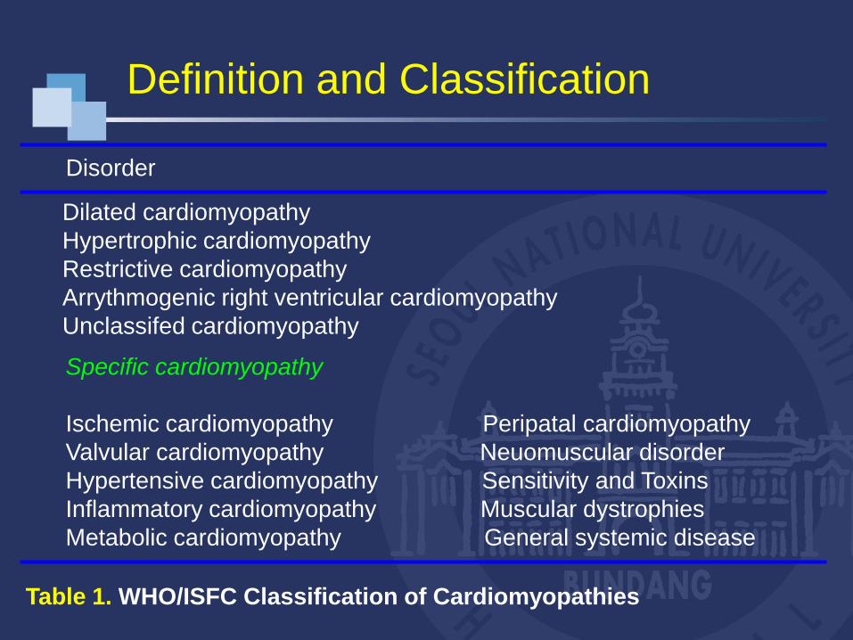
Table 1. WHO/ISFC Classification of Cardiomyopathies
Disorder
Dilated cardiomyopathy
Hypertrophic cardiomyopathy
Restrictive cardiomyopathy
Arrythmogenic right ventricular cardiomyopathy
Unclassifed cardiomyopathy
Specific cardiomyopathy
Ischemic cardiomyopathy Peripatal cardiomyopathy
Valvular cardiomyopathy Neuomuscular disorder
Hypertensive cardiomyopathy Sensitivity and Toxins
Inflammatory cardiomyopathy Muscular dystrophies
Metabolic cardiomyopathy General systemic disease
Definition and Classification

• Three basic types of functional impairment have been
described (Table 2).
- Dilated CMP
- Hypertrophic CMP
- Restrictive CMP
• Most of specific cardiomyopathies are characterized by the
dilated CMP pattern.
• Ischemic CMP has been used to describe condition in
which coronary artery disease causes multiple infarctions,
diffuse fibrosis, and left ventricular dysfucntion.
Functional Classification
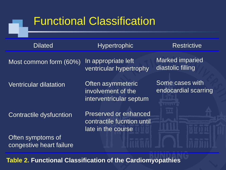
Table 2. Functional Classification of the Cardiomyopathies
Dilated
Most common form (60%)
Ventricular dilatation
Contractile dysfucntion
Often symptoms of
congestive heart failure
Functional Classification
Hypertrophic Restrictive
In appropriate left
ventricular hypertrophy
Often asymmeteric
involvement of the
interventricular septum
Preserved or enhanced
contractile fucntion until
late in the course
Marked imparied
diastolic filling
Some cases with
endocardial scarring

• Introduction
• Definition and Classification of Cardiomyopathy (CMP)
• MR Technique for Assessment of CMP
• Clinical Impact of Cardiac MRI
- Dilated CMP
- Hypertrophic CMP
- Restrictive CMP (Constrictive pericarditis)
- Arrythmogenic right ventricular dysplasia (ARVD) (RVOT)
- Specific CMP
(Stress-induced CMP, non-compaction, diverticulum, Myocarditis)
• Conclusion
Table of Contents

• The diagnosis of CMP is established by exclusion of
other cardiovascular etiologies and an accurate
characterization of the phenotype.
• Treatment is guided by the stage and hemodynamic
relevance of the disease and long-term follow-up after
therapy is needed.
• Thus, imaging techniques are important in the diagnosis
and therapy of cardiomyopathies.
MR Techniques for Assessment of CMP

• SSFP sequence:
morphologic and functional information (Table 3).
• VENC:
evaluation of diastolic and valvular function
• DE-MRI:
identification of myocardial necrosis and fibrosis
• Myocardial perfusion MR:
presence or extent of inducible ischemia
• Spin9Echo images (T1-, T2-weighted images):
identification of signal change of myocardium
• MR spectroscopy: for the evaluation of metabolic state
MR Techniques for Assessment of CMP

Table 3. Morphologic and Functional Abnormalities of the CMP
Dilated
Morphology
LV and RV size
Hypertrophy
Atrial dilatation
Pleural effusion
Pericardial effusion
ventricular thrombus
SVC/IVC dilatation
Myocardial function
Global dysfunction
Segmental dysfunction
Diastolic dysfunction
Valvular function
Mitral regurgitation
Tricuspid regurgitation
Hypertrophic Restrictive
MR Techniques for Assessment of CMP
+++ - -
+ +++ -/+
++ + ++
+ -/+ +
+ -/+ +
+ - -
+ - +++
+++ + +
+ +++ +
+ ++ +++
+ ++ +
+ - +

• Introduction
• Definition and Classification of Cardiomyopathy (CMP)
• MR Technique for Assessment of CMP
• Clinical Impact of Cardiac MRI
- Dilated CMP
- Hypertrophic CMP
- Restrictive CMP (Constrictive pericarditis)
- Arrythmogenic right ventricular dysplasia (ARVD) (RVOT)
- Specific CMP
(Stress-induced CMP, Non-compaction, Diverticulum, Myocarditis)
• Conclusion
Table of Contents

Clinical Impact of Cardiac MRI
• Recently, the use of MRI for the evaluation of CMP is
expanding, aided by the administration of paramagnetic
contrast agents.
• Cardiac MRI offers the accurate evaluation of morphology
and function, and it also offers characterization of various
CMP.
• Suspected myocardial ischemia and fibrosis are also
diagnosed by using dynamic first-pass and delayed-
enhancement MRI.

Table 4. Current indication of cardiac MRI for the evaluation of CMP
Clinical Impact of Cardiac MRI
Indication Class
Dilated cardiomyopathy:
Differentiation from dysfunction related coronary artery disease
Hypertrophic cardiomyopathy:
Apical
Non-apical
I
II
I
Restrictive cardiomyopathy:
Arrythmogenic RV dysplasia IIII
Non compaction II
*** Classification
Class I: provides clinically relevant information and is usually appropriate, may be used as first line imaging technique; usually supported by substantial literature
Class II: provides clinically relevant information and is frequently useful; other techniques may provide similar information; supported by limited literature
Reference: Eur Heart J (2004): Clinical Indications for Cardiac MRI: Consensus Panel Report

• Introduction
• Definition and Classification of Cardiomyopathy (CMP)
• MR Technique for Assessment of CMP
• Clinical Impact of Cardiac MRI
- Dilated CMP
- Hypertrophic CMP
- Restrictive CMP (Constrictive pericarditis)
- Arrythmogenic right ventricular dysplasia (ARVD) (RVOT)
- Specific CMP
(Stress-induced CMP, Non-compaction, Diverticulum, Myocarditis)
• Conclusion
Table of Contents
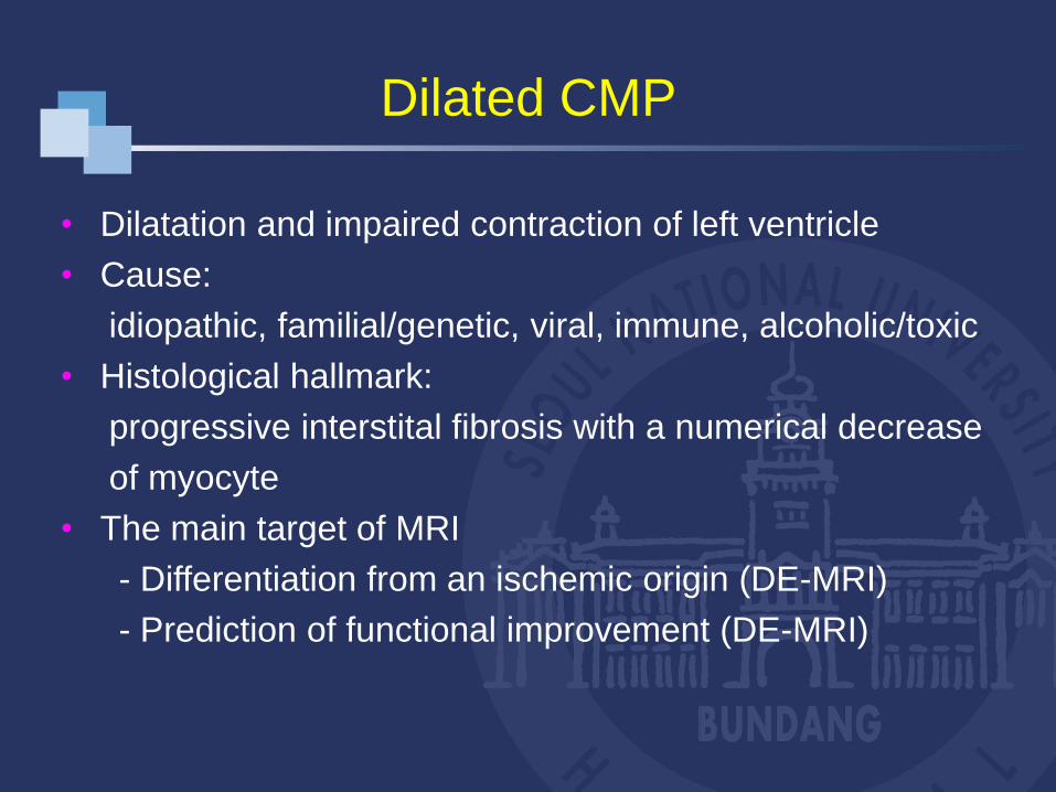
• Dilatation and impaired contraction of left ventricle
• Cause:
idiopathic, familial/genetic, viral, immune, alcoholic/toxic
• Histological hallmark:
progressive interstital fibrosis with a numerical decrease
of myocyte
• The main target of MRI
- Differentiation from an ischemic origin (DE-MRI)
- Prediction of functional improvement (DE-MRI)
Dilated CMP
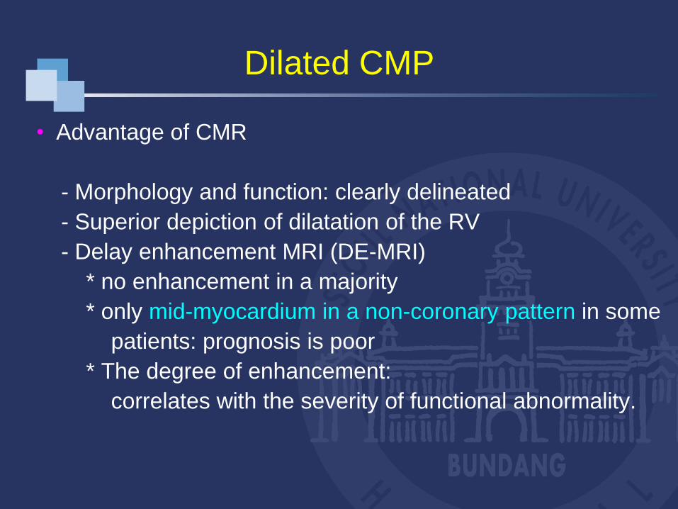
• Advantage of CMR
- Morphology and function: clearly delineated
- Superior depiction of dilatation of the RV
- Delay enhancement MRI (DE-MRI)
* no enhancement in a majority
* only mid-myocardium in a non-coronary pattern in some
patients: prognosis is poor
* The degree of enhancement:
correlates with the severity of functional abnormality.
Dilated CMP

Figure 1. No enhancement type on DE-MRI 39 year
old man with chest pain. (A) Cine MR showed globally
reduced systolic function (EF = 39.3%). (B) DE-MRI
shows no delayed enhancement. (C) MRI permits
accurate measurement of cardiac function and LV mass.
(D) Follow-up cine MRI after 3 month, wall motion is
normalized (EF = 52.9%) on cine MRI.
Dilated CMP
(A) (B)
(D)
(C)

Figure 2. Enhancement type on DE-MRI (Alcoholic
CMP) 36 year old man with dyspnea. (A) Cine MR showed
globally reduced systolic function (EF = 10. 6%). DE-MRI
view shows delayed enhancement at mid and epicardial
area of septal wall with non-coronary pattern. on short axis
view (B) and 4 chamber view (C). MR Spectroscopy (D)
was performed at septal wall and showed depletion of
creatine metabolism. Function was not improved during
the follow-up period.
Dilated CMP
(A) (B) (C)
(D)

Figure 3. Extensive Enhancement on DE-MRI 65 year old man with
dyspnea. Severe global systolic dysfunction (EF = 29.3%) with severe
dilated LV (A) (B) DE-MRI view shows extensive delayed enhancement at
apico to basal anterior and anterosepatal wall suggesting extensive fibrosis.
This finding is hallmark of end-stage of dilated cardiomyopathy.
Dilated CMP
(A) (B)

Dilated vs Ischemic CMP
• DE-MRI
* HF with CAD:
Subendocardial or Transmural
* HF related DCMP:
- no enhancement (59%)
- subendocaridal
or transmural (13%)
- Patchy of longitudinal striae
of midwall (28%)
Microphone JA, et al. Circulation 2003
Figure 4. Typical findings of DE-
MRI in patient with ischemic CMP
DE-MRI shows strong enhancement
along the LAD and RCA vascular
territory with transmural extent (75-
100%).

• Introduction
• Definition and Classification of Cardiomyopathy (CMP)
• MR Technique for Assessment of CMP
• Clinical Impact of Cardiac MRI
- Dilated CMP
- Hypertrophic CMP
- Restrictive CMP (Constrictive pericarditis)
- Arrythmogenic right ventricular dysplasia (ARVD) (RVOT)
- Specific CMP
(Stress-induced CMP, Non-compaction, Diverticulum, Myocarditis)
• Conclusion
Table of Contents

• Myocardial hypertrophy with impaired diastolic and
systolic function (mainly diastolic dysfucntion)
• Narrowing of the LVOT in obstructive cases
• Histology:
myocardial disarray as well as patches of myocardial
scarring
• The main target of MRI
- To determine phenotypes such as apical form
(cine MR using SSFP sequences)
- To assess regional myocardial hypertrophy
(cine MR using SSFP sequences)
Hypertrophic CMP

• Advantage of CMR
- Precise definition of the site and extent of hypertrophy,
especially LV apex (apcal HCM)
- Accurate assessment of flow dynamics of LV outflow tract
- Demonstration of myocardial scarring and fibrosis:
predominantly in the middle third of the ventricular wall
** The extent of hyperenhancement on DE-MRI may have
prognostic implications for the risk of progressive
ventricular dilation and sudden death
- Evaluation of post-surgical change
- Monitoring and quantification after septal ablation
Hypertrophic CMP
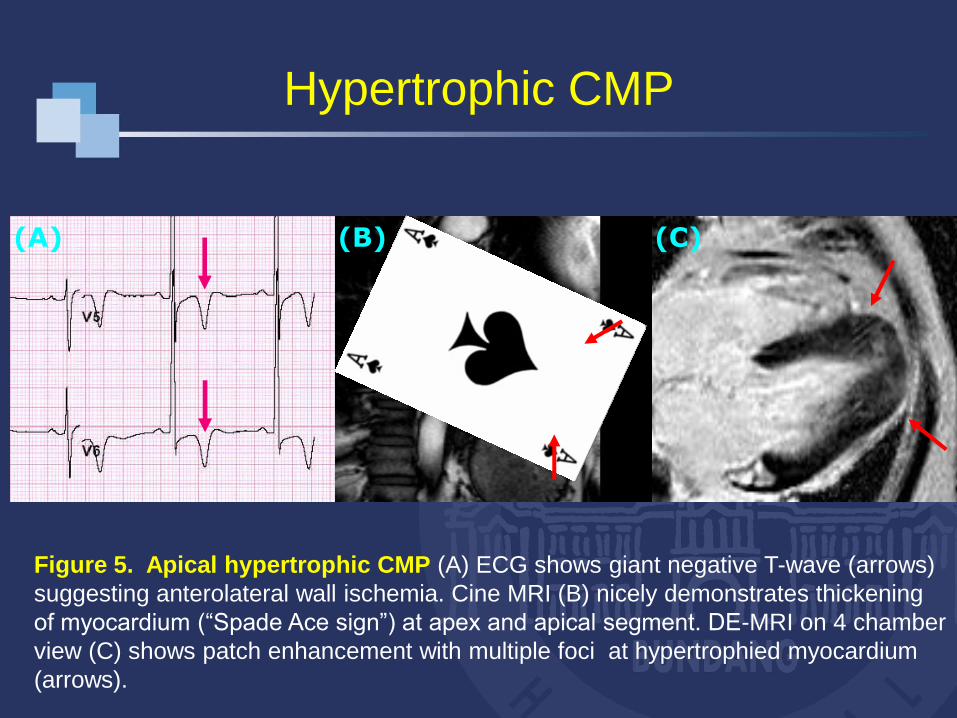
Figure 5. Apical hypertrophic CMP (A) ECG shows giant negative T-wave (arrows)
suggesting anterolateral wall ischemia. Cine MRI (B) nicely demonstrates thickening
of myocardium (“Spade Ace sign”) at apex and apical segment. DE-MRI on 4 chamber
view (C) shows patch enhancement with multiple foci at hypertrophied myocardium
(arrows).
(B) (C)(A)
Hypertrophic CMP
Hypertrophic CMP
(A) (B) (C)
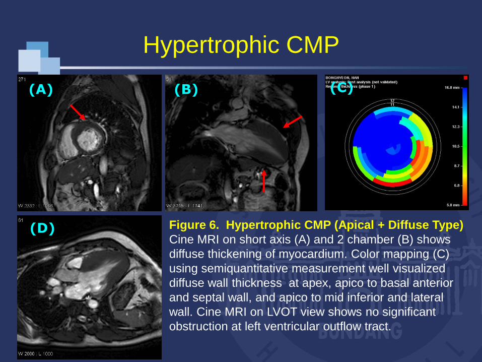
Figure 6. Hypertrophic CMP (Apical + Diffuse Type)
Cine MRI on short axis (A) and 2 chamber (B) shows
diffuse thickening of myocardium. Color mapping (C)
using semiquantitative measurement well visualized
diffuse wall thickness at apex, apico to basal anterior
and septal wall, and apico to mid inferior and lateral
wall. Cine MRI on LVOT view shows no significant
obstruction at left ventricular outflow tract.
(D)

Stress Perfusion
Rest Perfusion
(D)
(E)
(F)
Hypertrophic CMP
Figure 6. Hypertrophic CMP (Apical
+ Diffuse Type) Stress (D) and rest
(E) MR perfusion images shows
reversible subendocardial perfusion
defect at apical and mid anterior wall
(arrows) . DE-MRI (F) reveals
subcendocardial scarring at apico to
mid inferior wall (arrows) .
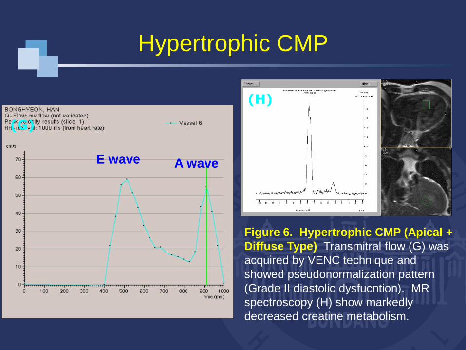
A waveE wave
Figure 6. Hypertrophic CMP (Apical +
Diffuse Type) Transmitral flow (G) was
acquired by VENC technique and
showed pseudonormalization pattern
(Grade II diastolic dysfucntion). MR
spectroscopy (H) show markedly
decreased creatine metabolism.
(G)
(H)
Hypertrophic CMP

• Introduction
• Definition and Classification of Cardiomyopathy (CMP)
• MR Technique for Assessment of CMP
• Clinical Impact of Cardiac MRI
- Dilated CMP
- Hypertrophic CMP
- Restrictive CMP (Constrictive pericarditis)
- Arrythmogenic right ventricular dysplasia (ARVD) (RVOT)
- Specific CMP
(Stress-induced CMP, Non-compaction, Diverticulum, Myocarditis)
• Conclusion
Table of Contents
• Restrictive filling and reduced diastolic size of eithr and
both ventricles with normal or near-normal systolic function
• Hallmark of restrictive CMP: Abnormal diastolic dysfunction
• Cause:
idiopathic or associated with other disease
(amyloidosis, sarcoidosis etc)
• The main target of MRI
- To determine phenotypes such as myocardial infiltrative
disease (Spin-echo Images, DE-MRI)
- To differentiate from constrictive pericarditis
(cine MR using SSFP sequences)
Restrictive CMP
• Advantage of CMR
- Clearly depict the anatomic and functional
abnormalities
- Define myocardial infiltrative disease such as
amyloidosis on the basis of typical findings on
DE-MRI
- Visualization of pericardial thickness
- Objective monitoring and quantification after treatment
Restrictive CMP

• A cause of restrictive CMP
• primary or secondary in origin. Infiltration with fibrillar
• MR findings:
- Variable, but usually increased SI on T1, T2 WI
- Thickening of Interatrial septum and right atrial wall
thickness (myocardial thickenss > 6 mm):
DDx point of symmetric hypertrophic CMP
Fattori et al, Am Heart J 1998
DE-MRI:
- Global and subendocardial enhancement (69%):
Increased LV and RV mass index, lower LV EF (57%)
Restrictive CMP: Amyloidosis
Maceira AM et al Circulation 2005
Restrictive CMP: Amyloidosis
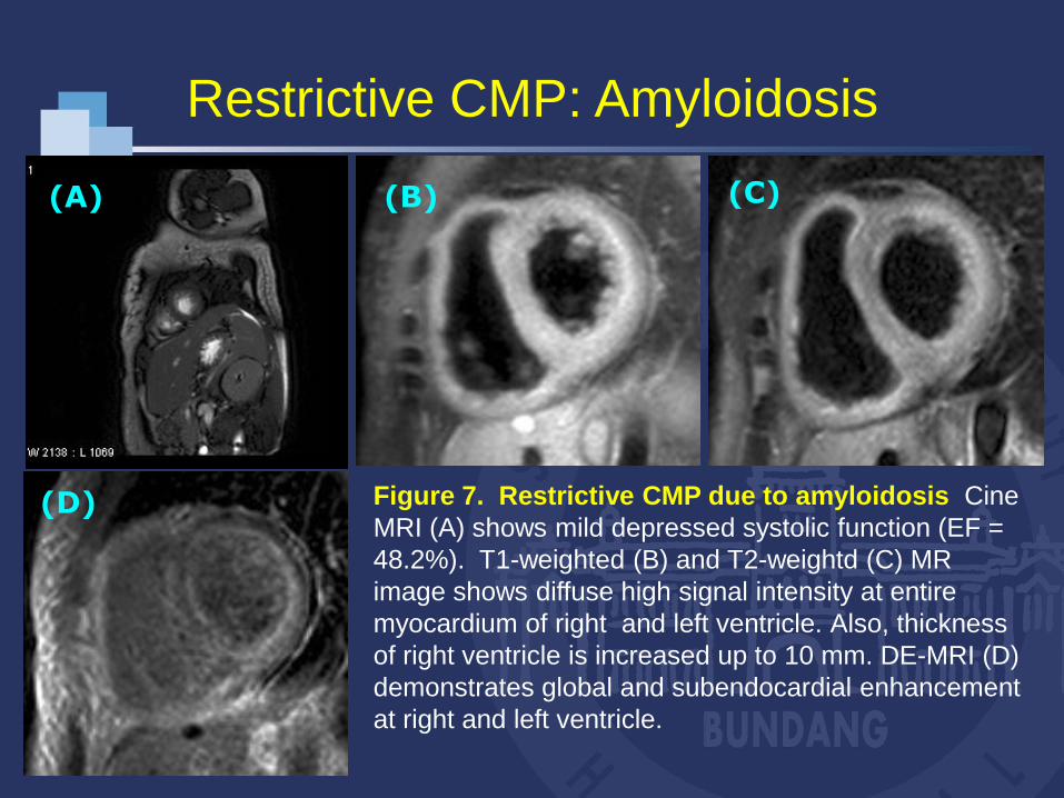
Figure 7. Restrictive CMP due to amyloidosis Cine
MRI (A) shows mild depressed systolic function (EF =
48.2%). T1-weighted (B) and T2-weightd (C) MR
image shows diffuse high signal intensity at entire
myocardium of right and left ventricle. Also, thickness
of right ventricle is increased up to 10 mm. DE-MRI (D)
demonstrates global and subendocardial enhancement
at right and left ventricle.
(A) (B) (C)
(D)

Restrictive CMP vs Constrictive CMP
Restrictive CMP
• Increased wall thickness
• Delayed ventricular filling
• Frequently combined MR, TR
Constrictive CMP
• Pericardial thickening (> 4mm)
Masui et al, Radiology 1992

• Introduction
• Definition and Classification of Cardiomyopathy (CMP)
• MR Technique for Assessment of CMP
• Clinical Impact of Cardiac MRI
- Dilated CMP
- Hypertrophic CMP
- Restrictive CMP (Constrictive pericarditis)
- Arrythmogenic right ventricular dysplasia (ARVD) (RVOT)
- Specific CMP
(Stress-induced CMP, Non-compaction, Diverticulum, Myocarditis)
• Conclusion
Table of Contents

Arrythmogenic RV dysplasia (ARVD)
• Ventricular tachycardia with LBBB from right ventricle
• Young males, variable symptoms
• The definite diagnosis is challenging
• Histology:
Fibro-fatty infiltration with thinning of RV free wall
• The main target of MRI
- Explicit demonstration of regional thinning and fatty
infiltration of right ventricular wall (Spin-echo, SSFP)
- Demonstration of myocardial fibrosis at right ventricular
wall (DE-MRI)

• Advantage of CMR
- Regional thinning and wall-motion abnormality of right
ventricle: clearly delineated
- Detailed differentiation between myocardium, epicardial fat,
trabeculae and myocardial fatty infiltration
- Delay enhancement MRI (DE-MRI):
noninvasive detection of myocardial fibrotic changes
Arrythmogenic RV dysplasia (ARVD)
Tandri et al, J Am Coll Cardiol 2005

Figure 8. T1-weighted MRI in
patient with Arrythmogenic
right ventricular dysplasia.
Axial MR image obtained at the
midventricular level shows
myocardial wall thinning and
fatty infiltration (arrows) at the
right ventricular free wall and
also there is global right
ventricular dilation. The
interventricular septum shows
convexity to the left ventricular
side.
Arrythmogenic RV dysplasia (ARVD)

Arrythmogenic RV dysplasia (ARVD)
Figure 9. A 19-month-old patient with abnormal
cardiac border. Chest PA (A) shows abnormal
enlarged left heart border. Cine MRI (B) reveals global
right ventricular dilatation with hypokinesia. DE-MRI
(C, D) nicely demonstrates diffuse thinning and
dilation with the strong enhanced wall of the right
ventricle (arrows) suggesting extensive fibrosis.
(A) (B) (C)
(D)

• Introduction
• Definition and Classification of Cardiomyopathy (CMP)
• MR Technique for Assessment of CMP
• Clinical Impact of Cardiac MRI
- Dilated CMP
- Hypertrophic CMP
- Restrictive CMP
- Arrythmogenic right ventricular dysplasia (ARVD)
- Specific CMP
(Stress-induced CMP, Non-compaction, Diverticulum, Myocarditis)
• Conclusion
Table of Contents

Stress-induced CMP (“Tako-tsubo")
• Definition: acute onset of a cardiovascular event, usually
associated with substernal chest pain, initially regarded as
ST-segment elevation myocardial infarction/evolving
coronary syndrome.
• Systolic dysfunction (ejection fraction 29±9%) ,
predominantly characterized by akinesia/hypokinesia of the
mid-to-distal portion of the LV chamber "apical ballooning" ,
with hypercontractile basal LV
• Absence of significant atherosclerotic luminal narrowing in
each of the 3 epicardial coronary arteries (0 to < 25%).

• Profound psychological stress immediately preceding and
triggering the cardiac events.
• Possible mechanism: catecholamine-mediated cardiotoxicity,
in which the distal LV chamber is selectively vulnerable
to a form of myocardial stunning
• DE-MRI: no enhancement
except focal infarction
at apex (5% )
Figure 10. “Tako-tsubo" cardiomyopathy
describes the resemblance of the LV
angiogram to an octopus trap
Stress-induced CMP (“Tako-tsubo")
Sharkey et al, Circulation 2005

Stress-induced CMP (“Tako-tsubo")
Figure 11. A 45-year-old female with Stress-induced Cardiomyopathy. She had
experienced psychological stress, and also suffered fromsevere rhadomyolysis
with ARF. Cine MRI on short axis view (A) and 2 chamber view shows
hypokinesia at apico to mid entire wall, but contractility at basal level is well
preserved. Note that there is no enhancement on DE-MRI (C). Conventional
coronary angiography revealed no significant steno-occlusive lesion.
(A) (B) (C)

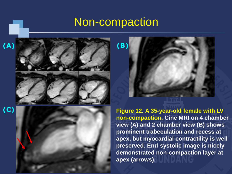
• Prominent trabeculation and recess
• Non-compact/compact layer > 2.0
on end-systolic phase
• Autosomal dominant inheritance.
• Failure of normal embryonic development of the
myocardium from loosely arranged muscle fibers to the
mature compacted form of myocardium.
• Microvascular dysfunction and ventricular arrhythmias.
Non-compaction
Non-compaction
Figure 12. A 35-year-old female with LV
non-compaction. Cine MRI on 4 chamber
view (A) and 2 chamber view (B) shows
prominent trabeculation and recess at
apex, but myocardial contractility is well
preserved. End-systolic image is nicely
demonstrated non-compaction layer at
apex (arrows).
(A) (B)
(C)

Diverticulum
• Morphology: saccular with narrow neck
• Location: apex, basal
• Two types
- muscular type:
saccular with narrow neck, contractility (+), DE-MRI (-).
- fibrous type: contractility (+), DE-MRI (+).
• Differential diagnosis
- True Aneurysm: Thin-walled with wide opening
contractility (akinetic or dyskinetic), DE-MRI (+).
- Pseudoaneurysm: saccular with narrow neck, but
contractility (akinetic or dyskinetic),
DE-MRI (acute stage - ?, chronic stage +).

Diverticulum
Figure 13. A 5-year-old boy with LV diverticulum. On
routine echocardiography, LV aneurysm was
suspected. Cine MRI (A,B,C) shows focal outpouching
lesion at basal inferior wall with narrow neck and
saccular shape. Note this lesion is normal contracted
during cardiac cycle. There is no enhancement (D) at
LV diverticulum on DE-MRI (arrows) suggesting
muscular type.
(A) (B) (C)
(D)
Myocarditis
• Myocardial inflammation caused by
- viral or postviral autoimmune response (primary)
- specific pathogen such as bacteria, drug, chemical etc
(secondary)
• DE-MRI
- enhancement predominantly in lateral wall
(associated with active inflammation)
- enhancement in 88% of patients with myocarditis
- follow-up: decreased extent of enhancement
Mahrholdt et al. Circulation 2004
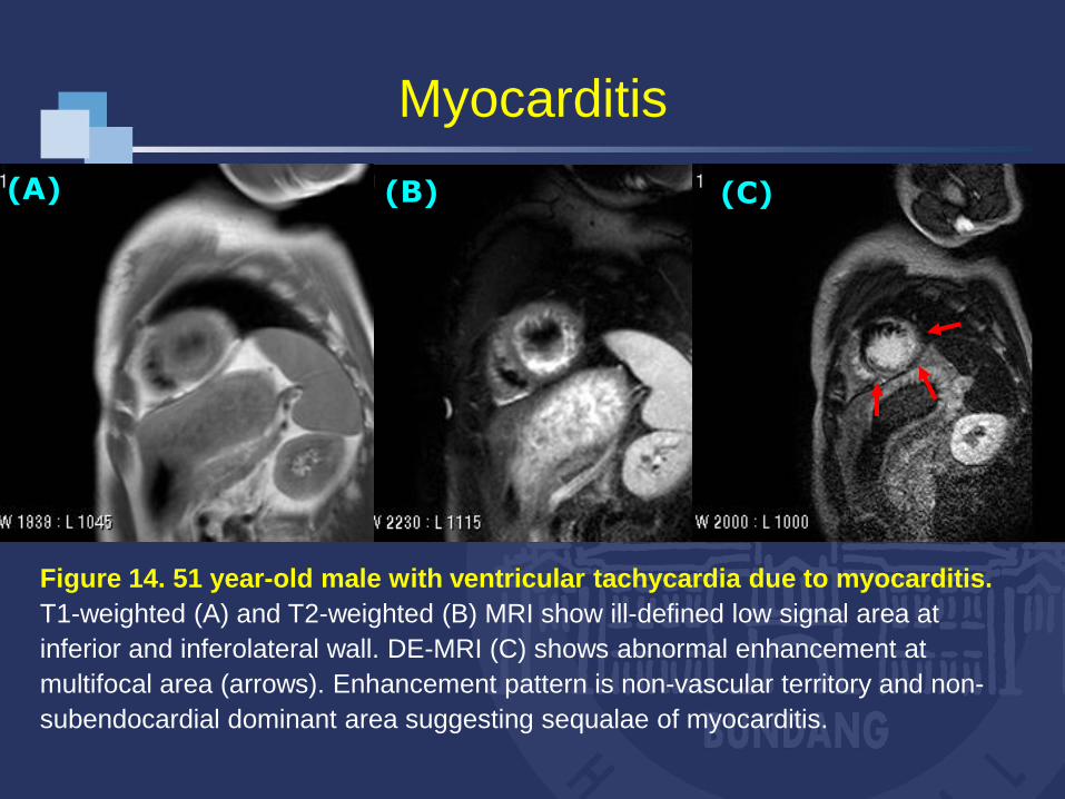
Figure 14. 51 year-old male with ventricular tachycardia due to myocarditis.
T1-weighted (A) and T2-weighted (B) MRI show ill-defined low signal area at
inferior and inferolateral wall. DE-MRI (C) shows abnormal enhancement at
multifocal area (arrows). Enhancement pattern is non-vascular territory and non-
subendocardial dominant area suggesting sequalae of myocarditis.
Myocarditis
(A) (B) (C)
• Introduction
• Definition and Classification of Cardiomyopathy (CMP)
• MR Technique for Assessment of CMP
• Clinical Impact of Cardiac MRI
- Dilated CMP
- Hypertrophic CMP
- Restrictive CMP
- Arrythmogenic right ventricular dysplasia (ARVD)
- Specific CMP
(Stress-induced CMP, Non-compaction, Diverticulum, Myocarditis)
• Conclusion
Table of Contents
• The understanding of various cardiomyopathies and
knowledge of characteristic MR findings is provided more
valuable information for the accurate diagnosis and proper
management.
• With the advances of MRI technology and, it will more
increase the role MRI for the assessment of various
cardiomyopathy.
Conclusion