Embed Size (px)
Citation preview

OPEN ACCESS | www.cell-stress.com 90 Cell Stress | NOVEMBER 2017 | Vol. 1 No. 2
www.cell-stress.com
Research Article
ABSTRACT Folding and packing of membrane proteins are highly influenced by the lipidic component of the membrane. Here, we explore how the hydro-phobic mismatch (the difference between the hydrophobic span of a trans-membrane protein region and the hydrophobic thickness of the lipid mem-brane around the protein) influences transmembrane helix packing in a cellu-lar environment. Using a ToxRED assay in Escherichia coli and a Bimolecular Fluorescent Complementation approach in human-derived cells complement-ed by atomistic molecular dynamics simulations we analyzed the dimerization of Glycophorin A derived transmembrane segments. We concluded that, bio-logical membranes can accommodate transmembrane homo-dimers with a wide range of hydrophobic lengths. Hydrophobic mismatch and its effects on dimerization are found to be considerably weaker than those previously ob-served in model membranes, or under in vitro conditions, indicating that bio-logical membranes (particularly eukaryotic membranes) can adapt to struc-tural deformations through compensatory mechanisms that emerge from their complex structure and composition to alleviate membrane stress. Re-sults based on atomistic simulations support this view, as they revealed that Glycophorin A dimers remain stable, despite of poor hydrophobic match, us-ing mechanisms based on dimer tilting or local membrane thickness perturba-tions. Furthermore, hetero-dimers with large length disparity between their monomers are also tolerated in cells, and the conclusions that one can draw are essentially similar to those found with homo-dimers. However, large dif-ferences between transmembrane helices length hinder the monomer/dimer equilibrium, confirming that, the hydrophobic mismatch has, nonetheless, biologically relevant effects on helix packing in vivo.
The role of hydrophobic matching on transmembrane helix packing in cells
Brayan Grau1, Matti Javanainen2,3, Maria Jesús García-Murria1, Waldemar Kulig2,3, Ilpo Vattulainen2,3,4, Ismael Mingarro1, Luis Martínez-Gil1,* 1 Departamento de Bioquímica y Biología Molecular, ERI BioTecMed, Universitat de València, E-46100 Burjassot, Spain.
2 Laboratory of Physics, Tampere University of Technology, FI-33101 Tampere, Finland.
3 Department of Physics, University of Helsinki, POB 64, FI-00014 Helsinki, Finland.
4 MEMPHYS - Centre for Biomembrane Physics.
* Corresponding Author: Luis Martínez-Gil, Departamento de Bioquímica y Biología Molecular, ERI BioTecMed, Universitat de València, E-46100 Burjassot, Spain; E-mail: [email protected]
INTRODUCTION Assembly of the native structure of most integral mem-brane proteins takes place in two main steps [1]. The first step includes targeting and insertion of the protein into a lipid membrane. In the case of alpha-helical membrane proteins this initial step occurs generally co-translationally (coupled with the translation of the protein) through the
translocon, a multiprotein complex that facilitates not only the insertion of integral membrane proteins into the lipid bilayer but also translocation of soluble proteins into the endoplasmatic reticulum (ER) lumen [2]. In the second stage, if required, the transmembrane (TM) segments in-teract to form the tertiary and quaternary structure of the mature functional membrane protein.
doi: 10.15698/cst2017.11.111 Received originally: 10.05.2017; in revised form: 13.10.2017, Accepted 16.10.2017, Published 02.11.2017. Keywords: hydrophobic match, mismatch, Glycophorin A, membrane protein folding, helix packing, transmembrane domain dimerization. Abbreviations: BiFC - Bimolecular Fluorescent Complementation, ER – endoplasmic recticulum, GpA – Glycophorin A, MBP – maltose binding protein, RFP – red fluorescent protein, TM – transmembrane, VFP – venus fluorescent protein.
B. Grau et al. (2017) In vivo hydrophobic matching
OPEN ACCESS | www.cell-stress.com 91 Cell Stress | NOVEMBER 2017 | Vol. 1 No. 2
While for water-soluble proteins the dynamics and en-ergetics underlying their folding have been studied thor-oughly, the extent of similar studies in the context of membrane proteins is much more modest. This is quite surprising, given that membrane proteins represent about 30% of the human proteome [3–5] and are involved in a significant fraction of key cellular processes such as signal-ing, energy production, etc. Due to their different envi-ronments, the forces that underlie the folding process are also distinct for water- and membrane-soluble proteins [6–8]. For water-soluble proteins, the folding is largely driven by hydrophobic interactions. In the folding of membrane proteins, the role of the hydrophobic effect is less relevant and applies mainly to the formation of secondary struc-tures [9]. Also, while salt bridges and aromatic interactions are important in the folding of water-soluble proteins, they do not contribute significantly to membrane protein fold-ing [10]. Meanwhile, there are forces such as inter-helical hydrogen bonding and especially van der Waals interac-tions that have only a minor role in the folding of soluble proteins, while they are considered major driving forces in protein folding within lipid bilayers [7, 8].
One of the means used by membranes to control TM domain conformation is hydrophobic matching, i.e, the matching between the hydrophobic span of a TM segment and the hydrophobic thickness of the lipid membrane around the protein [11, 12]. Given that exposure of hydro-phobic groups in proteins and lipids to water is highly un-
favorable, membranes tend to minimize their free energy by maximizing the matching between the hydrophobic length of the bilayer and the TM helices. However, in some cases, there is a disparity thus creating a hydrophobic mismatch (positive when the (hydrophobic) TM segment is longer than the hydrophobic thickness of membrane, and negative when the membrane non-polar region is thicker than the hydrophobic region of the peptide [13]). The re-sulting energetic penalty is thought to be compensated either by membrane or peptide rearrangements, including TM segment packing [13]. Intriguingly, while this concept has been explored quite extensively for individual TM do-mains (peptides) in model membranes and also under in vitro conditions [14, 15], it has received much less atten-tion in the more realistic setting of living cells.
Glycophorin A (GpA) represents one of the best suited and most studied models for alpha-helical TM segment packing and membrane protein folding [16, 17]. GpA ho-mo-dimerization relies exclusively on its unique TM domain [18]. The sequence motif within the TM segment driving the association can be reduced to five residues, namely G79VxxGVxxT87 (where x represents any hydrophobic res-idue) [19]. Amidst this motif, the glycine residues play a crucial role. Their disposition, coupled with the tilt of the helix, renders close packing of two monomers, thereby maximizing significant interactions between the TM seg-ments [20]. However, experimental results have shown that, at least in vitro, the formation of GpA dimers is not
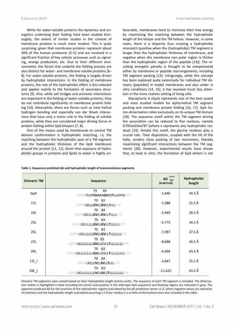
Table 1. Sequences predicted ΔG and hydrophobic length of transmembrane segments.
Chimeric TM segments were named based on their hydrophobic length (amino acids). The sequence of each TM segment is included. The dimeriza-tion motive is highlighted in bold (including the amino acid position in the wild-type GpA sequence) and flanking regions are indicated in gray. The apparent predicted ΔG for the insertion of the hydrophobic regions (calculated by the ΔG prediction server v1.0, where negative values are indicative of insertion) and the hydrophobic length (calculated assuming 1.5 Å per residue in a α-helix conformation) were also included in the table.
B. Grau et al. (2017) In vivo hydrophobic matching
OPEN ACCESS | www.cell-stress.com 92 Cell Stress | NOVEMBER 2017 | Vol. 1 No. 2
solely dependent on the protein sequence. The lipid envi-ronment can also make a significant contribution [21]. It has been shown that not only the lipid composition but also the hydrophobic mismatch between the GpA TM seg-ment and the surrounding hydrophobic environment of the lipid membrane can modify the monomer-dimer equilibri-um [19]. The above view based on experimental work is supported by molecular simulations of model systems, where GpA has served as a centerpiece. Hence, Molecular Dynamics (MD) simulations on GpA have been employed to investigate phenomena such as membrane insertion [22], dimer structure [23, 24], and dimerization energetics [25].
Since its introduction in the nineties [26], the concept of hydrophobic mismatch has received, extensive attention both experimentally (in vitro) [27–30] and computationally [31–33]. However, as we mentioned above, the implica-tions of hydrophobic matching on membrane protein fold-ing, packing, and oligomerization have not been investigat-ed in biological membranes of cells. In the present manu-script, we explore hydrophobic matching and its effects through GpA dimerization in prokaryotic and eukaryotic cells. To this end, we utilized an Escherichia coli fluores-cence-based assay (ToxRED system) and a Bimolecular Flu-orescent Complementation (BiFC) assay in mammalian cells. We show that even in an in vivo scenario with a com-plex membrane system, the hydrophobic matching does contribute to GpA dimerization, i.e., quaternary structure assembly. However, the significance of this effect in biolog-ical membranes is much weaker than under in vitro condi-tions, suggesting that biological membranes are far more adaptable to this stress than previously thought. This con-clusion is backed up by extensive atomistic simulations in a number of GpA-lipid membrane systems with varying pep-tide lengths and membrane thicknesses. These simulations provide an atom-scale interpretation of the mechanisms used by lipid membranes to stabilize dimers exposed to hydrophobic mismatch. To our knowledge, this is the first systematic in vivo study of hydrophobic matching, provid-ing a key step for a better understanding of membrane protein folding under native conditions.
RESULTS Packing of TM segments with different length in biological membranes To assess the influence of hydrophobic matching on the packing of TM segments, one should vary the length of either the TM segments or the model membrane system employed in the assay. Working in cell, the only modifiable variable is the length of the TM segment used. For this purpose, we constructed a series of chimeric stretches bearing the minimal dimerization domain found in GpA (GVxxGVxxT, where x represents an hydrophobic residue) [19] with increasing number of leucines forming TM seg-ments (Table 1). All the hydrophobic regions designed were identified as potential TM segments by the ΔG pre-diction server [34] (Table 1). Likewise, multiple TM protein prediction algorithms classified all the resultant chimeric
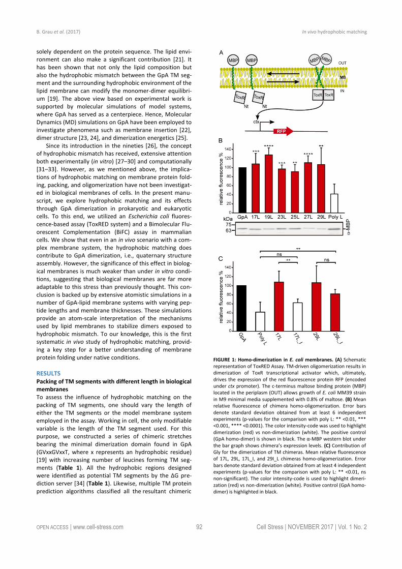
FIGURE 1: Homo-dimerization in E. coli membranes. (A) Schematic representation of ToxRED Assay. TM-driven oligomerization results in dimerization of ToxR transcriptional activator which, ultimately, drives the expression of the red fluorescence protein RFP (encoded under ctx promoter). The c-terminus maltose binding protein (MBP) located in the periplasm (OUT) allows growth of E. coli MM39 strain in M9 minimal media supplemented with 0.8% of maltose. (B) Mean relative fluorescence of chimera homo-oligomerization. Error bars denote standard deviation obtained from at least 6 independent experiments (p-values for the comparison with poly L: ** <0.01, *** <0.001, **** <0.0001). The color intensity-code was used to highlight dimerization (red) vs non-dimerization (white). The positive control (GpA homo-dimer) is shown in black. The α-MBP western blot under the bar graph shows chimera’s expression levels. (C) Contribution of Gly for the dimerization of TM chimeras. Mean relative fluorescence of 17L, 29L, 17L_I, and 29I_L chimeras homo-oligomerization. Error bars denote standard deviation obtained from at least 4 independent experiments (p-values for the comparison with poly L: ** <0.01, ns non-significant). The color intensity-code is used to highlight dimeri-zation (red) vs non-dimerization (white). Positive control (GpA homo-dimer) is highlighted in black.

B. Grau et al. (2017) In vivo hydrophobic matching
OPEN ACCESS | www.cell-stress.com 93 Cell Stress | NOVEMBER 2017 | Vol. 1 No. 2
proteins as membrane proteins. The designed TM seg-ments range from 17 to 29 residues long. The rise per resi-due along the axis in a canonical helix is 1.5Å. Therefore, a stretch of approximately 20 consecutive hydrophobic ami-no acids will span 30Å of the hydrocarbon core of a biologi-cal membrane. Indeed, the most prevalent length of TM helices is 21 residue [35]. By selecting TM segments that are either longer or shorter than 21 residues we can induce a discrepancy in the membrane thickness that allows us to investigate the role of this imbalance in the TM segment packing.
In order to understand the hydrophobic matching ef-fect in a cellular environment we first studied the ability of the aforementioned TM segments to homo-dimerize in E. coli membranes. To this end, we utilized a variation of the ToxCAT assay [36] known as ToxRED [37]. Briefly, this
methodology uses a chimeric construct composed of the N-terminus DNA binding domain of ToxR (a dimerization-dependent transcriptional activator), fused to the chal-lenged TM segment and a periplasmic anchor (the maltose binding protein, MBP) needed for the growth of the bacte-ria in minimal media supplemented with maltose [38]. Di-merization through the TM segments results in ToxRED-mediated activation of the ctx promoter which drives the synthesis of the Red Fluorescent Protein (RFP) (Figure 1A). RFP values were normalized using the absorbance of the bacteria culture (600 nm) to rule out culture growth differ-ences as the source of fluorescence variations (note that in this system the growth of the MM39 E. coli strain depends on the proper expression and insertion of the chimeric protein). Furthermore, the correct expression of all the constructs was assessed by western blot using an anti-MBP
FIGURE 2: Homo-dimerization in eukaryotic membranes. (A) Schematic representation of BiFC Assay. TM-driven oligomerization results in comple-mentation of two non-fluorescent halves (amino-terminus (VN) and carboxy-terminus (VC)) of the Venus Fluorescent Protein (VFP). (B) Mean relative fluorescence of chimera homo-oligomerization in HEK293T cells (GpA (VN-GpA/VC-GpA), 17L (VN-17L/VC-17L), 19L (VN-19L/VC-19L), 23L (VN-23L/VC-23L), 25L (VN-25L/VC-25L), 27L (VN-27L/VC-27L), 29L (VN-29L/VC-29L), H2 (VN-H2/VC-H2)). Error bars indicate standard deviation obtained from at least 4 independent replicates (H2 was used as a negative control, **** < 0.0001). A color intensity code is used to highlight dimerizing (green) and non-dimerizing (white) transmembrane segments, while positive dimerization control (GpA) is shown in black. (C) Western Blot show chimera’s expression levels detected by α-c-Myc antibody. (D) The contribution of Gly for the dimerization of TM chimeras. Relative fluorescence of chimera homo-oligomerization in human-derived HEK293T cells (GpA [VN-GpA/VC-GpA (depicted in black)], H2 [VN-H2/VC-H2] (white), 17L [VN-17L/VC-17L], 17L_I [VN-17L_I/VC-17L_I], 29L [VN-29L/VC-29L], 29L_I [VN-29L_I/VC-29L_I). The bars represent mean values of chimera homo-oligomerization, and error bars denote standard deviation obtained from 3 independent experiments (p-values of Student’s t-test: * < 0.05, ** < 0.01, ns (non-significant)). Color intensity is used to highlight dimerizing (significantly different from H2 control, green) and non-dimerizing (white). Light green is used to indicate those samples (where the Gly residues of the dimerization domain have been substituted with Ile) which fluorescence values are significantly higher than the H2 control and at the same time lower than the corresponding non-mutated control. (E) Cellular fractionation of chimera expressing eukaryotic cells. Subcellular fractionation of HEK293T cells expressing BiFC chimeras (c-Myc tagged) (VN-23L, VN-25L and VN-GpA) or EYFP (Flag-tagged) (used as a soluble marker). Soluble fraction (Sol) and Membrane fraction (MB).

B. Grau et al. (2017) In vivo hydrophobic matching
OPEN ACCESS | www.cell-stress.com 94 Cell Stress | NOVEMBER 2017 | Vol. 1 No. 2
antibody (Figure 1B, bottom). Our results show that, all the chimeras bearing the minimized GpA dimerization mot- if, despite their different hydrophobic lengths, were capa-ble of forming homo-dimers that rendered RFP levels simi-lar to those obtained using the wild-type GpA TM segment (a 23 hydrophobic residue long segment), and significantly higher than the negative controls (a 13 amino acid long stretch of leucines, poly L that efficiently inserts into the membranes [39, 40]) (Figure 1B). It has been shown that long TM hydrophobic segments can lead to oligomerization
[41, 42]. To isolate the contribution of the hydrophobic length on the oligomerization of our segments we mutated the Gly residues to Ile in the 17L (17L_I) and 29L (29L_I) constructs (Figure 1C) (the sequence, hydrophobic length, and predicted ΔG values of these segments are included in Table 1). While elimination of GxxxG motif in the 17L back-bone decreased the ToxRED associated fluorescence to background levels, the Gly to Ile substitutions had a minor effect on 29L, indicating that the positive mismatch can induce TM segment packing in E. coli membranes.
FIGURE 3: Stability of the dimeric structures from MD simulations. (A-D) Root mean squared deviation (RMSD) of the dimerization motif (5 residues per peptide) is shown on the top row and the RMSD of the whole dimer on the bottom row. The whole trajectory is included in the analyses. The 17L/29L hetero-dimer is labeled “HET”, and the polyleucine control in DOPC as “PolyL/DOPC”. (E and F) Helicity of the peptides during the full simu-lation time period (color code as in At to D). The 17L/29L hetero-dimer is labeled “HET”.
B. Grau et al. (2017) In vivo hydrophobic matching
OPEN ACCESS | www.cell-stress.com 95 Cell Stress | NOVEMBER 2017 | Vol. 1 No. 2
Additionally, we analyzed the formation of homo-dimers in eukaryotic cells utilizing a BiFC assay [43]. Briefly, the Venus Fluorescent Protein (VFP) was divided into two non-fluorescent parts: amino-terminus (VN) and carboxyl-terminus (VC). Each half was then fused to the hydrophobic segments previously designed (Table 1) and expressed in human-derived HEK293T cells as in [34]. Oligomerization of the TM domains allows the reconstitution of the full-length VFP and the recovery of its fluorescence properties (Figure 2A). Similarly to the ToxRED assay, we included native GpA TM homo-dimers as a positive control and normalization value. As a negative control, we used the second TM seg-ment of E. coli Leader peptidase (H2), a non-dimerizing protein widely used in membrane protein biogenesis stud-ies [44]. In eukaryotic membranes, we observed, once again, the strong dimerization of all the tested GpA chime-ras regardless of their hydrophobic length (from 17L to 29L) (Figure 2B), with fluorescence values comparable to those obtained when the wild-type TM segment of GpA was used. A western blot was included to monitor protein levels (Figure 2C). In the BiFC assay we also investigated the contribution of the GpA dimerization domain to the interaction between TM monomers. Once again, Gly resi-dues were substituted to Ile in the 17L and 29L constructs (VN and VC) [45]. In both cases, in contrast to the ToxRED results, the elimination of the Gly residue significantly re-duced the observed fluorescence (Figure 2D). Nonetheless, we could observe an increased fluorescence of the 29L_I as compared to the 17L_I, suggesting that positive but not negative hydrophobic mismatch can partially drive oli-gomerization in eukaryotic membranes. The resemblance between the BiFC and ToxRED assays suggest that biologi-cal membranes, despite their origin (eukaryotic or prokar-yotic), behave similarly, but not equally, when packing TM helices.
Contrarily to the ToxRED assay, in which the ToxR and MBP moieties have to face the cytosol and the periplasm, respectively, the BiFC approach cannot discern whether the chimeras are being inserted into the membrane or remain cytosolic. To distinguish between these two possi-bilities we performed subcellular fractionation treatments in which the membrane and cytosolic fractions were sepa-rated (Figure 2E). The chimeras bearing 23L, 25L, or wild-type GpA TM segments were located in the membrane
fraction (lanes 2, 4, and 6 respectively). Conversely, the EYFP (used as a soluble marker) was found exclusively in the cytosolic fraction (lane 7).
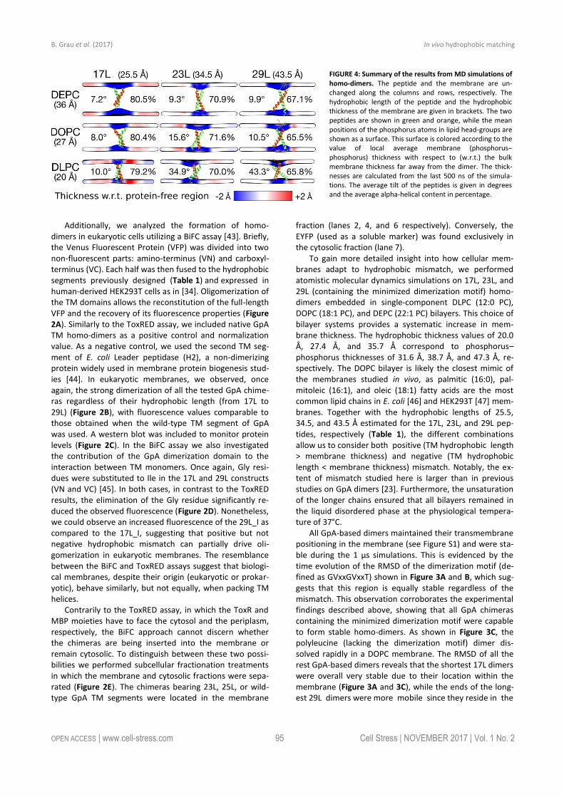
To gain more detailed insight into how cellular mem-branes adapt to hydrophobic mismatch, we performed atomistic molecular dynamics simulations on 17L, 23L, and 29L (containing the minimized dimerization motif) homo-dimers embedded in single-component DLPC (12:0 PC), DOPC (18:1 PC), and DEPC (22:1 PC) bilayers. This choice of bilayer systems provides a systematic increase in mem-brane thickness. The hydrophobic thickness values of 20.0 Å, 27.4 Å, and 35.7 Å correspond to phosphorus–phosphorus thicknesses of 31.6 Å, 38.7 Å, and 47.3 Å, re-spectively. The DOPC bilayer is likely the closest mimic of the membranes studied in vivo, as palmitic (16:0), pal-mitoleic (16:1), and oleic (18:1) fatty acids are the most common lipid chains in E. coli [46] and HEK293T [47] mem-branes. Together with the hydrophobic lengths of 25.5, 34.5, and 43.5 Å estimated for the 17L, 23L, and 29L pep-tides, respectively (Table 1), the different combinations allow us to consider both positive (TM hydrophobic length > membrane thickness) and negative (TM hydrophobic length < membrane thickness) mismatch. Notably, the ex-tent of mismatch studied here is larger than in previous studies on GpA dimers [23]. Furthermore, the unsaturation of the longer chains ensured that all bilayers remained in the liquid disordered phase at the physiological tempera-ture of 37°C.
All GpA-based dimers maintained their transmembrane positioning in the membrane (see Figure S1) and were sta-ble during the 1 µs simulations. This is evidenced by the time evolution of the RMSD of the dimerization motif (de-fined as GVxxGVxxT) shown in Figure 3A and B, which sug-gests that this region is equally stable regardless of the mismatch. This observation corroborates the experimental findings described above, showing that all GpA chimeras containing the minimized dimerization motif were capable to form stable homo-dimers. As shown in Figure 3C, the polyleucine (lacking the dimerization motif) dimer dis-solved rapidly in a DOPC membrane. The RMSD of all the rest GpA-based dimers reveals that the shortest 17L dimers were overall very stable due to their location within the membrane (Figure 3A and 3C), while the ends of the long-est 29L dimers were more mobile since they reside in the
FIGURE 4: Summary of the results from MD simulations of homo-dimers. The peptide and the membrane are un-changed along the columns and rows, respectively. The hydrophobic length of the peptide and the hydrophobic thickness of the membrane are given in brackets. The two peptides are shown in green and orange, while the mean positions of the phosphorus atoms in lipid head-groups are shown as a surface. This surface is colored according to the value of local average membrane (phosphorus–phosphorus) thickness with respect to (w.r.t.) the bulk membrane thickness far away from the dimer. The thick-nesses are calculated from the last 500 ns of the simula-tions. The average tilt of the peptides is given in degrees and the average alpha-helical content in percentage.

B. Grau et al. (2017) In vivo hydrophobic matching
OPEN ACCESS | www.cell-stress.com 96 Cell Stress | NOVEMBER 2017 | Vol. 1 No. 2
aqueous phase (Figure S2). The stability of the 23L dimers decreases upon increasing membrane thickness as their ends become more exposed to water and therefore more prone for unfolding. The increase in the fluctuations within the termini region due to increasing mismatch (negative to positive) was also evident in the RMSF data shown in Fig-ure S2, and explained by a lower overall alpha-helical con-tent of the peptides (see Figure 3E and F and Figure 4). Interestingly, the alpha-helical content of the peptides in a given dimer is independent of the thickness of the host membrane. However, when placed in the same membrane, the longer the peptides are, the lower their alpha-helical content is. Still, although the percentages of the alpha-helical content decrease as the peptides get longer, the absolute number of the amino acids in the alpha-helical conformation actually increases. Thus, some but not all
added Leu residues (17L→23L→29L), originally in a helical conformation, unfold rapidly during the short equilibration simulations, during which position restraints are turned off.
Despite the varying levels of mismatch, the stability of the dimers suggests that either the peptide dimer or the membrane is able to compensate for effects induced by the mismatch. The compensation likely takes place through structural adjustment. In the case of positive mismatch,
the tilt angle of the dimer shown in Figure 4 and Figure 5 reveals that the collective tilting of the dimer plays a role only in the case of the longer peptides (23L or 29L) simu-lated in the thinnest DLPC membrane, where the mismatch is larger, causing the dimer to tilt significantly (~40°) main-taining the crossing angle between monomers. In other cases, the dimer stands almost upright in the membrane (average tilt angle being <20°). As shown in Figure 3E and F, the peptides also maintain their alpha-helical content throughout the production simulation, and no stretching to the 310 helix structure is observed. Meanwhile, in the case of a negative mismatch, the tilt angle remains low (Figure 4 and Figure 5C). Notably, there is little adaptation to the hydrophobic mismatch by a scissor-like motion of the di-mers, as seen in the peptide crossing angles plots (Figure 5A and B).
For both positive and negative mismatch, the mem-brane thickness was perturbed only locally as demonstrat-ed in Figure 4. The spatial extent of the perturbation de-pends on the level of mismatch, though. For the 17L dimer, the effect was smallest in the case of DLPC membrane and increases systematically with increasing bilayer thickness (DLPC<DOPC<DEPC). For the 23L and 29L dimers, the smallest perturbation was observed in the case of DEPC
FIGURE 5: Tilt and crossing angle distribution. (A, B) Crossing angle distribution of the dimers calculated from the last 500 ns of the simulations. The 17L/29L hetero-dimer is labeled “HET”. (C-D) Tilt angle distribution of the dimers calculated from the last 500 ns of the simulations. The 17L/29L hetero-dimer is labeled “HET”.

B. Grau et al. (2017) In vivo hydrophobic matching
OPEN ACCESS | www.cell-stress.com 97 Cell Stress | NOVEMBER 2017 | Vol. 1 No. 2
membrane. The effect became more prominent in the DOPC membrane, which is thinner than DEPC, however, the effect did not increase further in the DLPC membrane as the tilting of the dimers began to dominate the mem-brane organization. Notably, the differences in the behav-ior of the studied peptides in the DOPC membrane, whose thickness resembles that of the bilayers studied here in vivo, are insignificant. All the described adaptation mecha-nisms can be observed in the movie available here.
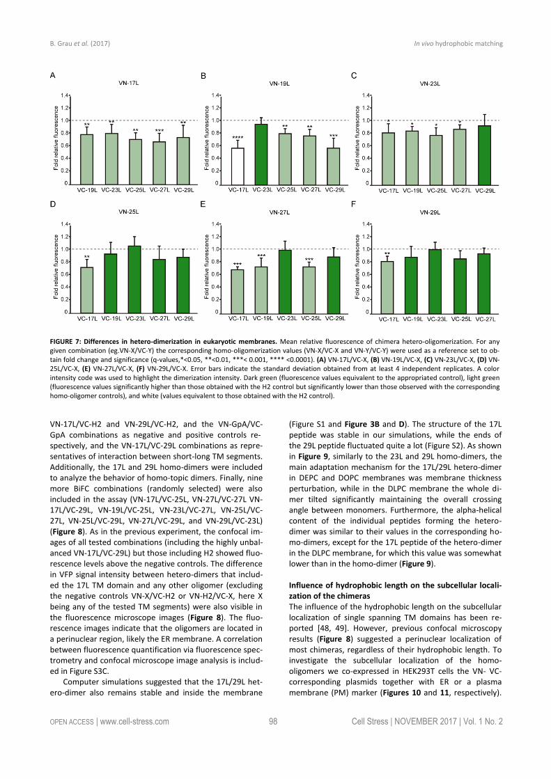
Influence of transmembrane hydrophobic length mis-match on heterotypic helix-helix packing Next, we analyzed the potential hetero-dimer formation between TM segments with different hydrophobic lengths. The GpA homo-dimer was used as a positive reference value set while the H2 was used as a negative control. Sur-prisingly, the majority of the tested combinations between VN- and VC- GpA-derived chimeras (all but the VN-19L/VC-17L) were capable of reconstituting the VFP and produced fluorescence values significantly higher than the negative control (H2) (Figure 6). To highlight differences among the tested combinations we re-analyzed our data using the values of the homo-dimers as a reference set. In this case, for any given combination (e.g., VN-X/VC-Y) the corre-
sponding homo-oligomerization values (VN-X/VC-X and VN-Y/VC-Y) were merged and used to obtain the fluorescence fold change unit (Figure 7). Light green bars indicate that the dimerization value for the selected heteromeric com-bination is significantly lower than its homo-dimer refer-ence set (the level of significance is depicted with the cor-responding number of asterisks on top of each bar). Con-trarily dark green bars show those heteromeric combina-tions in which the VFP fluorescence was as high as the ap-propriated homomeric reference set. The VN-19L/VC-17L was depicted in white to indicate that not only the fluores-cence was lower than in its controls but also not statistical-ly higher than in the non-dimerizing H2 control. Collectively, our data suggest that a different hydrophobic length be-tween the monomers hinders the formation of the dimer. Furthermore, a difficulty in hetero-dimer formation in bio-logical membranes can be observed either when a large disparity between the hydrophobic length of the mono-mers is found or when one of the GpA-derived chimeras contains a hydrophobic region below ~28 Å (17L). Heat map representations of the data in Figure 6 and 7 data are included in Figure S3A and S3B.
The aforementioned results were corroborated by con-focal microscopy (Figure 8). To this end, we selected the
FIGURE 6: Hetero-dimerization in eukaryotic membranes. Mean relative fluorescence of chimera hetero-oligomerization in HEK293T cells of all different combinations: (A) VN-17L/VC-X, (B) VN-19L/VC-X, (C) VN-23L/VC-X, (D) VN-25L/VC-X, (E) VN-27L/VC-X, (F) VN-29L/VC-X. Error bars indicate standard deviation obtained from at least 4 independent replicates. GpA homo-dimer (black bars) was used as positive control and normalization value while Lep H2 homo-oligomer was used as a negative control (white bars). For the experimental samples, a color intensity code was used to highlight dimerization (green, fluorescence values significantly higher than those obtained with the H2 control) and non-dimerization (white, values equivalents to those obtained with the negative control). Additionally asterisks were included to indicate the level of significance (**<0.01, ***< 0.001, **** <0.0001).
B. Grau et al. (2017) In vivo hydrophobic matching
OPEN ACCESS | www.cell-stress.com 98 Cell Stress | NOVEMBER 2017 | Vol. 1 No. 2
VN-17L/VC-H2 and VN-29L/VC-H2, and the VN-GpA/VC-GpA combinations as negative and positive controls re- spectively, and the VN-17L/VC-29L combinations as repre-sentatives of interaction between short-long TM segments. Additionally, the 17L and 29L homo-dimers were included to analyze the behavior of homo-topic dimers. Finally, nine more BiFC combinations (randomly selected) were also included in the assay (VN-17L/VC-25L, VN-27L/VC-27L VN-17L/VC-29L, VN-19L/VC-25L, VN-23L/VC-27L, VN-25L/VC-27L, VN-25L/VC-29L, VN-27L/VC-29L, and VN-29L/VC-23L) (Figure 8). As in the previous experiment, the confocal im-ages of all tested combinations (including the highly unbal-anced VN-17L/VC-29L) but those including H2 showed fluo-rescence levels above the negative controls. The difference in VFP signal intensity between hetero-dimers that includ-ed the 17L TM domain and any other oligomer (excluding the negative controls VN-X/VC-H2 or VN-H2/VC-X, here X being any of the tested TM segments) were also visible in the fluorescence microscope images (Figure 8). The fluo-rescence images indicate that the oligomers are located in a perinuclear region, likely the ER membrane. A correlation between fluorescence quantification via fluorescence spec-trometry and confocal microscope image analysis is includ-ed in Figure S3C.
Computer simulations suggested that the 17L/29L het-ero-dimer also remains stable and inside the membrane
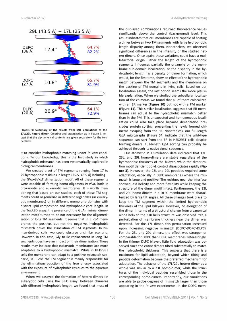
(Figure S1 and Figure 3B and D). The structure of the 17L peptide was stable in our simulations, while the ends of the 29L peptide fluctuated quite a lot (Figure S2). As shown in Figure 9, similarly to the 23L and 29L homo-dimers, the main adaptation mechanism for the 17L/29L hetero-dimer in DEPC and DOPC membranes was membrane thickness perturbation, while in the DLPC membrane the whole di-mer tilted significantly maintaining the overall crossing angle between monomers. Furthermore, the alpha-helical content of the individual peptides forming the hetero-dimer was similar to their values in the corresponding ho-mo-dimers, except for the 17L peptide of the hetero-dimer in the DLPC membrane, for which this value was somewhat lower than in the homo-dimer (Figure 9).
Influence of hydrophobic length on the subcellular locali-zation of the chimeras The influence of the hydrophobic length on the subcellular localization of single spanning TM domains has been re-ported [48, 49]. However, previous confocal microscopy results (Figure 8) suggested a perinuclear localization of most chimeras, regardless of their hydrophobic length. To investigate the subcellular localization of the homo-oligomers we co-expressed in HEK293T cells the VN- VC- corresponding plasmids together with ER or a plasma membrane (PM) marker (Figures 10 and 11, respectively).
FIGURE 7: Differences in hetero-dimerization in eukaryotic membranes. Mean relative fluorescence of chimera hetero-oligomerization. For any given combination (eg.VN-X/VC-Y) the corresponding homo-oligomerization values (VN-X/VC-X and VN-Y/VC-Y) were used as a reference set to ob-tain fold change and significance (q-values,*<0.05, **<0.01, ***< 0.001, **** <0.0001). (A) VN-17L/VC-X, (B) VN-19L/VC-X, (C) VN-23L/VC-X, (D) VN-25L/VC-X, (E) VN-27L/VC-X, (F) VN-29L/VC-X. Error bars indicate the standard deviation obtained from at least 4 independent replicates. A color intensity code was used to highlight the dimerization intensity. Dark green (fluorescence values equivalent to the appropriated control), light green (fluorescence values significantly higher than those obtained with the H2 control but significantly lower than those observed with the corresponding homo-oligomer controls), and white (values equivalent to those obtained with the H2 control).

B. Grau et al. (2017) In vivo hydrophobic matching
OPEN ACCESS | www.cell-stress.com 99 Cell Stress | NOVEMBER 2017 | Vol. 1 No. 2
The microscope-based assay revealed no considerable dif-ferences in subcellular localization among the tested TM segments and indicate that all the homo-oligomers tested, regardless of their hydrophobic length, are preferentially found in the ER membranes. Being our chimeras based on the GpA TM segment we were surprised to find them in the ER membranes and not in the plasma membrane [50]. To confirm that our results were not a mere artifact we transfected HEK293T cells with a plasmid bearing the full sequence of GpA (Flag tagged) and analyzed its colocaliza-tion with ER and PM markers. The micrographs clearly indi-cate that the full-length GpA localizes in the PM membrane (Figure S4). Therefore, since the subcellular localization of all TM segments studied is similar we can assume that the hydrophobic match between the hydrophobic length of the TM segments and the membrane stand as a major contrib-utor to the differences observed in our TM packing studies.
DISCUSSION Folding and packing of proteins depend on their sequence and the environment in which these processes take place. In the case of membrane proteins, it is the lipid bilayer that determines the type and strength of the interactions that will define the mature tertiary and/or quaternary structure. Hence, unlike the aqueous phase, cell membranes are very heterogeneous in terms of their lipid composition and or-ganization in the membrane plane. There is a wide diversi-ty of acyl chains and polar head groups complemented by a variety of domain structures within the bilayer plane. Thereby, membrane proteins are hosted by nanoscale en-
vironments that are dynamic and transient. In this complex scenario, the exposure of hydrophobic protein and lipid groups to water is highly unfavorable and therefore ex-pected to be reduced. Helices in TM domains are, on aver-age, 24 amino acids long (36 Å), ranging from 17 to 34 amino acids (25.5–51 Å) [35]. A stretch of approximately 20 consecutive hydrophobic amino acids can span 30Å of the hydrocarbon core of a biological membrane. Indeed, the most prevalent (~12%) length of TM helices is 21 residues [35]. Based on previous in vitro work, longer helices can span the bilayer with a concomitant tilting of the helix axis relative to the membrane plane [51]. Other options to ac-commodate the wide variety of TM segments range from lipid accommodation to polypeptide backbone defor-mation. Alternatively, each TM segment can be located in a lipid bilayer or a lipid bilayer sub-domain that matches its hydrophobic length, thereby minimizing peptide adapta-tions. In fact, the subcellular distribution of helical mem-brane proteins among multiple organelles based on their hydrophobic lengths is considered to occur [48].
GpA offers a valuable tool for the study of hydrophobic matching in biological membranes, even in living cells. Not only successful formation of a GpA dimer depends on the correct disposition of the TM segment, but it has also been demonstrated in vitro that the hydrophobic mismatch in-fluences TM helix packing in micelles [17, 19]. Furthermore, the NMR structure of the homo-dimer has been resolved in detergent micelles [52] and in membrane bilayers [53] and the dimerization motif has been thoroughly studied in vitro. Given the aforementioned characteristics of GpA, we used
FIGURE 8: Confocal microscopy analysis for membrane dimer formation. Confocal microscopy of DAPI stained (blue) HEK293T cells expressing rep-resentative VN-VC combinations (GpA [VN-GpA/VC-GpA], 17L [VN-17L/VC-17L], 29L [VN-29L/VC-29L], 17L/H2 [VN-17L/VC-H2], 29L/H2 [VN-29L/VC-H2], 17L/29L [VN-17L/VC-29L]). Successful TM-driven oligomerization results in VFP reconstitution and fluorescent signal (green). Scale bar size is 16 µm.
B. Grau et al. (2017) In vivo hydrophobic matching
OPEN ACCESS | www.cell-stress.com 100 Cell Stress | NOVEMBER 2017 | Vol. 1 No. 2
it to consider hydrophobic matching under in vivo condi-tions. To our knowledge, this is the first study in which hydrophobic mismatch has been systematically explored in biological membranes.
We created a set of TM segments ranging from 17 to 29 hydrophobic residues in length (25.5–43.5 Å) including the GVxxGVxxT dimerization motif. All of these segments were capable of forming homo-oligomers in vivo, both in prokaryotic and eukaryotic membranes. It is worth men-tioning that based on our studies, each of these TM seg-ments could oligomerize in different organelles (in eukary-otic membranes) or in different membrane domains with distinct lipid composition and hydrophobic core length. In the ToxRED assay, the presence of the GpA minimal dimer-ization motif turned to be not necessary for the oligomeri-zation of long TM segments. It seems that in E. coli mem-branes the positive, but not the negative, hydrophobic mismatch drives the association of TM segments. In hu-man-derived cells, we could observe a similar scenario. However, in this case, Gly to Ile replacement in long TM segments does have an impact on their dimerization. These results may indicate that eukaryotic membranes are more adaptable to a hydrophobic mismatch. While in HEK293T cells the membrane can adapt to a positive mismatch sce-nario, in E. coli the TM segment is mainly responsible for the elimination/reduction of the free energy associated with the exposure of hydrophobic residues to the aqueous environment.
When we assayed the formation of hetero-dimers (in eukaryotic cells using the BiFC assay) between chimeras with different hydrophobic length, we found that most of
the displayed combinations returned fluorescence values significantly above the control (background) level. This result indicates that cell membranes are capable of hosting a dimer between two TM segments with large hydrophobic length disparity among them. Nonetheless, we observed significant differences in the intensity of the studied het-ero-dimers. Once again, these variations could have a mul-ti-factorial origin. Either the length of the hydrophobic segments influences partially the organelle or the mem-brane sub-domain localization, or the disparity in the hy-drophobic length has a penalty on dimer formation, which would, for the first time, show an effect of the hydrophobic match between the TM segments and the membrane on the packing of TM domains in living cells. Based on our localization assays, the last option seems the more plausi-ble explanation. When we studied the subcellular localiza-tion of the chimeras we found that all of them colocalized with an ER marker (Figure 10) but not with a PM marker (Figure 11). This similar localization suggests that ER mem-branes can adjust to the hydrophobic mismatch better than in the PM. This unexpected and homogeneous locali-zation could also take place because dimerization pre-cludes protein sorting, preventing the newly formed chi-meras escaping from the ER. Nonetheless, our full-length GpA micrographs (Figure S4) indicate that the wild-type sequence can sort from the ER in HEK293T cells despite forming dimers. Full-length GpA sorting can probably be achieved through its native signal sequence.
Our atomistic MD simulation data indicated that 17L, 23L, and 29L homo-dimers are stable regardless of the hydrophobic thickness of the bilayer, while the dimeriza-tion motif deficient polyL control disassociates rapidly (Fig-ure 3). However, the 23L and 29L peptides required some adaptation, especially in DLPC membranes where the mis-match is large and positive. The residues near the interface showed less helicity and more flexibility while keeping the structure of the dimer motif intact. Furthermore, the 23L and 29L homo-dimers in a DLPC membrane were charac-terized by large tilt angles. All these changes took place to keep the TM segment within the limited hydrophobic thickness of the lipid bilayers. However, no elongation of the dimer in terms of a structural change from a canonical alpha helix to the 310 helix structure was observed. Yet, a perturbation of membrane thickness near the dimer was detected. For the 17L dimer, this perturbation increased upon increasing negative mismatch (DEPC>DOPC>DLPC). For the 23L and 29L dimers, the effect was stronger or comparable for DOPC than DEPC membranes. Interestingly, in the thinner DLPC bilayer, little lipid adaptation was ob-served since the entire dimers tilted substantially to match the hydrophobic thickness. This suggests that there is a maximum for lipid adaptation, beyond which tilting and peptide deformation become the preferred mechanism for adaptation. The behavior of the 17L/29L hetero-dimer as a whole was similar to a 23L homo-dimer, while the struc-tures of the individual peptides resembled those in the corresponding homo-dimers. Importantly, our simulations are able to probe degrees of mismatch larger than those appearing in the in vivo experiments. In the DOPC mem-
FIGURE 9: Summary of the results from MD simulations of the 17L/29L hetero-dimer. Coloring and organization as in Figure 3, ex-cept that the alpha-helical contents are given separately for the two peptides.

B. Grau et al. (2017) In vivo hydrophobic matching
OPEN ACCESS | www.cell-stress.com 101 Cell Stress | NOVEMBER 2017 | Vol. 1 No. 2
brane, the thickness of which likely matches that of the E. coli and HEK293T membranes studied in vivo, the differ-ences observed in dimer tilting were actually small. There-fore, the importance of this tilting in living membranes might not play a major role, and the perturbation of mem-brane thickness is likely the dominant adaption mechanism in vivo.
The stability of native GpA dimers in various lipid envi-ronments has been studied in previous works using both coarse-grained [24, 25, 54] and atomistic [55] simulations. These simulations reported dimerization free energies in the range of 6–10 kcal/mol. Notably, changing the mem-brane composition seems to have a minor effect on this value [54]. Furthermore, atomistic simulations reported small differences in the dimeric structure of native GpA embedded in membranes of different thicknesses [23]. Our simulation data complement these results from a novel perspective, now discussing the variation of the peptide lengths as a new variable, therefore covering larger values of mismatch and by reducing the importance of the struc-tural features of the GpA dimer to the minimalized dimeri-zation motif. Despite these extensions, our results agree with the earlier work on GpA.
The two adaptation mechanisms resolved here have been previously reported in systematic computational studies on KALP and WALP monomers of different lengths embedded in membranes of various thicknesses [32, 33]. It seems that in the case of stable dimers such as those stud-ied here, the adaptation mechanisms are similar to the ones observed previously for peptide monomers. It must be noted that some other suggested mechanisms [33], such as the formation of a non-lamellar phase or the expul-sion of the peptides from the membrane, are not feasible in our simulation study that is limited by periodic boundary conditions and achievable timescales. Furthermore, our atomistic study is limited to individual dimers. Therefore, we cannot rule out the possibility that membrane stress is relieved through the formation of larger oligomers.
Membrane proteins represent close to 30% of the hu-man proteome [3–5] and 60% of the approved drug targets [56]. Furthermore, processes crucial to life are regulated by elaborated interplays of membrane protein complexes, where TM domain interactions play an important role [57]. In this study, we have shown and discussed pioneering data for membrane protein folding and packing in living cells, aiming to unravel the role of hydrophobic mismatch
FIGURE 10: Homo-dimerization at the endoplasmic reticulum. Confocal microscopy of DAPI stained (blue) HEK293T cells expressing tested homo-dimers (17L [VN-17L/VC-17L], 19L [VN-19L/VC-19L], 23L [VN-23L/VC-23L], 25L [VN-25L/VC-25L], 27L [VN-27L/VC-27L], 29L [VN-29L/VC-29L]). TM-driven homo-oligomerization results in VFP reconstitution and fluorescent signal (green). Sec61α fused to mCherry fluorescent protein was used as ER marker (red). The presence of colocalization of red and green signals in the merge images was highlighted in yellow. Scale bar size was set to 16 µm.

B. Grau et al. (2017) In vivo hydrophobic matching
OPEN ACCESS | www.cell-stress.com 102 Cell Stress | NOVEMBER 2017 | Vol. 1 No. 2
in TM helix-helix packing. Our work not only highlights the capability of biological membranes to host TM homo-oligomers with hydrophobic regions ranging from 25.5 to 43.5 Å, but it also demonstrates the ability of lipids and peptide regions to cooperate in order to minimize the hy-drophobic mismatch. MATERIALS AND METHODS DNA constructs All chimeric TM segments were obtained adding the indicated number of extra leucines to the minimized GpA dimerization motif described by [19]. DNA coding for the chimeric TM seg-ments were introduced into the ToxRED plasmid (provided by Dr. Berger) using XhoI and HindIII restriction sites (the selected restrictions sites incorporated a Lys residue preceded by a Leu residue at the amino terminus of the constructs) or into modi-fied BiFC plasmids (pBiFC-VN155 and pBiFC-VC173, provided by Dr. Orzáez [57]) fused to the amino- or carboxyl-end of the Venus Fluorescent Protein using NotI site. All plasmid se-quences were corroborated by DNA sequencing (Macrogen, www.macrogen.com).
ToxRED assay The malE- E. coli (MM39) strain was transformed with the appropriate plasmids and plated onto Luria Bertani with 100 µg/ml ampicillin (LBA) plates. Individual colonies were inocu-lated into LBA medium and grown at 37°C o/n. The LBA cul-tures were diluted 1/100 in M9 minimal medium supplement-ed with 0.8% of maltose and grown at 37°C o/n. The optical density (λ595 nm) (used to normalize the fluorescence values) and the red fluorescence (λ620 nm) of the cultures expressing the chimeras were then measured using a Multimode Plate Reader Victor X3 (Perkin Elmer). For Western blots, MM39 colonies were inoculated into LBA medium and grown at 37°C until approximately OD420 of 0.6. At this point, cultures were harvested by centrifugation, resuspended in lysis buffer (TBS [Tris-HCl 20 mM pH7.5, NaCl 150 mM], 1% NP-40) and freeze-thaw (x3). The lysates were clarified by centrifugation (13,000 g). Supernatants were mixed with SDS-PAGE sample buffer, heated to 95°C for 5 min, separated on 12% (w/v) SDS gel and blotted onto nitrocellulose membranes. The constructs were detected with a HRP-conjugated anti-MBP monoclonal anti-body (New England Biolabs) and ECL reagent (GE Healthcare). Chemiluminescence was visualized using an ImageQuant LAS 4000 (GE Healthcare) Biomolecular Imager.
FIGURE 11: Homo-dimerization at the plasma membrane. Confocal microscopy of DAPI stained (blue) HEK293T cells expressing chimeras (17L [VN-17L/VC-17L], 19L [VN-19L/VC-19L], 23L [VN-23L/VC-23L], 25L [VN-25L/VC-25L], 27L [VN-27L/VC-27L], 29L [VN-29L/VC-29L]). TM-driven homo-oligomerization results in VFP reconstitution and fluorescent signal (green). Neuromodulin fused to mCherry fluorescent protein was used as plasma membrane marker (red). The presence of colocalization of red and green signals in the merge images was highlighted in yellow. Scale bar size was set to 16 µm.

B. Grau et al. (2017) In vivo hydrophobic matching
OPEN ACCESS | www.cell-stress.com 103 Cell Stress | NOVEMBER 2017 | Vol. 1 No. 2
Bimolecular Fluorescence Complementation assay (BiFC) HEK293T (ATCC) cells were cultured in Dulbecco’s modified Eagle medium (Gibco) supplemented with 10% fetal bovine serum in 24-well plates at 37°C. Plasmids encoding VN or VC fusion proteins were transfected into HEK293T using Lipofec-tamine 2000 transfection reagent (Invitrogen), according to manufacturer's instructions. A plasmid encoding Renilla lucif-erase under CMV promoter (pRL-CMV, Promega) was cotrans-fected for normalization purposes. For Renilla luciferase measuring, Renilla Luciferase Flash Assay Kit (Thermo Scien-tific) was used following the manufacturer’s instructions. The luminescence and the fluorescence of each sample were measured 48 hrs post-transfection using a Multimode Plate Reader Victor X3 (Perkin Elmer). Immuno-identification of the samples was done using α-c-Myc (VN) and α-HA tag (VC) rab-bit antibodies, respectively, followed by a secondary HRP-conjugated anti-rabbit antibody (Sigma). Chemiluminescence was visualized using an ImageQuant LAS 4000 (GE Healthcare) Biomolecular Imager. P-values were adjusted to compensate for a false discovery rate (q-values) using the Benjamini and Hoechberg approach. Confocal fluorescence microscopy Glass slides were treated with 30 µL of poly-L-Lysine 0.01% (Sigma-Aldrich) and washed 3 times with PBS. HEK293T cells were cultured in the glass slides and transfected as mentioned above. Cells were co-transfected with a plasmid encoding alternatively, an endoplasmatic reticulum marker (ER) (mCh-Sec61β, Addgene 49155) or a plasma membrane (PM) marker (mCh-Mem, Addgene 55779). After 2 days, cells were fixed (4% formaldehyde, Sigma-Aldrich) and DAPI stained (Fluoro-shield, Sigma-Aldrich). Images were captured using a FV1000 confocal microscope (Olympus). Mean gray value of each im-age was measured using ImageJ (NIH). Immunofluorescence HEK293T cells were transfected with a plasmid encoding full-length GpA protein with a Flag M2 tag in the amino terminus (after GpA signal peptide and cleavage site of the protease). Cells were also co-transfected with a plasmid encoding alter-natively the ER or PM markers. Forty-eight hours after trans-fection cells were fixed (4% formaldehyde in PBS, 15 min) and washed in PBS (x3). Permeabilization was done with PBS, 1% BSA, 0.1% Triton X-100 for 2 minutes. Immuno-stainings were done using a primary α-Flag M2 antibody (Sigma), followed by a secondary Alexa647-conjugated anti-Mouse antibody (Sig-ma). Additionally, cells were DAPI stained for nucleus staining. Subcellular fractionation HEK293T cells were transfected with plasmids encoding VN fusion proteins and cultured for 48h as mentioned above. As a soluble protein marker, we used the Enhanced Yellow Fluores-cent Protein (EYFP) bearing a Flag M2 tag. Cells were collected with 500 µL of a subcellular fractionation (SF) buffer (250mM Sucrose, 20 mM HEPES pH 7.4, 10 mM KCl, 1.5 mM MgCl2, 1 mM EDTA, 1 mM EGTA) and lysed by sonication. Lysates were incubated at 4°C for 30 min and centrifuged (10,000 g, 4°C, 10 min). Supernatants were carefully transferred to a new tube and ultracentrifuged at 10,0000g at 4°C for 1 h. The superna-tants of the ultracentrifugation (cytosolic fraction) were stored while the pellet was re-suspended in 500 µL of SF buffer and ultracentrifuged again using the same conditions. The final
pellet (membrane fraction) was re-suspended in 150 µL of 1x SDS-PAGE buffer. Structural models of the simulated system The structures of three peptide homo-dimers (17L, 23L, and 29L), one hetero-dimer (17L/29L), and a polyleucine control (corresponding to the 23L dimer without the dimerization motifs, PolyL) were prepared using the GpA dimer (PDB:1AFO, [52]) as a template. Using MODELLER [58], the structures of the dimers were aligned with the dimeric GpA NMR structure. The required mutations and/or the extension of the peptide were then performed. A total of 100 models were created and the ones with the best match to GpA were chosen to preserve the structure of the dimerization motif. The C-termini of these most appropriate models were left charged, while the N-termini were acetylated to mimic the chimeric polypeptides studied in cells. The dimers were embedded in three lipid bi-layers of varying thickness using CHARMM-GUI [59]: 1,2-dilauroyl-sn-glycero-3-phosphocholine (DLPC, di-12:0), 1,2-dioleoyl-sn-glycero-3-phosphocholine (DOPC, di-18:1), or 1,2-dierucoyl-sn-glycero-3-phosphocholine (DEPC, di-22:1). The polyleucine control (dimerization motif-less) was only studied in DOPC. These bilayers consisted of a total of 400 lipids (200 per leaflet), and they were solvated by a total of 24,000 water molecules and 134 mM of NaCl. Atomistic molecular dynamics simulations The peptides [60] and the lipids [61] were modeled using the CHARMM36 force field, while the CHARMM-specific TIP3P model was employed for water. The systems were equilibrat-ed following the standard protocol of CHARMM-GUI, in which the position restraints are stepwise reduced. For each of the systems, this was followed by 1 microsecond of unbiased sim-ulation, where the last 500 ns whenever averages of quanti-ties were calculated. The polyleucine control was only simu-lated for 500 ns. These simulations were performed in the NPT ensemble using a leap-frog integrator and a time step of 2 fs. The pressure was maintained semi-isotropically at 1 bar using the Parrinello–Rahman barostat [62] with a coupling time constant of 5 ps and a compressibility of 4.5x10
-5 bar
-1. The
temperatures of the peptides, lipids, and solvent were sepa-rately maintained at 37°C using the Nosé–Hoover thermostat [63] with a time constant of 1 ps. The smooth particle mesh Ewald algorithm was employed for electrostatics [64]. The Lennard-Jones interactions were (force-)switched to zero be-tween 10 and 12 Å. The buffered Verlet list cut-off scheme was employed [65]. The lengths of bonds involving hydrogen atoms were constrained using LINCS [66]. Trajectories were written every 100 ps for analysis. All simulations were per-formed using Gromacs v. 5.1.x [67]. All our simulation data are openly available online for everyone here and here with a movie of the simulations available here. Analyses of simulation data Root mean squared deviation (RMSD) and root mean square fluctuation (RMSF) were analyzed using the Gromacs tools gmx rms and gmx rmsf. Dimer tilt angles were evaluated using the gmx bundle tool. Two angles were used to describe the orientation of the peptides within the membrane, both were evaluated using the gmx bundle tool. The dimer tilt angle, describing the tilt of the dimer as a whole, was calculated as follows: the center of mass (COM) of the three subsequent

B. Grau et al. (2017) In vivo hydrophobic matching
OPEN ACCESS | www.cell-stress.com 104 Cell Stress | NOVEMBER 2017 | Vol. 1 No. 2
leucine residues right above the dimerization motifs in the two peptides defined the top of the dimerization motif. The bot-tom of the motif was defined similarly using the COM of the three leucine residues right below the motifs in the two pep-tides. The tilt angle of the dimer was defined as the angle be-tween a vector connecting the top and the bottom of the mo-tif and the z axis (normal to the bilayer). The crossing angle of the dimer, describing the mutual orientation of the two pep-tides, is defined as the angle between the two vectors con-necting the leucine residues right next to the dimerization motif in the individual peptides. The alpha-helical content of the peptides was evaluated using the gmx do_dssp and DSSP [68] tools. The thickness maps were calculated using g_lomepro [69]. Membrane thickness was defined as the in-ter-leaflet phosphorus–phosphorus distance, and membrane hydrophobic thickness as the inter-leaflet distance between the fatty acid chain alpha carbons. Both thicknesses were ana-lyzed employing a grid spacing of 0.33 nm, and the figures displaying the surfaces formed by the phosphorus atoms were rendered using VMD [70]. The thickness values of the mem-brane bulk were estimated as the average thickness of the membrane at the grid points furthest away from the protein.
ACKNOWLEDGEMENTS We would like to thank Ernesto Segredo for early work on the ToxRED system. CSC-IT Center for Science is thanked for computational resources. This work was supported by grants from the Spanish Ministry of Economy and Competi-tiveness (MINECO) (grant no. BFU2016-79487) and from the Generalitat Valenciana (GV/2016/139, Program Grupos
Emergentes; and PROMETEOII/2014/061, Program Grupos de Excelencia). BG is a recipient of a Pre-Doctoral fellow-ship from the Generalitat Valenciana. LMG is funded by the Spanish MiNECO (Program Juan de la Cierva) and MGM by the Generalitat Valenciana (PROMETEOII/2014/061). MJ, WK, and IV thank the Academy of Finland (Centre of Excel-lence and FiDiPro programs) and the European Research Council (Advanced Grant CROWDED-PRO-LIPIDS).
SUPPLEMENTAL MATERIAL All supplemental data for this article are available online at www.cell-stress.com.
CONFLICT OF INTEREST The authors declare no conflict of interest.
COPYRIGHT © 2017 Grau et al. This is an open-access article released under the terms of the Creative Commons Attribution (CC BY) license, which allows the unrestricted use, distribution, and reproduction in any medium, provided the original author and source are acknowledged.
Please cite this article as: Brayan Grau, Matti Javanainen, Maria Jesús García-Murria, Waldemar Kulig, Ilpo Vattulainen, Ismael Mingarro, Luis Martínez-Gil (2017). The role of hydrophobic matching on transmembrane helix packing in cells. Cell Stress 1(2): 90-106. doi: 10.15698/cst2017.11.111.
REFERENCES 1. Popot JL, and Engelman DM (1990). Membrane protein folding and oligomerization: the two-stage model. Biochemistry 29(17): 4031–4037. PMID: 1694455
2. Krieg UC, Johnson AE, and Walter P (1989). Protein translocation across the endoplasmic reticulum membrane: identification by photo-cross-linking of a 39-kD integral membrane glycoprotein as part of a putative translocation tunnel. J Cell Biol 109(5): 2033–2043. PMID: 2808520
3. Lander ES, Linton LM, Birren B, Nusbaum C, Zody MC, Baldwin J, Devon K, Dewar K, Doyle M, FitzHugh W, Funke R, Gage D, Harris K, Heaford A, Howland J, Kann L, Lehoczky J, LeVine R, McEwan P, McKernan K, Meldrim J, Mesirov JP, Miranda C, Morris W, Naylor J, Raymond C, Rosetti M, Santos R, Sheridan A, Sougnez C et al. (2001). Initial sequencing and analysis of the human genome. Nature 409(6822): 860–921. doi: 10.1038/35057062
4. Venter JC, Adams MD, Myers EW, Li PW, Mural RJ, Sutton GG, Smith HO, Yandell M, Evans CA, Holt RA, Gocayne JD, Amanatides P, Ballew RM, Huson DH, Wortman JR, Zhang Q, Kodira CD, Zheng XH, Chen L, Skupski M, Subramanian G, Thomas PD, Zhang J, Gabor Miklos GL, Nelson C, Broder S, Clark AG, Nadeau J, McKusick VA, Zinder N et al. (2001). The sequence of the human genome. Science 291(5507): 1304–1351. doi: 10.1126/science.1058040
5. Uhlén M, Fagerberg L, Hallström BM, Lindskog C, Oksvold P, Mardi-noglu A, Sivertsson Å, Kampf C, Sjöstedt E, Asplund A, Olsson I, Edlund K, Lundberg E, Navani S, Szigyarto CA, Odeberg J, Djureinovic D, Takanen JO, Hober S, Alm T, Edqvist PH, Berling H, Tegel H, Mulder J, Rockberg J, Nilsson P, Schwenk JM, Hamsten M, von Feilitzen K, Forsberg M et al. (2015). Proteomics. Tissue-based map of the human
proteome. Science 347(6220): 1260419. doi: 10.1126/science.1260419
6. Hong H (2014). Toward understanding driving forces in membrane protein folding. Arch Biochem Biophys 564C: 297–313. doi: 10.1016/j.abb.2014.07.031
7. Martínez-Gil L, Saurí A, Marti-Renom MA, and Mingarro I (2011). Membrane protein integration into the endoplasmic reticulum. FEBS J 278(20): 3846–3858. doi: 10.1111/j.1742-4658.2011.08185.x
8. Martinez-Gil L, and Mingarro I (2015). Viroporins, Examples of the Two-Stage Membrane Protein Folding Model. Viruses 7(7): 3462–3482. doi: 10.3390/v7072781
9. Cymer F, von Heijne G, and White SH (2015). Mechanisms of inte-gral membrane protein insertion and folding. J Mol Biol 427(5): 999–1022. doi: 10.1016/j.jmb.2014.09.014
10. Bañó-Polo M, Baeza-Delgado C, Orzáez M, Marti-Renom MA, Abad C, and Mingarro I (2012). Polar/Ionizable Residues in Transmembrane Segments: Effects on Helix-Helix Packing. PLoS ONE 7(9): e44263. doi: 10.1371/journal.pone.0044263
11. Andersen OS, and Koeppe RE (2007). Bilayer thickness and mem-brane protein function: an energetic perspective. Annu Rev Biophys Biomol Struct 36: 107–130. doi: 10.1146/annurev.biophys.36.040306.132643
12. Mouritsen OG, and Bloom M (1984). Mattress model of lipid-protein interactions in membranes. Biophys J 46(2): 141–153. doi: 10.1016/S0006-3495(84)84007-2

B. Grau et al. (2017) In vivo hydrophobic matching
OPEN ACCESS | www.cell-stress.com 105 Cell Stress | NOVEMBER 2017 | Vol. 1 No. 2
13. Killian JA (1998). Hydrophobic mismatch between proteins and lipids in membranes. Biochim Biophys Acta 1376(3): 401–415. PMID: 9805000
14. Jensen MØ, and Mouritsen OG (2004). Lipids do influence protein function-the hydrophobic matching hypothesis revisited. Biochim Biophys Acta 1666(1–2): 205–226. doi: 10.1016/j.bbamem.2004.06.009
15. Lee AG (2005). How lipids and proteins interact in a membrane: a molecular approach. Mol Biosyst 1(3): 203–212. doi: 10.1039/b504527d
16. Mackenzie KR (2006). Folding and stability of alpha-helical integral membrane proteins. Chem Rev 106(5): 1931–1977. doi: 10.1021/cr0404388
17. Orzáez M, Pérez-Payá E, and Mingarro I (2000). Influence of the C-terminus of the glycophorin A transmembrane fragment on the dimer-ization process. Protein Sci 9(6): 1246–1253. doi: 10.1110/ps.9.6.1246
18. Lemmon MA, Flanagan JM, Hunt JF, Adair BD, Bormann BJ, Demp-sey CE, and Engelman DM (1992). Glycophorin A dimerization is driven by specific interactions between transmembrane alpha-helices. J Biol Chem 267(11): 7683–7689. PMID: 1560003
19. Orzáez M, Lukovic D, Abad C, Pérez-Payá E, and Mingarro I (2005). Influence of hydrophobic matching on association of model trans-membrane fragments containing a minimised glycophorin A dimerisa-tion motif. FEBS Lett 579(7): 1633–1638. doi: 10.1016/j.febslet.2005.01.078
20. DeGrado WF, Gratkowski H, and Lear JD (2003). How do helix-helix interactions help determine the folds of membrane proteins? Per-spectives from the study of homo-oligomeric helical bundles. Protein Sci 12(4): 647–665. doi: 10.1110/ps.0236503
21. Kuznetsov AS, Volynsky PE, and Efremov RG (2015). Role of the Lipid Environment in the Dimerization of Transmembrane Domains of Glycophorin A. Acta Naturae 7(4): 122–127. PMID: 26798499
22. Bond PJ, and Sansom MSP (2006). Insertion and assembly of membrane proteins via simulation. J Am Chem Soc 128(8): 2697–2704. doi: 10.1021/ja0569104
23. Petrache HI, Grossfield A, MacKenzie KR, Engelman DM, and Woolf TB (2000). Modulation of glycophorin A transmembrane helix interac-tions by lipid bilayers: molecular dynamics calculations. J Mol Biol 302(3): 727–746. doi: 10.1006/jmbi.2000.4072
24. Flinner N, Mirus O, and Schleiff E (2014). The influence of fatty acids on the GpA dimer interface by coarse-grained molecular dynam-ics simulation. Int J Mol Sci 15(8): 14247–14268. doi: 10.3390/ijms150814247
25. Sengupta D, and Marrink SJ (2010). Lipid-mediated interactions tune the association of glycophorin A helix and its disruptive mutants in membranes. Phys Chem Chem Phys 12(40): 12987–12996. doi: 10.1039/c0cp00101e
26. Bloom M, Evans E, and Mouritsen OG (1991). Physical properties of the fluid lipid-bilayer component of cell membranes: a perspective. Q Rev Biophys 24(3): 293–397. PMID: 1749824
27. Anbazhagan V, and Schneider D (2010). The membrane environ-ment modulates self-association of the human GpA TM domain--implications for membrane protein folding and transmembrane signal-ing. Biochim Biophys Acta 1798(10): 1899–1907. doi: 10.1016/j.bbamem.2010.06.027
28. Grau-Campistany A, Strandberg E, Wadhwani P, Rabanal F, and Ulrich AS (2016). Extending the Hydrophobic Mismatch Concept to Amphiphilic Membranolytic Peptides. J Phys Chem Lett 7(7): 1116–1120. doi: 10.1021/acs.jpclett.6b00136
29. Muhle-Goll C, Hoffmann S, Afonin S, Grage SL, Polyansky AA, Windisch D, Zeitler M, Bürck J, and Ulrich AS (2012). Hydrophobic matching controls the tilt and stability of the dimeric platelet-derived growth factor receptor (PDGFR) β transmembrane segment. J Biol Chem 287(31): 26178–26186. doi: 10.1074/jbc.M111.325555
30. Soubias O, Teague WE, Hines KG, and Gawrisch K (2015). Rhodop-sin/lipid hydrophobic matching-rhodopsin oligomerization and func-tion. Biophys J 108(5): 1125–1132. doi: 10.1016/j.bpj.2015.01.006
31. de Jesus AJ, and Allen TW (2013). The determinants of hydropho-bic mismatch response for transmembrane helices. Biochim Biophys Acta 1828(2): 851–863. doi: 10.1016/j.bbamem.2012.09.012
32. Kim T, and Im W (2010). Revisiting hydrophobic mismatch with free energy simulation studies of transmembrane helix tilt and rota-tion. Biophys J 99(1): 175–183. doi: 10.1016/j.bpj.2010.04.015
33. Kandasamy SK, and Larson RG (2006). Molecular Dynamics Simula-tions of Model Trans-Membrane Peptides in Lipid Bilayers: A System-atic Investigation of Hydrophobic Mismatch. Biophys J 90(7): 2326–2343. doi: 10.1529/biophysj.105.073395
34. Hessa T, Meindl-Beinker NM, Bernsel A, Kim H, Sato Y, Lerch-Bader M, Nilsson I, White SH, and von Heijne G (2007). Molecular code for transmembrane-helix recognition by the Sec61 translocon. Nature 450(7172): 1026–1030. doi: 10.1038/nature06387
35. Baeza-Delgado C, Marti-Renom MA, and Mingarro I (2013). Struc-ture-based statistical analysis of transmembrane helices. Eur Biophys J EBJ 42(2–3): 199–207. doi: 10.1007/s00249-012-0813-9
36. Russ WP, and Engelman DM (1999). TOXCAT: A measure of trans-membrane helix association in a biological membrane. Proc Natl Acad Sci 96(3): 863–868. doi: 10.1073/pnas.96.3.863
37. Berger BW, Kulp DW, Span LM, DeGrado JL, Billings PC, Senes A, Bennett JS, and DeGrado WF (2010). Consensus motif for integrin transmembrane helix association. Proc Natl Acad Sci U S A 107(2): 703–708. doi: 10.1073/pnas.0910873107
38. Andreu-Fernández V, García-Murria MJ, Bañó-Polo M, Martin J, Monticelli L, Orzáez M, and Mingarro I (2016). The C-terminal Do-mains of Apoptotic BH3-only Proteins Mediate Their Insertion into Distinct Biological Membranes. J Biol Chem 291(48): 25207–25216. doi: 10.1074/jbc.M116.733634
39. Jaud S, Fernández-Vidal M, Nilsson I, Meindl-Beinker NM, Hübner NC, Tobias DJ, von Heijne G, and White SH (2009). Insertion of short transmembrane helices by the Sec61 translocon. Proc Natl Acad Sci U S A 106(28): 11588–11593. doi: 10.1073/pnas.0900638106
40. Baeza-Delgado C, von Heijne G, Marti-Renom MA, and Mingarro I (2016). Biological insertion of computationally designed short trans-membrane segments. Sci Rep 6: 23397. doi: 10.1038/srep23397
41. Wei P, Liu X, Hu M-H, Zuo L-M, Kai M, Wang R, and Luo S-Z (2011). The dimerization interface of the glycoprotein Ibβ transmembrane domain corresponds to polar residues within a leucine zipper motif. Protein Sci 20(11): 1814–1823. doi: 10.1002/pro.713
42. Li R, Gorelik R, Nanda V, Law PB, Lear JD, DeGrado WF, and Ben-nett JS (2004). Dimerization of the transmembrane domain of Integrin alphaIIb subunit in cell membranes. J Biol Chem 279(25): 26666–26673. doi: 10.1074/jbc.M314168200
43. Hu C-D, Chinenov Y, and Kerppola TK (2002). Visualization of inter-actions among bZIP and Rel family proteins in living cells using bimo-lecular fluorescence complementation. Mol Cell 9(4): 789–798. PMID: 11983170
44. White SH, and von Heijne G (2008). How translocons select trans-membrane helices. Annu Rev Biophys 37: 23–42. doi: 10.1146/annurev.biophys.37.032807.125904

B. Grau et al. (2017) In vivo hydrophobic matching
OPEN ACCESS | www.cell-stress.com 106 Cell Stress | NOVEMBER 2017 | Vol. 1 No. 2
45. Lemmon MA, Flanagan JM, Treutlein HR, Zhang J, and Engelman DM (1992). Sequence specificity in the dimerization of transmem-brane alpha-helices. Biochemistry 31(51): 12719–12725. PMID: 1463743
46. De Siervo AJ (1969). Alterations in the phospholipid composition of Escherichia coli B during growth at different temperatures. J Bacteriol 100(3): 1342–1349. PMID: 4902814
47. Dawaliby R, Trubbia C, Delporte C, Noyon C, Ruysschaert J-M, Van Antwerpen P, and Govaerts C (2016). Phosphatidylethanolamine Is a Key Regulator of Membrane Fluidity in Eukaryotic Cells. J Biol Chem 291(7): 3658–3667. doi: 10.1074/jbc.M115.706523
48. Cosson P, Perrin J, and Bonifacino JS (2013). Anchors aweigh: protein localization and transport mediated by transmembrane do-mains. Trends Cell Biol 23(10): 511–517. doi: 10.1016/j.tcb.2013.05.005
49. Sharpe HJ, Stevens TJ, and Munro S (2010). A comprehensive comparison of transmembrane domains reveals organelle-specific properties. Cell 142(1): 158–169. doi: 10.1016/j.cell.2010.05.037
50. Pang AJ, and Reithmeier RAF (2009). Interaction of anion exchang-er 1 and glycophorin A in human erythroleukaemic K562 cells. Bio-chem J 421(3): 345–356. doi: 10.1042/BJ20090345
51. Strandberg E, Esteban-Martín S, Ulrich AS, and Salgado J (2012). Hydrophobic mismatch of mobile transmembrane helices: Merging theory and experiments. Biochim Biophys Acta 1818(5): 1242–1249. doi: 10.1016/j.bbamem.2012.01.023
52. MacKenzie KR, Prestegard JH, and Engelman DM (1997). A trans-membrane helix dimer: structure and implications. Science 276(5309): 131–133. PMID: 9082985
53. Smith SO, Song D, Shekar S, Groesbeek M, Ziliox M, and Aimoto S (2001). Structure of the transmembrane dimer interface of glyco-phorin A in membrane bilayers. Biochemistry 40(22): 6553–6558. PMID: 11380249
54. Janosi L, Prakash A, and Doxastakis M (2010). Lipid-modulated sequence-specific association of glycophorin A in membranes. Biophys J 99(1): 284–292. doi: 10.1016/j.bpj.2010.04.005
55. Hénin J, Pohorille A, and Chipot C (2005). Insights into the recogni-tion and association of transmembrane alpha-helices. The free energy of alpha-helix dimerization in glycophorin A. J Am Chem Soc 127(23): 8478–8484. doi: 10.1021/ja050581y
56. Arinaminpathy Y, Khurana E, Engelman DM, and Gerstein MB (2009). Computational analysis of membrane proteins: the largest class of drug targets. Drug Discov Today 14(23–24): 1130–1135. doi: 10.1016/j.drudis.2009.08.006
57. Andreu-Fernández V, Sancho M, Genovés A, Lucendo E, Todt F, Lauterwasser J, Funk K, Jahreis G, Pérez-Payá E, Mingarro I, Edlich F, and Orzáez M (2017). Bax transmembrane domain interacts with prosurvival Bcl-2 proteins in biological membranes. Proc Natl Acad Sci U S A 114(2):310-315. doi: 10.1073/pnas.1612322114
58. Šali A, and Blundell TL (1993). Comparative Protein Modelling by Satisfaction of Spatial Restraints. J Mol Biol 234(3): 779–815. doi: 10.1006/jmbi.1993.1626
59. Lee J, Cheng X, Swails JM, Yeom MS, Eastman PK, Lemkul JA, Wei S, Buckner J, Jeong JC, Qi Y, Jo S, Pande VS, Case DA, Brooks CL, MacKe-rell AD, Klauda JB, and Im W (2016). CHARMM-GUI Input Generator for NAMD, GROMACS, AMBER, OpenMM, and CHARMM/OpenMM Simulations Using the CHARMM36 Additive Force Field. J Chem Theo-ry Comput 12(1): 405–413. doi: 10.1021/acs.jctc.5b00935
60. Huang J, and MacKerell AD (2013). CHARMM36 all-atom additive protein force field: validation based on comparison to NMR data. J Comput Chem 34(25): 2135–2145. doi: 10.1002/jcc.23354
61. Klauda JB, Venable RM, Freites JA, O’Connor JW, Tobias DJ, Mondragon-Ramirez C, Vorobyov I, MacKerell AD, and Pastor RW (2010). Update of the CHARMM all-atom additive force field for lipids: validation on six lipid types. J Phys Chem B 114(23): 7830–7843. doi: 10.1021/jp101759q
62. Parrinello M, and Rahman A (1981). Polymorphic transitions in single crystals: A new molecular dynamics method. J Appl Phys 52(12): 7182–7190. doi: 10.1063/1.328693
63. Hoover WG (1985). Canonical dynamics: Equilibrium phase-space distributions. Phys Rev A 31(3): 1695–1697. doi: 10.1103/PhysRevA.31.1695
64. Essmann U, Perera L, Berkowitz ML, Darden T, Lee H, and Peder-sen LG (1995). A smooth particle mesh Ewald method. J Chem Phys 103(19): 8577–8593. doi: 10.1063/1.470117
65. Páll S, and Hess B (2013). A flexible algorithm for calculating pair interactions on SIMD architectures. Comput Phys Commun 184(12): 2641–2650. doi: 10.1016/j.cpc.2013.06.003
66. Hess B, Bekker H, Berendsen HJC, and Fraaije JGEM (1997). LINCS: A linear constraint solver for molecular simulations. J Comput Chem 18(12): 1463–1472. doi: 10.1002/(SICI)1096-987X(199709)18:12<1463::AID-JCC4>3.0.CO;2-H
67. Abraham MJ, Murtola T, Schulz R, Páll S, Smith JC, Hess B, and Lindahl E (2015). GROMACS: High performance molecular simulations through multi-level parallelism from laptops to supercomputers. SoftwareX 1–2: 19–25. doi: 10.1016/j.softx.2015.06.001
68. Kabsch W, and Sander C (1983). Dictionary of protein secondary structure: pattern recognition of hydrogen-bonded and geometrical features. Biopolymers 22(12): 2577–2637. doi: 10.1002/bip.360221211
69. Gapsys V, de Groot BL, and Briones R (2013). Computational analy-sis of local membrane properties. J Comput Aided Mol Des 27(10): 845–858. doi: 10.1007/s10822-013-9684-0
70. Humphrey W, Dalke A, and Schulten K (1996). VMD: visual molecu-lar dynamics. J Mol Graph 14(1): 33–38, 27–28. PMID: 8744570